Embed Size (px)
Citation preview
Differential Diagnosis between Neoplastic andNon-Neoplastic Brain Lesions in Radiology
Diagnóstico diferencial entre lesões cerebraisneoplásicas e não neoplásicas em radiologia
Nícollas Nunes Rabelo1 Luciano José Silveira Filho1 Bruno Nascimento Bithencurt da Silva1
Christien Dannemberg Cunha1 Igor de Souza Furtado1 Daniel Alves Branco Valli1
Vitor Hugo Honorato Pereira1 George Santos dos Passos1 Marco Aurélio Ferrari Sant'Anna2
Neiffer Nunes Rabelo3 Luiz Antônio Araujo Dias4 Luiz Antônio Araujo Dias Junior5 Koji Tanaka5
Fernando Eduardo Plastina5
1Neurosurgery Resident, Department of Neurosurgery, Santa CasaHospital; Ribeirão Preto, São Paulo, Brazil
2Medical Student, Centro Universitário Barão de Mauá, RibeirãoPreto, São Paulo, Brazil
3Medical Student, Faculdade Atenas, Paracatu, Minas Gerais, Brazil4Neurosurgeon and Professor, Neurosurgery Department, Santa CasaHospital; Ribeirão Preto, SP, Brazil
5Neurosurgeon, Department of Neurosurgery, Santa Casa Hospital;Ribeirão Preto, São Paulo, Brazil
Arq Bras Neurocir 2016;35:45–61.
Address for correspondence Nícollas Nunes Rabelo, MD, Av. AntonioDiederichsen, 190/ap.193, Jardim América, Ribeirão Preto, SP, Brazil14020-250 (e-mail: [email protected]).
Keywords
► hypodensities► computed
tomography scan► neoplastic lesions► non-neoplastic
lesions► MRI
Abstract Introduction The observation of multiple lesions in a skull computed tomography(CT) scan is always cause for concern because of the frequent possibility of neoplasticetiology, although granulomatous, infectious, vascular, iatrogenic, demielinating,trauma, parasitic diseases, and strokes can produce a similar aspect on radiology. Awide range of non-neoplastic conditions can mimic a brain tumor, both clinically andradiologically, representing a potential pitfall for physicians involved in patient care.The study’s goal is to alert specialists to the possibility of other neoplastic and non-neoplastic etiologies in the differential diagnosis of hypodense lesions in non-contrast.Methods We performed a literature review using PubMed, Medline, Science Direct,Embase, Clinical Trials, Ebsco, and Scielo. Articles were selected in the period of 1986 to2015.Discussion Knowledge of various etiologies when with multiple lesions appear oncomputed tomography allows specialists to guide the diagnosis to appropriatetreatment, avoiding the irradiation of non-neoplastic lesions and unnecessary surger-ies. The most common lesions were the neoplasm (74% to 86%), especially gliomas,followed by infections (8% to 15%), and infarcts (0.6% to 6%), which represent non-neoplastic lesions.
receivedJune 9, 2015acceptedNovember 23, 2015published onlineFebruary 3, 2016
DOI http://dx.doi.org/10.1055/s-0035-1570362.ISSN 0103-5355.
Copyright © 2016 by Thieme PublicaçõesLtda, Rio de Janeiro, Brazil
THIEME
Review Article | Artigo de Revisão 45
Introduction
In this review, we discuss some clinically and radiologicallypresentations as intracranial mass lesions. In each case, theresearchers initially considered tumors. However, pathologyrevealed that a variety of neoplastic etiologies, such asgranulomatous, infectious, vascular, iatrogenic, demyelinat-ing, and parasitic diseases, as well as trauma and strokes canproduce a similar aspect on radiology. Although the tumor isoften the most likely diagnostic considered in a patientpresenting with a contrast-enhancing mass lesion withinthe brain parenchyma with surrounding edema and masseffect, it is not always the case. Not uncommonly, there canbe significant overlap in the radiologic presentation betweenneoplastic and nonneoplastic diseases. Both neoplastic andnonneoplastic diseases can produce abnormal contrast en-hancement, mass effect, and perilesional edema on both thecomputed tomography (CT), which is the most used, and themagnetic resonance imaging (MRI). Occasionally, some ofthese nonneoplastic etiologiesmay produce signs and symp-toms clinically mimicking tumoral disease.1 As such, thesesituations pose a diagnostic challenge to both the clinicianand radiologist, and often such patients undergo biopsy. Inmost cases, the pathologist can readily differentiate betweenneoplasia and nonneoplastic imitators. However, becausethe benign nature of some pseudoneoplastic lesions may not
be immediately apparent in the pathologic examination, thepathologist should be aware of their existence. Our study’sgoal is to alert pathologists, radiologists, neurologists, neuro-surgeons, and other physicians involved in the care of neuro-oncological patients, of the possibility of other neoplasticand non-neoplastic etiologies in the differential diagnosis ofhypodense lesions in non-contrast.2,3
Methods
We performed a literature review using PubMed, Medline,Science Direct, Embase, Clinical Trials, Ebsco, and Scielo.Articles were selected in the period of 1986 to 2015. Verifieddata records, clinical history and brought discussion of thecase in scientific service meetings. The search resulted intotal 42 papers that the inclusion criteria.
Infections
AspergillomaThe radiological finding of skull base erosion by this masslesion may contribute to its interpretation as a destructivetumor, with differential diagnosis including chondrosar-coma, metastatic lesion, osteosarcoma, and meningioma.1
Although most commonly occurring as an opportunisticinfection in immunocompromised patients, aspergilloma is
Conclusion Given the relatively high percentage of wrong neuroradiology diagnoses,most cases may require histological diagnosis, because even magnetic resonanceimaging (MRI) renders difficulties in distinguishing such lesions.
Resumo Introdução Observação de múltiplas lesões na tomografia computadorizada decrânio (TC) é sempre motivo de preocupação por causa da possibilidade frequentede etiologia neoplásica, embora as doenças granulomatosas, infecciosas, vascular,iatrogênica, desmielinizante, trauma, e parasitárias podem produzir aspecto seme-lhante na radiologia. Uma ampla gama de condições não neoplásicas pode mimetizarum tumor cerebral, tanto clínica, quanto radiologicamente, representando umaarmadilha potencial para os médicos envolvidos no cuidado ao paciente. O objetivodo estudo é alertar a possibilidade de outras etiologias neoplásicas e não neoplásicasno diagnóstico diferencial de lesões hipodensas em TC sem contraste.Métodos Revisão da literatura utilizando PubMed, MEDLINE, Google Scholar, EnsaiosClínicos, EBSCO, Scielo, Tópicos em radiologia. Foram selecionados por período 1986-2015.Discussão O conhecimento de várias etiologias, quando confrontado com múltiplaslesões na tomografia computadorizada permite o direcionamento do diagnóstico parao tratamento adequado, evitando a irradiação de lesões não neoplásicas e cirurgiasdesnecessárias. As lesões mais frequentes são neoplasias (74% a 86%), especialmentegliomas, seguido de infecções (8% a 15%) e infartos (0,6% a 6%), que representamlesões não neoplásicas.Conclusão Como um possível resultado da percentagem relativamente elevada dediagnósticos errados neurorradiológicos, o diagnóstico histológico faz necessário,porque mesmo Ressonância pode ser difícil na diferenciação de tais lesões.
Palavras-chave
► hipodensidades► tomografia de crânio► lesões neoplásicas► lesões não
neoplásicas► ressonância
magnética
Arquivos Brasileiros de Neurocirurgia Vol. 35 No. 1/2016
Differential Diagnosis between Neoplastic and Non-Neoplastic Brain Lesions Rabelo et al.46
well-described in immunocompetent patients, especially insituations where local infections of the ears or sinuses. Thegrowing incidence of Fungal Disease in the Central NervousSystem reported by physicians and pathologists can beattributed to the increasing number of immunocompro-mised patients, which is a consequence of widespread useof immunosuppressive drugs, diabetic populations and agrowing number of virus infection of survivor’s humanimmunodeficiency virus (HIV).2,3
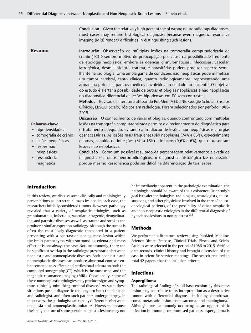
Usually, cerebral aspergillosis is a result of hematogenousdissemination or, alternatively, by direct extension from anadjacent focus. Aspergillus infection of the central nervoussystem (CNS) may present as an abscess or as cerebralinfarcts, but rarely as meningitis. The aspergilloma canalso present as an extra-axial mass extending inside thebrain parenchyma. Radiologically, granulomas with fungipresent appear as masses. Adjacent to dura mater, they cansimulate meningiomas.4 In computed tomography, they canbe associated with paranasal and small areas of bone de-struction sinusitis. Their appearance on MRI may vary de-pending on the organism involved. Aspergillomas tend toshow injury surrounded by perilesional edema in T2. Otherfungal infections, such as histoplasmosis, also radiologicallysimulate brain tumors. Treatment may require a combina-tion of surgical (abscess drainage) and clinical (drugs, am-photericin) procedures (►Fig. 1).5,6
CryptococcosisIn immunocompromised patients, infection by the fungusCryptococcus neoformans typically results in a diffuse men-ingitis and infiltration of the perivascular spaces. This canlead to the formation of small intraparenchymal gelatinouspseudocysts with little or no inflammatory response. Inimmunocompetent individuals with a chronic granuloma-tous reaction, the infection involves microorganisms with-out neovascular growth around the granulomas, whichproduces contrast enhancement similar to that of primaryor secondary brain tumors.7,8 The involvement of the centralnervous system by the fungus C. neoformansmost commonlyoccurs in the cerebellum, brain stem, and basal ganglia. Theradiological distinction between cerebral cryptococcosis and
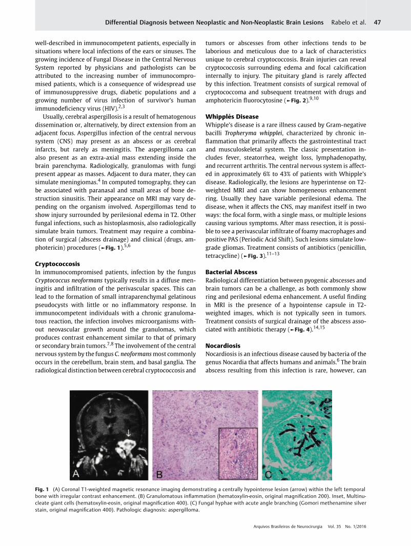
tumors or abscesses from other infections tends to belaborious and meticulous due to a lack of characteristicsunique to cerebral cryptococcosis. Brain injuries can revealcryptococcosis surrounding edema and focal calcificationinternally to injury. The pituitary gland is rarely affectedby this infection. Treatment consists of surgical removal ofcryptococcoma and subsequent treatment with drugs andamphotericin fluorocytosine (►Fig. 2).9,10
Whipples DiseaseWhipple’s disease is a rare illness caused by Gram-negativebacilli Tropheryma whipplei, characterized by chronic in-flammation that primarily affects the gastrointestinal tractand musculoskeletal system. The classic presentation in-cludes fever, steatorrhea, weight loss, lymphadenopathy,and recurrent arthritis. The central nervous system is affect-ed in approximately 6% to 43% of patients with Whipple’sdisease. Radiologically, the lesions are hyperintense on T2-weighted MRI and can show homogeneous enhancementring. Usually they have variable perilesional edema. Thedisease, when it affects the CNS, may manifest itself in twoways: the focal form, with a single mass, or multiple lesionscausing various symptoms. After mass resection, it is possi-ble to see a perivascular infiltrate of foamymacrophages andpositive PAS (Periodic Acid Shift). Such lesions simulate low-grade gliomas. Treatment consists of antibiotics (penicillin,tetracycline) (►Fig. 3).11–13
Bacterial AbscessRadiological differentiation between pyogenic abscesses andbrain tumors can be a challenge, as both commonly showring and perilesional edema enhancement. A useful findingin MRI is the presence of a hypointense capsule in T2-weighted images, which is not typically seen in tumors.Treatment consists of surgical drainage of the abscess asso-ciated with antibiotic therapy (►Fig. 4).14,15
NocardiosisNocardiosis is an infectious disease caused by bacteria of thegenus Nocardia that affects humans and animals.6 The brainabscess resulting from this infection is rare, however, can
Fig. 1 (A) Coronal T1-weighted magnetic resonance imaging demonstrating a centrally hypointense lesion (arrow) within the left temporalbone with irregular contrast enhancement. (B) Granulomatous inflammation (hematoxylin-eosin, original magnification 200). Inset, Multinu-cleate giant cells (hematoxylin-eosin, original magnification 400). (C) Fungal hyphae with acute angle branching (Gomori methenamine silverstain, original magnification 400). Pathologic diagnosis: aspergilloma.
Arquivos Brasileiros de Neurocirurgia Vol. 35 No. 1/2016
Differential Diagnosis between Neoplastic and Non-Neoplastic Brain Lesions Rabelo et al. 47
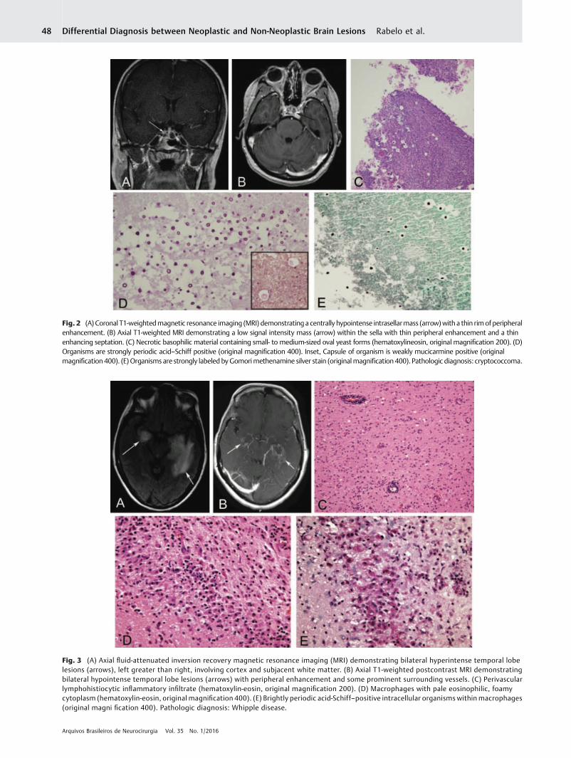
Fig. 3 (A) Axial fluid-attenuated inversion recovery magnetic resonance imaging (MRI) demonstrating bilateral hyperintense temporal lobelesions (arrows), left greater than right, involving cortex and subjacent white matter. (B) Axial T1-weighted postcontrast MRI demonstratingbilateral hypointense temporal lobe lesions (arrows) with peripheral enhancement and some prominent surrounding vessels. (C) Perivascularlymphohistiocytic inflammatory infiltrate (hematoxylin-eosin, original magnification 200). (D) Macrophages with pale eosinophilic, foamycytoplasm (hematoxylin-eosin, original magnification 400). (E) Brightly periodic acid-Schiff–positive intracellular organisms withinmacrophages(original magni fication 400). Pathologic diagnosis: Whipple disease.
Fig. 2 (A) Coronal T1-weightedmagnetic resonance imaging (MRI) demonstratinga centrally hypointense intrasellarmass (arrow)with a thin rimof peripheralenhancement. (B) Axial T1-weighted MRI demonstrating a low signal intensity mass (arrow) within the sella with thin peripheral enhancement and a thinenhancing septation. (C) Necrotic basophilic material containing small- to medium-sized oval yeast forms (hematoxylineosin, original magnification 200). (D)Organisms are strongly periodic acid–Schiff positive (original magnification 400). Inset, Capsule of organism is weakly mucicarmine positive (originalmagnification 400). (E)Organisms are strongly labeled byGomorimethenamine silver stain (originalmagnification 400). Pathologic diagnosis: cryptococcoma.
Arquivos Brasileiros de Neurocirurgia Vol. 35 No. 1/2016
Differential Diagnosis between Neoplastic and Non-Neoplastic Brain Lesions Rabelo et al.48
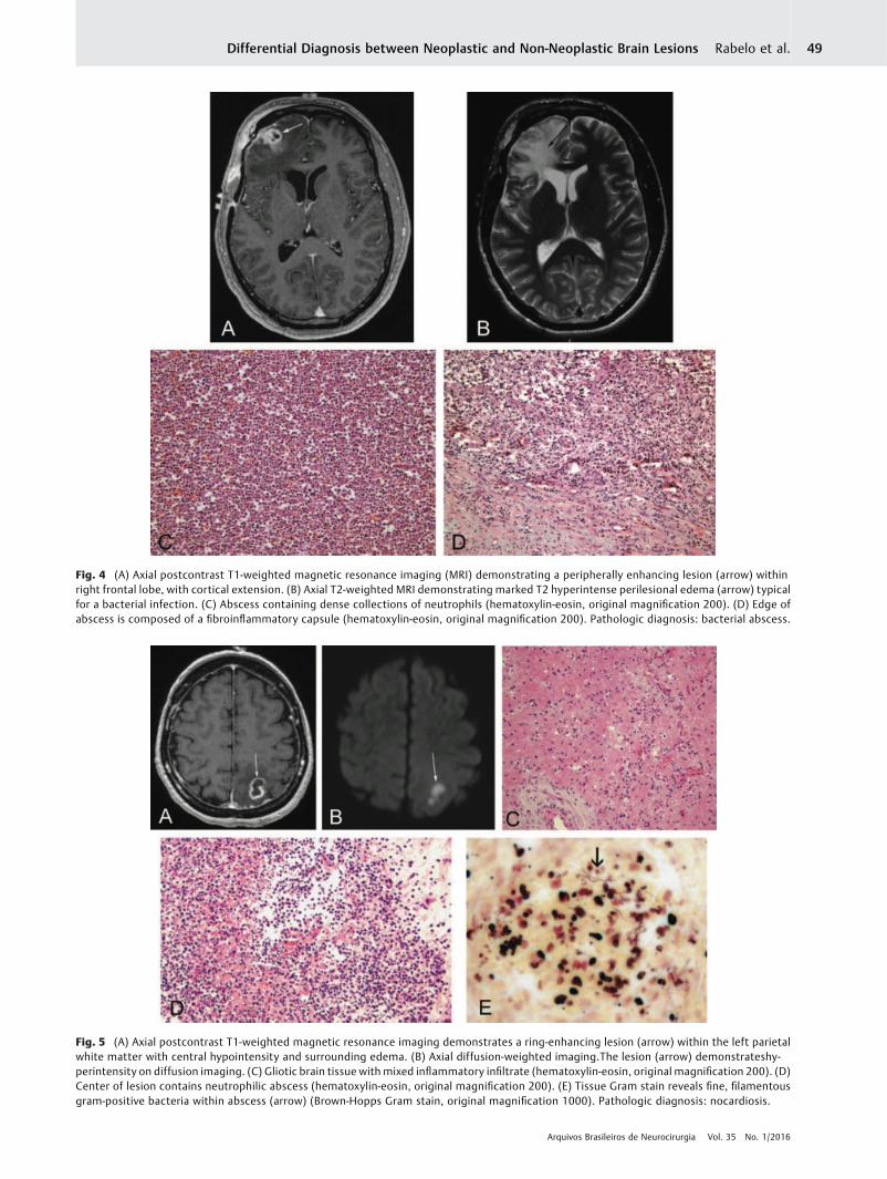
Fig. 5 (A) Axial postcontrast T1-weighted magnetic resonance imaging demonstrates a ring-enhancing lesion (arrow) within the left parietalwhite matter with central hypointensity and surrounding edema. (B) Axial diffusion-weighted imaging.The lesion (arrow) demonstrateshy-perintensity on diffusion imaging. (C) Gliotic brain tissue with mixed inflammatory infiltrate (hematoxylin-eosin, original magnification 200). (D)Center of lesion contains neutrophilic abscess (hematoxylin-eosin, original magnification 200). (E) Tissue Gram stain reveals fine, filamentousgram-positive bacteria within abscess (arrow) (Brown-Hopps Gram stain, original magnification 1000). Pathologic diagnosis: nocardiosis.
Fig. 4 (A) Axial postcontrast T1-weighted magnetic resonance imaging (MRI) demonstrating a peripherally enhancing lesion (arrow) withinright frontal lobe, with cortical extension. (B) Axial T2-weighted MRI demonstrating marked T2 hyperintense perilesional edema (arrow) typicalfor a bacterial infection. (C) Abscess containing dense collections of neutrophils (hematoxylin-eosin, original magnification 200). (D) Edge ofabscess is composed of a fibroinflammatory capsule (hematoxylin-eosin, original magnification 200). Pathologic diagnosis: bacterial abscess.
Arquivos Brasileiros de Neurocirurgia Vol. 35 No. 1/2016
Differential Diagnosis between Neoplastic and Non-Neoplastic Brain Lesions Rabelo et al. 49
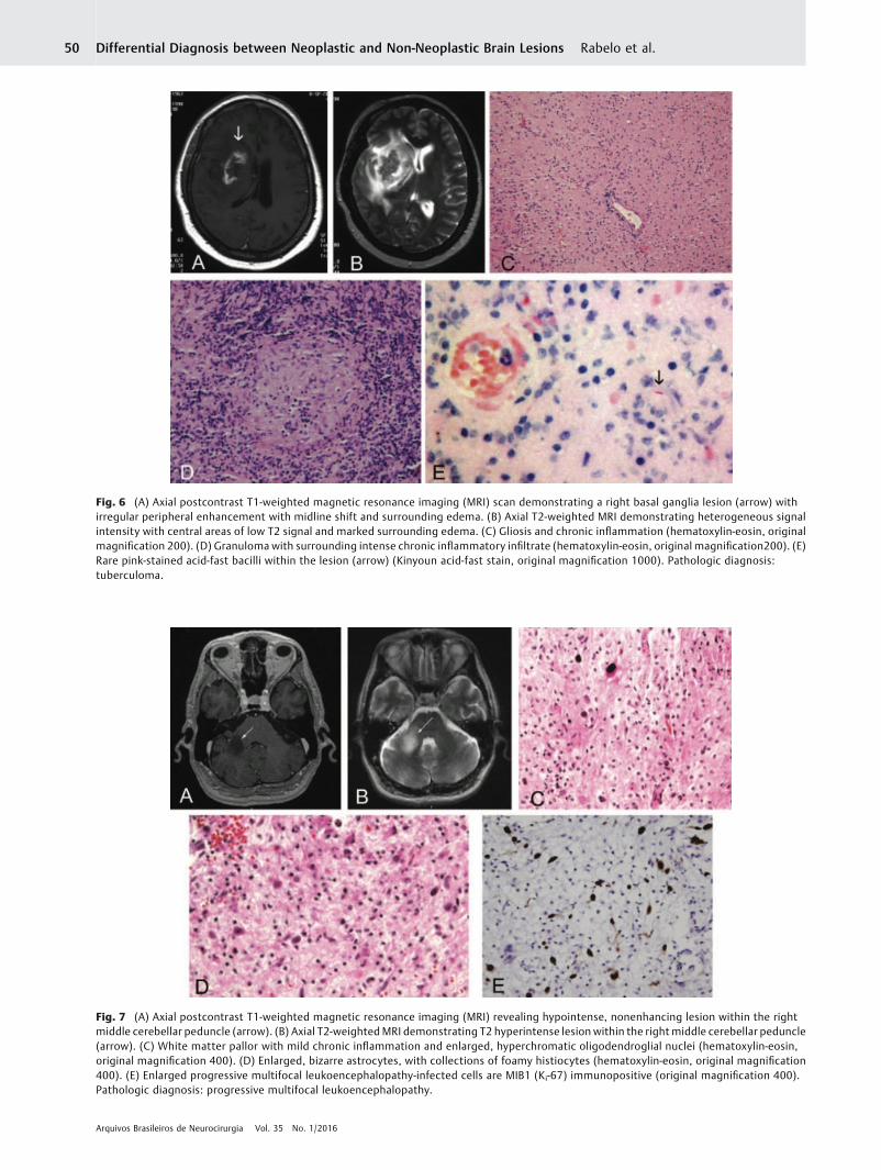
Fig. 6 (A) Axial postcontrast T1-weighted magnetic resonance imaging (MRI) scan demonstrating a right basal ganglia lesion (arrow) withirregular peripheral enhancement with midline shift and surrounding edema. (B) Axial T2-weighted MRI demonstrating heterogeneous signalintensity with central areas of low T2 signal and marked surrounding edema. (C) Gliosis and chronic inflammation (hematoxylin-eosin, originalmagnification 200). (D) Granuloma with surrounding intense chronic inflammatory infiltrate (hematoxylin-eosin, original magnification200). (E)Rare pink-stained acid-fast bacilli within the lesion (arrow) (Kinyoun acid-fast stain, original magnification 1000). Pathologic diagnosis:tuberculoma.
Fig. 7 (A) Axial postcontrast T1-weighted magnetic resonance imaging (MRI) revealing hypointense, nonenhancing lesion within the rightmiddle cerebellar peduncle (arrow). (B) Axial T2-weighted MRI demonstrating T2 hyperintense lesion within the right middle cerebellar peduncle(arrow). (C) White matter pallor with mild chronic inflammation and enlarged, hyperchromatic oligodendroglial nuclei (hematoxylin-eosin,original magnification 400). (D) Enlarged, bizarre astrocytes, with collections of foamy histiocytes (hematoxylin-eosin, original magnification400). (E) Enlarged progressive multifocal leukoencephalopathy-infected cells are MIB1 (Ki-67) immunopositive (original magnification 400).Pathologic diagnosis: progressive multifocal leukoencephalopathy.
Arquivos Brasileiros de Neurocirurgia Vol. 35 No. 1/2016
Differential Diagnosis between Neoplastic and Non-Neoplastic Brain Lesions Rabelo et al.50
affect both immunocompromised patients and immuno-competent patients. Radiologically, abscesses can be con-fused with intracranial tumors. There are several reportedcases transplant patients or patients with autoimmune dis-eases being treated with corticosteroids, brain abscesseswhich developed that simulate tumors. Histologically, no-cardiosis appears as a necrotic injury and acute inflamma-tion. Treatment consists of antibiotics and surgical drainagepossible (►Fig. 5).16,17
TuberculomaCerebral tuberculomas may present nodular or ring en-hancement in MRI images, similar to a primary or metastatictumor. When present in the brain, areas of caseous necrosismay correlate with isointense areas or those of mixedintensity on T1-weighted images. The onset of cerebraltuberculomas in MR imaging varies with the age of thelesion. In T2-weighted imaging, immature tuberculomasappear as multiple small isointense or hypointense areassurrounded by hyperintense edema. Mature tuberculomas,on the other hand, are heterogeneous, and can be isointenseor hypointense throughout the lesion with ring enhance-ment visible in the post-contrast T1.7 A magnetic resonancespectroscopy (MRS), generally inconclusive, does not help todistinguish the condition of a neoplasm. Histological appear-ance of a tuberculoma is characteristic, with granulomatousinflammation, caseous necrosis, and calcification. The pos-
sibility of lesion biopsy becomes important for diagnosisbecause the contribution in distinguishing a tuberculomawith a brain tumor. Treatment for tuberculoma consists ofantibiotic therapy and possible surgical intervention.(►Fig. 6).8
Progressive Multifocal Leukoencephalopathy (PML)The progressive multifocal leukoencephalopathy (PML) is arare infectious disease classically associated with immuno-suppressed individuals. PML is caused by reactivation of alatent virus belonging to the polyomavirus genus. Typically,patients with progressive multifocal leukoencephalopathypresent weakness, hemianopia or quadrantanopsia, andcognitive abnormalities. Radiologically, the lesions are usu-ally large and affect the subcortical white substance withoutmass effect.18 The most frequent location is in the parietallobe, but can also be seen in the occipital lobe, corpuscallosum, and thalamus. Some lesions have internal necrosisand the biopsy allows the pathologist to check areas ofdemyelination and oligodendrocyte with hyperchromaticnuclei. Finally, immunohistochemistry detects the viral cap-sid protein (►Fig. 7).19,20
NeurocysticercosisNeurocysticercosis develops as the larval form of Taeniasolium is implanted in the subject’s brain and causes aninflammatory and granulomatous reaction. It is
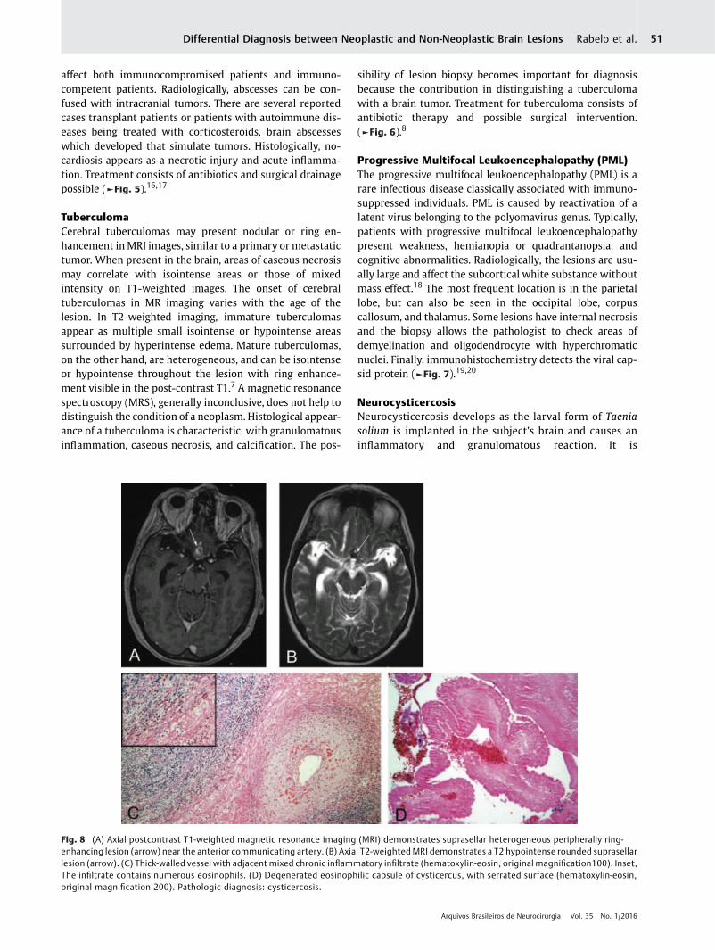
Fig. 8 (A) Axial postcontrast T1-weighted magnetic resonance imaging (MRI) demonstrates suprasellar heterogeneous peripherally ring-enhancing lesion (arrow) near the anterior communicating artery. (B) Axial T2-weightedMRI demonstrates a T2 hypointense rounded suprasellarlesion (arrow). (C) Thick-walled vessel with adjacent mixed chronic inflammatory infiltrate (hematoxylin-eosin, original magnification100). Inset,The infiltrate contains numerous eosinophils. (D) Degenerated eosinophilic capsule of cysticercus, with serrated surface (hematoxylin-eosin,original magnification 200). Pathologic diagnosis: cysticercosis.
Arquivos Brasileiros de Neurocirurgia Vol. 35 No. 1/2016
Differential Diagnosis between Neoplastic and Non-Neoplastic Brain Lesions Rabelo et al. 51
considered the most common cause of acquired seizuresworldwide. While epilepsy is the most common symptom,other symptoms include severe headache, meningitis,obstructive hydrocephalus, and cognitive problems. Theradiological diagnosis is usually straightforward, althoughappearance varies according to the stage of the parasitecycle and the age of the lesion. The injury may occur as asingle cyst or multiple cysts in the brain that undergocalcification, the latter representing dead larvae. Whenthe larvae invade the brain, they appear as small swollencysts and subsequently become larger lesions or nodules.7
Over time, the larvae die, undergo calcification, producingan inflammatory response around the cyst and contribut-ing to the onset of an annular capsule. Unlike what occurswith pyogenic abscesses, cysts are similar in strength tothe cerebrospinal fluid. The lesions rarely exceed 20 mmin diameter and a useful finding is the presence of thescolex within the cyst (the scolex is typically hyperintenseon T1 sequences). Solitary lesions are difficult to distin-guish from cancer due to surrounding edema and en-hancement of the cyst, and may be confused with aradiologically defined glioblastoma multiforme. Treat-ment consists of administering anti-parasitic drugs and
surgical removal, in cases where there is compression ofimportant structures (►Fig. 8).8,21
DemyelinationPseudotumoral features, also called tumefactive demyelin-ating plaques, are well-described in the literature. Multiplesclerosis and other demyelinating diseases, such as Shildersdisease and acute disseminated encephalomyelitis, maymanifest as tumefactive or pseudotumoral lesions. The pres-ence of complete solitary demyelinating lesions result inChallenge of differentiation between brain tumors, such asglioma.22,23Moreover, When the demyelinating lesions havea marked inflammatory component, they may be confusedwith primary central nervous system, like lymphoma. Thecharacteristics that favor a demyelinating injury includeapparent lack of mass effect, and vasogenic edema present(►Fig. 9).24,25
Vascular Diseases
InfarctCases inwhich high-grade gliomasmimic infarcts in contrastenhancement due to the mass effects are documented in the
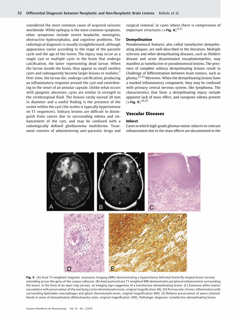
Fig. 9 (A) Axial T2-weighted magnetic resonance imaging (MRI) demonstrating a hyperintense bifrontal butterfly-shaped lesion (arrow)extending across the genu of the corpus callosum. (B) Axial postcontrast T1-weighted MRI demonstrates peripheral enhancement surroundingthe lesion, in the form of an open ring (arrow), an imaging sign suggestive of a tumefactive demyelinating lesion. (C) Extensive white mattervacuolation with preservation of the overlying cortex (hematoxylin-eosin, original magnification 40). (D) Perivascular chronic inflammation withsurrounding lipid-laden macrophages and gliosis (hematoxylin-eosin, original magnification 400). (E) Relative preservation of axons (stainedblack) in areas of demyelination (Bielschowsky stain, original magnification 200). Pathologic diagnosis: tumefactive demyelinating lesion.
Arquivos Brasileiros de Neurocirurgia Vol. 35 No. 1/2016
Differential Diagnosis between Neoplastic and Non-Neoplastic Brain Lesions Rabelo et al.52
literature. The medical practice of repeat images like MRIperfusions, in a short time, could help the distinction of acerebrovascular accident (CVA) and brain tumor. In mostcases, the diagnosis of ischemic stroke / heart attack is notproblematic. The clinical history of a sudden onset of neuro-logical symptoms and signs, in combination with typicalradiological findings, usually leads to the correct diagnosis.Confounding factors in the diagnosis of ischemic stroke thatare more suggestive of a brain tumor include an atypicalpresentation of neurological symptoms (slow evolution) anda bad brain injury defined in images associated with masseffect, as well as radiological similarity to glioma. An under-standing of the cerebral arterial vasculature is essential toreach the correct diagnosis (►Fig. 10).26
VasculitisThe clinical presentation and radiographic findings do notprovide unequivocal evidence in the diagnosis of vasculi-tis. Patients with sudden neurological focal deficit andring-enhancing lesions located in the white matter createsa situation where the diagnosis of high-grade gliomabecomes probable. In reality, there are relatively few casesof such conditions reported in the literature. However,other imaging techniques, such as magnetic resonanceangiography, may be useful in the differential diagnosis.26
Behcet’s disease (systemic inflammatory disease that cancause CNS vasculitis) can manifest in ambiguously inradiological images, because the images may appear as asolitary pseudotumor in one hemisphere, with low atten-uation on the T1-weighted MRI and extensive hyperin-tense fluid. However, this scenario is also rare, as Behcet’sdisease generally presents as multiple hyperintense le-sions in T2. The MR spectroscopy can be useful to distin-guish a vasculitis lesion from a neoplastic one. There is acase report of lymphocytic vasculitis simulating multifo-cal lesions in the right cerebral hemisphere, with signifi-cant vasogenic edema associated with injury.27
Radiologically, the lesions mimicked a multifocal glioma.However, the MRS pattern was more consistent with aninflammatory process (no elevated choline peak, markedelevation of glutamate/glutamine metabolites) ratherthan an aggressive neoplasm. The diagnosis was con-firmed by craniotomy and excisional biopsy (►Fig. 11).28
Traumatic
Chronic Subdural Hematoma (CSH)Chronic subdural hematoma (CSH) is a blood collection ofchronic evolution, located between the dura and arachnoid.It affectsmainlymales over sixty years old. Accidental fall hasbeen reported in most patients and their clinical picture isquite varied and can mimic other disease processes. Themeans for effective diagnosis has been computed tomogra-phy of the skull and its treatment of choice has beenprimarily surgical. In computed tomography, CSH is ex-pressed as a laminar, dynamic injury, and its appearancedepends on the time of evolution. Immediately after thehematoma, hemorrhage appears hyperdense. During thefollowing weeks, there is intense fibrinolysis and the hema-toma becomes isodense. After about a month, the picturebecomes hypodense due to liquefied blood inside the hema-toma. Other diagnoses that have been made, is higroma(►Fig. 12).
Amyloidosis
AmyloidomaThe challenges in the differential diagnosis related to theamyloid deposition are significant when there is no evi-dence of any systemic disease associated with amyloiddeposition. There are several reports of such in the centralnervous system. The most frequent presentation is asintracranial cerebral amyloid angiopathy, or as deposits
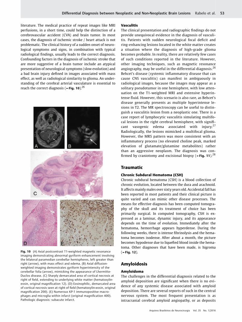
Fig. 10 (A) Axial postcontrast T1-weighted magnetic resonanceimaging demonstrating abnormal gyriform enhancement involvingthe bilateral paramedian cerebellar hemispheres, left greater thanright (arrow), with mass effect and edema. (B) Axial diffusion-weighted imaging demonstrates gyriform hyperintensity of thecerebellar folia (arrow), mimicking the appearance of Lhermitte-Duclos disease. (C) Sharply demarcated area of cortical necrosis atright of field, extending to underlying white matter (hematoxylin-eosin, original magnification 12). (D) Eosinophilic, demarcated areaof cortical necrosis seen at right of field (hematoxylin-eosin, originalmagnification 200). (E) Numerous KP-1 immunopositive macro-phages and microglia within infarct (original magnification 400).Pathologic diagnosis: subacute infarct.
Arquivos Brasileiros de Neurocirurgia Vol. 35 No. 1/2016
Differential Diagnosis between Neoplastic and Non-Neoplastic Brain Lesions Rabelo et al. 53
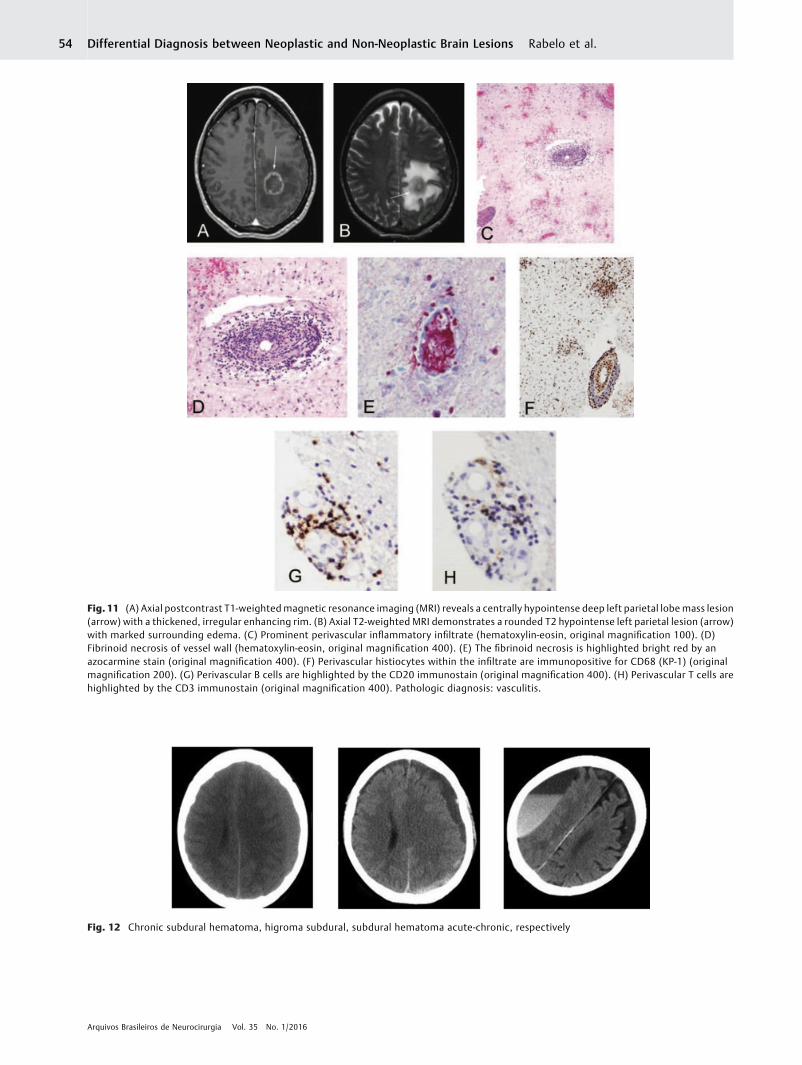
Fig. 11 (A) Axial postcontrast T1-weightedmagnetic resonance imaging (MRI) reveals a centrally hypointense deep left parietal lobe mass lesion(arrow) with a thickened, irregular enhancing rim. (B) Axial T2-weighted MRI demonstrates a rounded T2 hypointense left parietal lesion (arrow)with marked surrounding edema. (C) Prominent perivascular inflammatory infiltrate (hematoxylin-eosin, original magnification 100). (D)Fibrinoid necrosis of vessel wall (hematoxylin-eosin, original magnification 400). (E) The fibrinoid necrosis is highlighted bright red by anazocarmine stain (original magnification 400). (F) Perivascular histiocytes within the infiltrate are immunopositive for CD68 (KP-1) (originalmagnification 200). (G) Perivascular B cells are highlighted by the CD20 immunostain (original magnification 400). (H) Perivascular T cells arehighlighted by the CD3 immunostain (original magnification 400). Pathologic diagnosis: vasculitis.
Fig. 12 Chronic subdural hematoma, higroma subdural, subdural hematoma acute-chronic, respectively
Arquivos Brasileiros de Neurocirurgia Vol. 35 No. 1/2016
Differential Diagnosis between Neoplastic and Non-Neoplastic Brain Lesions Rabelo et al.54
in senile plaques in patients with Alzheimer’s disease.29
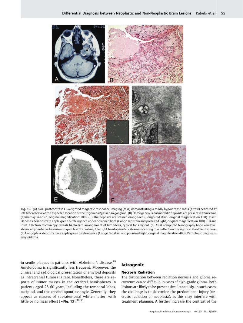
Amyloidoma is significantly less frequent. Moreover, theclinical and radiological presentation of amyloid depositsas intracranial tumors is rare. Nonetheless, there are re-ports of tumor masses in the cerebral hemispheres inpatients aged 28-60 years, including the temporal lobes,occipital, and the cerebellopontine angle. Generally, theyappear as masses of supratentorial white matter, withlittle or no mass effect (►Fig. 13).30,31
Iatrogenic
Necrosis RadiationThe distinction between radiation necrosis and glioma re-currence can be difficult. In cases of high-grade glioma, bothlesions are likely to be present simultaneously. In such cases,the challenge is to determine the predominant injury (ne-crosis radiation or neoplasia), as this may interfere withtreatment planning. A further increase the contrast of the
Fig. 13 (A) Axial postcontrast T1-weighted magnetic resonance imaging (MRI) demonstrating a mildly hypointense mass (arrow) centered atleft Meckel cave at the expected location of the trigeminal/gasserian ganglion. (B) Homogeneous eosinophilic deposits are present within lesion(hematoxylin-eosin, original magnification 100). (C) The deposits are stained orange-red (Congo red stain, original magnification 100). Inset,Deposits demonstrate apple green birefringence under polarized light (Congo red stain and polarized light, original magnification 100). (D) andinset, Electron microscopy reveals haphazard arrangement of 6-m fibrils, typical for amyloid. (E) Axial computed tomography bone windowshows a hyperdense biconvex-shaped lesion involving the right frontoparietal calvarium causing mass effect on the right cerebral hemisphere.(F) Congophilic deposits have apple green birefringence (Congo red stain and polarized light, original magnification 400). Pathologic diagnosis:amyloidoma.
Arquivos Brasileiros de Neurocirurgia Vol. 35 No. 1/2016
Differential Diagnosis between Neoplastic and Non-Neoplastic Brain Lesions Rabelo et al. 55
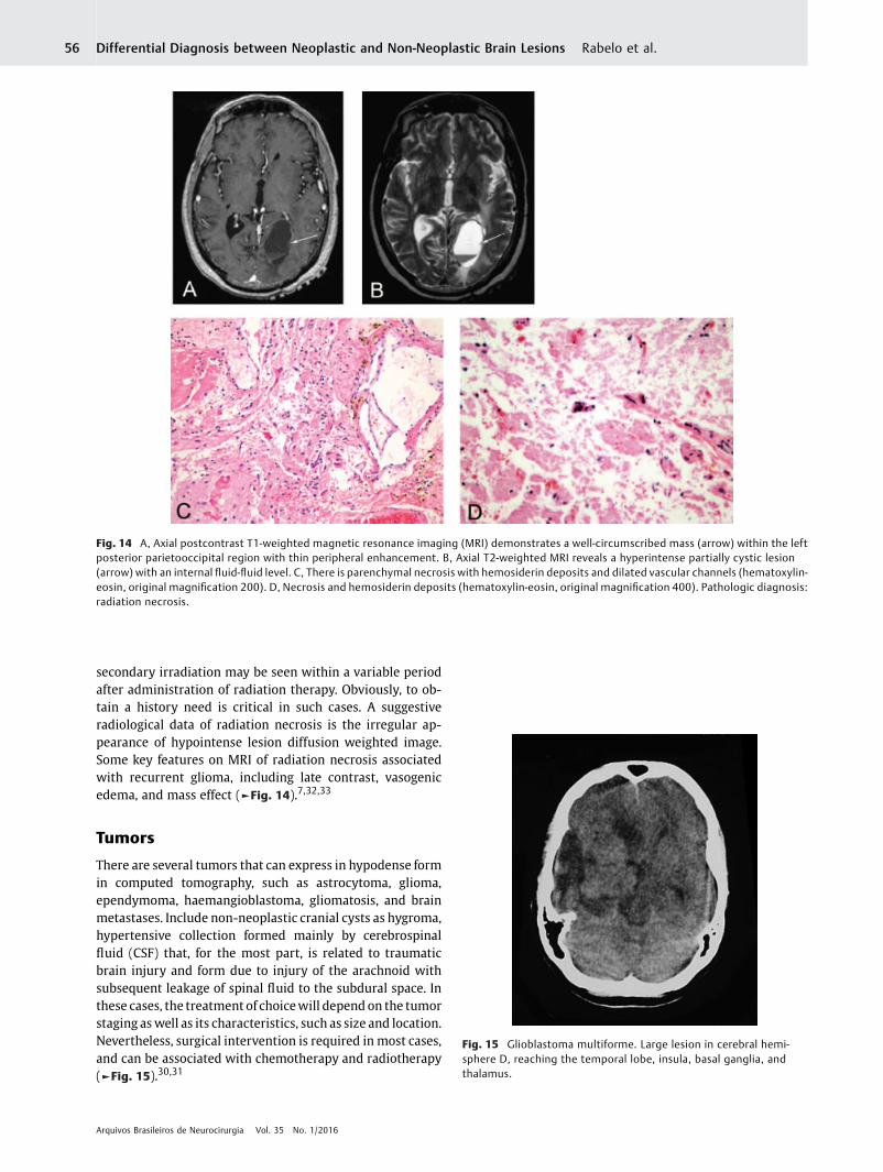
secondary irradiation may be seen within a variable periodafter administration of radiation therapy. Obviously, to ob-tain a history need is critical in such cases. A suggestiveradiological data of radiation necrosis is the irregular ap-pearance of hypointense lesion diffusion weighted image.Some key features on MRI of radiation necrosis associatedwith recurrent glioma, including late contrast, vasogenicedema, and mass effect (►Fig. 14).7,32,33
Tumors
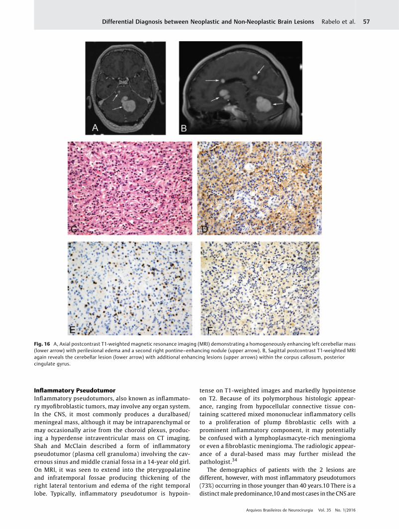
There are several tumors that can express in hypodense formin computed tomography, such as astrocytoma, glioma,ependymoma, haemangioblastoma, gliomatosis, and brainmetastases. Include non-neoplastic cranial cysts as hygroma,hypertensive collection formed mainly by cerebrospinalfluid (CSF) that, for the most part, is related to traumaticbrain injury and form due to injury of the arachnoid withsubsequent leakage of spinal fluid to the subdural space. Inthese cases, the treatment of choicewill depend on the tumorstaging aswell as its characteristics, such as size and location.Nevertheless, surgical intervention is required in most cases,and can be associated with chemotherapy and radiotherapy(►Fig. 15).30,31
Fig. 14 A, Axial postcontrast T1-weighted magnetic resonance imaging (MRI) demonstrates a well-circumscribed mass (arrow) within the leftposterior parietooccipital region with thin peripheral enhancement. B, Axial T2-weighted MRI reveals a hyperintense partially cystic lesion(arrow) with an internal fluid-fluid level. C, There is parenchymal necrosis with hemosiderin deposits and dilated vascular channels (hematoxylin-eosin, original magnification 200). D, Necrosis and hemosiderin deposits (hematoxylin-eosin, original magnification 400). Pathologic diagnosis:radiation necrosis.
Fig. 15 Glioblastoma multiforme. Large lesion in cerebral hemi-sphere D, reaching the temporal lobe, insula, basal ganglia, andthalamus.
Arquivos Brasileiros de Neurocirurgia Vol. 35 No. 1/2016
Differential Diagnosis between Neoplastic and Non-Neoplastic Brain Lesions Rabelo et al.56
Inflammatory PseudotumorInflammatory pseudotumors, also known as inflammato-ry myofibroblastic tumors, may involve any organ system.In the CNS, it most commonly produces a duralbased/meningeal mass, although it may be intraparenchymal ormay occasionally arise from the choroid plexus, produc-ing a hyperdense intraventricular mass on CT imaging.Shah and McClain described a form of inflammatorypseudotumor (plasma cell granuloma) involving the cav-ernous sinus and middle cranial fossa in a 14-year old girl.On MRI, it was seen to extend into the pterygopalatineand infratemporal fossae producing thickening of theright lateral tentorium and edema of the right temporallobe. Typically, inflammatory pseudotumor is hypoin-
tense on T1-weighted images and markedly hypointenseon T2. Because of its polymorphous histologic appear-ance, ranging from hypocellular connective tissue con-taining scattered mixed mononuclear inflammatory cellsto a proliferation of plump fibroblastic cells with aprominent inflammatory component, it may potentiallybe confused with a lymphoplasmacyte-rich meningiomaor even a fibroblastic meningioma. The radiologic appear-ance of a dural-based mass may further mislead thepathologist.34
The demographics of patients with the 2 lesions aredifferent, however, with most inflammatory pseudotumors(73%) occurring in those younger than 40 years.10 There is adistinctmale predominance,10 andmost cases in the CNS are
Fig. 16 A, Axial postcontrast T1-weighted magnetic resonance imaging (MRI) demonstrating a homogeneously enhancing left cerebellar mass(lower arrow) with perilesional edema and a second right pontine–enhancing nodule (upper arrow). B, Sagittal postcontrast T1-weighted MRIagain reveals the cerebellar lesion (lower arrow) with additional enhancing lesions (upper arrows) within the corpus callosum, posteriorcingulate gyrus.
Arquivos Brasileiros de Neurocirurgia Vol. 35 No. 1/2016
Differential Diagnosis between Neoplastic and Non-Neoplastic Brain Lesions Rabelo et al. 57
solitary (82%). Intraparenchymal heterogeneously enhanc-ing inflammatory pseudotumors may mimic a malignantbrain neoplasm (►Fig. 16).35
Noninfectious Inflammatory Conditions
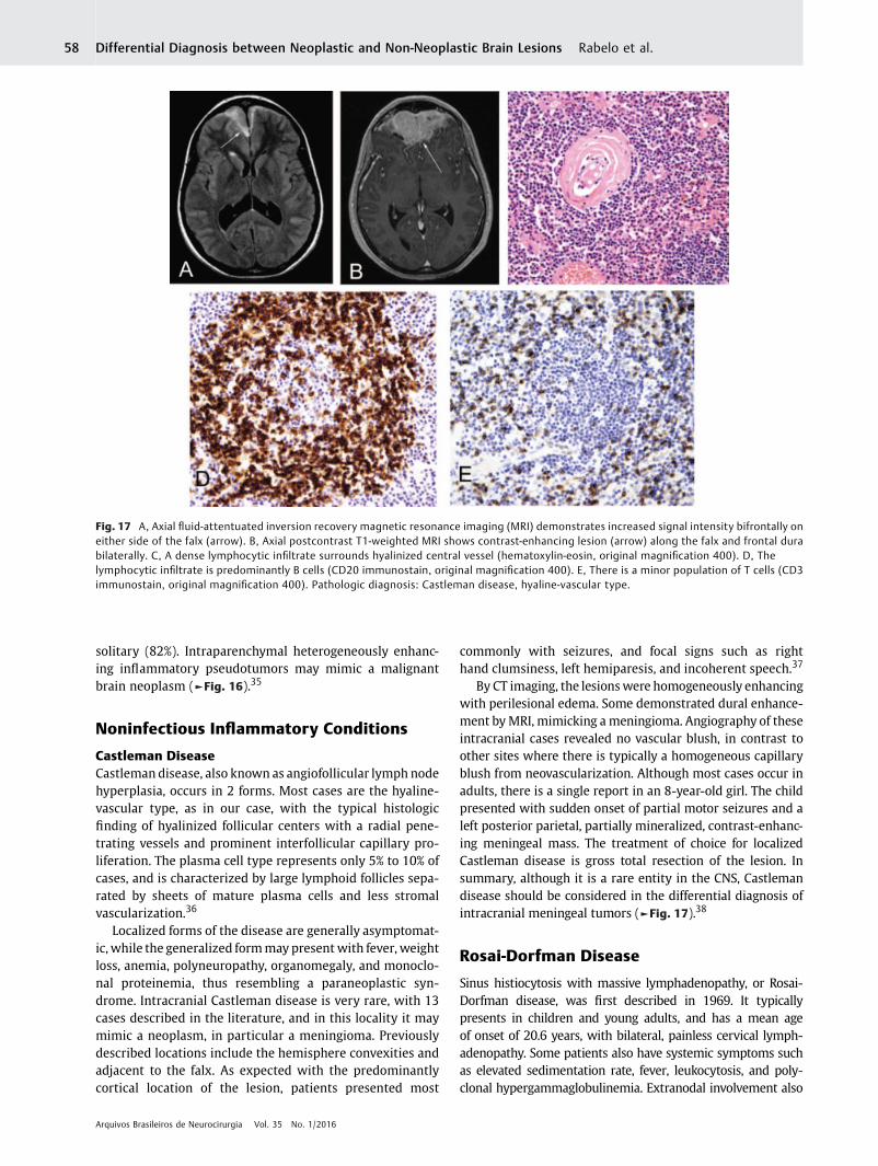
Castleman DiseaseCastleman disease, also known as angiofollicular lymph nodehyperplasia, occurs in 2 forms. Most cases are the hyaline-vascular type, as in our case, with the typical histologicfinding of hyalinized follicular centers with a radial pene-trating vessels and prominent interfollicular capillary pro-liferation. The plasma cell type represents only 5% to 10% ofcases, and is characterized by large lymphoid follicles sepa-rated by sheets of mature plasma cells and less stromalvascularization.36
Localized forms of the disease are generally asymptomat-ic, while the generalized formmaypresent with fever, weightloss, anemia, polyneuropathy, organomegaly, and monoclo-nal proteinemia, thus resembling a paraneoplastic syn-drome. Intracranial Castleman disease is very rare, with 13cases described in the literature, and in this locality it maymimic a neoplasm, in particular a meningioma. Previouslydescribed locations include the hemisphere convexities andadjacent to the falx. As expected with the predominantlycortical location of the lesion, patients presented most
commonly with seizures, and focal signs such as righthand clumsiness, left hemiparesis, and incoherent speech.37
By CT imaging, the lesionswere homogeneously enhancingwith perilesional edema. Some demonstrated dural enhance-ment byMRI, mimicking ameningioma. Angiography of theseintracranial cases revealed no vascular blush, in contrast toother sites where there is typically a homogeneous capillaryblush from neovascularization. Although most cases occur inadults, there is a single report in an 8-year-old girl. The childpresented with sudden onset of partial motor seizures and aleft posterior parietal, partially mineralized, contrast-enhanc-ing meningeal mass. The treatment of choice for localizedCastleman disease is gross total resection of the lesion. Insummary, although it is a rare entity in the CNS, Castlemandisease should be considered in the differential diagnosis ofintracranial meningeal tumors (►Fig. 17).38
Rosai-Dorfman Disease
Sinus histiocytosis with massive lymphadenopathy, or Rosai-Dorfman disease, was first described in 1969. It typicallypresents in children and young adults, and has a mean ageof onset of 20.6 years, with bilateral, painless cervical lymph-adenopathy. Some patients also have systemic symptoms suchas elevated sedimentation rate, fever, leukocytosis, and poly-clonal hypergammaglobulinemia. Extranodal involvement also
Fig. 17 A, Axial fluid-attentuated inversion recovery magnetic resonance imaging (MRI) demonstrates increased signal intensity bifrontally oneither side of the falx (arrow). B, Axial postcontrast T1-weighted MRI shows contrast-enhancing lesion (arrow) along the falx and frontal durabilaterally. C, A dense lymphocytic infiltrate surrounds hyalinized central vessel (hematoxylin-eosin, original magnification 400). D, Thelymphocytic infiltrate is predominantly B cells (CD20 immunostain, original magnification 400). E, There is a minor population of T cells (CD3immunostain, original magnification 400). Pathologic diagnosis: Castleman disease, hyaline-vascular type.
Arquivos Brasileiros de Neurocirurgia Vol. 35 No. 1/2016
Differential Diagnosis between Neoplastic and Non-Neoplastic Brain Lesions Rabelo et al.58
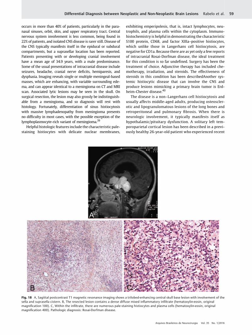
occurs in more than 40% of patients, particularly in the para-nasal sinuses, orbit, skin, and upper respiratory tract. Centralnervous system involvement is less common, being found in22% of patients, and isolated CNS disease is rarer still. Disease ofthe CNS typically manifests itself in the epidural or subduralcompartments, but a suprasellar location has been reported.Patients presenting with or developing cranial involvementhave a mean age of 34.9 years, with a male predominance.Some of the usual presentations of intracranial disease includeseizures, headache, cranial nerve deficits, hemiparesis, anddysphasia. Imaging reveals single or multiple meningeal-basedmasses, which are enhancing, with variable surrounding ede-ma, and can appear identical to a meningioma on CT and MRIscan. Associated lytic lesions may be seen in the skull. Onsurgical resection, the lesion may also grossly be indistinguish-able from a meningioma, and so diagnosis will rest withhistology. Fortunately, differentiation of sinus histiocytosiswith massive lymphadenopathy from meningioma presentsno difficulty in most cases, with the possible exception of thelymphoplasmocyte-rich variant of meningioma.39
Helpful histologic features include the characteristic pale-staining histiocytes with delicate nuclear membranes,
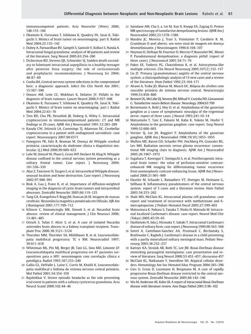
exhibiting emperipolesis, that is, intact lymphocytes, neu-trophils, and plasma cells within the cytoplasm. Immuno-histochemistry is helpful in demonstrating the characteristicS100 protein, CD68, and factor XIIIa–positive histiocytes,which unlike those in Langerhans cell histiocytosis, arenegative for CD1a. Because there are as yet only a few reportsof intracranial Rosai-Dorfman disease, the ideal treatmentfor this condition is so far undefined. Surgery has been thetreatment of choice. Adjunctive therapy has included che-motherapy, irradiation, and steroids. The effectiveness ofsteroids in this condition has been describedAnother sys-temic histiocytic disease that can involve the CNS andproduce lesions mimicking a primary brain tumor is Erd-heim-Chester disease.40
The disease is a non–Langerhans cell histiocytosis andusually affects middle-aged adults, producing osteoscler-otic and lipogranulomatous lesions of the long bones andretroperitoneal and pulmonary fibrosis. When there isneurologic involvement, it typically manifests itself ashypothalamic/pituitary dysfunction. A solitary left tem-poroparietal cortical lesion has been described in a previ-ously healthy 26-year-old patient who experienced recent
Fig. 18 A, Sagittal postcontrast T1 magnetic resonance imaging shows a trilobed-enhancing central skull base lesion with involvement of thesella and suprasella cistern. B, The resected lesion contains a dense diffuse mixed inflammatory infiltrate (hematoxylin-eosin, originalmagnification 100). C, Within the infiltrate, there are numerous pale-staining histiocytes and plasma cells (hematoxylin-eosin, originalmagnification 400). Pathologic diagnosis: Rosai-Dorfman disease.
Arquivos Brasileiros de Neurocirurgia Vol. 35 No. 1/2016
Differential Diagnosis between Neoplastic and Non-Neoplastic Brain Lesions Rabelo et al. 59
onset of seizures. The lesion was solitary, well-demarcatedand homogeneously enhancing onMRI examination. It hadminimal surrounding edema and no mass effect. Positronemission tomography scan suggested a low-grade neo-plasm, such as astrocytoma or pleomorphic xanthoastro-cytoma. Intraoperative squash preparations and frozensections revealed many multinucleate cells with a back-ground of fibrillary astrocytes, and a diagnosis of glialneoplasm was made. On permanent histology, clusters ofxanthomatous histiocytic cells were evident, many ofwhich were multinucleate with a wreathlike nuclear ar-rangement, surrounded by dense gliosis. The histiocyticcells were KP-1 (CD68) immunopositive and negative forCD1a and S100 (in contrast to those seenin Rosai-Dorfmandisease or Langerhans cell histiocytosis) (►Fig. 18).41,42
Conclusion
As a possible result of the relatively high percentage of wrongdiagnoses neuroradiological, patients with benign tumors,inflammatory lesions or vascular injury may be subjected tounnecessary radiation, chemotherapy, or both. CT is themostused exame, but even RMI make some indistinguishable
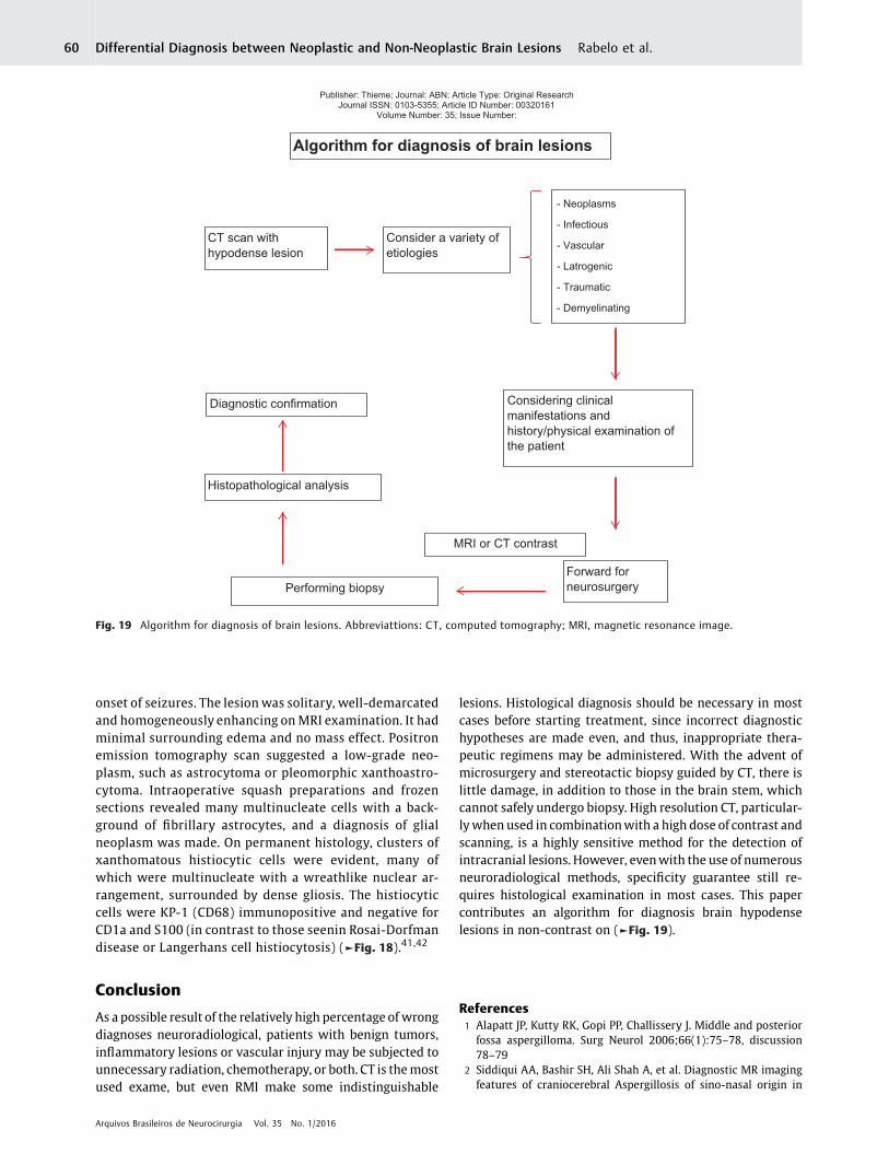
lesions. Histological diagnosis should be necessary in mostcases before starting treatment, since incorrect diagnostichypotheses are made even, and thus, inappropriate thera-peutic regimens may be administered. With the advent ofmicrosurgery and stereotactic biopsy guided by CT, there islittle damage, in addition to those in the brain stem, whichcannot safely undergo biopsy. High resolution CT, particular-lywhen used in combinationwith a high dose of contrast andscanning, is a highly sensitive method for the detection ofintracranial lesions. However, evenwith the use of numerousneuroradiological methods, specificity guarantee still re-quires histological examination in most cases. This papercontributes an algorithm for diagnosis brain hypodenselesions in non-contrast on (►Fig. 19).
References1 Alapatt JP, Kutty RK, Gopi PP, Challissery J. Middle and posterior
fossa aspergilloma. Surg Neurol 2006;66(1):75–78, discussion78–79
2 Siddiqui AA, Bashir SH, Ali Shah A, et al. Diagnostic MR imagingfeatures of craniocerebral Aspergillosis of sino-nasal origin in
Publisher: Thieme; Journal: ABN; Article Type: Original Research Journal ISSN: 0103-5355; Article ID Number: 00320161
Volume Number: 35; Issue Number:
Algorithm for diagnosis of brain lesions
CT scan with hypodense lesion
Consider a variety of etiologies
- Neoplasms
- Vascular
- Latrogenic
- Traumatic
- Demyelinating
- Infectious
Considering clinical manifestations and history/physical examination of the patient
Forward for neurosurgeryPerforming biopsy
Histopathological analysis
Diagnostic confirmation
MRI or CT contrast
Fig. 19 Algorithm for diagnosis of brain lesions. Abbreviattions: CT, computed tomography; MRI, magnetic resonance image.
Arquivos Brasileiros de Neurocirurgia Vol. 35 No. 1/2016
Differential Diagnosis between Neoplastic and Non-Neoplastic Brain Lesions Rabelo et al.60
immunocompetent patients. Acta Neurochir (Wien) 2006;148:155–166
3 Okamoto K, Furusawa T, Ishikawa K, Quadery FA, Sasai K, Toki-guchi S. Mimics of brain tumor on neuroimaging: part II. RadiatMed 2004;22:135–142
4 Dubey A, Patwardhan RV, Sampth S, Santosh V, Kolluri S, Nanda A.Intracranial fungal granuloma: analysis of 40 patients and reviewof the literature. Surg Neurol 2005;63:254–260
5 Dickerman RD, Stevens QE, Schneider SJ. Sudden death second-ary to fulminant intracranial aspegillosis in a healthy teenagerafter posterior fossa surgery: the role of corticosteroidsand prophylactic recommendations. J Neurosurg Sci 2004;48:87–89
6 Cunha BA. Central nervous system infections in the compromisedhost: a diagnostic approach. Infect Dis Clin North Am 2001;15:567–590
7 Omuro AM, Leite CC, Mokhtari K, Delattre JY. Pitfalls in thediagnosis of brain tumours. Lancet Neurol 2006;5:937–948
8 Okamoto K, Furusawa T, Ishikawa K, Quadery FA, Sasai K, Toki-guchi S. Mimics of brain tumor on neuroimaging: part I. RadiatMed 2004;22:63–76
9 Tien RD, Chu PK, Hesselink JR, Duberg A, Wiley C. Intracranialcryptococcosis in immunocompromised patients: CT and MRfindings in 29 cases. AJNR Am J Neuroradiol 1991;12:283–289
10 Kanaly CW, Selznick LA, Cummings TJ, Adamson DC. Cerebellarcryptococcoma in a patient with undiagnosed sarcoidosis: casereport. Neurosurgery 2007;60:E571
11 Panegyres PK, Edis R, Beaman M. Doença de Whipple cerebralprimária: caracterização da síndrome clínica e diagnóstico mo-lecular. Q J Med 2006;99:609–623
12 Lohr M, StenzelW, PlumG, GrossWP, Deckert M, Klug N.Whippledisease confined to the central nervous system presenting as asolitary frontal tumor. Case report. J Neurosurg 2004;101:336–339
13 Akar Z, TanrioverN, Tuzgen S, et al. IntracerebralWhipple disease:unusual location and bone destruction. Case report. J Neurosurg2002;97:988–991
14 Bink A, Gaa J, Franz K, et al. Importance of diffusion-weightedimaging in the diagnosis of cystic brain tumors and intracerebralabscesses. Zentralbl Neurochir 2005;66:119–125
15 Tung GA, Evangelista P, Rogg JM, Duncan JA III. Imagem demassascerebrais: Ressonância magnética ponderada emDifusão. AJR AmJ Roentgenol 2001;177:709–712
16 Kilincer C, Hamamcioglu MK, Simsek S, et al. Nocardial brainabscess: review of clinical management. J Clin Neurosci 2006;13:481–485
17 Ozturk S, Tufan F, Alisir S, et al. A case of isolated Nocardiaasteroides brain abscess in a kidney transplant recipient. Trans-plant Proc 2006;38:3121–3124
18 Thurnher MM, Thurnher SA, Mühlbauer B, et al. Leucoencefalo-patia multifocal progressiva: TC e RM. Neurorradiol 1997;39:611–618
19 Whiteman ML, Pós MJ, Berger JR, Tate LG, Sino MD, Limonte LP.Leucoencefalopatia multifocal progressiva em 47 pacientes sor-opositivos para o HIV: neuroimagem com correlação clínica epatológica. Radiol 1993;187:233–240
20 Gallia GL, DelValle L, Laine C, Curtis M, Khalili K. Leucoencefalo-patia multifocal e linfoma do sistema nervoso central primário.Mol Pathol 2001;54:354–359
21 Rajshekhar V. Severe episodic headache as the sole presentingictal event in patients with a solitary cysticercus granuloma. ActaNeurol Scand 2000;102:44–46
22 Saindane AM, Cha S, a. Lei M, Xue X, Knopp EA, Zagzag D. ProtonMR spectroscopyof tumefactive demyelinating lesions. AJNR Am JNeuroradiol 2002;23:1378–1386
23 Masdeu JC, Moreira J, Trasi S, Visintainer P, Cavaliere R. M.Grundman O anel aberto: um novo sinal de imagem em doençadesmielinizante. J Neuroimagem 1996;6:104–107
24 HeymanD, DelhayeM, Fournier D,Mercier P, RousseletMC,MeneiP. Pseudotumoral demyelination: a diagnosis pitfall (report ofthree cases). J Neurooncol 2001;54:71–76
25 Pakos EE, Tsekeris PG, Chatzidimou K, et al. Astrocytoma-likemultiple sclerosis. Clin Neurol Neurosurg 2005;107(2):152–157
26 Lie JT. Primary (granulomatous) angiitis of the central nervoussystem: a clinicopathologic analysis of 15 new cases and a reviewof the literature. Hum Pathol 1992;23:164–171
27 Alrawi A, Trobe JD, Blaivas M, Musch DC. Biópsia do cérebro comvasculite primária do sistema nervoso central. Neurocirurgia1999;53:858–860
28 BennettDL,McCabeDJ, Stevens JM,MifsudV,CozinhaND,GiovannoniG. Tumefactive neuro-Behcet disease. Neurology 2004;63:709
29 Bornemann A, Bohl J, Hey O, et al. Amyloidoma of the gasserianganglion as a cause of symptomatic neuralgia of the trigeminalnerve: report of three cases. J Neurol 1993;241:10–14
30 Matsumoto T, Tani E, Fukami M, Kaba K, Yokota M, Hoshii Y.Amyloidoma in the gasserian ganglion: case report. Surg Neurol1999;52:600–603
31 Vorster SJ, Lee JH, Ruggieri P. Amyloidoma of the gasserianganglion. AJNR Am J Neuroradiol 1998;19(10):1853–1855
32 Mullins ME, Barest GD, Schaefer PW, Hochberg FH, Gonzalez RG,Lev MH. Radiation necrosis versus glioma recurrence: conven-tional MR imaging clues to diagnosis. AJNR Am J Neuroradiol2005;26:1967–1972
33 Sugahara T, Koorogui Y, Tomiguchi S, et al. Posttherapeutic intra-axial brain tumor: the value of perfusion-sensitive contrast-enhanced MR imaging for differentiating tumor recurrencefrom nonneoplastic contrast-enhancing tissue. AJNR Am J Neuro-radiol 2000;21:901–909
34 Hausler M, Schaade L, Ramaekers VT, Doenges M, Heimann G,Sellhaus B. Inflammatory pseudotumors of the central nervoussystem: report of 3 cases and a literature review. Hum Pathol2003;34:253–262
35 Shah MD, McClain KL. Intracranial plasma cell granuloma: casereport and treatment of recurrence with methotrexate and 6-mercaptopurine. J Pediatr Hematol Oncol 2005;27:599–603
36 Matsumura K, Nakasu S, Tanaka T, Nioka H, Matsuda M. Intracra-nial localized Castleman’s disease: case report. Neurol Med Chir(Tokyo) 2005;45:59–65
37 Hashimoto H, Iida J, Hironaka Y, Sakaki T. Intracranial Castleman’sdisease of solitary form: case report. J Neurosurg1999;90:563–566
38 Sotrel A, Castellano-Sanchez AA, Prusmack C, Birchansky S,Brathwaite C, Ragheb J. Castleman’s disease in a child presentingwith a partly mineralized solitary meningeal mass. Pediatr Neu-rosurg 2003;38:232–237
39 Kattner KA, Stroink AR, Roth TC, Lee JM. Rosai-Dorfman diseasemimicking parasagittal meningioma: case presentation and re-view of literature. Surg Neurol 2000;53:452–457; discussion 457
40 McClain KL, Natkunam Y, Swerdlow SH. Atypical cellular disor-ders. Hematology Am Soc Hematol Educ Program 2004:283–296
41 Gies U, Gruia D, Lassmann H, Bergmann M. A case of rapidlyprogressive Rosai-Dorfman disease restricted to the central ner-vous system. Zentralbl Neurochir 2005;66:142–146
42 WuM, Anderson AE, Kahn LB. A report of intracranial Rosai-Dorfmandisease with literature review. Ann Diagn Pathol 2001;5:96–102
Arquivos Brasileiros de Neurocirurgia Vol. 35 No. 1/2016
Differential Diagnosis between Neoplastic and Non-Neoplastic Brain Lesions Rabelo et al. 61