Embed Size (px)
Citation preview

重症外傷性脳損傷(TBI)に 減圧開頭術は有効か
Journal Club 2016/11/01
聖マリアンナ医科大学 横浜市西部病院
PGY 5 堤 健

本日の論文
N Engl J Med 2016;375:1119-30.

背景

日本の頭部外傷の現状
平均年齢、57.0才(2009年)
47.4才(1998年)
原因:転倒・転落>交通外傷 (2009年)
転倒↑・交通外傷↓
頭部CT所見 びまん性脳損傷(34.4%)、局所性脳損傷(63.5%)
高齢者の転倒による脳挫傷・急性硬膜下血腫が増加
死亡率 45.4% (2009年)
1998年と比較して有意に減少
転帰良好率 21.5% (2009年)
転帰良好率→
植物状態↑
ICP測定群 28%(2009年)
ICPモニターの適応 ① GCS<8 ② SBP<90 mmHg ③ CTで正中偏位、脳槽の消失あり
(重症頭部外傷治療・管理のガイドライン 第3版、2013)
Jpn J Neurosurg (Tokyo) 25:214-219, 2016
米国 32% (1995) →78% (2005)

外傷性脳損傷のマネージメント
【脳圧亢進の治療】
過換気/血圧管理(昇圧)
高張食塩水
バルビツレート
低体温療法
減圧開頭術
血腫除去術・脳室開窓術(髄液ドレナージ)+ICPモニター
ICP <20mmHg, CPP 50-70 mmgに管理
ステロイドは推奨されない (Grade 1A)
低酸素(PaO2 <60 mmHg)・低血圧(SBP <90 mmHg)を回避
頭部挙上(30°)・マンニトール:脳圧亢進が疑われれば、すぐに投与(Grade 1B)
Euvolemia維持のための生食投与(Grade 1B)
短期間(1w)のearly seizure予防の抗てんかん薬投与
高体温・高血糖を回避。凝固異常を補正 静脈血栓症予防(Grade1A) 鎮静(バルビツレート、プロポフォール、
フェンタニル、ベンゾジアゼピン、モルヒネ)±筋弛緩
2016/11/1 Management of acute severe traumatic brain injury - UpToDate
https://www.uptodate.com/contents/management-of-acute-severe-traumatic-brain-injury/print?source=search_result&search=TBI&selectedTitle=2~86 1/29
Official reprint from UpToDate
www.uptodate.com ©2016 UpToDate
Management of acute severe traumatic brain injury
Authors: J Claude Hemphill, III, MD, MAS, Nicholas Phan, MD, FRCSC, FACS
Section Editors: Michael J Aminoff, MD, DSc, Maria E Moreira, MD
Deputy Editor: Janet L Wilterdink, MD
All topics are updated as new evidence becomes available and our peer review process is complete.
Literature review current through: Sep 2016. | This topic last updated: Feb 10, 2015.
INTRODUCTION — Traumatic brain injury (TBI) is the leading cause of death in North America for individuals
between the ages of 1 to 45 [1,2]. Many survivors live with significant disabilities, resulting in major
socioeconomic burden as well. In 2000, the economic impact of TBI in the United States was estimated to be
$9.2 billion in lifetime medical costs and $51.2 billion in productivity losses.
One of the major advances over the past two decades in the care of patients with severe head injury has
been the development of standardized approaches that follow international and national guidelines [36]. The
intent of these guidelines has been to use existing evidence to provide recommendations for current care in
order to lessen heterogeneity and improve patient outcomes. Unfortunately, the lack of randomized clinical
trials addressing many aspects of care of the severe TBI patient has meant that the strength of supporting
data for most treatment concepts is relatively weak. Despite this caveat, there is evidence that treatment in
centers with neurosurgical support, especially in settings where protocoldriven neurointensive care units
operate based on the abovereferenced guidelines, is associated with better patient outcomes [714]. Many
expert panels recommend that treatment of severe TBI should be centralized in large trauma centers that
offer neurosurgical treatment and access to specialized neurocritical care.
Patients with severe head injury may frequently have other traumatic injuries to internal organs, lungs, limbs,
or the spinal cord. Thus, the management of the patient with severe head injury is often complex and requires
a multidisciplinary approach and lends itself to protocolbased treatment and standardized hospital order
sets derived from the previously referenced guidelines.
This topic discusses the management of acute severe traumatic brain injury. The epidemiology and
pathophysiology of traumatic brain injury, the management of mild traumatic brain injury, acute spinal cord
injury, and other aspects of care of the trauma patient are discussed separately. (See "Traumatic brain injury:
Epidemiology, classification, and pathophysiology" and "Concussion and mild traumatic brain injury" and
"Acute traumatic spinal cord injury" and "Skull fractures in adults".)
INITIAL EVALUATION AND TREATMENT
Prehospital — The primary goal of prehospital management for severe head injury is to prevent hypotension
and hypoxia, two systemic insults known to be major causes of secondary injury after TBI [1520]. In a meta
analysis of clinical trials and populationbased studies, hypoxia (PaO2 <60 mmHg) and hypotension (systolic
BP <90 mmHg) were present in 50 and 30 percent of patients, respectively, and were each associated with a
higher likelihood of a poor outcome: hypoxia (OR 2.14); hypotension (OR 2.67) [16]. Changes in prehospital
management that aim to normalize oxygenation and blood pressure have improved outcomes [2125]:
®
®
Early endotracheal intubation is generally recommended for patients with a Glasgow coma scale score of
8 or less if performed by welltrained personnel (table 1). The value of prehospital intubation is
controversial, with studies finding conflicting results [26]. In one randomized trial of 312 patients with
severe TBI performed in Australia, prehospital rapid sequence intubation by paramedics was associated
with better functional outcome at six months compared with intubation in hospital (51 versus 39 percent
Management of acute severe
traumatic brain injury 2016/11/1

Metabolic suppression therapy (barbiturate coma)
バルビツレートには脳保護作用・脳圧降下作用あり 脳の代謝・血流↓ ⇒ 酸素消費↓+ 脳血流量↓ ⇒ ICP↓ 酸素フリーラジカルの抑制
全身麻酔下となるため、脳波持続モニタリングが必要となる
副作用 低血圧・心抑制・肺内シャント⇑によって、逆に脳血流低下を招き、逆効果になることあり
最大限の標準的内科・外科的治療を行って、治療抵抗性のICPに対して、高用量バルビツレート投与を推奨(Level ⅡB)
EEGのburst suppressionを達成するために、予防的にバルビツレートを投与することは、推奨しない(Level ⅡB)
Guidelines for the Management of SevereTraumatic Brain Injury 4th Edition
日本では施行困難な現状あり

バルビツレート
使用例
Pentobarbital (日本未承認)
①10mg/kg IV (30分かけて) ②5mg/kg/hrで3時間持続投与 ③1mg/kg/hrで持続投与
チオペンタール (ラボナール®) *代謝されてペントバルビタールになる
①2 mg/kg IV 20秒でbolus ②3mg/kg bolus ③5mg/kg bolusで目標達成できなければ、3mg/kg/hrで持続
Crit Care 2008;12(4):R112
Burst suppression
減圧開頭術 Decompressive Craniectomy
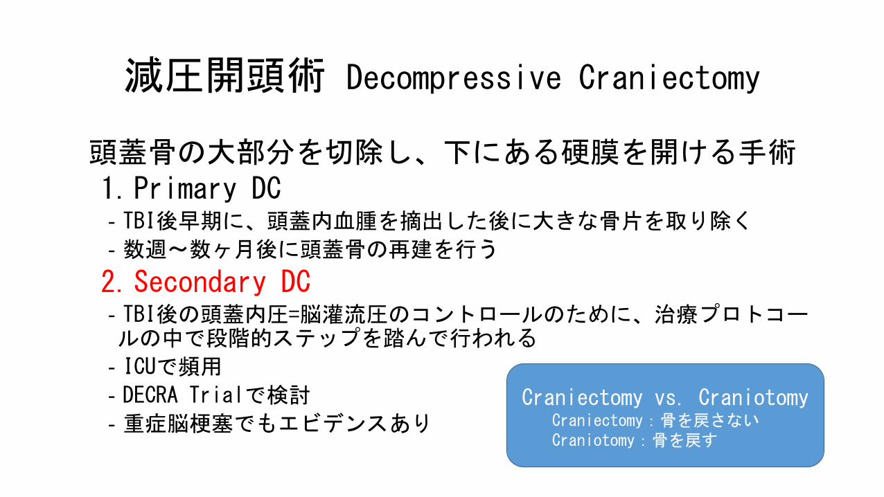
頭蓋骨の大部分を切除し、下にある硬膜を開ける手術 1.Primary DC ‐TBI後早期に、頭蓋内血腫を摘出した後に大きな骨片を取り除く
‐数週〜数ヶ月後に頭蓋骨の再建を行う
2.Secondary DC ‐TBI後の頭蓋内圧=脳灌流圧のコントロールのために、治療プロトコールの中で段階的ステップを踏んで行われる
‐ICUで頻用
‐DECRA Trialで検討
‐重症脳梗塞でもエビデンスあり Craniectomy vs. Craniotomy
Craniectomy:骨を戻さない Craniotomy:骨を戻す
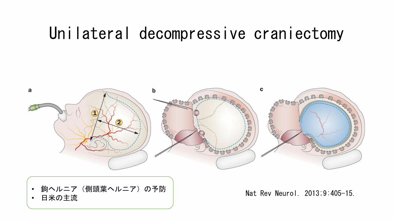
Unilateral decompressive craniectomy
Nat Rev Neurol. 2013;9:405-15. • 鉤ヘルニア(側頭葉ヘルニア)の予防 • 日米の主流
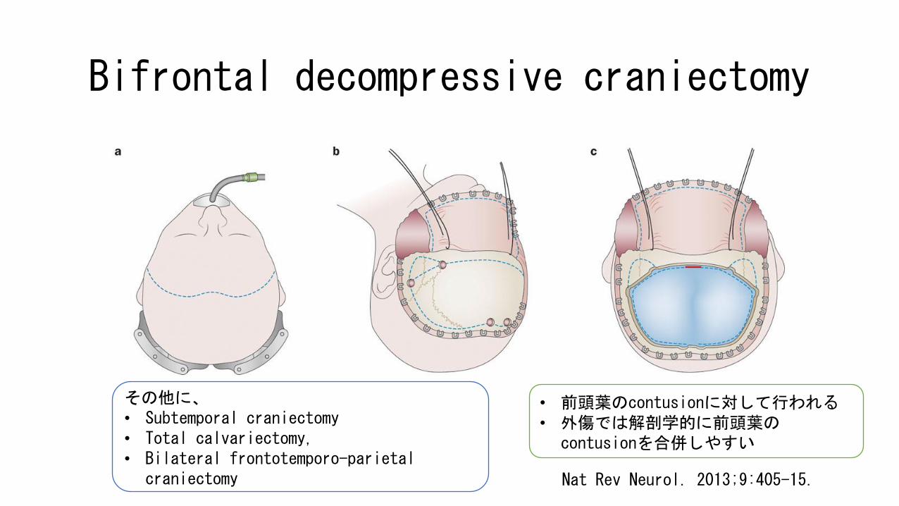
Bifrontal decompressive craniectomy
Nat Rev Neurol. 2013;9:405-15.
その他に、 • Subtemporal craniectomy • Total calvariectomy, • Bilateral frontotemporo-parietal
craniectomy
• 前頭葉のcontusionに対して行われる • 外傷では解剖学的に前頭葉の
contusionを合併しやすい

減圧開頭術の適応
1. 重症外傷性脳損傷 −脳軟部組織の不均一な病変 −局所(脳挫傷)・びまん性
2. 重症MCA梗塞
3. 動脈瘤性くも膜下出血
4. その他 −脳静脈血栓症 −脳炎 −代謝性脳症 −脳出血
Neurosurg Clin N Am 2013;24:375-391.

日本のガイドライン
【著しい脳腫脹に対する外科的治療方法】 初期のCT所見上、または保存的治療後やmass lesionの除去時術後に一側大脳半球または両側大脳半球の著しい脳腫脹を呈する症例に対して、外減圧術(一側または両側の広範囲減圧開頭術)を行うことがある
〔適応〕
保存的治療で脳圧亢進が制御不能 ( >30~35 mmHg) or 脳ヘルニアの進行
重篤な一次性脳幹損傷のある症例は通常適応外(例:GCS 3, 両側瞳孔散大固定)
若年者をよい適応とする傾向
〔時期〕
頭蓋内圧が持続的に≧40 mmHgを超える前に、可及的速やかに
重症頭部外傷 治療・管理のガイドライン 第2版, 2006

外傷性脳損傷(TBI)後に、頭蓋内血腫・脳挫傷・脳浮腫・水頭症といったmass effectによって頭蓋内圧が亢進する
N Engl J Med 2014;371:972.
TBI後の頭蓋内圧亢進⇒脳灌流圧↓⇒脳虚血 BMJ 2013;346:f1000.
TBI後の頭蓋内圧亢進は、死亡率と相関
Neurocrit Care 2006;4:8-13. Intensive Care Med 2012;38:1800-9.
頭蓋内圧のモニタリングと頭蓋内圧を低下させる処置は、TBI患者において、ルーチンで行われている。ただし、level 1エビデンスはなし。
Guidelines for the management of severe traumatic brain injury. J Neurotrauma 2007;24:Suppl 1:S55-8.

重症脳梗塞に対する減圧開頭術
重症MCA梗塞の発症48時間以内(早期)の減圧開頭術は、死亡率を減らし、良好な機能的転帰を増やす
Lancet Neurol 2007;6:215-22.
死亡率を下げるが、重度障害が残るため、症例は慎重に選択すべき。適応の患者選択に関しては、未検討。
Cochrane Database Syst Rev. 2012;1:CD003435.
重症脳梗塞(例:MCA梗塞)に対するDCは、予後を改善するが、植物状態や重度障害を増やす
Nat Rev Neurol 2013; 9:405-15.

The DECRA trial
減圧開頭術は、重症TBI後に治療不応性ICP亢進を呈した患者の予後・不幸な転帰を改善しない

P 重症の非穿通性頭部外傷の成人患者(15-59才) 初期治療 (1st tier therapy)に不応#のICP亢進あり
I early bifrontotemporoparietal decompressive craniectomy+保存的治療
C 保存的治療
O Unfavorable outcome (= GOS-E 1-4) 6ヶ月後のGOS-E*
*GOS-E: the Extended Glasgow Outcome Scale #1時間に15分以上、持続的 or 間欠的にICP >20mmHg
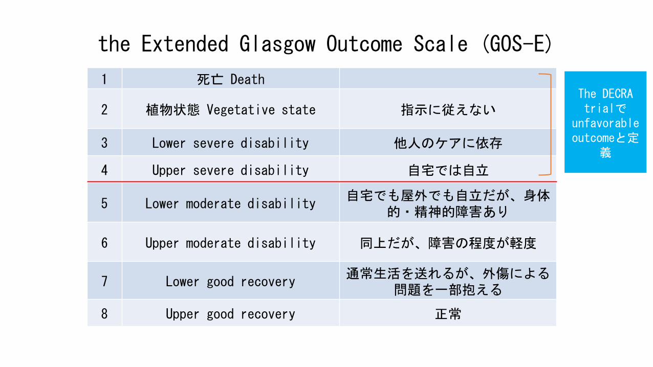
the Extended Glasgow Outcome Scale (GOS-E)
1 死亡 Death
2 植物状態 Vegetative state 指示に従えない
3 Lower severe disability 他人のケアに依存
4 Upper severe disability 自宅では自立
5 Lower moderate disability 自宅でも屋外でも自立だが、身体
的・精神的障害あり
6 Upper moderate disability 同上だが、障害の程度が軽度
7 Lower good recovery 通常生活を送れるが、外傷による
問題を一部抱える
8 Upper good recovery 正常
The DECRA trialで
unfavorable outcomeと定
義

Method & Characteristics of the Patients
多施設RCT 割付の隠蔽化の記載なし
期間:2002.12-2010.04
対象:155名の成人
年齢:23-24才(中央値)
手術群の方が、瞳孔の反応がある患者が少ない=重症(p=0.04)
標準治療群の18%が、cross overで救命のために手術を施行

Result
手術群 標準治療群 P値
死亡率* 19% 18% ?
不幸な転帰* 70% 51% P=0.02
(p=0.07)#
GOS-Eの 中央値* 3 4
P=0.03 (NS)#
平均ICP圧 (mmHg) 14.4 19.1 P<0.001
合併症 37% 17% ?
NNH 5
水頭症 10% 1% ?
NNH 11
#()内は、baselineの瞳孔の反応の影響に関して補正後
*全て6ヶ月の時点での評価

The DECRA trialの問題点
非常に限られた患者を対象としており、①この否定的な結果に外的妥当性があるか、②治療抵抗性頭蓋内圧亢進の定義に疑問が残る InclusionされたICP亢進の程度では、多くの臨床医はDCを検討しない
3478名スクリーニングされて、7年かけて155名しか集まっていないことは、TBI患者のうち、限られた集団を対象にしていることの表れ
N Engl J Med 2011; 364:1493.
N Engl J Med 2011;365:373.
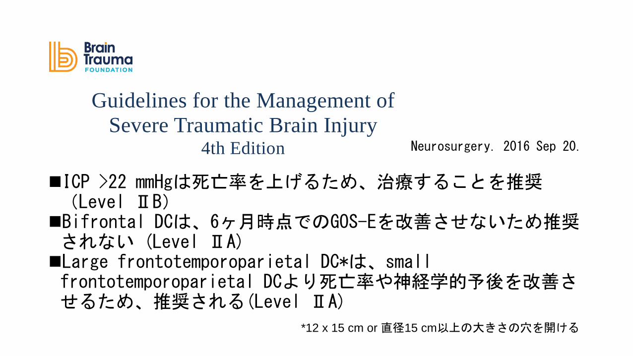
ICP >22 mmHgは死亡率を上げるため、治療することを推奨(Level ⅡB) Bifrontal DCは、6ヶ月時点でのGOS-Eを改善させないため推奨されない (Level ⅡA) Large frontotemporoparietal DC*は、small frontotemporoparietal DCより死亡率や神経学的予後を改善させるため、推奨される(Level ⅡA)
Guidelines for the Management of
Severe Traumatic Brain Injury 4th Edition
Nancy Carney, PhD Oregon Health & Science University, Portland, OR
Annette M. Totten, PhD Oregon Health & Science University, Portland, OR
Cindy O'Reilly, BS Oregon Health & Science University, Portland, OR
Jamie S. Ullman, MD Hofstra North Shore-LIJ School of Medicine, Hempstead, NY
Gregory W. J. Hawryluk, MD, PhD University of Utah, Salt Lake City, UT
Michael J. Bell, MD University of Pittsburgh, Pittsburgh, PA
Susan L. Bratton, MD University of Utah, Salt Lake City, UT
Randall Chesnut, MD University of Washington, Seattle, WA
Odette A. Harris, MD, MPH Stanford University, Stanford, CA
Niranjan Kissoon, MD University of British Columbia, Vancouver, BC
Andres M. Rubiano, MD El Bosque University, Bogota, Colombia; MEDITECH
Foundation, Neiva, Colombia
Lori Shutter, MD University of Pittsburgh, Pittsburgh, PA
Robert C. Tasker, MBBS, MD Harvard Medical School & Boston Children’s Hospital,
Boston, MA
Monica S. Vavilala, MD University of Washington, Seattle, WA
Jack Wilberger, MD Drexel University, Pittsburgh, PA
David W. Wright, MD Emory University, Atlanta, GA
Jamshid Ghajar, MD, PhD Stanford University, Stanford, CA
Reviewed for evidence-based integrity and endorsed by the American Association of
Neurological Surgeons and the Congress of Neurological Surgeons.
September 2016
Neurosurgery. 2016 Sep 20.
*12 x 15 cm or 直径15 cm以上の大きさの穴を開ける

今回の論文

The RESCUEicp Trial
N Engl J Med 2016;375:1119-30.
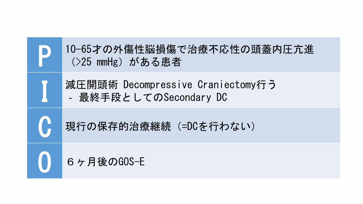
P 10-65才の外傷性脳損傷で治療不応性の頭蓋内圧亢進(>25 mmHg)がある患者
I 減圧開頭術 Decompressive Craniectomy行う ‐ 最終手段としてのSecondary DC
C 現行の保存的治療継続(=DCを行わない)
O 6ヶ月後のGOS-E

the Extended Glasgow Outcome Scale (GOS-E)
1 死亡 Death
2 植物状態 Vegetative state 指示に従えない
3 Lower severe disability 他人のケアに依存
4 Upper severe disability 自宅では自立
5 Lower moderate disability 自宅でも屋外でも自立だが、身体
的・精神的障害あり
6 Upper moderate disability 同上だが、障害の程度が軽度
7 Lower good recovery 通常生活を送れるが、外傷による
問題を一部抱える
8 Upper good recovery 正常
J Neurol Neurosurg Psychiatry 1981;44:285-93. J Neurotrauma 1998;15:573-85.
今回、 favorable outcomeと
定義

方法①
国際・多施設・parallel-group・優越性試験・RCT
重症TBIの急性期神経ケアを提供でき、脳外科が24時間体制で対応可能な病院で行われた
ICUで、プロトコールに従ってICPを25 mmHgに維持する様に、段階的な方法で行った(後述)
非劣性試験ではなく、通常の優越性を証明する研究
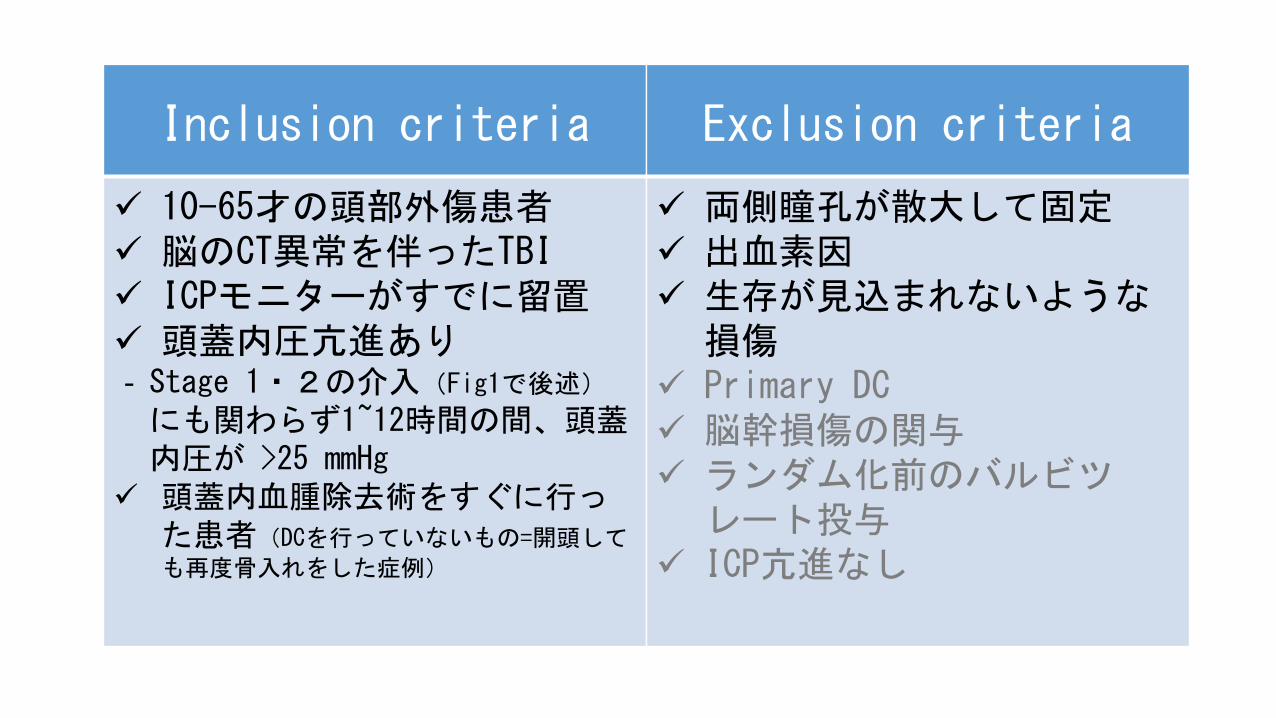
Inclusion criteria
10-65才の頭部外傷患者 脳のCT異常を伴ったTBI ICPモニターがすでに留置 頭蓋内圧亢進あり ‐Stage 1・2の介入(Fig1で後述)
にも関わらず1~12時間の間、頭蓋内圧が >25 mmHg
頭蓋内血腫除去術をすぐに行った患者(DCを行っていないもの=開頭して
も再度骨入れをした症例)
Exclusion criteria
両側瞳孔が散大して固定 出血素因 生存が見込まれないような
損傷 Primary DC 脳幹損傷の関与 ランダム化前のバルビツ
レート投与 ICP亢進なし

Stage 1
初期治療
頭部挙上 換気 鎮静 鎮痛 筋弛緩(オプション)
モニタリング
中心静脈圧 動脈圧 頭蓋内圧
その他の目標
脳灌流圧(=MAP – ICP) ≧60 mmHg
正常体温 正常血糖 軽度の低CO2血症(34-38 mmHg) 適切な酸素化(SpO2 >97%)
ICP >25 mmHg続くなら、
Stage 2へ

Stage 2
オプション治療
脳室開窓術 循環作動薬 マンニトール 高張生食 ループ利尿薬 低体温(<34は避ける) 中等度の低CO2血症 30-34 mmHg
ICP >25 mmHgが、1-12時間
持続するなら、Stage 3へ
Stage 1治療を継続 バルビツレートは許容しない 昇圧
浸透圧療法
浸透圧療法の効果増加

Stage 3
手術群と保存的治療群にランダムに割り付け
両群ともにStage 1, 2の治療は継続
手術群は、減圧開頭術を施行
保存的治療群はICPを下げるためのバルビツレート使用を許可
保存治療群でも、状態が悪化した場合には、手術を許可
手術群でも状態悪化時には、バルビツレート使用を許可
Stage 1での筋弛緩・すべてのStage 2治療・stage 3のバルビツレート投与は、研究をデザイン時にLevel 1 evidenceがなかったため、オプション治療とした
Cross overを許可

方法②
術式は以下の2つで、術者の自由裁量で決定
1. Hemi-craniectomy 片側半球の腫脹で推奨
2. Bifrontal craniectomy びまん性脳腫脹(両半球を障害)で推奨
手術はランダム化から4-6時間以内に行われるように推奨
ランダム化 1:1の置換ブロック法
臨床試験の場所に基づいた層別化
隠蔽化のためブロックは公開せず
Central telephone randamizaton serviceを使用
臨床試験のグループの割当の隠蔽化は確実 • Stage 3にいくまでrandomization codeが発行されないようにした

Outcome
Primary outcome(ランダム化から)6ヶ月後のGOS-E 2人の評価者により独立して評価 評価者は割付を知らない(盲検化)
Secondary outcome ‐12ヶ月・24ヶ月後のGOS-E ‐6ヶ月・12ヶ月・24ヶ月後の死亡率 ‐6ヶ月・12ヶ月・24ヶ月後のQOL ‐退院時GCS ‐ICPコントロールの評価 ‐ICU滞在時間 ‐在院日数 ‐経済的評価
簡易版健康調査 成人:36項目 小児:10項目
• 平均ICP圧 • ICP≧25 mmHgの時間 • 頭蓋内圧亢進Index20 or 25 • 脳低灌流Index

統計解析
2群間で15%の差が検出できるように設定 45% vs. 60%を想定
検出力 80%
フォローできなかった症例の割合を15%まで許容
サンプルサイズを算出⇒400症例
‘modified’ ITT解析 Follow upできなかった症例、同意が取れなかった症例を除外 Cross overは許容
GOS-Eの2群間比較でχ2検定を使用
連続変数に関しては、Mann-Whitney U testを使用

Result

Trial flowchart
期間:2004年1月〜2014年3月
2008名が参加
409名(52施設、20カ国)がランダム化へ 291名(71%がUK)
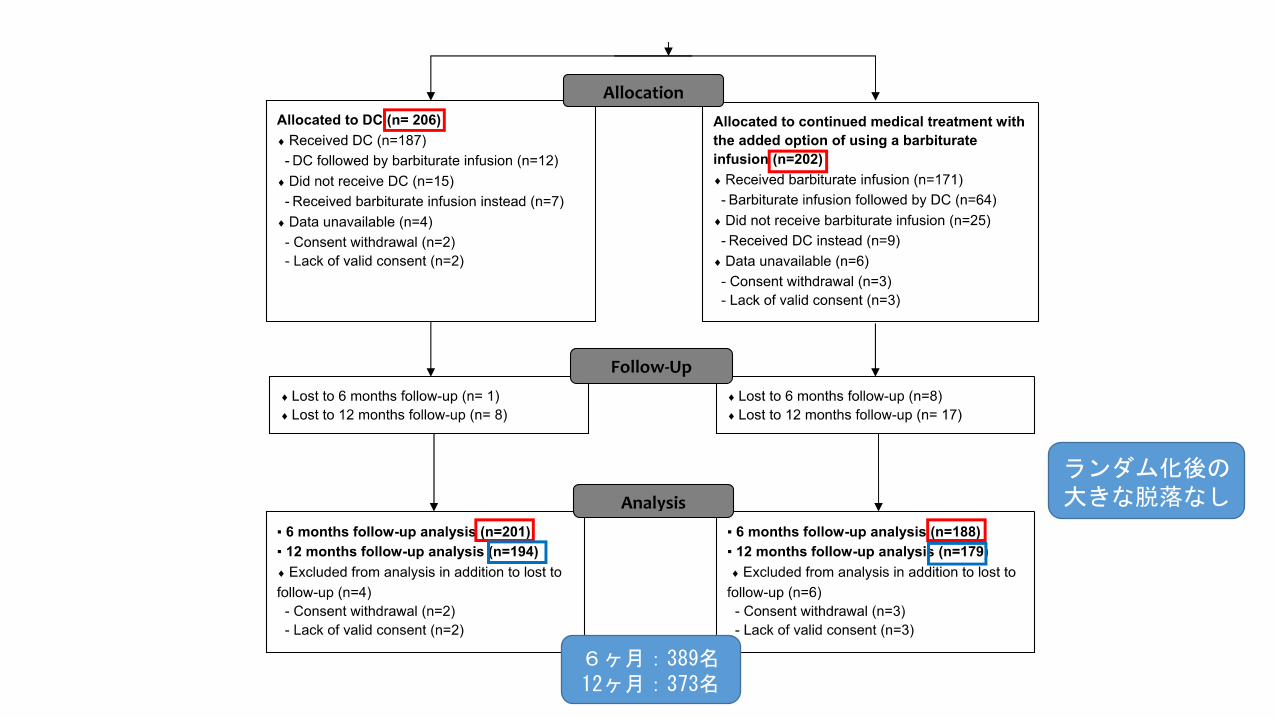
6ヶ月:389名 12ヶ月:373名
ランダム化後の大きな脱落なし

患者背景
平均30才で、男性が80% ‐≦40才が70%
GCSのMotor ≦2が50% ‐GCS≦8が70%
40%が頭以外の損傷を合併
80%にびまん性損傷あり
20%に占拠性病変あり ‐Marshall classification
若年男性の重症頭部外傷患者が中心
手術群で薬物・アルコール乱用の既往が少ない(p=0.02)
その他の背景は両群で同等
狙った患者層を抽出できた

介入
ランダム化前の治療(Stage 1・2のオプション)に差はなし
手術までの時間(中央値):2.2時間
バルビツレート投与 投与までの時間(中央値):1.5h
投与期間(中央値):53h
DCの方法 Bifrotal (63%) Unilateral (37%)
保存的治療群の約40%が最終的に、
DC施行!
手術できなかった理由 • 手術待機中に更に
ICP増悪 • 凝固異常 • 大量の鼻出血
ランダム化後
手術群でバルビツレート追加投与が必要となったのは9%のみ
保存的治療群の90%で追加投与が必要だった
ランダム化前
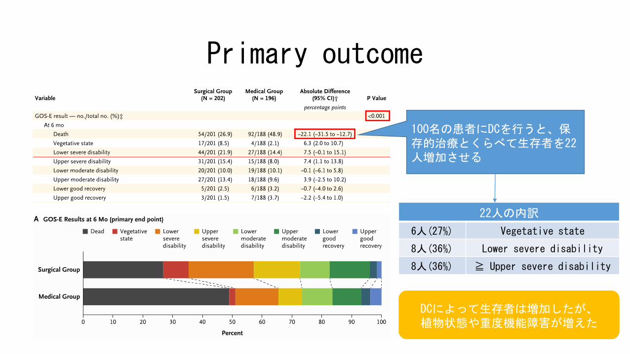
Primary outcome
100名の患者にDCを行うと、保存的治療とくらべて生存者を22人増加させる
22人の内訳
6人(27%) Vegetative state
8人(36%) Lower severe disability
8人(36%) ≧ Upper severe disability
DCによって生存者は増加したが、 植物状態や重度機能障害が増えた

Secondary outcome
100名の患者にDCを行うと、保存的治療とくらべて生存者を22人増加
22人の内訳
5人(23%) Vegetative state
4人(18%) Lower severe disability
13人(59%) ≧ Upper severe disability
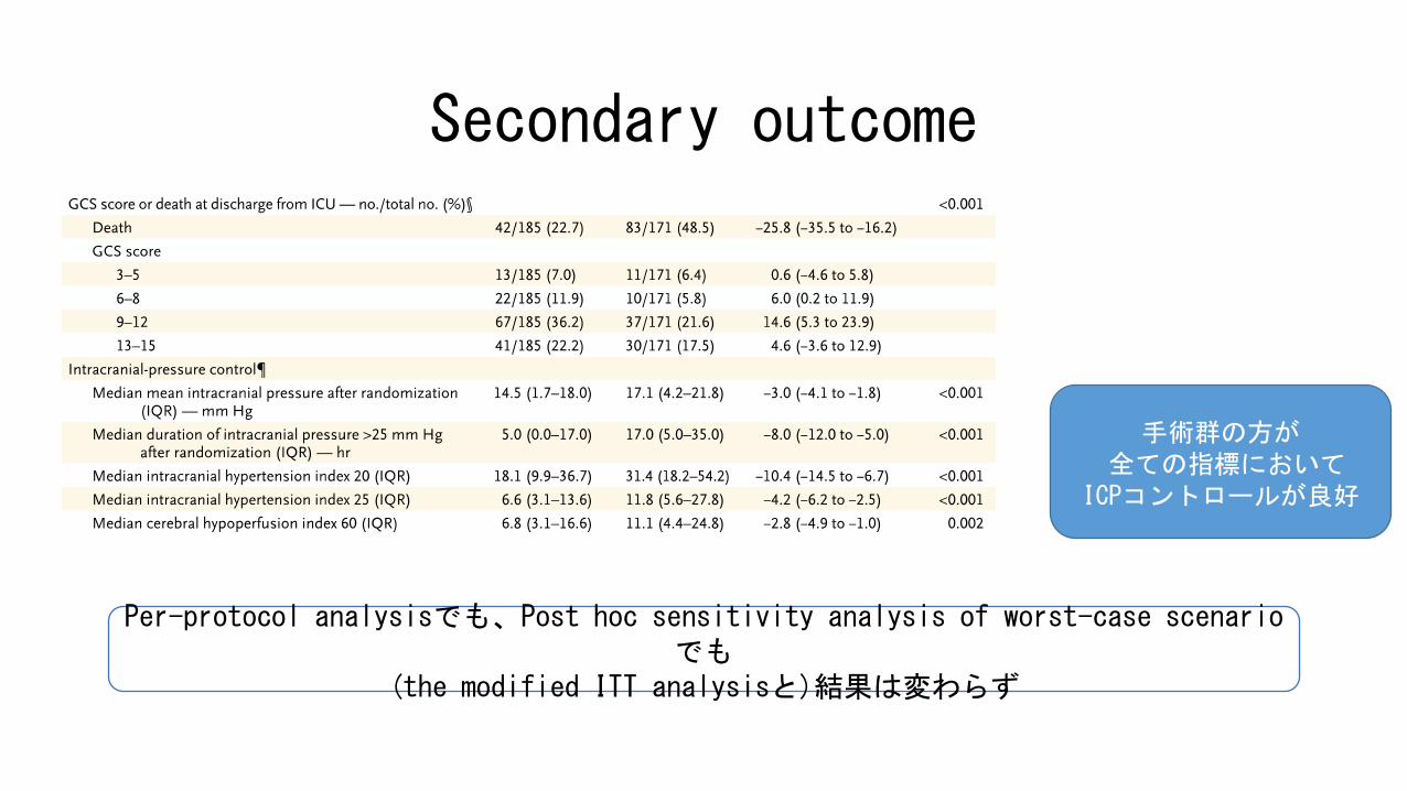
Secondary outcome
手術群の方が 全ての指標において ICPコントロールが良好
Per-protocol analysisでも、Post hoc sensitivity analysis of worst-case scenarioでも
(the modified ITT analysisと)結果は変わらず

Complications
17
Table S9 – Complications and adverse events
Surgical group (N = 202)
Medical group (N = 196)
P Value
No. (%) of patients with at least one reported complication or adverse event
33 (16.3) 18 (9.2) 0.033
Table S10 – Types of complications and adverse events
Types of reported complications and adverse events – no. of patients affected
Surgical group
Medical group
Acute kidney injury 1
Acute respiratory distress syndrome 2
Arrhythmia 1 1
Bleeding (surgical complication) 4
Bleeding (surgical complication; led to death) 1
Cardiac arrest 1 2
Cardiogenic shock 1
Cerebral infarction 2 2
Deep venous thrombosis 1
Diabetes insipidus 1
Disseminated intravascular coagulation 1
Hydrocephalus 1
Hypotension 2
Intra‐operative respiratory failure 2
Liver failure 1
Multiple organ dysfunction syndrome 1
Myocardial infarct 2
Pneumonia 4 6
Post‐operative hematoma 5
Pulmonary embolism 1
Seizures 1
Sepsis (not otherwise specified) 1 2
Shock (not otherwise specified) 1 2
Subdural collection 1 1
Subgaleal collection 3 1
Surgical site infection 5 1
Venous sinus injury 1
Ventriculostomy / CSF infection 1 2
A total number of 37 complications and adverse events were reported in 33 patients of the surgical
group. A total number of 32 complications and adverse events were reported in 18 patients of the
medical group.
手術群の方が合併症・有害事象を起こした患者「数」は多かった ただし、延べの発症率はおそらく同等 内訳から、合併症・有害事象の差は大きくないと考えられる
手術群 保存群 P値
1つ以上の合併症・有害事象の報告がされた患者
33 (16.3%)
18 (9.2%)
0.03
総報告数(延べ) 37 32 -

Discussion The DECRA trial The RESCUEicp trial
評価したDC Early craniectomy
stage2の治療として 受傷後72時間以内に施行
最終手段 stage 3としてのCraniectomy
対象としたICP亢進
中等度 1時間に15分以上、持続的 or 間
欠的にICP >20mmHg
重症 Stage2の治療でもICP >25 mmHgが、
1-12時間持続
頭蓋内血腫合併患者
除外 約20%含む
Hemi-craniectomy
行わない 選択肢の一つ
結果 死亡率:同等 GOS-E:悪化
死亡率:低下 GOS-E:同等〜悪化

Discussion
今回の治療プロトコールは、TBI後の頭蓋内圧亢進に対する低体温療法の研究のものに類似
N Engl J Med 2015;373:2403-12. (2015/11/24 Journal Club)
DCによる生存は、依存した生存も、自立した生存も増やす可能性あり⇒臨床家は、家族と慎重に相談する必要がある
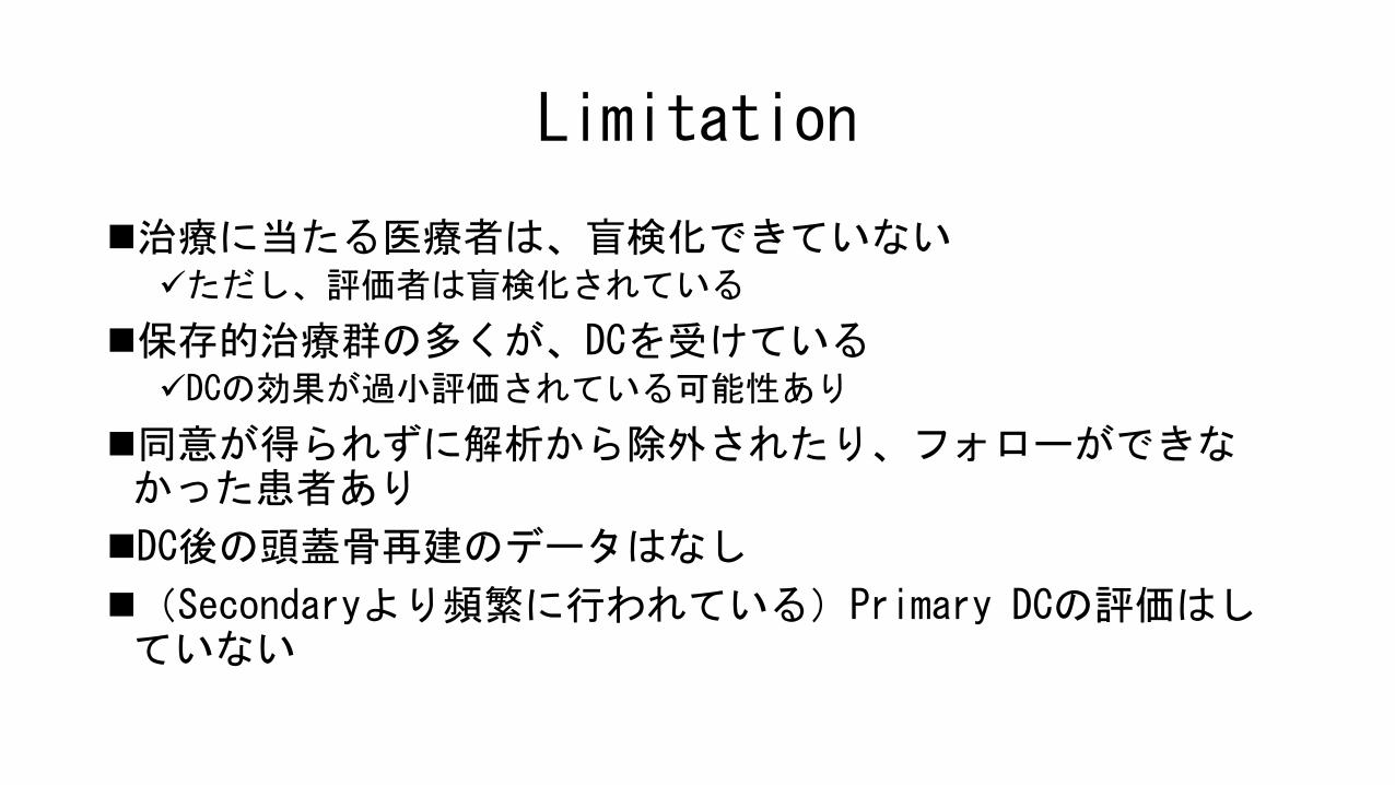
Limitation
治療に当たる医療者は、盲検化できていない ただし、評価者は盲検化されている
保存的治療群の多くが、DCを受けている DCの効果が過小評価されている可能性あり
同意が得られずに解析から除外されたり、フォローができなかった患者あり
DC後の頭蓋骨再建のデータはなし
(Secondaryより頻繁に行われている)Primary DCの評価はしていない

Conclusion
頭部外傷後の重症・治療抵抗性ICP亢進への減圧開頭術は、保存的治療と比べて死亡率を22%低下させた
手術によって保存的治療と比較して、植物状態・重度機能障害が増加し、中等度障害〜良好な回復は同等であった

治療不応性のICP亢進の定義が汎用化されやすい内容であり、外科的処置を速やかに行うことが可能であった
ある一定の制限内で、臨床医が治療選択を行うことが可能であった
① 外科医の手術方法
63% Bifrontal approach vs 37% Unilateral approach
※ 米国(少数);上記とは逆の割合
② Cross overによる結果
保存的加療群の37% → DCが必要
外科的治療群の9% → バルビツレート投与が必要
※ 保存的加療のみでは頭蓋内圧亢進が対応不十分なことがある
手術施行は生存予後の改善には寄与するが、神経予後を改善させるものではないかもしれない
日本でもUnilateralが主流

重症外傷性脳損傷に減圧開頭術を行うか
保存的治療を行った上でコントロールできないICP亢進に対して、植物状態や重度機能障害を呈する可能性があることを踏まえて、個々の症例に応じて検討
手術適応に関する更なる検証や費用対効果の報告が待たれる
24時間以内の再手術は日本では保険適応なし • 「ICPセンサー入れて、圧が高くて再手術」というのは非現実的
現在ICP亢進を予防する方向に進んでおり、Primary DCの方が現実的
脳外科医の意見