Embed Size (px)
Citation preview
Effect of Streptococcus salivarius K12 on the In Vitro Growth ofCandida albicans and Its Protective Effect in an Oral CandidiasisModel
Sanae A. Ishijima,a Kazumi Hayama,a Jeremy P. Burton,b Gregor Reid,d Masashi Okada,a Yuji Matsushita,c and Shigeru Abea
Teikyo University Institute of Medical Mycology, Tokyo, Japana; BLIS Technologies Ltd., Centre for Innovation, University of Otago, Dunedin, New Zealandb; Tradepia Co.,Tokyo, Japanc; and Department of Microbiology and Immunology, University of Western Ontario, London, Ontario, Canadad
Oral candidiasis is often accompanied by severe inflammation, resulting in a decline in the quality of life of immunosuppressedindividuals and elderly people. To develop a new oral therapeutic option for candidiasis, a nonpathogenic commensal oral pro-biotic microorganism, Streptococcus salivarius K12, was evaluated for its ability to modulate Candida albicans growth in vitro,and its therapeutic activity in an experimental oral candidiasis model was tested. In vitro inhibition of mycelial growth of C. al-bicans was determined by plate assay and fluorescence microscopy. Addition of S. salivarius K12 to modified RPMI 1640 culturemedium inhibited the adherence of C. albicans to the plastic petri dish in a dose-dependent manner. Preculture of S. salivariusK12 potentiated its inhibitory activity for adherence of C. albicans. Interestingly, S. salivarius K12 was not directly fungicidalbut appeared to inhibit Candida adhesion to the substratum by preferentially binding to hyphae rather than yeast. To determinethe potentially anti-infective attributes of S. salivarius K12 in oral candidiasis, the probiotic was administered to mice withorally induced candidiasis. Oral treatment with S. salivarius K12 significantly protected the mice from severe candidiasis. Thesefindings suggest that S. salivarius K12 may inhibit the process of invasion of C. albicans into mucous surfaces or its adhesion todenture acrylic resins by mechanisms not associated with the antimicrobial activity of the bacteriocin. S. salivarius K12 may beuseful as a probiotic as a protective tool for oral care, especially with regard to candidiasis.
The overgrowth of Candida albicans, which is one of the mem-bers of the oral microbial flora in a healthy human, causes
pathogenic symptoms such as oral candidiasis. Oral candidiasisaccompanied with severe inflammation can significantly degradethe quality of life of immunosuppressed individuals and elderlypeople (9). It can cause a variety of mucosal infections in thegastrointestinal, respiratory, and genital tracts and is a major causeof oral and esophageal infections (9, 23, 29). Oral candidiasis iscommon in patients with advanced AIDS, hyposalivation, anddiabetes mellitus, those on antibiotic therapy or immunosuppres-sive drugs, and those who have poor oral hygiene (9, 22, 23, 29).
The probiotic strain Streptococcus salivarius K12 was originallyisolated from the saliva of a healthy child and produces severalmegaplasmid-encoded bacteriocin-like inhibitory substances(BLISs), such as the lantibiotics salivaricin A and salivaricin B (11,13, 31). It has been used commercially as a probiotic for more thana decade and has numerous studies supporting its safety (3, 4, 5).S. salivarius strains have been reported to inhibit the biofilm for-mation of Streptococcus mutans (13, 19, 28), and Streptococcus sali-varius K12 has been shown to have the ability to inhibit variouspotentially deleterious upper respiratory tract bacteria, such asStreptococcus pyogenes and Streptococcus pneumoniae (13, 31), anddecrease oral malodor (2). These properties suggest that S. sali-varius K12 might be widely applied as a management tool for oralhealth applications.
C. albicans is a polymorphic yeast and grows predominantly asyeast, pseudohyphae, or hyphae (23). Mycelial growth of C. albi-cans is often observed in mucosal infection and is considered tocontribute to pathogenesis by biofilm formation (22). In thisstudy, we aimed to elucidate the potential mechanisms of Strepto-coccus salivarius K12 suppression of the mycelial growth of the C.
albicans first by in vitro analysis and then by testing an experimen-tal oral candidiasis model with mice with furry white tongues.
MATERIALS AND METHODSCandida albicans and Streptococcus salivarius. The C. albicans strainTIMM1768 was isolated clinically from the blood of a candidiasis patientand maintained at Teikyo University Institute of Medical Mycology; thisstrain, which was shown to induce oral candidiasis in a murine model, hasbeen used for animal experiments (12, 14). Cultures were stored at �80°Cin Sabouraud dextrose broth (Becton Dickinson, MD) containing 0.5%yeast extract (Becton Dickinson, MD) and 10% glycerol (vol/vol, finalconcentration) until use. Strain TIMM1768 was cultured on a Sabourauddextrose agar plate for 18 h at 37°C, and the cells were harvested with amicrospatula and suspended in RPMI 1640 medium containing 2.5% fetalcalf serum (RPMI 1640 medium). The cultured C. albicans cells were usedfor in vitro germ tube formation, the mycelial growth experiment, and alsoin vivo oral inoculation of Candida.
S. salivarius K12 is a commercially available probiotic that was origi-nally isolated from the oral cavity of a child. It was supplied as a freeze-dried powder at 2 � 1011 CFU per gram of material tested and was usedwith CAB K12 agar, which consisted of Columbia blood agar base (BectonDickinson, MD), 0.5% yeast extract (Becton Dickinson, MD), 0.25% glu-cose, and 0.1% calcium carbonate (pH 7.3 � 0.2).
Measurement of antimicrobial activity of bacteriocins produced byS. salivarius K12. To determine if the bacteriocins or other secretorymolecules from S. salivarius K12 inhibited C. albicans TIMM1768, a de-
Received 3 October 2011 Accepted 10 January 2012
Published ahead of print 20 January 2012
Address correspondence to Sanae A. Ishijima, [email protected].
Copyright © 2012, American Society for Microbiology. All Rights Reserved.
doi:10.1128/AEM.07055-11
2190 aem.asm.org 0099-2240/12/$12.00 Applied and Environmental Microbiology p. 2190–2199
on March 28, 2019 by guest
http://aem.asm
.org/D
ownloaded from
ferred antagonism assay was employed. This was conducted essentially asdescribed by Tagg and Bannister (25), in duplicate, using the nine bacte-rial indicator strains described to be positive controls for S. salivarius K12bacteriocin production and also applying the C. albicans TIMM1768strain. In brief, S. salivarius K12 was preliminarily cultured on a CAB agar(with 5% blood, 0.1% CaCO3) plate to form a 1-cm-wide streak. Afterincubation of the plate at 37°C under 5% CO2 for 18 to 24 h, the culture ofS. salivarius K12 was removed from the plate using a clean microscopeslide and sterilized with chloroform vapors for 30 min. The plates wereaired for 30 min in an extraction hood. Indicator bacterial strains as well asC. albicans were then inoculated horizontally across the original but nowsterile S. salivarius K12 streak. Plates were then reincubated for 18 h. Theinhibitory effect of microbial growth was evaluated as follows: �, no in-hibition of the test organism; �, inhibition of the test organism only overthe primary inoculation; ��, inhibition of the test organism just beyondthe primary inoculation; ���, inhibition of the test organism muchbeyond the primary inoculation.
In vitro assay of germ tube formation and mycelial growth of Can-dida albicans. The ability of C. albicans cells to undergo germ tube for-mation or mycelial growth with S. salivarius K12 was assessed as describedbelow.
(i) Germ tube formation analysis. One hundred microliters of C.albicans cells was aliquoted into 96-well microtiter plates (1 � 104 CFUper well for morphological analysis, 5 � 105 CFU per well for crystal violet[CV] staining), 100-�l serial dilutions of freeze-dried S. salivarius K12powder were then added to the plates, which made final concentrations of30 mg/ml (3.0 � 109 CFU/ml) to 0.12 mg/ml (1.2 � 107 CFU/ml), and theplates were incubated at 37°C in 5% CO2 in air for 3 h. Germ tube forma-tion was assessed microscopically: cells were fixed with 70% ethanol andstained with CV as described by Abe et al. (1) and Kamagata-Kiyoura et al.(14).
(ii) Mycelial growth analysis. Mycelial growth analysis was carriedout as described for the germ tube formation assay, except that the inoc-ulum per well was 500 cells in 100 �l and the culture period was length-ened to 16 h. Mycelial growth of C. albicans cells was determined as de-scribed by Abe et al. (1). Culture medium for in vitro assays was composedof diluted RPMI 1640 (1:3; Sigma Chemical Co., St. Louis, MO) contain-ing 0.8% fetal calf serum, 20 mM HEPES buffer, pH 7.2, 2 mM urea, and10 mg/ml D-glucose with or without antibiotics (60 �g/ml of benzylpen-icillin potassium [Wako, Japan] and kanamycin sulfate [Wako, Japan]),according to the nutritional requirements of S. salivarius (6). The plank-tonic cells were centrifuged, stained with 50% lactophenol blue solution(containing 1 mg/ml of methyl blue [C.I. 42780], 204 mg/ml of phenol,247 mg/ml of lactic acid, and 502 mg/ml of glycerol; Merck, Germany) insaline, and observed by microscopy.
Yeast viability assay using fluorescence microscopy. The effect of S.salivarius K12 on C. albicans viability was detected by use of a two-colorfluorescent probe (FUN1; F-7030; Molecular Probes, Eugene, OR), a live/dead yeast viability kit, and fungal surface labeling with a reagent of a thirdcolor (calcofluor white M2R; Molecular Probes, Eugene, OR). C. albicansand S. salivarius K12 were cultured as described above. In brief, C. albicansand S. salivarius K12 were combined in adequate culture medium andcultured for 1 to 3 h in a CO2 incubator. After centrifugation at 3,000 rpmfor 3 min and one-time washing with GH solution (2% glucose in 10 mMHEPES buffer, pH 7.2), the GH solution was replaced with GH solutioncontaining 20 �M FUN1 with 5 �M calcofluor white M2R. After incuba-tion for 30 min at room temperature, cells were observed with a fluores-cence microscope (BH50; Olympus, Japan) equipped with a WU (widerange of UV excitation), (WU), WBV (wide range of blue-violet excita-tion), WG (wide range of green excitation), and NB (narrow range of UVexcitation) filter assortment. Staining with FUN1 was observed using NBand calcofluor white WU. All images were taken as digital data with aDC200 camera (Leica, Germany), and the digital data were inserted intothe IM50 program and recorded.
Murine oral candidiasis model. All animal experiments were per-formed in accordance with the guidelines for the care and use of animalsapproved by Teikyo University. The derivation of the murine oral candi-diasis model has been described previously (15, 27). Six-week-old femaleICR mice (Charles River Japan, Inc., Yokohama, Kanagawa, Japan) wereused for all animal experiments. The mice were randomized, kept in cageshousing 3 to 4 individuals, and given food and water ad libitum. Duringthe experimental period, the photoperiods were adjusted to 12 h of lightand 12 h of darkness daily, and the environmental temperature was main-tained at 21°C. To induce an immunosuppressed condition, 100 mg ofprednisolone (Mitaka Pharmaceutical Co., Japan) per kg of body weightwas injected subcutaneously to mice 20 to 24 h before oral inoculation.Prior to prednisolone administration, 15 mg/ml of tetracycline hydro-chloride (Takeda Shering Purau Animal Health Co., Japan) was adminis-tered in drinking water for 24 h. On the day of infection, animals wereanesthetized by intramuscular injection with 14.4 mg/kg of chlorproma-zine chloride in the femur, after which they were orally inoculated with2.0 � 108 CFU/ml of C. albicans TIMM1768 in modified RPMI 1640medium. Oral inoculation was performed by means of rubbing and roll-ing a cotton swab (baby cotton buds; Johnson & Johnson Co., Tokyo,Japan) inside all parts of the mouth. The number of Candida cells inocu-lated in the oral cavity was calculated to be 1 � 106 CFU/mouse on thebasis of the difference in viable cell number adhering to the cotton swabsbefore and just after oral inoculation, as described by Takakura et al. (27).
Oral administration of Streptococcus salivarius K12. Fifty microli-ters of S. salivarius K12 solution, fluconazole (2 mg/ml), or distilled waterwas administered into the oral cavity of the Candida-inoculated mice atfive time points: 24 h and 3 h before and 3, 24, and 27 h after C. albicansinoculation. The total numbers of mice in each group during two differenttrials were as follows: water control, n � 15; S. salivarius K12 at 7.5 mg/ml,n � 7; S. salivarius K12 at 15 mg/ml, n � 12; 30 mg/ml, n � 15; andfluconazole at 2 mg/ml, n � 6. Administration was undertaken using arounded-top needle to spread the treatment over all parts of the mouth.An active control of 50 �l of fluconazole solution (2 mg/ml) was similarlyadministered.
Scoring severity of oral infection. The procedure of scoring the sever-ity of oral infection was performed as described previously (27). Forty-eight hours after inoculation, mice were sacrificed by cervical dislocationand the severity of the lesion of the tongue was evaluated by scoring the furcoating on each tongue and the squamous disorder as follows: 0, normal;1, fur on less than 20% of the tongue; 2, fur on more than 21% but lessthan 90% of the tongue; 3, fur on more than 91% of the tongue and on thesquamous layer; 4, thick fur on more than 91% of the tongue and on thesquamous layer (12, 27).
Evaluation of number of viable Candida cells on murine tongues. At48 h after inoculation, the cheek, tongue, and soft palate of each mousewas swabbed uniformly using a cotton swab, and the swab was used formicrobiological evaluation. After swabbing, the cotton end was cut offand placed in 3 ml of sterile saline. Candida cells were resuspended bymixing on a vortex mixer and diluted with a series of 20-fold and 100-folddilutions of sterile saline. Fifty microliters of each dilution was incubatedon a Candida GS agar plate (selection medium for Candida; Eiken Chem-ical Co. Ltd., Japan) for 20 h at 37°C. The Candida cells were counted, andthen the total numbers per swab were calculated and reported as numbersof CFU.
Histology. For histological study, the tongues were resected at the baseof the tongue, fixed with 4% paraformaldehyde (pH 7.4) at 4°C, dehy-drated by ethanol series, and embedded in paraffin in accordance withcommon procedure. Specimens were sectioned to an 8-�m thicknessalong the longitudinal center line. Sections on the slide were deparaf-finized by xylene, rehydrated by ethanol series, and stained with periodicacid-Schiff (PAS).
Statistical analysis. The score data were compared by the nonparametricMann-Whitney U test. Statistical analysis of the log10 number of CFU of C.albicans isolated from each mouse part was compared using a Student’s t test.
Effect of Probiotic S. salivarius K12 on Candidiasis
April 2012 Volume 78 Number 7 aem.asm.org 2191
on March 28, 2019 by guest
http://aem.asm
.org/D
ownloaded from
P values of �0.05 were considered statistically significant. All mean valuesgiven in the text include the standard deviation of the mean.
RESULTSInhibition of Candida albicans attachment to plastic substra-tum by S. salivarius BLIS K12. Mycelial growth of C. albicans isconsidered to contribute to the pathogenesis of mucosal candidi-
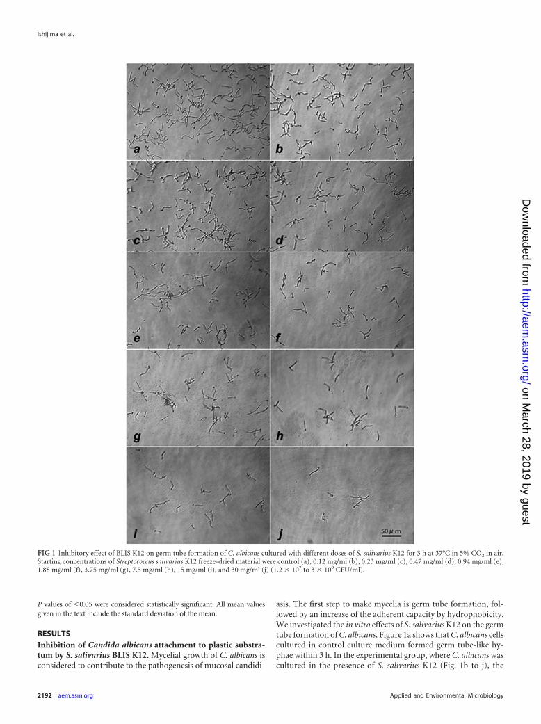
asis. The first step to make mycelia is germ tube formation, fol-lowed by an increase of the adherent capacity by hydrophobicity.We investigated the in vitro effects of S. salivarius K12 on the germtube formation of C. albicans. Figure 1a shows that C. albicans cellscultured in control culture medium formed germ tube-like hy-phae within 3 h. In the experimental group, where C. albicans wascultured in the presence of S. salivarius K12 (Fig. 1b to j), the
FIG 1 Inhibitory effect of BLIS K12 on germ tube formation of C. albicans cultured with different doses of S. salivarius K12 for 3 h at 37°C in 5% CO2 in air.Starting concentrations of Streptococcus salivarius K12 freeze-dried material were control (a), 0.12 mg/ml (b), 0.23 mg/ml (c), 0.47 mg/ml (d), 0.94 mg/ml (e),1.88 mg/ml (f), 3.75 mg/ml (g), 7.5 mg/ml (h), 15 mg/ml (i), and 30 mg/ml (j) (1.2 � 107 to 3 � 109 CFU/ml).
Ishijima et al.
2192 aem.asm.org Applied and Environmental Microbiology
on March 28, 2019 by guest
http://aem.asm
.org/D
ownloaded from

morphological shape and size of the Candida cells appeared to bealmost the same as those for the control group; however, the ad-herence of the mycelial form to the plastic substratum was weakerand the mycelial numbers on the plastic bottom were dose-de-pendently reduced in the presence of more than 0.94 mg/ml offreeze-dried S. salivarius K12 starting material, equating to ap-proximately 1 � 108 CFU/ml.
Figure 2 shows the results of the number of viable C. albicanscells growing in planktonic form, which were found to increase,according to the concentration of S. salivarius K12, to more than
1.9 to 3.8 mg/ml (approximately 2.0 � 108 to 4.0 � 108 CFU/ml).These results indicate that S. salivarius K12 increased the numberof planktonic Candida cells in culture medium. The planktoniccells, including unattached mycelia, were centrifuged and stainedwith lactophenol blue. Figure 3 shows that mycelial cells of Can-dida attached to and were surrounded by S. salivarius K12.
Effective inhibition of C. albicans attachment to substratumby viable S. salivarius K12. Although S. salivarius K12 was shownto bind to mycelial growth of Candida at 3 h of culture and inhibitCandida adherence to plastic plates, it is not clear whether these
FIG 2 Number of planktonic cells after 3 h culture of C. albicans with various concentrations of S. salivarius K12. C. albicans was cultured with different dosesof S. salivarius K12 for 3 h at 37°C in 5% CO2 in air. After the cultured plate was shaken, the supernatant was collected, diluted, and seeded on a GS agar plate fordetermining the number of planktonic cells. The experiments were performed in duplicate.
FIG 3 Microscopic observation of interaction between S. salivarius K12 and Candida planktonic cells after 3 h culture and staining with lactophenol blue. Blackarrows, Candida cells; white arrows, S. salivarius.
Effect of Probiotic S. salivarius K12 on Candidiasis
April 2012 Volume 78 Number 7 aem.asm.org 2193
on March 28, 2019 by guest
http://aem.asm
.org/D
ownloaded from
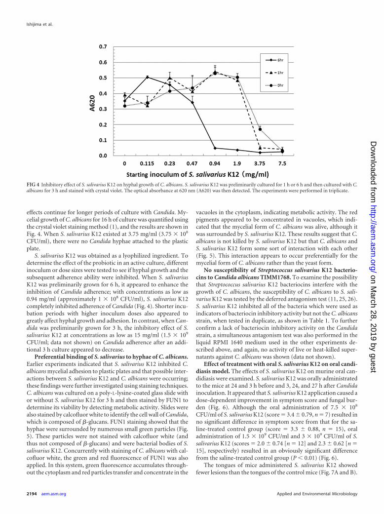
effects continue for longer periods of culture with Candida. My-celial growth of C. albicans for 16 h of culture was quantified usingthe crystal violet staining method (1), and the results are shown inFig. 4. When S. salivarius K12 existed at 3.75 mg/ml (3.75 � 108
CFU/ml), there were no Candida hyphae attached to the plasticplate.
S. salivarius K12 was obtained as a lyophilized ingredient. Todetermine the effect of the probiotic in an active culture, differentinoculum or dose sizes were tested to see if hyphal growth and thesubsequent adherence ability were inhibited. When S. salivariusK12 was preliminarily grown for 6 h, it appeared to enhance theinhibition of Candida adherence; with concentrations as low as0.94 mg/ml (approximately 1 � 108 CFU/ml), S. salivarius K12completely inhibited adherence of Candida (Fig. 4). Shorter incu-bation periods with higher inoculum doses also appeared togreatly affect hyphal growth and adhesion. In contrast, when Can-dida was preliminarily grown for 3 h, the inhibitory effect of S.salivarius K12 at concentrations as low as 15 mg/ml (1.5 � 109
CFU/ml; data not shown) on Candida adherence after an addi-tional 3 h culture appeared to decrease.
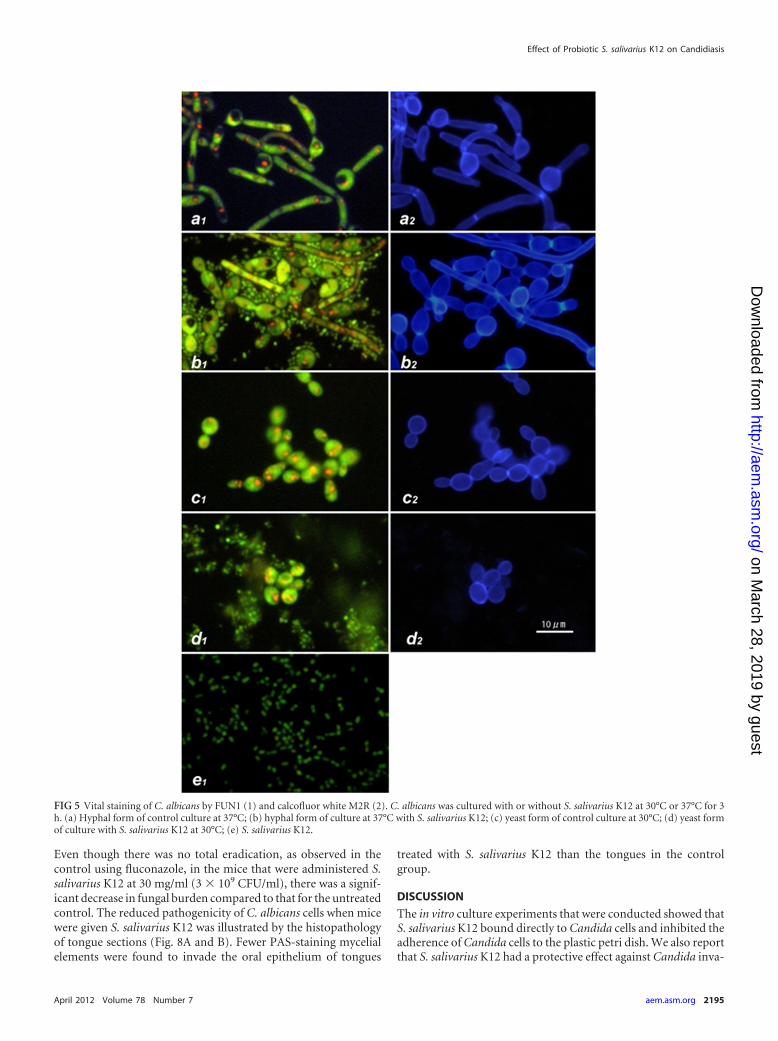
Preferential binding of S. salivarius to hyphae of C. albicans.Earlier experiments indicated that S. salivarius K12 inhibited C.albicans mycelial adhesion to plastic plates and that possible inter-actions between S. salivarius K12 and C. albicans were occurring;these findings were further investigated using staining techniques.C. albicans was cultured on a poly-L-lysine-coated glass slide withor without S. salivarius K12 for 3 h and then stained by FUN1 todetermine its viability by detecting metabolic activity. Slides werealso stained by calcofluor white to identify the cell wall of Candida,which is composed of �-glucans. FUN1 staining showed that thehyphae were surrounded by numerous small green particles (Fig.5). These particles were not stained with calcofluor white (andthus not composed of �-glucans) and were bacterial bodies of S.salivarius K12. Concurrently with staining of C. albicans with cal-cofluor white, the green and red fluorescence of FUN1 was alsoapplied. In this system, green fluorescence accumulates through-out the cytoplasm and red particles transfer and concentrate in the
vacuoles in the cytoplasm, indicating metabolic activity. The redpigments appeared to be concentrated in vacuoles, which indi-cated that the mycelial form of C. albicans was alive, although itwas surrounded by S. salivarius K12. These results suggest that C.albicans is not killed by S. salivarius K12 but that C. albicans andS. salivarius K12 form some sort of interaction with each other(Fig. 5). This interaction appears to occur preferentially for themycelial form of C. albicans rather than the yeast form.
No susceptibility of Streptococcus salivarius K12 bacterio-cins to Candida albicans TIMM1768. To examine the possibilitythat Streptococcus salivarius K12 bacteriocins interfere with thegrowth of C. albicans, the susceptibility of C. albicans to S. sali-varius K12 was tested by the deferred antagonism test (11, 25, 26).S. salivarius K12 inhibited all of the bacteria which were used asindicators of bacteriocin inhibitory activity but not the C. albicansstrain, when tested in duplicate, as shown in Table 1. To furtherconfirm a lack of bacteriocin inhibitory activity on the Candidastrain, a simultaneous antagonism test was also performed in theliquid RPMI 1640 medium used in the other experiments de-scribed above, and again, no activity of live or heat-killed super-natants against C. albicans was shown (data not shown).
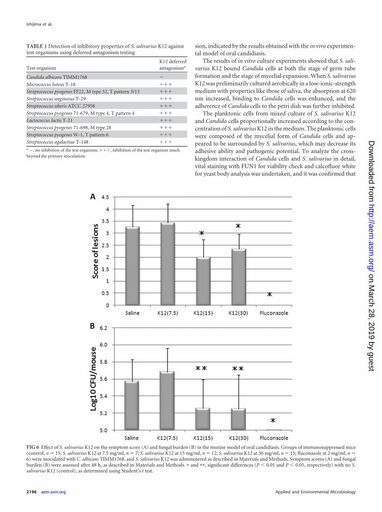
Effect of treatment with oral S. salivarius K12 on oral candi-diasis model. The effects of S. salivarius K12 on murine oral can-didiasis were examined. S. salivarius K12 was orally administratedto the mice at 24 and 3 h before and 3, 24, and 27 h after Candidainoculation. It appeared that S. salivarius K12 application caused adose-dependent improvement in symptom score and fungal bur-den (Fig. 6). Although the oral administration of 7.5 � 108
CFU/ml of S. salivarius K12 (score � 3.4 � 0.79, n � 7) resulted inno significant difference in symptom score from that for the sa-line-treated control group (score � 3.3 � 0.88, n � 15), oraladministration of 1.5 � 109 CFU/ml and 3 � 109 CFU/ml of S.salivarius K12 (scores � 2.0 � 0.74 [n � 12] and 2.3 � 0.62 [n �15], respectively) resulted in an obviously significant differencefrom the saline-treated control group (P � 0.01) (Fig. 6).
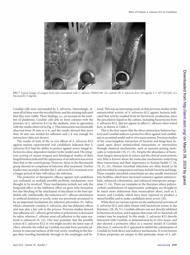
The tongues of mice administered S. salivarius K12 showedfewer lesions than the tongues of the control mice (Fig. 7A and B).
FIG 4 Inhibitory effect of S. salivarius K12 on hyphal growth of C. albicans. S. salivarius K12 was preliminarily cultured for 1 h or 6 h and then cultured with C.albicans for 3 h and stained with crystal violet. The optical absorbance at 620 nm (A620) was then detected. The experiments were performed in triplicate.
Ishijima et al.
2194 aem.asm.org Applied and Environmental Microbiology
on March 28, 2019 by guest
http://aem.asm
.org/D
ownloaded from
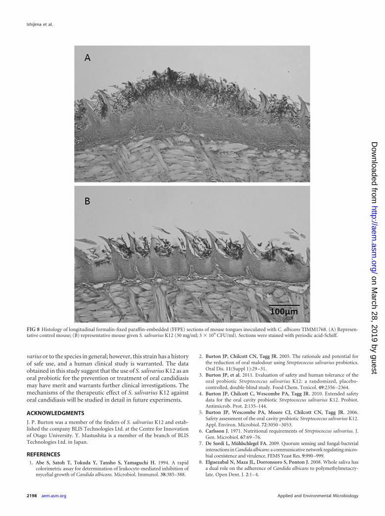
Even though there was no total eradication, as observed in thecontrol using fluconazole, in the mice that were administered S.salivarius K12 at 30 mg/ml (3 � 109 CFU/ml), there was a signif-icant decrease in fungal burden compared to that for the untreatedcontrol. The reduced pathogenicity of C. albicans cells when micewere given S. salivarius K12 was illustrated by the histopathologyof tongue sections (Fig. 8A and B). Fewer PAS-staining mycelialelements were found to invade the oral epithelium of tongues
treated with S. salivarius K12 than the tongues in the controlgroup.
DISCUSSION
The in vitro culture experiments that were conducted showed thatS. salivarius K12 bound directly to Candida cells and inhibited theadherence of Candida cells to the plastic petri dish. We also reportthat S. salivarius K12 had a protective effect against Candida inva-
FIG 5 Vital staining of C. albicans by FUN1 (1) and calcofluor white M2R (2). C. albicans was cultured with or without S. salivarius K12 at 30°C or 37°C for 3h. (a) Hyphal form of control culture at 37°C; (b) hyphal form of culture at 37°C with S. salivarius K12; (c) yeast form of control culture at 30°C; (d) yeast formof culture with S. salivarius K12 at 30°C; (e) S. salivarius K12.
Effect of Probiotic S. salivarius K12 on Candidiasis
April 2012 Volume 78 Number 7 aem.asm.org 2195
on March 28, 2019 by guest
http://aem.asm
.org/D
ownloaded from
sion, indicated by the results obtained with the in vivo experimen-tal model of oral candidiasis.
The results of in vitro culture experiments showed that S. sali-varius K12 bound Candida cells at both the stage of germ tubeformation and the stage of mycelial expansion. When S. salivariusK12 was preliminarily cultured aerobically in a low-ionic-strengthmedium with properties like those of saliva, the absorption at 620nm increased, binding to Candida cells was enhanced, and theadherence of Candida cells to the petri dish was further inhibited.
The planktonic cells from mixed culture of S. salivarius K12and Candida cells proportionally increased according to the con-centration of S. salivarius K12 in the medium. The planktonic cellswere composed of the mycelial form of Candida cells and ap-peared to be surrounded by S. salivarius, which may decrease itsadhesive ability and pathogenic potential. To analyze the cross-kingdom interaction of Candida cells and S. salivarius in detail,vital staining with FUN1 for viability check and calcofluor whitefor yeast body analysis was undertaken, and it was confirmed that
TABLE 1 Detection of inhibitory properties of S. salivarius K12 againsttest organisms using deferred antagonism testing
Test organismK12 deferredantagonisma
Candida albicans TIMM1768 �
Micrococcus luteus T-18 ���
Streptococcus pyogenes FF22, M type 52, T pattern 3/13 ���
Streptococcus anginosus T-29 ���
Streptococcus uberis ATCC 27958 ���
Streptococcus pyogenes 71-679, M type 4, T pattern 4 ���
Lactococcus lactis T-21 ���
Streptococcus pyogenes 71-698, M type 28 ���
Streptococcus pyogenes W-1, T pattern 6 ���
Streptococcus agalactiae T-148 ���a �, no inhibition of the test organism; ���, inhibition of the test organism muchbeyond the primary inoculation.
FIG 6 Effect of S. salivarius K12 on the symptom score (A) and fungal burden (B) in the murine model of oral candidiasis. Groups of immunosuppressed mice(control, n � 15; S. salivarius K12 at 7.5 mg/ml, n � 7; S. salivarius K12 at 15 mg/ml, n � 12; S. salivarius K12 at 30 mg/ml, n � 15; fluconazole at 2 mg/ml, n �6) were inoculated with C. albicans TIMM1768, and S. salivarius K12 was administered as described in Materials and Methods. Symptom scores (A) and fungalburden (B) were assessed after 48 h, as described in Materials and Methods. � and ��, significant differences (P � 0.01 and P � 0.05, respectively) with no S.salivarius K12 (control), as determined using Student’s t test.
Ishijima et al.
2196 aem.asm.org Applied and Environmental Microbiology
on March 28, 2019 by guest
http://aem.asm
.org/D
ownloaded from
Candida cells were surrounded by S. salivarius. Interestingly, al-most all of these were the mycelial form, and the staining indicatedthat they were viable. These findings, i.e., an increase in the num-ber of planktonic Candida cells able to form colonies with thepresence of S. salivarius K12 in the medium, were in agreementwith the results observed in Fig. 2. This interaction was kineticallyobserved from 30 min to 6 h, and the results showed that morethan 30 min was needed for adhesion and 1 h was enough forinteraction (data not shown).
The results of tests of the in vivo effects of S. salivarius K12against murine experimental oral candidiasis indicated that S.salivarius K12 had the ability to protect against severe fungal in-fection in a dose-dependent manner in the model used. The symp-tom scoring of mouse tongues and histological studies of theirfungal burdens indicated the appearance of an infection less severethan that in the control group. However, those in the fluconazolegroup showed no symptoms of infection after treatment. Furtherstudies may ascertain whether the S. salivarius K12 treatment overa longer period of time will reduce the infection.
The protective or therapeutic efficacy against oral candidiasiswas evaluated, as multiple possible probiotic mechanisms werethought to be involved. These mechanisms include not only thefungicidal effect or the inhibitory effect on germ tube formationbut also blocking of the attachment of mycelium to the host epi-thelial cells. Additionally, the reduction of C. albicans attachmentto artificial dentition or acrylic resin was previously presumed tobe an important mechanism for infection prevention (9). Saliva,which commonly contains S. salivarius, also has defensive effectsand may play a key role in the process. Previous studies showedthat adhesion of C. albicans germ tubes to polystyrene is decreasedby saliva, whereas C. albicans yeast cell adhesion to the same ma-terial is enhanced (8, 14). One may postulate that the possibleaction of S. salivarius K12 in vivo might involve the latter type ofeffect, whereby the rolled up Candida mycelial form prevents ad-hesion to mucosal surfaces of the oral cavity, resulting in the fun-gus then traveling harmlessly through to the esophagus and be-
yond. This was an interesting result, in that previous studies of theantimicrobial activity of S. salivarius K12 against bacteria indi-cated that activity resulted from its bacteriocin production, sincethe percolation liquid in the culture, including bacteriocins fromS. salivarius K12, did not appear to affect C. albicans when testedhere, as shown in Table 1.
This is the first report that the direct interaction between bac-teria and Candida induces a protective effect against oral candidi-asis in an animal model and in vitro assay systems. Previous studiesof the cross-kingdom interaction of bacteria and fungi have fo-cused upon direct antimicrobial interactions or interactionsthrough chemical mechanisms, such as quorum-sensing mole-cules or terpenoids (10, 17, 18). Despite the abundance of bacte-rium-fungus interactions in nature and the clinical environment,very little is known about the molecular mechanisms underlyingthese interactions and their importance to human health (7, 16,19, 21, 24). Human microbial infections are often found to bepolymicrobial in composition and may include bacteria and fungi.These complex microbial consortiums are also usually structuredinto biofilms, which have increased resistance against antimicro-bials, enhanced colonization, and enhanced interspecies antago-nism (7, 16). There are examples in the literature where polymi-crobial combinations of opportunistic pathogens are thought tobe much more deleterious than monoculture alone, such as S.mutans and Candida, which have been reported to produce amixed biofilm and to make candidiasis more severe (20).
While there are various reports on the antibacterial activities ofS. salivarius K12 and other strains with bacteriocin action in theliterature (28, 30, 31), Candida albicans was not directly inhibitedby bacteriocin action, and it appears that yeast cell-to-bacterial cellcontact may be required. In this study, S. salivarius K12 directlyinteracted with Candida, as demonstrated by in vitro assays, andalso showed a protective effect in a murine model of Candidainfection. S. salivarius K12 appeared to inhibit the colonization ofCandida by both direct and indirect mechanisms. It is not knownif these properties are unique to this particular strain of S. sali-
FIG 7 Typical images of tongues from mice inoculated with C. albicans TIMM1768. (A) control; (B) S. salivarius K12 (30 mg/ml; 3 � 109 CFU/ml); (C)fluconazole (2 mg/ml).
Effect of Probiotic S. salivarius K12 on Candidiasis
April 2012 Volume 78 Number 7 aem.asm.org 2197
on March 28, 2019 by guest
http://aem.asm
.org/D
ownloaded from
varius or to the species in general; however, this strain has a historyof safe use, and a human clinical study is warranted. The dataobtained in this study suggest that the use of S. salivarius K12 as anoral probiotic for the prevention or treatment of oral candidiasismay have merit and warrants further clinical investigations. Themechanisms of the therapeutic effect of S. salivarius K12 againstoral candidiasis will be studied in detail in future experiments.
ACKNOWLEDGMENTS
J. P. Burton was a member of the finders of S. salivarius K12 and estab-lished the company BLIS Technologies Ltd. at the Centre for Innovationof Otago University. Y. Mastushita is a member of the branch of BLISTechnologies Ltd. in Japan.
REFERENCES1. Abe S, Satoh T, Tokuda Y, Tansho S, Yamaguchi H. 1994. A rapid
colorimetric assay for determination of leukocyte-mediated inhibition ofmycelial growth of Candida albicans. Microbiol. Immunol. 38:385–388.
2. Burton JP, Chilcott CN, Tagg JR. 2005. The rationale and potential forthe reduction of oral malodour using Streptococcus salivarius probiotics.Oral Dis. 11(Suppl 1):29 –31.
3. Burton JP, et al. 2011. Evaluation of safety and human tolerance of theoral probiotic Streptococcus salivarius K12: a randomized, placebo-controlled, double-blind study. Food Chem. Toxicol. 49:2356 –2364.
4. Burton JP, Chilcott C, Wescombe PA, Tagg JR. 2010. Extended safetydata for the oral cavity probiotic Streptococcus salivarius K12. Probiot.Antimicrob. Prot. 2:135–144.
5. Burton JP, Wescombe PA, Moore CJ, Chilcott CN, Tagg JR. 2006.Safety assessment of the oral cavity probiotic Streptococcus salivarius K12.Appl. Environ. Microbiol. 72:3050 –3053.
6. Carlsson J. 1971. Nutritional requirements of Streptococcus salivarius. J.Gen. Microbiol. 67:69 –76.
7. De Sordi L, Mühlschlegel FA. 2009. Quorum sensing and fungal-bacterialinteractions in Candida albicans: a communicative network regulating micro-bial coexistence and virulence. FEMS Yeast Res. 9:990–999.
8. Elguezabal N, Maza JL, Dorronsoro S, Ponton J. 2008. Whole saliva hasa dual role on the adherence of Candida albicans to polymethylmetacry-late. Open Dent. J. 2:1– 4.
FIG 8 Histology of longitudinal formalin-fixed paraffin-embedded (FFPE) sections of mouse tongues inoculated with C. albicans TIMM1768. (A) Represen-tative control mouse; (B) representative mouse given S. salivarius K12 (30 mg/ml; 3 � 109 CFU/ml). Sections were stained with periodic acid-Schiff.
Ishijima et al.
2198 aem.asm.org Applied and Environmental Microbiology
on March 28, 2019 by guest
http://aem.asm
.org/D
ownloaded from
9. Ellepola AN, Samaranayake LP. 2000. Oral candidal infections and an-timycotics. Crit. Rev. Oral Biol. Med. 11:172–198.
10. Hogan DA, Vik A, Kolter R. 2004. A Pseudomonas aeruginosa quorum-sensing molecule influences Candida albicans morphology. Mol. Micro-biol. 54:1212–1223.
11. Hyink O, et al. 2007. Salivaricin A2 and the novel lantibiotic salivaricin Bare encoded at adjacent loci on a 190-kilobase transmissible megaplasmidin the oral probiotic strain Streptococcus salivarius K12. Appl. Environ.Microbiol. 73:1107–1113.
12. Ishijima SA, et al. 2011. N-Acetylglucosamine increases symptoms andfungal burden in a murine model of oral candidiasis. Med. Mycol [Epubahead of print.] doi:10.3109/13693786.2011.598194.
13. James SM, Tagg JR. 1991. The prevention of dental caries by BLIS-mediated inhibition of mutans streptococci. N. Z. Dent. J. 87:80 – 83.
14. Kamagata-Kiyoura Y, Abe S, Yamaguchi H, Nitta T. 2003. Detachmentactivity of human saliva in vitro for Candida albicans cells attached to aplastic plate. J. Infect. Chemother. 9:215–220.
15. Kamagata-Kiyoura Y, Abe S. 2005. Recent studies on oral candidiasisusing a murine model. J. Oral Biosci. 47:60 – 64.
16. Kumamoto CA, Vinces MD. 2005. Contributions of hyphae and hypha-co-regulated genes to Candida albicans virulence. Cell. Microbiol. 7:1546 –1554.
17. Morales DK, Hogan DA. 2010. Candida albicans interactions with bac-teria in the context of human health and disease. PLoS Pathog. 6:1– 4.
18. Nickerson KW, Atkin AL, Hornby JM. 2006. Quorum sensing in dimor-phic fungi: farnesol and beyond. Appl. Environ. Microbiol. 72:3805–3813.
19. Ogawa A, et al. 2011. Inhibition of Streptococcus mutans biofilm forma-tion by Streptococcus salivarius FruA. Appl. Environ. Microbiol. 77:1572–1580.
20. Peleg AY, Hogan DA, Mylonakis E. 2010. Medically important bacterial-fungal interactions. Nat. Rev. Microbiol. 8:340 –349.
21. Pereira-Cenci T, et al. 2008. The effect of Streptococcus mutans and Can-dida glabrata on Candida albicans biofilms formed on different surfaces.Arch. Oral Biol. 53:755–764.
22. Sardi JC, et al. 2010. Candida spp. in periodontal disease: a brief review.J. Oral Sci. 52:177–185.
23. Shapiro RS, Robbins N, Cowen LE. 2011. Regulatory circuitry governingfungal development, drug resistance, and disease. Microbiol. Mol. Biol.Rev. 75:213–267.
24. Shirtliff ME, Peters BM, Jabra-Rizk MA. 2009. Cross-kingdom interac-tions: Candida albicans and bacteria. FEMS Microbiol. Lett. 299:1– 8.
25. Tagg JR, Bannister LV. 1979. “Fingerprinting” beta-haemolytic strepto-cocci by their production of and sensitivity to bacteriocine-like inhibitors.J. Med. Microbiol. 12:397– 411.
26. Tagg JR, Dajani AS, Wannamaker LW. 1976. Bacteriocins of gram-positive bacteria. Bacteriol. Rev. 40:722–756.
27. Takakura N, et al. 2003. A novel murine model of oral candidiasis withlocal symptoms characteristic of oral thrush. Microbiol. Immunol. 47:321–326.
28. Tamura S, et al. 2009. Inhibiting effects of Streptococcus salivarius oncompetence-stimulating peptide-dependent biofilm formation by Strep-tococcus mutans. Oral Microbiol. Immunol. 24:152–161.
29. Thompson GR, III, et al. 2010. Oropharyngeal candidiasis in the era ofantiretroviral therapy. Oral Surg. Oral Med. Oral Pathol. Oral Radiol.Endod. 109:488 – 495.
30. Walls T, Power DA, Tagg JR. 2003. Bacteriocin-like inhibitory substance(BLIS) production by the normal flora of the nasopharynx: potential toprotect against otitis media? J. Med. Microbiol. 52:829 – 833.
31. Wescomebe PA, Heng NCK, Burton JP, Chilcott CN, Tagg R. 2009.Streptococcus bacteriocins and the case for Streptococcus salivarius asmodel oral probiotics. Future Microbiol. 4:819 – 835.
Effect of Probiotic S. salivarius K12 on Candidiasis
April 2012 Volume 78 Number 7 aem.asm.org 2199
on March 28, 2019 by guest
http://aem.asm
.org/D
ownloaded from


![Candidiasis Oral[1]](https://img.pdfslide.tips/doc/110x75/5571f81c49795991698ca796/candidiasis-oral1.jpg)