Embed Size (px)
Citation preview

Research ArticleEffect of Vitamin D3 in combination with Omega-3Polyunsaturated Fatty Acids on NETosis in Type 2 DiabetesMellitus Patients
Liliya Yu. Basyreva , Tatyana V. Vakhrusheva, Zoya V. Letkeman, Dmitry I. Maximov,Evgeniya A. Fedorova, Оleg M. Panasenko , Evgeny M. Ostrovsky, and Sergey A. Gusev
Federal Research and Clinical Center of Physical-Chemical Medicine of Federal Medical Biological Agency, Moscow 119435, Russia
Correspondence should be addressed to Liliya Yu. Basyreva; [email protected]
Received 29 July 2021; Revised 7 September 2021; Accepted 27 September 2021; Published 22 October 2021
Academic Editor: Adrian Sturza
Copyright © 2021 Liliya Yu. Basyreva et al. This is an open access article distributed under the Creative Commons AttributionLicense, which permits unrestricted use, distribution, and reproduction in any medium, provided the original work isproperly cited.
An understanding of the consequences of oxidative/halogenative stress triggered by neutrophil activation is impossible withoutconsidering NETosis. NETosis, formation of neutrophil extracellular traps (NETs), is known to promote microthrombusformation and impair wound healing in type 2 diabetes mellitus (T2DM) patients. Therefore, there is a need to search fordrugs and treatment approaches that could prevent excessive NET formation. We aimed to evaluate the effect of vitaminD3 in combination with omega-3 polyunsaturated fatty acids (vitamin D3/omega-3 PUFAs) on NETosis in T2DM patientswith purulent necrotizing lesions of the lower extremities. Patients and healthy subjects had vitamin D3 deficiency. Patientsreceived, beyond standard treatment, 6000 IU of vitamin D3 and 480mg of omega-3 PUFAs, and healthy subjects 1000 IUof vitamin D3 and 240mg of omega-3 PUFAs daily for seven days. Neutrophil activation in ex vivo blood by phorbol-12-myristate-13-acetate (PMA) was used as a NETosis model. The percentage of blood NETs relative to leukocytes(NETbackground) before vitamin D3/omega-3 PUFA supplementation was 3.2%-4.9% in healthy subjects and 1.7%-10.8% inpatients. These values rose, respectively, to 7.7%-9.1% and 4.0%-17.9% upon PMA-induced NETosis. In addition, theleukocyte count decreased by 700-1300 per 1μL in healthy subjects and 700-4000 per 1 μL in patients. For both patientsand healthy subjects, taking vitamin D3/omega-3 PUFAs had no effect on NETbackground but completely inhibited PMA-induced NET formation, though neutrophils exhibited morphological features of activation. Also, leukocyte loss wasreduced (to 500 per 1μL). For patients on standard treatment alone, changes occurred neither in background NETs andleukocytes nor in their amount after PMA stimulation. The decreased ability of neutrophils to generate NETs, which canbe achieved by vitamin D3/omega-3 PUFA supplementation, could have a positive effect on wound healing in T2DMpatients and reduce the incidence and severity of complications.
1. Introduction
Neutrophils, which are the largest population of circulatingleukocytes (~60% in adults), play a key role in cellular innateimmunity. Neutrophil granules contain a rich antimicrobial“arsenal,” which allows these cells to efficiently destroyphagocytosed pathogens [1]. During degranulation, antibac-terial proteins and enzymes (myeloperoxidase (MPO), lacto-ferrin, lysozyme, neutrophil elastase (NE), etc.) can also bepartially secreted from the cell [1]. The functioning mainly
of NADPH oxidase and MPO leads to the formation of reac-tive oxygen- and halogen-containing compounds, the so-called reactive oxygen species (ROS) and reactive halogenspecies (RHS), which are directly involved in the destructionof pathogens both inside and outside the cell [2]. Anotherantimicrobial mechanism of neutrophils, which was discov-ered relatively recently, has been termed NETosis [3].
In NETosis, neutrophils release the so-called neutrophilextracellular traps (NETs), which are tangles of decondensedchromatin fibers and attached bactericidal agents including
HindawiOxidative Medicine and Cellular LongevityVolume 2021, Article ID 8089696, 10 pageshttps://doi.org/10.1155/2021/8089696
MPO, NE, histones, and defensins. To date, multipleinducers of NET formation have been described, amongwhich are bacteria, fungi, viruses, immune complexes, lipo-polysaccharides, phorbol-12-myristate-13-acetate (PMA),etc. NETs allow neutrophils to destroy extracellular patho-gens while causing minimal damage to the host cells [4].
Although ROS and RHS are useful to kill microbes, theirexcessive production (as it can occur upon neutrophil activa-tion at the inflamed site) is unfavorable, since it causes dam-age to biologically important molecules and cellular andtissue structures of the host body, leads to oxidative/halo-genative stress, and provokes the occurrence ofinflammation-associated diseases [5–8]. NETosis contrib-utes to these harmful processes, since, firstly, it is activatedby ROS produced with the participation of NADPH oxidase[4, 9], and secondly, MPO, which catalyzes the formation ofRHS [2, 7], is among NET components [10, 11]. It has beenindeed repeatedly observed that excessive NET formation islinked to the development of autoimmune, cardiovascular,and endocrine diseases, including diabetes mellitus. In par-ticular, our data have shown that in type 2 diabetes mellitus(T2DM) patients with purulent necrotizing lesions of thelower extremities, a more severe clinical course is associatedwith a higher blood NET level [12, 13]. NETosis promotesmicrothrombosis as well as causes delayed wound healing[9, 14, 15], which could have a negative effect on the healingprocess in T2DM patients.
It has been found that some medications can induceNETosis [16, 17]. Therefore, it is worth checking the effectof drugs on NETosis as well as searching for new therapeuticoptions that prevent excessive NET formation [17–19]. Thisis currently especially important, since it has been demon-strated that when infected with COVID-19, people showan increase in NET blood concentration, and comorbid dia-betes mellitus is associated with a severe course of pneumo-nia and higher mortality [20, 21].
Vitamin D3 or omega-3 polyunsaturated fatty acid(omega-3 PUFA) supplements can promote wound repair,as shown in T2DM patients with purulent necrotizing footlesions [22, 23]. What is more, this effect was accompaniedby a decrease in the blood plasma level of lipid peroxidationproducts, an increase in plasma reduced glutathione, animportant antioxidant in the body [22], and an increase inthe plasma total antioxidant activity [23], which indicatesthe involvement of oxidative stress in the development ofpurulent necrotizing wounds in T2DM as well as showsthe antioxidant effect of vitamin D3 and omega-3 PUFAs.
A large number of data are available on the effects ofvitamin D3 and omega-3 PUFAs on the functioning of var-ious components of the immune system [24, 25], but onlytwo studies investigated the effect of vitamin D3 on NETosis.In one of them, vitamin D3 inhibited [26], whereas, in theother, it activated NETosis [27]. No studies have examinedhow NET formation in blood is affected by vitamin D3 incombination with omega-3 PUFAs (vitamin D3/omega-3PUFAs). Since the wound character state and healing pro-cess depend on the activation status of neutrophils, in partic-ular, their predisposition to form NETs, the present work isaimed at assessing the effect of oral intake of vitamin
D3/omega-3 PUFAs on the blood NET level as well as onPMA-stimulated NETosis in the ex vivo blood samples fromT2DM patients with purulent necrotizing lesions of thelower extremities.
2. Materials and Methods
2.1. Subjects. The study included healthy subjects (n = 4) andT2DM patients with purulent necrotizing lesions of thelower extremities (n = 14) hospitalized at the Center forPurulent Surgery and Regenerative Technologies of the Fed-eral Research and Clinical Center of Physical-ChemicalMedicine of Federal Medical Biological Agency of Russia(FRCC PCM). Both patients and healthy subjects enrolledin the study had vitamin D3 deficiency.
2.2. Experimental Design. All patients received standardtreatment, including glycemia correction, antibacterial ther-apy (based on bacteriologic microflora results, antibiotic sus-ceptibility testing, and resistance PCR results), andanticoagulant, detoxification, rheological, anti-inflamma-tory, and immunomodulating therapy. Patients with comor-bid disease were consulted by an endocrinologist, a therapist,and other specialists. All patients underwent treatment ofthe purulent site by hydrosurgery with the Versajet II hydro-surgery system (Smith & Nephew Inc., USA) and ultrasonicwound debridement with the Sonoca-190 ultrasonic genera-tor (Söring, Germany). Slowly granulating wounds weresubjected to negative pressure therapy with the VivanoTecsystem (Hartmann, Germany). In addition to standard treat-ment, 8 patients were given an oral water-soluble form ofvitamin D3 (6000 IU) and omega-3 PUFAs (eicosapentae-noic acid 288mg, docosahexaenoic acid 192mg) daily dur-ing the entire hospital stay. Healthy subjects received anoral water-soluble form of vitamin D3 (1000 IU) andomega-3 PUFAs (eicosapentaenoic acid 144mg, docosahex-aenoic acid 96mg) daily for 7 days. The supplement doseswere determined based on the results of preliminary experi-ments (data not shown). For patients, higher doses wererequired to produce the effect on NET formation.
Blood from healthy subjects was taken before and 1, 4,and 7 days after the beginning of vitamin D3/omega-3PUFA supplementation. Blood from patients was taken atadmission to the hospital and after 7 days of either standardtreatment or taking vitamin D3/omega-3 PUFAs in additionto it.
Peripheral venous blood was collected into sodium cit-rate vacutainers (MiniMed, Russia). Biochemical and hema-tological blood analyses included, among others, erythrocytesedimentation rate (ESR), glycated hemoglobin (HbA1c)level, baseline blood glucose level, creatinine, urea, aspartateaminotransferase (AST) and alanine aminotransferase(ALT) activities, and 25-hydroxyvitamin D3 level (measuredby HPLC).
2.3. Analysis of Blood Smears. Standardized blood smearswere prepared immediately after drawing the blood samplesand after 2 h of blood incubation at 37°C in the presence orabsence of 100nM PMA (Sigma-Aldrich, USA). Blood
2 Oxidative Medicine and Cellular Longevity
smears were examined using a Motic B3 microscope (MoticAsia, Hong Kong). A quantitative assessment of NETs wasperformed as described previously [16]. Briefly, bloodsmears were stained by the Romanowsky method. NETsamong 300-500 leukocytes were counted in the middle thirdpart of the smear, and the NETs-to-leukocyte percentageratio was calculated. The ratio in the collected blood is here-inafter designated as NETbackground, and it is designated asNETPMA for blood incubated with PMA and NETcontrol forblood incubated without PMA. Blood leukocyte concentra-tion was determined using a Mythic 18 flow hematologyanalyzer (Orphee, Switzerland) immediately after bloodsampling (Lbackground) and after 2 h of blood incubation at37°C in the presence of 100nM PMA (LPMA) or withoutPMA (Lcontrol).
2.4. Data Analysis. The statistical analysis of results was per-formed with STATISTICA 6.0 software (StatSoft Inc., USA).The statistical significance for differences between thegroups was assessed using Student’s t-test and Mann–Whit-ney U-test.
3. Results
All healthy subjects enrolled in the study were deficient inserum 25-hydroxyvitamin D3. Their hematological and bio-chemical parameters were inside the reference limits, exceptfor this vitamin D3 metabolite. Blood parameters in patientsare shown in Table 1. No significant differences were foundin the measured parameters between patients who receivedor not vitamin D3/omega-3 PUFAs. Worthy of note is amarked deficiency of 25-hydroxyvitamin D3 in all patients(<17.1 ng/mL, with a normal range of 30-100 ng/mL).
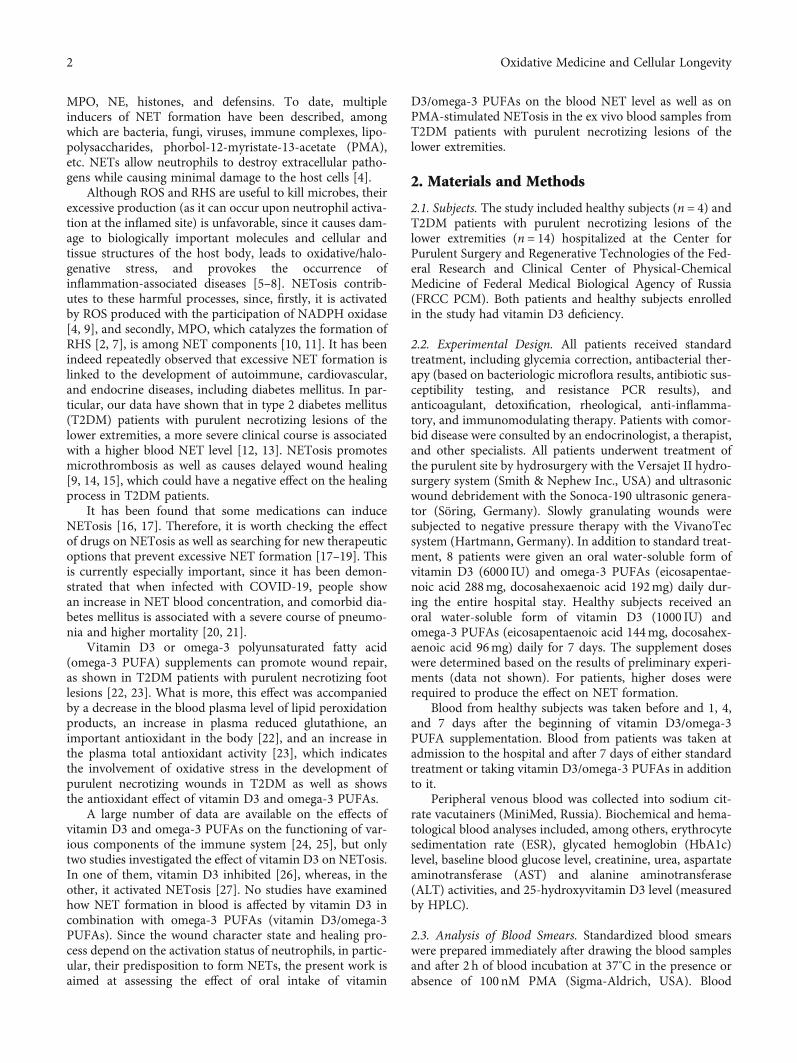
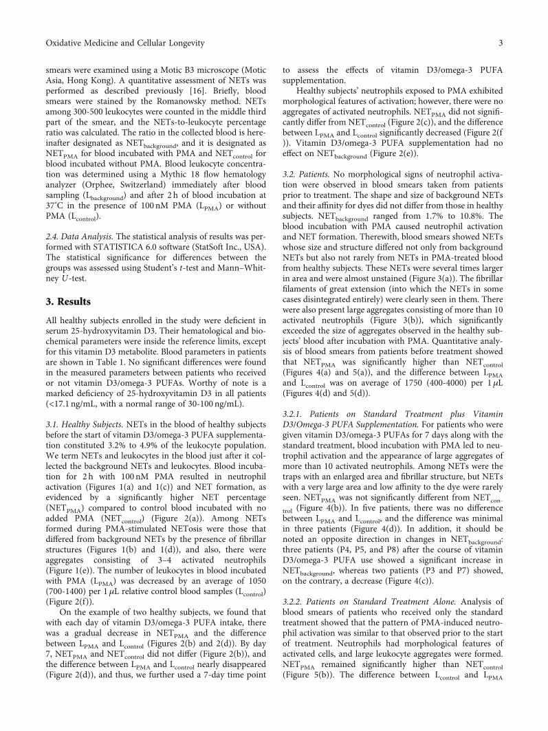
3.1. Healthy Subjects. NETs in the blood of healthy subjectsbefore the start of vitamin D3/omega-3 PUFA supplementa-tion constituted 3.2% to 4.9% of the leukocyte population.We term NETs and leukocytes in the blood just after it col-lected the background NETs and leukocytes. Blood incuba-tion for 2 h with 100 nM PMA resulted in neutrophilactivation (Figures 1(a) and 1(c)) and NET formation, asevidenced by a significantly higher NET percentage(NETPMA) compared to control blood incubated with noadded PMA (NETcontrol) (Figure 2(a)). Among NETsformed during PMA-stimulated NETosis were those thatdiffered from background NETs by the presence of fibrillarstructures (Figures 1(b) and 1(d)), and also, there wereaggregates consisting of 3–4 activated neutrophils(Figure 1(e)). The number of leukocytes in blood incubatedwith PMA (LPMA) was decreased by an average of 1050(700-1400) per 1μL relative control blood samples (Lcontrol)(Figure 2(f)).
On the example of two healthy subjects, we found thatwith each day of vitamin D3/omega-3 PUFA intake, therewas a gradual decrease in NETPMA and the differencebetween LPMA and Lcontrol (Figures 2(b) and 2(d)). By day7, NETPMA and NETcontrol did not differ (Figure 2(b)), andthe difference between LPMA and Lcontrol nearly disappeared(Figure 2(d)), and thus, we further used a 7-day time point
to assess the effects of vitamin D3/omega-3 PUFAsupplementation.
Healthy subjects’ neutrophils exposed to PMA exhibitedmorphological features of activation; however, there were noaggregates of activated neutrophils. NETPMA did not signifi-cantly differ from NETcontrol (Figure 2(c)), and the differencebetween LPMA and Lcontrol significantly decreased (Figure 2(f)). Vitamin D3/omega-3 PUFA supplementation had noeffect on NETbackground (Figure 2(e)).
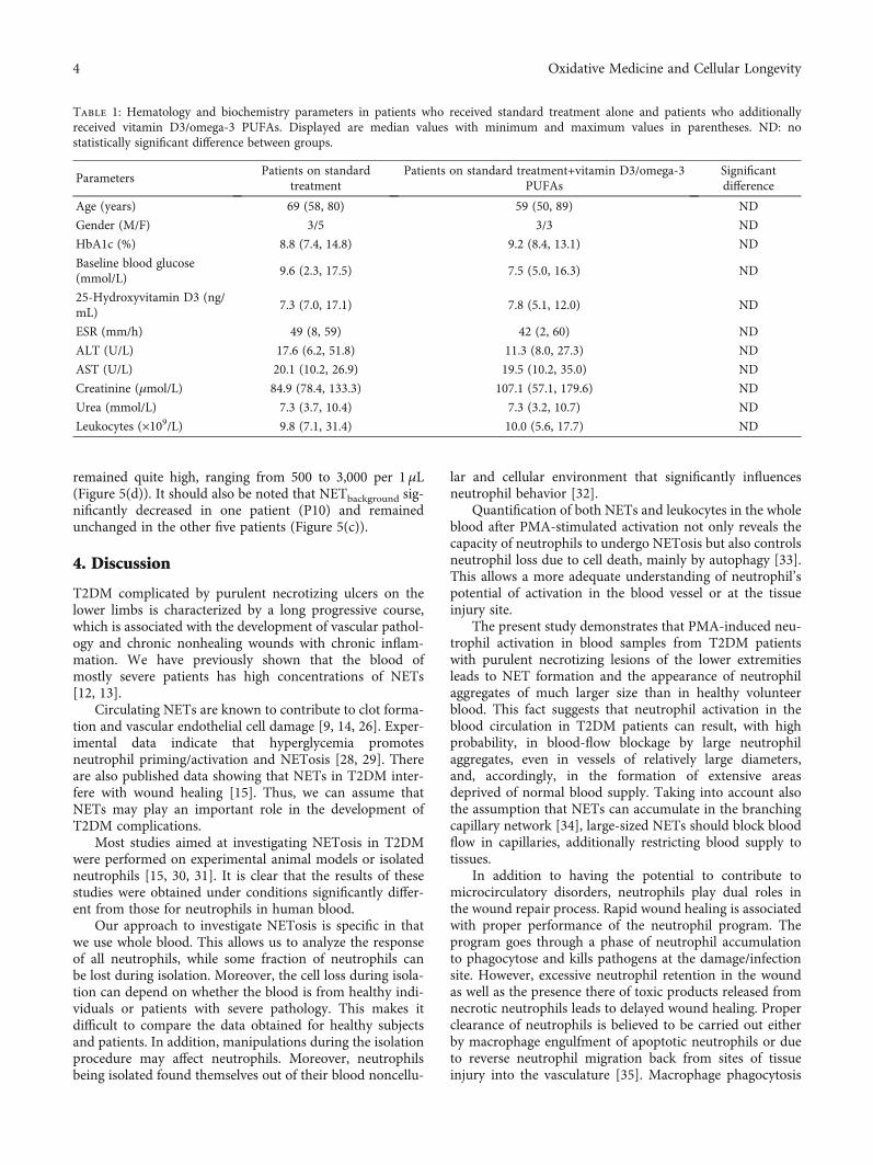
3.2. Patients. No morphological signs of neutrophil activa-tion were observed in blood smears taken from patientsprior to treatment. The shape and size of background NETsand their affinity for dyes did not differ from those in healthysubjects. NETbackground ranged from 1.7% to 10.8%. Theblood incubation with PMA caused neutrophil activationand NET formation. Therewith, blood smears showed NETswhose size and structure differed not only from backgroundNETs but also not rarely from NETs in PMA-treated bloodfrom healthy subjects. These NETs were several times largerin area and were almost unstained (Figure 3(a)). The fibrillarfilaments of great extension (into which the NETs in somecases disintegrated entirely) were clearly seen in them. Therewere also present large aggregates consisting of more than 10activated neutrophils (Figure 3(b)), which significantlyexceeded the size of aggregates observed in the healthy sub-jects’ blood after incubation with PMA. Quantitative analy-sis of blood smears from patients before treatment showedthat NETPMA was significantly higher than NETcontrol(Figures 4(a) and 5(a)), and the difference between LPMAand Lcontrol was on average of 1750 (400-4000) per 1μL(Figures 4(d) and 5(d)).
3.2.1. Patients on Standard Treatment plus VitaminD3/Omega-3 PUFA Supplementation. For patients who weregiven vitamin D3/omega-3 PUFAs for 7 days along with thestandard treatment, blood incubation with PMA led to neu-trophil activation and the appearance of large aggregates ofmore than 10 activated neutrophils. Among NETs were thetraps with an enlarged area and fibrillar structure, but NETswith a very large area and low affinity to the dye were rarelyseen. NETPMA was not significantly different from NETcon-
trol (Figure 4(b)). In five patients, there was no differencebetween LPMA and Lcontrol, and the difference was minimalin three patients (Figure 4(d)). In addition, it should benoted an opposite direction in changes in NETbackground:three patients (P4, P5, and P8) after the course of vitaminD3/omega-3 PUFA use showed a significant increase inNETbackground, whereas two patients (P3 and P7) showed,on the contrary, a decrease (Figure 4(c)).
3.2.2. Patients on Standard Treatment Alone. Analysis ofblood smears of patients who received only the standardtreatment showed that the pattern of PMA-induced neutro-phil activation was similar to that observed prior to the startof treatment. Neutrophils had morphological features ofactivated cells, and large leukocyte aggregates were formed.NETPMA remained significantly higher than NETcontrol(Figure 5(b)). The difference between Lcontrol and LPMA
3Oxidative Medicine and Cellular Longevity
remained quite high, ranging from 500 to 3,000 per 1μL(Figure 5(d)). It should also be noted that NETbackground sig-nificantly decreased in one patient (P10) and remainedunchanged in the other five patients (Figure 5(c)).
4. Discussion
T2DM complicated by purulent necrotizing ulcers on thelower limbs is characterized by a long progressive course,which is associated with the development of vascular pathol-ogy and chronic nonhealing wounds with chronic inflam-mation. We have previously shown that the blood ofmostly severe patients has high concentrations of NETs[12, 13].
Circulating NETs are known to contribute to clot forma-tion and vascular endothelial cell damage [9, 14, 26]. Exper-imental data indicate that hyperglycemia promotesneutrophil priming/activation and NETosis [28, 29]. Thereare also published data showing that NETs in T2DM inter-fere with wound healing [15]. Thus, we can assume thatNETs may play an important role in the development ofT2DM complications.
Most studies aimed at investigating NETosis in T2DMwere performed on experimental animal models or isolatedneutrophils [15, 30, 31]. It is clear that the results of thesestudies were obtained under conditions significantly differ-ent from those for neutrophils in human blood.
Our approach to investigate NETosis is specific in thatwe use whole blood. This allows us to analyze the responseof all neutrophils, while some fraction of neutrophils canbe lost during isolation. Moreover, the cell loss during isola-tion can depend on whether the blood is from healthy indi-viduals or patients with severe pathology. This makes itdifficult to compare the data obtained for healthy subjectsand patients. In addition, manipulations during the isolationprocedure may affect neutrophils. Moreover, neutrophilsbeing isolated found themselves out of their blood noncellu-
lar and cellular environment that significantly influencesneutrophil behavior [32].
Quantification of both NETs and leukocytes in the wholeblood after PMA-stimulated activation not only reveals thecapacity of neutrophils to undergo NETosis but also controlsneutrophil loss due to cell death, mainly by autophagy [33].This allows a more adequate understanding of neutrophil’spotential of activation in the blood vessel or at the tissueinjury site.
The present study demonstrates that PMA-induced neu-trophil activation in blood samples from T2DM patientswith purulent necrotizing lesions of the lower extremitiesleads to NET formation and the appearance of neutrophilaggregates of much larger size than in healthy volunteerblood. This fact suggests that neutrophil activation in theblood circulation in T2DM patients can result, with highprobability, in blood-flow blockage by large neutrophilaggregates, even in vessels of relatively large diameters,and, accordingly, in the formation of extensive areasdeprived of normal blood supply. Taking into account alsothe assumption that NETs can accumulate in the branchingcapillary network [34], large-sized NETs should block bloodflow in capillaries, additionally restricting blood supply totissues.
In addition to having the potential to contribute tomicrocirculatory disorders, neutrophils play dual roles inthe wound repair process. Rapid wound healing is associatedwith proper performance of the neutrophil program. Theprogram goes through a phase of neutrophil accumulationto phagocytose and kills pathogens at the damage/infectionsite. However, excessive neutrophil retention in the woundas well as the presence there of toxic products released fromnecrotic neutrophils leads to delayed wound healing. Properclearance of neutrophils is believed to be carried out eitherby macrophage engulfment of apoptotic neutrophils or dueto reverse neutrophil migration back from sites of tissueinjury into the vasculature [35]. Macrophage phagocytosis
Table 1: Hematology and biochemistry parameters in patients who received standard treatment alone and patients who additionallyreceived vitamin D3/omega-3 PUFAs. Displayed are median values with minimum and maximum values in parentheses. ND: nostatistically significant difference between groups.
ParametersPatients on standard
treatmentPatients on standard treatment+vitamin D3/omega-3
PUFAsSignificantdifference
Age (years) 69 (58, 80) 59 (50, 89) ND
Gender (M/F) 3/5 3/3 ND
HbA1c (%) 8.8 (7.4, 14.8) 9.2 (8.4, 13.1) ND
Baseline blood glucose(mmol/L)
9.6 (2.3, 17.5) 7.5 (5.0, 16.3) ND
25-Hydroxyvitamin D3 (ng/mL)
7.3 (7.0, 17.1) 7.8 (5.1, 12.0) ND
ESR (mm/h) 49 (8, 59) 42 (2, 60) ND
ALT (U/L) 17.6 (6.2, 51.8) 11.3 (8.0, 27.3) ND
AST (U/L) 20.1 (10.2, 26.9) 19.5 (10.2, 35.0) ND
Creatinine (μmol/L) 84.9 (78.4, 133.3) 107.1 (57.1, 179.6) ND
Urea (mmol/L) 7.3 (3.7, 10.4) 7.3 (3.2, 10.7) ND
Leukocytes (×109/L) 9.8 (7.1, 31.4) 10.0 (5.6, 17.7) ND
4 Oxidative Medicine and Cellular Longevity

of apoptotic neutrophils is part of the normal resolution ofinflammation.
Thus, the proper clearance of neutrophils implies thatactivated neutrophils after they have completed their func-tions at the inflamed site do not die by mechanisms otherthan apoptosis, e.g., NETosis, autophagy, or necrosis.
Our results show that taking vitamin D3/omega-3PUFAs completely “switches off” NETosis and death of neu-trophils upon their activation with PMA in whole bloodex vivo. This suggests that upon vitamin D3/omega-3 PUFA
supplementation, neutrophil death in the wound site will notoccur through NETosis, autophagy, or otherwise, whichincreases the possibility of neutrophil clearance by reversemigration into the bloodstream or macrophage phagocyto-sis. This should support faster wound repair.
The data reported in the literature suggest that vitaminD3/omega-3 PUFA supplement can significantly reduceROS/RHS generation and increase the total antioxidantcapacity of blood serum [22, 23, 36, 37] and that the preven-tion of NET formation enhances neutrophil phagocytic
(a) (b)
(c) (d)
(e)
Figure 1: Neutrophils and NETs in blood smears from a healthy subject before vitamin D3/omega-3 PUFA supplementation. The blood wasincubated (37°C, 2 h) with no added PMA (a, b) or with 100 nM PMA (c–e): (a) a resting neutrophil; (c) an activated neutrophil; (b, d) NETs(indicated by arrows); (e) a neutrophil aggregate (indicated by arrows). Scale bar: 20 μm.
5Oxidative Medicine and Cellular Longevity

0HS1 HS2 HS3 HS4
2
4
6
8
10
NET
s (%
)
⁎⁎
⁎⁎
(a)
Before0
2
4
6
8
10
1 d 4 d 7 d
NET
s (%
)
⁎
⁎
⁎
(b)
0HS1 HS2 HS3 HS4
2
4
6
8
10
NET
s (%
)
(c)
Before0
–200
–400
–600
–800
–1000Le
ukoc
ytes
(per
𝜇L)
1 d 4 d 7 d
⁎
⁎ ⁎
(d)
0HS1 HS2 HS3 HS4
2
4
6
8
10
NET
s (%
)
(e)
–1600–1400–1200–1000
–800–600–400–200
0
Leuk
ocyt
es (p
er 𝜇
L)
⁎⁎
⁎⁎
HS1 HS2 HS3 HS4
(f)
Figure 2: NETs and leukocytes in blood from healthy subjects (HS) before and after vitamin D3/omega-3 PUFA supplementation. Resultsof measurements done before (NETbackground, background NETs) or after blood incubation (37°C, 2 h) with no added PMA (NETcontrol andLcontrol, control NETs, and control leukocytes, respectively) or with 100 nM PMA (NETPMA and LPMA).
∗P < 0:001: (a) NETcontrol (■) andNETPMA (□) before supplementation; (b) changes in NETcontrol (- - -) and NETPMA (—) for 7 days of supplementation; (c) NETcontrol (■)and NETPMA (□) after 7 days of supplementation; (d) changes in the difference between LPMA and Lcontrol for 7 days of supplementation; (e)NETbackground before (■) and after (□) 7 days of supplementation; (f) the difference between LPMA and Lcontrol before (■) and after (□) 7days of supplementation.
(a) (b)
Figure 3: NET (a) and a neutrophil aggregate (b) in blood smears from a patient before vitamin D3/omega-3 PUFA supplementation. Theblood was incubated (37°C, 2 h) with 100 nM PMA. Scale bar: 20 μm.
6 Oxidative Medicine and Cellular Longevity

activity [38]. All these may potentially favor wound healingin T2DM patients with purulent necrotizing injuries on thelower limbs.
It should be noted that the ending background level ofNETs in T2DM patients after 7 days of vitaminD3/omega-3 PUFA intake remained the same as the starting
P1 P2 P3 P4 P5 P6 P7 P80
5
10
15
20
NET
s (%
)⁎⁎
⁎
⁎
⁎
⁎
⁎
⁎⁎
(a)
P1 P2 P3 P4 P5 P6 P7 P80
5
10
15
20
NET
s (%
)
(b)
P1 P2 P3 P4 P5 P6 P7 P80
5
10
15
20
NET
s (%
)
⁎⁎
⁎⁎
⁎⁎
⁎
(c)Le
ukoc
ytes
(per
𝜇L)
P1 P2
–3500
–2500
–1500
–500
P3 P4 P5 P6 P7 P8#####
⁎
⁎
⁎ ⁎ ⁎⁎
⁎
⁎
(d)
Figure 4: NETs and leukocytes in blood from patients (P) before and after vitamin D3/omega-3 PUFA supplementation in addition tostandard treatment. Results of measurements done before (NETbackground, background NETs) or after blood incubation (37°C, 2 h) withno added PMA (NETcontrol and Lcontrol, control NETs, and control leukocytes, respectively) or with 100 nM PMA (NETPMA and LPMA).
∗
P < 0:001; ∗∗ − 0:01 < P < 0:05. (a) NETcontrol (■) and NETPMA (□) before supplementation; (b) NETcontrol (■) and NETPMA (□) after 7days of supplementation; (c) NETbackground before (■) and after (□) 7 days of supplementation; (d) the difference between LPMA andLcontrol before (■) and after (□) 7 days of supplementation.
P9 P10 P11 P12 P13 P140
5
10
15
20
NET
s (%
)
⁎⁎
⁎
⁎
⁎
⁎
⁎
(a)
P9 P10 P11 P12 P13 P140
5
10
15
20
NET
s (%
)
⁎
⁎
⁎⁎
⁎⁎
(b)
P9 P10 P11 P12 P13 P140
5
10
15
20
NET
s (%
)
⁎
(c)
P9 P10 P11 P12 P13 P14
–5000
Leuk
ocyt
es (p
er 𝜇
L)
–4000
–3000
–1000
0
–2000
⁎
⁎
(d)
Figure 5: NETs and leukocytes in blood from patients (P) at admission to hospital and after 7 days of standard treatment alone. Results ofmeasurements done before (NETbackground, background NETs) or after blood incubation (37°C, 2 h) with no added PMA (NETcontrol andLcontrol, control NETs, and control leukocytes, respectively) or with 100 nM PMA (NETPMA and LPMA).
∗P < 0:001; ∗∗ − 0:01 < P < 0:05.(a) NETcontrol (■) and NETPMA (□) in patents before standard treatment alone; (b) NETcontrol (■) and NETPMA (□) in patients after 7days of standard treatment alone; (c) NETbackground in patients before (■) and after (□) 7 days of standard treatment alone; (d) thedifference between LPMA and Lcontrol in patients before (■) and after (□) 7 days of standard treatment alone.
7Oxidative Medicine and Cellular Longevity

level or changed in opposite directions. Probably, the back-ground NET formation was not blocked by a vitaminD3/omega-3 PUFA dosage used. It may be possible thatbackground NETosis can be reduced by an increase in thedose and/or supplement-taking interval. However, it cannotbe ruled out that there is a NETosis-inducing mechanismthat remains active despite taking the supplement. Furtherstudies should answer questions regarding supplement doseand course-length as well as the inducers and mechanisms ofbackground NET formation.
It seems interesting to carry out analogous measure-ments also in T2DM patients who caught COVID-19, sinceT2DM in COVID-19 patients is associated with more severepneumonia and higher mortality compared to nondiabeticsubjects [21]. Sera from COVID-19 patients have beenshown to have elevated levels of cell-free DNA, MPO/DNAcomplex, and citrullinated histone H3 (Cit-H3), which areNETosis specific markers. Importantly, both cell-free DNAand MPO/DNA complex were higher in hospitalizedpatients who were on a ventilator compared with patientswith no need for lung ventilation. Finally, sera from people
with COVID-19 showed an ability to induce NET formationin control neutrophils in vitro [39].
Some T2DM patients in our study displayed high back-ground NET levels (P3, P7) (Figure 4(c)), while others withlower levels showed a higher level of neutrophil activation inresponse to PMA (P1, P4, P10, P11, and P14) (Figures 4(a)and 5(a)).
If we assume that NET formation provoked by COVID-19 infection is superimposed on the T2DM-mediatedincrease in NETs and neutrophil activation, it will lead toextremely high levels of circulating NETs and NET accumu-lation in the capillaries of lungs and other organs, causingreduced blood flow in the microcirculatory bed and therebycausing a more severe course of pneumonia and develop-ment of multiple organ failure.
Complete blocking of NET formation with vitaminD3/omega-3 PUFAs in T2DM patients, as we expect, shouldpromote a more favorable course of pneumonia in the caseof COVID-19 infection. The idea that blood NET levelsand the degree of neutrophil activation can be consideredpredictors of pneumonia severity and risk of developing
Oxidative/
halogenative
stress
Activation ofneutrophils
by PMA
1
NETs
2
5
Neutrophil
4
9Vitamin D3 +omega-3 PUFAs
3
RHS
ROS
Tissue damage Thrombosis
Purulent-necrotic complications
7 6
88
Deadneutrophil
Figure 6: Summary scheme for the effect of vitamin D3/omega-3 PUFAs on T2DM complications such as purulent necrotizing ulcers,which may arise from neutrophil activation, NETosis, and oxidative/halogenative stress. Explanations are in the text.
8 Oxidative Medicine and Cellular Longevity

multiple organ failure in COVID-19 infection is also note-worthy. Taking into account the fact that vitaminD3/omega-3 PUFAs block NET formation not only inT2DM patients but also in healthy subjects, the use of thissupplement should be considered a potential preventivemeasure and/or adjuvant therapy against COVID-19. Thisassumption is indirectly supported by the data that highervitamin D3 levels in the population correspond to a signifi-cantly lower incidence rate of COVID-19 disease [40].Clearly, these assumptions need to be tested in a double-blind, placebo-controlled study.
Figure 6 gives a schematic summary of the data dis-cussed above. Neutrophil activation (1 in Figure 6) leads toROS/RHS production (2 in Figure 6), NET formation (3 inFigure 6), and cell death (4 in Figure 6). All these processescontribute to the development of oxidative/halogenativestress (5 in Figure 6) and can cause vascular thrombosis (6in Figure 6), tissue damage (7 in Figure 6), and, as a conse-quence, delay of purulent necrotizing wound healing (8 inFigure 6), thereby considerably aggravating the diseasecourse. Administration of an appropriate dosage of vitaminD3/omega-3 PUFAs reduces ROS/RHS generation,“switches off” the ability of neutrophils to form NETs, andprevents cell death (9 in Figure 6). This should reduce thelikelihood and/or intensity of oxidative/halogenative stress,microcirculation alteration, and tissue damage, therebyreducing the severity or frequency of complications in dis-eases associated with impaired neutrophil function.
5. Conclusions
The intake of vitamin D3/omega-3 PUFAs in the dosageused in this study prevented PMA-stimulated NET forma-tion and cell death in whole blood samples from bothhealthy subjects and T2DM patients. These changes in neu-trophils should reduce the likelihood and/or intensity of oxi-dative/halogenative stress, prevent thrombosis in themicrovasculature, and reduce the severity and frequency ofcomplications in T2DM. In the future, it seems reasonableto assess the extent to which the possibility to regulate neu-trophil function with vitamin D3/omega-3 PUFAs can beused to reduce the severity or frequency of complicationsin diseases associated with impaired neutrophil function.This pilot study, which included only a small number ofpatients and healthy subjects, should be followed by adouble-blind placebo-controlled study of a sufficiently largenumber of participants.
Data Availability
The data used to support the findings of this study areincluded within the article. Additional information may beobtained from the corresponding author upon request.
Ethical Approval
The study was approved by the ethical committee of the Fed-eral Research and Clinical Center of Physical-ChemicalMedicine of Federal Medical Biological Agency of Russia.
Consent
All patients and healthy subjects were volunteers and gaveinformed consent.
Conflicts of Interest
The authors declare no competing financial and nonfinan-cial interests.
Acknowledgments
We express gratitude to the staff of the clinical-diagnosticlaboratory, Hospital No. 123, Federal Research and ClinicalCenter of Physical-Chemical Medicine of Federal MedicalBiological Agency of Russia. This study was supported bythe Russian Science Foundation (grant No. 20-15-00390).
References
[1] J. B. Cowland and N. Borregaard, “Granulopoiesis and gran-ules of human neutrophils,” Immunological Reviews, vol. 273,no. 1, pp. 11–28, 2016.
[2] S. J. Klebanoff, “Myeloperoxidase: friend and foe,” Journal ofLeukocyte Biology, vol. 77, no. 5, pp. 598–625, 2005.
[3] V. Brinkmann, U. Reichard, C. Goosmann et al., “Neutrophilextracellular traps kill bacteria,” Science, vol. 303, no. 5663,pp. 1532–1535, 2004.
[4] N. V. Vorobjeva and B. V. Chernyak, “NETosis: molecularmechanisms, role in physiology and pathology,” Biochemistry(Moscow), vol. 85, no. 10, pp. 1178–1190, 2020.
[5] E. Cadenas and H. Sies, “Oxidative stress: excited oxygen spe-cies and enzyme activity,” Advances in Enzyme Regulation,vol. 23, pp. 217–237, 1985.
[6] O. M. Panasenko and V. I. Sergienko, “Halogenizing stress andits biomarkers,” Vestnik Rossiiskoi akademii meditsinskikhnauk, no. 1, pp. 27–39, 2010.
[7] O. M. Panasenko, I. V. Gorudko, and A. V. Sokolov, “Hypo-chlorous acid as a precursor of free radicals in living systems,”Biochemistry (Moscow), vol. 78, no. 13, pp. 1466–1489, 2013.
[8] O. M. Panasenko, T. I. Torkhovskaya, I. V. Gorudko, and A. V.Sokolov, “The role of halogenative stress in atherogenic mod-ification of low-density lipoproteins,” Biochemistry, vol. 85,Suppl. 1, pp. 34–S55, 2020.
[9] V. Papayannopoulos, “Neutrophil extracellular traps in immu-nity and disease,”Nature Reviewers Immunology, vol. 18, no. 2,pp. 134–147, 2018.
[10] V. Papayannopoulos, R. D. Metzler, A. Hakkim, andA. Zychlinsky, “Neutrophil elastase and myeloperoxidase reg-ulate the formation of neutrophil extracellular traps,” Journalof Cell Biology, vol. 191, no. 3, pp. 677–691, 2010.
[11] K. D. Metzler, T. A. Fuchs, W. M. Nauseef et al., “Myeloperox-idase is required for neutrophil extracellular trap formation:implications for innate immunity,” Blood, vol. 117, no. 3,pp. 953–959, 2011.
[12] E. V. Mikhalchik, D. I. Maximov, E. M. Ostrovsky et al., “Neu-trophils as a source of factors increasing duration of theinflammatory phase of wound healing in patients with type 2diabetes mellitus,” Biochemistry (Moscow), Supplement SeriesB: Biomedical Chemistry, vol. 13, no. 1, pp. 68–73, 2019.
9Oxidative Medicine and Cellular Longevity

[13] D. I. Maximov, L. Y. Basyreva, A. A. Gusev et al., “Circulatingneutrophil extracellular traps in type 2 diabetes mellituspatients with suppurative and necrotic complications,” Medi-cal Academic Journal, vol. 18, no. 2, pp. 72–77, 2018.
[14] J. Collison, “Preventing NETosis to reduce thrombosis,”Nature Reviews Rheumatology, vol. 15, no. 6, p. 317, 2019.
[15] S. L. Wong, M. Demers, K. Martinod et al., “Diabetes primesneutrophils to undergo NETosis, which impairs wound heal-ing,” Nature Medicine, vol. 21, no. 7, pp. 815–819, 2015.
[16] L. Y. Basyreva, I. B. Brodsky, A. A. Gusev et al., “The effect ofintravenous immunoglobulin (IVIG) on ex vivo activation ofhuman leukocytes,” Human Antibodies, vol. 24, no. 3–4,pp. 39–44, 2017.
[17] L. Y. Basyreva, E. V. Voinova, A. A. Gusev et al., “Fluorouracilneutrophil extracellular traps formation inhibited by polymernanoparticle shielding,” Materials Science & Engineering. C,Materials for Biological Applications, vol. 108, article 110382,2020.
[18] L. Menegazzo, V. Scattolini, R. Cappellari et al., “The antidia-betic drug metformin blunts NETosis in vitro and reduces cir-culating NETosis biomarkers in vivo,” Acta Diabetologica,vol. 55, no. 6, pp. 593–601, 2018.
[19] N. V. Vorobjeva and B. V. Pinegin, “Effects of the antioxidantsTrolox, Tiron and Tempol on neutrophil extracellular trap for-mation,” Immunobiology, vol. 221, no. 2, pp. 208–219, 2016.
[20] B. J. Barnes, J. M. Adrover, A. Baxter-Stoltzfus et al., “Target-ing potential drivers of COVID-19: neutrophil extracellulartraps,” The Journal of Experimental Medicine, vol. 217, no. 6,article e20200652, 2020.
[21] M. B.Whyte, P. Vas, C. Heiss, andM. D. Feher, “The contribu-tion of diabetic micro-angiopathy to adverse outcomes inCOVID-19,” Diabetes Research and Clinical Practice,vol. 164, article 108217, 2020.
[22] R. Razzaghi, H. Pourbagheri, M. Momen-Heravi et al., “Theeffects of vitamin D supplementation on wound healing andmetabolic status in patients with diabetic foot ulcer: A ran-domized, double-blind, placebo- controlled trial,” Journal ofDiabetes and its Complications, vol. 31, no. 4, pp. 766–772,2017.
[23] Z. Soleimani, F. Hashemdokht, F. Bahmani et al., “Clinical andmetabolic response to flaxseed oil omega-3 fatty acids supple-mentation in patients with diabetic foot ulcer: a randomized,double-blind, placebo-controlled trial,” Journal of Diabetesand its Complications, vol. 31, no. 9, pp. 1394–1400, 2017.
[24] E. Hoe, J. Nathanielsz, Z. Q. Toh et al., “Anti-inflammatoryeffects of vitamin D on human immune cells in the contextof bacterial infection,” Nutrients, vol. 8, no. 12, p. 806, 2016.
[25] S. Gutiérrez, S. L. Svahn, and M. E. Johansson, “Effects ofomega-3 fatty acids on immune cells,” International Journalof Molecular Sciences, vol. 20, no. 20, p. 5028, 2019.
[26] K. Handono, Y. O. Sidarta, B. A. Pradana et al., “Vitamin Dprevents endothelial damage induced by increased neutrophilextracellular traps formation in patients with systemic lupuserythematosus,” Acta Medica Indonesiana, vol. 46, no. 3,pp. 189–198, 2014.
[27] J. M. Agraz-Cibrian, D. M. Giraldo, and S. Urcuqui-Inchima,“1,25-Dihydroxyvitamin D3 induces formation of neutrophilextracellular trap-like structures and modulates the transcrip-tion of genes whose products are neutrophil extracellulartrap-associated proteins: A pilot study,” Steroids, vol. 141,pp. 14–22, 2019.
[28] L. Menegazzo, S. Ciciliot, N. Poncina et al., “NETosis isinduced by high glucose and associated with type 2 diabetes,”Acta Diabetologica, vol. 52, no. 3, pp. 497–503, 2015.
[29] E. V. Mikhalchik, V. A. Lipatova, L. Y. Basyreva, O. M. Pana-senko, S. A. Gusev, and V. I. Sergienko, “Hyperglycemia andsome aspects of leukocyte activation in vitro,” Bulletin ofExperimental Biology and Medicine, vol. 170, no. 6, pp. 748–751, 2021.
[30] G. P. Fadini, L. Menegazzo, M. Rigato et al., “NETosis delaysdiabetic wound healing in mice and humans,” Diabetes,vol. 65, no. 4, pp. 1061–1071, 2016.
[31] J. C. Segoviano-Ramirez, D. F. Lopez-Altamirano, J. Garcia-Juarez, J. E. S. Aguirre-Garza, E. Cárdenas-Estrada, andJ. Ancer-Rodriguez, “The diethylcarbamazine delays anddecreases the NETosis of polymorphonuclear cells of humanswith DM type 2,” Journal of Diabetes Research, vol. 2020, Arti-cle ID 4827641, 8 pages, 2020.
[32] A. Lizcano, I. Secundino, S. Döhrmann et al., “Erythrocyte sia-loglycoproteins engage Siglec-9 on neutrophils to suppressactivation,” Blood, vol. 129, no. 23, pp. 3100–3110, 2017.
[33] Q. Remijsen, T. V. Vanden Berghe, E. Wirawan et al., “Neutro-phil extracellular trap cell death requires both autophagy andsuperoxide generation,” Cell Research, vol. 21, no. 2, pp. 290–304, 2011.
[34] L. Boneschansker, Y. Inoue, R. Oklu, and D. Irimia, “Capillaryplexuses are vulnerable to neutrophil extracellular traps,” Inte-grative Biology: quantitative biosciences from nano to macro,vol. 8, no. 2, pp. 149–155, 2016.
[35] S. Oliveira, E. E. Rosowski, and A. Huttenlocher, “Neutrophilmigration in infection and wound repair: going forward inreverse,” Nature Reviews Immunology, vol. 16, no. 6,pp. 378–391, 2016.
[36] L. Lu, Q. Lu, W. Chen, J. Li, C. Li, and Z. Zheng, “Vitamin D3Protects against Diabetic Retinopathy by Inhibiting High-Glu-cose- Induced Activation of the ROS/TXNIP/NLRP3 Inflam-masome Pathway,” Journal of Diabetes Research, vol. 2018,Article ID 8193523, 11 pages, 2018.
[37] J. Heshmati, M. Morvaridzadeh, S. Maroufizadeh et al.,“Omega-3 fatty acids supplementation and oxidative stressparameters: a systematic review and meta-analysis of clinicaltrials,” Pharmacological Research, vol. 149, article 104462,2019.
[38] N. Branzk and V. Papayannopoulos, “Molecular mechanismsregulating NETosis in infection and disease,” Seminars inImmunopathology, vol. 35, no. 4, pp. 513–530, 2013.
[39] Y. Zuo, S. Yalavarthi, S. A. Navaz et al., “Autoantibodies stabi-lize Neutrophil extracellular traps in COVID-19,” Journal ofClinical Investigation Insight, vol. 5, no. 11, 2021.
[40] W. B. Grant, H. Lahore, S. L. McDonnell et al., “Evidence thatvitamin D supplementation could reduce risk of influenza andCOVID-19 infections and deaths,” Nutrients, vol. 12, no. 4,p. 988, 2020.
10 Oxidative Medicine and Cellular Longevity