Embed Size (px)
Citation preview
Effects of Structure onRC-H Bond Enthalpies of Amino Acid Residues:Relevance to H Transfers in Enzyme Mechanisms and in Protein Oxidation†
A. Rauk,* D. Yu, J. Taylor, G. V. Shustov, D. A. Block, and D. A. Armstrong
Department of Chemistry, UniVersity of Calgary, Calgary, Alberta, Canada T2N 1N4
ReceiVed February 2, 1999; ReVised Manuscript ReceiVed April 21, 1999
ABSTRACT: The bond dissociation enthalpies (BDE) of all of the amino acid residues, modeled by HC-(O)NHCH(R)C(O)NH2 (PH(res)), were determined at the B3LYP/6-31G*//B3LYP/6-31G* level, coupledwith isodesmic reactions. The results for neutral side chains withφ,ψ angles∼180°,∼180° in ascendingorder, to an expected accuracy of(10 kJ mol-1, are Asn 326; cystine 330; Asp 332; Gln 334; Trp 337;Arg 340; Lys 340; Met 343; His 344; Phe 344; Tyr 344; Leu 344; Ala 345; Cys 346; Ser 349; Gly 350;Ile 351; Val 352; Glu 354; Thr 357; Pro-cis358; Pro-trans369. BDEs calculated at the ROMP2/6-31G*//B3LYP/6-31G* level exhibit the same trends but are∼7 kJ mol-1 higher. All BDEs are smaller thanthose of typical secondary or tertiary C-H bonds due to the phenomenon of captodative stabilization.The stabilization is reduced by changes in theφ,ψ angles. As a result the BDEs increase by about 10 kJmol-1 in â-sheet and 40 kJ mol-1 in R-helical environments, respectively. In effect theRC-H BDEs canbe “tuned” from about 345 to 400 kJ mol-1 by adjusting the local environment. Some very significanteffects of this are seen in the current literature on H-transfer processes in enzyme mechanisms and inoxidative damage to proteins. These observations are discussed in terms of the findings of the presentstudy.
During the past decade interest in protein-based freeradicals has literally exploded. There are two reasons forthis. First, it is now well-established that such radicals areinvolved as intermediates in the mechanisms of reaction ofseveral enzymes (1). For example, the class 1 and 2ribonucleotide reductases involve thiyl (RS•) radicals derivedfrom the side chains of cysteine residues, while pyruvateformate lyase and anaerobic ribonucleotide reductases makeuse ofRC-centered radicals, which are part of glycine residuesin the backbone structures. The second reason for interestin protein-based radicals arises from the fact thatRC-centeredradicals on the peptide backbone (2, 3) or other types ofradicals in the side chains (4-6) of amino acid residues maybe created by the reactions of reactive oxygen species (ROS).ROS can be produced by the actions of toxic chemicals andradiation on cells and by the detoxification of extraneouschemicals in the liver (3, 7, 8). More important is the factthat they are also byproducts of the normal respiratory chain(7, 9). Although the complex structures of that system aredesigned to carry out transfers of electrons with a minimumnumber of errors, there are, nevertheless, side reactions whichproduce ROS. These are principally superoxide (O2
•-),peroxyl (ROO•), and hydroxyl (•OH) radicals and hydrogenperoxide (H2O2). ROS react with membranes and nucleicacids, as well as proteins, and their overall effects on theliving cell are quite far-reaching (7, 9). Indeed ROS fromthe side reactions of normal metabolism may be the ultimatecause of aging (10, 11), and ROS damage has been
implicated in apoptosis in both animals (12, 13) and plants(14).
The roles of protein radicals in enzyme mechanisms andin the oxidative damage caused by ROS cannot be properlyunderstood in the absence of knowledge of the structuresand relative stabilities of radicals derived from differentamino acid residues. A considerable body of informationrelating to side chain radicals, such as those of cysteine,tyrosine, and tryptophan, has been built up from studies ofpeptides and smaller model compounds (15, 16). Results forbackboneRC-centered radicals, however, are not extensive.Key studies of their mechanisms of decay and reactions withoxygen have been reported (2, 17), but data relating to therelative stabilities or bond dissociation enthalpies is sparse.One reason for that is the special feature of captodativestabilization which they possess (18, 19). The extent of thisvaries with the Ramachandran anglesφ andψ at the radicalsite (20), and this will complicate determinations of ther-modynamic properties by experiment. To date the onlyrelevant experimental thermochemical study has been donewith the cyclic anhydrides of glycine and alanine, in whichthe φ andψ both have values close to 0° (21).
Modern theoretical/computational techniques are now ableto provide structures and energies of model peptides, whichare large enough to reflect the protein environment. We havepreviously used these methods to obtain values ofDCH, theenthalpy of bond dissociation (BDE) at 298 K, for theresidues of glycine (19) and the amino acids with smalleraliphatic side chains: alanine, serine, threonine (20), proline(22), and cysteine (23). Application of the same methods tothe anhydrides of glycine and alanine gave good agreementwith experiment (21). Here we apply this procedure to model
† Financial support for this work was provided by the NaturalSciences and Engineering Council of Canada.
* To whom correspondence should be addressed.
9089Biochemistry1999,38, 9089-9096
10.1021/bi990249x CCC: $18.00 © 1999 American Chemical SocietyPublished on Web 06/22/1999

peptides of all of the remaining amino acids. The mainobjective was to determine how the larger amino acid sidechains, including those with aromatic groups, affect thecaptodative stabilization. The procedure uses neutral glycinein an isodesmic reaction, and theRC-H BDEs and thestabilities of the radicals are all determined relative to thissame standard. Captodative stabilization was found in all ofthe amino acid residues. Results from the literature showthat it has important consequences. These are discussed interms of the present results.
METHODS
The RC-H BDE for the model PH(res) is defined as theheat of reaction (eq 1),∆H(1)°, where the specific amino acidresidue can be indicated by inserting the conventionalabbreviation (Gly, Ala, etc.) in place of “res” in parentheses.
If calculated directly,∆H(1)° is subject to substantial com-putational errors. Instead, as a means of reducing errors dueto basis set and correlation effects, BDEs were derived fromthe heats of isodesmic reactions (24). Here reaction 2 wasused with H2NCH2COOH () 331.0 kJ mol-1) (19) as thereference molecule, AH.
Then one has
where∆H(2)° was calculated from the energies of the fourspecies in eq 2 each computed at the B3LYP/6-31G* levelof theory. This procedure has been shown to yieldDCH(PH)values with an accuracy within 10 kJ mol-1 (19, 21).Structures of several of the most stable conformations of theparent residue model peptides and the derivedRC-centeredradicals were obtained by full optimization at this level oftheory, as implemented in the Gaussian 94 suite of quantumchemistry codes (25). In a study of radical stabilizationenergies of a number of multiply substituted C-centeredradicals, it was demonstrated that energies calculated at thelevel of restricted open shell Moller-Plesset perturbationtheory up to second order on the B3LYP-optimized geom-etries (ROMP2/6-31G*//B3LYP/6-31G*) are closer in aabsolute sense to higher level theoretical results than B3LYPitself (26). In this study the BDEs of a representative numberof residues were also derived at this level within theisodesmic reaction scheme explained above.
Only those structures were considered in which the“backbone” peptide chain conformation is extended, asshown by the examples in Figures 2-4. This correspondsto φ,ψ angles in the vicinity of 180°,180°, the predominantconformation in disordered regions of a protein and not farfrom typical values for an antiparallelâ-sheet (-150°,+150°).The extended conformation permits an intraresidue H-bond
between the carbonyl oxygen and the vicinal N-H. The sidechains of Lys, Arg, His, Asp, and Glu were examined inboth their neutral and charged forms. In the latter case, theextended conformation of the side chain was adopted asrepresentative of the structure in aqueous solution.
RESULTS
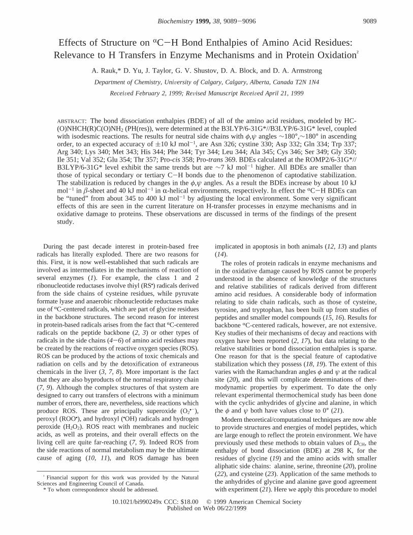
In Table 1 are listed theRC-H BDEs for all of the aminoacid residues at both the B3LYP and ROMP2 levels oftheory, based on the present model system which adequatelyincorporates the local internal stereoelectronic effects expe-rienced by a midchain residue (19). The data are showngraphically in Figure 1, arranged from left to right in orderof increasing BDE (by B3LYP) of the neutral amino acidresidue. The vertical scale is in kJ mol-1, and the horizontalbold line denotes the BDE of the S-H bond of glutathione(GSH) or cysteine, 367 kJ mol-1 (23). Figure 1 also ordersin a visual way the relative stabilities of theRC radicalspecies, from most stable (Asn) to least (trans-Pro).
Although this was not done in the present instance, onemay note that the BDEs can be used to estimate the reductionpotentials for theRC-centered radicals in reaction 4 (20):
TheE° (RP•(res),H+/PH(res)) values are measures of relativestabilities of the radicals or ease of oxidation of their parentsin solution.
DISCUSSION
Assessment of Theoretical Reliability.The B3LYP andROMP2 BDEs of the residues in Table 1 and Figure 1 followthe same trends, with the largest BDEs occurring for Proand Thr and the weakest for Asn and Asp. The results forGly by the two methods are identical. For all others theROMP2 BDEs are higher than those obtained with B3LYP,the average difference being 7.5 kJ mol-1. However, thedifferences are within the(10 kJ mol-1 uncertainties of thetwo procedures. Both sets of results are based on isodesmicreactions with H2NCH2COOH, taking a BDE of 331 kJ mol-1
for that molecule. There are as yet no experimental valuesfor this species, but methylene C-H BDEs for R2NCH2-COR compounds determined from pKHA and oxidationpotential data are 320 kJ mol-1 (27), and the value for Me2-NCH2COOEt has been shown by photoacoustic calorimetryto lie in the range 331-347 kJ mol-1 (28). The onlyexperimental BDEs, which are available in systems relatedto the residues, are those for the cyclic anhydrides of glycineand alanine, 340 and 325 kJ mol-1, respectively (21). Theseare shown by the black hexagons in Figure 1. The valueshave an absolute error of(15 kJ mol-1, and agreement withthe B3LYP results (glycine, 351 kJ mol-1; alanine, 335 kJmol-1) is satisfactory. More importantly, the B3LYP valuesreproduce the experimental difference of 15 kJ mol-1
between the two systems, which is much more accurate thanthe absolute values. The ROMP2 values for the anhydrides,350 and 340 kJ mol-1 for glycine and alanine, respectively,do not reproduce the experimental difference quite as wellbut are also in accord with the measured BDEs. Thus, in anoverall sense the theoretical results are consistent withavailable experimental data. Since the ROMP2 and B3LYP
RP•(res)+ AH h PH(res)+ A• (2)
DCH(PH) ) DCH(AH) - ∆H(2)° (3)
RP•(res)+ e- + H+ h PH(res) (4)
9090 Biochemistry, Vol. 38, No. 28, 1999 Rauk et al.
BDEs show the same trends, the remaining discussion isbased on the B3LYP data. However, the minor differencesare noted where relevant. As already mentioned, for Glyresidues the ROMP2 and B3LYP results are identical.
Factors That Affect the Stability of theRC Radical.All ofthe RC radical species are exceptionally stable compared tomost C-centered radicals. For example, the glycine residuehas aRC-H BDE of 350 kJ mol-1. This value is to be
Table 1: RC-H Bond Dissociation Energies of Amino Acid Residues in the Peptide Model
E(PH) (hartree) ZPE (kJ mol-1) E(RP•) (hartree) ZPE (kJ mol-1) BDEa (kJ mol-1) BDEb (kJ mol-1)
Nonpolar Aliphatic R groupsglycine -377.90145 266 -377.26202 235 350c 350alanine -417.21814 342 -416.58038 309 345d 352valine -495.84487 -495.20400 352leucine -535.15898 567 -534.52174 535 344 352isoleucine -535.15696 567 -534.51748 536 351 360prolinee cis -533.95219 -533.30896 358f 366
trans -533.95938 -533.31201 369f
Polar Uncharged R groupsserine -492.42896 356 -491.78998 326 349d 352threonine -531.74456 432 -531.10256 400 357d 364cysteine -815.40336 343 -814.76563 310 346g 353cystineh -1252.90711 424 -1252.27536 393 330g 338methionine -894.02856 -893.39101 343asparagine -585.92249 -585.29148 326 331glutamine -625.22897 -624.59461 334 342
Aromatic R Groupsphenylalanine -648.26675 -647.62876 344tyrosine -723.48279 -722.84487 344 356tryptophan -779.83622 -779.20100 337
Positively Charged R Groupslysine (0) -590.49829 -589.86182 340
(+) -590.86959 -590.23383 338arginine (0) -700.00074 -699.36422 340
(+) -700.40863 -699.77169 341histidine (0) -642.24515 -641.60646 346 353
(+) -642.63173 -641.99381 344 356
Negatively Charged R Groupsaspartate (0) -605.78119 -605.14768 332
(-) -605.23293 -604.60547 316glutamate (0) -645.09727 -644.46028 341
(-) -644.52919 -643.89605 331a B3LYP. b ROMP2.c Reference19. d Reference20. e N-Acetyl rather thanN-formyl. f Reference22. g Reference23. h PH(CysSSCH3), i.e., SH
of PH(Cys) replaced by SSCH3.
FIGURE 1: RC-H bond dissociation energies of the amino acid residues in a fully relaxed model peptide (see text). CysSSCH3 is a modelfor the cystine residue. TheRC-H BDEs of residues with ionized side chains are designated with (+) and (-). All results are at the B3LYPlevel, except white circles and hexagons are at ROMP2. Results for Gly and Ala anhydride are from ref21.
RC-H Bond Enthalpies of Amino Acid Residues Biochemistry, Vol. 38, No. 28, 19999091

compared with a secondary C-H BDE of 412 kJ mol-1 inC3H8 (29), 396 kJ mol-1 for C-H next to OH in CH3CH2-OH (30), and 388 kJ mol-1 for CH next to N (CH3NH2)
(31). The underlying reason has been calledcaptodatiVestabilization (18), a special feature of radicals which can enterinto conjugative delocalization with at least oneπ-acceptor
FIGURE 2: Asparagine model peptide andRC radical showing the H-bonding network. Hydrogen bond lengths are in angstroms and dihedralangles in degrees.
FIGURE 3: Isoleucine model peptide andRC radical showing steric crowding. Dimensions as in Figure 2.
FIGURE 4: Trytophan model peptide andRC radical showing steric crowding. Dimensions as in Figure 2.
9092 Biochemistry, Vol. 38, No. 28, 1999 Rauk et al.
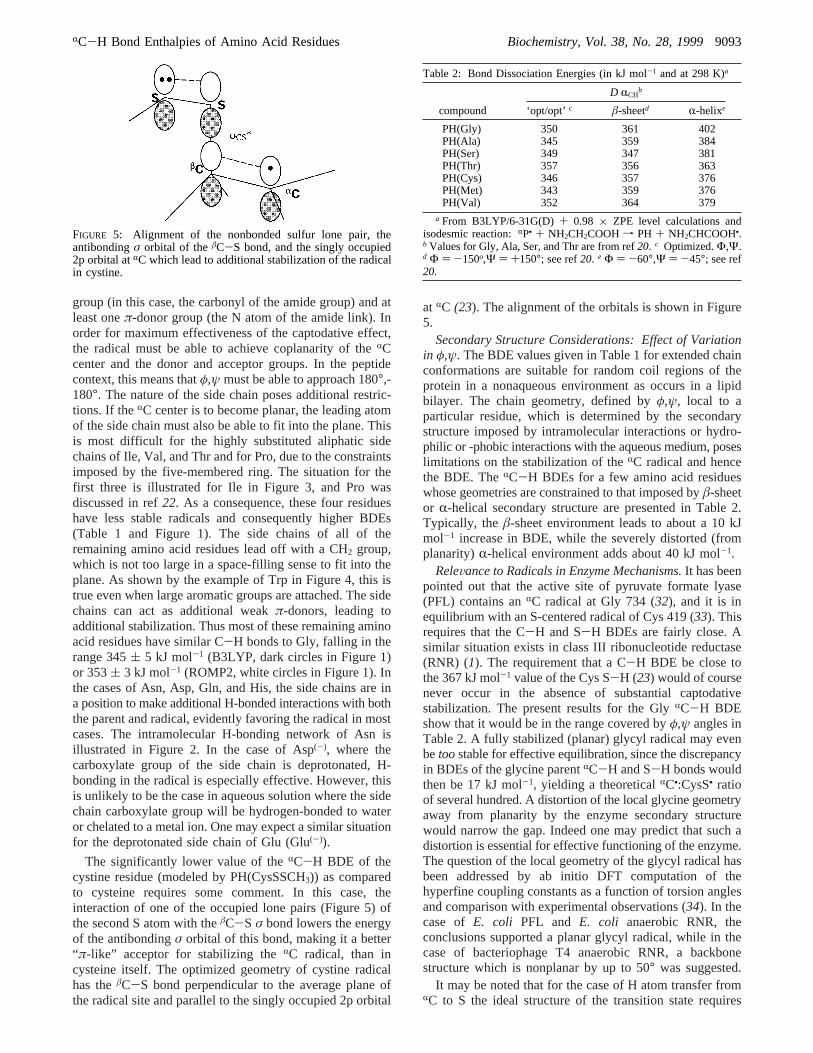
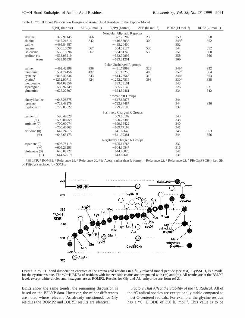
group (in this case, the carbonyl of the amide group) and atleast oneπ-donor group (the N atom of the amide link). Inorder for maximum effectiveness of the captodative effect,the radical must be able to achieve coplanarity of theRCcenter and the donor and acceptor groups. In the peptidecontext, this means thatφ,ψ must be able to approach 180°,-180°. The nature of the side chain poses additional restric-tions. If theRC center is to become planar, the leading atomof the side chain must also be able to fit into the plane. Thisis most difficult for the highly substituted aliphatic sidechains of Ile, Val, and Thr and for Pro, due to the constraintsimposed by the five-membered ring. The situation for thefirst three is illustrated for Ile in Figure 3, and Pro wasdiscussed in ref22. As a consequence, these four residueshave less stable radicals and consequently higher BDEs(Table 1 and Figure 1). The side chains of all of theremaining amino acid residues lead off with a CH2 group,which is not too large in a space-filling sense to fit into theplane. As shown by the example of Trp in Figure 4, this istrue even when large aromatic groups are attached. The sidechains can act as additional weakπ-donors, leading toadditional stabilization. Thus most of these remaining aminoacid residues have similar C-H bonds to Gly, falling in therange 345( 5 kJ mol-1 (B3LYP, dark circles in Figure 1)or 353( 3 kJ mol-1 (ROMP2, white circles in Figure 1). Inthe cases of Asn, Asp, Gln, and His, the side chains are ina position to make additional H-bonded interactions with boththe parent and radical, evidently favoring the radical in mostcases. The intramolecular H-bonding network of Asn isillustrated in Figure 2. In the case of Asp(-), where thecarboxylate group of the side chain is deprotonated, H-bonding in the radical is especially effective. However, thisis unlikely to be the case in aqueous solution where the sidechain carboxylate group will be hydrogen-bonded to wateror chelated to a metal ion. One may expect a similar situationfor the deprotonated side chain of Glu (Glu(-)).
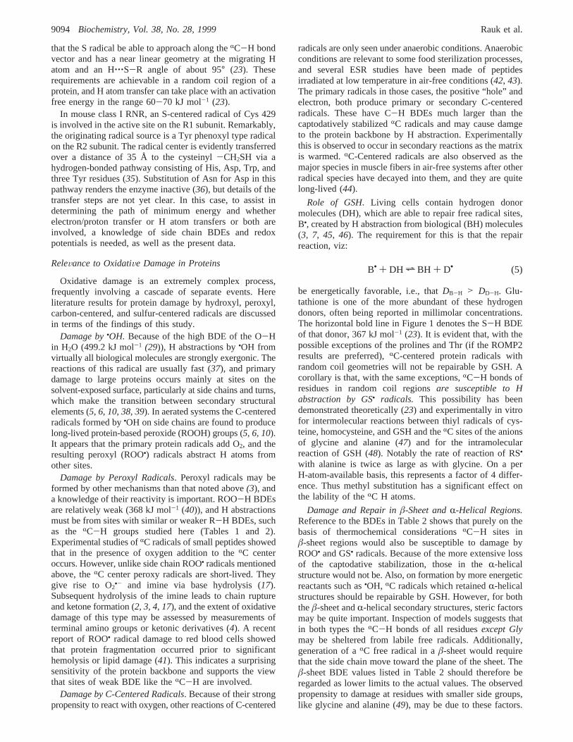
The significantly lower value of theRC-H BDE of thecystine residue (modeled by PH(CysSSCH3)) as comparedto cysteine requires some comment. In this case, theinteraction of one of the occupied lone pairs (Figure 5) ofthe second S atom with theâC-S σ bond lowers the energyof the antibondingσ orbital of this bond, making it a better“π-like” acceptor for stabilizing theRC radical, than incysteine itself. The optimized geometry of cystine radicalhas theâC-S bond perpendicular to the average plane ofthe radical site and parallel to the singly occupied 2p orbital
at RC (23). The alignment of the orbitals is shown in Figure5.
Secondary Structure Considerations: Effect of Variationin φ,ψ. The BDE values given in Table 1 for extended chainconformations are suitable for random coil regions of theprotein in a nonaqueous environment as occurs in a lipidbilayer. The chain geometry, defined byφ,ψ, local to aparticular residue, which is determined by the secondarystructure imposed by intramolecular interactions or hydro-philic or -phobic interactions with the aqueous medium, poseslimitations on the stabilization of theRC radical and hencethe BDE. TheRC-H BDEs for a few amino acid residueswhose geometries are constrained to that imposed byâ-sheetor R-helical secondary structure are presented in Table 2.Typically, theâ-sheet environment leads to about a 10 kJmol-1 increase in BDE, while the severely distorted (fromplanarity)R-helical environment adds about 40 kJ mol-1.
ReleVance to Radicals in Enzyme Mechanisms.It has beenpointed out that the active site of pyruvate formate lyase(PFL) contains anRC radical at Gly 734 (32), and it is inequilibrium with an S-centered radical of Cys 419 (33). Thisrequires that the C-H and S-H BDEs are fairly close. Asimilar situation exists in class III ribonucleotide reductase(RNR) (1). The requirement that a C-H BDE be close tothe 367 kJ mol-1 value of the Cys S-H (23) would of coursenever occur in the absence of substantial captodativestabilization. The present results for the GlyRC-H BDEshow that it would be in the range covered byφ,ψ angles inTable 2. A fully stabilized (planar) glycyl radical may evenbetoostable for effective equilibration, since the discrepancyin BDEs of the glycine parentRC-H and S-H bonds wouldthen be 17 kJ mol-1, yielding a theoreticalRC•:CysS• ratioof several hundred. A distortion of the local glycine geometryaway from planarity by the enzyme secondary structurewould narrow the gap. Indeed one may predict that such adistortion is essential for effective functioning of the enzyme.The question of the local geometry of the glycyl radical hasbeen addressed by ab initio DFT computation of thehyperfine coupling constants as a function of torsion anglesand comparison with experimental observations (34). In thecase of E. coli PFL and E. coli anaerobic RNR, theconclusions supported a planar glycyl radical, while in thecase of bacteriophage T4 anaerobic RNR, a backbonestructure which is nonplanar by up to 50° was suggested.
It may be noted that for the case of H atom transfer fromRC to S the ideal structure of the transition state requires
FIGURE 5: Alignment of the nonbonded sulfur lone pair, theantibondingσ orbital of theâC-S bond, and the singly occupied2p orbital atRC which lead to additional stabilization of the radicalin cystine.
Table 2: Bond Dissociation Energies (in kJ mol-1 and at 298 K)a
D RCHb
compound ‘opt/opt’c â-sheetd R-helixe
PH(Gly) 350 361 402PH(Ala) 345 359 384PH(Ser) 349 347 381PH(Thr) 357 356 363PH(Cys) 346 357 376PH(Met) 343 359 376PH(Val) 352 364 379
a From B3LYP/6-31G(D)+ 0.98 × ZPE level calculations andisodesmic reaction:RP• + NH2CH2COOH f PH + NH2CHCOOH•.b Values for Gly, Ala, Ser, and Thr are from ref20. c Optimized.Φ,Ψ.d Φ ) -150o,Ψ ) +150°; see ref20. e Φ ) -60°,Ψ ) -45°; see ref20.
RC-H Bond Enthalpies of Amino Acid Residues Biochemistry, Vol. 38, No. 28, 19999093
that the S radical be able to approach along theRC-H bondvector and has a near linear geometry at the migrating Hatom and an H‚‚‚S-R angle of about 95° (23). Theserequirements are achievable in a random coil region of aprotein, and H atom transfer can take place with an activationfree energy in the range 60-70 kJ mol-1 (23).
In mouse class I RNR, an S-centered radical of Cys 429is involved in the active site on the R1 subunit. Remarkably,the originating radical source is a Tyr phenoxyl type radicalon the R2 subunit. The radical center is evidently transferredover a distance of 35 Å to the cysteinyl-CH2SH via ahydrogen-bonded pathway consisting of His, Asp, Trp, andthree Tyr residues (35). Substitution of Asn for Asp in thispathway renders the enzyme inactive (36), but details of thetransfer steps are not yet clear. In this case, to assist indetermining the path of minimum energy and whetherelectron/proton transfer or H atom transfers or both areinvolved, a knowledge of side chain BDEs and redoxpotentials is needed, as well as the present data.
ReleVance to OxidatiVe Damage in Proteins
Oxidative damage is an extremely complex process,frequently involving a cascade of separate events. Hereliterature results for protein damage by hydroxyl, peroxyl,carbon-centered, and sulfur-centered radicals are discussedin terms of the findings of this study.
Damage by•OH. Because of the high BDE of the O-Hin H2O (499.2 kJ mol-1 (29)), H abstractions by•OH fromvirtually all biological molecules are strongly exergonic. Thereactions of this radical are usually fast (37), and primarydamage to large proteins occurs mainly at sites on thesolvent-exposed surface, particularly at side chains and turns,which make the transition between secondary structuralelements (5, 6, 10, 38, 39). In aerated systems the C-centeredradicals formed by•OH on side chains are found to producelong-lived protein-based peroxide (ROOH) groups (5, 6, 10).It appears that the primary protein radicals add O2, and theresulting peroxyl (ROO•) radicals abstract H atoms fromother sites.
Damage by Peroxyl Radicals. Peroxyl radicals may beformed by other mechanisms than that noted above(3), anda knowledge of their reactivity is important. ROO-H BDEsare relatively weak (368 kJ mol-1 (40)), and H abstractionsmust be from sites with similar or weaker R-H BDEs, suchas the RC-H groups studied here (Tables 1 and 2).Experimental studies ofRC radicals of small peptides showedthat in the presence of oxygen addition to theRC centeroccurs. However, unlike side chain ROO• radicals mentionedabove, theRC center peroxy radicals are short-lived. Theygive rise to O2
•- and imine via base hydrolysis (17).Subsequent hydrolysis of the imine leads to chain ruptureand ketone formation (2, 3, 4, 17), and the extent of oxidativedamage of this type may be assessed by measurements ofterminal amino groups or ketonic derivatives (4). A recentreport of ROO• radical damage to red blood cells showedthat protein fragmentation occurred prior to significanthemolysis or lipid damage (41). This indicates a surprisingsensitivity of the protein backbone and supports the viewthat sites of weak BDE like theRC-H are involved.
Damage by C-Centered Radicals. Because of their strongpropensity to react with oxygen, other reactions of C-centered
radicals are only seen under anaerobic conditions. Anaerobicconditions are relevant to some food sterilization processes,and several ESR studies have been made of peptidesirradiated at low temperature in air-free conditions (42, 43).The primary radicals in those cases, the positive “hole” andelectron, both produce primary or secondary C-centeredradicals. These have C-H BDEs much larger than thecaptodatively stabilizedRC radicals and may cause damgeto the protein backbone by H abstraction. Experimentallythis is observed to occur in secondary reactions as the matrixis warmed.RC-Centered radicals are also observed as themajor species in muscle fibers in air-free systems after otherradical species have decayed into them, and they are quitelong-lived (44).
Role of GSH. Living cells contain hydrogen donormolecules (DH), which are able to repair free radical sites,B•, created by H abstraction from biological (BH) molecules(3, 7, 45, 46). The requirement for this is that the repairreaction, viz:
be energetically favorable, i.e., thatDB-H > DD-H. Glu-tathione is one of the more abundant of these hydrogendonors, often being reported in millimolar concentrations.The horizontal bold line in Figure 1 denotes the S-H BDEof that donor, 367 kJ mol-1 (23). It is evident that, with thepossible exceptions of the prolines and Thr (if the ROMP2results are preferred),RC-centered protein radicals withrandom coil geometries will not be repairable by GSH. Acorollary is that, with the same exceptions,RC-H bonds ofresidues in random coil regionsare susceptible to Habstraction by GS• radicals. This possibility has beendemonstrated theoretically (23) and experimentally in vitrofor intermolecular reactions between thiyl radicals of cys-teine, homocysteine, and GSH and theRC sites of the anionsof glycine and alanine (47) and for the intramolecularreaction of GSH (48). Notably the rate of reaction of RS•
with alanine is twice as large as with glycine. On a perH-atom-available basis, this represents a factor of 4 differ-ence. Thus methyl substitution has a significant effect onthe lability of theRC H atoms.
Damage and Repair inâ-Sheet andR-Helical Regions.Reference to the BDEs in Table 2 shows that purely on thebasis of thermochemical considerationsRC-H sites inâ-sheet regions would also be susceptible to damage byROO• and GS• radicals. Because of the more extensive lossof the captodative stabilization, those in theR-helicalstructure would not be. Also, on formation by more energeticreactants such as•OH, RC radicals which retainedR-helicalstructures should be repairable by GSH. However, for boththeâ-sheet andR-helical secondary structures, steric factorsmay be quite important. Inspection of models suggests thatin both types theRC-H bonds of all residuesexcept Glymay be sheltered from labile free radicals. Additionally,generation of aRC free radical in aâ-sheet would requirethat the side chain move toward the plane of the sheet. Theâ-sheet BDE values listed in Table 2 should therefore beregarded as lower limits to the actual values. The observedpropensity to damage at residues with smaller side groups,like glycine and alanine (49), may be due to these factors.
B• + DH h BH + D• (5)
9094 Biochemistry, Vol. 38, No. 28, 1999 Rauk et al.
CONCLUSIONS
Captodatively stabilized radicals are produced by removalof the RC-H bond of amino acid residues in proteins. Inrandom coil regions, where theφ,ψ angles can aproach 180°,-180°, the stabilization is at a maximum, and most aminoacid residues are predicted to haveRC-H BDEs in the range345( 10 kJ mol-1. Stronger bonds occur in threonine (357)and proline (cis 358,trans369). Exceptionally weakRC-Hbonds occur in glutamine (334), aspartate (332), cystine(330), and asparagine (326). The stabilization is reduced bychanges in theφ,ψ angles, and the BDEs increase by about10 kJ mol-1 in â-sheet and 40 kJ mol-1 in R-helicalenvironments, respectively.
The existence of the captodative stabilization and itsdependence onφ,ψ means that theRC-H BDEs can be“tuned” from about 345 to 400 kJ mol-1 by adjusting thelocal environment. This has important consequences forH-transfer processes in enzyme mechanisms and in oxidativedamage to proteins. In the latter case, for instance, it makesRC-H sites in random coil regions susceptible to H abstrac-tion by relatively weak oxidants, like GS• and ROO•, as wellas the highly damaging•OH.
REFERENCES
1. Stubbe, J., and van der Dork, W. A. (1998)Chem. ReV. 98,705-762.
2. Garrison, W. M. (1987)Chem. ReV. 87, 381-398.3. von Sonntag, C. (1987)The Chemical Basis of Radiation
Biology, Taylor and Francis, London.4. Stadman, E. R. (1993)Annu. ReV. Biochem. 62, 797-821.5. Gebicki, S., and Gebicki, J. M. (1993)Biochem. J. 289, 743-
749.6. Davies, M. J., Fu, S., and Dean, R. T. (1995)Biochem. J.
305, 643-649.7. Davies, K. J. A., Ed. (1991)OxidatiVe Damage and Repair:
Chemical, Biological and Medical Aspects, Pergamon Press,New York.
8. Cheeseman, K. H. (1995) inImmunopharmacology of FreeRadical Species(Blake, D., and Winyard, P. G., Eds.)Academis Press, New York.
9. (a) Simic, M. G., Taylor, K. A., Ward, J. F., and von Sonntag,C., Eds. (1988)Oxygen Radicals in Biology and Medicine,Plenum Press, New York. (b) Sies, H., Ed. (1991)OxidatiVeStresssOxidants and Anti-Oxidants, Academic Press, London.
10. Dean, R. T., Fu, S., Stocker, R., and Davies, M. J. (1997)Biochem. J. 324, 1-18.
11. (a) Ames, B. N., Shigenaga, M. K., and Hagen, T. N. (1993)Proc. Natl. Acad. Sci. U.S.A. 90, 7915-7922. (b) Orr, W. C.,and Sohal, R. S. (1994)Science 263, 1128-1130.
12. Borek, C. (1987)Br. J. Cancer Suppl. 8, 74-86.13. (a) Vatissiere, J. L., Petit, P. X., Risler, Y., and Mignotte, B.
(1994) Proc. Natl. Acad. Sci. U.S.A. 91, 11752-11756. (b)Johnson, T. M., Yu, Z.-X., Ferrans, V. J., Lowenstein, R. A.,and Finkel, T. (1996)Proc. Natl. Acad. Sci. U.S.A. 93, 11848-11852.
14. Greenberg, J. T. (1996)Proc. Natl. Acad. Sci. U.S.A. 93,12094-12097.
15. Wardman, P. (1989)J. Phys. Chem. Ref. Data 18, 1637-1755.16. Klapper, M. H., and Farragi, M. (1994)J. Am. Chem. Soc.
116, 1414-1422.17. (a) Mieden, O. J., and von Sonntag, C. (1989)Z. Naturforsch.
44b, 959-974. (b) Mieden, O. J., and von Sonntag, C. (1989)J. Chem. Soc., Perkin Trans. 2,2071-2078. (c) Mieden, O.J., and von Sonntag, C. (1993)J. Phys. Chem. 97, 3783-3790.
18. Leroy, G., Sana, M., and Wilante, C. (1991)J. Mol. Struct.228, 37-45.
19. Armstrong, D. A., Yu, D., and Rauk, A. (1996)Can. J. Chem.74, 1192-1199.
20. Armstrong, D. A., Rauk, A., and Yu, D. (1997)J. Am. Chem.Soc. 119, 208-217.
21. Jonsson, M., Wayner, D. D. M., Armstrong, D. A., Yu, D.,and Rauk, A. (1998)J. Chem. Soc. Perkin Trans. 2,1967-1972.
22. Block, D. A., Yu, D., Armstrong, D. A., and Rauk, A. (1998)Can. J. Chem. 76, 1042-1049.
23. Rauk, A., Yu, D., and Armstrong, D. A. (1998)J. Am. Chem.Soc. 120, 8848-8855.
24. Hehre, W. J., Ditchfield, R., Radom, L., and Pople, J. A. (1970)J. Am. Chem. Soc. 92, 4796-4816.
25. Frisch, M. J., Trucks, G. W., Schlegel, H. B., Gill, P. M. W.,Johnson, B. G., Robb, M. A., Cheeseman, J. R., Keith, T. A.,Petersson, G. A., Montgomery, J. A., Raghavachari, K., Al-Laham, M. A., Zakrewski, V. G., Ortiz, J. V., Foresman, J.B., Cioslowski, J., Stefanov, B. B., Nanayakkara, A., Chal-lacombe, M., Peng, C. Y., Ayala, P. Y., Chen, W., Wong, M.W., Andres, J. L., Replogle, E. S., Gomperts, R., Martin, R.L., Fox, D. J., Binkley, J. S., Defrees, D. J., Baker, J., Stewart,J. P., Head-Gordon, M., Gonzalez, C., and Pople, J. A. (1995)Gaussian 94 (SGI-ReVision B.3), Gaussian, Inc., Pittsburgh,PA.
26. (a) Parkinson, C. J., Mayer, P. M., and Radom, L.Theoret.Chem. Acc. (in press). (b) Parkinson, C. J., Mayer, P. M., andRadom, L. To be published. (c) For application to simpleamino acid radicals, see also Croft, A. K., Easton, C. J.,Radom, L. To be published. The high-level CBS-RAD theorywas taken as yielding reference RSEs, and the application wasto radicals with some features in common with the presentset, including captodative stabilization.
27. Bordwell, F. G., Gallagher, T., and Zhang, X. (1991)J. Am.Chem. Soc. 113, 3495-3497.
28. Clark, K. B., Wayner, D. D. M., Demirjdji, S. H., and Koch,T. H. (1993)J. Am. Chem. Soc. 115, 2447-2453.
29. Berkowitz, J., Ellison, G. B., and Gutman, D. (1994)J. Phys.Chem. 98, 2744-2765.
30. Block, D. A., Armstrong, D. A., and Rauk, A. Submitted forpublication.
31. Wayner, D. D. M., Clark, K. B., Rauk, A., Yu, D., andArmstrong, D. A. (1997)J. Am. Chem. Soc. 119, 8925-8932.
32. Wagner, A. V. F., Frey, M., Neugebauer, F. A., Schafer, W.,and Knappe, J. (1992)Proc. Natl. Acad. Sci. U.S.A. 89, 996-1000.
33. Reddy, S. G., Wong, K. K., Parast, C. V., Peisach, J.,Magliozzo, R. S., and Kozarich, J. W. (1998)Biochemistry37, 558-563.
34. Himo, F., and Eriksson, L. A. (1998)J. Chem. Soc., PerkinTrans. 2, 305-308.
35. Schmidt, P. P., Rova, U., Katterle, B., Thelander, L., andGraslund, A. (1998)J. Biol. Chem. 273, 21463-21472.
36. Ekberg, M., Potsch, S., Sandin, E., Thunnissen, M., Nordlund,P., Sahlin, M., and Sjoberg, B.-M. (1998)J. Biol. Chem. 273,21003-21008.
37. Buxton, G. V., Greenstock, C. L., Helman, W. P., and Ross,A. B. (1988)J. Phys. Chem. Ref. Data 17, 513-886.
38. Schuessler, H., and Schilling, K. (1984)Int. J. Radiat. Biol.45, 267.
39. Puchala, M., and Schuessler, K. (1993)Int. J. Radiat. Biol.64, 149-156.
40. Merenyi, G., Lind, J., and Engman, L. (1994)J. Chem. Soc.,Perkin Trans. 2,2551-2553.
41. Celedon, G., Lips, V., Alvarado, C., Cortes, M., Lissis, E. A.,and Gonzalez, G. (1997)Biochem. Mol. Biol. Internat. 43,1121-1127.
42. (a) Sevilla, M. D., D′Arcy, J. B., and Morehouse, K. M. (1979)J. Phys. Chem. 83, 2887-2892. (b) Sevilla, M. D., D’Arcy,J. B., and Morehouse, K. M. (1979)J. Phys. Chem. 83, 2893-2897.
43. Burlinska, G., Michalik, J., and Bobrowski, K. (1994)Radiat.Phys. Chem. 43, 425-432.
44. Iskakov, A. A., and L’Vov, K. M. (1997)Biophysics 38, 405-410.
RC-H Bond Enthalpies of Amino Acid Residues Biochemistry, Vol. 38, No. 28, 19999095

45. (a) Wardman, P. (1993) inAtmospheric Oxidation andAntioxidants(Scott, G., Ed.) Vol. III, pp 101-127, Elsevier,New York. (b) Wardman, P., and von Sonntag, C. (1995)Methods Enzymol. 251, 31-45.
46. Wayner, D. D. M., Burton, G. W., Ingold, K. U., Barclay, L.R. C., and Kocke, J. S. (1987)Biochim. Biophys. Acta408-419.
47. Zhao, R., Lind, J., Merenyi, G., and Eriksen, T. E. (1994)J.Am. Chem. Soc. 116, 12010-12015.
48. Zhao, R., Lind, J., Merenyi, G., and Eriksen, T. E. (1997)J.Chem. Soc., Perkin Trans. 2, 569-574.
49. Sharpatyi, V. A. (1995)High Energy Chem. 29, 85-100.
BI990249X
9096 Biochemistry, Vol. 38, No. 28, 1999 Rauk et al.