Embed Size (px)
Citation preview
TitleElucidation of the central role of long-chain fatty acids in thepalatability of dietary fat by neuroscientific and animalbehavioral studies( Dissertation_全文 )
Author(s) Adachi, Shinichi
Citation Kyoto University (京都大学)
Issue Date 2015-01-23
URL https://doi.org/10.14989/doctor.k18695
Right
Type Thesis or Dissertation
Textversion ETD
Kyoto University
Elucidation of the central role of long-chain fatty acids
in the palatability of dietary fat
by neuroscientific and animal behavioral studies
Shinichi Adachi
2015

CONTENTS
GENERAL INTRODUCTION • • • • • • • • • • • • 1
CHAPTER 1 Effect of long chain fatty acid on dopamine level increase in the
nucleus accumbens by ingesting dietary fat • • • • • • • • • • • • 7
CHAPTER 2 Involvement of GPR120-agonistic activity of long chain fatty acid
in the palatability of dietary fat • • • • • • • • • • • • 24
SUMMARY • • • • • • • • • • • • 49
ACKNOWLEDGEMENTS • • • • • • • • • • • • 50
LIST OF PUBLICATIONS • • • • • • • • • • • • 51

1
GENERAL INTRODUCTION Most animals, including rodents and humans prefer fat-rich foods (1, 2). The
ingesting of corn oil has been reported to produce a reward effect in mice, and the
dopaminergic pathway in the nervous system has been implicated in the manifestation
of this effect (3, 4). Additionally, sham feeding of corn oil were reported to induce the
activation of the midbrain dopamine (DA) system, which is involved in reward behavior
(5). The mesolimbic system is thought to play a critical role in the reward effect, and the
release of DA has been demonstrated when a natural and drug reward is acquired or
when its acquisition is anticipated (6, 7) The midbrain dopaminergic circuits originate
from the ventral tegmental area (VTA) and project to different sites, such as the nucleus
accumbens (NAc), amygdala, and the prefrontal area, which are related to motivation,
palatability, and addiction (8–10). Beta-endorphin is known as an endogenous opioid
peptide and µ-opioid receptor ligand. Tanda et al. demonstrated that intra-VTA infusion
of naloxonazine, µ1 opioid receptor antagonist, prevented the NAc DA increase by
ingestion of palatable food and suggested that µ1 receptors in the VTA depend on the
activation of the midbrain dopamine system by the palatability food intake (11).
Matsumura et al. found fat emulsion ingestion increased c-fos expression in
beta-endorphin neurons of the mouse hypothalamus (12). Mizushige et al. demonstrated
corn oil ingestion induced beta-endorphin levels in the serum and cerebrospinal fluid in
the rats (13). Iwakura et al. showed that the secretion of ghrelin known as a hormone to
stimulate eating was induced by DA in a ghrelin-producing cell line MGN3-1 (14).
Since the extracellular concentration of DA in the NAc of the rat has been increased
dose-dependently by self-administration of cocaine or consumption of sucrose, release
of DA in the NAc could be considered as a kind of index for the palatability or
motivational drive (15, 16). However, which property of fat caused the increase in DA
release has not been elucidated. Fat palatability could be explained by such factors as
the unique texture of fat, flavor, high caloric density per unit mass, and chemical
reception of fatty acids on the tongue (17–19), all of which might result in increased DA
release during fat ingestion.
Recent studies have revealed that chemoreceptions of long-chain fatty acids
(LCFAs) are involved in the recognition of fatty foods. Kawai et al. reported that, when
fat was introduced into the oral cavity of rats, a certain percentage of triacylglycerides
2
was hydrolyzed to LCFAs by lingual lipase (20). Gilbertson et al. demonstrated the
regulation of K+ channels in type II taste cells by unsaturated LCFAs and suggested the
presence of fatty acid chemoreceptors in taste cells (21). Fukuwatari et al. found that
CD36 fatty acid transporter was expressed on the apical side of taste cells in the
circumvallate papillae (22). In addition, CD36-deficient mice were reported to show a
low taste preference for fat (23). Moreover, Matsumura et al. reported that G
protein-coupled receptor GPR120 was also expressed on the apical side of taste cells in
the circumvallate papillae (24). The unsaturated LCFAs, such as oleic acid and linolenic
acid, induced a rise in concentration of intracellular calcium ion ([Ca2+]i) in Human
Embryonic Kidney 293 (HEK293) cells stably expressing GPR120. On the other hand,
LCFA esters and capric acid did not induce its rise (25).In a licking behavior test, it was
found the mice exhibited equally strong preference for a low concentration of linoleic
acid as that for 100% corn oil (26). Additionally, the mice and rats displayed a similar
strong preference for LCFAs, such as oleic, linolenic, and linoleic acid, whereas they
did not show any preference for LCFA esters nor long-chain fatty alcohols (27, 28).
Compared with wild-type mice, GPR120 knock-out mice displayed a lower preference
for LCFAs (29). These findings lead us to postulate that, in the oral cavity, fat is
hydrolyzed to LCFA by lingual lipase and that chemoreception of this LCFA by
receptor proteins, such as CD36 and GPR120 expressed on the taste cells, could be
involved in the oral recognition and palatability of fats. The purpose of the present
study were to elucidate the involvement of LCFA, which is a component of fat, in the
increased release of DA in NAc during corn oil ingestion and to examine which
structural characteristics of LCFA are responsible for the palatability of fat.
In chapter 1, we studied the change in the extracellular concentration of DA in NAc
by in vivo microdialysis when rats ingested a low concentration of LCFA. Similar
measurements were made in the basolateral amygdala (BLA), which is projected by the
dopaminergic nerve system from VTA, as well as to NAc. Since the DA level in BLA
increased with the stimulation of reward expectation (30) and cue-responsive neurons in
BLA encode the motivating or reinforcing properties of a cue previously associated
with a reward (31), the DA response in BLA is thought to be related to reward
acquisition.
In chapter 2, we examined the relationship between the GPR120-agonistic
properties of ligands and the palatability of LCFAs. First, using HEK 293 cells stably
3
expressing human GPR120, we examined the effect of various LCFAs on the
concentration of [Ca2+]i by using a fluorescence spectrophotometer. We then assessed
the palatability for a variety of LCFAs at a low concentration by testing the licking
behavior of the mice. Next, we studied the change in the extracellular concentration of
DA in the NAc of the mice by in vivo microdialysis after ingestion of various
low-concentration LCFAs.
REFERENCES
1. Drewnowski A, Greenwood MR. (1983). Cream and sugar: human preferences
for high-fat foods. Physiol Behav. 30,:629–33
2. Takeda M, Imaizumi M, Fushiki T. (2000). Preference for vegetable oils in the
two-bottle choice test in mice. Life Sci. 67,:197–204
3. Imaizumi M, Takeda M, Fushiki T. (2000). Effects of oil intake in the
conditioned place preference test in mice. Brain Res. 870,:150–56
4. Yoneda T, Taka Y, Okamura M, Mizushige T, Matsumura S, Manabe Y, Tsuzuki
S, Inoue K, Fushiki T. (2007). Reinforcing effect for corn oil stimulus was
concentration dependent in an operant task in mice. Life Sci. 81,:1585–92
5. Liang N-C, Hajnal A, Norgren R. (2006). Sham feeding corn oil increases
accumbens dopamine in the rat. Am J Physiol Regul Integr Comp Physiol.
291,:R1236–9
6. Wise RA, Rompre PP. (1989). Brain dopamine and reward. Annu Rev Psychol.
40,:191–225
7. Wise RA. (2006). Role of brain dopamine in food reward and reinforcement.
Philos Trans R Soc Lond B Biol Sci. 361,:1149–58
4
8. Geisler S, Zahm DS. (2005). Afferents of the ventral tegmental area in the
rat-anatomical substratum for integrative functions. J Comp Neurol. 490,:270–94
9. Meye FJ, Adan RA. (2014). Feelings about food: the ventral tegmental area in
food reward and emotional eating. Trends Pharmacol Sci. 35,:31–40
10. Willuhn I, Wanat MJ, Clark JJ, Phillips PEM. (2010). Dopamine signaling in the
nucleus accumbens of animals self-administering drugs of abuse. Curr Top
Behav Neurosci. 3,:29–71
11. Tanda G, Di Chiara G. (1998). A dopamine-mu1 opioid link in the rat ventral
tegmentum shared by palatable food (fonzies) and non-psychostimulant drugs of
abuse. Eur J Neurosci. 10,:1179–87
12. Matsumura S, Eguchi A, Okafuji Y, Tatsu S, Mizushige T, Tsuzuki S, Inoue K,
Fushiki T. (2012). Dietary fat ingestion activates β-endorphin neurons in the
hypothalamus. FEBS Lett. 586,:1231–35
13. Mizushige T, Saitoh K, Manabe Y, Nishizuka T, Taka Y, Eguchi A, Yoneda T,
Matsumura S, Tsuzuki S, Inoue K, Fushiki T. (2009). Preference for dietary fat
induced by release of beta-endorphin in rats. Life Sci. 84,:760–65
14. Iwakura H, Ariyasu H, Hosoda H, Yamada G, Hosoda K, Nakao K, Kangawa K,
Akamizu T. (2011). Oxytocin and dopamine stimulate ghrelin secretion by the
ghrelin-producing cell line, mgn3-1 in vitro. Endocrinology. 152,:2619–25
15. Hajnal A, Smith GP, Norgren R. (2004). Oral sucrose stimulation increases
accumbens dopamine in the rat. Am J Physiol Regul Integr Comp Physiol.
286,:R31–7
16. Pettit HO, Justice JB. (1991). Effect of dose on cocaine self-administration
behavior and dopamine levels in the nucleus accumbens. Brain Res. 539,:94–102
17. Mindell S, Smith GP, Greenberg D. (1990). Corn oil and mineral oil stimulate
sham feeding in rats. Physiol Behav. 48,:283–87
5
18. Ramirez I. (1993). Role of olfaction in starch and oil preference. Am J Physiol.
265,:R1404–9
19. Matsumura S, Yoneda T, Shoji A, Eguchi A, Manabe Y, Tsuzuki S, Inoue K,
Fushiki T. (2010). Intragastric infusion of glucose enhances the rewarding effect
of sorbitol fatty acid ester ingestion as measured by conditioned place preference
in mice. Physiol Behav. 99,:509–14
20. Kawai T, Fushiki T. (2003). Importance of lipolysis in oral cavity for orosensory
detection of fat. Am J Physiol Regul Integr Comp Physiol. 285,:R447–54
21. Gilbertson TA, Fontenot DT, Liu L, Zhang H, Monroe WT. (1997). Fatty acid
modulation of k+ channels in taste receptor cells: gustatory cues for dietary fat.
Am J Physiol. 272,:C1203–10
22. Fukuwatari T, Kawada T, Tsuruta M, Hiraoka T, Iwanaga T, Sugimoto E,
Fushiki T. (1997). Expression of the putative membrane fatty acid transporter
(fat) in taste buds of the circumvallate papillae in rats. FEBS Lett. 414,:461–64
23. Laugerette F, Passilly-Degrace P, Patris B, Niot I, Febbraio M, Montmayeur JP,
Besnard P. (2005). Cd36 involvement in orosensory detection of dietary lipids,
spontaneous fat preference, and digestive secretions. J Clin Invest. 115,:3177–84
24. Matsumura S, Eguchi A, Mizushige T, Kitabayashi N, Tsuzuki S, Inoue K,
Fushiki T. (2009). Colocalization of gpr120 with phospholipase-cbeta2 and
alpha-gustducin in the taste bud cells in mice. Neurosci Lett. 450,:186–90
25. Hirasawa A, Tsumaya K, Awaji T, Katsuma S, Adachi T, Yamada M, Sugimoto
Y, Miyazaki S, Tsujimoto G. (2005). Free fatty acids regulate gut incretin
glucagon-like peptide-1 secretion through gpr120. Nat Med. 11,:90–94
26. Yoneda T, Saitou K, Mizushige T, Matsumura S, Manabe Y, Tsuzuki S, Inoue K,
Fushiki T. (2007). The palatability of corn oil and linoleic acid to mice as
measured by short-term two-bottle choice and licking tests. Physiol Behav.
91,:304–9
6
27. Yoneda T, Saitou K, Asano H, Mizushige T, Matsumura S, Eguchi A, Manabe Y,
Tsuzuki S, Inoue K, Fushiki T. (2009). Assessing palatability of long-chain fatty
acids from the licking behavior of balb/c mice. Physiol Behav. 96,:735–41
28. Tsuruta M, Kawada T, Fukuwatari T, Fushiki T. (1999). The orosensory
recognition of long-chain fatty acids in rats. Physiol Behav. 66,:285–88
29. Cartoni C, Yasumatsu K, Ohkuri T, Shigemura N, Yoshida R, Godinot N, le
Coutre J, Ninomiya Y, Damak S. (2010). Taste preference for fatty acids is
mediated by gpr40 and gpr120. J Neurosci. 30,:8376–82
30. Weiss F, Maldonado-Vlaar CS, Parsons LH, Kerr TM, Smith DL, Ben-Shahar O.
(2000). Control of cocaine-seeking behavior by drug-associated stimuli in rats:
effects on recovery of extinguished operant-responding and extracellular
dopamine levels in amygdala and nucleus accumbens. Proc Natl Acad Sci U S A.
97,:4321–26
31. Tye KM, Janak PH. (2007). Amygdala neurons differentially encode motivation
and reinforcement. J Neurosci. 27,:3937–45
Brain Dopamine Release by Ingesting a Long-Chain Fatty Acid
7
CHAPTER 1
Effect of long chain fatty acid on dopamine level increase in the
nucleus accumbens by ingesting dietary fat
Animals, including rodents and human beings, prefer fat-rich foods (1, 2). The
ingestion of corn oil has a strong reward effect on mice, and the involvement of
dopaminergic pathways in the nervous system has been implicated in the manifestation
of this effect (3, 4). The mesolimbic system in the brain is thought to play a critical role
in the reward effect, and the release of dopamine (DA) has been demonstrated when a
natural or drug reward is acquired or its acquisition is anticipated (5, 6). The midbrain
dopaminergic circuits originate from the ventral tegmental area (VTA), and project to
such sites as the nucleus accumbens (NAc), amygdala, and prefrontal area which are
respectively related to motivation, palatability, and addiction (7–9). Since the
extracellular concentration of DA in NAc has been increased dose-dependently by
self-administering cocaine and ingesting sucrose in rats, the DA release in NAc could be
considered as a kind of index of palatability or motivational drive (10, 11). In fact, sham
feeding corn oil or sucrose has been reported to increase the DA level in NAc of rats
(12), and a similar increase in DA released by cocaine administration has also been
reported to occur in the amygdala (13). However, which property of fat caused the
increase in DA release has not been elucidated. Fat palatability could be explained by
such factors as the unique texture of fat, odor, high caloric density per unit mass, and
chemical reception of fatty acids on the tongue (14–16), all of which might result in
increased DA release during fat ingestion.
We focused the present study on the chemoreception of fatty acids on the tongue.
Kawai et al. have reported that when fat was introduced into the oral cavity of rats, a
certain percentage of triacylglycerides was hydrolyzed to long-chain fatty acids
(LCFAs) by lingual lipase (17). Gilbertson et al. have shown the regulation of K+ ion
channels in type II taste cells by long-chain unsaturated fatty acids and suggested the
presence of fatty acid chemoreceptors in taste cells (18). The protein candidates related
to the recognition of LCFAs in the oral cavity are CD36 and G-protein-coupled receptor
120 (GPR120). We have found the expression of CD36 and GPR120 on the apical side
of taste cells in the circumvallate papillae (19, 20). In addition, CD36- or GPR120 gene-
Brain Dopamine Release by Ingesting a Long-Chain Fatty Acid
8
deficient mice have been reported to show a low taste preference for fat (21, 22). These
findings lead us to postulate that fat introduced into the oral cavity is hydrolyzed into
LCFA by lingual lipase and that chemoreception of this LCFA by proteins such as
CD36 and GPR120 expressed on the taste cells would function as energy sensors which
transduce the presence of fat in the oral cavity. The purpose of the present study was to
elucidate the involvement of LCFA, which is a component of fat, in the increased
release of DA in NAc during corn oil ingestion. We studied the change in the
extracellular concentration of DA in NAc by in vivo microdialysis when rats ingested a
low concentration of LCFA. We used 1% linoleic acid diluted with mineral oil in the
experiments. This concentration of fatty acid was in the range that could be released
from fat by lingual lipase and had only a 1/100 calorie content when compared with the
same weight of fat. Similar measurements were made in the basolateral amygdala
(BLA), which is projected by the dopaminergic nerve system from VTA, as well as to
NAc. Since the DA level in BLA increased with the stimulation of reward expectation
(23) and cue-responsive neurons in BLA encode the motivating or reinforcing properties
of a cue previously associated with a reward (24), the DA response in BLA is thought to
be related to reward acquisition.
Materials and Methods
Animals. This study was conducted in accordance with the ethical guidelines of the
Kyoto University Animal Experimentation Committee and in complete compliance with
the National Institutes of Health Guide for the Care and Use of Laboratory Animals, and
was approved by the above-mentioned committee. Male Wistar rats (Japan SLC,
Hamamatsu, Japan) at 8 weeks old were housed in stainless wire mesh cages in a room
controlled by a 12-h light–dark cycle (dark phase of 18:00–6:00) and constant
temperature (24±1 ºC). They were separately housed for a week for acclimatization to
the environment. The animals were fed tap water and regular MF rat food (Oriental
Yeast, Tokyo, Japan) ad libitum.
Materials. Corn oil was purchased from Ajinomoto (Tokyo, Japan), and mineral oil
was purchased from Kaneda Company (Tokyo, Japan). Linoleic acid from Sigma (St.
Louis, MO, USA) was 99% pure, stored at –20 ºC until needed and then diluted 1%
Brain Dopamine Release by Ingesting a Long-Chain Fatty Acid
9
with mineral oil for use in this experiment. The other reagents were purchased from
Nacalai Tesque (Kyoto, Japan).
Training protocols for oil ingestion. To allow the rats to get accustomed to ingesting
corn oil, mineral oil, and 1% linoleic acid, the rats were fed these liquids in their home
cages before surgery and then in the microdialysis cage after recovery from surgery.
The rats were deprived of water and food for 4 h and the liquids presented for 30 min.
Before surgery, the rats were presented with corn oil and mineral oil at the same time on
days 1 and 4, with mineral oil and 1% linoleic acid on days 2 and 5, and with 1%
linoleic acid and corn oil on days 3 and 6. To confirm the rats’ preference for corn oil
versus mineral oil, the rats were subjected on day 7 to a 2-bottle preference test for corn
oil vs. mineral oil. The bottle of each liquid was positioned randomly. After recovering
from the surgery, the rats were presented one kind of liquid in the microdialysis cage as
follows: corn oil on days 1 and 4, mineral oil on days 2 and 5, and 1% linoleic acid on
days 3 and 6. The rats were then subjected to a microdialysis test on day 7.
Microdialysis. Surgery: The animals were anesthetized with pentobarbital sodium
(Nembutal; Dainippon Pharmaceutical Co., Tokyo, Japan) and placed in a stereotaxic
frame adapted for rat surgery. The skull was subsequently exposed, and holes for micro-
dialysis were drilled. The respective coordinates for the NAc shell and amygdala guide
cannula (AG-10; Eicom, Kyoto, Japan) were AP, 1.7; ML, 0.2; and DV, 8.0; and AP,
–2.8; ML, 5.0; and DV, 7.5 from the bregma. All coordinates were determined
according to the stereotaxic atlas of Paxinos and Watson (25). The cannulas were
secured to the skull with a LOCTITE 454 adhesive bond (Henkel Japan, Yokohama,
Japan). A dummy AD-10 cannula (Eicom) was inserted into the guide cannula and
secured with an AC-1 cap nut (Eicom). The rats were allowed 5–7 d to recover from the
surgery. Each rat implanted with a probe in NAc or BLA was provided for one-time
microdialysis with one kind of test liquid.
Procedure: The experiments were conducted during the light period. The dummy
cannula was removed on the day of the experiment, and the AI-10-2 microdialysis probe
(Eicom, 2.0mm membrane length) was inserted into NAc or the amygdala via the guide
cannula. The rats were placed in the microdialysis cage at 10:00 a.m. for 4 h without
food and water, and then presented with the test liquid at 02:00 p.m. for 10 min. The
Brain Dopamine Release by Ingesting a Long-Chain Fatty Acid
10
amount of liquid ingested was also recorded. The rats remained in the microdialysis
cage for another 110 min after presenting the test liquid. Ringer’s solution, containing
147mM Na+, 4mM K+, 2.3mM Ca2+, and 155.6mM Cl- was perfused at 2 mL/min by
using an ESP-64 micro-syringe pump (Eicom), dialysate collection being started 20 min
before the liquids were presented and conducted every 5 min for a total of 140 min
thereafter. To quantify the DA levels in the dialysate, samples were analyzed by
reversed-phase HPLC with an electrochemical detector, using an Eicompak PP-ODS
column (4.6 i.d. × 30 mm long; Eicom). The applied voltage was set at 450mV
(relative to an Ag/AgCl reference electrode). The mobile phase at a flow rate of 500
mL/min consisted of a 99% (v/v) 0.1M phosphate buffer at pH 6.0, 1% (v/v) methanol,
500 mg/L of sodium decane sulfate, and 50 mg/L of 2Na-EDTA. The mean value
obtained from 3 samples from –20 to –5 min was set as the 100% baseline level, and all
subsequent sample values were expressed as a percentage of the baseline value.
Histological analysis. After completing the experiment, the rats were deeply
anesthetized with sodium pentobarbital. The brain was removed from the skull, frozen
and cut into 30-µm sections. The placement of the microdialysis probe was verified by
thionine blue staining. Data obtained from the rats with inappropriate probe placement
were excluded from the analysis.
Statistics. Data are expressed as the mean ± SEM. Data from the 2-bottle preference
test were analyzed by a paired t-test. Changes in DA levels were compared with the
corresponding baseline value by one-way ANOVA and Tukey’s multiple-comparison
test as a post-hoc test. Mean differences among 3 groups at each time point were
analyzed by two-way repeated-measures ANOVA and Bonferroni’s
multiple-comparison test as a post-hoc test. The amounts of each fluid ingested during
microdialysis were analyzed with one-way ANOVA and the Tukey–Kramer test as a
post-hoc test. p values of 5% or less were considered statistically significant. Statistical
analyses were conducted by using the Prism 4 software package (GraphPad, San Diego,
CA, USA).
Brain Dopamine Release by Ingesting a Long-Chain Fatty Acid
11
Results
Validation of the preference for oil after ingestion training
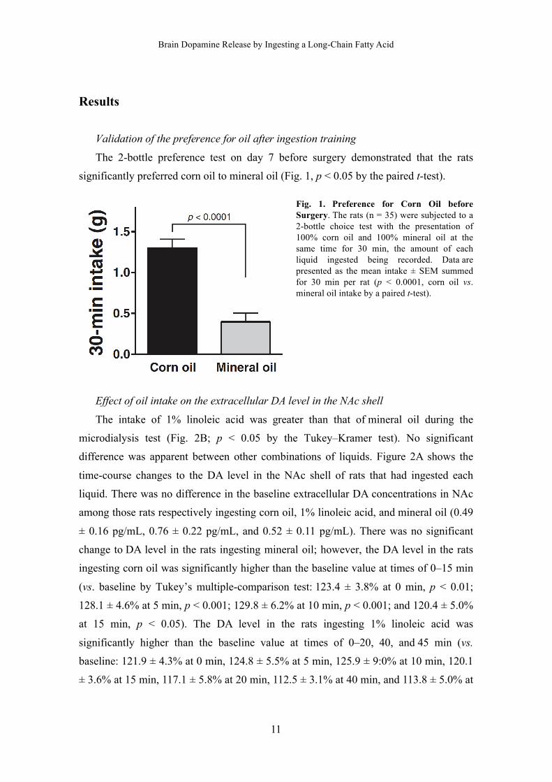
The 2-bottle preference test on day 7 before surgery demonstrated that the rats
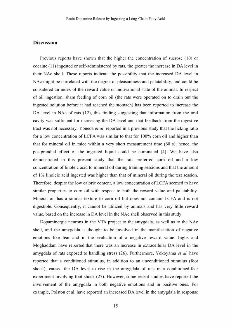
significantly preferred corn oil to mineral oil (Fig. 1, p < 0.05 by the paired t-test).
Fig. 1. Preference for Corn Oil before Surgery. The rats (n = 35) were subjected to a 2-bottle choice test with the presentation of 100% corn oil and 100% mineral oil at the same time for 30 min, the amount of each liquid ingested being recorded. Data are presented as the mean intake ± SEM summed for 30 min per rat (p < 0.0001, corn oil vs. mineral oil intake by a paired t-test).
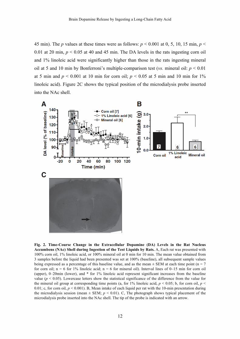
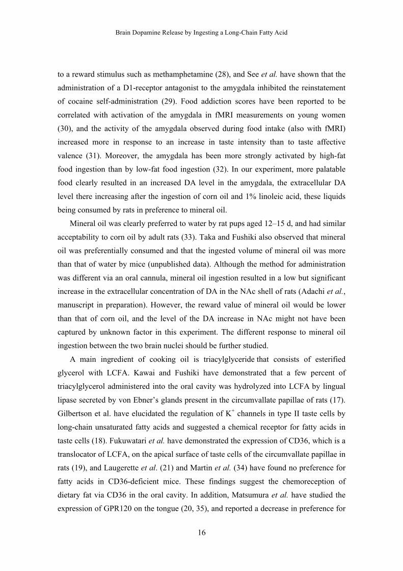
Effect of oil intake on the extracellular DA level in the NAc shell
The intake of 1% linoleic acid was greater than that of mineral oil during the
microdialysis test (Fig. 2B; p < 0.05 by the Tukey–Kramer test). No significant
difference was apparent between other combinations of liquids. Figure 2A shows the
time-course changes to the DA level in the NAc shell of rats that had ingested each
liquid. There was no difference in the baseline extracellular DA concentrations in NAc
among those rats respectively ingesting corn oil, 1% linoleic acid, and mineral oil (0.49
± 0.16 pg/mL, 0.76 ± 0.22 pg/mL, and 0.52 ± 0.11 pg/mL). There was no significant
change to DA level in the rats ingesting mineral oil; however, the DA level in the rats
ingesting corn oil was significantly higher than the baseline value at times of 0–15 min
(vs. baseline by Tukey’s multiple-comparison test: 123.4 ± 3.8% at 0 min, p < 0.01;
128.1 ± 4.6% at 5 min, p < 0.001; 129.8 ± 6.2% at 10 min, p < 0.001; and 120.4 ± 5.0%
at 15 min, p < 0.05). The DA level in the rats ingesting 1% linoleic acid was
significantly higher than the baseline value at times of 0–20, 40, and 45 min (vs.
baseline: 121.9 ± 4.3% at 0 min, 124.8 ± 5.5% at 5 min, 125.9 ± 9:0% at 10 min, 120.1
± 3.6% at 15 min, 117.1 ± 5.8% at 20 min, 112.5 ± 3.1% at 40 min, and 113.8 ± 5.0% at
Brain Dopamine Release by Ingesting a Long-Chain Fatty Acid
12
45 min). The p values at these times were as follows: p < 0.001 at 0, 5, 10, 15 min, p <
0.01 at 20 min, p < 0.05 at 40 and 45 min. The DA levels in the rats ingesting corn oil
and 1% linoleic acid were significantly higher than those in the rats ingesting mineral
oil at 5 and 10 min by Bonferroni’s multiple-comparison test (vs. mineral oil: p < 0.01
at 5 min and p < 0.001 at 10 min for corn oil; p < 0.05 at 5 min and 10 min for 1%
linoleic acid). Figure 2C shows the typical position of the microdialysis probe inserted
into the NAc shell.
Fig. 2. Time-Course Change in the Extracellular Dopamine (DA) Levels in the Rat Nucleus Accumbens (NAc) Shell during Ingestion of the Test Liquids by Rats. A, Each rat was presented with 100% corn oil, 1% linoleic acid, or 100% mineral oil at 0 min for 10 min. The mean value obtained from 3 samples before the liquid had been presented was set at 100% (baseline), all subsequent sample values being expressed as a percentage of this baseline value, and as the mean ± SEM at each time point (n = 7 for corn oil; n = 6 for 1% linoleic acid; n = 6 for mineral oil). Interval lines of 0–15 min for corn oil (upper), 0–20min (lower), and * for 1% linoleic acid represent significant increases from the baseline value (p < 0.05). Lowercase letters show the statistical significance of the difference from the value for the mineral oil group at corresponding time points (a, for 1% linoleic acid, p < 0.05; b, for corn oil, p < 0.01; c, for corn oil, p < 0.001). B, Mean intake of each liquid per rat with the 10-min presentation during the microdialysis session (mean ± SEM; p < 0.01). C, The photograph shows typical placement of the microdialysis probe inserted into the NAc shell. The tip of the probe is indicated with an arrow.
Brain Dopamine Release by Ingesting a Long-Chain Fatty Acid
13
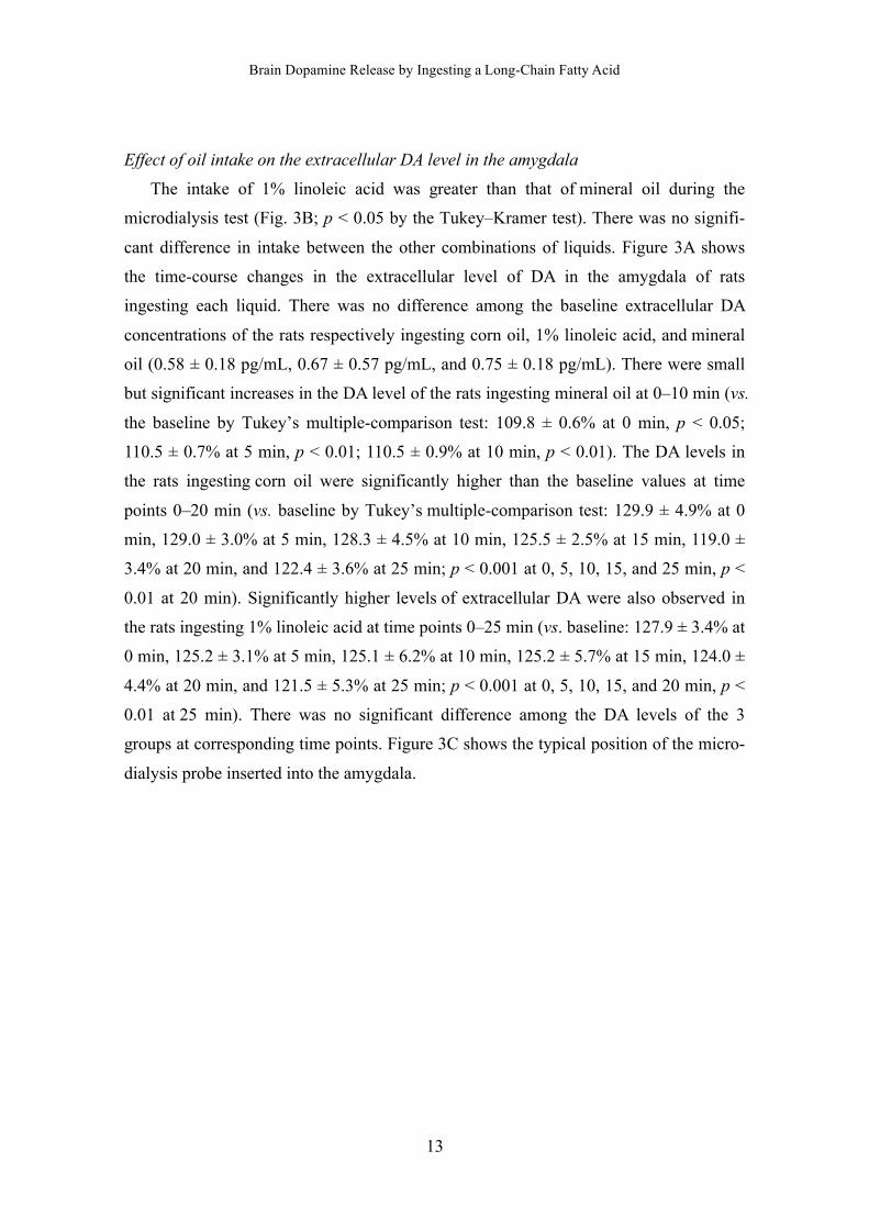
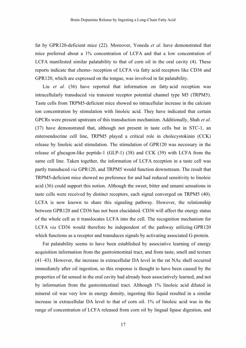
Effect of oil intake on the extracellular DA level in the amygdala
The intake of 1% linoleic acid was greater than that of mineral oil during the
microdialysis test (Fig. 3B; p < 0.05 by the Tukey–Kramer test). There was no signifi-
cant difference in intake between the other combinations of liquids. Figure 3A shows
the time-course changes in the extracellular level of DA in the amygdala of rats
ingesting each liquid. There was no difference among the baseline extracellular DA
concentrations of the rats respectively ingesting corn oil, 1% linoleic acid, and mineral
oil (0.58 ± 0.18 pg/mL, 0.67 ± 0.57 pg/mL, and 0.75 ± 0.18 pg/mL). There were small
but significant increases in the DA level of the rats ingesting mineral oil at 0–10 min (vs.
the baseline by Tukey’s multiple-comparison test: 109.8 ± 0.6% at 0 min, p < 0.05;
110.5 ± 0.7% at 5 min, p < 0.01; 110.5 ± 0.9% at 10 min, p < 0.01). The DA levels in
the rats ingesting corn oil were significantly higher than the baseline values at time
points 0–20 min (vs. baseline by Tukey’s multiple-comparison test: 129.9 ± 4.9% at 0
min, 129.0 ± 3.0% at 5 min, 128.3 ± 4.5% at 10 min, 125.5 ± 2.5% at 15 min, 119.0 ±
3.4% at 20 min, and 122.4 ± 3.6% at 25 min; p < 0.001 at 0, 5, 10, 15, and 25 min, p <
0.01 at 20 min). Significantly higher levels of extracellular DA were also observed in
the rats ingesting 1% linoleic acid at time points 0–25 min (vs. baseline: 127.9 ± 3.4% at
0 min, 125.2 ± 3.1% at 5 min, 125.1 ± 6.2% at 10 min, 125.2 ± 5.7% at 15 min, 124.0 ±
4.4% at 20 min, and 121.5 ± 5.3% at 25 min; p < 0.001 at 0, 5, 10, 15, and 20 min, p <
0.01 at 25 min). There was no significant difference among the DA levels of the 3
groups at corresponding time points. Figure 3C shows the typical position of the micro-
dialysis probe inserted into the amygdala.
Brain Dopamine Release by Ingesting a Long-Chain Fatty Acid
14
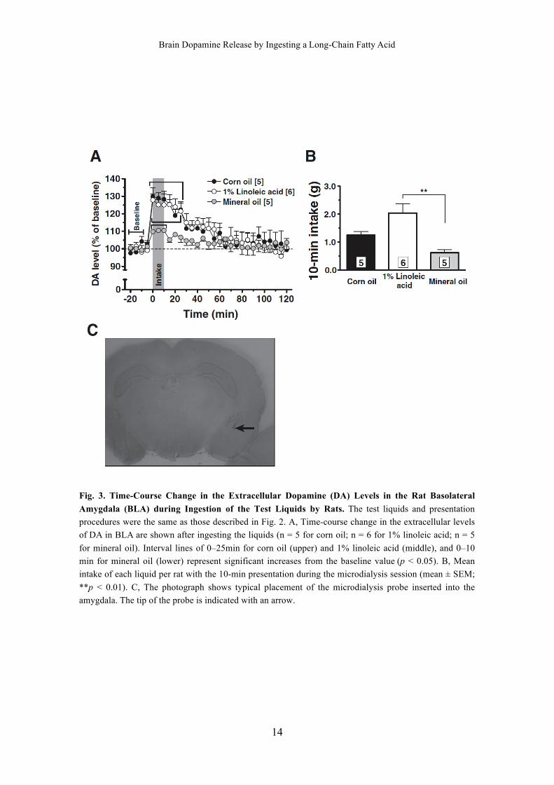
Fig. 3. Time-Course Change in the Extracellular Dopamine (DA) Levels in the Rat Basolateral
Amygdala (BLA) during Ingestion of the Test Liquids by Rats. The test liquids and presentation
procedures were the same as those described in Fig. 2. A, Time-course change in the extracellular levels
of DA in BLA are shown after ingesting the liquids (n = 5 for corn oil; n = 6 for 1% linoleic acid; n = 5
for mineral oil). Interval lines of 0–25min for corn oil (upper) and 1% linoleic acid (middle), and 0–10
min for mineral oil (lower) represent significant increases from the baseline value (p < 0.05). B, Mean
intake of each liquid per rat with the 10-min presentation during the microdialysis session (mean ± SEM;
**p < 0.01). C, The photograph shows typical placement of the microdialysis probe inserted into the
amygdala. The tip of the probe is indicated with an arrow.
Brain Dopamine Release by Ingesting a Long-Chain Fatty Acid
15
Discussion
Previous reports have shown that the higher the concentration of sucrose (10) or
cocaine (11) ingested or self-administered by rats, the greater the increase in DA level in
their NAc shell. These reports indicate the possibility that the increased DA level in
NAc might be correlated with the degree of pleasantness and palatability, and could be
considered an index of the reward value or motivational state of the animal. In respect
of oil ingestion, sham feeding of corn oil (the rats were operated on to drain out the
ingested solution before it had reached the stomach) has been reported to increase the
DA level in NAc of rats (12), this finding suggesting that information from the oral
cavity was sufficient for increasing the DA level and that feedback from the digestive
tract was not necessary. Yoneda et al. reported in a previous study that the licking ratio
for a low concentration of LCFA was similar to that for 100% corn oil and higher than
that for mineral oil in mice within a very short measurement time (60 s); hence, the
postprandial effect of the ingested liquid could be eliminated (4). We have also
demonstrated in this present study that the rats preferred corn oil and a low
concentration of linoleic acid to mineral oil during training sessions and that the amount
of 1% linoleic acid ingested was higher than that of mineral oil during the test session.
Therefore, despite the low caloric content, a low concentration of LCFA seemed to have
similar properties to corn oil with respect to both the reward value and palatability.
Mineral oil has a similar texture to corn oil but does not contain LCFA and is not
digestible. Consequently, it cannot be utilized by animals and has very little reward
value, based on the increase in DA level in the NAc shell observed in this study.
Dopaminergic neurons in the VTA project to the amygdala, as well as to the NAc
shell, and the amygdala is thought to be involved in the manifestation of negative
emotions like fear and in the evaluation of a negative reward value. Inglis and
Moghaddam have reported that there was an increase in extracellular DA level in the
amygdala of rats exposed to handling stress (26). Furthermore, Yokoyama et al. have
reported that a conditioned stimulus, in addition to an unconditioned stimulus (foot
shock), caused the DA level to rise in the amygdala of rats in a conditioned-fear
experiment involving foot shock (27). However, some recent studies have reported the
involvement of the amygdala in both negative emotions and in positive ones. For
example, Polston et al. have reported an increased DA level in the amygdala in response
Brain Dopamine Release by Ingesting a Long-Chain Fatty Acid
16
to a reward stimulus such as methamphetamine (28), and See et al. have shown that the
administration of a D1-receptor antagonist to the amygdala inhibited the reinstatement
of cocaine self-administration (29). Food addiction scores have been reported to be
correlated with activation of the amygdala in fMRI measurements on young women
(30), and the activity of the amygdala observed during food intake (also with fMRI)
increased more in response to an increase in taste intensity than to taste affective
valence (31). Moreover, the amygdala has been more strongly activated by high-fat
food ingestion than by low-fat food ingestion (32). In our experiment, more palatable
food clearly resulted in an increased DA level in the amygdala, the extracellular DA
level there increasing after the ingestion of corn oil and 1% linoleic acid, these liquids
being consumed by rats in preference to mineral oil.
Mineral oil was clearly preferred to water by rat pups aged 12–15 d, and had similar
acceptability to corn oil by adult rats (33). Taka and Fushiki also observed that mineral
oil was preferentially consumed and that the ingested volume of mineral oil was more
than that of water by mice (unpublished data). Although the method for administration
was different via an oral cannula, mineral oil ingestion resulted in a low but significant
increase in the extracellular concentration of DA in the NAc shell of rats (Adachi et al.,
manuscript in preparation). However, the reward value of mineral oil would be lower
than that of corn oil, and the level of the DA increase in NAc might not have been
captured by unknown factor in this experiment. The different response to mineral oil
ingestion between the two brain nuclei should be further studied.
A main ingredient of cooking oil is triacylglyceride that consists of esterified
glycerol with LCFA. Kawai and Fushiki have demonstrated that a few percent of
triacylglycerol administered into the oral cavity was hydrolyzed into LCFA by lingual
lipase secreted by von Ebner’s glands present in the circumvallate papillae of rats (17).
Gilbertson et al. have elucidated the regulation of K+ channels in type II taste cells by
long-chain unsaturated fatty acids and suggested a chemical receptor for fatty acids in
taste cells (18). Fukuwatari et al. have demonstrated the expression of CD36, which is a
translocator of LCFA, on the apical surface of taste cells of the circumvallate papillae in
rats (19), and Laugerette et al. (21) and Martin et al. (34) have found no preference for
fatty acids in CD36-deficient mice. These findings suggest the chemoreception of
dietary fat via CD36 in the oral cavity. In addition, Matsumura et al. have studied the
expression of GPR120 on the tongue (20, 35), and reported a decrease in preference for
Brain Dopamine Release by Ingesting a Long-Chain Fatty Acid
17
fat by GPR120-deficient mice (22). Moreover, Yoneda et al. have demonstrated that
mice preferred about a 1% concentration of LCFA and that a low concentration of
LCFA manifested similar palatability to that of corn oil in the oral cavity (4). These
reports indicate that chemo- reception of LCFA via fatty acid receptors like CD36 and
GPR120, which are expressed on the tongue, was involved in fat palatability.
Liu et al. (36) have reported that information on fatty acid reception was
intracellularly transduced via transient receptor potential channel type M5 (TRPM5).
Taste cells from TRPM5-deficient mice showed no intracellular increase in the calcium
ion concentration by stimulation with linoleic acid. They have indicated that certain
GPCRs were present upstream of this transduction mechanism. Additionally, Shah et al.
(37) have demonstrated that, although not present in taste cells but in STC-1, an
enteroendocrine cell line, TRPM5 played a critical role in cholecystokinin (CCK)
release by linoleic acid stimulation. The stimulation of GPR120 was necessary in the
release of glucagon-like peptide-1 (GLP-1) (38) and CCK (39) with LCFA from the
same cell line. Taken together, the information of LCFA reception in a taste cell was
partly transduced via GPR120, and TRPM5 would function downstream. The result that
TRPM5-deficient mice showed no preference for and had reduced sensitivity to linoleic
acid (36) could support this notion. Although the sweet, bitter and umami sensations in
taste cells were received by distinct receptors, each signal converged on TRPM5 (40).
LCFA is now known to share this signaling pathway. However, the relationship
between GPR120 and CD36 has not been elucidated. CD36 will affect the energy status
of the whole cell as it translocates LCFA into the cell. The recognition mechanism for
LCFA via CD36 would therefore be independent of the pathway utilizing GPR120
which functions as a receptor and transduces signals by activating associated G-protein.
Fat palatability seems to have been established by associative learning of energy
acquisition information from the gastrointestinal tract, and from taste, smell and texture
(41–43). However, the increase in extracellular DA level in the rat NAc shell occurred
immediately after oil ingestion, so this response is thought to have been caused by the
properties of fat sensed in the oral cavity had already been associatively learned, and not
by information from the gastrointestinal tract. Although 1% linoleic acid diluted in
mineral oil was very low in energy density, ingesting this liquid resulted in a similar
increase in extracellular DA level to that of corn oil. 1% of linoleic acid was in the
range of concentration of LCFA released from corn oil by lingual lipase digestion, and
Brain Dopamine Release by Ingesting a Long-Chain Fatty Acid
18
this would be matched by the properties of oil which had been associatively learned by
the rats. This would be the reason for a similar response being caused with 1% linoleic
acid ingestion, as was observed in the case of corn oil, and suggests that the reward
obtained by 1% linoleic acid stimulation in the oral cavity was from its palatability, and
not from its calorie content.
To summarize, the results of the present study corroborate the increase in
extracellular DA levels in the NAc shell and BLA of rats after ingesting 1% linoleic acid,
which has a very low calorie level, these levels being comparable to those of full caloric
100% corn oil. These data support the notion that the chemoreception of LCFA released
from fat by lingual lipase plays a critical part in the detection of fat in the oral cavity
and the manifestation of a reward effect.
Brain Dopamine Release by Ingesting a Long-Chain Fatty Acid
19
REFERENCES
1. Drewnowski A, Greenwood MR. (1983). Cream and sugar: human preferences
for high-fat foods. Physiol Behav. 30,:629–33
2. Takeda M, Imaizumi M, Fushiki T. (2000). Preference for vegetable oils in the
two-bottle choice test in mice. Life Sci. 67,:197–204
3. Imaizumi M, Takeda M, Fushiki T. (2000). Effects of oil intake in the
conditioned place preference test in mice. Brain Res. 870,:150–56
4. Yoneda T, Saitou K, Mizushige T, Matsumura S, Manabe Y, Tsuzuki S, Inoue K,
Fushiki T. (2007). The palatability of corn oil and linoleic acid to mice as
measured by short-term two-bottle choice and licking tests. Physiol Behav.
91,:304–9
5. Wise RA. (2006). Role of brain dopamine in food reward and reinforcement.
Philos Trans R Soc Lond B Biol Sci. 361,:1149–58
6. Wise RA, Rompre PP. (1989). Brain dopamine and reward. Annu Rev Psychol.
40,:191–225
7. Willuhn I, Wanat MJ, Clark JJ, Phillips PEM. (2010). Dopamine signaling in the
nucleus accumbens of animals self-administering drugs of abuse. Curr Top
Behav Neurosci. 3,:29–71
8. Vucetic Z, Reyes TM. (2010). Central dopaminergic circuitry controlling food
intake and reward: implications for the regulation of obesity. Wiley Interdiscip
Rev Syst Biol Med. 2,:577–93
9. Geisler S, Zahm DS. (2005). Afferents of the ventral tegmental area in the
rat-anatomical substratum for integrative functions. J Comp Neurol. 490,:270–94
10. Hajnal A, Smith GP, Norgren R. (2004). Oral sucrose stimulation increases
accumbens dopamine in the rat. Am J Physiol Regul Integr Comp Physiol.
286,:R31–7
Brain Dopamine Release by Ingesting a Long-Chain Fatty Acid
20
11. Pettit HO, Justice JB. (1991). Effect of dose on cocaine self-administration
behavior and dopamine levels in the nucleus accumbens. Brain Res. 539,:94–102
12. Liang N-C, Hajnal A, Norgren R. (2006). Sham feeding corn oil increases
accumbens dopamine in the rat. Am J Physiol Regul Integr Comp Physiol.
291,:R1236–9
13. Hurd YL, Pontén M. (2000). Cocaine self-administration behavior can be
reduced or potentiated by the addition of specific dopamine concentrations in the
nucleus accumbens and amygdala using in vivo microdialysis. Behav Brain Res.
116,:177–86
14. Mindell S, Smith GP, Greenberg D. (1990). Corn oil and mineral oil stimulate
sham feeding in rats. Physiol Behav. 48,:283–87
15. Ramirez I. (1993). Role of olfaction in starch and oil preference. Am J Physiol.
265,:R1404–9
16. Matsumura S, Yoneda T, Shoji A, Eguchi A, Manabe Y, Tsuzuki S, Inoue K,
Fushiki T. (2010). Intragastric infusion of glucose enhances the rewarding effect
of sorbitol fatty acid ester ingestion as measured by conditioned place preference
in mice. Physiol Behav. 99,:509–14
17. Kawai T, Fushiki T. (2003). Importance of lipolysis in oral cavity for orosensory
detection of fat. Am J Physiol Regul Integr Comp Physiol. 285,:R447–54
18. Gilbertson TA, Fontenot DT, Liu L, Zhang H, Monroe WT. (1997). Fatty acid
modulation of k+ channels in taste receptor cells: gustatory cues for dietary fat.
Am J Physiol. 272,:C1203–10
19. Fukuwatari T, Kawada T, Tsuruta M, Hiraoka T, Iwanaga T, Sugimoto E,
Fushiki T. (1997). Expression of the putative membrane fatty acid transporter
(fat) in taste buds of the circumvallate papillae in rats. FEBS Lett. 414,:461–64
Brain Dopamine Release by Ingesting a Long-Chain Fatty Acid
21
20. Matsumura S, Mizushige T, Yoneda T, Iwanaga T, Tsuzuki S, Inoue K, Fushiki
T. (2007). Gpr expression in the rat taste bud relating to fatty acid sensing.
Biomed Res. 28,:49–55
21. Laugerette F, Passilly-Degrace P, Patris B, Niot I, Febbraio M, Montmayeur JP,
Besnard P. (2005). Cd36 involvement in orosensory detection of dietary lipids,
spontaneous fat preference, and digestive secretions. J Clin Invest. 115,:3177–84
22. Cartoni C, Yasumatsu K, Ohkuri T, Shigemura N, Yoshida R, Godinot N, le
Coutre J, Ninomiya Y, Damak S. (2010). Taste preference for fatty acids is
mediated by gpr40 and gpr120. J Neurosci. 30,:8376–82
23. Weiss F, Maldonado-Vlaar CS, Parsons LH, Kerr TM, Smith DL, Ben-Shahar O.
(2000). Control of cocaine-seeking behavior by drug-associated stimuli in rats:
effects on recovery of extinguished operant-responding and extracellular
dopamine levels in amygdala and nucleus accumbens. Proc Natl Acad Sci U S A.
97,:4321–26
24. Tye KM, Janak PH. (2007). Amygdala neurons differentially encode motivation
and reinforcement. J Neurosci. 27,:3937–45
25. Paxinos G, Charles W. (2004). “The Rat Brain in Stereotaxic Coordinates.” San
Diego: Academic Press
26. Inglis FM, Moghaddam B. (1999). Dopaminergic innervation of the amygdala is
highly responsive to stress. J Neurochem. 72,:1088–94
27. Yokoyama M, Suzuki E, Sato T, Maruta S, Watanabe S, Miyaoka H. (2005).
Amygdalic levels of dopamine and serotonin rise upon exposure to conditioned
fear stress without elevation of glutamate. Neurosci Lett. 379,:37–41
28. Polston JE, Rubbinaccio HY, Morra JT, Sell EM, Glick SD. (2011). Music and
methamphetamine: conditioned cue-induced increases in locomotor activity and
dopamine release in rats. Pharmacol Biochem Behav. 98,:54–61
Brain Dopamine Release by Ingesting a Long-Chain Fatty Acid
22
29. See RE, Kruzich PJ, Grimm JW. (2001). Dopamine, but not glutamate, receptor
blockade in the basolateral amygdala attenuates conditioned reward in a rat
model of relapse to cocaine-seeking behavior. Psychopharmacology (Berl).
154,:301–10
30. Gearhardt AN, Yokum S, Orr PT, Stice E, Corbin WR, Brownell KD. (2011).
Neural correlates of food addiction. Arch Gen Psychiatry. 68,:808–16
31. Small DM, Gregory MD, Mak YE, Gitelman D, Mesulam MM, Parrish T. (2003).
Dissociation of neural representation of intensity and affective valuation in
human gustation. Neuron. 39,:701–11
32. Grabenhorst F, Rolls ET, Parris B a, d’Souza A a. (2010). How the brain
represents the reward value of fat in the mouth. Cereb Cortex. 20,:1082–91
33. Ackroff K, Vigorito M, Sclafani A. (1990). Fat appetite in rats: the response of
infant and adult rats to nutritive and non-nutritive oil emulsions. Appetite.
15,:171–88
34. Martin C, Passilly-Degrace P, Gaillard D, Merlin JF, Chevrot M, Besnard P.
(2011). The lipid-sensor candidates cd36 and gpr120 are differentially regulated
by dietary lipids in mouse taste buds: impact on spontaneous fat preference.
PLoS One. 6,:e24014
35. Matsumura S, Eguchi A, Mizushige T, Kitabayashi N, Tsuzuki S, Inoue K,
Fushiki T. (2009). Colocalization of gpr120 with phospholipase-cbeta2 and
alpha-gustducin in the taste bud cells in mice. Neurosci Lett. 450,:186–90
36. Liu P, Shah BP, Croasdell S, Gilbertson TA. (2011). Transient receptor potential
channel type m5 is essential for fat taste. J Neurosci. 31,:8634–42
37. Shah BP, Liu P, Yu T, Hansen DR, Gilbertson TA. (2012). Trpm5 is critical for
linoleic acid-induced cck secretion from the enteroendocrine cell line, stc-1. Am J
Physiol Cell Physiol. 302,:C210–9
Brain Dopamine Release by Ingesting a Long-Chain Fatty Acid
23
38. Hirasawa A, Tsumaya K, Awaji T, Katsuma S, Adachi T, Yamada M, Sugimoto
Y, Miyazaki S, Tsujimoto G. (2005). Free fatty acids regulate gut incretin
glucagon-like peptide-1 secretion through gpr120. Nat Med. 11,:90–94
39. Tanaka T, Katsuma S, Adachi T, Koshimizu T, Hirasawa A, Tsujimoto G. (2008).
Free fatty acids induce cholecystokinin secretion through gpr120. Naunyn
Schmiedebergs Arch Pharmacol. 377,:523–27
40. Zhang Y, Hoon MA, Chandrashekar J, Mueller KL, Cook B, Wu D, Zuker CS,
Ryba NJP. (2003). Coding of sweet, bitter, and umami tastes: different receptor
cells sharing similar signaling pathways. Cell. 112,:293–301
41. Suzuki A, Yamane T, Imaizumi M, Fushiki T. (2003). Integration of orosensory
and postingestive stimuli for the control of excessive fat intake in mice. Nutrition.
19,:36–40
42. Suzuki A, Yamane T, Fushiki T. (2006). Inhibition of fatty acid beta-oxidation
attenuates the reinforcing effects and palatability to fat. Nutrition. 22,:401–7
43. Mizushige T, Saitoh K, Manabe Y, Nishizuka T, Taka Y, Eguchi A, Yoneda T,
Matsumura S, Tsuzuki S, Inoue K, Fushiki T. (2009). Preference for dietary fat
induced by release of beta-endorphin in rats. Life Sci. 84,:760–65
Fat Palatability Mediated via GPR120
24
CHAPTER 2
Involvement of GPR120-agonistic activity of long chain fatty acid in
the palatability of dietary fat
Most animals, including rodents, as well as humans prefer fat-rich foods (1, 2). The
consumption of corn oil has been reported to produce a reward effect in mice, and the
dopaminergic pathway in the nervous system has been implicated in the manifestation
of this effect (3, 4). Additionally, sham and real feeding of corn oil were reported to
induce the activation of the midbrain dopamine (DA) system, which is involved in
reward behavior (5,6, and chap.1). The mesolimbic system is thought to play a critical
role in the reward effect, and the release of DA has been demonstrated when a natural
and drug reward is acquired or when its acquisition is anticipated (7, 8). The midbrain
dopaminergic circuits originate from the ventral tegmental area (VTA) and project to
different sites, such as the nucleus accumbens (NAc), amygdala, and the prefrontal area,
which are related to motivation, palatability, and addiction (9–11). Iwakura et al.
showed that the secretion of ghrelin, a hormone that stimulates food intake, is induced
by DA in a ghrelin-producing cell line MGN3-1 (12). Since the extracellular
concentration of DA in the NAc of the rat increases in a dose-dependent manner after
self-administration of cocaine or consumption of sucrose, release of DA in the NAc
could be considered as a form of index for the palatability or motivational drive (13,
14).
Recent studies have revealed that chemoreceptors of long-chain fatty acids (LCFAs)
are involved in the recognition of fatty foods. Kawai et al. reported that, when fat was
introduced into the oral cavity of rats, a certain percentage of triacylglycerides was
hydrolyzed to LCFAs by lingual lipase (15). Gilbertson et al. demonstrated the
regulation of K+ channels in type II taste cells by unsaturated LCFAs and suggested that
fatty acid chemoreceptors were present within taste cells (16). Fukuwatari et al. found
that CD36 fatty acid transporter was expressed on the apical side of taste cells in the
circumvallate papillae (17). Additionally, CD36-deficient mice were reported to show a
low taste preference for fat (18). Moreover, Matsumura et al. reported that GPR120 was
also expressed on the apical side of taste cells in the circumvallate papillae (19). The
Fat Palatability Mediated via GPR120
25
unsaturated LCFAs, such as oleic acid and linolenic acid, induced a rise in
concentration of intracellular calcium ion ([Ca2+]i) in Human Embryonic Kidney 293
(HEK293) cells stably expressing GPR120. On the other hand, LCFA esters and capric
acid did not induce this increase in concentration (20). When the licking behavior of the
mice was tested, it was found that the mice exhibited equally strong preference for a
low concentration of linoleic acid as that for 100% corn oil (21). In addition, the mice
exhibited a similar strong preference for LCFAs, such as oleic, linolenic, and linoleic
acid, whereas they did not display any preference for LCFA esters nor long-chain fatty
alcohols (22). Moreover, ingesting linoleic acid at a low concentration increased
extracellular DA release in the NAc of rats (6 and chap.1). Compared with wild-type
mice, GPR120 knock-out mice showed a lower preference for LCFAs and lower
response of the chorda tympani and glossopharyngeal nerve to LCFAs (23). These
findings lead us to postulate that, in the oral cavity, fat is hydrolyzed to LCFA by
lingual lipase and that chemoreception of this LCFA by receptor proteins, such as CD36
and GPR120 expressed on the taste cells, could be involved in the oral recognition and
palatability of fats. However, it remains unclear as to which structural characteristics of
LCFA are responsible for the GPR120-agonistic activity and if these activities could be
implicated in the palatability of LCFA. Therefore, in this study, we examined the
relationship between the GPR120-agonistic properties of ligands and the palatability of
LCFAs. First, using HEK 293 cells stably expressing human GPR120, we examined the
effect of various LCFAs on the concentration of [Ca2+]i by using a fluorescence
spectrophotometer. We then assessed the palatability for a variety of LCFAs at a low
concentration by testing the licking behavior of the mice. Next, we studied the change
in the extracellular concentration of DA in the NAc of the mice by in vivo microdialysis
after ingestion of various low-concentration LCFAs. In both the tests, we used
0.0322 mol/L fatty acid in mineral oil. The molar concentration was equal to a
volume/volume% concentration of 1% linoleic acid. This concentration of fatty acid
was in the range that could be released from fat by lingual lipase and had only 1/100th of
the calorie content of fats of the same weight.
Fat Palatability Mediated via GPR120
26
Materials and Methods
[Ca2+]i analysis
Cell Culture. HEK293 cells were maintained in Dulbecco's Modified Eagle Medium
(DMEM) containing 10% fetal bovine serum (FBS) and 1% penicillin-streptomycin in a
humidified, 5% CO2 atmosphere at 37°C. Human GPR120 cDNA from the lung was
provided by Pharmafoods International Co., Ltd. (Kyoto, Japan).
Transfection into cells. Human GPR120 was transfected into HEK293 cells using the
lipofection method as per the manufacturer’s instructions. For the control, an empty
vector was transfected into the HEK293 cells (Empty). Forty-eight hours after
transfection, the medium was replaced with fresh medium containing 400 µg/mL G418
(Wako, Osaka, Japan) for the selection of the transfected cells, following which a single
clone was selected by the standard limiting dilution method. GPR120 expression in
cells was confirmed by reverse transcriptase polymerase chain reaction (RT-PCR).
Transfected cells were maintained in DMEM containing 10% FBS, 1%
penicillin-streptomycin, and G-418 (400 µg/mL).
Reagents for [Ca2+]i analysis. All fatty acids—caprylic acid, C8:0; capric acid, C10:0;
lauric acid, C12:0; myristic acid, C14:0; myristoleic acid, C14:1; palmitic acid, C16:0;
palmitoleic acid, C16:1; stearic acid, C18:0; oleic acid, C18:1; linoleic acid, C18:2;
linolenic acid, C18:3; stearidonic acid, C18:4; arachidic acid, C20:0; arachidonic acid,
C20:4; eicosapentaenoic acid (EPA), C20:5; behenic acid, C22:0; docosahexaenoic acid
(DHA), C22:6; methyl oleate, methyl linoleate, and methyl linolenate—were purchased
from Sigma (St. Louis, MO, USA) and stored at -20°C until use. All cell culture
reagents (HEPES, Hanks buffer, DMEM, FBS, penicillin-streptomycin, and
Lipofectamine) were purchased from Invitrogen (Carlsbad, CA, USA). All other
chemicals, unless stated otherwise, were purchased from Sigma.
Ca2+ mobilization assay. Ca2+ loading buffer comprised of 5 µL Fluo-3AM (1 µM;
Dojindo, Kumamoto, Japan) and 10 µL pluronic F-127 (Wako), diluted to yield 10 mL
Ca2+ assay buffer (20 mM HEPES pH 7.6, 0.01% BSA, 1 mM Probenecid [Wako] in
Hanks solution) (24). On the day before the assay, 5 × 10⁴ cells were seeded in 96-well,
Fat Palatability Mediated via GPR120
27
poly-D-lysine-coated plates (BD BioCoat, Franklin Lakes, NJ, USA). The cells were
washed once with phosphate buffered saline (PBS) and incubated in a final volume of
100 µL/well in Ca2+ loading buffer for 60 min at 37°C. Then, the cells were then
washed twice with Ca2+ assay buffer and the assay was carried out in 100 µL of Ca2+
assay buffer. Changes in Ca2+ levels were monitored by a fluorescence
spectrophotometer (Powerscan HT, DS Pharma Biomedical Co., Ltd., Osaka, Japan) at
an excitation wavelength of 485 nm and emission wavelength of 528 nm. The
maximum intracellular Ca2+ ([Ca2+]i) fluorescence intensity was obtained as the mean of
triplicate assays. Test samples for the Ca2+ mobilization assay were prepared by
sonication in Ca2+ buffer just prior to the assay.
Behavioral Test
Animals. This study was conducted in accordance with the ethical guidelines of the
Kyoto University Animal Experimentation Committee, in complete compliance with the
National Institutes of Health Guide for the Care and Use of Laboratory Animals, and it
was approved by the above-mentioned committee. Male BALB/c mice (Japan SLC,
Hamamatsu, Japan) at 8 weeks of age were housed in plastic cages in a room with a
12-h light–dark cycle (dark phase of 18:00–6:00) and constant temperature (24 ± 1°C).
They were separately housed for > 5 days for acclimatization to the environment. The
animals were provided with tap water and regular MF mouse food (Oriental Yeast,
Tokyo, Japan) ad libitum.
Materials. Corn oil was purchased from Ajinomoto (Tokyo, Japan) and mineral oil was
purchased from Kaneda Company (Tokyo, Japan). All fatty acids—caproic acid, C6:0;
caprylic acid, C8:0; capric acid, C10:0; lauric acid, C12:0; myristic acid, C14:0;
myristoleic acid, C14:1; palmitic acid, C16:0; palmitoleic acid, C16:1; stearic acid,
C18:0; oleic acid, C18:1 cis-9; elaidic acid, C18:1 trans-9; cis-vaccenic acid, C18:1
cis-11; trans-vaccenic acid, C18:1 trans-11; linoleic acid, C18:2; linolenic acid, C18:3;
stearidonic acid, C18:4; arachidic acid, C20:0; arachidonic acid, C20:4; behenic acid,
C22:0; docosahexaenoic acid (DHA), C22:6; lignoceric acid, C24:0; methyl oleate,
methyl linoleate, and methyl linolenate—were purchased from Sigma (St. Louis, MO,
USA). They were 99% pure, stored at -20°C until use and then diluted in mineral oil to
0.0322 mol/L, which is equivalent to a v/v% concentration of 1% linoleic acid. Capric
Fat Palatability Mediated via GPR120
28
acid, lauric acid, myristic acid, palmitic acid, stearic acid, elaidic acid, trans-vaccenic
acid, arachidic acid, behenic acid, and lignoceric acid do not dissolve at room
temperature. Therefore, we heated each fatty acid solution to 75°C to eliminate the
effects of difference in temperature. For testing the licking behavior, we used all the
above fatty acids. In the microdialysis test, we used capric acid, lauric acid, stearic acid,
oleic acid, linoleic acid, docosahexaenoic acid, and methyl linoleate. The other reagents
were purchased from Nacalai Tesque (Kyoto, Japan).
Evaluation of the licking behavior
Apparatus for the test. Licking behavior was evaluated in a custom-made licking test
chamber (Muromachi Kikai, Tokyo, Japan) previously described (25). In brief, the test
chamber (150 × 120 × 130 mm) was made of Plexiglas with an automatic shutter placed
on the front wall, 1.5 cm above the metal-grid floor. When the shutter opened, mice
gained access to a stainless steel drinking spout. The licking response was recorded by a
computer. The licking rate was calculated for 60 s starting from the first lick. Given this
very short period, we can rule out any contribution of post-ingestive feedback to the
licking behavior.
Evaluation of the licking behavior. To allow the mice to be habituated to the test
environment and to get accustomed to ingesting corn oil and mineral oil, they were kept
in the test chamber for 30 min and offered corn oil and mineral oil for 30 min. This
training lasted until the mice could discriminate corn oil from mineral oil to the same
degree as the previous report (21). After training, the mice were offered linoleic acid
and stearic acid. We confirmed that the preference for linoleic acid was high and that
for stearic acid was low, similar to the previous report (22). The licking behavior of the
mice was then tested. In the test, the mice were offered the test fluids for 30 min once a
day in the test chamber. We recorded the licking rate for 1 min from the first lick and
the intake for 30 min from the start of presentation of the test fluid. To avoid order
effects, each mouse was offered the test fluids in a different order.
Microdialysis test
Training protocols for oil ingestion in the microdialysis test. To allow the mice to get
accustomed to ingesting corn oil, mineral oil, and fatty acid fluid, the mice were fed
Fat Palatability Mediated via GPR120
29
these liquids in their cages before surgery and then in the microdialysis cage after
recovery from surgery. The mice were deprived of water and food for 30 min, and then
the liquids were kept in front of the mice for 10 min. Before surgery, the mice were
presented with corn oil and mineral oil at the same time on days 1 and 2. To confirm
their preference for corn oil over mineral oil, the mice were subjected to a 2-bottle
preference test for corn oil vs. mineral oil on day 3. The liquid bottles were positioned
randomly. The mice were randomly presented with mineral oil, lauric acid, and linoleic
acid on days 4 to 6 and capric acid, stearic acid, oleic acid, methyl linoleate, and
docosahexaenoic acid on days 7 to 11. After recovering from the surgery, the mice were
presented with corn oil on day 1 and all other liquids in random order on days 2 to 9 in
the microdialysis cage. The mice were then subjected to a microdialysis test on day 10.
Microdialysis surgery. The animals were anesthetized with pentobarbital sodium
(Nembutal; Dainippon Pharmaceutical Co., Tokyo, Japan) and placed in a stereotaxic
frame modified for surgery in mice. The skulls of the mice were subsequently exposed
and holes were drilled for microdialysis. The coordinates for the NAc guide cannula
(AG-5; Eicom, Kyoto, Japan) were AP, 1.2; ML, 0.6; and DV, 3.2 from the bregma. The
coordinates were determined according to the stereotaxic atlas of Paxinos and Franklin
(26). The cannulas were secured to the skull with a LOCTITE 454 adhesive bond
(Henkel Japan, Yokohama, Japan). A dummy AD-5 cannula (Eicom) was inserted into
the guide cannula and secured with an AC-1 cap nut (Eicom). The mice were allowed 3
to 5 days to recover from the surgery. Each mouse implanted with a probe in the NAc
was used for a single microdialysis procedure with a single test liquid.
Procedure. The experiments were conducted during the light period of the light-dark
cycle. The dummy cannula was removed on the day of the experiment, and the AI-5-1.5
microdialysis probe (Eicom, 1.5 mm membrane length) was inserted into the NAc via
the guide cannula. The mice were placed in the microdialysis cage at 8:00 a.m. for 3.5 h
without food and water, and then presented with the test liquid at 11:30 p.m. for 10 min.
The amount of liquid ingested was also recorded. The rats remained in the microdialysis
cage for another 80 min after presenting the test liquid. Ringer’s solution containing
147 mM Na+, 4 mM K+, 2.3 mM Ca2+, and 155.6 mM Cl- was perfused at 3 µL/min by
an ESP-64 micro-syringe pump (Eicom). Dialysate collection was started 30 min before
Fat Palatability Mediated via GPR120
30
the liquids were presented, and the collection conducted every 10 min for a total of
120 min thereafter. To quantify DA and 5-HT levels in the dialysate, samples were
analyzed by reversed-phase high performance liquid chromatography (HPLC) with an
electrochemical detector, using an Eicompak PP-ODS II column (4.6 i.d. × 30 mm long;
Eicom). The voltage applied was set at 400 mV (relative to an Ag/AgCl reference
electrode). The mobile phase at a flow rate of 500 µL/min consisted of a 98% (v/v)
0.1 M phosphate buffer at pH 6.0, 2% (v/v) methanol, 500 mg/L sodium decane sulfate,
and 50 mg/L EDTA-2Na. The mean value obtained from 3 samples from -30 to -10 min
was set as the 100% baseline level, and all subsequent sample values were expressed as
a percentage of the baseline value.
Histological analysis. Upon completion of the experiment, the mice were deeply
anesthetized with sodium pentobarbital. The brain was removed from the skull, frozen,
and cut into 20-µm sections. The placement of the microdialysis probe was verified by
thionine blue staining. Data obtained from the mice with inappropriate probe placement
were excluded from the analysis.
Statistics. Data are expressed as the mean ± SEM. Data from [Ca2+]i assay were
analyzed using a one-way ANOVA and Dunnett’s post hoc test. Data obtained by testing
the licking behavior were analyzed using one-way repeated ANOVA and Dunnett’s post
hoc test. Data from the 2-bottle preference test were analyzed by a paired t-test.
Changes in DA and 5-HT levels were compared with the corresponding baseline value
by one-way repeated ANOVA and Tukey’s multiple-comparison test as a post-hoc test.
Mean differences among the 3 groups at each time point were analyzed by two-way
repeated-measures ANOVA and Bonferroni’s multiple-comparison as a post-hoc test.
The amount of each fluid ingested during microdialysis was analyzed by a one-way
ANOVA and Dunnett’s test as a post-hoc test. Correlation coefficient was obtained by
Pearson correlation test. p values of 5% or less were considered statistically significant.
Statistical analyses were conducted by using the Prism 6 software package (GraphPad,
San Diego, CA, USA).
Fat Palatability Mediated via GPR120
31
RESULTS
Intracellular Ca2+ Assay in HEK293 cells
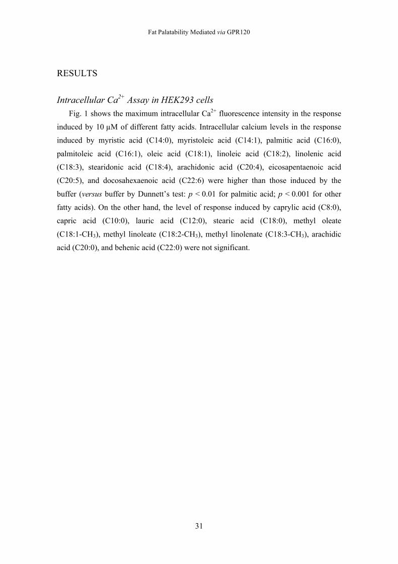
Fig. 1 shows the maximum intracellular Ca2+ fluorescence intensity in the response
induced by 10 µM of different fatty acids. Intracellular calcium levels in the response
induced by myristic acid (C14:0), myristoleic acid (C14:1), palmitic acid (C16:0),
palmitoleic acid (C16:1), oleic acid (C18:1), linoleic acid (C18:2), linolenic acid
(C18:3), stearidonic acid (C18:4), arachidonic acid (C20:4), eicosapentaenoic acid
(C20:5), and docosahexaenoic acid (C22:6) were higher than those induced by the
buffer (versus buffer by Dunnett’s test: p < 0.01 for palmitic acid; p < 0.001 for other
fatty acids). On the other hand, the level of response induced by caprylic acid (C8:0),
capric acid (C10:0), lauric acid (C12:0), stearic acid (C18:0), methyl oleate
(C18:1-CH3), methyl linoleate (C18:2-CH3), methyl linolenate (C18:3-CH3), arachidic
acid (C20:0), and behenic acid (C22:0) were not significant.
Fat Palatability Mediated via GPR120
32
Fig. 1 Intracellular Ca2+ ([Ca2+]i) level increases induced by 10 µM of various fatty acids in HEK293 cells stably expressing GPR120. [Ca2+]i response induced by 10 µM test fatty acid in HEK 293 cells GPR120 was monitored by a fluorescence spectrophotometer for 1 min at an excitation wavelength of 485 nm and an emission wavelength of 528 nm. [Ca2+]i level was expressed as the maximum fluorescence intensity observed in 1 min. Data are presented as the mean ± SEM (n = 3, **p < 0.01, ***p < 0.001, versus control buffer).
Buf
fer
Cap
rylic
aci
d (C
8:0)
Cap
ric a
cid
(C10
:0)
Laur
ic a
cid
(C12
:0)
Myr
istic
aci
d (C
14:0
)
Myr
istle
ic a
cid
(C14
:1)
Pal
miti
c ac
id (
C16
:0)
Pal
mito
leic
aci
d (C
16:1
)
Ste
aric
aci
d (C
18:0
)
Ole
ic a
cid
(C18
:1)
Lino
leic
aci
d (C
18:2
)
Lino
leni
c ac
id (
C18
:3)
Ste
arid
onic
aci
d (C
18:4
)
Met
hyl o
leat
e (C
18:1
-CH
3)
Met
hyl l
inol
eate
(C
18:2
-CH
3)
Met
hyl l
inol
enat
e (C
18:3
-CH
3)
Ara
chid
ic a
cid
(C20
:0)
Ara
chid
onic
aci
d (C
20:4
)
Eic
osap
enta
enoi
c ac
id (
C20
:5)
Beh
enic
aci
d (C
22:0
)
Doc
osah
exae
noic
aci
d (C
22:6
)0
100
200
300
Arb
itra
ry fl
uo
resc
en
ce in
ten
sity
******
**
***
***
***
***
***
******
***
Fat Palatability Mediated via GPR120
33
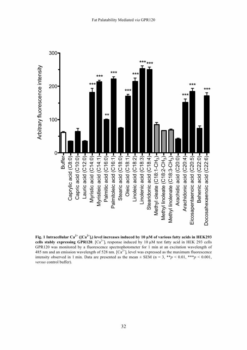
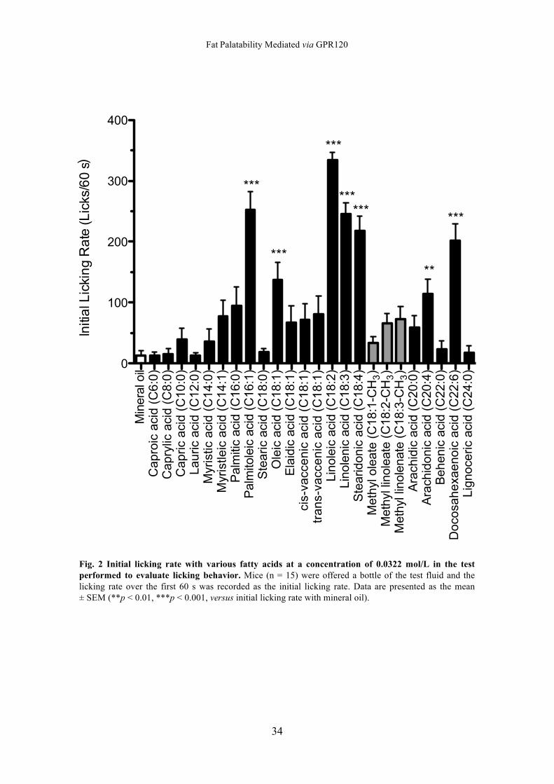
Evaluation of the Licking behavior Fig. 2 shows the initial licking rate with various fatty acids. The mice exhibited a
significantly higher licking rate with palmitoleic acid (C16:1), oleic acid (C18:1 cis-9),
linoleic acid (C18:2), linolenic acid (C18:3), stearidonic acid (C18:4), arachidonic acid
(C20:4), and docosahexaenoic acid (C22:6) than with mineral oil (versus mineral oil by
Dunnett’s test: p < 0.01 for arachidonic acid; p < 0.001 for other fatty acids). On the
other hand, the mice did not respond significantly to caproic acid (C6:0), caprylic acid
(C8:0), capric acid (C10:0), lauric acid (C12:0), myristic acid (C14:0), myristoleic acid
(C14:1), palmitic acid (C16:0), stearic acid (C18:0), elaidic acid (C18:1 trans-9),
cis-vaccenic acid (C18:1 cis-11), trans-vaccenic acid (C18:1 trans-11), methyl oleate
(C18:1-CH3), methyl linoleate (C18:2-CH3), methyl linolenate, (C18:3-CH3), arachidic
acid (C20:0), behenic acid (C22:0), and lignoceric acid (C24:0).
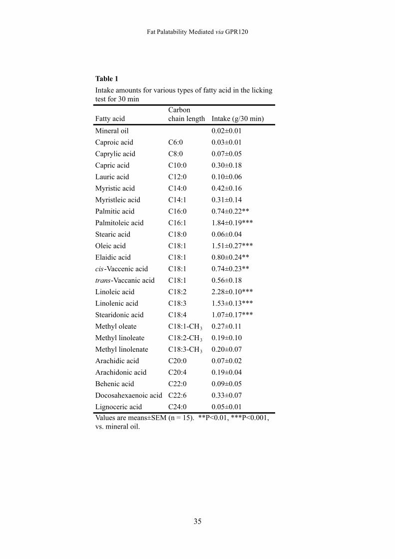
Similar to the result for the licking rate, the intake amount for palmitic acid (C16:0),
palmitoleic acid (C16:1), oleic acid (C18:1 cis-9), elaidic acid (C18:1 trans-9), linoleic
acid (C18:2), cis-vaccenic acid (C18:1 cis-11), linolenic acid (C18:3), and stearidonic
acid (C18:4) were significantly greater than that of mineral oil (versus mineral oil by
Dunnett’s test: p < 0.01 for palmitic acid and elaidic acid; p < 0.001 for other fatty
acids) (Table 1). However, the intake amount for all other fatty acids was not
significant.
Fat Palatability Mediated via GPR120
34
Fig. 2 Initial licking rate with various fatty acids at a concentration of 0.0322 mol/L in the test performed to evaluate licking behavior. Mice (n = 15) were offered a bottle of the test fluid and the licking rate over the first 60 s was recorded as the initial licking rate. Data are presented as the mean ± SEM (**p < 0.01, ***p < 0.001, versus initial licking rate with mineral oil).
Min
eral
oil
Cap
roic
aci
d (C
6:0)
Cap
rylic
aci
d (C
8:0)
Cap
ric a
cid
(C10
:0)
Laur
ic a
cid
(C12
:0)
Myr
istic
aci
d (C
14:0
)M
yris
tleic
aci
d (C
14:1
)P
alm
itic
acid
(C
16:0
)P
alm
itole
ic a
cid
(C16
:1)
Ste
aric
aci
d (C
18:0
)O
leic
aci
d (C
18:1
)E
laid
ic a
cid
(C18
:1)
cis-
vacc
en
ic a
cid
(C
18
:1)
tra
ns-
vacc
en
ic a
cid
(C
18
:1)
Lino
leic
aci
d (C
18:2
)Li
nole
nic
acid
(C
18:3
)S
tear
idon
ic a
cid
(C18
:4)
Met
hyl o
leat
e (C
18:1
-CH
3)
Met
hyl l
inol
eate
(C
18:2
-CH
3)
Met
hyl l
inol
enat
e (C
18:3
-CH
3)
Ara
chid
ic a
cid
(C20
:0)
Ara
chid
onic
aci
d (C
20:4
)B
ehen
ic a
cid
(C22
:0)
Doc
osah
exae
noic
aci
d (C
22:6
)Li
gnoc
eric
aci
d (C
24:0
)0
100
200
300
400
Initi
al L
icki
ng
Ra
te (L
icks
/60
se
c)
***
*****
***
****** ***
Fat Palatability Mediated via GPR120
35
Table 1
Fatty acidCarbonchain length Intake (g/30 min)
Mineral oil 0.02±0.01
Caproic acid C6:0 0.03±0.01
Caprylic acid C8:0 0.07±0.05
Capric acid C10:0 0.30±0.18
Lauric acid C12:0 0.10±0.06
Myristic acid C14:0 0.42±0.16
Myristleic acid C14:1 0.31±0.14
Palmitic acid C16:0 0.74±0.22**
Palmitoleic acid C16:1 1.84±0.19***
Stearic acid C18:0 0.06±0.04
Oleic acid C18:1 1.51±0.27***
Elaidic acid C18:1 0.80±0.24**
cis-Vaccenic acid C18:1 0.74±0.23**
trans-Vaccanic acid C18:1 0.56±0.18
Linoleic acid C18:2 2.28±0.10***
Linolenic acid C18:3 1.53±0.13***
Stearidonic acid C18:4 1.07±0.17***
Methyl oleate C18:1-CH3 0.27±0.11
Methyl linoleate C18:2-CH3 0.19±0.10
Methyl linolenate C18:3-CH3 0.20±0.07
Arachidic acid C20:0 0.07±0.02
Arachidonic acid C20:4 0.19±0.04
Behenic acid C22:0 0.09±0.05
Docosahexaenoic acid C22:6 0.33±0.07
Lignoceric acid C24:0 0.05±0.01
Values are means±SEM (n = 15). **P<0.01, ***P<0.001,vs. mineral oil.
Intake amounts for various types of fatty acid in the lickingtest for 30 min
Fat Palatability Mediated via GPR120
36
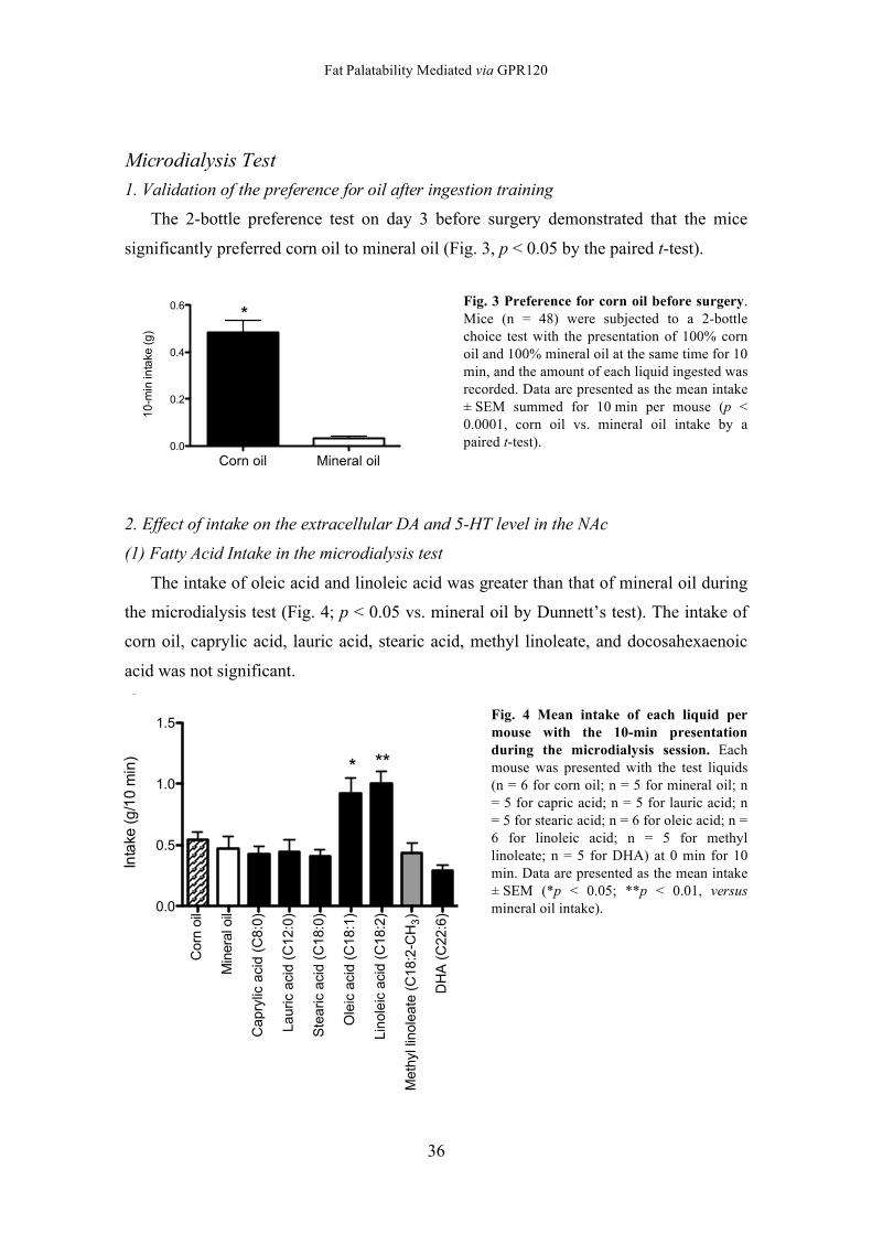
Microdialysis Test 1. Validation of the preference for oil after ingestion training
The 2-bottle preference test on day 3 before surgery demonstrated that the mice
significantly preferred corn oil to mineral oil (Fig. 3, p < 0.05 by the paired t-test).
Fig. 3 Preference for corn oil before surgery. Mice (n = 48) were subjected to a 2-bottle choice test with the presentation of 100% corn oil and 100% mineral oil at the same time for 10 min, and the amount of each liquid ingested was recorded. Data are presented as the mean intake ± SEM summed for 10 min per mouse (p < 0.0001, corn oil vs. mineral oil intake by a paired t-test).
2. Effect of intake on the extracellular DA and 5-HT level in the NAc
(1) Fatty Acid Intake in the microdialysis test
The intake of oleic acid and linoleic acid was greater than that of mineral oil during
the microdialysis test (Fig. 4; p < 0.05 vs. mineral oil by Dunnett’s test). The intake of
corn oil, caprylic acid, lauric acid, stearic acid, methyl linoleate, and docosahexaenoic
acid was not significant.
Fig. 4 Mean intake of each liquid per mouse with the 10-min presentation during the microdialysis session. Each mouse was presented with the test liquids (n = 6 for corn oil; n = 5 for mineral oil; n = 5 for capric acid; n = 5 for lauric acid; n = 5 for stearic acid; n = 6 for oleic acid; n = 6 for linoleic acid; n = 5 for methyl linoleate; n = 5 for DHA) at 0 min for 10 min. Data are presented as the mean intake ± SEM (*p < 0.05; **p < 0.01, versus mineral oil intake).
Corn oil Mineral oil0.0
0.2
0.4
0.6
10
-min
inta
ke (g
)
*
!"# !$# !%# # %# $# "# &# '# (# )# *##
*#
%##
%$#
%&#
%(#
%*#
+,-./0-,12
34/5.6.5/07/89/:;<.5,1.2
=8>1/8,5
?,1.>;5/8,5
=;@>,A/;A,B/0=%#C#2
D;E>,A/;A,B/0=%$C#2
FG.;>,A/;A,B/0=%*C#2
H5.,A/;A,B/0=%*C%2
D,185.,A/;A,B/0=%*C$2
?.GIJ5/5,185.;G./0=%*C$!=K"2
3K4/0=$$C(2
L;<.5,1.
M1G;N.
!
;:O
O
O
OOOO
!"# !$# !%# # %# $# "# &# '# (# )# *##
*#
%##
%$#
%&#
%(#
%*#
+,-./0-,12
'!K+/5.6.5/07/89/:;<.5,1.2
=8>1/8,5
?,1.>;5/8,5
=;@>,A/;A,B/0=%#C#2
D;E>,A/;A,B/0=%$C#2
FG.;>,A/;A,B/0=%*C#2
H5.,A/;A,B/0=%*C%2
D,185.,A/;A,B/0=%*C$2
?.GIJ5/5,185.;G./0=%*C$!=K"2
3K4/0=$$C(2
L;<.5,1.
M1G;N.
"
=8>1/8,5
?,1.>;5/8,5
=;@>,A/;A,B/0=%#C#2
D;E>,A/;A,B/0=%$C#2
FG.;>,A/;A,B/0=%*C#2
H5.,A/;A,B/0=%*C%2
D,185.,A/;A,B/0=%*C$2
?.GIJ5/5,185.;G./0=%*C$!=K"2
3K4/0=$$C(2#
%##
%$#
%&#
%(#
%*#
34/5.6.5/07/89/:;<.5,1.2
OOOO
O
#
=8>1/8,5
?,1.>;5/8,5
=;@>J5,A/;A,B/0=*C#2
D;E>,A/;A,B/0=%$C#2
FG.;>,A/;A,B/0=%*C#2
H5.,A/;A,B/0=%*C%2
D,185.,A/;A,B/0=%*C$2
?.GIJ5/5,185.;G./0=%*C$!=K"2
3K4/0=$$C(2#P#
#P'
%P#
%P'
M1G;N./0QR%#/-,12
O OO
$
%
!"# !$# !%# # %# $# "# &# '# (# )# *##
*#
%##
%$#
%&#
%(#
%*#
+,-./0-,12
34/5.6.5/07/89/:;<.5,1.2
=8>1/8,5
?,1.>;5/8,5
=;@>,A/;A,B/0=%#C#2
D;E>,A/;A,B/0=%$C#2
FG.;>,A/;A,B/0=%*C#2
H5.,A/;A,B/0=%*C%2
D,185.,A/;A,B/0=%*C$2
?.GIJ5/5,185.;G./0=%*C$!=K"2
3K4/0=$$C(2
L;<.5,1.
M1G;N.
!
;:O
O
O
OOOO
!"# !$# !%# # %# $# "# &# '# (# )# *##
*#
%##
%$#
%&#
%(#
%*#
+,-./0-,12
'!K+/5.6.5/07/89/:;<.5,1.2
=8>1/8,5
?,1.>;5/8,5
=;@>,A/;A,B/0=%#C#2
D;E>,A/;A,B/0=%$C#2
FG.;>,A/;A,B/0=%*C#2
H5.,A/;A,B/0=%*C%2
D,185.,A/;A,B/0=%*C$2
?.GIJ5/5,185.;G./0=%*C$!=K"2
3K4/0=$$C(2
L;<.5,1.
M1G;N.
"
=8>1/8,5
?,1.>;5/8,5
=;@>,A/;A,B/0=%#C#2
D;E>,A/;A,B/0=%$C#2
FG.;>,A/;A,B/0=%*C#2
H5.,A/;A,B/0=%*C%2
D,185.,A/;A,B/0=%*C$2
?.GIJ5/5,185.;G./0=%*C$!=K"2
3K4/0=$$C(2#
%##
%$#
%&#
%(#
%*#
34/5.6.5/07/89/:;<.5,1.2
OOOO
O
#
=8>1/8,5
?,1.>;5/8,5
=;@>J5,A/;A,B/0=*C#2
D;E>,A/;A,B/0=%$C#2
FG.;>,A/;A,B/0=%*C#2
H5.,A/;A,B/0=%*C%2
D,185.,A/;A,B/0=%*C$2
?.GIJ5/5,185.;G./0=%*C$!=K"2
3K4/0=$$C(2#P#
#P'
%P#
%P'
M1G;N./0QR%#/-,12
O OO
$
%
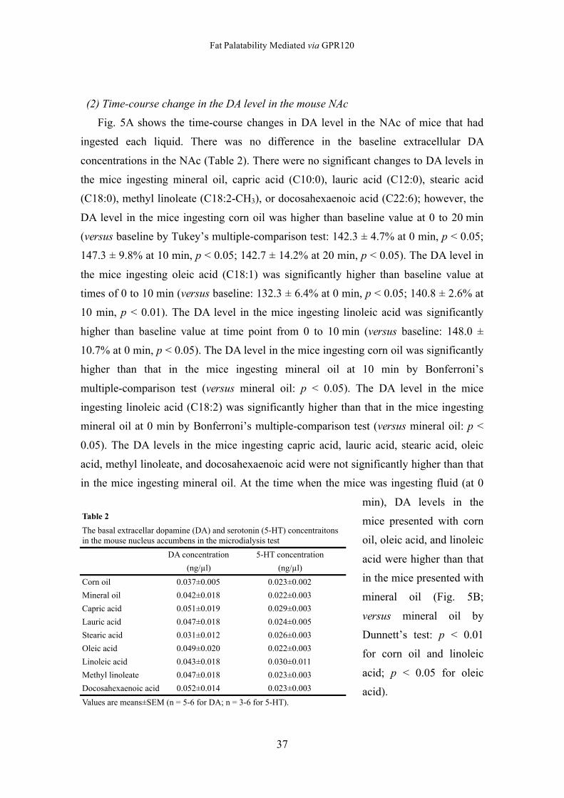
Fat Palatability Mediated via GPR120
37
(2) Time-course change in the DA level in the mouse NAc
Fig. 5A shows the time-course changes in DA level in the NAc of mice that had
ingested each liquid. There was no difference in the baseline extracellular DA
concentrations in the NAc (Table 2). There were no significant changes to DA levels in
the mice ingesting mineral oil, capric acid (C10:0), lauric acid (C12:0), stearic acid
(C18:0), methyl linoleate (C18:2-CH3), or docosahexaenoic acid (C22:6); however, the
DA level in the mice ingesting corn oil was higher than baseline value at 0 to 20 min
(versus baseline by Tukey’s multiple-comparison test: 142.3 ± 4.7% at 0 min, p < 0.05;
147.3 ± 9.8% at 10 min, p < 0.05; 142.7 ± 14.2% at 20 min, p < 0.05). The DA level in
the mice ingesting oleic acid (C18:1) was significantly higher than baseline value at
times of 0 to 10 min (versus baseline: 132.3 ± 6.4% at 0 min, p < 0.05; 140.8 ± 2.6% at
10 min, p < 0.01). The DA level in the mice ingesting linoleic acid was significantly
higher than baseline value at time point from 0 to 10 min (versus baseline: 148.0 ±
10.7% at 0 min, p < 0.05). The DA level in the mice ingesting corn oil was significantly
higher than that in the mice ingesting mineral oil at 10 min by Bonferroni’s
multiple-comparison test (versus mineral oil: p < 0.05). The DA level in the mice
ingesting linoleic acid (C18:2) was significantly higher than that in the mice ingesting
mineral oil at 0 min by Bonferroni’s multiple-comparison test (versus mineral oil: p <
0.05). The DA levels in the mice ingesting capric acid, lauric acid, stearic acid, oleic
acid, methyl linoleate, and docosahexaenoic acid were not significantly higher than that
in the mice ingesting mineral oil. At the time when the mice was ingesting fluid (at 0
min), DA levels in the
mice presented with corn
oil, oleic acid, and linoleic
acid were higher than that
in the mice presented with
mineral oil (Fig. 5B;
versus mineral oil by
Dunnett’s test: p < 0.01
for corn oil and linoleic
acid; p < 0.05 for oleic
acid).
Table 2
DA concentration 5-HT concentration
(ng/µl) (ng/µl)
Corn oil 0.037±0.005 0.023±0.002
Mineral oil 0.042±0.018 0.022±0.003
Capric acid 0.051±0.019 0.029±0.003
Lauric acid 0.047±0.018 0.024±0.005
Stearic acid 0.031±0.012 0.026±0.003
Oleic acid 0.049±0.020 0.022±0.003
Linoleic acid 0.043±0.018 0.030±0.011
Methyl linoleate 0.047±0.018 0.023±0.003
Docosahexaenoic acid 0.052±0.014 0.023±0.003
Values are means±SEM (n = 5-6 for DA; n = 3-6 for 5-HT).
The basal extracellar dopamine (DA) and serotonin (5-HT) concentraitonsin the mouse nucleus accumbens in the microdialysis test
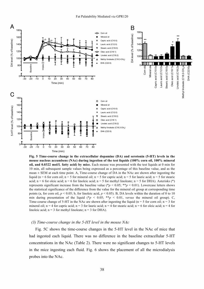
Fat Palatability Mediated via GPR120
38
Fig. 5 Time-course change in the extracellular dopamine (DA) and serotonin (5-HT) levels in the mouse nucleus accumbens (NAc) during ingestion of the test liquids (100% corn oil, 100% mineral oil, and 0.0322 mol/L fatty acid) by mice. Each mouse was presented with the test liquids at 0 min for 10 min, all subsequent sample values being expressed as a percentage of this baseline value, and as the mean ± SEM at each time point. A, Time-course change of DA in the NAc are shown after ingesting the liquid (n = 6 for corn oil; n = 5 for mineral oil; n = 5 for capric acid; n = 5 for lauric acid; n = 5 for stearic acid; n = 6 for oleic acid; n = 6 for linoleic acid; n = 5 for methyl linoleate; n = 5 for DHA). Asterisks (*) represents significant increase from the baseline value (*p < 0.05; **p < 0.01). Lowercase letters shows the statistical significance of the difference from the value for the mineral oil group at corresponding time points (a, for corn oil, p < 0.05; b, for linoleic acid, p < 0.05). B, DA levels within the duration of 0 to 10 min during presentation of the liquid (*p < 0.05; **p < 0.01, versus the mineral oil group). C, Time-course change of 5-HT in the NAc are shown after ingesting the liquid (n = 5 for corn oil; n = 3 for mineral oil; n = 4 for capric acid; n = 3 for lauric acid; n = 4 for stearic acid; n = 6 for oleic acid; n = 4 for linoleic acid; n = 3 for methyl linoleate; n = 3 for DHA).
(3) Time-course change in the 5-HT level in the mouse NAc
Fig. 5C shows the time-course changes in the 5-HT level in the NAc of mice that
had ingested each liquid. There was no difference in the baseline extracellular 5-HT
concentrations in the NAc (Table 2). There were no significant changes to 5-HT levels
in the mice ingesting each fluid. Fig. 6 shows the placement of all the microdialysis
probes into the NAc.
!"# !$# !%# # %# $# "# &# '# (# )# *##
*#
%##
%$#
%&#
%(#
%*#
+,-./0-,12
34/5.6.5/07/89/:;<.5,1.2
=8>1/8,5
?,1.>;5/8,5
=;@>,A/;A,B/0=%#C#2
D;E>,A/;A,B/0=%$C#2
FG.;>,A/;A,B/0=%*C#2
H5.,A/;A,B/0=%*C%2
D,185.,A/;A,B/0=%*C$2
?.GIJ5/5,185.;G./0=%*C$!=K"2
3K4/0=$$C(2
L;<.5,1.
M1G;N.
!
;:O
O
O
OOOO
!"# !$# !%# # %# $# "# &# '# (# )# *##
*#
%##
%$#
%&#
%(#
%*#
+,-./0-,12
'!K+/5.6.5/07/89/:;<.5,1.2
=8>1/8,5
?,1.>;5/8,5
=;@>,A/;A,B/0=%#C#2
D;E>,A/;A,B/0=%$C#2
FG.;>,A/;A,B/0=%*C#2
H5.,A/;A,B/0=%*C%2
D,185.,A/;A,B/0=%*C$2
?.GIJ5/5,185.;G./0=%*C$!=K"2
3K4/0=$$C(2
L;<.5,1.
M1G;N.
"
=8>1/8,5
?,1.>;5/8,5
=;@>,A/;A,B/0=%#C#2
D;E>,A/;A,B/0=%$C#2
FG.;>,A/;A,B/0=%*C#2
H5.,A/;A,B/0=%*C%2
D,185.,A/;A,B/0=%*C$2
?.GIJ5/5,185.;G./0=%*C$!=K"2
3K4/0=$$C(2#
%##
%$#
%&#
%(#
%*#
34/5.6.5/07/89/:;<.5,1.2
OOOO
O
#
=8>1/8,5
?,1.>;5/8,5
=;@>J5,A/;A,B/0=*C#2
D;E>,A/;A,B/0=%$C#2
FG.;>,A/;A,B/0=%*C#2
H5.,A/;A,B/0=%*C%2
D,185.,A/;A,B/0=%*C$2
?.GIJ5/5,185.;G./0=%*C$!=K"2
3K4/0=$$C(2#P#
#P'
%P#
%P'
M1G;N./0QR%#/-,12
O OO
$
%
!"# !$# !%# # %# $# "# &# '# (# )# *##
*#
%##
%$#
%&#
%(#
%*#
+,-./0-,12
34/5.6.5/07/89/:;<.5,1.2
=8>1/8,5
?,1.>;5/8,5
=;@>,A/;A,B/0=%#C#2
D;E>,A/;A,B/0=%$C#2
FG.;>,A/;A,B/0=%*C#2
H5.,A/;A,B/0=%*C%2
D,185.,A/;A,B/0=%*C$2
?.GIJ5/5,185.;G./0=%*C$!=K"2
3K4/0=$$C(2
L;<.5,1.
M1G;N.
!
;:O
O
O
OOOO
!"# !$# !%# # %# $# "# &# '# (# )# *##
*#
%##
%$#
%&#
%(#
%*#
+,-./0-,12
'!K+/5.6.5/07/89/:;<.5,1.2
=8>1/8,5
?,1.>;5/8,5
=;@>,A/;A,B/0=%#C#2
D;E>,A/;A,B/0=%$C#2
FG.;>,A/;A,B/0=%*C#2
H5.,A/;A,B/0=%*C%2
D,185.,A/;A,B/0=%*C$2
?.GIJ5/5,185.;G./0=%*C$!=K"2
3K4/0=$$C(2
L;<.5,1.
M1G;N.
"
=8>1/8,5
?,1.>;5/8,5
=;@>,A/;A,B/0=%#C#2
D;E>,A/;A,B/0=%$C#2
FG.;>,A/;A,B/0=%*C#2
H5.,A/;A,B/0=%*C%2
D,185.,A/;A,B/0=%*C$2
?.GIJ5/5,185.;G./0=%*C$!=K"2
3K4/0=$$C(2#
%##
%$#
%&#
%(#
%*#
34/5.6.5/07/89/:;<.5,1.2
OOOO
O
#
=8>1/8,5
?,1.>;5/8,5
=;@>J5,A/;A,B/0=*C#2
D;E>,A/;A,B/0=%$C#2
FG.;>,A/;A,B/0=%*C#2
H5.,A/;A,B/0=%*C%2
D,185.,A/;A,B/0=%*C$2
?.GIJ5/5,185.;G./0=%*C$!=K"2
3K4/0=$$C(2#P#
#P'
%P#
%P'
M1G;N./0QR%#/-,12
O OO
$
%
!"# !$# !%# # %# $# "# &# '# (# )# *##
*#
%##
%$#
%&#
%(#
%*#
+,-./0-,12
34/5.6.5/07/89/:;<.5,1.2
=8>1/8,5
?,1.>;5/8,5
=;@>,A/;A,B/0=%#C#2
D;E>,A/;A,B/0=%$C#2
FG.;>,A/;A,B/0=%*C#2
H5.,A/;A,B/0=%*C%2
D,185.,A/;A,B/0=%*C$2
?.GIJ5/5,185.;G./0=%*C$!=K"2
3K4/0=$$C(2L;<.5,1.
M1G;N.
!
;:O
O
O
OOOO
!"# !$# !%# # %# $# "# &# '# (# )# *##
*#
%##
%$#
%&#
%(#
%*#
+,-./0-,12
'!K+/5.6.5/07/89/:;<.5,1.2
=8>1/8,5
?,1.>;5/8,5
=;@>,A/;A,B/0=%#C#2
D;E>,A/;A,B/0=%$C#2
FG.;>,A/;A,B/0=%*C#2
H5.,A/;A,B/0=%*C%2
D,185.,A/;A,B/0=%*C$2
?.GIJ5/5,185.;G./0=%*C$!=K"2
3K4/0=$$C(2
L;<.5,1.
M1G;N.
"
=8>1/8,5
?,1.>;5/8,5
=;@>,A/;A,B/0=%#C#2
D;E>,A/;A,B/0=%$C#2
FG.;>,A/;A,B/0=%*C#2
H5.,A/;A,B/0=%*C%2
D,185.,A/;A,B/0=%*C$2
?.GIJ5/5,185.;G./0=%*C$!=K"2
3K4/0=$$C(2#
%##
%$#
%&#
%(#
%*#
34/5.6.5/07/89/:;<.5,1.2
OOOO
O
#
=8>1/8,5
?,1.>;5/8,5
=;@>J5,A/;A,B/0=*C#2
D;E>,A/;A,B/0=%$C#2
FG.;>,A/;A,B/0=%*C#2
H5.,A/;A,B/0=%*C%2
D,185.,A/;A,B/0=%*C$2
?.GIJ5/5,185.;G./0=%*C$!=K"2
3K4/0=$$C(2#P#
#P'
%P#
%P'
M1G;N./0QR%#/-,12
O OO
$
%
!"# !$# !%# # %# $# "# &# '# (# )# *##
*#
%##
%$#
%&#
%(#
%*#
+,-./0-,12
34/5.6.5/07/89/:;<.5,1.2
=8>1/8,5
?,1.>;5/8,5
=;@>,A/;A,B/0=%#C#2
D;E>,A/;A,B/0=%$C#2
FG.;>,A/;A,B/0=%*C#2
H5.,A/;A,B/0=%*C%2
D,185.,A/;A,B/0=%*C$2
?.GIJ5/5,185.;G./0=%*C$!=K"2
3K4/0=$$C(2
L;<.5,1.
M1G;N.
!
;:O
O
O
OOOO
!"# !$# !%# # %# $# "# &# '# (# )# *##
*#
%##
%$#
%&#
%(#
%*#
+,-./0-,12
'!K+/5.6.5/07/89/:;<.5,1.2
=8>1/8,5
?,1.>;5/8,5
=;@>,A/;A,B/0=%#C#2
D;E>,A/;A,B/0=%$C#2
FG.;>,A/;A,B/0=%*C#2
H5.,A/;A,B/0=%*C%2
D,185.,A/;A,B/0=%*C$2
?.GIJ5/5,185.;G./0=%*C$!=K"2
3K4/0=$$C(2
L;<.5,1.
M1G;N.
"
=8>1/8,5
?,1.>;5/8,5
=;@>,A/;A,B/0=%#C#2
D;E>,A/;A,B/0=%$C#2
FG.;>,A/;A,B/0=%*C#2
H5.,A/;A,B/0=%*C%2
D,185.,A/;A,B/0=%*C$2
?.GIJ5/5,185.;G./0=%*C$!=K"2
3K4/0=$$C(2#
%##
%$#
%&#
%(#
%*#
34/5.6.5/07/89/:;<.5,1.2
OOOO
O
#
=8>1/8,5
?,1.>;5/8,5
=;@>J5,A/;A,B/0=*C#2
D;E>,A/;A,B/0=%$C#2
FG.;>,A/;A,B/0=%*C#2
H5.,A/;A,B/0=%*C%2
D,185.,A/;A,B/0=%*C$2
?.GIJ5/5,185.;G./0=%*C$!=K"2
3K4/0=$$C(2#P#
#P'
%P#
%P'
M1G;N./0QR%#/-,12
O OO
$
%
Fat Palatability Mediated via GPR120
39
Fig. 6 Placement of all the microdialysis probes into the NAc. Black bar represents the tip of the probe. The number on each section is the distance in millimeters anterior from the bregma according to the stereotaxic atlas of Paxinos and Franklin.
Correlations between Ca2+ Fluorescence Changes and Initial Licking Rate between,
and Accumbens Dopamine Level
Fig. 7A shows a positive correlation between initial licking rate and the
consumption of the different fatty acids in mice and maximum intracellular Ca2+ level
estimated in cultured cells induced by corresponding fatty acid (Pearson correlation; r =
0.8005, p < 0.0001, n = 19). Additionally, there was a positive correlation between the
30 min intake amounts for fatty acids and [Ca2+]i levels (data not shown; Pearson
correlation; r = 0.7308, p = 0.0004, n = 19). Fig. 7B shows a positive correlation
between cumulative value of DA levels in the NAc at 0 to 80 min in the mice offered
various types of fatty acid and [Ca2+]i levels (Pearson correlation; r = 0.8688, p = 0.0111,
n = 7). Additionally, there was a positive correlation between DA levels at the time (0 to
10 min) when the mice were offered the fluids and [Ca2+]i levels (data not shown;
Pearson correlation; r = 0.7829, p = 0.0374, n = 7).
!"# !$# !%# # %# $# "# &# '# (# )# *##
*#
%##
%$#
%&#
%(#
%*#
+,-./0-,12
34/5.6.5/07/89/:;<.5,1.2
=8>1/8,5
?,1.>;5/8,5
=;@>,A/;A,B/0=%#C#2
D;E>,A/;A,B/0=%$C#2
FG.;>,A/;A,B/0=%*C#2
H5.,A/;A,B/0=%*C%2
D,185.,A/;A,B/0=%*C$2
?.GIJ5/5,185.;G./0=%*C$!=K"2
3K4/0=$$C(2
L;<.5,1.
M1G;N.
!
;:O
O
O
OOOO
!"# !$# !%# # %# $# "# &# '# (# )# *##
*#
%##
%$#
%&#
%(#
%*#
+,-./0-,12
'!K+/5.6.5/07/89/:;<.5,1.2
=8>1/8,5
?,1.>;5/8,5
=;@>,A/;A,B/0=%#C#2
D;E>,A/;A,B/0=%$C#2
FG.;>,A/;A,B/0=%*C#2
H5.,A/;A,B/0=%*C%2
D,185.,A/;A,B/0=%*C$2
?.GIJ5/5,185.;G./0=%*C$!=K"2
3K4/0=$$C(2
L;<.5,1.
M1G;N.
"
=8>1/8,5
?,1.>;5/8,5
=;@>,A/;A,B/0=%#C#2
D;E>,A/;A,B/0=%$C#2
FG.;>,A/;A,B/0=%*C#2
H5.,A/;A,B/0=%*C%2
D,185.,A/;A,B/0=%*C$2
?.GIJ5/5,185.;G./0=%*C$!=K"2
3K4/0=$$C(2#
%##
%$#
%&#
%(#
%*#
34/5.6.5/07/89/:;<.5,1.2
OOOO
O
#
=8>1/8,5
?,1.>;5/8,5
=;@>J5,A/;A,B/0=*C#2
D;E>,A/;A,B/0=%$C#2
FG.;>,A/;A,B/0=%*C#2
H5.,A/;A,B/0=%*C%2
D,185.,A/;A,B/0=%*C$2
?.GIJ5/5,185.;G./0=%*C$!=K"2
3K4/0=$$C(2#P#
#P'
%P#
%P'
M1G;N./0QR%#/-,12
O OO
$
%
Fat Palatability Mediated via GPR120
40
Fig. 7 Correlations of the maximum intracellular Ca2+ ([Ca2+]i) fluorescence intensities and the initial licking rates (A), and of the maximum [Ca2+]i fluorescence intensities and the cumulative values of DA level (B). A, X axis shows the averaged initial licking rate with various fatty acids and Y axis shows the averaged maximum [Ca2+]i fluorescence intensities in the response induced by corresponding fatty acid (Pearson correlation; r = 0.7421, p = 0.0002, n = 19). B, X axis shows the cumulative value of averaged DA level at 0 to 80 min in the NAc of mice after ingesting various fatty acids and Y axis shows the averaged maximum [Ca2+]i fluorescence intensities in the response induced by corresponding fatty acid (Pearson correlation; r = 0.8688, p = 0.0111, n = 7).
0 100 200 300 4000
100
200
300
Initial licking rate (Licks/60 sec)
Arb
itra
ry fl
uo
resc
en
ce in
ten
sity
C8:0
C10:0
C12:0
C14:0
C14:1
C16:0
C16:1
C18:0
C18:1
C18:2
C18:3
C18:4
C18:1-CH3
C18:2-CH3
C18:3-CH3
C20:0
C20:4
C22:0
C22:6
A
800 900 1000 1100 1200 13000
50
100
150
200
250
Arb
itra
ry fl
uo
resc
en
ce in
ten
sity
Cumulative value of DA level
C10:0
C12:0
C18:0
C18:1
C18:2
C18:2-CH3
C22:6
B
Fat Palatability Mediated via GPR120
41
DISCUSSION 1. Intracellular Ca2+ Assay
Since there was no response to fatty acids in the HEK293 cells transfected with an
empty vector, it was confirmed that the fluorescence changes stimulated by fatty acids
were the GPR120-specific responses (data not shown).
The addition of saturated and unsaturated 14- and 16-carbon fatty acids induced a
significantly higher increase in [Ca2+]i than that of the control buffer in the HEK293
cells stably expressing GPR120. Additionally, in the 18-, 20-, and 22-carbon fatty acids,
increases in [Ca2+]i induced by unsaturated fatty acids were significantly higher than
those induced by control buffer, whereas saturated forms of these fatty acids caused no
significant response. These results demonstrate that saturated 14- and 16-carbon fatty
acids and unsaturated 14-, 16-, 18-, 20-, and 22-carbon fatty acids are strong ligands for
GPR120. Moreover, in 18- and 20-carbon fatty acids, [Ca2+]i levels tended to increase
due to poly unsaturated fatty acids like stearic acid (C18:0), oleic acid (C18:1), linoleic
acid (C18:2), linolenic acid (C18:3), stearidonic acid (C18:4), arachidic acid (C20:0),
arachidonic acid (C20:4), and EPA (C20:5). This result shows that the structure of poly
unsaturated fatty acids is more suitable for GPR120 ligands in the 18- and 20-carbon
acids than in other fatty acids. Further, [Ca2+]i levels stimulated by methyl esters of
unsaturated 18-carbon fatty acid were not significant when compared with that of
control. This result implies that the terminal carboxyl group of the fatty acids plays an
important role in the recognition of fatty acid structure by GPR120. The results of
[Ca2+]i assay in this study correspond well with previous reports by Hirasawa et al. and
Galindo et al. (20, 27).
2. Evaluation of Licking Behavior
During the evaluation of the licking behavior with middle-chain fatty acids such as
6-carbon caproic acid, 8-C caprylic acid, and 10-C capric acid, as well as shorter-chain
LCFAs such as 12-C lauric acid, 14-C myristic acid, and myristoleic acid, the initial
licking rates were not significantly different from those observed by using mineral oil.
On the other hand, in the test with longer LCFAs such as 16-C palmitoleic acid, 18-C
oleic acid, linoleic acid, linolenic acid, stearidonic acid, 20-C arachidonic acid, and
22-C DHA, the initial licking rates were significantly higher than that with mineral oil.
This result suggests that carbon chain length is involved in the palatability of fatty acid.
Fat Palatability Mediated via GPR120
42
In addition, since the mice displayed high licking response to unsaturated LCFAs
(palmitic acid, 16:1; oleic acid, 18:1; linoleic acid, 18:2; linolenic acid, 18:3; stearidonic
acid, 18:4; arachidonic acid, 20:4; and DHA, 22:6) and low licking response to
saturated LCFAs (palmitic acid, 16:0; stearic acid, 18:0; arachidic acid, 20:0; and
behenic acid, 22:0), these results indicate that palatability of fatty acids is affected by
the saturation state of the fatty acid. Moreover, the mice showed a significantly higher
licking rate with oleic acid, linoleic acid, and linolenic acid. On the other hand, the mice
did not respond significantly to corresponding methyl esters, namely, methyl oleate,
methyl linoleate, and methyl linolenate. Yoneda et al. reported that the mice exhibited
low palatability for not only fatty acid ester but also fatty alcohol (22). These results
imply that terminal carboxyl group of fatty acids is involved in their palatability.
Further, the licking rate with oleic acid (cis-9-octadecenoic acid) was significantly
higher than that with mineral oil. However, there was no difference in the licking rates
with geometric isomer of oleic acid, elaidic acid (trans-9-octadecenoic acid) and
mineral oil. Additionally, the licking rates with cis-vaccenic acid (cis-11-octadecenoic
acid) and trans-vaccenic acid (trans-11-octadecenoic acid) which are regioisomers of
oleic acid and elaidic acid, respectively, had no such effect. The results show that
conformation of the fatty acid is important in the manifestation of palatability, in at least
C18:1 fatty acids.
In this study, we observed that the amount of fatty acid intake over 30 min did not
necessarily correlate with the initial licking rate. For example, the licking rates of
myristoleic acid and elaidic acid were not significantly higher than that of mineral oil;
however, their intake was greater than that of mineral oil. A possible explanation for this
difference is that the post-ingestive effects of these acids may have promoted their
consumption. In contrast, the licking rate of DHA was significantly higher than that of
mineral oil, while the intake amount was not. Harden et al. reported that DHA
stimulated significantly more release of cholecystokinin (CCK) than other LCFAs such
as linoleic acid and oleic acid in secretin tumor (STC-1) cells (28). Therefore, release of
CCK from the duodenum, which is suppressed after DHA ingestion, may suppress the
overall intake of this fatty acid over 30 min.
3. Change in DA Level in Microdialysis Test
Previously, we have reported that extracellular DA level in the NAc of rats was
Fat Palatability Mediated via GPR120
43
elevated by ingesting 1% (v/v) linoleic acid, which had a very low calorie level
(approximately 0.09 kcal/g), as compared to the similar increase observed with as high
as 100% corn oil (9 kcal/g) ingestion (6 and chap.1). This showed that the reward value
of fat, at least as estimated from the increase in DA level in the NAc, was determined by
the chemoreception of LCFAs in the oral cavity, not by its caloric density. Similarly, in
this study using mice, 100% corn oil and 1% linoleic acid significantly increased the
NAc DA levels. Oleic acid also resulted in a significant increase in DA level. On the
other hand, capric acid, lauric acid, stearic acid, methyl linoleate, and DHA caused no
significant change in the DA levels. This result suggests that 18-carbon length, saturated
state of fatty acid, and terminal carboxyl group are important for the rise in DA level
observed upon the ingestion of these LCFAs. Further, the intake amount of oleic acid
and linoleic acid during the 10 min of presentation in the microdialysis test was
significantly greater than that of mineral oil, whereas other fatty acids presented in the
microdialysis test showed no difference in consumption than that of mineral oil. These
results indicated that the characteristics of fatty acids that caused an increase in the DA
level corresponded well with the palatability of LCFAs.
4. Correlation between [Ca2+]i level, licking rate, and DA level
In this study, the GPR120-agonistic activity of LCFA as estimated in cell culture
correlated with the initial licking rate and the increase in DA level. This suggests that
the chemoreception of LCFA via GPR120 is the first step in the manifestation of this
effect. [Ca2+]i levels stimulated by myristic acid and myristoleic acid were higher than
those achieved by using the control buffer, and the mice did not show significantly
higher licking response to these fatty acids as compared to mineral oil. This difference
may be attributed to the influence of the flavors in these fatty acids, which may have
resulted in the mice avoiding their consumption. Godinot et al. reported that non-fatty
acid agonists of GPR120 activated the glossopharyngeal nerve of mice and that the mice
did not show any preference for non-fatty acid agonists of GPR120 (29). These findings
imply that the palatability of LCFAs at low concentration may not consist of only
GPR120 and that composition of the palatability may include other mechanisms such as
chemoreception via CD36 and GPR40. Both of CD36 and GPR40 are reported to be
expressed in the taste cell, while GPR120 is reported to be involved in the recognition
and palatability of fat in the oral cavity (30). Further studies using GPR120 knockout
Fat Palatability Mediated via GPR120
44
mice are necessary to determine whether the expression of GPR120 is implicated in the
palatability of fat and increase in DA level when ingesting fat. The agonistic activity of
receptor proteins expressed in the oral cavity, such as CD36 and GPR40 require
investigation and a detailed study is warranted, on the relationship between agonistic
activity and palatability using animal models.
To summarize, we found that saturated 14- and 16-carbon fatty acids and
unsaturated 14-, 16-, 18-, 20-, and 22-carbon fatty acids are strong ligands for GPR120
that cause an increase in [Ca2+]i levels in the cultured cells. We also revealed that mice
have high palatability for unsaturated 16-, 18-, 20-, and 22-carbon fatty acids. Moreover,
DA level in the NAc of mice was elevated after ingestion of unsaturated 18-carbon acid
to the same extent as 100% corn oil. Further, the agonistic activity of GPR120 ligands
correlated with the palatability of LCFAs in the oral cavity and the reward values based
on DA levels in the NAc. These findings suggest that the chemoreception of LCFAs via
GPR120 is implicated in the palatability of LCFAs, and they support the notion that the
chemoreception of LCFAs released from fat by lingual lipase plays an important role in
the detection of fat in the oral cavity as well as fat palatability, including the reward
effect.
Fat Palatability Mediated via GPR120
45
REFERENCES
1. Drewnowski A, Greenwood MR. (1983). Cream and sugar: human preferences
for high-fat foods. Physiol Behav. 30,:629–33
2. Takeda M, Imaizumi M, Fushiki T. (2000). Preference for vegetable oils in the
two-bottle choice test in mice. Life Sci. 67,:197–204
3. Imaizumi M, Takeda M, Fushiki T. (2000). Effects of oil intake in the
conditioned place preference test in mice. Brain Res. 870,:150–56
4. Yoneda T, Taka Y, Okamura M, Mizushige T, Matsumura S, Manabe Y, Tsuzuki
S, Inoue K, Fushiki T. (2007). Reinforcing effect for corn oil stimulus was
concentration dependent in an operant task in mice. Life Sci. 81,:1585–92
5. Liang N-C, Hajnal A, Norgren R. (2006). Sham feeding corn oil increases
accumbens dopamine in the rat. Am J Physiol Regul Integr Comp Physiol.
291,:R1236–9
6. Adachi S, Endo Y, Mizushige T, Tsuzuki S, Matsumura S, Inoue K, Fushiki T.
(2013). Increased levels of extracellular dopamine in the nucleus accumbens and
amygdala of rats by ingesting a low concentration of a long-chain fatty acid.
Biosci Biotechnol Biochem. 77,:2175–80
7. Wise RA, Rompre PP. (1989). Brain dopamine and reward. Annu Rev Psychol.
40,:191–225
8. Wise RA. (2006). Role of brain dopamine in food reward and reinforcement.
Philos Trans R Soc Lond B Biol Sci. 361,:1149–58
9. Geisler S, Zahm DS. (2005). Afferents of the ventral tegmental area in the
rat-anatomical substratum for integrative functions. J Comp Neurol. 490,:270–94
10. Meye FJ, Adan RA. (2014). Feelings about food: the ventral tegmental area in
food reward and emotional eating. Trends Pharmacol Sci. 35,:31–40
Fat Palatability Mediated via GPR120
46
11. Willuhn I, Wanat MJ, Clark JJ, Phillips PEM. (2010). Dopamine signaling in the
nucleus accumbens of animals self-administering drugs of abuse. Curr Top
Behav Neurosci. 3,:29–71
12. Iwakura H, Ariyasu H, Hosoda H, Yamada G, Hosoda K, Nakao K, Kangawa K,
Akamizu T. (2011). Oxytocin and dopamine stimulate ghrelin secretion by the
ghrelin-producing cell line, mgn3-1 in vitro. Endocrinology. 152,:2619–25
13. Hajnal A, Smith GP, Norgren R. (2004). Oral sucrose stimulation increases
accumbens dopamine in the rat. Am J Physiol Regul Integr Comp Physiol.
286,:R31–7
14. Pettit HO, Justice JB. (1991). Effect of dose on cocaine self-administration
behavior and dopamine levels in the nucleus accumbens. Brain Res. 539,:94–102
15. Kawai T, Fushiki T. (2003). Importance of lipolysis in oral cavity for orosensory
detection of fat. Am J Physiol Regul Integr Comp Physiol. 285,:R447–54
16. Gilbertson TA, Fontenot DT, Liu L, Zhang H, Monroe WT. (1997). Fatty acid
modulation of k+ channels in taste receptor cells: gustatory cues for dietary fat.
Am J Physiol. 272,:C1203–10
17. Fukuwatari T, Kawada T, Tsuruta M, Hiraoka T, Iwanaga T, Sugimoto E,
Fushiki T. (1997). Expression of the putative membrane fatty acid transporter
(fat) in taste buds of the circumvallate papillae in rats. FEBS Lett. 414,:461–64
18. Laugerette F, Passilly-Degrace P, Patris B, Niot I, Febbraio M, Montmayeur JP,
Besnard P. (2005). Cd36 involvement in orosensory detection of dietary lipids,
spontaneous fat preference, and digestive secretions. J Clin Invest. 115,:3177–84
19. Matsumura S, Eguchi A, Mizushige T, Kitabayashi N, Tsuzuki S, Inoue K,
Fushiki T. (2009). Colocalization of gpr120 with phospholipase-cbeta2 and
alpha-gustducin in the taste bud cells in mice. Neurosci Lett. 450,:186–90
Fat Palatability Mediated via GPR120
47
20. Hirasawa A, Tsumaya K, Awaji T, Katsuma S, Adachi T, Yamada M, Sugimoto
Y, Miyazaki S, Tsujimoto G. (2005). Free fatty acids regulate gut incretin
glucagon-like peptide-1 secretion through gpr120. Nat Med. 11,:90–94
21. Yoneda T, Saitou K, Mizushige T, Matsumura S, Manabe Y, Tsuzuki S, Inoue K,
Fushiki T. (2007). The palatability of corn oil and linoleic acid to mice as
measured by short-term two-bottle choice and licking tests. Physiol Behav.
91,:304–9
22. Yoneda T, Saitou K, Asano H, Mizushige T, Matsumura S, Eguchi A, Manabe Y,
Tsuzuki S, Inoue K, Fushiki T. (2009). Assessing palatability of long-chain fatty
acids from the licking behavior of balb/c mice. Physiol Behav. 96,:735–41
23. Cartoni C, Yasumatsu K, Ohkuri T, Shigemura N, Yoshida R, Godinot N, le
Coutre J, Ninomiya Y, Damak S. (2010). Taste preference for fatty acids is
mediated by gpr40 and gpr120. J Neurosci. 30,:8376–82
24. Iino T, Sugimoto H, Watanabe A, Encinas JA, Liu N, Floeckner J, Bacon KB.
(2002). Molecular cloning and functional characterization of cynomolgus
monkey (macaca fascicularis) cc chemokine receptor, ccr3. Cytokine. 19,:276–86
25. Saitou K, Yoneda T, Mizushige T, Asano H, Okamura M, Matsumura S, Eguchi
A, Manabe Y, Tsuzuki S, Inoue K, Fushiki T. (2009). Contribution of gustation
to the palatability of linoleic acid. Physiol Behav. 96,:142–48
26. Paxinos G, Franklin KBJ. “the mouse brain in stereotaxic coordinates, second
edition” San Diego: Academic Press
27. Galindo MM, Voigt N, Stein J, van Lengerich J, Raguse JD, Hofmann T,
Meyerhof W, Behrens M. (2012). G protein-coupled receptors in human fat taste
perception. Chem Senses. 37,:123–39
28. Harden CJ, Jones AN, Maya-jimenez T, Barker ME, Hepburn NJ, Garaiova I,
Plummer SF, Corfe BM, Limited C, Road C, Park BI, Talbot P, Sa WG. (2012).
Fat Palatability Mediated via GPR120
48
Effect of different long-chain fatty acids on cholecystokinin release in vitro and
energy intake in free-living healthy males. Br J Nutr. 108,:755–58
29. Godinot N, Yasumatsu K, Barcos ME, Pineau N, Ledda M, Viton F, Ninomiya Y,
le Coutre J, Damak S. (2013). Activation of tongue-expressed gpr40 and gpr120
by non caloric agonists is not sufficient to drive preference in mice. Neuroscience.
250,:20–30
30. Tucker RM, Mattes RD, Running C a. (2014). Mechanisms and effects of fat
taste in humans. BioFactors. 40,:313–26
49
SUMMARY
Chapter 1
Changes in the extracellular concentration of dopamine (DA) in the nucleus
accumbens (NAc) shell and the basolateral amygdala (BLA) resulting from the volun-
tary ingestion of either corn oil, mineral oil, or 1% linoleic acid diluted with mineral oil
as a vehicle were measured in rats by using in vivo microdialysis after they had been
trained to establish a preference for corn oil. Ingesting the mineral oil caused no
significant change in DA level in the NAc shell, whereas corn oil ingestion significantly
increased the DA level during 0–15 min of the test session, reaching the maximum level
of 129.8 ± 6.2% compared with the baseline after 10 min. Ingesting linoleic acid also
resulted in a significant increase in DA level during 0–20 min, reaching 125.9 ± 9.0%
after 10 min. Similar results were obtained in the BLA. Despite its very low calorie
content, a low concentration of non-esterified fatty acid increased the DA levels
equivalent to those resulting from corn oil in the brain’s reward system.
Chapter 2
Mechanism underlying the involvement of the GPR120-agonistic activity of LCFA
in the palatability of dietary fat remains elusive. Therefore, we examined the association
between the GPR120-agonistic activity and palatability of LCFA. First, we measured
Ca2+ signaling in HEK293 cells stably expressing GPR120 under stimulation by various
LCFAs. We then assessed the palatability of the various LCFAs by testing the licking
behavior in mice and measured the changes in the NAc-DA level by in vivo
microdialysis. Consequently, 14- to 22-carbon unsaturated LCFAs showed strong
GPR120-agonistic activity. Additionally, mice displayed high licking response to
unsaturated 16- and 18-carbon LCFAs, and unsaturated 18-carbon LCFA significantly
increased the DA level. The licking rate and the LCFA-dependent increase in DA level
also correlated well with the GPR120-agonistic activity. These findings demonstrate
that chemoreception of LCFA by GPR120 is involved in the recognition and palatability
of dietary fat.
50
ACKNOWLEDGEMENT
The author wishes to show his grateful acknowledgement to Dr. Tohru Fushiki,
Professor of Nutrition Chemistry, Division of Food Science and Biotechnology,
Graduate School of Agriculture, Kyoto University, for useful advice, helpful discussions
and his counting interest and encouragement of this research.
The author would like to express his sincere gratitude to Dr. Kazuo Inoue, Associate
Professor of Nutrition Chemistry for his useful advice, valuable discussion, critical
review of this thesis, and kind encouragement of this research.
The author would like to express his sincere gratitude to Dr. Satoshi Tsuzuki,
Assistant Professor of Nutrition Chemistry for his kind advice and suggestion.
The author would like to express his sincere gratitude to Dr. Shigenobu Matsumura,
Assistant Professor of Nutrition Chemistry for his useful advice and encouragement of
this research.
The author would like to express special thanks to Dr. Ai Eguchi, the senior member
of the same research group, for acquisition of excellent data (calcium ion assay).
The author is deeply grateful to Mr. Hiroki Asano, the member of the same research
group, for acquisition of excellent data (licking test).
The author would like to sincerely appreciate to Dr. Yasuko Manabe, Dr. Takafumi
Mizushige, Mr. Yuki Endo and Mr. Kazuhiro Sakamoto, the members of the same
research member for their valuable experimental support and discussion.
The author would like to express special thanks Ms. Maki Ishii and Ms. Satoko
Hanabuchi, the staff of the laboratory, for their kind support and encouragement.
The author would acknowledge all members of the laboratory for their helpful
support and assistance.
Finally, the author would like to express the great appreciation to his wife, Keiko
Adachi, his parents and his family for heartful and warm encouragement and continuous
support.
Kyoto 2015
Shinichi Adachi
51
List of publications
1. Adachi S, Endo Y, Mizushige T, Tsuzuki S, Matsumura S, Inoue K, Fushiki T.
Increased levels of extracellular dopamine in the nucleus accumbens and amygdala
of rats by ingesting a low concentration of a long-chain Fatty Acid.
Biosci Biotechnol Biochem. 2013;77(11):2175-80.
2. Adachi S, Eguchi A, Sakamoto K, Asano H, Manabe Y, Matsumura S, Tsuzuki S,
Inoue K, Fushiki T.
Behavioral Palatability of Dietary Fatty Acids Correlates with the Intracellular
Calcium Ion Levels Induced by the Fatty Acids in GPR120-Expressing Cells
Biomedical Research. In press 2014


![Structure Elucidation of Benzhexol-β-Cyclodextrin Complex ... · of inclusion complex, but also provides information useful for detailed structure elucidation of the complex [13]](https://img.pdfslide.tips/doc/110x75/5e7e1d38e07ed352d60daf63/structure-elucidation-of-benzhexol-cyclodextrin-complex-of-inclusion-complex.jpg)