Embed Size (px)
Citation preview
JPET #171520 1
Endogenous luminal surface adenosine signaling regulates duodenal
bicarbonate secretion in rats
Maggie Ham, Misa Mizumori, Chikako Watanabe, Joon-Ho Wang, Takuya Inoue, Takanari
Nakano, Paul H Guth, Eli Engel, and Jonathan D Kaunitz, and Yasutada Akiba
Department of Medicine, School of Medicine (M.H., M.M., C.W., J.H.W, T.I., T.N., J.D.K.,
Y.A.), Department of Biomathematics (E.E.), University of California Los Angeles,
Greater Los Angles Veterans Affairs Healthcare System (P.H.G, J.D.K, Y.A.) and
Brentwood Biomedical Research Institute (J.D.K., Y.A.), Los Angeles, CA 90073, USA
JPET Fast Forward. Published on August 30, 2010 as DOI:10.1124/jpet.110.171520
Copyright 2010 by the American Society for Pharmacology and Experimental Therapeutics.
This article has not been copyedited and formatted. The final version may differ from this version.JPET Fast Forward. Published on August 30, 2010 as DOI: 10.1124/jpet.110.171520
at ASPE
T Journals on M
ay 6, 2019jpet.aspetjournals.org
Dow
nloaded from
JPET #171520 2
Running title: Adenosine-induced duodenal HCO3- secretion
Corresponding to: Jonathan D. Kaunitz,
Bldg. 114, Suite 217,
West Los Angeles VA Medical Center,
11301 Wilshire Blvd., Los Angeles, CA 90073 USA
Phone: 310-268-3879
Fax: 310-268-4811
e-mail: [email protected]
Pages 26
Tables 0
Figures 7
References 45
Words in Abstract 250, Introduction 715, Discussion 1147
Abbreviations used
ADA: adenosine deaminase, ADO: adenosine, CFTR: cystic fibrosis transmembrane
conductance regulator, CNT: concentrative nucleoside transporter, CPA: N6-
cyclopentyladenosine, DPCPX: 8-cyclopentyl-1,3-dipropylxanthine, CSC: 8-(3-
chlorostyryl)caffeine, DBS: duodenal bicarbonate secretion, DCF: 2’-deoxycoformycin,
ENT: equilibrative nucleoside transporter, ForB: formycin B, IAP: intestinal alkaline
phosphatase, IB-MECA: N6-(3-iodobenzyl)adenosine-5’-N-methyluronamide, INO:
inosine, NBTI: S-(4-nitrobenzyl)-6-thioinosine, NECA: 5’-(N-ethylcarboxamido)-
adenosine, NT: nucleoside transporter.
This article has not been copyedited and formatted. The final version may differ from this version.JPET Fast Forward. Published on August 30, 2010 as DOI: 10.1124/jpet.110.171520
at ASPE
T Journals on M
ay 6, 2019jpet.aspetjournals.org
Dow
nloaded from
JPET #171520 3
ABSTRACT
Luminal ATP increases duodenal bicarbonate secretion (DBS) via brush border P2Y receptors.
Since ATP sequentially dephosphorylates ATP to adenosine (ADO), and since the brush border
highly expresses adenosine deaminase (ADA), we hypothesized that luminal [ADO] regulators
and sensors, including P1 receptors, ADA , and nucleoside transporters (NT) regulate DBS. We
measured DBS with pH and CO2 electrodes, perfusing ADO +/- adenosine receptor agonists or
antagonists, or the cystic fibrosis transmembrane conductance regulator (CFTR) inhibitor
CFTRinh-172 on DBS. Furthermore, we examined the effect of inhibitors of ADA or NT on DBS.
Perfusion of AMP or ADO (0.1 mM) uniformly increased DBS, whereas inosine had no effect.
The A1/2 receptor agonist 5’-(N-ethylcarboxamido)-adenosine (NECA, 0.1 mM) increased DBS,
whereas ADO-augmented DBS was inhibited by the potent A2B receptor antagonist MRS1754
(10 µM). Other selective adenosine receptor agonists or antagonists had no effect. The A2B
receptor was immunolocalized to the brush border membrane of duodenal villi, whereas the A2A
receptor was primarily immunolocalized to the vascular endothelium. Furthermore, ADO-
induced DBS was enhanced by 2’-deoxycoformycin (DCF, 1 µM), formycin B (0.1 mM), but not
by S-(4-nitrobenzyl)-6-thioinosine (NBTI, 0.1 mM), and was abolished by CFTRinh-172
pretreatment (1 mg/kg, i.p). Moreover, ATP (0.1 mM)-induced DBS was partially reduced by
MRS2500 or PSB603 and abolished by both, suggesting that ATP is sequentially degraded to
ADO. Luminal ADO stimulates DBS via A2B receptors and CFTR. ATP release, ecto-
phosphohydrolases, ADA and concentrative NT may coordinately regulate luminal surface ADO
concentration in order to modulate ADO-P1 receptor signaling in rat duodenum.
This article has not been copyedited and formatted. The final version may differ from this version.JPET Fast Forward. Published on August 30, 2010 as DOI: 10.1124/jpet.110.171520
at ASPE
T Journals on M
ay 6, 2019jpet.aspetjournals.org
Dow
nloaded from
JPET #171520 4
INTRODUCTION
Adenosine (ADO) is a purinergic signaling molecule that profoundly affects gut function,
including motility, ion secretion, and the modulation of inflammation (Antonioli, et al.,
2008;Ye and Rajendran, 2009). Most of the research regarding extracellular ADO
signaling in the gut has addressed enteric neurons, smooth muscle, afferent neurons,
and the immune system, with relatively few studies addressing its effect on epithelial
secretory function. Of these, even fewer have reported the effect of luminal ADO on ion
secretion in intact epithelia in vivo, since most published studies reported short-circuit
current (Isc) measured in intestinal T84 monolayers (Barrett, et al., 1989;Strohmeier, et
al., 1995) or mounted mammalian intestinal tissues (Grasl and Turnheim, 1984;Ghanem,
et al., 2005;Dobbins, et al., 1984). Since apical or basolateral ADO stimulated Isc, ADO
receptors were predicted to be expressed on both epithelial surfaces (Barrett, et al.,
1989;Strohmeier, et al., 1995). Luminal AMP and ADO increase glucose transport in
intact mouse small intestine in vitro and in vivo, consistent with apical ADO receptors
(Kimura, et al., 2005).
The duodenal enterocyte actively secretes bicarbonate as part of a system that protects
the mucosa from acid injury. We have recently reported that duodenal bicarbonate
secretion (DBS) is regulated by a luminal purinergic regulatory system comprised of the
brush border intestinal alkaline phosphatase (IAP), non-lytic ATP release, and
enterocyte P2Y receptors, which, when activated, augment epithelial DBS (Mizumori, et
al., 2009). Although not directly tested, this purinergic regulatory system likely functions
to regulate duodenal surface microclimate pH, which prior studies had identified with the
use of microelectrodes (Flemström and Kivilaakso, 1983;Flemström, et al., 1982). We
This article has not been copyedited and formatted. The final version may differ from this version.JPET Fast Forward. Published on August 30, 2010 as DOI: 10.1124/jpet.110.171520
at ASPE
T Journals on M
ay 6, 2019jpet.aspetjournals.org
Dow
nloaded from
JPET #171520 5
have also demonstrated in vivo that IAP activity at the epithelial surface is correlated
with the rate of DBS (Akiba, et al., 2007), further suggesting the importance of IAP in
surface microclimate pH regulation. Despite of strong evidence supporting the
involvement of P2Y receptors in this regulatory system, potent P2Y receptor antagonists
only partially inhibit DBS augmented by the perfusion of exogenous ATP or by inhibition
of IAP (Mizumori, et al., 2009). Since surface phosphohydrolases including IAP and the
combination of ecto-nucleoside triphosphate diphosphohydrolase (ENTPDase or CD39)
and 5'-nucleotidase (CD73), hydrolyze ATP to ADO, another known purinergic signaling
compound, endogenously produced ADO may also mediate intestinal ion secretion.
Unlike ATP, which activates P2Y Gq/11 receptors followed by the increase of cellular
Ca2+, ADO activates P1 Gi receptors (A1 and A3) or Gs receptors (A2A and A2B). The
latter receptors increase adenylate cyclase activity which elevates cellular cAMP,
directly activating the cystic fibrosis transmembrane regulator (CFTR), an important
component of active DBS (Hogan, et al., 1997;Seidler, et al., 1997;Hirokawa, et al.,
2004;Akiba, et al., 2005). There is, however, no published study addressing the effect of
luminal ADO on bicarbonate secretion.
An ADO degrading enzyme adenosine deaminase (ADA) is highly expressed and active
in the enterocyte brush border (Witte, et al., 1991;Mohamedali, et al., 1993).
Furthermore, luminal ADO is absorbed via the enterocyte nucleoside transporters (NTs),
such as concentrative NT (CNT or SLC28) and the equilibrative NT (ENT or SLC29)
(Pastor-Anglada, et al., 2007). These observations suggest that the luminal ADO
signaling activity may be affected by ADA and NT activity, as reported in the airway
epithelium (Hirsh, et al., 2007). Furthermore, apical CNT activity is thought to mediate
This article has not been copyedited and formatted. The final version may differ from this version.JPET Fast Forward. Published on August 30, 2010 as DOI: 10.1124/jpet.110.171520
at ASPE
T Journals on M
ay 6, 2019jpet.aspetjournals.org
Dow
nloaded from
JPET #171520 6
ADO uptake into the epithelial cells, where it can be recycled to ATP, metabolized, or
released across the basolateral membrane (Ritzel, et al., 1998).
On the basis of these observations, we formulated a hypothesis that in addition to the
ATP-P2Y purinergic signaling, DBS is also regulated by a related ADO-based system,
in which ADO is generated from released ATP by brush border phosphohydrolases and
interacts with P1 receptors on the enterocyte brush border. Brush border ADA and NT
activity may regulate luminal surface ADO concentration, affecting luminal surface ADO
signaling. We thus studied the presence and mechanism of a luminal surface ADO
signaling system in rat duodenum, testing the hypothesis that DBS is regulated by ADO.
We further hypothesized that the ADO-P1 signaling complements the ATP-P2Y
signaling in co-regulation of DBS. Since physiological, endogenous ADO signaling in
the gut has not been reported previously, and since orally-ingested P1 receptor ligands
are of current interest in the therapy of intestinal inflammation, an integrative study of
intestinal brush border ADO signaling is likely to be of current interest.
This article has not been copyedited and formatted. The final version may differ from this version.JPET Fast Forward. Published on August 30, 2010 as DOI: 10.1124/jpet.110.171520
at ASPE
T Journals on M
ay 6, 2019jpet.aspetjournals.org
Dow
nloaded from
JPET #171520 7
MATERIALS AND METHODS
Chemicals and animals
CFTRinh-172 was synthesized by Dr. Samedy Ouk in the Department of Chemistry,
UCLA (Akiba, et al., 2005). Formycin B (ForB) was purchased from Berry & Associates,
Inc. (Dexter, MI, USA). 2’-deoxycoformycin (DCF), MRS2500, and PSB603 (8-[4-[4-(4-
Chlorophenzyl)piperazide-1-sulfonyl)phenyl]]-1-propylxanthine) was obtained from
Tocris Bioscience (Ellisville, MO, USA). ADO, adenosine 5’-monophosphate (AMP), ATP,
inosine (INO), N6-cyclopentyladenosine (CPA), 5’-(N-ethylcarboxamido)-adenosine
(NECA), N6-(3-iodobenzyl)adenosine-5’-N-methyluronamide (IB-MECA), CGS21680 (2-
p-(2-carboxyethyl)phenethylamino-5’-N-ethylcarboxamidoadenosine), 8-cyclopentyl-1,3-
dipropylxanthine (DPCPX), 8-(3-chlorostyryl)caffeine (CSC), S-(4-nitrobenzyl)-6-
thioinosine (NBTI), MRS1754, MRS1523, HEPES and other chemicals were obtained
from Sigma Chemical (St. Louis, MO, USA). Krebs solution contained (in mM) 136 NaCl,
2.6 KCl, 1.8 CaCl2, and 10 HEPES at pH 7.0. All studies were performed with approval
of the Veterans Affairs Institutional Animal Care and Use Committee (VA IACUC). Male
Sprague-Dawley rats weighing 200-250 g (Harlan, San Diego, CA, USA) were fasted
overnight, but had free access to water.
Measurement of DBS
Duodenal loops were prepared and perfused as previously described (Akiba, et al.,
2007;Mizumori, et al., 2006). Under isoflurane anesthesia (1.5-2.0 %), the proximal
duodenal loop (perfused length 2 cm) was perfused with pH 7.0 Krebs buffer by using a
peristaltic pump (Fisher Scientific, Pittsburgh, PA, USA) at 1 ml/min. The perfusate was
This article has not been copyedited and formatted. The final version may differ from this version.JPET Fast Forward. Published on August 30, 2010 as DOI: 10.1124/jpet.110.171520
at ASPE
T Journals on M
ay 6, 2019jpet.aspetjournals.org
Dow
nloaded from
JPET #171520 8
bubbled with 100% O2, and stirred and warmed at 37 ˚C with a heating stirrer
(Barnstead Int., Dubuque, IA, USA). To eliminate the buffer action of agonists or
antagonists, which would over- or under-estimate the titration volume using pH-stat, two
sets of flow-through pH and CO2 electrodes were connected in the perfusion loop where
pH and CO2 concentration ([CO2]) were simultaneously and continuously measured.
Since the input (perfusate) [CO2] is ~ 0, the effluent [CO2] and pH were used to
calculate the total CO2 output equivalent to the secreted HCO3- as previously described
(Akiba, et al., 2007;Mizumori, et al., 2006). After stabilization with continuous perfusion
of pH 7.0 Krebs buffer for ~ 30 min, the time was set as t = 0. The duodenal loop was
perfused with pH 7.0 Krebs from t = 0 min until t = 10 min (basal period). The perfusate
was then changed to pH 7.0 Krebs buffer containing agonists or antagonists from t = 10
min until t = 35 min (challenge period), with or without agonists or antagonists.
Experimental protocol
We first examined the effect of perfusion of AMP, ADO or INO on DBS. The duodenal
loop was perfused with AMP, ADO or INO (0.1 mM), the same concentration used for
ATP-induced stimulation of DBS (Mizumori, et al., 2009), dissolved in pH 7.0 Krebs
buffer during the challenge period. Some animals were pretreated with the potent
selective CFTR inhibitor CFTRinh-172 (1 mg/kg, i.p) 1 hr prior to the experiments.
Pretreatment with CFTRinh-172 at this dose eliminates acid-induced HCO3- secretion in
rat duodenum (Akiba, et al., 2005).
To determine which P1 adenosine receptor subtype (A1, A2A, A2B, or A3) is involved in
DBS, we examined the effect of perfusion of P1 receptor agonists at concentrations
close to the ED50 for each receptor on DBS: a selective A1 receptor agonist CPA (0.1
This article has not been copyedited and formatted. The final version may differ from this version.JPET Fast Forward. Published on August 30, 2010 as DOI: 10.1124/jpet.110.171520
at ASPE
T Journals on M
ay 6, 2019jpet.aspetjournals.org
Dow
nloaded from
JPET #171520 9
mM), a potent A2A receptor agonist CGS21680 (10 µM), a non-selective A1/A2 receptor
agonist NECA (0.1 mM) or a selective A3 receptor agonist IB-MECA (10 µM).
Furthermore, a potent P1 receptor antagonist was co-perfused with ADO (0.1 mM): a
selective A1 receptor antagonist DPCPX (0.1 mM), a selective A2A receptor antagonist
CSC (0.1 mM), a potent A2B receptor antagonist MRS1754 (10 µM) or a selective A3
receptor antagonist MRS1523 (10 µM). Antagonist concentrations were chosen to be at
concentrations near the ID50 of each receptor.
To test the contribution of the ADO degrading enzyme ADA and the ADO absorbing
nucleoside transporter CNT or ENT to DBS, we perfused a highly potent ADA inhibitor
DCF (1 µM), a CNT inhibitor ForB (0.1 mM) or an ENT inhibitor NBTI (0.1 mM) with or
without ADO (0.1 mM).
Since we have shown that luminally released ATP from duodenal mucosa stimulates
HCO3- secretion partially via P2Y1 receptor activation (Mizumori, et al., 2009) and ATP is
degraded to ADO by IAP and ENTPDase/5’-nucleotidase (Zimmermann, 2000), we
examined the effect of a highly potent P2Y1 receptor antagonist MRS2500 (1 µM) or a
highly selective A2B receptor antagonist PSB603 (10 µM) on ATP (0.1 mM)-induced
HCO3- secretion in order to clarify the contribution of ATP-P2Y and ADO-P1 signals to
the ATP-induced DBS.
Expression of P1 receptor subtypes in rat duodenum
Immunofluorescence staining was performed as previously described (Akiba, et al.,
2006) on the cryostat sections of proximal duodenum fixed with 4% paraformaldehyde,
using primary antibodies for A1, A2A, A2B, and A3 receptors (rabbit polyclonal, 1: 100,
Alomone Labs, Ltd., Jerusalem, Israel), CFTR (M3A7 mouse monoclonal, 1: 50,
This article has not been copyedited and formatted. The final version may differ from this version.JPET Fast Forward. Published on August 30, 2010 as DOI: 10.1124/jpet.110.171520
at ASPE
T Journals on M
ay 6, 2019jpet.aspetjournals.org
Dow
nloaded from
JPET #171520 10
Thermo Scientific, Waltham, MA, USA) or ADA (goat polyclonal, 1 : 100, Santa Cruz
Biotechnology, Inc., Santa Cruz, CA, USA). Negative controls were examined by
omitting the primary antibody or by the pre-incubation of antibody with the immunized
peptide. The sections were observed under a fluorescence microscope (Zeiss, Jena,
Germany) and the images were captured and recorded using a charge-coupled device
color video camera (Hamamatsu Photonics, Hamamatsu, Japan) with imaging software,
Simple PCI® (Compix Inc. Imaging Systems, Cranberry Township, PA, USA), or using
Zeiss confocal laser scanning microscope (LSM 710).
Statistics
All data are expressed as means ± SEM. Data were derived from six rats in each group.
Comparisons between groups were made by one-way ANOVA followed by Fischer’s
least significant difference test. P values less than 0.05 were taken as significant.
This article has not been copyedited and formatted. The final version may differ from this version.JPET Fast Forward. Published on August 30, 2010 as DOI: 10.1124/jpet.110.171520
at ASPE
T Journals on M
ay 6, 2019jpet.aspetjournals.org
Dow
nloaded from
JPET #171520 11
RESULTS
Effect of ADO on duodenal HCO3- secretion
In order to determine nucleotide or nucleoside specificity, we initially examined the effect
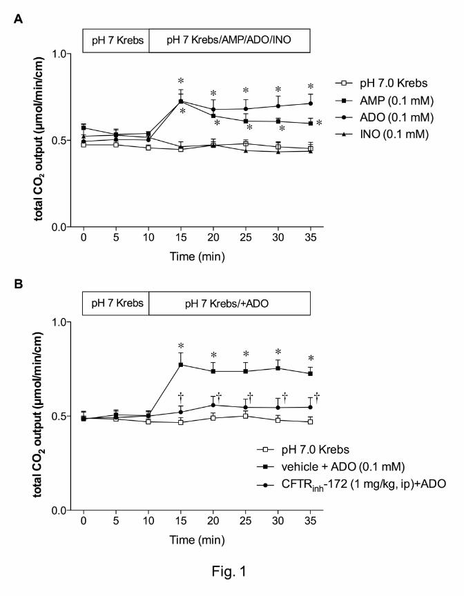
of AMP, ADO or INO (0.1 mM) on DBS. During perfusion of pH 7.0 Krebs buffer, HCO3-
secretion (measured as total CO2 output) was stable over time (Fig. 1). AMP and ADO
uniformly increased HCO3- secretion, whereas INO had no effect (Fig. 1A), suggesting
that ADO is a predominant signaling molecule among the three for HCO3- secretion.
To test the role of CFTR in ADO-induced DBS, rats were pretreated with CFTRinh-172 (1
mg/kg, i.p). CFTR inhibition abolished ADO-induced HCO3- secretion (Fig. 1B),
suggesting that ADO-induced DBS is mediated via CFTR.
Effect of P1 receptor agonists or antagonists on duodenal HCO3- secretion
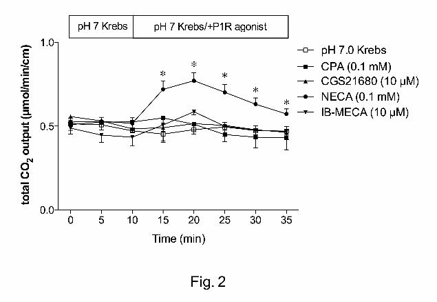
Next, we examined the effect of P1 receptor agonists on DBS. The A1/A2 receptor
agonist NECA (0.1 mM) increased HCO3- secretion, whereas CPA (A1, 0.1 mM),
CGS21680 (A2, 10 µM), or IB-MECA (A3, 10 µM) had no effect (Fig. 2).
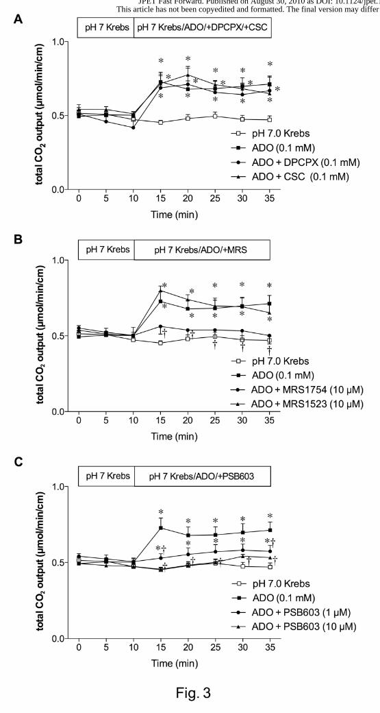
We also examined the effect of P1 receptor antagonists on ADO-induced HCO3-
secretion. The selective A1 receptor antagonist DPCPX (0.1 mM) or selective A2A
receptor antagonist CSC (0.1 mM) failed to affect ADO-induced HCO3- secretion (Fig.
3A). The potent A2B receptor antagonist MRS1754 (10 µM) abolished ADO-induced
HCO3- secretion, whereas the selective A3 receptor antagonist MRS1523 (10 µM) had
no effect (Fig. 3B). To confirm the inhibitory selectivity for A2B receptors, a highly
selective A2B receptor antagonist PSB603 (1 or 10 µM) was perfused with ADO.
This article has not been copyedited and formatted. The final version may differ from this version.JPET Fast Forward. Published on August 30, 2010 as DOI: 10.1124/jpet.110.171520
at ASPE
T Journals on M
ay 6, 2019jpet.aspetjournals.org
Dow
nloaded from
JPET #171520 12
PSB603 dose-dependently inhibited ADO-induced HCO3- secretion (Fig. 3C). These
results suggest that A2B receptor is involved in ADO-induced DBS.
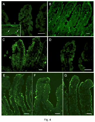
Expression of P1 receptors in the duodenal epithelium
Immunofluorescence for P1 receptors showed that A2B receptor was expressed on the
brush border membrane of duodenal villous cells (Fig. 4C). A2A receptor was
predominantly recognized in the endothelium in the lamina propria of villi (Fig. 4B). A1
or A3 receptor was not observed in the villi (Fig. 4A, D), whereas A1 receptor was
recognized in the myenteric plexus (Fig. 4A inset). This result supports our hypothesis
that luminal ADO stimulates duodenal HCO3- secretion via A2B receptor. To further
demonstrate the presence of the components of luminal ADO-P1 signaling on the
duodenal brush border, immunostaining for CFTR, A2B receptor and ADA was also
examined. The brush border membranes of duodenal villous cells expressed CFTR (Fig.
4E), A2B receptor (Fig. 4F) and ADA (Fig. 4G), further supporting our hypothesis.
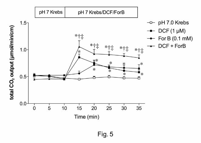
Effect of inhibition of ADA or NT on DBS
The highly potent ADA inhibitor DCF (1 µM) increased HCO3- secretion, whereas a CNT
inhibitor ForB (0.1 mM) gradually increased HCO3- secretion (Fig. 5). Furthermore, co-
perfusion of DCF and ForB additively enhanced HCO3- secretion (Fig. 5). This result
suggests that endogenous ADO stimulates HCO3- secretion and that luminal surface
ADO concentration is regulated by the brush border ADA activity and CNT.
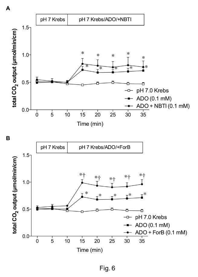
Furthermore, we examined the role of ENT and CNT in ADO-induced HCO3- secretion.
The ENT inhibitor NBTI (0.1 mM) had no effect on ADO-induced HCO3- secretion (Fig.
6A), whereas the CNT inhibitor ForB (0.1 mM) enhanced the effect of ADO (Fig. 6B),
This article has not been copyedited and formatted. The final version may differ from this version.JPET Fast Forward. Published on August 30, 2010 as DOI: 10.1124/jpet.110.171520
at ASPE
T Journals on M
ay 6, 2019jpet.aspetjournals.org
Dow
nloaded from
JPET #171520 13
further suggesting that CNT, not ENT regulates luminal surface ADO-induced HCO3-
secretion in rat duodenum.
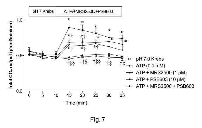
Effect of P2Y1 or A2B receptor antagonist on ATP-induced DBS
We next tested our hypothesis that endogenous ADO is produced by brush border
phosphohydrolases, since ATP is released from the duodenal mucosa in response to
physiological secretory stimuli such as luminal acid perfusion, or ATP release is
unmasked under the inhibition of IAP or ENTPDase (Mizumori, et al., 2009).
Furthermore, exogenous ATP activates P2Y1 receptors on the enterocyte brush border,
augmenting DBS (Mizumori, et al., 2009). We hence studied the sequential effect of
exogenous luminal ATP on DBS. Luminal perfusion of ATP (0.1 mM) increased HCO3-
secretion (Fig. 7) as previously reported (Mizumori, et al., 2009). ATP-induced HCO3-
secretion was partially reduced by the addition of a highly potent P2Y1 receptor
antagonist MRS2500 (1 µM). A highly selective A2B receptor antagonist PSB603 (10
µM) also reduced ATP-induced HCO3- secretion. Co-perfusion of MRS2500 and
PSB603 abolished ATP-induced HCO3- secretion (Fig. 7). These data suggest that
luminal ATP stimulates HCO3- secretion partially via P2Y1 receptor; simultaneously
sequential degradation of ATP by brush border phosphohydrolases supplies luminal
surface ADO in situ to activate the A2B receptors. ATP release thus appears to be the
sole source of extracellular purines, which sequentially activate brush border ATP and
ADO receptors.
This article has not been copyedited and formatted. The final version may differ from this version.JPET Fast Forward. Published on August 30, 2010 as DOI: 10.1124/jpet.110.171520
at ASPE
T Journals on M
ay 6, 2019jpet.aspetjournals.org
Dow
nloaded from
JPET #171520 14
DISCUSSION
Exogenous luminal AMP and ADO, not INO, augmented DBS, consistent with the
activation of brush border P1 receptors. Studies with selective P1 agonists and
antagonists were consistent with brush border A2B receptors mediating ADO-induced
DBS. The presence of A2B receptors, CFTR and ADA at the enterocyte brush border
was confirmed by immunohistochemistry. Additive augmentation of DBS by the ADA
inhibitor DCF and by the CNT inhibitor ForB implicated these proteins in the regulation
of luminal surface ADO abundance whereas the ENT does not appear to affect luminal
ADO concentrations. Finally, DBS augmented by perfusion of exogenous ATP was
abolished by co-perfusion of P1 and P2 receptor antagonists, but partially by perfusion
of either antagonist singly, providing the novel observation that luminal ADO is derived
from ATP in the intestine. This is the first study demonstrating that luminal ADO
stimulates DBS measured in vivo in an intact preparation, that ADO-induced DBS is
mediated in intestine by brush border A2B receptors and by CFTR, and that ADO is
endogenously generated in the duodenal lumen.
Despite its considerable importance to neuromuscular signaling, differentiation, and
inflammatory modulation, few in vivo descriptions of ADO effects on epithelial secretory
function exist. In the 1970s and 1980s, ATP but not ADO was reported to increase
intestinal ion secretion (Gerencser and Armstrong, 1972;Kohn, et al., 1970;Korman, et
al., 1982). The earliest descriptions of the pro-secretory effect of ADO in intestine were
reported in 1984, in which basolateral addition of mM ADO to chambered rabbit ileum or
colon in vitro elicited an increase in electrogenic net Cl- secretion (Dobbins, et al.,
1984;Grasl and Turnheim, 1984). More recently, numerous groups have reported the
This article has not been copyedited and formatted. The final version may differ from this version.JPET Fast Forward. Published on August 30, 2010 as DOI: 10.1124/jpet.110.171520
at ASPE
T Journals on M
ay 6, 2019jpet.aspetjournals.org
Dow
nloaded from
JPET #171520 15
pro-secretory effects of ADO in cultured epithelial cells or mounted intestinal tissues
through activation of A2 or A1 receptors, respectively (Rajagopal and Pao, 2010;Wang,
et al., 2008;Novak, et al., 2008;Ghanem, et al., 2005).
The ready availability of potent and selective adenosine receptor agonists and
antagonists enabled us to pharmacologically characterize ADO-induced DBS. CPA,
CGS21680 and IB-MECA, A1, A2A and A3 selective agonists respectively, did not affect
DBS. Nevertheless, NECA increased DBS, indicating that either receptor A1 or A2 is
involved in ADO-mediated DBS. Given the nonselective nature of NECA, and the lack of
specific A2B agonists, we utilized specific A1, A2A, A2B, and A3 selective antagonists,
DPCPX, CSC, MRS1754 and MRS1523, respectively. Of these antagonists, only
MRS1754, the A2B antagonist inhibited ADO-induced DBS. Immunostaining confirmed
the presence of A2B receptors on the villous brush border, consistent with the
pharmacological observations. In contrast, A2A receptors predominantly localized on the
endothelial cells, consistent with the role of ADO in the regulation of blood flow via A2A
receptors (Belardinelli, et al., 1998;Pennanen, et al., 1994). A1 or A3 receptor-like
immunoreactivity was not observed on the villous brush border, whereas A1 receptors
were located in the myenteric plexus, as reported previously in human jejunum
(Christofi, et al., 2001). No staining for A3 receptor-like immunoreactivity was observed
in the duodenum, although the antibody used recognized the A3 receptor in the
esophageal mucosa (data not shown).
DCF, a potent ADA inhibitor, and ForB, a CNT inhibitor increased DBS. These data
demonstrate the presence of endogenous ADO generation in the lumen. Furthermore,
ForB, not NBTI, enhanced ADO-induced DBS, suggesting that brush border CNT,
This article has not been copyedited and formatted. The final version may differ from this version.JPET Fast Forward. Published on August 30, 2010 as DOI: 10.1124/jpet.110.171520
at ASPE
T Journals on M
ay 6, 2019jpet.aspetjournals.org
Dow
nloaded from
JPET #171520 16
unlike ENT, is related to nucleoside transport from the lumen to the enterocytes as
predicted (Pastor-Anglada, et al., 2007). Present on the apical membrane of epithelial
cells, CNTs may absorb luminal nucleosides into the enterocytes. Of the three known
paralogs belonging to the SLC28A family, CNT1-3, CNT2 is the most likely duodenal
brush border ADO transporter. Since no selective CNT inhibitor is available, data
supporting this hypothesis include the high relative duodenal expression of CNT2 and
the relative affinity of CNT2 for purines compared with CNT1 or CNT3 (Gray, et al.,
2004;Kim, et al., 2007;Lu, et al., 2004;Larráyoz, et al., 2004). Similar to our data, an
‘adenosine scavenging’ mechanism affects basolateral extracellular ADO
concentrations in cultured T84 cell monolayers (Tally, et al., 1996). Pharmacological
inhibition of the proteins implicated in adenosine scavenging increases extracellular
ADO concentration, increasing Isc via the basolateral A2B receptor. Since this report was
published, many of the proteins involved in the adenosine scavenging mechanism have
been identified; the phenomenon described was likely due to ENTs localized on the T84
cell basolateral membrane, which transport in either direction in a concentration-
dependent fashion and may regulate intracellular nucleoside concentrations (Pastor-
Anglada, et al., 2007). Furthermore, our findings are consistent with the previous report
that luminal ADA and CNT, not ENT, regulate luminal ADO concentrations in the human
airway epithelia (Hirsh, et al., 2007).
MRS2500, a P2Y1 antagonist, and PSB603, an A2B antagonist, each decreased DBS
augmented by luminal perfusion of ATP, and co-perfusion of both antagonists
completely suppressed ATP-induced DBS. Therefore, luminal ATP and its metabolite
ADO may simultaneously activate P2Y and P1 receptors, respectively. We have
This article has not been copyedited and formatted. The final version may differ from this version.JPET Fast Forward. Published on August 30, 2010 as DOI: 10.1124/jpet.110.171520
at ASPE
T Journals on M
ay 6, 2019jpet.aspetjournals.org
Dow
nloaded from
JPET #171520 17
previously reported that IAP inhibition increases DBS as a probable consequence of
increasing ATP output into the lumen due to reduced ATP hydrolysis. This ATP release
is partially reduced by CFTR inhibition (Mizumori, et al., 2009), whereas CFTR inhibition
abolished ADO-induced DBS, suggesting that the CFTR-dependent effect of ATP on
DBS may be explained by ADO-A2B signaling. Nevertheless, CFTR inhibition abolished
the secretory effect of ADO, fully consistent with ADO-induced CFTR activation via A2B
Gs receptors that increases intracellular cAMP (Dobbins, et al., 1984).
The source of endogenous ADO is controversial. The non-lytic release of ATP from
many epithelia has been reported in numerous publications (Seminario-Vidal, et al.,
2009;Woo, et al., 2008). The released ATP is enzymatically hydrolyzed by ENTPDase
to AMP, which is then converted by ecto-5’-nucleotidase to ADO (Zimmermann, 2000).
Alternatively, ATP is sequentially dephosphorylated to ADO by alkaline phosphatase
(Yegutkin, 2008). ADO can then be further degraded to INO by ADA. We have
previously reported non-lytic ATP release and brush border IAP and CD39 activity in rat
duodenum (Mizumori, et al., 2009), suggesting that luminal surface ADO is derived from
released ATP, as predicted earlier in T84 cell monolayers (Stutts, et al., 1995). Our data
in the present study support this hypothesis, since luminal ATP-induced DBS was
inhibited by the A2B receptor antagonist. Another possible source of extracellular ADO is
the extracellular cAMP-ADO pathway, consisting of cAMP transporter, ecto-
phosphodiesterase and ecto-5’-nucleotidase (Godecke, 2008). This pathway is
observed in the skeletal muscle (Chiavegatti, et al., 2008) and ileal muscle strip (Giron,
et al., 2008). Perfusion of the rat duodenum with cAMP, however, did not increase the
This article has not been copyedited and formatted. The final version may differ from this version.JPET Fast Forward. Published on August 30, 2010 as DOI: 10.1124/jpet.110.171520
at ASPE
T Journals on M
ay 6, 2019jpet.aspetjournals.org
Dow
nloaded from
JPET #171520 18
rate of DBS, inconsistent with the presence of the cAMP-ADO pathway in rat duodenum
(unpublished observations).
In summary, we have reported for the first time that endogenously produced luminal
surface ADO increases HCO3- secretion in an intact epithelium in vivo through the
activation of A2B receptors. Our data complement the ‘adenosine scavenging’
hypothesis wherein extracellular ADO concentrations are regulated by apical nucleoside
transporters and ADA, which in turn regulate intestinal anion secretion.
ACKNOWLEDGEMENTS
We thank Coleen Palileo for her assistance with manuscript preparation.
This article has not been copyedited and formatted. The final version may differ from this version.JPET Fast Forward. Published on August 30, 2010 as DOI: 10.1124/jpet.110.171520
at ASPE
T Journals on M
ay 6, 2019jpet.aspetjournals.org
Dow
nloaded from
JPET #171520 19
REFERENCES
Akiba Y, Ghayouri S, Takeuchi T, Mizumori M, Guth PH, Engel E, Swenson ER and Kaunitz JD (2006) Carbonic anhydrases and mucosal vanilloid receptors help mediate the hyperemic response to luminal CO2 in rat duodenum. Gastroenterology 131:142-152.
Akiba Y, Jung M, Ouk S and Kaunitz JD (2005) A novel small molecule CFTR inhibitor attenuates HCO3
- secretion and duodenal ulcer formation in rats. Am J Physiol Gastrointest Liver Physiol 289:G753-G759.
Akiba Y, Mizumori M, Guth PH, Engel E and Kaunitz JD (2007) Duodenal brush border intestinal alkaline phosphatase activity affects bicarbonate secretion in rats. Am J Physiol Gastrointest Liver Physiol 293:G1223-G1233.
Antonioli L, Fornai M, Colucci R, Ghisu N, Tuccori M, Del TM and Blandizzi C (2008) Regulation of enteric functions by adenosine: pathophysiological and pharmacological implications. Pharmacol Ther 120:233-253.
Barrett KE, Huott PA, Shah SS, Dharmsathaphorn K and Wasserman SI (1989) Differing effects of apical and basolateral adenosine on colonic epithelial cell line T84. Am J Physiol 256:C197-C203.
Belardinelli L, Shryock JC, Snowdy S, Zhang Y, Monopoli A, Lozza G, Ongini E, Olsson RA and Dennis DM (1998) The A2A adenosine receptor mediates coronary vasodilation. J Pharmacol Exp Ther 284:1066-1073.
Chiavegatti T, Costa VL, Jr., Araujo MS and Godinho RO (2008) Skeletal muscle expresses the extracellular cyclic AMP-adenosine pathway. Br J Pharmacol 153:1331-1340.
Christofi FL, Zhang H, Yu JG, Guzman J, Xue J, Kim M, Wang YZ and Cooke HJ (2001) Differential gene expression of adenosine A1, A2a, A2b, and A3 receptors in the human enteric nervous system. J Comp Neurol 439:46-64.
Dobbins JW, Laurenson JP and Forrest JN, Jr. (1984) Adenosine and adenosine analogues stimulate adenosine cyclic 3', 5'-monophosphate-dependent chloride secretion in the mammalian ileum. J Clin Invest 74:929-935.
Flemström G, Garner A, Nylander O, Hurst BC and Heylings JR (1982) Surface epithelial HCO3
- transport by mammalian duodenum in vivo. Am J Physiol 243:G348-G358.
Flemström G and Kivilaakso E (1983) Demonstration of a pH gradient at the luminal surface of rat duodenum in vivo and its dependence on mucosal alkaline secretion. Gastroenterology 84:787-794.
This article has not been copyedited and formatted. The final version may differ from this version.JPET Fast Forward. Published on August 30, 2010 as DOI: 10.1124/jpet.110.171520
at ASPE
T Journals on M
ay 6, 2019jpet.aspetjournals.org
Dow
nloaded from
JPET #171520 20
Gerencser GA and Armstrong WM (1972) Sodium transfer in bullfrog small intestine. Stimulation by exogenous ATP. Biochim Biophys Acta 255:663-674.
Ghanem E, Lovdahl C, Dare E, Ledent C, Fredholm BB, Boeynaems JM, Van DW and Beauwens R (2005) Luminal adenosine stimulates chloride secretion through A1 receptor in mouse jejunum. Am J Physiol Gastrointest Liver Physiol 288:G972-G977.
Giron MC, Bin A, Brun P, Etteri S, Bolego C, Florio C and Gaion RM (2008) Cyclic AMP in rat ileum: evidence for the presence of an extracellular cyclic AMP-adenosine pathway. Gastroenterology 134:1116-1126.
Godecke A (2008) cAMP: fuel for extracellular adenosine formation? Br J Pharmacol 153:1087-1089.
Grasl M and Turnheim K (1984) Stimulation of electrolyte secretion in rabbit colon by adenosine. J Physiol 346:93-110.
Gray JH, Owen RP and Giacomini KM (2004) The concentrative nucleoside transporter family, SLC28. Pflügers Arch 447:728-734.
Hirokawa M, Takeuchi T, Chu S, Akiba Y, Wu V, Guth PH, Engel E, Montrose MH and Kaunitz JD (2004) Cystic fibrosis gene mutation reduces epithelial cell acidification and injury in acid-perfused mouse duodenum. Gastroenterology 127:1162-1173.
Hirsh AJ, Stonebraker JR, van Heusden CA, Lazarowski ER, Boucher RC and Picher M (2007) Adenosine deaminase 1 and concentrative nucleoside transporters 2 and 3 regulate adenosine on the apical surface of human airway epithelia: implications for inflammatory lung diseases. Biochemistry 46:10373-10383.
Hogan DL, Crombie DL, Isenberg JI, Svendsen P, Schaffalitzky de Muckadell OB and Ainsworth MA (1997) Acid-stimulated duodenal bicarbonate secretion involves a CFTR-mediated transport pathway in mice. Gastroenterology 113:533-541.
Kim HR, Park SW, Cho HJ, Chae KA, Sung JM, Kim JS, Landowski CP, Sun D, Abd El-Aty AM, Amidon GL and Shin HC (2007) Comparative gene expression profiles of intestinal transporters in mice, rats and humans. Pharmacol Res 56:224-236.
Kimura Y, Turner JR, Braasch DA and Buddington RK (2005) Lumenal adenosine and AMP rapidly increase glucose transport by intact small intestine. Am J Physiol Gastrointest Liver Physiol 289:G1007-G1014.
Kohn PG, Newey H and Smyth DH (1970) The effect of adenosine triphosphate on the transmural potential in rat small intestine. J Physiol 208:203-220.
Korman LY, Lemp GF, Jackson MJ and Gardner JD (1982) Mechanism of action of ATP on intestinal epithelial cells: Cyclic AMP-mediated stimulation of active ion transport. Biochim Biophys Acta 721:47-54.
This article has not been copyedited and formatted. The final version may differ from this version.JPET Fast Forward. Published on August 30, 2010 as DOI: 10.1124/jpet.110.171520
at ASPE
T Journals on M
ay 6, 2019jpet.aspetjournals.org
Dow
nloaded from
JPET #171520 21
Larráyoz IM, Casado FJ, Pastor-Anglada M and Lostao MP (2004) Electrophysiological characterization of the human Na+/nucleoside cotransporter 1 (hCNT1) and role of adenosine on hCNT1 function. J Biol Chem 279:8999-9007.
Lu H, Chen C and Klaassen C (2004) Tissue distribution of concentrative and equilibrative nucleoside transporters in male and female rats and mice. Drug Metab Dispos 32:1455-1461.
Mizumori M, Ham M, Guth PH, Engel E, Kaunitz JD and Akiba Y (2009) Intestinal alkaline phosphatase regulates protective surface microclimate pH in rat duodenum. J Physiol 587:3651-3663.
Mizumori M, Meyerowitz J, Takeuchi T, Lim S, Lee P, Supuran CT, Guth PH, Engel E, Kaunitz JD and Akiba Y (2006) Epithelial carbonic anhydrases facilitate PCO2 and pH regulation in rat duodenal mucosa. J Physiol 573:827-842.
Mohamedali KA, Guicherit OM, Kellems RE and Rudolph FB (1993) The highest levels of purine catabolic enzymes in mice are present in the proximal small intestine. J Biol Chem 268:23728-23733.
Novak I, Hede SE and Hansen MR (2008) Adenosine receptors in rat and human pancreatic ducts stimulate chloride transport. Pflügers Arch 456:437-447.
Pastor-Anglada M, Errasti-Murugarren E, Aymerich I and Casado FJ (2007) Concentrative nucleoside transporters (CNTs) in epithelia: from absorption to cell signaling. J Physiol Biochem 63:97-110.
Pennanen MF, Bass BL, Dziki AJ and Harmon JW (1994) Adenosine: differential effect on blood flow to subregions of the upper gastrointestinal tract. J Surg Res 56:461-465.
Rajagopal M and Pao AC (2010) Adenosine activates a2b receptors and enhances chloride secretion in kidney inner medullary collecting duct cells. Hypertension 55:1123-1128.
Ritzel MW, Yao SY, Ng AM, Mackey JR, Cass CE and Young JD (1998) Molecular cloning, functional expression and chromosomal localization of a cDNA encoding a human Na+/nucleoside cotransporter (hCNT2) selective for purine nucleosides and uridine. Mol Membr Biol 15:203-211.
Seidler U, Blumenstein I, Kretz A, Viellard-Baron D, Rossmann H, Colledge WH, Evans M, Ratcliff R and Gregor M (1997) A functional CFTR protein is required for mouse intestinal cAMP-, cGMP- and Ca2+-dependent HCO3
- secretion. J Physiol 505:411-423.
Seminario-Vidal L, Lazarowski ER and Okada SF (2009) Assessment of extracellular ATP concentrations. Methods Mol Biol 574:25-36.
This article has not been copyedited and formatted. The final version may differ from this version.JPET Fast Forward. Published on August 30, 2010 as DOI: 10.1124/jpet.110.171520
at ASPE
T Journals on M
ay 6, 2019jpet.aspetjournals.org
Dow
nloaded from
JPET #171520 22
Strohmeier GR, Reppert SM, Lencer WI and Madara JL (1995) The A2b adenosine receptor mediates cAMP responses to adenosine receptor agonists in human intestinal epithelia. J Biol Chem 270:2387-2394.
Stutts MJ, Lazarowski ER, Paradiso AM and Boucher RC (1995) Activation of CFTR Cl- conductance in polarized T84 cells by luminal extracellular ATP. Am J Physiol 268:C425-C433.
Tally KJ, Hrnjez BJ, Smith JA, Mun EC and Matthews JB (1996) Adenosine scavenging: a novel mechanism of chloride secretory control in intestinal epithelial cells. Surgery 120:248-254.
Wang D, Sun Y, Zhang W and Huang P (2008) Apical adenosine regulates basolateral Ca2+-activated potassium channels in human airway Calu-3 epithelial cells. Am J Physiol Cell Physiol 294:C1443-C1453.
Witte DP, Wiginton DA, Hutton JJ and Aronow BJ (1991) Coordinate developmental regulation of purine catabolic enzyme expression in gastrointestinal and postimplantation reproductive tracts. J Cell Biol 115:179-190.
Woo K, Dutta AK, Patel V, Kresge C and Feranchak AP (2008) Fluid flow induces mechanosensitive ATP release, calcium signalling and Cl- transport in biliary epithelial cells through a PKCζ-dependent pathway. J Physiol 586:2779-2798.
Ye JH and Rajendran VM (2009) Adenosine: an immune modulator of inflammatory bowel diseases. World J Gastroenterol 15:4491-4498.
Yegutkin GG (2008) Nucleotide- and nucleoside-converting ectoenzymes: Important modulators of purinergic signalling cascade. Biochim Biophys Acta 1783:673-694.
Zimmermann H (2000) Extracellular metabolism of ATP and other nucleotides. Naunyn Schmiedebergs Arch Pharmacol 362:299-309.
This article has not been copyedited and formatted. The final version may differ from this version.JPET Fast Forward. Published on August 30, 2010 as DOI: 10.1124/jpet.110.171520
at ASPE
T Journals on M
ay 6, 2019jpet.aspetjournals.org
Dow
nloaded from
JPET #171520 23
FOOTNOTES
This work was supported by the Department of Veterans Affairs Merit Review Award
(J.D.K.); the National Institute of Health-National Institute of Diabetes and Digestive and
Kidney Diseases [R01 DK54221] (J. D. K.); and the animal core of the National Institute
of Health-National Institute of Diabetes and Digestive and Kidney Diseases [P30
DK0413] (J. E. Rozengurt).
This article has not been copyedited and formatted. The final version may differ from this version.JPET Fast Forward. Published on August 30, 2010 as DOI: 10.1124/jpet.110.171520
at ASPE
T Journals on M
ay 6, 2019jpet.aspetjournals.org
Dow
nloaded from
JPET #171520 24
FIGURE LEGENDS
Fig. 1. Effect of adenosine (ADO) on duodenal HCO3- secretion in rats. A: Duodenal
HCO3- secretion was measured as total CO2 output with flow-through pH and CO2
electrodes. Perfusion of adenosine 5’-monophosphate (AMP, 0.1 mM) or ADO (0.1 mM)
similarly increased total CO2 output, whereas inosine (INO, 0.1 mM) had no effect. Each
data presents mean ± SEM (n = 6 rats). *p < 0.05 vs. pH 7.0 Krebs group. B: CFTR was
inhibited by CFTRinh-172 (1 mg/kg, i.p) 1 hr prior to the experiment. CFTR inhibition
abolished the ADO effect. Each data presents mean ± SEM (n = 6 rats). *p < 0.05 vs.
pH 7.0 Krebs group, †p < 0.05 vs. ADO group.
Fig. 2. Effect of P1 receptor agonists on duodenal HCO3- secretion in rats. Perfusion of
N6-cyclopentyladenosine (CPA, 0.1 mM), CGS21680 (10 µM), or N6-(3-
iodobenzyl)adenosine-5′-N-methyluronamide (IB-MECA, 10 µM) had no effect, whereas
5’-(N-ethylcarboxamido)-adenosine (NECA, 0.1 mM) increased HCO3- secretion. Each
data presents mean ± SEM (n = 6 rats). *p < 0.05 vs. pH 7.0 Krebs group.
Fig. 3. Effect of P1 receptor antagonists on adenosine (ADO)-induced augmented
HCO3- secretion in rat duodenum. A: Co-perfusion of 8-cyclopentyl-1,3-dipropylxanthine
(DPCPX, 0.1 mM) or 8-(3-chlorostyryl)caffeine (CSC, 0.1 mM) had no effect on ADO-
induced increase of HCO3- secretion. B: Co-perfusion of MRS1754 (10 µM) abolished
the ADO effect, but MRS1523 (10 µM) had no effect. C: Co-perfusion of PCB603 (1 or
10 µM) inhibited the effect of ADO. Each data presents mean ± SEM (n = 6 rats). *p <
0.05 vs. pH 7.0 Krebs group, †p < 0.05 vs. ADO group.
This article has not been copyedited and formatted. The final version may differ from this version.JPET Fast Forward. Published on August 30, 2010 as DOI: 10.1124/jpet.110.171520
at ASPE
T Journals on M
ay 6, 2019jpet.aspetjournals.org
Dow
nloaded from
JPET #171520 25
Fig. 4. Expression of P1 receptors, CFTR and adenosine deaminase (ADA) in rat
duodenal mucosa. Cryostat sections of fixed rat duodenum were reacted with primary
antibodies for P1 receptors, CFTR and ADA. A2B receptor-like immunoreactivity was
recognized on the brush border membranes of villous cells (C, F). A2A receptor-like
immunoreactivity was mainly observed on the endothelial cells of vasculature (B). No
specific staining was observed for A1 (A) or A3 (D) receptor in the villi. A1 receptor-like
immunoreactivity was recognized in the myenteric plexus (arrows, A, inset). CFTR (E),
A2B receptor (F) and ADA (G) were co-expressed on the enterocyte brush border. A-D:
conventional microscopic images, E-G: confocal laser scanning microscopic images.
Bar = 50 µm.
Fig. 5. Effect of inhibitors of adenosine deaminase (ADA) or concentrative nucleoside
transporter (CNT) on duodenal HCO3- secretion in rats. 2’-deoxycoformycin (DCF, 1 µM),
ForB (0.1 mM) or both was perfused. DCF and ForB augmented HCO3- secretion. *p <
0.05 vs. pH 7.0 Krebs group, †p < 0.05 vs. DCF group, ‡p < 0.05 vs. ForB group.
Fig. 6. Effect of equilibrative (ENT) or concentrative nucleoside transporter (CNT) on
adenosine (ADO)-induced HCO3- secretion in rat duodenum. A: The ENT inhibitor S-(4-
nitrobenzyl)-6-thioinosine (NBTI, 0.1 mM) had no effect on ADO-induced HCO3-
secretion. B: ADO-induced HCO3- secretion was enhanced by the addition of formycin B
(ForB, 0.1 mM). Each data presents mean ± SEM (n = 6 rats). *p < 0.05 vs. pH 7.0
Krebs group, †p < 0.05 vs. ADO group, *p < 0.05 vs. ADO + ForB group. Each data
presents mean ± SEM (n = 6 rats). *p < 0.05 vs. pH 7.0 Krebs group, †p < 0.05 vs. ADO
group.
This article has not been copyedited and formatted. The final version may differ from this version.JPET Fast Forward. Published on August 30, 2010 as DOI: 10.1124/jpet.110.171520
at ASPE
T Journals on M
ay 6, 2019jpet.aspetjournals.org
Dow
nloaded from
JPET #171520 26
Fig. 7. Effect of P2Y1 or A2B receptor antagonist on ATP-induced HCO3- secretion in rat
duodenum. The potent P2Y1 receptor antagonist MRS2500 (1 µM) or selective A2B
receptor antagonist PSB603 (10 µM) was co-perfused with ATP (0.1 mM). ATP-induced
HCO3- secretion was partially via P2Y1 or A2B receptor. Each data presents mean ± SEM
(n = 6 rats). *p < 0.05 vs. pH 7.0 Krebs group, †p < 0.05 vs. ATP group, ‡p < 0.05 vs.
ATP + MRS2500 group, §p < 0.05 vs. ATP + PSB603 group.
This article has not been copyedited and formatted. The final version may differ from this version.JPET Fast Forward. Published on August 30, 2010 as DOI: 10.1124/jpet.110.171520
at ASPE
T Journals on M
ay 6, 2019jpet.aspetjournals.org
Dow
nloaded from
This article has not been copyedited and formatted. The final version may differ from this version.JPET Fast Forward. Published on August 30, 2010 as DOI: 10.1124/jpet.110.171520
at ASPE
T Journals on M
ay 6, 2019jpet.aspetjournals.org
Dow
nloaded from
This article has not been copyedited and formatted. The final version may differ from this version.JPET Fast Forward. Published on August 30, 2010 as DOI: 10.1124/jpet.110.171520
at ASPE
T Journals on M
ay 6, 2019jpet.aspetjournals.org
Dow
nloaded from
This article has not been copyedited and formatted. The final version may differ from this version.JPET Fast Forward. Published on August 30, 2010 as DOI: 10.1124/jpet.110.171520
at ASPE
T Journals on M
ay 6, 2019jpet.aspetjournals.org
Dow
nloaded from
This article has not been copyedited and formatted. The final version may differ from this version.JPET Fast Forward. Published on August 30, 2010 as DOI: 10.1124/jpet.110.171520
at ASPE
T Journals on M
ay 6, 2019jpet.aspetjournals.org
Dow
nloaded from
This article has not been copyedited and formatted. The final version may differ from this version.JPET Fast Forward. Published on August 30, 2010 as DOI: 10.1124/jpet.110.171520
at ASPE
T Journals on M
ay 6, 2019jpet.aspetjournals.org
Dow
nloaded from
This article has not been copyedited and formatted. The final version may differ from this version.JPET Fast Forward. Published on August 30, 2010 as DOI: 10.1124/jpet.110.171520
at ASPE
T Journals on M
ay 6, 2019jpet.aspetjournals.org
Dow
nloaded from
This article has not been copyedited and formatted. The final version may differ from this version.JPET Fast Forward. Published on August 30, 2010 as DOI: 10.1124/jpet.110.171520
at ASPE
T Journals on M
ay 6, 2019jpet.aspetjournals.org
Dow
nloaded from


![JPET #201616jpet.aspetjournals.org/content/jpet/early/2013/01/08/jpet.112.201616.full.pdf[D-Ala2, NMe-Phe4, Gly-ol5]-This article has not been copyedited and formatted. The final version](https://img.pdfslide.tips/doc/110x75/5e3960ce75216306724b28d2/jpet-d-ala2-nme-phe4-gly-ol5-this-article-has-not-been-copyedited-and-formatted.jpg)