Embed Size (px)
Citation preview

ORIGINAL ARTICLE
Annals of Nuclear Medicine Vol. 9, No. 2, 75-80, 1995
Error analysis of table look-up method for cerebral blood flowmeasurement by 123I-IMP brain SPECT:
Comparison with conventional microsphere model method
Hiroshi ITO,* Kiyoshi Isxtt,** Hiroto ATsuMi,** Toshifumi KINOSxrrA,** Ryuta KAWASHIMA,* Shuichi ONo,*Seiro YOSHIOKA,* Hidehiro ImA,*** Kazuo UEMURA*** and Hiroshi FuKUDA*
*Department of Nuclear Medicine and Radiology, Division of Brain Sciences, Institute of Development,Aging and Cancer, Tohoku University
**Department of Radiology, Sendai City Hospital** *Department of Radiology and Nuclear Medicine, Research Institute for Brain and Blood Vessels, Akita
While N-isopropyl-p- [ 123I]iodoamphetamine (IMP) is commonly used as a flow tracer, significantclearance from the brain causes underestimation of CBF as compared with true CBF whenconventional microsphere model analysis is applied. We previously reported a simple "table look-up" method for CBF measurement using IMP taking into account this clearance effect. The methodis based on a two-compartment model, the K, (corresponding to CBF) and k 2 constants beingobtained from a table from the ratio of the 1st SPECT (40 min) to the 2nd SPECT (180 min) counts.Arterial input data used were obtained by one point blood sampling 10 min after IMP infusionagainst the standard input function. In the present study, this approach was compared with conven-tional microsphere model analysis. For 30 subjects, the latter method entailed 8 min continuousarterial blood sampling after IMP infusion and the use of SPECT data at the end of this period,calibrated by a count ratio of 8 min/40 min planar images of whole brains. A good correlation wasobserved between the two methods (r = 0.88), but an overestimation of table look-up method CBFas compared with microsphere model CBF was observed contrary to theoretical predictions. Limi-tations in the estimation of SPECT data at 8 min, obtained with SPECT data at 40 min for calibrationof the count ratio of 8 min/40 min whole brain planar images, might be responsible for this.
Key words: IMP, SPECT, cerebral blood flow, table look-up method
INTRODUCTION
IODINE- 123 ( 123I) labeled N-isopropyl-p-iodoamphetamine(IMP) is used as a cerebral blood flow (CBF) tracer forsingle photon emission computed tomography (SPECT)due to its large extraction fraction and high affinity for thebrain. 1 ' 2 But, significant clearance from the brain causeschange in IMP distribution 3 '4 and underestimation of CBFwhen a conventional microsphere model analysis' isapplied to prolonged data acquisition.' We previouslyreported a "table look-up method," a new simple ap-proach to measurement of CBF with IMP, taking into
Received October 17, 1994, revision accepted November 25,1994.
For reprint contact: Hiroshi Ito, M.D., Department of NuclearMedicine and Radiology, Division of Brain Sciences, Instituteof Development, Aging and Cancer, Tohoku University, 4-1Seiryo-machi, Aoba-ku, Sendai 980, JAPAN.
account this clearance effect.'°' 2 The approach is basedon a two-compartment model (influx: K,, efflux: k 2), inwhich K, (taken to represent CBF) and k 2 are obtainedfrom a table read with the count ratio of first SPECT scan(mid-scan time: 40 min) to second SPECT scan (mid-scantime: 180 min). Arterial input function is obtained bycalibrating against the standard input function from onepoint arterial blood sampling at 10 min after intravenousinfusion of IMP. The purpose of the present study was tocompare this method with the conventional microspheremodel method 13 for a major patient series.
MATERIALS AND METHODS
Subjects
SPECT studies were performed on 30 subjects including19 patients suffering from cerebral contusion, 3 withcerebrovascular disease, 2 with hypoxic brain, 2 with
Vol. 9, No. 2, 1995 Original Article 75
tion for the table look-up method. 10-12 Continuous arterialblood sampling at a constant rate from the brachial arterywas also performed during the first 8 min after IMPinfusion and octanol extracted radioactivity was meas-ured for the conventional microsphere model analysis(Fig. 1).
A cross calibration scan was performed using an ellip-tic cylindrical uniform phantom (long axis: 19 cm, shortaxis: 14 cm inner diameter) for calibrating the relativesensitivities of the SPECT scanner and the well countersystem.
Fig. 1 The experimental protocol design shows the timeschedule of scan and blood sampling. The one point arterialblood sampling from the brachial artery was performed at 10min after IMP infusion for determination of arterial inputfunction for the table look-up method. Continuous arterial bloodsampling from the brachial artery was also performed over the8 min after IMP infusion for the microsphere model analysis.Two SPECT scans were performed at mid-scan time of 40 and180 min after IMP infusion for the table look-up method (40min, 180 min) and the microsphere model analysis (40 min).Brain planar images over 50 sec were obtained at 8 min afterIMP infusion for the microsphere model analysis.
carbon monoxide toxicosis and 4 normal volunteers.None of the patients had any heart or lung disease andinformed consent was obtained from all subjects afterproper explanation of the study being conducted.
SPECT studyTwo SPECT scans were performed, at 40 min and 180min of mid-scan time, after intravenous infusion of 222MBq IMP lasting 1 min. Fifty sec planar brain imageswere obtained 8 min after IMP infusion for the conven-tional.microsphere model analysis (Fig. 1). The SPECTscanner used was a Neurocam (Yokogawa Medical Sys-tems Corp., Tokyo, Japan), 14 equipped with a three-headrotating gamma camera. In-plane resolution was 9 mmfull width at half maximum (FWHM), and axial resolu-tion was 10 mm FWHM. The SPECT scan protocolacquired 64 projections at 50 sec per projection with 120°rotation of the camera. Reconstruction was performed byfiltered backprojection using a Butterworth filter (cutofffrequency 0.45 cycle/cm, power factor 10). Attenuationcorrection was made numerically by assuming the objectshape to be circular or elliptical and the attenuationcoefficient to be uniform. Image slices were setup parallelto the orbitomeatal (OM) line and obtained at 8 mmintervals through the whole brain.
One point arterial blood sampling from the brachialartery was performed at 10 min after IMP infusion.Radioactivity of the whole blood was measured with awell counter and was used for calibration against thestandard input function to provide an arterial input func-
Image analysisRegions-of-interest in the cerebellum, pons, thalamus,putamen, centrum semiovale and cerebral cortex includ-ing frontal, temporal, parietal and occipital lobes wereoutlined on the 40 and 180 min SPECT images. The shapeof regions-of-interest was circular with a 35 mm diameterfor the cerebellum, and elliptic with a short axes of 16-25mm and long axes of 25-50 mm for other region.
TheoryTable look-up method 10 '2 :In this method, a two-compartment model was employedin line with previous reports. 6•
7.15
dCb(t) = Ki- Ca(t) — k2'Cb(t) (1)
dt
where
Cb(t): concentration of radioactivity in the brainCa(t): arterial input functionK,: influx rate constant (ml/ml/min)k2 : efflux rate constant (1/min)
In this study, we assumed the first-pass extraction fractionof IMP to be equal to 1 1,2,16 and therefore, K, equals CBF.The ratio of K, to k2 is called the distribution volume ofIMP in the brain (Yd (ml/ml)).
Solving Eq. 1 provides:
Cb(t) = KrCa(t) © e2 1(2)
where © denotes the convolution integral.For this method, two SPECT scans are performed. The
model equation (Eq. 2) can therefore be expressed foreach scan.
Cb(te) = Ki . Ca(te) ®e ° (3a)
Cb(ta) = KrCa(ta) 0 e° (3b)(3b)
where to and td are mid-scan times at first and second scans,respectively. Calculating the ratio of Eq. 3a to Eq. 3bgives:
Cb(te) _ Ca(te) ©e krk
Cb(td) — Ca(td) ®e kztd
For a given input function, Ca(t), the radioactivity ratio
(4)
76 Hiroshi Ito, Kiyoshi Ishii, Hiroto Atsumi, et al Annals of Nuclear Medicine

of the first to second scans (the right side of Eq. 4) can beconsidered to tabulate as a function of k 2 . For a givenradioactivity ratio of first to second scans, the table look-up procedure then provides a corresponding k 2 value. Byinserting this k2 value into Eq. 3a or 3b, a K, value thatcorresponds to CBF can be calculated. The arterial inputfunction, C a(t) is obtained by calibration against thestandard input function by using the arterial blood radio-activity gained from the one point sampling.
Microsphere model method 13 :CBF values were also calculated by microsphere modelanalysis as follows:
Cb Cb•Rf= _ (5)
f8 min
c Ca(t)dt a min
where
f: CBF (ml/ml/min)Cb : concentration of radioactivity in the brain at 8
min after IMP infusionCa(t): arterial input functionR: constant arterial blood sampling rate (ml/min)Ca : total octanol extracted radioactivity of the blood
withdrawn over 8 min
In this study, Cb was obtained as follows:
Cb(Planar8)Cb =
Cb(SPECT^) (6)
Cb(Planar)
where
Cb(Planar8): the whole brain radioactivity of the pla-nar image at 8 min after IMP infusion
Cb(Planar4o): the whole brain radioactivity of the pla-nar image at 40 min which is one of theprojections of SPECT scans
Cb(SPECT40): the brain radioactivity concentration ofthe SPECT scan with the mid-scan timeof 40 min after IMP infusion
Simulation of CBF correlation between the two compart-ment model and the microsphere modelFor prediction of systematic underestimation of CBF bymicrosphere model analysis, a simulation of the correla-tion between CBF values evaluated by the two-compart-ment model analysis and those from microsphere modelanalysis was performed. CBF values from the microspheremodel were calculated as follows: Firstly, the brain radio-activity curve, C b(t) was generated for a CBF range of 0 to100 ml/100 ml/min according to the two-compartmentmodel equation (Eq. 2) where the V d values were 20, 30,40 or 50. The standard input function used in the tablelook-up method was employed for the arterial input func-tion, Ca(t). Secondly, for each calculated C b(t), the micro-
sphere model CBF values were calculated using C b(t) at 8min and integrated with C a(t) for the time period [0, 8 min](Eq. 5). The resultant microsphere model CBF valueswere compared with those generated by two-compart-ment model analysis.
Simulation of the effects of gray-white matter mixtureThe limited spatial resolution of SPECT scanners causesgray-white matter mixture in regions-of-interest. Theeffects of gray-white matter mixing on CBF values calcu-lated by the table look-up method were evaluated." Theheterogeneous tissue radioactivities at first and secondSPECT scans were generated as mixtures of gray andwhite matter. CBF values of the gray and white matterwere assumed to be 80 and 20 ml/100 ml/min, respec-tively. The Vd value of gray and white matter was as-sumed to be the same as 30, 35, 40, 45 or 50 ml/ml. Thedifference between true CBF values (= 80 ml/100 ml/min x gray matter fraction + 20 ml/100 ml/min x whitematter fraction) and CBF values calculated by table look-up method with the generated heterogeneous tissue radio-activity were estimated where the fraction of gray matterper given region-of-interest varied from 0 to 100%. In thissimulation, the arterial input function was the standardinput function used for the table look-up method.
Simulation of CBF correlation between the two-compart-ment model and the microsphere model method in which8 min SPECT data were obtained from the 40 min SPECTand 8 min/40 min whole brain ratioIn this study, in the microsphere model method, theSPECT data at 8 min were obtained from count ratios of8 min/40 min whole brain planar images and 40 minSPECT data. But the radioactivities of 40 min SPECTscans are non-linear for CBF due to a significant clear-ance of IMP, and this could cause error. For this reason,a simulation of the CBF correlation between the two-compartment model analysis and the microsphere modelmethod with count ratios of 8 min/40 min was alsoperformed. Firstly, the brain radioactivity at 40 min,Cb(40 min) was generated for each CBF according to thetwo-compartment model equation (Eq. 2) with the stand-ard input function as an arterial input function with the Vd
value assumed to be 50 ml/ml. Secondly, for each countratio of 8 min/40 min planar images, i.e., 0.6. 0.7, 0.8, or0.9, CBF values were calculated by the microspheremodel method (Eq. 5 and 6). The resultant microspheremodel method CBF values were compared with thosegenerated by two-compartment model analysis.
RESULTS
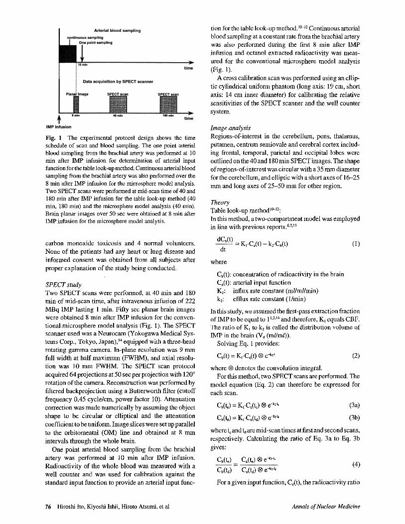
Figure 2 shows the simulation of the CBF correlationbetween the two-compartment model and the micro-sphere model. This indicated systematic underestimationof CBF values evaluated by the microsphere model anal-
Vol. 9, No. 2, 1995 Original Article 77

Ly 100
mU9 80
C_OEE g 60
acG1 — 40
aEo " 20v_
0
0 20 40 60 80 100
2 compartment model CBF(ml/1 00ml/min)
Distribution volumeof IMP (ml/ml)
-- Vd=20- Vd=30
Vd=40Vd=50
20
O 10t!CL
0v
-10
-20
Vd=30.......... Vd=35
_ Vd=40----""" Vd=45
Vd=50
0 20 40 60 80 100
Gray matter fraction (%)
Fig. 2 Simulation of the CBF correlation between two-com-partment model and microsphere model analyses, indicatingsystematic underestimation of CBF values evaluated by themicrosphere model analysis as compared with those from thetwo-compartment model analysis (5.0% underestimation forCBF of 50 ml/100 ml/min and V d of 50 ml/ml).
Fig. 3 Simulation of the effects of gray-white matter mixingon CBF values calculated by table look-up method. When the V d
value of gray and white matter was more than 40 ml/ml, CBFvalues calculated were systematically overestimated (9.8% forgray matter fraction of 50% and V4 of 50 ml/ml).
u 100mU
80CofB £ 60do_ = 40
wE2 20U_
0
8min/40min = 0.6--- 8min/40min = 0.7
8min/40min = 0.8_...._ ._ 8min/40min = 0.9
LL 80
mU3 C 60
OE
00 40
o _ 20
2
Y=0.92X+7.34r=0.88
0 20 40 60 80 100
2 compartment model CBF(m1/100mUrnin)
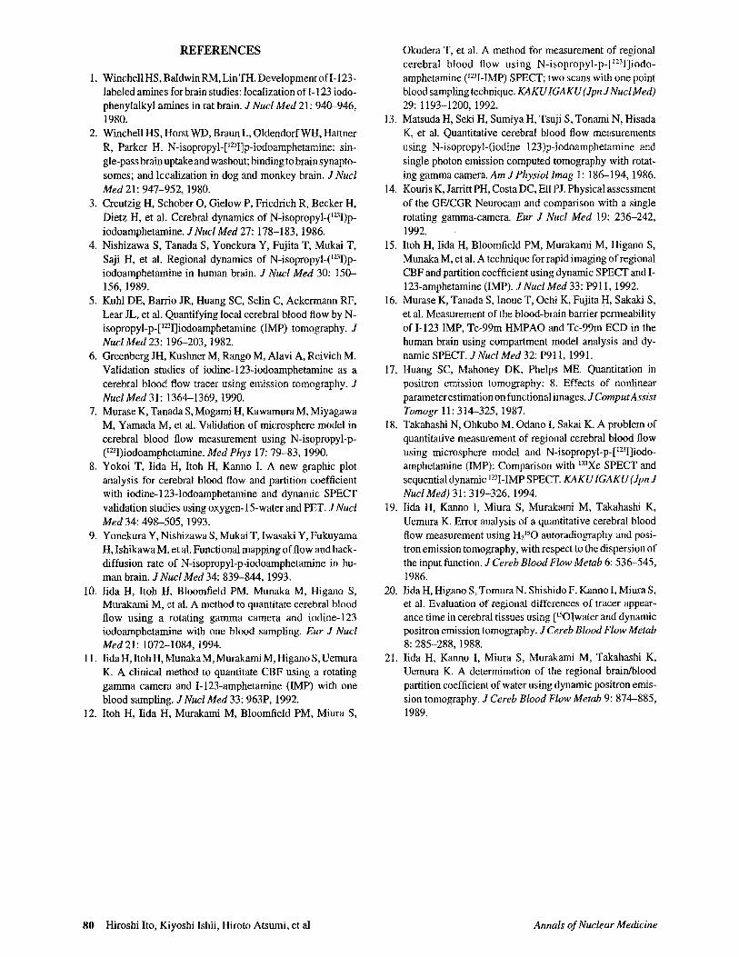
Fig. 4 Simulation of the CBF correlation between the two-compartment model analysis and microsphere model methodusing count ratios of 8 min/40 min planar images, i.e., 0.6, 0.7,0.8, or 0.9. Overestimation of CBF values evaluated by themicrosphere model method as compared with those from thetwo-compartment model analysis was observed in CBF rangesfor the two-compartment model of 0.0-30.0, 0.0-45.0, 0.0-70.0 and 0.0-95.0 ml/100 ml/min with 8 min/40 min planarimage count ratios of 0.6, 0.7, 0.8 and 0.9, respectively.
0 20 40 60 80
Table look-up method CBF(mi100mi/min)
Fig. 5 Correlation between CBF values evaluated by the tablelook-up method and those from the microsphere model method.Underestimation of CBF values evaluated by the table look-upmethod as compared with those from the microsphere modelmethod was observed (table look-up method: mean CBF ± S.D.= 37.4 ± 8.09 ml/100 ml/min, microspheremodel method: meanCBF ± S.D. = 41.8 ± 8.46 ml/l00 ml/min).
ysis as compared with those from the two-compartmentmodel analysis. The magnitude of CBF underestimationwas thus expected to be 5.0% for two-compartmentmodel CBF of 50 ml/l00 ml/min and V 4 of 50 ml/ml.
Figure 3 shows the effects of gray-white matter mixingfor CBF values calculated by the table look-up method.When the V d value for gray and white matter was 30 ml/ml, calculated CBF values were systematically underes-timated when the fraction of gray matter varied from 0 to100%. But, when the Vd value for gray and white matterwas more than 40 ml/ml, calculated CBF values weresystematically overestimated (9.8% for gray matter frac-tion of 50% and V4 of 50 ml/ml).
Figure 4 shows the simulation of the CBF correlationbetween the two-compartment model and the microspheremodel method in which 8 min SPECT data were obtainedfrom the 40 min SPECT and 8 min/40 min whole brain
count ratio. In this simulation, comparative overestima-tion of CBF values by the microsphere model method wasobserved in CBF ranges for the two-compartment modelof 0.0-30.0,0.0-45.0,0.0-70.0 and 0.0-95.0 ml/100 ml/min with 8 min/40 min planar image count ratio of 0.6,0.7, 0.8 and 0.9, respectively. In this study, the actualmean count ratio of the 8 min/40 min planar image was0.762 ± 0.072 (± S.D.).
A good correlation was obtained between CBF valuesevaluated by the table look-up method and those from themicrosphere model method (Y = 0.92X + 7.34, X: tablelook-up method, r = 0.88) (Fig. 5), but overestimation ofCBF values was observed with the microsphere modelmethod as compared with those from the table look-upmethod. Mean CBF values evaluated by the table look-upmethod were 10.6% lower than those from the microspheremodel method (table look-up method: mean CBF ± S.D.
78 Hiroshi Ito, Kiyoshi Ishii, Hiroto Atsumi, et al Annals of Nuclear Medicine

= 37.4 ± 8.09 ml/100 ml/min, microsphere model method:mean CBF ± S.D. = 41.8 ± 8.46 ml/100 ml/min).
Vd values were not uniform in the brain, especially lowVd values being observed in lesions, i.e., cerebralinfarctions and contusions. The mean V d value in X-rayCTnormal density regions was 48.7 ± 9.15 ml/ml (± S.D.).There was no significant difference between gray andwhite matter in Vd values.
DISCUSSION
The microsphere model analysis has been routinely usedas a method for measuring CBF using IMP, but under-estimation of CBF is caused by significant clearance ofIMP from the brain, especially when data acquisition isprolonged." In this study, the simulation study similarlyindicates systematic underestimation of CBF values withevaluation by microsphere model analysis as comparedwith those from the two-compartment model analysiseven when data acquisition is limited to the early phasei.e., within 8 min after IMP infusion (5.0% underestima-tion for a two-compartment model CBF of 50 ml/100ml/min and Vd of 50 ml/ml) (Fig. 2).
The simulation of the effects of gray-white mattermixture also indicated differences between true CBFvalues (= 80 ml/100 ml/min x gray matter fraction + 20ml/ 100 ml/min x white matter fraction) and table look-upmethod CBF values. In this study, the mean Vd value was48.7 ± 9.15 ml/ml (± S.D.) for normal regions on X-rayCT. When the Vd value of gray and white matter was morethan 40 ml/ml, the table look-up method CBF was system-atically overestimated (9.8% for a gray matter fraction of50% and a Vd of 50 ml/ml) (Fig. 3). On the other hand,there were no effects of gray-white matter mixing on CBFvalues calculated by the microsphere model analysis,because the correlation between the brain radioactivityand CBF value is linear in the microsphere model (Eq.5) . 17
A good correlation was obtained between CBF valuesevaluated by the table look-up method and those from theconventional microsphere model method (Fig. 5), sug-gesting equivalent applicability, but while the two simu-lation studies (Figs. 2 and 3) indicated that CBF valuesobtained from the table look-up method would be higherthan those from microsphere model analysis, in fact theopposite was the case. The microsphere model methodvalues were thus actually 10.6% higher than the tablelook-up method CBF values (Fig. 5). As reasons for this,a number of factors must be considered.
One possibility is error in estimating the SPECT braincounts at 8 min in the microsphere model method withcount ratios 8 min/40 min whole brain planar images and40 min SPECT data." This error would be caused bynon-linearity of 40 min SPECT radioactivities due to asignificant clearance of IMP. The simulation study (Fig.4) revealed comparative overestimation of CBF values
by the microsphere model method in CBF ranges for thetwo-compartment model of 0.0-30.0,0.0-45.0,0.0-70.0and 0.0-95.0 ml/100 ml/min with 8 min/40 min planarimage count ratios of 0.6, 0.7, 0.8 and 0.9, respectively.In this study, the actual mean count ratio of the 8 min/40min planar image was 0.762 ± 0.072 (± S.D.), and there-fore this could have been responsible for the overestima-tion of CBF values determined by the microsphere modelmethod. In addition, other unknown errors due to radio-activities from extracerebral arteries included in 8 and 40min planar images could have played roles.
Another potential source of error is in the determina-tion of the arterial input function. For accurate CBFmeasurement, accurate determination of this functionincluding corrections for time delay and dispersion ofinput is required. It has been shown in the H2 150 PETstudies that no correction for these is associated withgreater overestimation of CBF when the scan duration isshorter. 19-2 ' The standard input function used in tablelook-up method does not feature these corrections, be-cause errors from time delay and dispersion would not besignificant due to the sufficient delay until the mid-scantime of the two SPECT scans, i.e., 40 and 180 min, 10-12 butthese errors in the microsphere model analysis case willbe more significant, because the scan time was very earlyat 8 min. This could have directly caused the overestima-tion of CBF values determined by the microsphere modelanalysis. With the table look-up method, on the otherhand, there might have been unknown errors due todifference in the arterial input curve shape for eachsubject.
In conclusion, a relatively good correlation was ob-tained between CBF values gained by table look-upmethod and those from the conventional microspheremodel method. Since the table look-up method is simple,and does not require a continuous arterial blood sampl-ing, it can be recommended for routine application.Possible reasons for the contrast to expectations fromtheoretical considerations, higher CBF values from themicrosphere model method than the table look-up methodare:
1. Errors in estimation of SPECT data at 8 min bycalibration of SPECT scan at 40 min with count ratios of8 min/40 min whole brain planar images in the micro-sphere model method.
2. Errors in determination of arterial input functionwith both methods.
ACKNOWLEDGMENTS
We are greatly indebted to the staff of Sendai City Hospital andthe Institute of Development, Aging and Cancer, Tohoku Uni-versity, particularly Messrs. Yoshimasa Inukai, Shigeto Abe,Masami Sato for operating the SPECT scanner.
This study was supported by aGrant-in-Aid No. 05454297 forScientific Research from the Japanese Ministry of Education,Science and Culture.
Vol. 9, No. 2, 1995 Original Article 79
REFERENCES
1. Winchell HS, BaIdwinRM, LinTH. Development of 1-123-labeled amines for brain studies: localization of I-123 iodo-phenylalkyl amines in rat brain. J Nucl Med 21: 940-946,1980.
2. Winchell HS, Horst WD, Braun L, Oldendorf WH, HattnerR, Parker H. N-isopropyl-[ d231]p-iodoamphetamine: sin-gle-pass brain uptake and washout; binding to brain synapto-somes; and localization in dog and monkey brain. J NuclMed 21: 947-952, 1980.
3. Creutzig H, Schober 0, Gielow P, Friedrich R, Becker H,Dietz H, et al. Cerebral dynamics of N-isopropyl-( mI)p-iodoamphetamine. J Nucl Med 27: 178-183, 1986.
4. Nishizawa S. Tanada S, Yonekura Y, Fujita T, Mukai T,Saji H, et al. Regional dynamics of N-isopropyl-( 123I)p-iodoamphetamine in human brain. J Nucl Med 30: 150-156, 1989.
5. Kuhl DE, Barrio JR, Huang SC, Selin C, Ackermann RF,Lear JL, et al. Quantifying local cerebral blood flow by N-isopropyl-p-["'I]iodoamphetamine (IMP) tomography. JNucl Med 23: 196-203, 1982.
6. Greenberg JH, Kushner M, Rango M, Alavi A, Reivich M.Validation studies of iodine-123-iodoamphetamine as acerebral blood flow tracer using emission tomography. JNucl Med 31: 1364-1369, 1990.
7. Murase K, Tanada S. Mogami H, Kawamura M, MiyagawaM, Yamada M, et al. Validation of microsphere model incerebral blood flow measurement using N-isopropyl-p-(' 23I)iodoamphetamine. Med Phys 17: 79-83, 1990.
8. Yokoi T, Iida H, Itoh H, Kanno I. A new graphic plotanalysis for cerebral blood flow and partition coefficientwith iodine-123-Iodoamphetamine and dynamic SPECTvalidation studies using oxygen-15-water and PET. JNuclMed 34: 498-505, 1993.
9. Yonekura Y, Nishizawa S. Mukai T, Iwasaki Y, FukuyamaH, Ishikawa M, et al. Functional mapping of flow and back-diffusion rate of N-isopropyl-p-iodoamphetamine in hu-man brain. J Nucl Med 34: 839-844, 1993.
10. Iida H, Itoh II, Bloomfield PM, Munaka M, Higano S,Murakami M, et al. A method to quantitate cerebral bloodflow using a rotating gamma camera and iodine-123iodoamphetamine with one blood sampling. Eur J NuclMed 21: 1072--1084, 1994.
11. lids H, Itoh H, Munaka M, Murakami M, Higano S, UemuraK. A clinical method to quantitate CBF using a rotatinggamma camera and I-123-amphetamine (IMP) with oneblood sampling. JNucl Med 33: 963P, 1992.
12. Itch H, lids H, Murakami M, Bloomfield PM, Miura S,
Okudera T, et al. A method for measurement of regionalcerebral blood flow using N-isopropyl-p-[ 123I]iodo-amphetamine ( 1 Z11-IMP) SPECT; two scans with one pointblood sampling technique. KAKUIGAKU (Jpn JNucl Med)29: 1193-1200, 1992.
13. Matsuda H, Seki H, Sumiya H, Tsuji S. Tonami N, HisadaK, et al. Quantitative cerebral blood flow measurementsusing N-isopropyl-(iodine 123)p-iodoamphetamine andsingle photon emission computed tomography with rotat-ing gamma camera. Am J Physiol Imag 1: 186-194, 1986.
14. Kouris K, Jarritt PH, Costa DC, Ell PJ. Physical assessmentof the GE/CGR Neurocam and comparison with a singlerotating gamma-camera. Eur J Nucl Med 19: 236-242,1992.
15. Itoh H, Iida H, Bloomfield PM, Murakami M, Higano S,Munaka M, et al. A technique for rapid imaging of regionalCBF and partition coefficient using dynamic SPECT and I-123-amphetamine (IMP). JNucl Med 33: P911, 1992.
16. Murase K, Tanada S, Inoue T, Ochi K, Fujita H, Sakaki S.et al. Measurement of the blood-brain barrier permeabilityof I-123 IMP, Tc-99m HMPAO and Tc-99m ECD in thehuman brain using compartment model analysis and dy-namic SPECT. J Nucl Med 32: P911, 1991.
17. Huang SC, Mahoney DK, Phelps ME. Quantitation inpositron emission tomography: 8. Effects of nonlinearparameter estimation on functional images. J ComputAssistTomogr 11: 314-325, 1987.
18. Takahashi N, Ohkubo M, Odano I, Sakai K. A problem ofquantitative measurement of regional cerebral blood flowusing microsphere model and N-isopropyl-p-[ 1231]iodo-amphetamine (IMP): Comparison with 13Xe SPECT andsequential dynamic ° 31-IMP SPELT. KAKUIGAKU(Jpn JNucl Med) 31: 319-326, 1994.
19. Iida H, Kanno I, Miura S, Murakami M, Takahashi K,Uemura K. Error analysis of a quantitative cerebral bloodflow measurement using H 2 150 autoradiography and posi-tron emission tomography, with respect to the dispersion ofthe input function. J Cereb Blood Flow Metab 6: 536-545,1986.
20. Iida H, Higano S, Tomura N, Shishido F, Kanno I, Miura S,et al. Evaluation of regional differences of tracer appear-ance time in cerebral tissues using [ 150]water and dynamicpositron emission tomography. J Cereb Blood Flow Metab8: 285-288, 1988.
21. lida H, Kanno I, Miura S. Murakami M, Takahashi K,Uemura K. A determination of the regional brain/bloodpartition coefficient of water using dynamic positron emis-sion tomography. J Cereb Blood Flow Metab 9: 874-885,1989.
80 Hiroshi Ito, Kiyoshi Ishii, Hiroto Atsumi, et al Annals of Nuclear Medicine