Embed Size (px)
DESCRIPTION
Â
Citation preview

i

Marmara Üniversitesi Yayınları / No. 832
Istanbul Endoscopic Ultrasound and Advanced Endoscopy Days ISTEUS 2015 Syllabus
Editör: Deniz Güney Duman
© Marmara Üniversitesi Yayınevi, 2015
e-ISBN: 978-975-400-393-2
Aralık, 2015
Marmara Üniversitesi Yayınevi
Göztepe Kampüsü, Kadıköy 34722 İstanbul
Tel: +90 216 348 43 79 Fax: +90 216 348 43 79
E-Posta: [email protected]
This meeting has been supported by Marmara University Scientific Research Projects Committee
(BAPKO)
Istanbul Endoscopic Ultrasound and Advanced Endoscopy Days syllabus (IstEUS), December,
10-11, 2015/editör Deniz Güney Duman.__ İstanbul : Marmara Üniversitesi, 2015.
84 s ; 24 sm.__( Marmara Üniversitesi yayınları; No. 832)
978-975-400-393-2
1. Endoscopic ultrasonography 2. Endoskopik ultrasonografi 3. Gastrointestinal system -- Diseases -- Diagnosis 4. Gastrointestinal Sistem – Tanı 5. Endoscopy -- Congresses 6. Endoskopi – Kongreler
RC 804 .E59
616.075 43

iii
Contents
Preface .......................................................................................................................... iv
Scientefic Programme .................................................................................................... v
Organising committee .................................................................................................. vii
Scientefic Committee ................................................................................................... viii
Summary of speakers’ presentations ..............................................................................1
Oral Abstracts ............................................................................................................. 49
Poster Abstracts .......................................................................................................... 56
Author Index ............................................................................................................... 74

iv
Dear colleagues
Following the success of the EGEUS 2013 congress accomplished by our group with partnership of EGEUS organization, we would like to move forward our experience higher with IstEUS meeting. We aimed to improve your EUS practice and help you to meet the experts in this field.
The objective of “Istanbul Endoscopic Ultrasound and Advanced Endoscopy Days (IstEUS 2015)” is to bring together EUS experts around the world in order to generate synergy in EUS developments, new techniques, EUS guided advanced endoscopic interventions, research, to encourage young endoscopists and investigators from diverse backgrounds to study and practice EUS.
Our specific aims have been:1. Teach the fundamentals of EUS to beginners who are interested in EUS.2. Give insight in the fields of indications, contraindications, advantages and
limitations of EUS3. EUS guided advanced endoscopic interventions such as difficult to canulate ERCP,
tumor treatment, pain management in chronic pancreatitis and pancreatic cancer, confocal endomicroscopy, elastography.
4 Provide support, networking and potential collaboration to young investigators. 5. Admission of oral and poster presentations as well as videos for video marathon
session. We gathered the summaries of the speakers’ lectures, oral and poster presentations
together in this e-book aiming to provide a reference for your future studies. We would like to thank the Marmara University Library & Documentation
Department and the Publishing house for giving us support in the preparation of this e-book and enabling its publication.
On behalf of the organizing committeeDeniz Guney DUMAN, MD, Prof.
Congress President
iv
vv

vi
viivii
organization Commitee
DENIz GUNEY DUMAN (PRESIDENt)YESIM ALAHDAB (SECREtARY)CEM KALAYCIADNAN GIRALOSMAN OzDOGANOMER GUNAL

viiiviii
AHMEt AYDINAHMEt BEKtAŞAHMEt DANALIOĞLUAHMEt UYANIKOĞLUALİ tÜzÜN İNCEALİYE SOYLUBAHAttİN ÇİÇEKBİLLUR CANABAKANBİROL BAYSALCAN GÖNENCEM AYGÜNCEMİL SAVAŞCENGİz PAtAÇEtİN KARACADİLEK OĞUzEMRAH ALPERENDER DULUNDUENGİN ALtINtAŞFAtİH ASLANFİLİz AKYÜzHACI MEHMEt SÖKMENHAKAN ŞENtÜRKHÜSEYİN AtASEVENİRFAN KORUKKADİR BALKAMİL ÖzDİL
MARC GIOVANNINIMEHMEt ARHANMEHMEt BEKtAŞMEHMEt CİNDORUKMEHMEt İBİŞMELtEM ERGUNMÜJDE SOYtÜRKNEVİN ORUÇORHAN tARÇINORHAN. K POYRAzOĞLUOSMAN ERSOYÖMER ŞENtÜRKÖMER YILMAzSAADEttİN HÜLAGÜSABİtE KAÇARSAİt BAĞCISEDEF KURANSELİM AYDERMİRtARKAN KARAKANtAYLAN KAVYASEMİN H. BALABANYILDIRAN SONGÜRYOSUKE NAKAIYUSUF zİYA ERzİNYÜCEL ÜStÜNDAĞYÜKSEL GÜMÜRDÜLÜ
Scientefic Commitee
ix
SUMMARY OFSPEAKERS’
PRESENtAtIONS

1
Istanbul Endoscopic Ultrasound and Advanced Endoscopy Days, December 10-11 2015,ss.1-3 • e-ISBN: 978-975-400-393-2
What to do with the incidental asymptomatic small gastric subepithelial lesions. Facts? Recommendations?
Deniz Guney DUMAN, MD., Prof.Internal medicine & Gastroenterology, Marmara University, School of Medicine
Since upper endoscopy is widely available, clinicians more often encounter the bulges arising beneath the epithelium, with variable clinical significance ranging from insignificant to malignant lesions. Endoscopic biopsies can unlikely determine the diagnosis because these lesions lie deep in the GI wall. For the subepithelial lesions (SELs) larger than 10 mm, evaluation with EUS is recommended to ascertain the size, layer of origin, features of echogenicity, and high-risk features for malignancy suspicion (1).
Evaluation of the small gastric subepithelial lesionsIn most cases, EUS findings only allow a presumptive diagnosis and determine the
need for further explorations like tissue sampling, surgery, or follow-up. Muscularis propria (MP) (fourth layer) hypoechoic lesions are mostly GISTs if they are localized in the stomach, but EUS alone is not sufficient to differentiate GIST from other causes of hypoechoic MP masses like schwannoma, leiomyoma, lymphoma, etc. therefore, a tissue diagnosis with EUS guided-fine needle aspiration (EUS-FNA) is generally performed.
At present, although EUS-FNA is considered the procedure of choice for preoperative diagnosis of GIST by immunohistochemical analysis of the sample for c-KIT (2), it provides inadequate material in up to 33.3% of the cases. Particularly, smaller tumors are technically more difficult to obtain adequate histological samples compared to larger ones (3). So, the diagnosis of small GISTs may be only presumptive based on the EUS appearance but also performing EUS-FNA and management decision may be challenging. Nevertheless, GIST appeared to be the most common diagnosis among the MP-SELs in many studies evaluating the post-operative results. In one study, post-operative histological examination revealed GIST in 16 of 19 (84.2%) resected gastric MP-SELs ≤3 cm in size (the others were 2
SUMMARY OF SPEAKERS’ PRESENTATIONS

Deniz Güney DUMAN
2
schwannomas and 1 leiomyoma) (4). Even in the case of a GIST diagnosis accomplished by EUS-FNA, evaluation of the malignant potential of the tumor based on the mitotic index may not be possible due to the lack of as sufficient material as required for prompt investigation. Thus EUS-FNA may not change the management strategy at least in a significant subset of patients with asymptomatic small MP-SELs.
The National Institutes of Health (NIH) consensus (5) classified GISTs into very low, low-, intermediate-, and high-risk categories by using the size and mitotic count of the lesions. Furthermore, Miettinen and Lasota(6) indicated that GISTs ⩽20 mm with a mitotic index of ⩽5/50 HPF have no metastasis risk, thus they defined these lesions as benign, although NIH has avoided such a category. Nevertheless, obtaining sufficient material from a small SEL in an attempt to scrutinize its malignant potential by assessing the mitotic count may be technically difficult.
How to follow up the small muscularis propria subepithelial lesionsThe frequency of follow-up duration of small SELs suggestive of GIST is controversial
in different guidelines. The American Gastroenterological Association (AGA) recommends surveillance for <30 mm lesions without concerning the endosonographic features. (7) The US National Comprehensive Cancer Network (NCCN) avoids making a clear statement for the small gastric GISTs (<20 mm) because of the insufficient data, but recommends resection of the small lesions with high-risk EUS features, and endoscopic surveillance at 6- to 12-month intervals for the lesions without high-risk features. (8) European Society for Medical Oncology (ESMO) guidelines suggest a short-term first control (e.g. at 3 months) continuing with a longer interval follow-up schedule in case of no growth, if follow-up strategy is chosen for small lesions. (9) Japanese guidelines recommend that lesions <20 mm in size and without ulceration or surface depression can be managed with endoscopic follow-up once or twice a year. (10) There are no large scale investigations providing evidence for the effectiveness of these follow-up schedules. The study published by our group has shown that small hypoechoic SELs of <2 cm originating from MP in stomach suggestive of GIST lesions with EUS survive disease free for a mean duration of 4 years although most patients in our series had much longer surveillance. (11). Therefore we support the notion of conservative management of small hypoechoic SELs originating from MP of gastric wall. Our results suggest that following up of those small asymptomatic MP-SELs detected incidentally during upper gastrointestinal endoscopy without any high risk echogenic features for malignancy not less than 4 years is sufficient. We believe, the guidelines regarding the follow-up recommendations of small hypoechoic MP SELs of stomach suggestive of GISTs should be revised as they currently recommend EUS follow-ups every 3 to 12 months after the initial examination. (12)

Istanbul Endoscopic Ultrasound and Advanced Endoscopy Days • December 10-11 2015, ss. 1-3
3
In conclusion, drawing definite conclusions for small gastric GISTs may be limited by the insufficient sample size of studies, mostly due to the fact that GISTs are rare tumors. Available data support the notion of a conservative management strategy, rather than a surgical approach, for small gastric MP-SELs, but in the light of the uncertainties, final decision for these small lesions must be individualized after options are thoroughly discussed with the patient. More accurate non-invasive characterization with the newer imaging techniques that allow a more targeted puncture, improvements in design of needles providing larger and safer tissue acquisition, and identification of the molecular and genetic aspects of malignant transformation profiles at the early stage for the small gastric MP-SELs may provide superior risk estimation and refinement of the management strategies in the future.
References1. Hwang JH, Kimmey MB. The Incidental Upper Gastrointestinal Subepithelial Mass.
Gastroenterology. 2004;126(1):301-7.2. Akahoshi K, Oya M. Gastrointestinal stromal tumor of the stomach: How to manage? World
J Gastrointest Endosc. 2010;2(8):271-7.3. Akahoshi K, Sumida Y, Matsui N, Oya M, Akinaga R, Kubokawa M, et al. Preoperative
diagnosis of gastrointestinal stromal tumor by endoscopic ultrasound-guided fine needle aspiration. World J Gastroenterol 2007; 13(14): 2077-2082.
4. Kim MY, Jung HY, Choi KD, Song HJ, Lee JH, Kim do H, et al. Natural history of asymptomatic small gastric subepithelial tumors. J Clin Gastroenterol. 2011;45(4):330-6.
5. Fletcher CD, Berman JJ, Corless C, Gorstein F, Lasota J, Longley BJ, Miettinen M, O’Leary TJ, Remotti H, Rubin BP, Shmookler B, Sobin LH, Weiss SW. Diagnosis of gastrointestinal stromal tumors: A consensus approach. Hum Pathol 2002; 33:459-465.
6. Miettinen M, Lasota J. Gastrointestinal stromal tumors: pathology and prognosis at different sites. Semin Diagn Pathol 2006;23:70– 83.
7. Hwang JH, Rulyak SD, Kimmey MB; American Gastroenterological Association Institute. American Gastroenterological Association Institute technical review on the management of gastric subepithelial masses. Gastroenterology. 2006;130(7):2217-28.
8. George D. Demetri, Margaret von Mehren, Cristina R. Antonescu, Ronald P. DeMatteo, Kristen N. Ganjoo, Robert G. Maki, et al. NCCN Task Force Report: Update on the Management of Patients with Gastrointestinal Stromal Tumors. JNCCN 2010; 8[Suppl 2]:S1–S40.].
9. The ESMO / European Sarcoma Network Working Group. Gastrointestinal stromal tumors: ESMO Clinical Practice Guidelines for diagnosis, treatment and follow-up. Annals of Oncology 2012; 23 (Supplement 7): vii49–vii55, 2012.
10. Nishida T, Hirota S, Yanagisawa A, Sugino Y, Minami M, Yamamura Y, Otani Y, Shimada Y, Takahashi F, Kubota T. Clinical practice guidelines for gastrointestinal stromal tumor (GIST) in Japan: English version. Int J Clin Oncol 2008; 13: 416-430.
11. Yegin EG, Kani T, Banzragch M, Kalayci C, Bicakci E, Duman DG. Survival in patients with hypoechoic muscularis propria lesions suggestive of gastrointestinal stromal tumors in gastric wall. Acta Gastroenterol Belg. 2015;78(1):12-7.
12. Yegin EG, Duman D. Small EUS-suspected gastrointestinal stromal tumors of the stomach: an overview for the current state of management. EUS Journal 2015(article in press)

4
Istanbul Endoscopic Ultrasound and Advanced Endoscopy Days, December 10-11 2015,ss. 4-8 • e-ISBN: 978-975-400-393-2
Pankreasın Nöroendokrin Tümörlerinde Tanı
Y. Doç. Dr. Birol BaYsal
Bezmialem Üniversitesi Gastroenteroloji Bilim Dalı, İstanbulTel: 0505 775 85 85, [email protected]
Pankreasın nöroendokrin tümörleri (PNET) pankrasın adacık hücrelerinden kaynaklanır ve nöroendokrin tümörlerin (NET) bir alt grubudur. Tüm pankreas tümörleri içinde %1.3 oranında saptanır. PNET’ler hormon üretimine, biyolojik etkilerine ve semptomlarına bağlı olarak fonksiyonel veya fonksiyonel olmayanlar şeklinde 2 gruba ayrılır. PNET’lerinin yaklaşık %10-30’u fonksiyoneldir. İnsülinomalar, gastrinomalar, VIPomalar, somatostatinomalar, glukagonomalar ve büyüme hormonu salgılayan tümörler (GRFomalar) sık olarak saptanan fonksiyonel pankreatik nöroendokrin tümörlerdir (F-PNET) (Tablo-1). Daha nadir olarak saptanan F-PNET’ler ise: ACTH sekresyonu yapan ve Cushing sendromuna neden olan ACTHomalar, karsinoid sendroma neden olan PNET’ler, hiperkalsemiye neden olan PNET’ler ve çok seyrek olmakla birlikte luteinizan hormon, renin ya da eritropoetin salgılayan PNET’lerdir. Fonksiyonel olmayan pankreatik nöroendokrin tümörler (NF-PNET) tüm PNET’lerin %30-50’si oluşturur. Karakteristik olarak, NF-PNET’ler büyük boyutludur ve %60-85’inde tanı konduğu sırada karaciğer metastazı vardır. NF-PNET’ler %60-90 oranlarındaki malign seyirleri nedeni ile F-PNET’ler den farklıdırlar ve genellikle NF-PNET’lerde tümör boyutu malign seyirle ilişkilidir. Fonksiyonel PNET’ler ve NF-PNET’ler sıklıkla başka kimyasal maddeler de salgılayabilirler (kromagranin, nöron spesifik enolaz, nörotensin, ghrelin). PNET’ler sporadik saptanabileceği gibi, çeşitli kalıtsal hastalıklar veya sendromlara eşlik edebilirler (Tablo 2). Multipl endokrinoplazi tip 1’li (MEN1) hastaların %80-100’ünde; von Hippel-Lindau sendromlu hastaların %10-17’sinde; von Recklinghausen hastalarının yaklaşık %10’unda (nörofibromatoz 1) ve nadirende tübero sklerozisli hastalarda PNET’ler saptanır. Otozomal dominat MEN1 ve PNET birlikteliği en sık rastlanan tablodur. MEN1’li hastaların çoğunda (%80-100) NF-PNET saptanır ve çoğu küçük, multifokal ve mikroskobik olup, hastaların sadece %0-13’ünde semptomlara yol açmaktadır. MEN1’li hastaların %54’ünde gastrinomalar
sUMMaRY OF sPEaKERs’ PREsENTaTIONs

Istanbul Endoscopic Ultrasound and Advanced Endoscopy Days • December 10-11 2015, ss. XX-XX
5
(>%80 duodenal), %18’inde insülinomalar ve daha nadir olarak (<%5) glukagonomalar, VIPomalar, GRFomalar ve somatostatinomalar gelişir.
Tablo 1. Fonksiyonel Pankreatik Nöroendokrin Tümörler ve ÖzellikleriTümör tipi ve sendrom
Pankreas lokalizasyonu Bulgu ve semptomlar Dolaşımdaki biyomarkırlar
Insulinoma (Whipple triadı)
Baş, gövde, kuyruk (eşit dağılım gösterir)
Hipoglisemi, baş dönmesi, terleme, taşikardi, titreme, konfüzyon, nöbet
CgA ve CgB, kan glukoz düzeyi için uygunsuz insülin, proinsülin, C-peptid
Gastrinoma (Zollinger–Ellison)
Gastrinoma üçgeni. Sıklıkla ekstrapankreatiktir (duodenal); pankreas üzerinde herhangi bir yerde olabilir.
Aşırı gastrik asit sekresyonu, peptik ülser, diyare, özofajit, epigastrik ağrı
CgA, gastrin, PP (%35)
VIPoma (Verner– Morrison sendromu, WDHA)
Distal pankreas (gövde ve kuyruk) Sıklıkla pankreas dışına yayılır
Sulu diyare, hipokalemi, aklorhidri (veya asidoz)
CgA, VIP
Glukagonoma Gövde ve kuyruk. Genellikle büyük boyutludur ve pankreas dışına yayılım gösterir
Diyabet (hiperglisemi), nekrolitik migratuar eritem, stomatit, glosit,
CgA, glukagon, glisentin
Somatostatinoma Pankreatoduodenal alan, ampuller, periampuller
Safra taşı, diyabet (hiperglisemi), steatore
CgA, somatostatin
CgA, kromogranin A; CgB, kromogranin B, PP, pankreatik polipeptid; VIP, vazoaktif intestinal peptid; WDHA, sulu diyare, hipokalemi ve aklorhidri.
PNET’li hastaların tanısında klasik görüntüleme yöntemleri (BT, MR, ultrason, anjiyografi) yanı sıra endoskopik ultrason (EUS), intraoperatif ultrason ve son zamanlarda pozitron emisyon tomografisi sık kullanılmaya başlanılmıştır. EUS pankreasın görüntülemesinde diğer görüntüleme yöntemlerine göre özellikle 2 cm den küçük lezyonların saptanmasında oldukça üstündür. 2-3 mm boyutundaki lezyonları dahil görüntülemek mümkün olabilmektedir ve PNET tesbitinde yüksek sensiviteye sahiptir. Ancak bu tekniğin başarısı endoskopiste bağlıdır ve özellikle pankreasın kuyruk kısmındaki lezyonları görüntülemede yetersiz kalabilir. EUS ve EUS-ince iğne aspirasyon biyopsisi özellikle NF-PNET’in adenokarsinomlardan veya diğer pankreatik kitleye yol açan sebeplerden ayırt edilmesinde çok önemlidir. Genellikle klinik, hormon ve biyomarkerların tayinleri F-PNET varlığını düşündürmesine karşın

Birol BAYSAL
6
tümörün lokalizasyonun belirlenmesi özellikle tedavinin planlanmasında önemli bir yer tutar. Ancak F-PNET’ler (özellikle insülinomalar, duodenal gastrinomalar) genellikle küçük boyutlu olduğundan görüntülenmeleri güç olabilmektedir. EUS, duodenal gastrinomalar veya somatostatinomalar gibi ekstrapankreatik PNET’lere kıyasla pankreatik PNET’lerin yerinin belirlenmesinde çok daha etkilidir. Özellikle küçük, hemen hemen her zaman intrapankreatik ve sıklıkla klasik görüntüleme çalışmalarında saptanmayan insülinomaların tesbit edilmesinde yararlıdır. EUS ile olguların yaklaşık %90’ında intrapankreatik PNET tespit edilebilinir. EUS ayrıca MEN1 ve von Hippel-Lindau hastalarında saptanan, genellikle küçük boyutlu ve tedavileri konusunda kesin fikir birliği saptanmamış NF-PNET’lerin tanı ve takibinde oldukça önemli role sahiptir.
Tablo 2. Pankreatik nöroendokrin tümörlerle ilişkilendirilen kalıtsal bozukluklar
sendromlar İlişkilendirilen klinik özellikler Pankreatik nöroendokrin tümör tipi
MEN1 Primer hiperparatiroidizmHipofiz tümörleriDaha az yaygın:• Adrenokortikal tümörler• Karsinoid tümörler• Non-medüller tiorid tümörleri
NF-PNETGastrinomaİnsülinomaÇeşitli
Von Hippel-Lindau hastalığı (VHL)
Feokromositoma (genellikle çift yönlü)Retinal ve serebellar hemanjiyoblastomalarRenal hücreli karsinomlar
NF-PNETKistik tümörler dahil çeşitli
Nörofibromatozis 1 (von Recklinghausen hastalığı)
NörofibromalarCafe au lait lekeleriFeokromastoma
Tübero sklerozis Kardiyak rabdomyomalarRenal kistlerAngiomyolipomalar

Istanbul Endoscopic Ultrasound and Advanced Endoscopy Days • December 10-11 2015, ss. XX-XX
7
Tablo 3. Sindirim sisteminin nöroendokrin tümörlerinde ENETS/WHO isimlendirmeleri ve sınıflandırması Farklılaşma Grade Mitotik
sayı*Ki-67 indeksi
Tümör ENETS, WHO
İyi differansiye Düşük grade (G1)
10 HPF başına <2
<%3 Karsinoid, adacık hücreli, pankreatik (nöro)endokrin tümör
Nöroendokrin tümör, grade 1
Orta grade (G2)
10 HPF başına 2-20 arasında
%3-20 Karsinoid, atipik karsinoid, adacık hücreli, pankreatik (nöro)endokrin tümör
Nöroendokrin tümör, grade 2
Kötü differansiye
Yüksek grade (G3)
10 HPF başına >20
>%20 Küçük hücreli karsinom Nöroendokrin karsinom, grade 3, küçük hücreli
Büyük hücreli nöroendokrin karsinom
Nöroendokrin karsinom, grade 3, büyük hücreli
ENETS: Avrupa Nöroendokrin Tümör Topluluğu; WHO: Dünya Sağlık Örgütü*10 büyük büyütme sahasında sayılmış (HPF). 10 HPF = 2 mm2, en yüksek mitotik yoğunluğa sahip alanlarda en az 40 saha (400x büyütme ile) değerlendirilmiş.
Referanslar1. Vinik A, Casellini C, Perry RR, Feliberti E, MD HV. Diagnosis and Management of Pancreatic
Neuroendocrine Tumors (PNETS). 2014; 14. In: De Groot LJ, Beck-Peccoz P, Chrousos G, Dungan K, Grossman A, Hershman JM, Koch C, McLachlan R, New M, Rebar R, Singer F, Vinik A, Weickert MO, editors. Endotext [Internet]. South Dartmouth (MA): MDText.com
2. Metz DC, Jensen RT: Gastrointestinal neuroendocrine tumors: pancreatic endocrine tumors. Gastroenterology 2008; 135:1469-92.
3. Kloppel G, Anlauf M: Epidemiology, tumour biology and histopathological classification of neuroendocrine tumours of the gastrointestinal tract. Best Pract Res Clin Gastroenterol 2005; 19: 507-17.
4. Oberg K, Eriksson B: Endocrine tumours of the pancreas. Best Pract Res Clin Gastroenterol 2005; 19: 753-81.
5. Liakakos T, Roukos DH: Everolimus and sunitinib: from mouse models to treatment of pancreatic neuroendocrine tumors. Future Oncol. 2011; 7: 1025-29.
6. Falconi M, Plockinger U, Kwekkeboom DJ, Manfredi R, Korner M, Kvols L, Pape UF, Ricke J, Goretzki PE, Wildi S, Steinmuller T, Oberg K, Scoazec JY: Well-differentiated pancreatic nonfunctioning tumors/carcinoma. Neuroendocrinology 2006; 84: 196-211.
7. Dralle H, Krohn SL, Karges W, Boehm BO, Brauckhoff M, Gimm O: Surgery of resectable nonfunctioning neuroendocrine pancreatic tumors. World J Surg. 2004; 28: 1248-60.
8. Fendrich V, Waldmann J, Bartsch DK, Langer P: Surgical management of pancreatic endocrine tumors. Nat Rev Clin Oncol. 2009; 6: 419-28.
9. Bettini R, Partelli S, Boninsegna L, Capelli P, Crippa S, Pederzoli P, Scarpa A, Falconi M:

Birol BAYSAL
8
Tumor size correlates with malignancy in nonfunctioning pancreatic endocrine tumor. Surgery 2011; 150: 75-82.
10. Jensen RT, Berna MJ, Bingham DB, Norton JA: Inherited pancreatic endocrine tumor syndromes: advances in molecular pathogenesis, diagnosis, management, and controversies. Cancer 2008; 113:1807-43.
11. Gibril F, Schumann M, Pace A, Jensen RT: Multiple endocrine neoplasia type 1 and Zollinger-Ellison syndrome: a prospective study of 107 cases and comparison with 1009 cases from the literature. Medicine (Baltimore) 2004; 83: 43-83.
12. Rockall AG, Reznek RH: Imaging of neuroendocrine tumours (CT/MR/US). Best Pract Res Clin Endocrinol Metab. 2007; 21: 43-68.
13. McLean AM, Fairclough PD: Endoscopic ultrasound in the localisation of pancreatic islet cell tumours. Best Pract Res Clin Endocrinol Metab. 2005; 19: 177-93.
14. Shin LK, Brant-Zawadzki G, Kamaya A, Jeffrey RB: Intraoperative ultrasound of the pancreas. Ultrasound Q 2009; 25: 39-48.
15. Eriksson B, Orlefors H, Oberg K, Sundin A, Bergstrom M, Langstrom B: Developments in PET for the detection of endocrine tumours. Best Pract Res Clin Endocrinol Metab. 2005; 19:311-24.
16. Noone TC, Hosey J, Firat Z, Semelka RC: Imaging and localization of islet-cell tumours of the pancreas on CT and MRI. Best Pract Res Clin Endocrinol Metab. 2005; 19: 195-211.
17. Libutti SK, Choyke PL, Bartlett DL, Vargas H, Walther M, Lubensky I, Glenn G, Linehan WM, Alexander HR: Pancreatic neuroendocrine tumors associated with von Hippel Lindau disease: diagnostic and management recommendations. Surgery 1998; 124:1153-59.
18. Langer P, Kann PH, Fendrich V, Richter G, Diehl S, Rothmund M, Bartsch DK: Prospective evaluation of imaging procedures for the detection of pancreaticoduodenal endocrine tumors in patients with multiple endocrine neoplasia type 1. World J Surg. 2004; 28:1317-22.

9
Istanbul Endoscopic Ultrasound and Advanced Endoscopy Days, December 10-11 2015,ss. 9-13 • e-ISBN: 978-975-400-393-2
Tesadüfi Pankreatik Kistler ve Ipmn’e Yaklaşım
Prof. Dr. Çetin KARACAİstanbul Üniversitesi, İstanbul Tıp Fakültesi, Gastroenteroloji Bilim Dalı
Pankreasın kistik lezyonları görüntüleme yöntemlerinin yaygın olarak kullanılması ile birlikte oldukça sık saptanmaktadır (1). Pankreas kistleri sıklıkla başka bir nedenle yapılan görüntülemeler sırasında tesadüfen saptanırlar. Abdominal görüntülemelerde %2-3 olan insidental pankreas kisti prevalansı yaşla birlikte artmakta ve 8. dekatta %8’e kadar çıkmaktadır (2). Otopsi serilerinde %25’e varan oranlar bildirilmiştir (3). Son yıllarda kesitsel görüntüleme yöntemleri ve EUS kullanımının artması ile birlikte pankreas kistleri insidansında 10 kat, kistik tümörler ile ilişkili pankreatik rezeksiyon sayısında iki kat artış olmuştur (4,5).
Pankreas kistleri neoplastik veya non-neoplastik olabilir. Psödokist, retansiyon kisti, lenfoepitelyal kist, duplikasyon kistleri ve infeksiyöz kistler non-neoplastik kistlerdir. Neoplastik kistler; müsinöz ve non-müsinöz olarak kabaca iki gruba ayrılmaktadır. Seröz kistik neoplazi (SKN) ve solip psödopapiller tümör non-müsinöz neoplastik kistleri oluşturmaktadır. Müsinöz kistler ise premalign lezyonlardır ve müsinöz kistik neoplazi (MKN) ve intraduktal papiller müsinözneoplazi (IPMN)’yi içermektedir (Tablo1). Doğru sınıflandırma çok önemlidir. Çünkü non-neoplastik kistler semptomatik olmadığı sürece takip edilirken, pankreatik kistik neoplazilerin malignite potansiyeli nedeniyle rezeke edilmeleri gerekebilir.
SUMMARY OF SPEAKERS’ PRESENTATIONS
Çetin KARACA
10
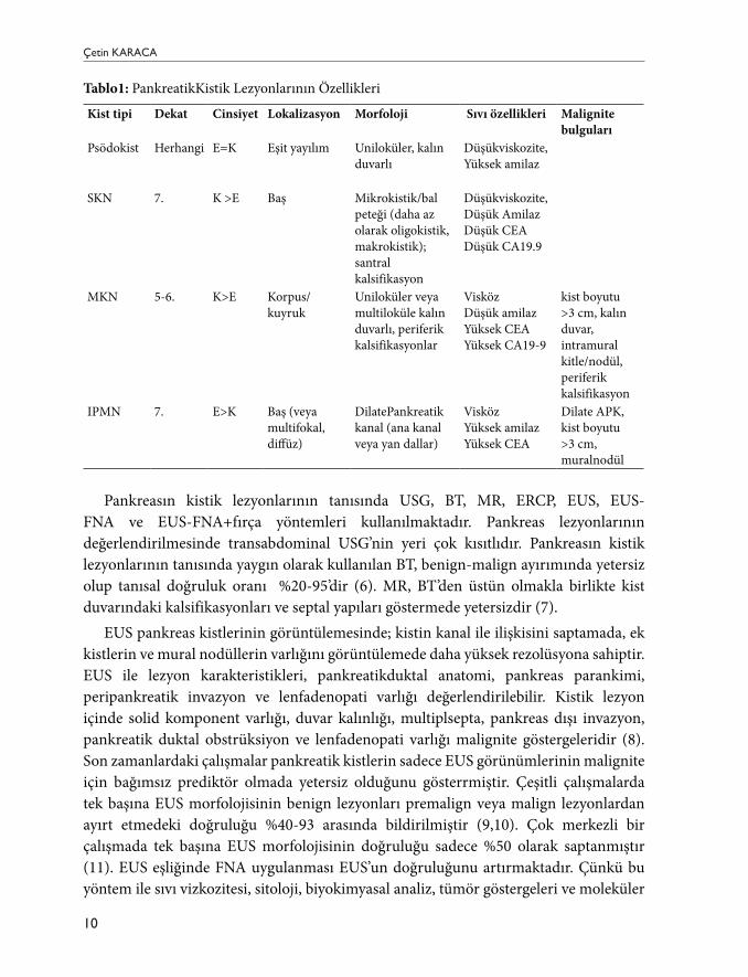
Tablo1: PankreatikKistik Lezyonlarının Özellikleri
Kist tipi Dekat Cinsiyet Lokalizasyon Morfoloji Sıvı özellikleri Malignite bulguları
Psödokist Herhangi E=K Eşit yayılım Uniloküler, kalın duvarlı
Düşükviskozite,Yüksek amilaz
SKN 7. K >E Baş Mikrokistik/bal peteği (daha az olarak oligokistik, makrokistik); santral kalsifikasyon
Düşükviskozite, Düşük AmilazDüşük CEADüşük CA19.9
MKN 5-6. K>E Korpus/kuyruk
Uniloküler veya multiloküle kalın duvarlı, periferik kalsifikasyonlar
VisközDüşük amilazYüksek CEAYüksek CA19-9
kist boyutu >3 cm, kalın duvar, intramural kitle/nodül, periferik kalsifikasyon
IPMN 7. E>K Baş (veya multifokal, diffüz)
DilatePankreatik kanal (ana kanal veya yan dallar)
VisközYüksek amilazYüksek CEA
Dilate APK, kist boyutu >3 cm, muralnodül
Pankreasın kistik lezyonlarının tanısında USG, BT, MR, ERCP, EUS, EUS-FNA ve EUS-FNA+fırça yöntemleri kullanılmaktadır. Pankreas lezyonlarının değerlendirilmesinde transabdominal USG’nin yeri çok kısıtlıdır. Pankreasın kistik lezyonlarının tanısında yaygın olarak kullanılan BT, benign-malign ayırımında yetersiz olup tanısal doğruluk oranı %20-95’dir (6). MR, BT’den üstün olmakla birlikte kist duvarındaki kalsifikasyonları ve septal yapıları göstermede yetersizdir (7).
EUS pankreas kistlerinin görüntülemesinde; kistin kanal ile ilişkisini saptamada, ek kistlerin ve mural nodüllerin varlığını görüntülemede daha yüksek rezolüsyona sahiptir. EUS ile lezyon karakteristikleri, pankreatikduktal anatomi, pankreas parankimi, peripankreatik invazyon ve lenfadenopati varlığı değerlendirilebilir. Kistik lezyon içinde solid komponent varlığı, duvar kalınlığı, multiplsepta, pankreas dışı invazyon, pankreatik duktal obstrüksiyon ve lenfadenopati varlığı malignite göstergeleridir (8).Son zamanlardaki çalışmalar pankreatik kistlerin sadece EUS görünümlerinin malignite için bağımsız prediktör olmada yetersiz olduğunu gösterrmiştir. Çeşitli çalışmalarda tek başına EUS morfolojisinin benign lezyonları premalign veya malign lezyonlardan ayırt etmedeki doğruluğu %40-93 arasında bildirilmiştir (9,10). Çok merkezli bir çalışmada tek başına EUS morfolojisinin doğruluğu sadece %50 olarak saptanmıştır (11). EUS eşliğinde FNA uygulanması EUS’un doğruluğunu artırmaktadır. Çünkü bu yöntem ile sıvı vizkozitesi, sitoloji, biyokimyasal analiz, tümör göstergeleri ve moleküler

Istanbul Endoscopic Ultrasound and Advanced Endoscopy Days • December 10-11 2015, ss. XX-XX
11
inceleme yapılabilir. Bu nedenle Amerikan Gastrointestinal Endoskopi Derneği pankreas kistlerinin tanısında EUS-FNA’yi önerir (12). EUS-FNA pankreas kistik lezyonlarında yüksek özgüllüğe sahiptir, ancak duyarlılığı sadece %20-50’dir. Düşük duyarlılıkaspiratta yeterli hücre olmaması, yetersiz örnek volümü, gastrointestinal duvar hücrelerinin kontaminasyonu gibi nedenlerden kaynaklanabilir (13).Duyarlılığı artırmak için hedeften biyopsi alınması, fırça sitoloji, farklı preperasyon teknikleri, sitopatoloji uzmanlarının dahil edilmesi gibi yaklaşımlar uygulansa da halen duyarlılık düşük olarak seyretmektedir. Kist aspirasyonu nispeten kolay bir işlemdir. Çoğu laboratuvarda CEA analizi için 1-2 ml sıvı yeterlidir. Sıvının vizkozitesine göre 19 veya 22 G iğne kullanılabilir. Tek bir giriş ile kist tamamen boşaltılmaya çalışılır. Alınan sıvının makroskopik görünümüne ve vizkozistesine dikkat edilmelidir. İşlem öncesi IV antibiyotik verilmesi ve işlem sonrası 3-5 gün oral antibiyotiğe devam edilmesi önerilir. CEA, amilaz ve/veya lipaz ve sitoloji için örnek alınır.
Sitoloji: Pankreatik kistlerin tanısında en doğru yöntemlerden biri sitolojidir. Ancak yeterli hücre elde edilememesi nedeniyle tanı sıklıkla zordur ve genel duyarlılığı %50’den düşüktür (14). Çok merkezli bir çalışmada MKN tanısı için sitolojinin duyarlılığı %35, özgüllüğü %83 ve EUS morfolojisi ile benzer olarak doğruluğu %59 olarak saptanmıştır (11).
Tümör Göstergeleri: Pankreatik kist sıvısı, epitelden sekrete edilen CEA, CA 19-9, CA 15-3, CA 72-4 gibi glikoproteinler içerebilir. Bunlardan pratikte en fazla kullanılanı ve faydalı olanı CEA’dır. Yüksek CEA, müsinöz kistleri non-müsinöz olanlardan ayırır ancak benign-malign ayırımı yapamaz. Müsinöz-nonmüsinöz ayırımında CEA için gerçek kestirim değeri bilinmiyor. Brugge ve arkadaşlarının yaptığı prospektif bir çalışmada 192 ng/ml’nin %79 doğrulukla ayırım yaptığı gösterilmiş ve duyarlılığı % 73, özgüllüğü %84 saptanmıştır. Başka bir çalışmada 5 ng/ml kestirim değerinin non-müsinöz kistler için %95 özgüllükte, 800 ng/ml kestirim değerinin müsinöz kistler için %98 özgüllükte olduğu gösterilmiştir (11). Pankres kistlerinin sınıflandırılmasında yol gösterici olabilecek diğer bir gösterge olan CA-19.9 ise CEA kadar değerli değildir. 37 U/ml’den düşük değerlerin benign lezyonları düşündürebileceğini öne süren çalışmalar bulunmaktadır (14).Üzerinde çalışılan bir diğer gösterge olan CA-72.4 için Hammel ve arkadaşları, >40 U/ml değerin %63 duyarlılık ve %98 özgüllük ile müsinöz kistleri ve kistadenokarsinomları psödokistlerden ayırabileceğini saptamışlardır (15). Brugge ve arkadaşları ise CA72.4’ün müsinöz kistleri non-müsinöz olanlardan ayırdetmedeki değerini 7 U/ml kestirim değeri için %80 duyarlılık, % 61 özgüllük, % 72 doğrulukla saptamışlardır (11).
Amilaz: Yapılan çalışmalarda amilazın pankreas kistlerinin ayırıcı tanısındaki yerine dair bilgiler çelişkilidir. Özelllikle pankreatit öyküsü varlığında psödokist tanısını konfirme etmede faydalıdır. Ek olarak SKN’de genellikle amilaz seviyesi düşüktür. IPMN’de sıklıkla yüksek olup psödokistten ayırımını tek başına yapması mümkün değildir (16).

Çetin KARACA
12
Moleküler Analiz: Preoperatif tanı; radyoloji, kist sıvısı analizi ve/veya sitolojiyi içine alan multidisipliner bir yaklaşımı gerektirmektedir. Buna rağmen bir kısım hastada benign-malign ayırımı yapılamamaktadır. Bu nedenle, mevcut multidisipliner yaklaşıma moleküler analiz sağlayan testlerin eklenmesi kistlerin büyük bir kısmını oluşturan malign, benign müsinöz ve benign non-müsinöz kistler için duyarlılık ve özgüllüğü arttırmaktadır. Bu amaçla kist sıvısından mutasyon analizi ve DNA ölçümleri yapılabilmektedir. K-ras mutasyonu müsinöz kistlerin tanısında yüksek özgüllüğe sahiptir(%93-100), ancak duyarlılık düşüktür (%11-57) (17). Yüksek DNA miktarı müsinöz kistlerin bir göstergesidir. Allelik imbalans müsinöz kistlerin tanısı için orta derecede duyarlılık (%43-70) ve geniş aralıklı bir özgüllüğe (%66-100) sahiptir (18,19).
EUS-FNA+Fırçalama: EUS-FNA’inverimini artırmak için fırça sitoloji uygulanabilir. İşlemde kist iğne ile geçilerek karşı duvara ilerlenir. Sendino ve arkadaşlarının 30 pankreas kistine fırça sitoloji uyguladıkları çalışmada hastaların %27(8)’sinde işlem teknik olarak başarısız olmuş. İşlemin başarılı olduğu hastaların %91’inde (20/22) bir tanı sağlanabilmiş ve fırça sitoloji sıvı aspirasyonundan üstün bulunmuştur (sırasıyla %73,%36; p=0.08)(20).
Sonuç olarak; görüntüleme yöntemlerinin yaygın kullanılması ile birlikte pankreas kistleri ile sık karşılaşılmaktadır. Pankreas kistlerinin karekterizasyonunda EUS eşliğnde alınan sıvının sitolojik ve biyokimyasal incelemesi önemlidir. Klinisyenin öncelikle pankreas kistinin neoplastik veya nonneoplastik ayırımını daha sonra da neoplastik kistin premalign veya malign olup olmadığını ortaya koymalıdır.
Kaynaklar1. Brugge WR, Lauwers GY, Sahani D, et al. Cysticneoplasms of thepancreas. N Engl J Med
2004; 351: 1218-26.2. Laffan TA, Horton KM, Klein AP, et al. Prevalence of unsuspectedpancreaticcysts on MDCT.
AJR Am J Roentgenol. 2008;191:802Y807.3. Kimura W, Nagai H, Kuroda A, et al. Analysis of smallcysticlesions of thepancreas. Int J
Pancreatol. 1995;18:197Y206.4. Carpizo DR, Allen PJ, Brennan MF: Currentmanagement of cysticneoplasms of thepancreas.
Surgeon 2008; 6: 298–307.5. Tseng JF, Castillo CF, Warshaw AL: Cysticneoplasms of thepancreas; in Blumgart LH, Fong
Y (eds): Surgery of theLiverandBilaryTract, 3rd ed. 858–866, WB Saunders, 2000.6. Curry CA, Eng J, Horton KM, et al. CT of primarycysticpancreaticneoplasms: can CT be
usedforpatienttriageandtreatment? AJR Am J Roentgenol 2000; 175(1):99–103.7. Mathieu D, Guigui B, Valette PJ, et al. Pancreaticcysticneoplasms. RadiolClin North Am
1989; 27: 163-768. Ahmad NA, Kochman ML, Lewis JD, Ginsberg GG: Can EUS
alonedifferentiatebetweenmalignantandbenigncysticlesions of thepancreas? Am J Gastroenterol 2001; 96: 3295–3300.
9. Ahmad NA, Kochman ML, Brensinger C, Brugge WR, Faigel DO, Gress FG, et al. Interobserveragreementamongendosonographersforthediagnosis of neoplasticversusnon-neoplasticpancreaticcysticlesions. GastrointestEndosc 2003; 58:59-64.

Istanbul Endoscopic Ultrasound and Advanced Endoscopy Days • December 10-11 2015, ss. XX-XX
13
10. Sedlack R, Affi A, Vazquez-Sequeiros E, Norton ID, Clain JE, Wiersema MJ. Utility of EUS in theevaluation of cysticpancreaticlesions. GastrointestEndosc 2002; 56:543-7.
11. Brugge WR, Lewandrowski K, Lee-Lewandrowski E, et al. Diagnosis of pancreaticcysticneoplasms: a report of thecooperativepancreaticcyststudy. Gastroenterology 2004; 126:1330–1336
12. Jacobson BC, Baron TH, Adler DG, et al. ASGE guideline: the role of endoscopy in thediagnosisandthemanagement of cysticlesionsandinflammatoryfluidcollections of thepancreas. GastrointestEndosc 2005;61(3):363–70.
13. Genevay M, Mino-Kenudson M, Yaeger K, et al. Cytologyaddsvaluetoimagingstudiesfor risk assessment of malignancy in pancreaticmucinouscysts. AnnSurg 2011;254:977–983.
14. van der Waaij LA, vanDullemen HM, Porte RJ. Cystfluidanalysis in thedifferentialdiagnosis of pancreaticcysticlesions: a pooledanalysis. GastrointestEndosc 2005;62(3):383–9.
15. Hammel P, Voitot H, Vilgrain V, et al. Diagnosticvalue of CA 72-4 andcarcinoembryonicantigendetermination in thefluid of pancreaticcysticlesions. Eur J GastroenterolHepatol 1998;10(4):345–8.
16. Pelaez-Luna M, Chari ST. Cystfluidanalysistodiagnosepancreaticcysticlesions: an as yet unfulfilledpromise. Gastroenterology. 2006; 130:1007–1009.
17. Garud SS1, Willingham FF.Molecularanalysis of cystfluidaspiration in thediagnosisand risk assessment of cysticlesions of thepancreas.ClinTranslSci. 2012 Feb;5(1):102-7
18. ShenJ , Brugge WR , Dimaio CJ , Pitman MB . Molecularanalysis of pancreaticcystfluid: a comparativeanalysiswithcurrentpractice of diagnosis .CancerCytopathol .Jun 25 2009 ; 117 ( 3 ): 217–227.
19. KhalidA, Zahid M, Finkelstein SD, LeBlanc JK, Kaushik N, Ahmad N, Brugge WR, Edmundowicz SA, Hawes RH, McGrath KM. Pancreaticcystfluid DNA analysis in evaluatingpancreaticcysts: a report of the PANDA study .GastrointestEndosc. May 2009 ; 69 ( 6 ): 1095–1102 .
20. Sendino O, Fernández-Esparrach G, Solé M, et al. Endoscopicultrasonography- guidedbrushingincreasescellulardiagnosis of pancreaticcysts: a prospectivestudy. DigLiverDis 2010;42:877-881.

Istanbul Endoscopic Ultrasound and Advanced Endoscopy Days, December 10-11 2015,ss.14-23 • e-ISBN: 978-975-400-393-2
Endoskopik ultrasound elastografi
Prof. Dr. Dilek OğuzKırıkkale Üniversitesi Tıp Fakültesi, Gastroenteroloji Bilim Dalı
Endoskopik Ultrasonografi(EUS)1990’lı yıllardan itibaren dünyada yoğun olarak kullanıma girmeye başlamış olup son yıllarda Türkiye’de de önemi giderek artan oranlarda anlaşılmış ve yapan merkezlerin sayısı artmıştır.Gastrointestinal traktusa ve pankreatobiliyer sisteme başlangıçta tanısal anlamda ışık tutmaya başlamış ve bu alanda da kullanılmaya başlamış. Dünyada bugün geldiği noktada tanısal ve tedavi edici anlamda ileri işlemlerin yapılabilmesi ile de önem kazanmaya başlamıştır.
Özellikle son yıllarda gastrointestinal tümörlerin evrelendirilmesi, subepitelyal tümörlerin değerlendirilmesi, pankreatikobiliyer sistemin değerlendirilmesinde giderek gelişen bir endoskopik yöntem olmaya başlamıştır(1). Buraya kadar bahsettiğimiz teknik için konvansiyonel EUS terimini kullanırsak son yıllarda Kontrast Enhanced Ultrasonografi ve EUS ve Sonoelastografi yeni gelişen EUS teknikleri olarak tanımlanabilir.
Lineer ekoendoskopların kullanım alanına girmesi ile birlikte EUS eşliğinde ince iğne aspirasyonu(EUS-FNA) yapılmaya başlanmış ve böylece tekniğin tanısal değeri artmıştır. EUS-FNA’nın toplam duyarlılığı ve özgüllüğü yüksektir( sırasıyla % 80 ve %100)(2,3). Ancak ayırtedici tanı her zaman bu kadar kolay olmamaktadır. Konvansiyonel EUS için kullandığımız B-mode görüntü ve EUS-FNA için hala gri noktalarımız vardır(4,5). Özellikle solid pankreas kitleleri ve lenf nodlarının değerlendirilmesi açısından sorunlar ortaya çıkmıştır. Şüpheli görülen lenf nodunu belirlemek ve aspirasyon yapmak göründüğü kadar kolay değildir(6).
SONOELASTOGRAFİ(SE)Lezyonu daha doğru karekterize edebilmek için kullanılan yeni yöntemlerden birisi
eş zamanlı elastografidir(7). Sonoelastografi(SE) B-mod görüntülemeden daha yararlı görünmektedir. Elastografi doku sertliğinin değerlendirilmesi demek olup önceleri meme ve prostat gibi yüzeyel dokuların değerlendirilmesi için kullanılmıştır(8,9).Bunun için Tsukuba skoru denen bir skor tanımlanmıştır(8).Dokuların elastisitesinin ölçülmesi normal dokudan daha sert olan tümör dokusun tanınmasına olanak sağlar(10).
SUMMARY OF SPEAKERS’ PRESENTATIONS
14

Istanbul Endoscopic Ultrasound and Advanced Endoscopy Days • December 10-11 2015, ss. 14-23
15
Kanser dokusunun doku sertliğinin değiştiği önceden beri bilinen bir konudur. Bu görüntüleme ilk önce eş zamanlı renk skalası olarak ölçülmüş olup son yıllarda da kantitatif olarak ikinci jenerasyon ekoendoskoplar ile ölçülebilmektedir. Alınan görüntüler sırasında elde edilen doku elastisitesine ait skala histopataloji ile ilişkilidir(10).
Elastografinin temel ilkeleriEUS elastografinin dayandığı temel prensip basit bir yay modeliyle açıklanabilir
(Şekil 1a,b). Tümör dokusu, çevre dokular ile birlikte ardışık tek düzlemde bağlanmış farklı sertlikte yaylar olarak modellenebilir. Bu yay sistemine uygulanan bir stres altında sert yaylarda sıkışma ve deformasyon, yumuşak yaylara oranla daha az gerçekleşir. Deformasyondaki bu farklılık, yaylardaki yer değiştirmenin de farklılık göstermesine sebep olur. Boyutsal diferansiyel üzerinden ölçülebilen bu yer değişim farklılığı bilgisi de yayların elastikliği hakkında bilgi sağlamış olur.
Şekil 1: 1a: Yumuşak yay modeline basınç uygulandığında sıkışma ve deformasyon daha kolaydır, 1b: sert yay modelinde uygulanan basınç ile sıkışma ve deformasyon daha azdır. Sonoelastografide bu karşımıza farklı renk skalası olarak çıkar.

Dilek OĞUZ
16
Genellikle doku sertliği malignite ile doğru orantılıdır.Malign tümörler benign tümörlerden daha serttir. Elastik özellikler benign lezyonlarda uniform olarak dağılım gösterir. Öte yandan kanser dokusunda elastic özellikler tümör boyunca heterojendir(11). İşte bu temel prensip esas alınarak doku elastisitesinin EUS eşliğinde eş zamanlı olarak üç boyutlu değerlendirilmesi kullanıma girmiş ve EUS Elastografi olarak tanımlanmıştır. Elastografik pattern özelliklerine göre birinci nesil elastografi renk skalasının homojen veya heterojen oluşuna ve baskın renke göre kalitatif olarak doku sertliğini belirler. Yeni nesil elastografi ise kantitatif ölçümü sağlayabilmektedir(7).
Kalitatif SonoelastografiEndosonografi cihazlarındaki elastografi modülü (HITACHI SİSTEMLERİ)
eş zamanlı elastografik değerlendirme ve kayıt edebilmeyi sağlayacak şekilde tasarlanmıştır. Teknoloji B-mod görüntüde basıncın neden olduğu yapısal küçük deformasyonları saptama esasına dayanır,güce karşın esneme sert dokularda yumuşak dokulardan daha azdır(8). Deformasyonun derecesi doku sertliğinin indikatörüdür. Farklı elastisite değerleri (1-255) farklı renklerle yansır ve farklı doku elastisite paternleri olarak değerlendirilir. Renkler B-mod görüntü üzerine süperpoze olur. Sistem renk tonlamalarını(kırmız, yeşil, mavi) kullanmak üzere kurulur. Sert doku alanları koyu mavi,orta sertlikteki dokular yeşile çalan mavi, aradaki dokular yeşil, orta yumuşaklıktaki dokular sarı ve yumuşak dokular kırmızı olarak görülür. İnceleme sırasında iki panel yanyana görülür(7,12). Konvansiyonel gri skala B-mod görüntü sağ tarafta elastografik görüntü sol tarafta yer alır(Resim 1). Doğru elastografik görüntüyü sağlamak için probe 7,5 MHz B- mod görüntüde duvara yapışmalıdır.
Resim1: Normal pankreas üzerinden alınan görüntüde sağ tarafta gri skala B-mod görüntü, solda ise aynı resmin üzerine süperpoze renk skalası ile elastografik görünüm ( HITACHI Lineer ekoendoskop)

Istanbul Endoscopic Ultrasound and Advanced Endoscopy Days • December 10-11 2015, ss. 14-23
17
İlgilenilen bölge(the region of interest-ROI) denilen alan inceleme sırasında işlemi yapan endoskopist tarafından cihaza yön verilerek belirlenir. Hedeflenen lezyon çevre dokularla birlikte görüntü içine alınmaya çalışılır. Görüntüleme sırasında sürekli renk değişecektir. Elastografik final değeri belirlemek için ise sabit görüntü en az 5 saniye sağlanmalıdır(13).
Kantitatif SonoelastografiBu inceleme için iki seçenek vardır.Renk histogramının hesaplanması veya yeni
elastografi modülü kullanarak esneme oranı hesaplanmasıdır. Renk histogramı(Hue-histogram);dijital görüntünün en yararlı uygulamalarından
biri renk dağılımının grafiğinin gösterilebilmesidir. Spesifik bilgisayar programı ile bu grafik sağlanabilmektedir. Bu standart inceleme sırasında kalitatif elastografik görüntü sağlandıktan sonra hesaplanabilir. Görüntü oluşunca hue-histogram seçeneği kullanılarak x ve y eksenlerinden oluşan bir grafik elde edilir. X ekseni 0 (yumuşak)-255 (sert) arası elastisite değerini gösterirken, y ekseni sıçramaların yüksekliğini gösterir. Bilgisayar programı ilgilenilen alanın (ROI) elastisite düzeyini ortalama skor olarak verir.
Strain Ratio(Esneme oranı);kantitatif diğer yöntemdir. Bireysel değişme göstermeyen bağ ve yağ dokusunu referans alarak ölçüme dayanır. Hesaplama kalitatif elastografik görüntüleme temel alınarak yapılır. Hedeflenen bölgeden iki alan seçilir(A ve B). A hedeflenen alandır,B referans alınan kırmızı tercih edilen alandır örneğin barsak duvarı. B/A strain ratio denilen oranı bize hesaplayarak dokunun elastisitesini kantitatif olarak ölçer(15).
Sonoelastografi günümüzde pankreas lezyonlarının değerlendirilmesinde kullanılmakta olup bir diğer yaygın kullanım alanı lenf nodlarının değerlendirilmesidir. Ancak bu konuda deneyim arttıkça başka alanlarda da doku elastisitesinin değerlendirilmesi gelecekte önem kazanacaktır.
Pankreasın değerlendirilmesinde SonoelastografiEndoskopik Ultrasonografi, pankreasın tüm lezyonlarının değerlendirilmesinde en
yüksek çözünürlükte görüntü sağlayan bir yöntemdir. Konvansiyonel Ultrasonografi ve diğer görüntüleme yöntemlerinde(CT, MR) net ayırtedilemeyen özellikle de klinik olarak malign olduğundan şüphelenilen lezyonlarda EUS ile en iyi görüntü sağlanmaktadır. Elastogarfi gelişmeden önce bu lezyonlara tanıda tek yöntem EUS-FNA olarak kabul edilirdi. Ancak yalancı pozitif ve negatiflik oranları tanıda zorlandığımız noktaları oluşturmaktadır. Sonoelastografi bu Alana yeni bir sayfa açmaktadır.
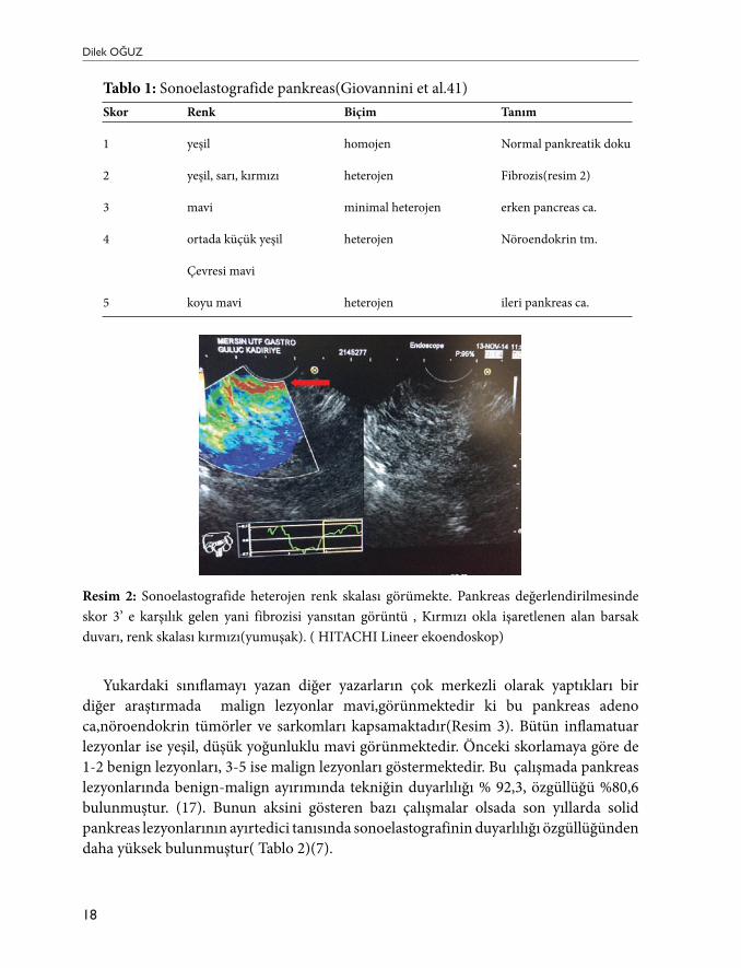
Normal pankreas; elastografide renk skalsı yeşil olarak görülür. Bir araştırmada renk skalası kullanılarak yapılan incelemede pankreas için 5 skor tanımlanmıştır(16)( Tablo 1). Bu skorlamada özellikle ileri pankreas kanserinde daha yumuşak alanların heterojen olarak görüntülenmesinin nekrozu yansıttığı bilinmektedir.
Dilek OĞUZ
18
Tablo 1: Sonoelastografide pankreas(Giovannini et al.41)Skor Renk Biçim Tanım
1 yeşil homojen Normal pankreatik doku
2 yeşil, sarı, kırmızı heterojen Fibrozis(resim 2)
3 mavi minimal heterojen erken pancreas ca.
4 ortada küçük yeşil heterojen Nöroendokrin tm.
Çevresi mavi
5 koyu mavi heterojen ileri pankreas ca.
Resim 2: Sonoelastografide heterojen renk skalası görümekte. Pankreas değerlendirilmesinde skor 3’ e karşılık gelen yani fibrozisi yansıtan görüntü , Kırmızı okla işaretlenen alan barsak duvarı, renk skalası kırmızı(yumuşak). ( HITACHI Lineer ekoendoskop)
Yukardaki sınıflamayı yazan diğer yazarların çok merkezli olarak yaptıkları bir diğer araştırmada malign lezyonlar mavi,görünmektedir ki bu pankreas adeno ca,nöroendokrin tümörler ve sarkomları kapsamaktadır(Resim 3). Bütün inflamatuar lezyonlar ise yeşil, düşük yoğunluklu mavi görünmektedir. Önceki skorlamaya göre de 1-2 benign lezyonları, 3-5 ise malign lezyonları göstermektedir. Bu çalışmada pankreas lezyonlarında benign-malign ayırımında tekniğin duyarlılığı % 92,3, özgüllüğü %80,6 bulunmuştur. (17). Bunun aksini gösteren bazı çalışmalar olsada son yıllarda solid pankreas lezyonlarının ayırtedici tanısında sonoelastografinin duyarlılığı özgüllüğünden daha yüksek bulunmuştur( Tablo 2)(7).

Istanbul Endoscopic Ultrasound and Advanced Endoscopy Days • December 10-11 2015, ss. 14-23
19
Tablo 2. Solid pankreas lezyonlarında sonoelastografinin değeri(7 numaralı yayından modifiye edilmiştir)
Yazar Yıl Duyarlılık(%) Özgüllük(%)
Kalitatif SE Giovannini(16) 2006 100 67
Giovannini(17) 2009 92,3 80
Janssen(19) 2007 93,8 65,4
Sãftoiu(14) 2006 91,7 94,4
Iglesias-Garcia(13) 2009 100 85,5
Kantitatif SE Sãftoiu(18) 2008 91,4 87,9
Iglesias-Garcia(15) 2010 100 92,9
Sãftoiu(20) 2011 93,4 66
Son yıllarda yapılan bir metaanalizde pankreas adenokarsinomu ile inflamatuar kitlelerin sonoelastografisi değerlendirilmiştir. Buna göre kalitatif ölçümde duyarlılık % 99, özgüllük % 76 bulunmuştur(21).
Resim 3. Pankreas adeno ca: hasta batında assit ile kliniğimize başvuruyor. Omentum kalınlığı ile mezotelyoma düşünülürken çekilen tomografide pankreasa ait spesifik patoloji tanımlanmamış. Sadece GGT yüksek ve koledok 10 mm bulunması üzerine yapılan EUS ve Sono elastografide pancreas başında 18 mm heterojen-hipoekoik , kenarları irregüler kitle( sarı ok). Sol görüntüde kalitatif elastografi görüntülenmekte. Lezyon mavi renk skalasında( beyaz ok).( HITACHI Lineer ekoendoskop)

Dilek OĞUZ
20
Sonuç olarak pankreasın solid lezyonlarının değerlendirilmesinde elimizdeki veriler önceki zorluklarımızı sonoelastografi ile aşacağımızı göstermektedir. Bizim deneyimimiz bu konu ile ilgili olarak çok yeni olmakla birlikte solid lezyon olan 10 hastada EUS-FNA ve cerrahi referans alındığında tanı oranı % 100 bulunmuştur( yayınlanmamış veri- Mersin Üniversitesi Gastroenteroloji Bilim Dalı). Bu konu ile ilgili çalışmaların artması gerekmektedir.
Lenf nodlarının değerlendirilmesinde SonoelastografiLenf nodlarının değerlendirilmesinde EUS çok önemli bir endoskopik tekniktir.
Ancak patolojik lenf nodu diyebilmek her zaman kolay olmamaktadır. Çoğunlukla transabdominal ultrasonografide ve konvansiyonel EUS’da malign lenf nodlarını tanımlamada; yuvarlak şekil, hipoekojenite, çapın > 1 cm oluşu, belirgin sınırlarının oluşu gibi özellikler kullanılır. Ancak bu özelliklerin spesifitesi düşük olup benign lezyonlarda da buna benzer görünümler saptanabilir(6). Doğruluğu tanımlamada son yıllara kadar kullandığımız yöntem EUS-FNA’dir. Teknik zorluklar ve zaman zaman az da olsa ortaya çıkan komplikasyonlar bilinir(22).Ayrıca aynı hastada pek çok lenf nodu bulunabilir hangisinin patolojik olduğu konvansiyonel EUS’da zorlanılan noktadır. Gastrointestinal tümörlerin evrelemesinde saptanabilen sonografinin ulaştığı her alandaki lenf nodlarının değerlendirilmesi prensip olarak esastır. Sonoelastografi bu lenf nodlarının doğru değerlendirilmesinde yeni ve değerli bir yöntemdir. Malign lenf nodları renk skalasında doku sertliği nedeni ile mavi görünür(Resim 4). Patolojik olmayan lenf nodu ise yeşil olarak görülür(Resim 5,6). Kantitatif ölçümü ile ilgili az sayıda çalışma vardır. Lenf nodlarının patolojik olarak belirlenmesi ile ilgili yapılan çalışmalarda duyarlılık % 85-100, özgüllük ise % 50-94,4 olarak bulunmuştur(14,16,17,19).
Resim 4. Çölyak önü konglomere lenf nodları arasında sağdaki beyaz ok B- mod görüntüde lenf nodu, solda kalitatif elastografi görünümü mavi renk skalası( sarı ok).

Istanbul Endoscopic Ultrasound and Advanced Endoscopy Days • December 10-11 2015, ss. 14-23
21
Resim 5. Sağda B-mod EUS’da şüpheli lenf nodu( mavi ok), solda kalitatif elastografide yeşil renk skalası görünümü (sarı ok)
Resim 6. Sağdaki EUS B-mod görüntü küçük boyutta lenf nodu (kırmızı ok), solda kalitatif elastografi görüntüsü yeşil renk skalası (sarı ok).
Sonoelastografinin gelecekteki diğer kullanım alanları ?Elimizde bu konu ile ilgili veriler şu anda yetersiz görünmekle birlikte tekniğin temel
felsefesi dikkate alındığında pek çok malign dokunun değerlendirilmesine açık gibi görünmektedir. EUS’un ulaştığı tüm alanların solid lezyonlarının değerlendirilmesinde

Dilek OĞUZ
22
tartışılması ve araştırılması gerekmektedir. Bunların arasında sol sürrenal adenomların malignitelerden ayırt edilmesi, karaciğer sol lobunda yer alan karmaşık solid lezyonların değerlendirilmesi, özofagus ve gastrik kanserin lokal evrelemesinde çevre dokuların elastisitesinin değerlendirilmesi düşünülebilir. Öte yandan pek çok subepitelyal lezyonun doku sertliğinin değerlendirilmesi ve böylece lezyonların tanımlanabilmesi (pankreatik rest, lipom …) mümkün görünmektedir. Bu alanlar araştırılmaya açık alanlardır.
ÖzetSonoelastografi yeni gelişen bir tekniktir. Kalitatif ve kantitatif olarak kullanılarak
fibröz ve benign dokuları malign dokulardan ayırtedebilmektedir. Günümüzde pankreasın solid lezyonları ve lenf nodları için kullanılmaktadır. Araştırmaya açık bir sahadır. Ülkemizde pek çok referans merkezde EUS kullanım alanına yaygın bir şekilde girmiştir. Sonoelastografinin öğrenilmesi ve kullanılması tanı ve tedavi yaklaşımlarımızı iyileştirecektir.
Kaynaklar
1- Lee Tae Hee , Cha Sang-Woo, Cho Young Deok . EUS Elastography: Advances in Diagnostic EUS of the Pancreas Korean J Radiol 2012 ;13(Suppl 1), Jan/Feb:12-16
2- Erickson RA. EUS-guided FNA. Gastrointest Endosc 2004;60(2):267–79.3- Dumonceau JM, Polkowski M, Larghi A, et al. Indications, results, and clinical impact of
endoscopic ultrasound (EUS)-guided sampling in gastroenterology European Society of Gastrointestinal Endoscopy (ESGE) Clinical Guideline.Endoscopy 2011;43(10):897–912.
4- Erickson RA, Sayage-Rabie L, Beisner RS. Factors’ predicting the number of EUS-guided fine-needle passes for diagnosis of pancreatic malignancies. Gastrointest Endosc 2000;51:184–90.
5- Binmoeller KF, Rathod VD. Difficult pancreatic mass FNA: tips for success.Gastrointest Endosc 2002;56:S86–93.
6- Bhutani MS, Hawes RH, Hoffman BJ. A comparison of the accuracy of echofeatures during endoscopic ultrasound (EUS) and EUS-guided fine needle aspiration for diagnosis of malignant lymph node invasion. Gastrointest Endosc 1997;45:474–9.
7- Iglesias-Garcia J,Domínguez-Muñoz J E.Endoscopic Ultrasound Image Enhancement Elastography Gastrointest Endoscopy Clin N Am 22 (2012) 333–348
8- Itoh A, Ueno E, Tohno E, Kamma H, Takahashi H, Shiina T, et al. Breast disease: clinical application of US elastography for diagnosis. Radiology 2006;239:341-350
9- Cochlin DL, Ganatra RH, Griffiths DF. Elastography in the detection of prostatic cancer. Clin Radiol 2002;57:1014-1020
10- Krouskop TA, Wheeler TM, Kallel F, Garra BS, Hall T. Elastic moduli of breast and prostate tissues under compression. Ultrason Imaging 1998;20:260-274
11- Ophir J, Céspedes I, Ponnekanti H, Yazdi Y, Li X. Elastography: a quantitative method for imaging the elasticity of biological tissues. Ultrason Imaging 1991;13:111-134

Istanbul Endoscopic Ultrasound and Advanced Endoscopy Days • December 10-11 2015, ss. 14-23
23
12- Frey H. Real-time elastography. A new ultrasound procedure for the reconstruction of tissue elasticity. Radiologie 2003;43:850–5.
13- Iglesias-Garcia J, Larin˜o-Noia J, Abdulkader I, et al. Endoscopic ultrasound elastography for the characterization of solid pancreatic masses. Gastrointest Endosc 2009;70:1101–8.
14- Saftoiu A, Vilmann P, Gorunescu F, et al. Neural network analysis of dynamic sequences of EUS elastography used for the differential diagnosis of chronic pancreatitis and pancreatic cancer. Gastrointest Endosc 2008;68:1086–94.
15- Iglesias-Garcia J, Larin˜o-Noia J, Abdulkader I, et al. Quantitative endoscopic ultrasound elastography: an accurate method for the differentiation of solid pancreatic masses. Gastroenterol 2010;139:1172–80.
16- Giovannini M, Hookey LC, Bories E, et al. Endoscopic ultrasound elastography: the first step towards virtual biopsy? Preliminary results in 49 patients. Endoscopy 2006;38:344–8.
17- Giovannini M, Botelberge T, Bories E, et al. Endoscopic ultrasound elastography for evaluation of lymph nodes and pancreatic masses: a multicenter study. WorldJ Gastroenterol 2009;15:1587–93.
18- Saftoiu A, Vilmann P, Hassan H, et al. Analysis of endoscopic ultrasound elastography used for characterisation and differentiation of benign and malignant lymph nodes. Ultraschall Med 2006;27(6):535–42.
19- Janssen J, Dietrich CF, Will U, et al. Endosonographic elastography in the diagnosis of mediastinal lymph nodes. Endoscopy 2007;39(11):952–7.
20- Saftoiu A, Vilmann P, Gorunescu F, et al. Accuracy of endoscopic ultrasound elastography used for differential diagnosis of focal pancreatic masses: a multicenter study. Endoscopy 2011;43:596–603.
21- Xiang Li, Wei Xu, Jian Shi, Yong Lin, Xin Zeng. Endoscopic ultrasound elastography for differentiating between pancreatic adenocarcinoma and inflammatory masses: A meta-analysis World J Gastroenterol 2013 October 7; 19(37): 6284-6291
22- Micames CG, McCrory DC, Pavey DA, et al. Endoscopic ultrasound-guided fineneedle aspiration for non-small cell lung cancer staging: A systematic review and metaanalysis. Chest 2007;131(2):539–48.

Istanbul Endoscopic Ultrasound and Advanced Endoscopy Days, December 10-11 2015,ss.24-28 • e-ISBN: 978-975-400-393-2
Eus Klavuzluğunda Doku Elde Edilmesi: Sonuçları Nasıl Daha İyi Hale Getirebiliriz? İğne Performansı?
Doç. Dr. Emrah ALPER
Endosonografi (EUS) ile ekoskopun ulaştığı tüm noktalara en fazla 5-6 cm uzaklıktaki tüm lezyonlardan doku ve sıvı örneklemesi elde etme şansı mevcuttur. Özöfagusa komşu orta ve alt posterior mediasten lezyonları, mide ve duodenuma komşu pankreas kistik ya da solit lezyonları, rektuma komşu perirektal lezyonlar, üst gastrointestinal sistem subepitelyal lezyonları, üst abdomende yerleşik kitleler, sol böbrek- sol sürrenal gland- prostat- sol karaciğer lobunda yerleşik lezyonlar EUS FNA yada core biyopsi iğneleri ile örnekleme yapılabilen lezyonlardır.
EUS ile örnekleme çeşitli tip ve yapıda iğneler kullanılarak sitolojik ve doku örneklemesi şeklinde yapılabilmektedir. Bu amaçlarla çeşitli çaplarda (19-22-25 Gauge) aspirasyon iğneleri (FNA) ya da çeşitli çaplarda ( 19- 20- 22 Gauge) doku kesici core biyopsi iğneleri ( procore ve tru-cut iğneler) kullanılmaktadır. Lezyonun tipi ( solit- kistik), yerleşim yeri (mediasten, subepitelyal, pankreatik vb), ön tanı ( lenfoma, metastaz, nöroendokrin tm vb) kullanılacak örnekleme ve iğne türünü belirlememizde önem taşımaktadır.
Bu konuda ilk akla gelen soru; Hangi tip EUS iğnesi kullanırsak daha yüksek diagnostik etkinlik sağlayabiliriz? Etkinliği belirlemekte ilk basamak hücre- hücre bloğu mu yoksa doku mu elde etmek istiyoruz. Eğer lenfoma ya da metastaz ayırıcı tanısıyla lenfadenopatiden EUS ile örnekleme yapılacaksa, prostat ca ön tanı ile prostattan örnekleme yapılacaksa, pankreatik kronik pankreatit nodülü, metastatik pankreas kitlesi yada karaciğer kitlesi düşünülüyorsa ana amaç doku bloğu elde edilmesi olacaktır. Bu nedenle doku örneklemesini iyi yapabilen procore ya da tru-cut core EUS biyopsi iğnelerinin kullanılması daha iyi olabilir. Ancak adenokarisnom düşünülen pankreas kitlelerinin natürünün saptanması, subepitelyal lezyonların tanısının konması, kistik lezyonlarda sıvı aspirasyonu ile beraber hücre elde edilmesi planlanıyorsa aspirasyon EUS iğneleri ( FNA) kullanılması daha iyi olacaktır. Na ve ark. (1)
GIST ön tanılı hastalarda yaptıkları çalışmada 19 G tru-cut core biyopsi iğnesinin FNA iğneye göre çok daha etkin olduğu görülmektedir. Subepitelyal lezyonlarda 22 G FNA ile 22 G Tru-cut iğnenin karşılatırıldığı diğer bir çalışmada da tru-cut cor biopsinin
SUMMARY OF SPEAKERS’ PRESENTATIONS
24

Istanbul Endoscopic Ultrasound and Advanced Endoscopy Days • December 10-11 2015, ss. 24-28
25
tanısal etkinliğin yüksek olduğu belirtilmektedir (2). Nagula ve ark. (3) ile De La Mora Levy ve ark. (4) solit kitlelerinin tanısının konmasında procore iğne ve FNA iğneleri karşılaştırdıkları çalışmalarda her iki iğnenin etkinliğin benzer olduğu görülmektedir. Strand ve ark. (5) pankreas kitlelerinde procore ve FNA iğneleri karşılaştırdıkları çalışmada FNA aspirasyon iğnesi daha etkin saptanmış iken Choi ve ark. (6) benzer hasta grubunda yaptıkları çalışmada procore iğnenin daha etkin olduğu saptanmıştır. Ramay ve ark. (7) lenf nodu örneklemesinde procore ve FNA iğneleri karşılaştırdıkları retrospektif çalışmada her iki iğnenin benzer tanısal etkinlikte olduğu belirtilmektedir. O’Connor ve ark.( 8) gastrointestinal sisteme komşu solit lesyonlardan yaptıkları FNA örneklemelerde diagnostik başarı oranı % 98 olarak belirtilmektedir. Yukarıda atıf yapılan çalışmalardan da anlaşılacağı üzere eğer hücre ya da hücre bloğu elde eilmek isteniyorsa iğne yapıları arasında çok afrk yoktur. Ancak doku elde edilmesi isteniyorsa core biyopsi iğnelerin kullanılması tercih edilmelidir.
Akla gelen diğer bir soru; Hangi çap iğne kullanırsak daha yüksek diagnostik etkinlik sağlayabiliriz? Şu anda kullanımda olan endosonografik ekoskopların çalışma kanalları en kalın 19 Gauge iğne kullanılmasına imkan vermektedir. Çeşitli firmaların 19-22-25 Gauge çapında FNA, 19-20-22 G çapında procore core biyopsi iğneleri ve 19 G çapında tru-cut core biyopsi iğneleri bulunmaktadır. İğne çapı arttıkça elde edilecek örneğin daha yüksek diagnostik başarı elde edeceği düşünülse de yapılan çalışmaların çoğunda iğne çaplarının diagnostik başarıda belirgin farka neden olmadığı görülmektedir. Vilmann P ve ark. (9) 22 G ve 25 G iğne ile intraabdominal solit kitelelerden yaptıkları örneklemelerde her iki iğne arasında diagnostik başarı açısından fark saptanmamıştır. Benzer şekilde 22 ve 25 G iğnelerin etkinliklerinin karşılaştırıldığı Camellini ve ark (10) çalışmasında da istatistiksel anlamlı fark saptanmamıştır. Mediastinal lenf nodu EUS biyopsi örneklerinde 19 ve 22 G iğne ile yapılan çalışmada her iki iğne benzer etkinlikte saptanmıştır (11). Bu paragrafta atıf yapılan çalışmalardan anlaşıldığı üzere iğne çapının lezyon yeri ve yapısından bağımsız olarak diagnostik etkinlik açısından belirleyici olmadığı görülmektedir. Ancak özellikle derin duodenumdan yapılacak pankreas baş ve uncinat kesim kitle örneklemelerinde 25 G iğnenin tercih edilmesi kullanıcı kolaylığı ve cihaz güvenliği sağlayacaktır. Özellikle lenf nodundan alınacak biyopsilerde 19 G FNA iğne kullanıldığında eirtrosit artefaktının daha fazla olacağı gözardı edilmemelidir.
Hangi FNA örnekleme tekniğini ( slow pull teknik- standart vakum aspirasyon) kullanırsak daha çok hücre ve hücre bloğu elde edebiliriz? sorusu diğer önemli bir sorudur. Standart vakum aspirasyon tekniğinde doku içine yerleştirilen FNA iğnesinin styleti çekildikten sonra 10 ya da 20 ml hacimli vakumlu aspiratörler iğnenin proksimaline yerleştirilerek apirasyon yapılarak işleme devam edilmektedir. Slow pull teknikte ise stylet iğne hareketine uygun olarak yavaşça geri çekilerek aspiratör takılmadan kapillerizasyon kuralı kullanılarak örnekleme yapılmaktadır. Bu konuda yapılmış üç çalışmada slow

Emrah ALPER
26
pull teknik kullanıldığında daha az eritrosit artefaktı olduğu ve tanısal etkinliğin arttığı belirtilmektedir. (12,13,14).Slow pull teknikte styletin her iğne vuruşunu takiben yaklaşık 10 cm geri çekilmesi dikkat edilmesi gereken önemli bir ayrıntıdır. Vakum aspirasyon teknikte ise mide suyunun çekilmemesi için aspirasyon işlemi iğne dokudan çekilmeden önce sonlandırılmalıdır.
Örnekleme işlemi sırasında onsite sitolojik değerlendirme önemli mi? On site sitopatolog olmadan işlemi yaparsak nelere dikkat etmeliyiz? diğer iki önemli soru olarak karşımıza çıkmaktadır. Hızlı onsite sitolojik değerlendirme işlem sürenizi çok fazla uzatmadan tanısal yeterlilik oranınızı %100 e çıkaracaktır (15). Onsite inceleme sırasında Diff-Quick boyama kullanılmaktadır. Sitoloji teknisyeni tarafından kurallara uygun şekilde hazırlanan preparat ilk olarak metil alkol ile fikse edilmekte takiben L xsanthene, metilen blue ve azure boyalar ile boyanmaktadır. Uygun sürede açık havada kurutulan preparat konusunda deneyimli sitopatolog tarafından işlem odasında hızlıca değerlendirilmekte tanısal yeterli örnekleme yapılıp yapılmadığı belirlenmektedir. Eğer ilk pass vuruşlarda yeterli örnek alınmamışsa ikinci pass vuruş yapılmaktadır. Sitopatolog hücresel yeterliliği ve kabaca ön tanıyı aynı işlem seansında saptayabilmektedir. Onsite sitopatolojik değerlendirmenin en önemli kısıtlayıcı yönü sitpatologa ulaşmadaki sorundur. Sitopatolog olmaksızın işlem yapılacaksa aynı örnekleme seansında 4-6 pass vuruş yapılması tanısal etkinliği arttıracaktır (16). İşlem sırasında 4-6 kez pass vuruş yapılması 2. İğne kullanımına neden olarak maliyeti arttırabilmektedir. Ayrıca işlem süresinin uzaması anestezi ve işleme bağlı komplikasyon riskinde artışa neden olabilmektedir. Onsite sitopatolog olmadan yapılan örneklemelerde dikkat edilmesi gereken en önemli nokta preperatların sitolojik değerlendirme için uygun hazırlanmasıdır. Slaytlar ne çok ince ne de çok kalın olmalıdır. Preparatların bir kısmı kuru havada, bir kısmı alkollü şale içinde transfer edilmelidir. Doku ve hücre bloğu hazırlanması için elde edilen solit kısım formaldehit içeren tüpe konarak transfer edilmelidir.
Endosonographerın dikkat etmesi gereken önemli bir nokta dokunun birçok yerinden örnekleme yapılmasıdır. Doku içinde nekroz alanı bulunuyorsa bu alandan örnekleme yapılması tanısal yeterliliği belirgin şekilde düşürecektir. Lezyonun merkez ve periferinden örnekleme yapılması tanısal etkinliği belirgin arttırmaktadır. Aynı pass vuruş ya da diğer pass vuruşlarda iğnenin lezyonun farklı noktalarından örnekleme yapması sağlanmalıdır.
Sonuçta; EUS ile lezyondan FNA ya da core biyopsi örneklemede tanısal etkinliği belirleyen en önemli faktörün endoskopistin ve sitopatologun deneyimi olduğu unutulmamalıdır. Deneyimli endoskopistin ve sitopatologun varlığında iğne çapının tanısal başarı üzerinde belirgin etkisi yoktur. Core biyopsi iğne ya da aspirasyon (FNA) iğne seçimi lezyonun yapısına, lezyonun yerleşim yerine ve düşünülen ön tanıya göre değişmelidir. Mümkünse onsite sitopatolog eşliğinde örnekleme yapılmalı eğer yapılamıyorsa örnekleme işlemi sırasında 4-6 pass vuruş yapılmalıdır. Elde edilen örnekler kurallara uygun hazırlanmalı ve en kısa sürede sitopatoloji laboratuvarına ulaştırılmalıdır.
Istanbul Endoscopic Ultrasound and Advanced Endoscopy Days • December 10-11 2015, ss. 24-28
27
Kaynaklar1. Na HK, Lee JH, Park YS, Ahn JY, Choi KS, Kim do H, Choi KD, Song HJ, Lee GH,Jung HY,
Kim JH. Yields and Utility of Endoscopic Ultrasonography-Guided 19-Gauge Trucut Biopsy versus 22-Gauge Fine Needle Aspiration for Diagnosing Gastric Subepithelial Tumors. Clin Endosc. 2015 Mar;48(2):152-7
2. Kim GH, Cho YK, Kim EY, Kim HK, Cho JW, Lee TH, Moon JS; Korean EUS Study Group. Comparison of 22-gauge aspiration needle with 22-gauge biopsy needle in endoscopic ultrasonography-guided subepithelial tumor sampling. Scand J Gastroenterol. 2014 Mar;49(3):347-54
3. Nagula S, Pourmand K, Aslanian HR, et al. EUS-fine needle aspiration (FNA) vs. EUS-fine needle biopsy (FNB) for solid mass lesions: interim analysis of a large multicenter, randomized clinical trial. Gastrointest Endosc 2013;77
4. De La Mora-Levy JG, Florez-Sarmiento CF, Alonso-Larraga JO, Campuzano JO, Del Monte JS, Hernandez-Guerrero A. Direct comparison between Procore™, and non-Procore™ fine needle aspiration biopsy needles: does it make any difference in expert hands? Gastrointest Endosc 2013;77(5 Suppl):AB360
5. Strand DS, Shami VM, Sauer BG, Wang AY. EUS-guided 22-gauge fine needle aspiration is superior to EUS-guided 22-gauge core needle biopsy in the evaluation of solid pancreatic neoplasms. Gastrointest Endosc 2013;77(5 Suppl):AB403.
6. Choi HJ, Moon JH, Kim HK, et al. Comparison of EUS-fine needle biopsy with EUS-fine needle aspiration as a historical control for diagnosis of pancreatic solid masses. Gastrointest Endosc 2013;77(5 Suppl): AB401.
7. Ramay F, Singh M, Sood V. Retrospective study comparing yield of EUS 22G FNA/FNB of abnormal lymph nodes: single tertiary referral center experience. Gastrointest Endosc 2013;77(5 Suppl):AB360
8. O’’Connor K, Cheriyan DG, Li-Chang HH, Kalloger SE, Garrett J, Byrne MF, Weiss AA, Donnellan F, Schaeffer DF. Gastrointestinal Endoscopic Ultrasound-Guided Fine-Needle Aspiration Biopsy Specimens: Adequate Diagnostic Yield and Accuracy Can Be Achieved without On-Site Evaluation. Acta Cytol. 2015 Oct;59(4):305-10
9. Vilmann P, Săftoiu A, Hollerbach S, Skov BG, Linnemann D, Popescu CF, WellmannA, Gorunescu F, Clementsen P, Freund U, Flemming P, Hassan H, Gheonea DI, Streba L, Ioncică AM, Streba CT. Multicenter randomized controlled trial comparing the performance of 22 gauge versus 25 gauge EUS-FNA needles in solid masses. Scand J Gastroenterol. 2013 Jul;48(7):877-83
10. Camellini L, Carlinfante G, Azzolini F, Iori V, Cavina M, Sereni G, Decembrino F, Gallo C, Tamagnini I, Valli R, Piana S, Campari C, Gardini G, Sassatelli R. A randomized clinical trial comparing 22G and 25G needles in endoscopic ultrasound-guided fine-needle aspiration of solid lesions. Endoscopy. 2011 Aug;43(8):709-15
11. Songür N, Songür Y, Bırcan S, Kapucuoğlu N. Comparison of 19- and 22-gauge needles in EUS-guided fine needle aspiration in patients with mediastinal masses and lymph nodes. Turk J Gastroenterol. 2011;22(5):472-8
12. Nakai Y, Isayama H, Chang KJ, Yamamoto N, Hamada T, Uchino R, Mizuno S,Miyabayashi K, Yamamoto K, Kawakubo K, Kogure H, Sasaki T, Hirano K, Tanaka M,Tada M, Fukayama M, Koike K. Slow pull versus suction in endoscopic ultrasound-guided fine-needle aspiration of pancreatic solid masses. Dig Dis Sci. 2014 Jul;59(7):1578-85
13. Paik WH, Park Y, Park do H, Hong SM, Lee BU, Choi JH, Lee SS, Seo DW, Lee SK,Kim MH. Prospective evaluation of new 22 gauge endoscopic ultrasound core needle using capillary sampling with stylet slow-pull technique for intra-abdominal solid masses. J Clin Gastroenterol. 2015 Mar;49(3):199-205
14. Kin T, Katanuma A, Yane K, Takahashi K, Osanai M, Takaki R, Matsumoto K, Gon K,

Emrah ALPER
28
Matsumori T, Tomonari A, Maguchi H, Shinohara T, Nojima M. Diagnostic ability of EUS-FNA for pancreatic solid lesions with conventional 22-gauge needle using the slow pull technique: a prospective study. Scand J Gastroenterol. 2015 Jul;50(7):900-7
15. Collins BT, Murad FM, Wang JF, Bernadt CT. Rapid on-site evaluation for endoscopic ultrasound-guided fine-needle biopsy of the pancreas decreases the incidence of repeat biopsy procedures. Cancer Cytopathol. 2013 Sep;121(9):518-24
16. Iglesias-Garcia J, Lariño-Noia J, Abdulkader I, Domínguez-Muñoz JE. Rapid on-site evaluation of endoscopic-ultrasound-guided fine-needle aspiration diagnosis of pancreatic masses. World J Gastroenterol. 2014 Jul 28;20(28):9451-7

29
Istanbul Endoscopic Ultrasound and Advanced Endoscopy Days, December 10-11 2015,ss.29-30 • e-ISBN: 978-975-400-393-2
Treatment of Pancreatic Neuroendocrine Tumors
Dr. Faysal DaneMarmara University School of Medicine Division of Medical Oncology
Pancreatic neuroendocrine tumors (pNETs) are rare neoplasms that arise in the endocrine tissues of the pancreas. They are low to intermediate grade neoplasms and have a more indolent course compared to more frequently seen pancreatic adenocarcinoma. These tumors can secrete a variety of peptide hormones resulting in different clinical syndromes. However, between 50 to75 percent of pNETs are nonfunctioning.
Surgery is currently the only treatment modality that may achieve cure. Patients with advanced, unresectable pNETs, cure is generally not possible. The goals of oncologic treatment include palliation or prevention of symptoms and cytoreduction of bulky tumors to improve survival. For patients with potentially resectable liver-isolated metastatic pNETs, surgical resection of the hepatic metastases along with the primary tumor is recommended. Although the majority of cases will not be cured by surgery, given the slow growing nature of the tumor, extended survival is sometimes possible. Other treatment options for patients with unresectable hepatic predominant symptomatic metastatic disease include embolization, chemoembolization, radiofrequency ablation (RFA), and cryoablation. Occasionally, systemic therapy may also convert cases wherein surgery may render the patients disease free.
Poorly differentiated neuroendocrine carcinomas have a bad prognosis. They are generally treated with platinum based chemotherapy. Patients having low to intermediate grade tumors with symptoms of hormone hypersecretion from a pNET should be managed with somatostatin analogs and other agents as appropriate to the specific syndrome. In general, initial therapy for insulinomas consists of carbohydrates and diazoxide, which directly inhibits the release of insulin from insulinoma cells. Everolimus can also be highly effective in improving glycemic control in patients with insulinoma. For patients with gastrinoma, high doses of oral proton pump inhibitors are the treatment of choice. For asymptomatic patients with low volume disease, observation
SUMMARY OF SPEAKERS’ PRESENTATIONS
Faysal DANE
30
alone rather than early administration of a somatostatin analog is suggested. For patients who are felt to require therapy because of progressive disease or symptoms related to hormone production, initiation of therapy with a somatostatin analog, everolimus, or sunitinib is suggested that have all been shown to improve progression free survival (PFS) compared to best supportive care alone, although none of these agents have been compared directly to each other. In the absence of comparative trials, the choice of initial agent may be influenced by the expected toxicity profile. Given a favorable toxicity profile, a somatostatin analog is an appropriate first choice for many patients with use of a targeted agent if there is subsequent disease progression. Everolimus may be of particular value in patients with functioning insulinomas and refractory hypoglycemia. For patients who are symptomatic from tumor bulk, initiation of a somatostatin analog with either a targeted agent or chemotherapy is offered. For patients who are highly symptomatic or who have rapidly enlarging metastases, chemotherapy can be used as an initial treatment because of the higher objective response rate compared to other approaches. There are two acceptable options for therapy, a streptozocin based combination or a temozolomide containing regimen. In the absence of comparative trials, the choice of regimen should be individualized, considering the convenience of oral rather than intravenous treatment, performance status, and the anticipated side effect profile of both combinations. The role of peptide receptor radioligand therapy (PRRT) in patients with progressive advanced pancreatic NETs is unclear. Targeted radiotherapy using radiolabeled somatostatin analogs could be considered for patients with disease that is otherwise refractory to medical therapy.
In conclusion; pNETs are rare; consequently, randomized trial data to form a robust evidence are limited. Treatment should aim to cure the patient but is often palliative because most patients have evidence of metastasis at presentation. In patients with incurable disease, treatment should aim to maintain a good quality of life as long as possible, recognising that this can be achieved for a number of years in many NETs, even in the presence of metastatic disease. As a result, management of patients with pNETs requires an understanding of the disease process and the importance of a multimodality approach.

31
Istanbul Endoscopic Ultrasound and Advanced Endoscopy Days, December 10-11 2015,ss.31-33 • e-ISBN: 978-975-400-393-2
Akılcı İlaç Kullanımı -Antibiyotik İlişkili İshalin Önlenmesi
Prof. Dr. Müjde SOYTÜRKDokuz Eylül Üniversitesi, Tıp Fakültesi, Gastroenteroloji B.D.
Akılcı İlaç Kullanımı tanımı ilk defa 1985 yılında Dünya Sağlık Örgütü tarafından yapılmıştır. Kişilerin klinik bulgularına ve bireysel özelliklerine göre; uygun ilacı, uygun süre ve dozda, en düşük fiyata ve kolayca sağlayabilmeleri olarak tanımlanmaktadır. Akılcı ilaç kullanımında sorumluluk sahibi taraflar; hekim, eczacı, hemşire, diğer sağlık personeli, hasta ve hasta yakını, üretici, düzenleyici otorite ve diğerleridir (medya, akademi vb).
Dünya’da ilaçların yaklaşık %50’si uygunsuz şekilde kullanılmaktadır. Uygun olmayan ilaç kullanımı örnekleri; çoklu ilaç kullanımı, ilaçların gereksiz ve aşırı kullanımı, klinik rehberlere uyumsuz tedavi seçimi, piyasaya yeni çıkan ilaçların uygunsuz tercihi, ilaç kullanımında özensiz davranılması (uygulama yolu, süre, doz vb), uygunsuz kişisel tedavilere başvurulması, gereksiz yere antibiyotik tüketimi, gereksiz yere enjeksiyon önerilmesi, gereksiz ve uygunsuz vitamin kullanımı, bilinçsiz gıda takviyesi ve bitkisel ürünlerin kullanımı, ilaç-ilaç etkileşimleri ve besin-ilaç etkileşimlerinin göz ardı edilmesidir.
Akılcı olmayan ilaç kullanımı, akılcı ilaç kullanımı tanımında yer alan maddelerden herhangi birinin veya bir kaçının karşılanamaması durumudur. Hastaların tedaviye uyumunun azalmasına, ilaç etkileşimlerine, bazı ilaçlara karşı direnç gelişmesine, hastalıkların tekrarlamasına ya da uzamasına, yan etki görülme sıklığının ve tedavi maliyetlerinin artmasına neden olur.
Tanı ve tedavi sürecinde akılcı ilaç kullanımı için hastanın sorununun tanımlanması, hekim tarafından doğru tanının konulması, ilaçlı veya ilaçsız etkili ve güvenilir tedavinin belirlenmesi, tedavinin uygulanabilirliği ve maliyetinin, tedavinin başarısı ve hasta uyumunun değerlendirilmesi önemlidir. Eğer ilaçla tedavi uygulanacaksa uygun
SUMMARY OF SPEAKERS’ PRESENTATIONS

Müjde SOYTÜRK
32
ilaçların seçimi, çoklu ilaç kullanımında ilaç etkileşimlerinin öngörülmesi, her bir ilaç için uygun dozun ve uygulama süresinin belirlenmesi ve uygun reçetenin yazılması gerekmektedir. Bu aşamada güncel tanı ve tedavi kılavuzları esas alınmalıdır. Hasta ve hasta yakını tedavi hakkında bilgilendirilmelidir.
Tedavi seçimini etkileyen faktörler; ekonomik faktörler, ilaç endüstrisinin etkileri, alışkanlıklar, iş yükü ve personel sıkıntısı, yasal etmenler, yetersiz ve eksik bilgi ile sosyal ve kültürel etmenlerdir.
Dünya Sağlık Örgütü’nün akılcı ilaç kullanımının teşvik edilmesi ile ilgili temel önerileri şunlardır: 1. İlaç kullanım politikalarını koordine edecek ve bunların etkilerini izleyecek kurum, 2. Eğitim denetim ve karar alma süreçlerinin desteklenmesine yönelik Klinik Tanı ve Tedavi Rehberlerinden yararlanma, 3. İlk seçenek tedavileri esas alan temel ilaçlar listesi oluşturma, 4. Bölgelerde ve hastanelerde ilaç ve tedavi kurulları kurma, 5. Mezuniyet öncesi müfredat programında probleme dayalı farmakoterapi eğitimi vermeyi sağlama, 6. Bir gereklilik olarak hizmet içi sürekli tıp eğitimleri düzenlenmesi, 7. Kurumsal çerçevede izleme, denetim ve geri bildirim sistemlerinin geliştirilmesi, 8. İlaçlar konusunda bağımsız (tarafsız bilgi) bilgi kaynaklarını kullanma, 9. Kamuoyunun ilaçlar hakkında eğitilmesi, 10. Etik olmayan mali girişimlerden kaçınılması, 11. Uygun ve zorunlu düzenlemeleri hayata geçirme, 12. İlaçların ve personellerin mevcudiyetini güvence altına almaya yönelik yeterli devlet harcamasının sağlanması.
Ülkemizde Sağlık Bakanlığı bünyesinde, akılcı ilaç kullanımı ile ilgili çalışmalar, yaklaşık 20 yıldır sürmektedir. 2010 yılında Akılcı İlaç Kullanımı Birimi kurulmuştur. 2012 yılında Türkiye İlaç ve Tıbbi Cihaz Kurumu bünyesinde Akılcı İlaç Kullanımı, İlaç Tedarik Yönetimi ve Tanıtım Dairesi kurulmuştur. İllerde koordinasyonu sağlamak amacıyla 81 ilde İl Sağlık Müdürlüklerinde “akılcı ilaç kullanımı il temsilcisi” bulunmaktadır. Hastane hizmet kalite standartları gereğince, hastanelerde planlama yapmak ve faaliyetlerde bulunmak amacıyla Akılcı İlaç Kullanımı ekipleri oluşturulmuştur.
Antibiyotik ilişkili ishal, geniş spektrumlu antibiyotik kullanımının sık görülen bir komplikasyonudur. Antibiyotik kullanımı sırasında ya da tedavi sonlandıktan 2 hafta sonrasına kadar hastaların %5-30 kadarında ortaya çıkabilmektedir. Özellikle penisilin, sefalosporin ve klindamisin grubu antibiyotiklerin kullanımından sonra görülebilmektedir. Klinik tablo, hafif bir ishalden fulminan psödomembranöz kolite dek değişen bir spektrum gösterebilmektedir. Antibiyotik ilişkili ishale en sık neden olan patojen Clostridium difficile’dir. Bunu Salmonella, Candida albicans ve enterotoksijenik Clostridium perfringens izler. Antibiyotik ilişkili ishalden korunmada anahtar nokta; antibiyotikleri yukarıda bahsedilen akılcı ilaç kullanımı ilkelerine uygun kullanmak ve özellikle gereksiz antibiyotik kullanımından kaçınmaktır. Antibiyotik ilişkili ishalin yönetiminde sorumlu antibiyotik mümkünse kesilmeli ve hastaya destek tedavi yapılmalıdır. C. difficile tedavisinde metranidazol ve vankomsin kullanılabilir. Antibiyotik ilişkili ishal çoğunlukla normal intestinal floranın dengesinin bozulması

Istanbul Endoscopic Ultrasound and Advanced Endoscopy Days • December 10-11 2015, ss. 31-33
33
sonucunda ortaya çıkmaktadır. Buradan yola çıkarak probiyotiklerin (Ör; Lactobacillus acidophilus, L casei GG, L bulgaricus, Bifidobacterium bifidum, B longum, Enterococcus faecium, Streptococcus thermophilus, Saccharomyces boulardii) antibiyotik ilişkili ishalin önlenmesi ve tedavisinde denendiği çok sayıda çalışma yapılmıştır. Bu çalışmaların sonuçları çelişkili olmakla birlikte probiyotik kullanımı faydalı görünmektedir. Helicobacter pylori eradikasyon tedavisi verilen hastalarda da antibiyotik ilişkili ishal gelişebilmektedir. H. pylori eradikasyon tedavisi verilen hastalarda Saccharomyces boulardii’nin antibiyotik ilişkili ishali önlemede etkinliği ve güvenilirliğinin araştırıldığı bir çalışmaya 389 hasta dahil edilmiş ve 376 hasta çalışmayı tamamlamıştır. Tedavi sırasında ve sonrasında S. boulardii verilen hastalarda sırasıyla %5.9 ve %1 oranında ishal görülürken kontrol grubunda bu oran %11.5 ve %3.8 olmuştur (p<0.05). Sonuç olarak S. boulardii, H. pylori eradikasyonu verilen hastalarda, antibiyotik ilişkili ishali önlemede etkili ve güvenli bulunmuştur.

Istanbul Endoscopic Ultrasound and Advanced Endoscopy Days, December 10-11 2015,ss.34-35 • e-ISBN: 978-975-400-393-2
Rectal cancer: Is MR sufficient for staging? Any advantage with EUS?
Nevin Oruç (Izmir)
Accurate locoregional staging is crucial in rectal cancer for deciding patient management. The administration of neoadjuvant therapy and decision for surgery depends on TN stage of the disease. Disease free survival and prognosis is determined by also stage of the disease (1).
Endoscopic ultrasound ( EUS) and magnetic resonance imaging (MRI) are used in the pre therapeutic workup of rectal cancer. Endoscopic ultrasound (EUS) was initial examination for staging. However because of the lack of facilities in many centres and also the progress in imaging techniques shifted the initial preference of EUS to other methods in rectal cancer staging. EUS is better then CT or MRI for local staging of the disease. However it is operator dependent and invasive techniques. Device is usually could not pass strictures due to tumour. MRI compared the EUS is more easily tolerated, non-invasive and widely available. MRI also does staging in stenotic tumours and it can assess the resection margin. It also detects T stage better then EUS since it shows extramural spread better(2). In advanced cases with distant metastases, computed tomography (CT) is the primary approach used to evaluate the disease
EUS is preferred method in early rectal cancer which is eligible for local excision. Recent prospective study compared EUS versus MRI in reliability for rectal cancer staging. Groups were compared for staging accuracy of depth of penetration (T), lymph nodes positive (N) and combined T and N (TN) result. EUS had higher sensitivity in T1 (p=0.044 < 0.05) and specificity in T2 (p = 0.039 < 0.05) than MRI. MRI had higher sensitivity in N staging (p = 0.046 < 0.05) In another retropective comparison trial MRI was not able detect any of T 1 tumous . (3,4)
Endorectal ultrasound is useful for tumour (T) staging; hence EUS is a useful imaging modality for early rectal cancer. Magnetic resonance imaging (MRI) is useful for assessing the mesorectum and the mesorectal fascia which has useful prognostic significance and for early identification of local recurrence. Most of the common imaging techniques
SUMMARY OF SPEAKERS’ PRESENTATIONS
34

Istanbul Endoscopic Ultrasound and Advanced Endoscopy Days • December 10-11 2015, ss. 34-35
35
are suboptimal for imaging following chemoradiation as they struggle to differentiate fibrotic changes and tumor. In this situation, EUS and MRI may provide complementary information to decide further treatment. After neoadjuvant chemoradiation, EUS and MRI correctly classified 46% (18/39) and 44% (17/39) of patients, respectively, in line with their histological T stage (P > 0.05). EUS was better than MRI for patients with T0-T2 (44% vs 33%, P > 0.05) and N0 disease (87% vs 52%, P = 0.013). However, MRI was more accurate than EUS in T and N staging for patients with more advanced disease after radiotherapy, (5).
In rectum cancer staging both EUS and MRI have some drawbacks and advantages. EUS is preferred method in early rectal cancer while MRI helps more accurate staging in advanced cases.
References1 Cesmeli E. Anorectal staging: is EUS necessary? Minerva Med. 2014 Oct;105(5):423-36.2 Xiao Y, Lu J, Xiong G, Wu B, Lin G, Zhao L, Liang Z, Zhong G, Hu K, Pan W, Qiu H.
[Postoperative pathological staging correlates the prognosis of rectal cancer after neoadjuvant chemoradiotherapy]. Zhonghua Wai Ke Za Zhi. 2014 Feb;52(2):99-104.
3 Fernández-Esparrach G, Ayuso-Colella JR, Sendino O, Pagés M, Cuatrecasas M, Pellisé M, Maurel J, Ayuso-Colella C, González-Suárez B, Llach J, Castells A, Ginès A. EUS and magnetic resonance imaging in the staging of rectal cancer: a prospective and comparative study. Gastrointest Endosc. 2011 Aug;74(2):347-54.
4 Mezzi G, Arcidiacono PG, Carrara S, Perri F, Petrone MC, De Cobelli F, Gusmini S, Staudacher C, Del Maschio A, Testoni PA. Endoscopic ultrasound and magnetic resonance imaging for re-staging rectal cancer after radiotherapy. World J Gastroenterol. 2009 Nov 28;15(44):5563-7
5 Samee A, Selvasekar CR. Current trends in staging rectal cancer. World J Gastroenterol. 2011 Feb 21;17(7):828-34.

Istanbul Endoscopic Ultrasound and Advanced Endoscopy Days, December 10-11 2015,ss.36-42 • e-ISBN: 978-975-400-393-2
How to achieve most from your EUS platform &Latest situation for diagnostic and therapeutic EUS in
Japan
Yousuke NAKAIThe University of Tokyo, Japan
SUMMARY OF SPEAKERS’ PRESENTATIONS
Every EUS is not the same: What’s the indication? -Pancreas? Biliary tract?
-Screening? Any findings in prior cross sectional imaging?
Always check CT/MRI “image” (not report) -Physicians who know clinical information are better than radiologists
36

Istanbul Endoscopic Ultrasound and Advanced Endoscopy Days • December 10-11 2015, ss. 36-42
37
°
°Distal end øInsertion tube ø
° °
° °
Operation channel ø°
i What’s on EUS image

Yousuke NAKAI
38

Istanbul Endoscopic Ultrasound and Advanced Endoscopy Days • December 10-11 2015, ss. 36-42
39

Yousuke NAKAI
40

Istanbul Endoscopic Ultrasound and Advanced Endoscopy Days • December 10-11 2015, ss. 36-42
41
≃ %

Yousuke NAKAI
42
…but this is the reality

43
Istanbul Endoscopic Ultrasound and Advanced Endoscopy Days, December 10-11 2015,ss.43-47 • e-ISBN: 978-975-400-393-2
Kronik pankreatit (KP) and Endosonografi(EUS):Teşhis
Yücel ÜSTÜNDAĞZonguldak BEU Tıp Fakültesi İç Hastalıkları ABD Gastroenteroloji Bilim Dalı
SUMMARY OF SPEAKERS’ PRESENTATIONS
KP-Teşhis • KP-geri d���şs�� / ilerleyici inflamasyon • İleri evre – kolay teşhis • Erken KP zor teşhis • Patoloji-ercp-sekretin • EUS-morfolojik teşhis – Parenkimal and duktal değişiklikler – FNA/FNB – ��st�l�ğ�� şiddeti – Elastografi ve kontrast tek�iği – KP ilişkili k���lik�s���l�r�� tedavisi
KP-EUS • EUS-yüksek rezolusyonlu görüntüleme • EUS teşhis sps kriterler Abbaken L, et al. Endoscopy 2009
• >3-4 EUS kriter
Duktal 1-Pankreas kanal �e�iş�e�e 2-Pankreatik kanal ���e�si��i�i 3-Hiperekojenik duktal yapı 4-Görünür pankreatik yan dallar 5-intraduktal kalsifikasyonlar
• Rosement sı�ı��a��ı��ası Catalono ����et�a�����������
– KP kriterlerin önemine göre p�a��a��ası
– Major A, Major B and Minor kriterler
Parenkimal 1-Kalsifikasyon 2-Lobularite 3-Kistler 4-Hiperekoik fokus 5-Hiperekoik strand
Özellik Tarifler Major Minor
*Hiperekojenik fokus +gölgeli
>2 mm ekojenik yapılar +gölge Major A
Lobularite >5 mm iyi sınırlı parlayan halka-eko fakir merkez
*a. Balpeteği + Ardısıra >3 lobuller Major B b. Balpeteği - Ardışık olmayan lobuller Evet Hiperekoik fokus gölgesiz
Ekojenik yapılar >2 mm Evet
Kistler Aneokik, yuvarlak-eliptik yapılar-septasyonsuz
Evet
Strand *Ana kanal taş Düzensiz ana kanal kontur Dilate yan dallar Ana pankreatik kanal genişlemesi Hiperekojenik ana kanal sınırları
3 mm den uzun hiperekoik çizgiler Akustik gölgeli ekojenik yapılar Düzensiz kontur-ektazik 3 veya daha fazla tübüler aneoik yapılar >1 mm >3.5 mm korpus veya >1.5 mm kuyruk Hiperekoik Ana pankreatik
Major A
Evet Evet Evet Evet Evet
EUS-KP
• ROSEMENT KP ile uyumlu 1 Major A + >3 Minör 1 Major A + Major B 2 Major A KP telkin ediyor 1Major A + <3 Minör 1 Major B +>3 Minor >5 Minör KP kararsız (indetermine) 3-4 Minör Major yok Majör B tek �a�ı�a veya + <3 minor Normal
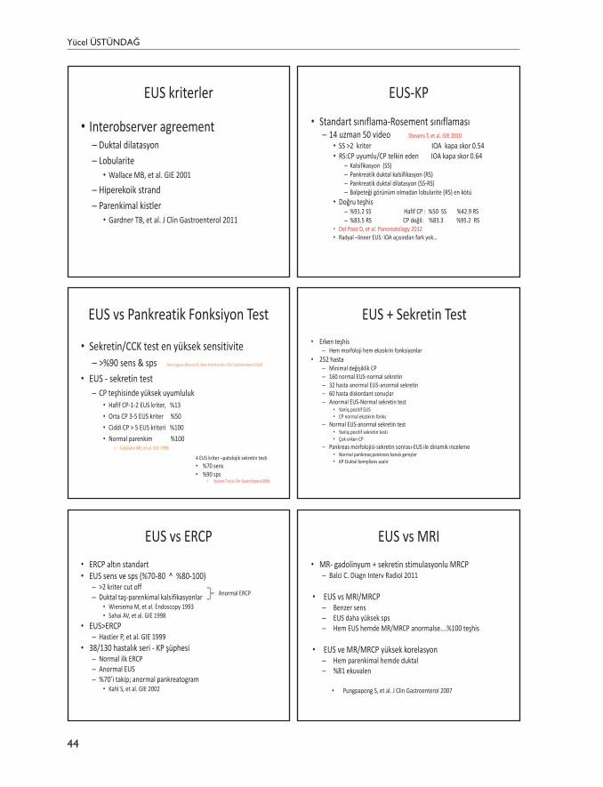
Yücel ÜSTÜNDAĞ
44
EUS kriterler
• Interobserver agreement – Duktal dilatasyon – Lobularite • Wallace MB, et al. GIE 2001
– Hiperekoik strand – Parenkimal kistler • Gardner TB, et al. J Clin Gastroenterol 2011
EUS-KP • Standart s����la�a-Rosement s����la�as� – 14 uzman 50 video Stevens T, et al. GIE 2010
• SS >2 kriter IOA kapa skor 0.54 • RS:CP uyumlu/CP telkin eden IOA kapa skor 0.64
– Kalsifikasyon (SS) – Pankreatik duktal kalsifikasyon (RS) – Pankreatik duktal dilatasyon (SS-RS) – Balpeteği görünüm olmadan lobularite (RS) en kötü
• Doğru teşhis – %91.2 SS Hafif CP : %50 SS %42.9 RS – %83.5 RS CP değil: %83.3 %95.2 RS
• Del Pozo D, et al. Pancreatology 2012 • Radyal –lineer EUS: IOA a��s��da� fark yok…
EUS vs Pankreatik Fonksiyon Test
• Sekretin/CCK test en yüksek sensitivite – >%90 sens & sps Dominguez Munoz JE. Best Practice Res Clin Gastroenterol 2010
• EUS - sekretin test – CP ���������� yüksek uyumluluk
• Hafif CP-1-2 EUS kriter, %13 • Orta CP 3-5 EUS kriter %50 • Ciddi CP > 5 EUS kriteri %100 • Normal parenkim %100
– Catalano MF, et al. GIE 1998
4 EUS kriter –patolojik sekretin testi • %70 sens • %90 sps
• Stevens T et al. Clin Gastro Hepatol 2009
EUS vs ERCP • ERCP altı� standart • EUS sens ve sps (%70-80 ^ %80-100)
– >2 kriter cut off – Duktal taş-parenkimal kalsifikasyonlar
• Wiersema M, et al. Endoscopy 1993 • Sahai AV, et al. GIE 1998
• EUS>ERCP – Hastier P, et al. GIE 1999
• 38/130 hastalık seri - KP şüphesi – Normal ilk ERCP – Anormal EUS – ����i�takip; anormal pankreatogram
• Kahl S, et al. GIE 2002
Anormal ERCP
EUS + Sekretin Test • Erken teşhis
– Hem morfoloji hem ekzokrin fonksiyonlar • 252 hasta
– Minimal değişiklik CP – 160 normal EUS-normal sekretin – 32 hasta anormal EUS-anormal sekretin – 60 hasta diskordant sonuçlar – Anormal EUS-Normal sekretin test
• �a�lış pozitif EUS • CP normal ekzokrin fonks
– Normal EUS-anormal sekretin test • �a�lış pozitif sekretin testi • Çok erken CP
– Pankreas morfolojisi-sekretin s��rası-EUS ile dinamik inceleme • Normal pankreas;pankreas ka�alı �e�işler • KP-Duktal komplians azalır
EUS vs MRI • MR- gadolinyum + sekretin stimulasyonlu MRCP
– Balci C. Diagn Interv Radiol 2011
• EUS vs MRI/MRCP – Benzer sens – EUS daha yüksek sps – Hem EUS hemde MR/MRCP anormalse
• EUS ve MR/MRCP yüksek korelasyon – Hem parenkimal hemde duktal – %81 ekuvalen
• Pungpapong S, et al. J Clin Gastroenterol 2007

Istanbul Endoscopic Ultrasound and Advanced Endoscopy Days • December 10-11 2015, ss. 43-47
45
EUS vs Histoloji
• >3 EUS kriter; histolojiyi öngörebilir…. • Chong AK, et al. GIE 2007
– Asemptomatik alkol tüketicileri-yaşlı insanlar • Stevens T, et al. AJG 2006 • Bhutani M, et al. Pancreas 2009
• Hiperekojenik odak, çizgilenme, lobularite, duktal aberasyon ≈ histoloji – 4 kriter ; cut off • Varadarajulu S, et al. GIE 2007
EUS vs Histoloji
Bazı EUS kriterleri-Ciddi KP histolojisi
• Bal peteği �e��i��e lobulasyon • Hiperekoik odaklar-gölgeli • �e�i� ana kanal • Düzensiz ana kanal konturu • �e�i� yan dallar
– Leblanc JK, et al. Pancreas 2014
EUS-FNA/FNB
• Erken KP �a�ı�ı�da EUS FNA/FNB? – Riskler-���i� histolojik �a�ı�ı
• EUS FNA, EUS negatif prediktivitesini – Hollerbach S, et al. Endoscopy 2001
• Alkoli ilişkili KP-14 hasta-EUS FNA – Tüm örneklerde inflamatuvar hücreler – Asinar yapı korunuyor –hafif/orta değişiklikli EUS – Duktal epitel ve fibrosiz-8-10 EUS kriteri • Iglesias-Garcia J , et al. Gut 2006
EUS elastografi-kontrast enhancement
• EUS kriterleri ile QE korele • Rosement kriterleri – Normal – KP indetermine – KP telkin ediyor – KP uyumlu
• Iglasias-Garcia J, et al. Gastroenterol Heepatol 2011 • Iglasias-Garcia J, et al. Endoscopy 2013
QE DEĞERLERİ
EUS elastografi-kontrast enhancement
• Doku strain • Doku sertse strain azalır • Strain farklı renklerle gösterilir – Mavi sert doku – �ır�ızı y���şak – Normal pankreas homojen �����l�kla yeşil ve sarı
• Strain kantifiye edilir-strain ratio • Kantitatif elastografi (QE)
EUS-QE-KP+PEI
• Domingues Munoz JE, et al. GIE 2015 – 115 KP hasta-35 pankreas ekzokrin yetmezlik (PEI) • KP EUS-MR/MRCP • PEI C13 trigliserid nefes testi • Strain ratio-PEI olan 35 hasta ve olmayan 80 hasta
– SR 4.89 (4.36-5.41) – SR 2.99 (2.82-3.16)
» SR<2.5 (%4.5 PEI) » SR>5.5 (%92.5PEI)

Yücel ÜSTÜNDAĞ
46
EUS-KP şiddeti • ERCP
– görünür yan dallar – Duktal dilatasyon – Duktal düzensizlik – Kalsifikasyon
• Irisawa A, et al. J Gastroenterol 2007 • Samer M, et al. GUT 1984
• EUS
– Intraduktal kalsifikasyonlar %80 PEI – Gölgeli hiperekojenik odaklar – Ana pankreatik kanal dilatasyonu %82 PEI
• Domingues-Munoz JE, et al. Pancreas 2012
KP şiddetli…
Kitle �l�ş����� KP-Pankreas ca • Solid pankreatik mass – Primer-sekonder tm, fokal KP, AIP
• EUS FNA/FNB – KP (+) %54 sns KP (-) %92 sns – Düşük kaliteli örneklem –teknik sorunlar – Tru cut veya yeni procore TM ����l�� deneniyor
• Kontrast enhanced harmonik EUS (CEHEUS)-mikrobubles
– Izoenhanced lezyon – Düşük enhancement-hızlı washout (%89-96) – Time intensity curve analiz
Barthel M, et al. Endoscopy 1996 Ardengh JC, et al. JOP 2007 Fritscher Ravers A, et al.AJG 2002 Varadarajulu S, et al. GIE 2005
Napoleon B, et al. Endoscopy 2010 Fusaroli P, et al. Clin Gastoenterol Hepatol 2010 Gheonea DI, et al. BMC Gastroenterol 2013
KP-kitle ������r�� vs pankreas ca
• EUS elastografi – Kalitatif elastografi: Sens-Sps, PPV, NPV, Accuracy
%100 %85 %90.7 %100 % 94 – Kantitatif elastografi: Bening malign ��r��� %100-
%92.9 – Ortalama hue histogram değeri • Sens %91.4 • Sps %87.9 • Accuracy %89.7
– Saftoi A, et al. Endoscopy 2011
1-Ağrı ! Duktal HT / interstisiyel HT
Lokal oksidatif stres
Lokal inflamasyon
Nociception
Pankreatik nöropati/nöroelastisite
Santral hiperaljezi
Duktal HT/Interstisiyel HT
Oksidatif stres
KP – Ağrı palyasyonu Enzim replasman tedavisi
KP- ağrı • �a�ı doğru mu? • Diğer nedenler
– Gastrik, duodenal veya biliyer tıka�ıklık BT-MR – Pseudokist – Sekonder kanser
• �ğrı�ı� ciddiyeti kantifiye edilir-karekteri �etleştirilir 1. Alkol/sigara kesilir 2. Düşük potans analjezikler verilir 3. Yetersiz ağrı kontrolü varsa �e�iş kanal hastalık varsa endoskopik
ve veya cerrahi tedaviler �la�la�ır 4. ERT ve antioksidanlar 5. Daha potent ajanlar +SSRI veya gabepentin 6. Kanal dilate değilse medikal tedavi devam + total pankreatektomi
+ islet cell transplantasyon

Istanbul Endoscopic Ultrasound and Advanced Endoscopy Days • December 10-11 2015, ss. 43-47
47
Enzim replasman tedavisi (ERT)
• Pankreas-Biyofeedback kontrol – Proteaz-Barsakta CCK salan faktör 1 !
• CCK serum düzeyi artar – pankreatik duktal ���ı�� – Interstisiyel ���ı��
• ERT Hiperstimulasyon - Ağrı kontrolü • Ağrılı CP �l��l�rı�ı� ���ıl�rı��� serum CCK • Nöral/humoral mekanizmalar N
KP-ERT • 6 randomize çift kör plasebo �alış�a – 2 �alış�ada non-enterik kaplı preperat
• �adı�, idyopatik pankreatit, erken evre KP – Staff J, et al. Gastroenterology 1984 – Isaksson G, et al. Dig Dis Sci 1983
– 4 �alış�a enterik kaplı mikrosferik preperatlar • Halgreen H, et al. Scand J Gastro 1986 • Mossner J, et al. Digestion 1982 • Malesci A, et al. Scand J Gastro 1995 • Larvin M, et al. Gastroenterology 1991
• Non-enterik preperatlar-mide asidite – PPI ile beraber kullan
• İleri evre ��e�iş ka�allı KP de etki düşük
iŞE YARAMIYOR !
İŞE YARIYOR !
Malabsorbsiyon • CHO • Protein Enzimler yemek boyunca tüketilmelidirler • Yağ 30 000 IU lipaz/her yemekte
• Yağda çözünür vitaminler • D vit düzeyi • Kemik dansitometri
• Kilo a�ı�ı-gayta kı�a�ı/dışkıda yağ görünümü
Enzyme Products for the Treatment of Chronic Pancreatitis
Product Formulation Lipase Content per Pill or Capsule (USP units) Creon Enteric-coated capsule 3000; 6000; 12,000; 24,000; 36,000 Zenpep Enteric-coated capsule 3000; 5000; 10,000; 15,000; 20,000; 25,000 Pancreaze Enteric-coated capsule 4200; 10,500; 16,800; 21,000 Ultresa Enteric-coated capsule 13,800; 20,700; 23,000 Pertzye Enteric-coated with bicarbonate 8000; 16,000 Viokase Non–enteric-coated tablet* 10,440; 20,880
Nutrisyon-kilo - kalitesi-
Malabsorbsiyon
• Başarısızlık….! – 72 saatlık gayta yağ tayini – Daha potent ajan! – Doz arttırı�ı – Asit supresyonu….���-enterik kaplı prep – Daha sık, küçük miktar �ğ��l�r – Enterik kaplı-nonenterik kaplı formulasyon- shift – Başka ta�ı? – Orta zincirli yağasit r�plas�a�ı

ORAL ABSTRACTS

49
Istanbul Endoscopic Ultrasound and Advanced Endoscopy Days, December 10-11 2015,ss.49 • e-ISBN: 978-975-400-393-2
Imaging Of Pancreatic Cystic Lesions With Confocal Laser Endomicroscopy: An Ex Vivo Pilot Study
A. Kadayifci* 1,2,, M. Atar2, M. Yang3, C. Fernandez-del Castillo4, M. Mino-Kenudson3,W. R. Brugge2
1Gastroenterology, University of Gaziantep, Gaziantep, Turkey2Gastroenterology, 3Pathology, 4Surgery, Mass General Hospital, Harvard Medical School, Boston,
United States
Introduction: The differential diagnosis of pancreatic cystic lesions (PCLs) is an increasingly common clinical challenge. A needle based confocal laser endomicroscopy probe (nCLE, Cellvisio, Mauna Kea) through a 19G needle has been shown to be helpful for differential diagnosis of PCLs by imaging the cyst wall during endoscopic ultrasound-fine needle aspiration. However, clinical experience is still limited, and better image definition and characterization of the cyst wall from different lesions are needed.
Aims & Methods: The aim of this pilot study was to develop a reference standard in CLE characterization of various types of PCLs. Patients who underwent surgery of a PCL were enrolled to study if they gave their consent. During surgery, intravenous fluorescein (2.5 ml of 10%) was injected just prior to the ligation of blood vessels supplying the pancreas. The injected fluorescein was expected to retain for at least an hour after the injection. The freshly specimens were transected along the long axis to fully expose the luminal surface. A Gastroflex-UHD CLE probe (pCLE) was used manually to acquire images directly from the surface of the cyst wall. The cyst walls were screened longitudinally in 2 millimeter ranges by the tip of the probe and sequences recorded for a total 15 minutes. The entire resected specimen subsequently underwent cross sectional histology. All recorded data were analyzed by two investigators for predefined and original image findings of PCLs.
Results: During the 4 months study period, 10 cases (4 male and 6 female) were recruited into the study. The median age of the patients was 77 (range, 33-88). All patients underwent surgery because of a mucinous cyst with worrisome features or a symptomatic PCL. The median duration between fluorescein injection and confocal imaging was 55 minutes (range, 40-90). Imaging was successful in all patients, and various papillary projections with a vascular core and mucinous epithelial borders were visualized in 8 of the patients. In 2 patients, typical vascular network were visualized without papillary structures. The conventional pathological examination confirmed 6 cases with Intraductal Papillary Mucinous Neoplasm (IPMN), 2 cases with Mucinous Cystic Neoplasm (MCN) and 2 cases with serous cysts. Pancreatic ductal adenocarcinoma arising in high grade dysplasia was found in one patient, and high grade dysplasia in 2 patients. The sensitivity and specificity of ex-vivo confocal imaging to demonstrate papillary structures was 100%.
Conclusion: Pancreatic cyst epithelial wall can be visualized successfully by pCLE in ex vivo surgical specimens. Various papillary projections have been seen in all cases of IPMN and MCN in this examination. The variety of images which have been acquired in this model will be helpful for the definition of similar structures during in vivo examination of pancreatic cysts. To develop a reference standard for definition of IPMN subtypes and for grade of differentiation would be the ultimate aim of this model.
Keywords: Confocal endoscopy, pancreas cysts, IPMN
ORAL ABSTRACTS

Istanbul Endoscopic Ultrasound and Advanced Endoscopy Days, December 10-11 2015,ss.50-51 • e-ISBN: 978-975-400-393-2
Endosonography; Differentiation of Malign and Benign Pancreatic Cysts
Aliye Soylu, Hakan Yıldız, Serdal Çakmak, İsa Sevindir, Deniz Koç, Genco Gençdal
Backround: Pancreatic cystic lesions (PCLs) are increasingly found in clinicalpractice because of the widespread use of imaging modalities. Although differantial diagnosis of malign and bening of pancreas cysts is not certain markers; endosonography which has been taken place in cyst management showing morphologic speciality of cyst and of cyst fluids by fine-needle aspiration(FNA). In our clinical EUS and EUS FNA performed patients were evaluated to pancreas cyst features for differenti-ation of premalign and malingn.Method: In our clinic, between may 2014-july 2015 , 60 patients with PCLs undergoing EUS were enrolled this study and retrospectively evaluated. Patients were recorded with undergoing pancreatitis, diabetes mellitus , hematologic and biochemical parameters. EUS evaluation of PCLs were noted according to size, focal irregularity, wall thickness, any papillary projections, relationship of main pancreatic duct, and echo-dens mucus or debris. EUS-FNA fluid was evaluated with biochemical(amylase, CEA, CA 19-9) and cytology.Findins: In this study, patients that enrolled were main age 58,6±14,6 and 26( %43.3) female. EUS-FNA were performed in %40 of patients. Finally, 73.3% of benign cases, 26.7% were found to have malignant or premalignant. In benign and malign/pre-malign groups compared to each others were described value of age(0.002), alkaline phosphatase (p.0.001) and gamma glutamyl transferase (p:0.004) high and hemotoc-rit(p.0.001) and albumin(p.0.001) low in malign groups.Blood tumor markers were not different for the groups, CA 19,9(P.0518), CEA(P.0.376) and CA 125(P.0.518).(Table 1-2-3). Differentiation of PCLs in the bening group were atypical pseudocyst (%61.3), serous cystadenoma(%20.4) and, simple cyst(18.1). In malign group were IPMN(%26.7), %18 mucinous cystic neoplasm , and %25 adeno cancer. The Septation in the benign group were 25% and 56.2% in patients with the malignant group (p=0.023). Cyst lobulation was %25 in the bening groups and %56.2 in the malign group(p=0.014). According to mural nodularity was %2.5 in the benign group and %26.7 in malign group(p=0.005)Conclusion: In majority of patients needed EUS-FNA that eus and clinical investigation were insufficient for final diagnosis were benign. This situation is thought that management of PCLs for clinician poses still problems. At same time , applicable FNA in managemant of EUS patients helped to differentiation of between malign and benign situation.Cholestasis findings in the malign PCLs are more common than the benign groupPCLS with localisation of pancreas head and accompanying anemia were associated with rates of higher malignancyIn our study, presence of mural nodularity and cyst septation in PCLs were found effective in differenti-ation of pre-malign and benign as in other studies. In addition, We think that cyst lobularity may help to differentation of premalignant and benign.
ORAL ABSTRACTS
50

Istanbul Endoscopic Ultrasound and Advanced Endoscopy Days • December 10-11 2015, ss. 50-51
51
Variables Benign(n.44) Malign(n:16) P value
Age 55,6±15,1 67±8,92 0,008
Sex(F/M) 26(59,1)/18(40,9) 8(50)/8(509 0,530
Pancreatitis 20(50) 4(26,7) 0,120
Diabetes 7(18,4) 4(30,8) 0,350
Cyst localization
Head 17(%41,5) 9(%64,3) 0.140
Korpus 16(%39) 4(%28,6) 0.483
Tail 5(%12,2) 1(%7,1) 0.601
Uncinat 3(%7,3) 0 0.298
Cyst Size 29,1±21,1 30,4±17,4 0.598
Cyst lobulation 10(%22,7) 9(56.2) 0.014
Cyst septation 11(%25) 9(%56.2) 0,023
Cyst nodularity 1(%2.5) 4(%25) 0.005
Htc 39,3±3,7 31,8±5,1 0.001
Plt 242±70 276±99,6 0,106
GGT 32(6-107) 80(16-351) 0,004
ALP 82(58-144) 179(82-432) 0,001
ALB 4,3±0,49 3,4±0,65 0.001
Cyst Size 29,1±21,1 30,4±17,4 0.598
Cyst lobulation 10(%22,7) 9(56.2) 0.014
Cyst septation 11(%25) 9(%56.2) 0,023
Cyst nodularity 1(%2.5) 4(%25) 0.005

Istanbul Endoscopic Ultrasound and Advanced Endoscopy Days, December 10-11 2015,ss.52 • e-ISBN: 978-975-400-393-2
Endosonographic evaluation of esophageal and gastric submucosal lesions: Histopathological and
long-term results
Alpaslan Tanoglu1, Orhan Tarçın2, Mustafa Kaplan1
1Gastroenterology, Gata Haydarpasa Training Hospital, Istanbul, Turkey2Gastroenterology, Yeditepe University, Istanbul, Turkey
Background and Aim: Gastrointestinal submucosal lesions (SML) include benign, potentially malignant, and malignant lesions. Usually SML’s are asymptomatic and noticed incidentally. Endosonography (EUS) is the key evaluation method for these lesions. EUS-FNA and EUS-guided tru-cut biopsy has the pivotal role in evaluation of SMLs. In this study, we evaluated the endosonographic features, fine needle biopsy and endoscopic submucosal dissection and/or surgical outcomes of SMLs for a 5-year lasting period. Methods: Patients who were referred for EUS were divided according to lesion size. Large lesions were examined by with radial system and small lesions were evaluated with miniprob endosonography. The lesions originated by the first 3 layer and requiring dissection were referred to endoscopic submucosal dissection. Fine needle aspiration procedure was performed to the fourth layer originated lesions or these patients underwent surgical operation. Suitable patients were included in the follow-up program. Results: 4 of 33 suspected submucosal esophageal lesions (12%) were found to be endosonographically normal, and 10 of 33 lesions (30.3%) were detected compression of the external sources. 5 of all lesions (15%) were mucosal, 2 (6%) were originated from Layer 2, 4 (12%) were from Layer 3 and 8 (24%) were from Layer 4. 4 of 57 (7%) suspected submucosal gastric lesions were found to be endosonographically normal, and 5 of 57 lesions (8.7%) were detected compression of the external sources. 11 of all lesions (19%) were mucosal, 5 (8.7%) were originated from Layer 2, 23 (40%) were from Layer 3 and 8 (14%) were from Layer 4. 5 of 11 mucosal gastric lesion was MALT lymphoma. One lesion was diagnosed as linitis plastica. Due to classification easiness, layer 4 enhancing pancreatic rest lesions were added to lesions of Layer 3. Discussion: SML can occur anywhere in the gastrointestinal tract and are located beneath the normal epithelial layer, which explains why a tissue diagnosis is difficult to obtain with routine biopsies. EUS can distinguish intramural lesion from extramural compression. Moreover, it allows allocation of intramural lesions to a specific layer of the GI wall and offers additional information as to whether a lesion could be benign or malignant. Endosonography also provides the optimal means of tissue acquisition.Keywords: endosonography, gastrointestinal submucosal lesions
ORAL ABSTRACTS
52

53
Istanbul Endoscopic Ultrasound and Advanced Endoscopy Days, December 10-11 2015,ss.53 • e-ISBN: 978-975-400-393-2
Endoscopic ultrasound elastography for differantiation of benign and malign pancreatic lesions
Dilek Oguz1, Engin Altıntas2, Enver Uçbilek3, Fehmi Ates4, Orhan Sezgin5
1dilek oguz, 2engin altintas, 3enver ucbilek, 4fehmi ateş, 5orhan sezgin
Introduction: The diagnosis of focal pancreatic lesions remains challenging despite improvement in imaging methods. Recent data suggest that endoscopic ultrasoun(EUS) elastography can accurately differentiate malignant lesions from benign masses.To aim of this study was to evaluate the accuracy of EUS elastography for diagnosis of focal pancreatic masses
Methods: We analysed 49 consecutive patients solid focal pancreatic lesions. 24 of 49 patients were included prospectively over a year period and studied by EUS with real time elastography. We used color pattern to description of tissue stiffness. Different elasticity values are marked with different colors resulting in different tissue elasticity patterns,represented in color superimposed over the conventional B-mode.The system is set-up hue color map (red-green-blue),where hard tissue areas are shown in dark blue, intermediate tissue areas in green,medium soft tissue areas in yellow, and soft tissue areas in red (Giovannini classification-GESS*).Final diagnosis was based on histopathology, Fine-needle aspiration (FNA) cytology
Results: Blue color pattern and malign histopathology/cytology were found in 15 of 24 patients.Yellow/green color pattern and benign histopathology/cytology were found in 5 of 24 patients.A ROC curve was performed to evaluate the overall predictive value of Giovannini elastography score(GESS).This analysis showed a statistically significant area under the curve of 0.842 with a standard error of 0.129 and p =0.008 (95% CI (0.604-0.966).Maximal Youden index calculated the Giovanni Elastography Grading Scale cut-off value at 4 with a specificity of 75.0% and sensitivity of 93.3%.
Conclusion: EUS elastography is a novel technique for the discrimination of solid pancreatic masses and may be useful complementary tool for EUS-guided FNA
Keywords: pancreas, benign/malign lesions, eus-elastography
ORAL ABSTRACTS
Istanbul Endoscopic Ultrasound and Advanced Endoscopy Days, December 10-11 2015,ss.54 • e-ISBN: 978-975-400-393-2
Endoscopic Ultrasound Guided Fine Needle Aspiration of Lymph Nodes
Mehmet Bektaş1, Atilla Tan2, Orhan Tarçın3, Hasan Erdal1, Koray Ceyhan4, Fatih Karataş1,
Aysun Çalışkan1, Cihan Yurtaydın1
1Ankara University School of Medicine Division of Gastroenterology, Ankara,Turkey2Koc University, School of Medicine Division of Gastroenterology, Istanbul,Turkey
3Yeditepe University, School of Medicine Division of Gastroenterology, Istanbul, Turkey4Ankara University School of Medicine Division of Cytopathology, Ankara,Turkey
Introduction: Endosonographic features for prediction of lymph node malignancy (short axis size > 10mm, round shape, well-demarcated borders, homogeneously hypoechoic) is not satisfactory. Only 25% of malignant lymph nodes have all these features. Endoscopic ultrasound guided fine needle aspiration (EUS-FNA) of lymph nodes increases accuracy.
Aim: Our aim is to evaluate the impact of EUS-FNA on the accurate diagnosis of lymph nodes.
Method: The demographics, lymph node (LN) morphologic features (size, echopattern, borders, shape), location, needle type, number of passes, cytology results and EUS-FNA outcomes of patients with abdominal and mediastinal LNs were retrospectively analyzed. The study period was between 01/2010-06/2015.
Findings: Fifty-eight patients (40 male, 18 female, average age: 56.6 years) with LNs underwent EUS-FNA. The average long axis of lymph nodes was 24.3 mm (8-70mm) and short axis was 16 mm (6-50 mm). 22 ga needle was used in 42 patients, 25 ga needle was used in 17 patients. Average number of passes was 2.19 (range: 1 – 5). The sample obtained for cytologic evaluation was adequate in 93.1% (54/58) of cases. In 4 cases, the sample was inadequate or non-diagnostic for cytologic evaluation. Specific cytopathologic diagnosis was reached in 43.1% (25/58): 14 malignant diseases (adenocarcinoma 7, squamous cell carcinoma 4, lymphoma 2, neuroendocrine tumor 1) and 11 benign diseases (Granulomatous disease 10, Castelman disease 1) were reported. Eight out of 14 malignant LNs (57.1%) had endosonographic malignancy criteria. No complication related to the EUS-FNA reported.
Conclusion:EUS-FNA of LNs is a safe and effective modality with a significant impact in patients with lymphadenopathy. Adequate material for cytopathologic evaluation was obtained in 93.1% of cases. Endosonographic criteria are not adequate to obtain the correct diagnosis. Therefore, EUS-FNA should be performed for every suspicious lymphadenopathy.
Keywords: EUS, FNA, Lymph Nodes
ORAL ABSTRACTS
54

POSTER ABSTRACTS

Istanbul Endoscopic Ultrasound and Advanced Endoscopy Days, December 10-11 2015,ss.56 • e-ISBN: 978-975-400-393-2
POSTER ABSTRACTS
Prospective Evaluation of the Patients with Acute Pancreatitis
Arash Turan, Rovshan Ibishov, Yeşim Alahdab, Haldun Akoğlu, Deniz Güney DumanGastroenterology, Marmara University, Istanbul, Turkey
Description of the characteristics of the patients with acute pancreatitis (AP) in prospective series is lacking in Turkey. Most of the studies rely upon the retrospective review of the hospital files. Aim: To outline the clinical and demographic findings of patients admitted to the emergency room with the diagnosis of AP.Materials-Methods: Patients consecutively admitted to the emergency room with the diagnosis of acute pancreatitis between August and November 2015 were enrolled. The definition of AP required at least 2 of the following criteria: (1) characteristic abdominal pain, (2)amylase and/or lipase more than 3 times the upper limit of normal, or (3) positive results of characteristic cross sectional imaging. Their demographic and clinical variables (age, gender, confounding factors like alcohol usage, concomitant disease, number of previous attacks) were noted. We prospectively studied the severity of their acute pancreatitis using the systemic inflammatory response syndrome (SIRS) criteria and Apache II scores within the first 24 hours of admission.Results: We evaluated a total of 32 patients (10 males, 22 females; mean age, 55 (25-90) years. Mean body mass index was 28.34 (21.45-45.18) 9.3% of patients were alcohol users. Half of the patients had a history of previous pancreatitis attack, 18.75% of patients had cholecystectomy. All patients had Apache II score of 3 or less. Only 7 (21.9%) patients had SIRS but none of the patients had severe pancreatitis according to the Atlanta criteria. Conclusion: Majority of patients with acute pancreatitis admitted to a tertiary reference center in Turkey had mild pancreatitis although one forth of them has fullfilled the SIRS criteria.Keywords: Acute Pancreatitis, Prospective Evaluation
56

57
Istanbul Endoscopic Ultrasound and Advanced Endoscopy Days, December 10-11 2015,ss.57 • e-ISBN: 978-975-400-393-2
POSTER ABSTRACTS
Intracystic hemorrhage during EUS-FNA of a cystic lesion of the pancreas
Atakan Topçu1, Davut Tüney2, Deniz Güney Duman1
1Marmara University Faculty of Medicine, Department of Internal Medicine and Gastroenterology2Marmara University Faculty of Medicine, Department of Radiology
Introduction: Hemorrhage into cystic space is an infrequent complication of EUS guided fine needle aspiration of pancreatic cystic lesions. This should be recognized by the endosonographist because of the risk of fatal outcome. Herein, we present a patient suffered from with immediate bleeding into cyst during an EUS fine needle aspiration (EUS-FNA )procedure and its EUS appearance.Case presentation: Fifty-two year old woman was referred to our EUS clinic because of the 23x33 mm diameter, unilocular, thick-walled, non-contrast enhancing cyst located in the head and the uncinate process of the pancreas diagnosed by abdominal CT. On EUS examination, 24,1x 22,7 mm isolated single clear cyst with regular but thick wall was visible. There was no mass or nodule associated with cyst, and main pancreatic duct was of normal size and appearance. Color flow and Doppler US were performed to exclude vascular structures in the path of the needle and ciprofloxacin was administered intravenously before EUS-FNA. EUS-FNA with 22 G needle was performed. After the aspiration of the initial 2 cc of clear cystic fluid, blood-tinged fluid was observed. The hypoechoic appearance of the cyst (figure 1a) suddenly has turned to hyperechoic dense nature on ultrasound (figure 1b).We have stopped the procedure because of the risk of hemorrhage into cyst and patient experienced abdominal pain despite the conscious sedation. She was observed in the emergency ambulatory care unit for 12 hours and discharged with orally administered ciprofloxacin for 5 days (500 mg, twice a day). In the evaluation of the aspirated material, low CEA (2,5 ng/ml) and high amylase (714 U/L) levels suggestive of pseudocyst were detected. Cytology results were suggestive of pseudocyst.Abdominal computerized tomography taken after the EUS procedure revealed a 37x30 mm diameter thick-walled cystic mass with a bleeding area between the head and the uncinate process was present. The platelet count and the international normalized ratio, determined before EUS, were within normal limits. No history of nonsteroidal anti-inflammatory drug (NSAID), including aspirin, were present during the week preceding EUS-FNA Additionally hematology consultation for the rare causes of bleeding disorders did not reveal any abnormality.Discussion: EUS-FNA is mainstay of diagnosis for the pancreatic cysts. The opacification of the cyst during aspiration is rare condition described in up to 6 % of patients undergoing EUS-FNA (1). During the EUS-FNA sudden change of hypoechoic content of the cyst into hyperechoic pattern should bring to mind the possibility of bleeding into the cystic space.Keywords: Pancreatic cyst, hemorrhage, Eus
References1 Varadarajulu S, Eloubeidi MA. Frequency and significance of acute intracystic hemorrhage during EUS-FNA of
cystic lesions of the pancreas. Gastrointest Endosc. 2004 Oct;60:631-5.
Figure 1Pancreatic cyst in eusa.Before aspiration. b.After aspiration

Istanbul Endoscopic Ultrasound and Advanced Endoscopy Days, December 10-11 2015,ss.56 • e-ISBN: 978-975-400-393-2
Medical treatment of chylothorax and chylous ascites as a late complication of acute necrotizing pancreatitis
Dilek Oguz1, Arif Ozdemir4, Elvan Uludag2, Ismail Hakkı Kalkan1, Burhan Sahin3
1Kırıkkale Üniversitesi2The private hospital of 19 May
3AGEM medical center4Memorial Hospital Ankara
Several late period complications can occur according to the severity of the pancreatic necrosis. Chylothorax, chylous ascites are extremely rare complications of acute necrotizing pancreatitis. These complications are always hard to overcome for clinicians. We presented the case with severe acute necrotizing pancreatitis. that developed chylothorax and chylous ascites.Case presentation: 55-year-old male patient was admitted to our intensive care unit (ICU) due to acute necrotizing biliary pancreatitis.Supportive therapy was initiated in ICU.In the follow-up of the patient in the ICU, an abdominal ultrasonography revealed a pseudocyst of size 16x10x9 cm covering whole.A cystogastrostomy and cholecystectomy operation was performed. After a week it was performed stent insertion to pseudocyst by endoscopy. Two months later was resolved pseudocyst and the patient was discharged.In follow up period( amonth later) the patient was complained vomiting and performed gastroscopy and barium graphy. Gastroscopy and barium graphy was showed us obstruction of second part of duodenum due to intraperitoneal inflammation. Surgeons was performed by pass surgery then the patient was follow up.After six-month asymptomatic period chylothorax and chylous ascites was detected in the patient who was admitted to outpatient clinic because of dyspnea and abdominal distension. A chest tube was placed due to chylothorax. The patient’s oral intake was stopped and total parenteral nutrition (TPN) was started. Initially 700 ml chylous fluid was drained and it gradually decreased to 100 ml/day in the follow-up. After the beginning of oral nutrition chylous fluid was increased again.To avoid disadvantages of TPN, Golden Goat and Peptisorb oral solutions- rich in medium-chain triacylglycerols was started.The volume of chyle drained from chest tube cessated on the 15th day with this treatment and the patient was discharged.Conclusion: Chylothorax and chylous ascites are rare conditions that can be observed after acute necrotizing pancreatitis. There is no consensus on the treatment of chyothorax and chylous ascites.Transition to a low-fat diet with MCT and parenteral nutrition can be effective in the treatment of this disease with high morbidityKeywords: Acute pancreatitis, chylous ascites
POSTER ABSTRACTS
58

59
Istanbul Endoscopic Ultrasound and Advanced Endoscopy Days, December 10-11 2015,ss.59-60 • e-ISBN: 978-975-400-393-2
Role of Endosonography in the Upper Gastrointestinal Subepithelial Lesions
Hakan Yıldız, Aliye Soylu, Serdal Çakmak, İsa Sevindir, Deniz Koç, Genço Gençdal
Introduction: Subepithelial lesions(SEL) are often covered with normal mucosa, asymptomatic and found incidentally during endoscopy. SEL is encountered in approximately 1 of every 500 endoscopy. Endosonography(EUS) is the most important diagnostic tool for optimal treatment and management of the SEL. In this study, we published the results of our clinic that patients underwent EUS and follow-up for the SEL.Method: In our clinic, EUS were performed to patients by Olympus Linear / Radial endosonography device. All patients were recorded by sex, age, lesion site, size, layer, echo pattern, calcification, cystic cavities, infiltration of serosa and lesion- edge characteristics. EUS fine needle aspiration (FNA), and operations data were found from medical recordsFindings: A total of 79 patients with SEL preliminary diagnosis were performed by EUS . The mean age 52.9 ± 15.3, 38 (48.1%) were female and 41 (51.9%) were male patients with an average of 16 months (3-22 months) was observed. Mean lesion size were 20.9(6-90) mm. EUS-FNA was performed in 17(%26.9) patients.. In the upper gastrointestinal system, the most common SEL in encountered with EUS were first gastrointestinal stromal tumor(GIST) and second extraluminal compression 16(%20.3)(table-1). When compared to extraluminal compression according to the age and sex of SMT was no statistically significant difference(table-2). When SEL was divided according to the anatomical region.The most frequent lesion in the esophagus leiomyoma 61.1%, GIST %58.5 in the stomach and duodenum was lipoma with 75% (table 3).Results: EUS that due to description of layer, size ,echo pattern and biopsy sampling in SEL is the most effective tool in the diagnosis and treatment decisions SEL. We believe the diagnosis and treatment is carried out effectively, when Patients with SEL are evaluated by EUS/EUS-FNA
Table 1
EUS results n:79GIST 26(%32.9)Leiomyoma 14(%17.7)Lipoma 8(%10.1)Carsinoid 1(%1.3)Cavernous hemangioma 1(%1.3)Duplication cyst 3(%3.8)Granular hc tumor 1(%1.3)Aberrant pancreas 9(%11.4)External pressure 16(%20.3)
POSTER ABSTRACTS

Hakan Yıldız, Aliye SoYlu, Serdal ÇAkmAk, İsa Sevİndİr, deniz koÇ, Genço Gençdal
60
Table-2
Variables SEL External pressure P valueAGE 51±15.5 49.4±14.3 0.280SEX(F/M) 31(81.6)/32(%78) 7(18.4)/9(22) 0.696
Table-3
Esophagus (n:18) Stomach(n:41) Duodenum (n:4)GIST/leimyom %66.7 %65.8 %25Lipoma %16.7 %4.9 %75Karsinoid 0 %2.4Cavernöz hemangioma %5.6 0 0Duplication cyst %5.6 %4.9 0Granular hc tumor %5.6 0 0Aberrant pancreas 0 %22 0

61
Istanbul Endoscopic Ultrasound and Advanced Endoscopy Days, December 10-11 2015,ss.61-62 • e-ISBN: 978-975-400-393-2
The Diagnostic Reliability of Endoscopic Ultrasonography (EUS) Guided Fine Needle Aspiration
Biopsy (FNA) of Pancreatic Lesions
Hande Mollamemişoğlu1, Gamze Akbaş1, Ender Dulundu3, Deniz Güney Duman2, Pelin Bağcı1, Çiğdem Ataizi Çelikel1
1pathology; marmara university school of medicine; istanbul; turkey2gastroenterology; marmara university school of medicine; istanbul; turkey3general surgery; marmara univercity school of medicine; istanbul; turkey
Introduction: The diagnostic usage of EUS procedure in pancreas has been fairly increased in our daily practice. We documented EUS guided FNA cases, and compared the diagnostic concordance between EUS guided FNA and tissue biopsiesMaterial and Method: Archives of our department screened between 2010-2015 for pancreatic EUS guided FNAs.Results: The diagnostic distribution of EUS guided FNA (n=108) and following biopsy/resections are shown in the table.F/M ratio was 0,8 (49/59) and the mean age was 53 (84-13). The cases diagnosed as malign were; 16% (n=17) solid tumors (adenocarcinoma, neuroendocrine tumor, metastatic carcinoma, GIST, lymphoma); and 3% (n=3) cystic lesions (mucinous cystic neoplasia).The reasons of nondiagnostic FNA results (n=39) were; crush artefact in 3% (n=1), hypocellularity in 36% (n=14), and acellularity in 62% (n=24). The localization of the lesion of these unsatisfactory cases were; 23% (n=9) pancreatic head, 15% (n=6) pancreatic corpus, and 10% (n=4) pancreatic tail.Discussion: Pancreatic EUS-FNA is a problematic procedure, mainly due to the nature of the lesions; as such the lesions can be both cystic and solid; and cystic lesions have a wide spectrum from benign to malignant. Another factor for diagnostic difficulty is the contamination from the adjacent regions (stomach/duodenum).We had the opportunity to study 44 resections out of 108 FNA biopsies. Our diagnostic concordance was 100% for the cases that were positive for malignancy on FNA. Seventy-five percent (n=3/4) of the cases classified as “presence of neoplastic cells”; turned out to be benign cystic lesions in the resection, such as serous cystadenoma and lymphoepithelial cyst. This heterogeneity could be explained as, this group contains both benign and malignant cystic lesions by definition.Eighteen percent (n=2/11) of the cases classified as “negative for malignancy”; turned out to be malignant at the resection. The FNA slides of these cases showed peritumoral nonspesific chronic pancreatitis. These results shows us; the group “presence of neoplastic cells; benign/malignant” is a very heterogenous one, and could be misinterpreted or misunderstood easily by the clinicians.Keywords: EUS, FNA, pancreas, adenocarcinoma, diagnostic
POSTER ABSTRACTS

Hande MollaMeMişoğlu, Gamze akbaş, Ender DulunDu, Deniz Güney DuMan, Pelin bağcı, Çiğdem ataizi Çelikel
62
Table 1. Diagnostic Distribution of FNA & Resections
FNA(n=108) FNA(n=108) RESECTION(n=44) RESECTION(n=44)DIAGNOSIS n (%) Malignant,n (%) Benign,n (%)NON DIAGNOSTIC 39 (36) 6 (37) 10 (63)NEGATIVE FOR MALIGNANCY 30 (28) 2 (18) 9 (82)PRESENCE OF ATYPICAL CELLS 4 (4) 1 (50) 1 (50)PRESENCE OF NEOPLASTIC CELLS; BENIGN/ MALIGNANT 11 (10) 1 (25) 3 (75)
SUSPICIOUS FOR MALIGNANCY 3 (3) 0 0POSITIVE FOR MALIGNANCY 21 (19) 8 (100) 0

63
Istanbul Endoscopic Ultrasound and Advanced Endoscopy Days, December 10-11 2015,ss.63 • e-ISBN: 978-975-400-393-2
Esophageal Abscess: A Rare Condition Mimicking a Subepithelial Lesion
Mehmet Bektaş1, Koray Ceyhan2, Atilla Tan3, Orhan Tarçın4, Fatih Karataş1,Aysun Çalışkan1, Cihan Yurtaydın1
1Ankara University School of Medicine Division of Gastroenterology, Ankara,Turkey2Ankara University School of Medicine Division of Cytopathology, Ankara,Turkey3Koc University, School of Medicine Division of Gastroenterology, Istanbul,Turkey
4Yeditepe University, School of Medicine Division of Gastroenterology, Istanbul,Turkey
Introduction: The endoscopic frequency of a subepithelial lesion is 1/300. Approximately, 10% of submucosal lesions are located within the esophagus. Esophageal submucosal lesions can be classified as leiomyoma, granular cell tumor, duplication cyst, lipoma and varices. An abscess mimicking an esophageal submucosal lesion was not previously reported. Case: A 38 year-old male patient had an upper endoscopy to evaluate sudden onset of odynophagia and throat pain. It revealed a 2 cm subepithelial lesion with a normal overlying esophageal mucosa, which was located 3 cm below the upper esophageal sphincter. Radial endoscopic ultrasound evaluation showed a 6 mm x 11.5 mm partially heterogeneous, hypoechoic, well-demarcated lesion likely originating from muscularis mucosa. Doppler evaluation was negative for vascularity, and no peri-esophageal lymph nodes noted. Elastrographic evaluation revealed a dark blue color. EUS-FNA was performed with a 22 ga needle (2 passes). A soft consistency material was noted on the slide. Cytopathologic evaluation revealed active suppurative inflammatory findings consistent with abscess. A subsequent magnetic resonance imaging showed a 5 mm x 12 mm lesion at the proximal esophagus consistent with an abscess. The patient subsequently mentioned that his symptoms started after eating fish almost a week ago. It was thought that the abscess was possibly developed following a fish bone penetration to the proximal esophagus. The patient’s odynophagia improved with two-week treatment with ciprofloxacin and metronidazole. In conclusion, esophageal abscess should be kept in the differential diagnosis in patients with a history of sudden onset of odynaphagia and a subepithelial lesion on endoscopyKeywords: Esophageal Abscess, Subepithelial Lesion, EUS
POSTER ABSTRACTS

Istanbul Endoscopic Ultrasound and Advanced Endoscopy Days, December 10-11 2015,ss.64 • e-ISBN: 978-975-400-393-2
Peripancreatic Unicentric Castelman’s Disease Diagnosed with Endoscopic Ultrasound Guided Fine
Needle Aspiration
Mehmet Bektaş1, Atilla Tan2, Koray Ceyhan3, Orhan Tarçın4, Fatih Karataş1,Aysun Çalışkan1, Cihan Yurtaydın1
1Ankara University School of Medicine Division of Gastroenterology, Ankara,Turkey2Koc University, School of Medicine Division of Gastroenterology, Istanbul,Turkey3Ankara University School of Medicine Division of Cytopathology, Ankara,Turkey
4Yeditepe University, School of Medicine Division of Gastroenterology, Istanbul,Turkey
Introduction: Castelman’s disease is a rare disease with usually mediastinally located lymphoproliferative disease. Peripancreatic types are even quite rare. Endoscopic ultrasound guided Fine-needle aspiration is a tool for the diagnosis of Castelman’s disease. Case: A 42 year-old female patient had an abdominal ultrasound for the evaluation of left abdominal pain and nausea revealed a 33 mm x 39 mm solid lesion at the tail region of the pancreas. A computerized tomography showed a contrast enhancing mass (37 mm x 47 mm) extending from the inferior of the pancreas to the left para-aortic left renal vein. Radial endosonographic evaluation revealed a perigastric 31 mm x 35 mm hypoechoic, well-demarcated lesion, which was similar to a lymph node. A smaller lymph node was also noted at the proximity of the large lesion. EUS-FNA of the large lesion with a 22 ga needle was performed (3 passes). Adequate samples were obtained for cytology, cell-block and FISH. Cytology revealed relative hyperplasia. FISH showed mixed type lymphoid population without a monoclonality (58% B cells, 42% T cells). Immunehistochemistry was suspicious for a low grade lymphoproliferative disease, but a lymphoid malignity could not be proved. The patient subsequently underwent laparoscopic lymph node excision, which revealed a hyaline type Castelman’s disease. In conclusion, a unicentric Castelman’s disease endosonographically appear as a homogeneous, well-demarcated, hypoechoic lesion. Cyopathologic differentiation from other lymphoma types with EUS-FNA samples is quite difficult.Keywords: Castelman’s Disease, EUS, FNA
POSTER ABSTRACTS
64

65
Istanbul Endoscopic Ultrasound and Advanced Endoscopy Days, December 10-11 2015,ss.65 • e-ISBN: 978-975-400-393-2
Thyroid Follicular Cell Carcinoma Diagnosed with Endoscopic Ultrasound Guided Fine Needle Aspiration
Mehmet Bektaş1, Orhan Tarçın2, Atilla Tan3, Koray Ceyhan4, Fatih Karataş1,Aysun Çalışkan1, Cihan Yurtaydın1
1Ankara University School of Medicine Division of Gastroenterology, Ankara,Turkey2Yeditepe University, School of Medicine Division of Gastroenterology, Istanbul,Turkey
3Koc University, School of Medicine Division of Gastroenterology, Istanbul,Turkey4Ankara University School of Medicine Division of Cytopathology, Ankara,Turkey
Introduction: Follicular cell-derived carcinomas of the thyroid gland comprise a heterogeneous group of malignant neoplasms of the thyroid gland with varied histologic appearance and molecular profiles. Fine-needle aspiration is considered an essential tool for the diagnosis of suspicious thyroid nodules.Case: A 71 year-old female patient with a history of multiple myeloma underwent a magnetic resonance imaging (MR) for the evaluation of neck pain and cough. MR revealed a 41 mm x 28 mm lobulated soft tissue lesion with homogeneous contrast enhancement at the superior mediastinum, posterior of trachea. The lesion could not be separated from the right lobe of the thyroid gland and the signal intensity was similar to the thyroid gland’s signal. Given the fact that the fat plane between the trachea and esophagus was lost, the patient was referred for endosonographic evaluation. The upper endoscopy revealed normal esophageal mucosa without any submucosal lesions or extrinsic compression. Radial endosonographic evaluation revealed an upper mediastinal (18-20 cm from incisors), well-demarcated, homogeneously hypoechoic solid lesion (28 mm x 35 mm). The lesion was out of the esophagus. The echo-texture was similar to the thyroid gland’s echo-texture. The elastrographic evaluation of the lesion revealed a yellow-green heterogenous pattern, with a 1.5 strain ratio. Doppler was used to avoid vascular structures. The cytologic evaluation of the sample obtained with a 25 ga needle revealed follicular (hurtle cell) thyroid neoplasia. Immunehistochemical evaluation confirmed the diagnosis. Conclussion: EUS-FNA is a safe tool that can be used for detection and diagnosis of primary and metastatic thyroid lesions.Keywords: Thyroid Follicular Cell Carcinoma, EUS, FNA
POSTER ABSTRACTS

Istanbul Endoscopic Ultrasound and Advanced Endoscopy Days, December 10-11 2015,ss.66 • e-ISBN: 978-975-400-393-2
Large pedunculated esophageal lipoma presented with dysphagia and reflux symptoms
Mete Akin1, Altug Senol2
1Akdeniz University, School of Medicine, Department of Gastroenterology, Antalya, Turkey2Süleyman Demirel University, School of Medicine, Department of Gastroenterology, Isparta, Turkey
Introduction: Lipoma of the esophagus is a rare lesion and usually detect incidentally during endoscopy. It is generally asymptomatic and requried no therapy. However, large esophageal lipoma may cause disphagy and reflux sypmtoms. Endoscopic or surgical excision may require in symptomatic cases. In this report, a large pedunculated esophageal lipoma was reported, which caused esopgaheal obstruction and treated by endoscopic excision.Case: A 55-year-old female patient applied to the Gastroenterology clinic because of dysphagia, reflux symptoms and weight loss for the last 6 months. She had Type 2 diabetes mellitus in her medical history. Physical examination and laboratory findings were normal. Upper gastrointestinal endoscopy showed an approximately 4 cm in diameter, pedunculated, polypoid mass lesion with a normal mucosal lining, which located and caused partially obstruction at the proxymal esophagus (Figure 1). Endoscopic ultrasonography (EUS) examination revealed a iso-hyprechoic mass lesion originating from submucosa and a major blood vessel was not observed in pedicle (Figure 2). Due to these findings, endoscopic excision with polypectomy snare was performed without any complication. Histopatologically the lesion was reported as a lipoma. The patient’s symptoms improved after the procedure. Conclusion: Lipoma is the second most common benign tumor of the gastrointestinal tract and usually located in colon and gastric antrum. It may be rarely seen in esophagus and usually detect as small submucosal lesion. However, lesions larger than 2 cm in size may cause symptoms such as bleeding, abdominal pain,obstruction and intussusception. EUS is an important method for differantial diagnosis and characterisation of lesions and guidance to therapy. Treatment options can be endoscopic or surgical excision according to the size and localisation of the lesion.Keywords: Esophagus, lipoma, endoscopic ultrasonography, endoscopic excision
POSTER ABSTRACTS
Figure 1. Large pedunculated polypoid mass lesion with a normal mucosallining at the proxymal esophagus in the upper gastrointestinal endoscopy
Figure 2. A iso-hyprechoic mass lesion originating from submucosa in the lineer EUS examination
66

67
Istanbul Endoscopic Ultrasound and Advanced Endoscopy Days, December 10-11 2015,ss.67 • e-ISBN: 978-975-400-393-2
Peritoniotlerde peritonial mohteviyyatdageden biofizik prosesler
Mushvig Celal Hasanli
Azerbaycan Tibb Universteti,Tedris cerrahiyye klinikasi,Medi-lux klinikasi.
Karın boşluğunun acil cerrahi müdahale gerektiren hastalıklarından en tehlikelisi yayılmış irinli peritonitlerdır. Cünki bu zaman periton boşluğuna toplanan muhteva yanlışlığının bünyesinde gerçekleşen biyokimyasal ve biofiziki işlemler sonucunda yüksek toksik etkiye sahip maddeler oluşur ve bunların vücuda emilmesi nedeniyle endotoksikoz oluşur.Amaç: Laparotomik ve Laparoskopik ameliyatlar sırasında karın boşluğundan alınan peritoneal muhteva yanlışlığının bünyesinde yaşanan biofiziki prossesleri araştırmak olmuştur. Malzeme ve Yöntemler: Araştırma endotosikozla eşlik edilen 40 kişi hastada peritonit nedeniyle yapılan laparotomik ve laparoskopik cerrahi işlem sırasında karın boşluğundan alınmış sıvıda aparılmışdır.Hastaların büyük çoğunluğu 35-70 yaş arasında olmuşdur. Peritonitin oluşma nedeni
1.Mide ülserinin perforasiyası 8 (% 20)2.Safra kasasının perforasiyasi 9 (22,5%)3.Apendiksin perforasiyasi 5 (12,5%)4. İnce bagırsak kecmemezliyi 8 (% 20)5.Yumurtalıkların katlanmış kistasının patlaması 4 (10%)6.12 parmak bağırsağın tikişlərinin tutarsızlığı 3 (% 7.5)7.Rahim ekstripasiyasından sonraki dikiş tutarsızlığı 3 (% 7.5)
Toplam 40 hasta- 17 si kadın, 23’ü erkek olmuşdur.Sıvının toksikliyi TL Pafomov yöntemi ile, en yüksek toksik etkiye sahip maddelerden Triasilgliseridlər, doymuş ve doymamış yağ asitleri (LMOsagraya 1982) yöntemi ve lipidler peroksidləşməsi ürünlerini ise Uchiyama, Michara, Stalnayanin önerdiği yöntemlerle tayin edilmisdir.Arasdırma sonucunda anlaşılmıştır ki, sıvıda malon dialdehidinin katılığı 130-150,5 mmol / l, hidrojen peroksidinin katılığı 0,142-0,236mmol / l, dk katılığı 2,61-3,0 mmol / l tespit edilmiştir. Nəticə.Peritonitler sırasında karın boşluğundaki sıvıda çok fazlı biofiziki ve biyokimyasal prossesler oluşur.Ilkin aşamada lipidler serbest peroksidleşmesi ürünleri yükseliyor, sonradan ayrilan triasilgliseridlər ve doymuş doymamış yağ asitleri amino asitler hidrolize uğradır. Bu göstergeler endotoksikozu ve abdominal sepsisin ilk proqnostik göstergesi gibi deyerlendirilebiliy.Anahtar kelimeler: Peritonis.abdomenKeywords: Peritonits, abdomen
POSTER ABSTRACTS

Istanbul Endoscopic Ultrasound and Advanced Endoscopy Days, December 10-11 2015,ss.68 • e-ISBN: 978-975-400-393-2
Peritonitle agirlasmis akut apandisitlerde Laporoskopik apendiktomiye gosterisler
Mushvig Celal Hasanli
Azerbaycan Tibb Universteti,Tedris cerrahiyye klinikasi,Medi-lux klinikasi.
Amac: Tagdim etdiyimiz elmi arasdirmada akut apandisitlin deyisik formalarinda,ayni zamanda peritonitle agirlasmis formalarinda Laparoskopik apendiktomiyanin gostericilernin secimi,yerine yetirilme texnikasi ve neticelerin arasdirilmasidir. Material ve metod.Arasdirma :2006-2014 cu illerde klinikamiza akut apendisit diagnozu ile kabul edilmis ve bunlardan 186(50,18%)uzerinde laporoskopik apendiktomiya yapilmisdir.Bunlardan kataral formali apendisit 2-hastada(1,07%),flegmanoz apendisit 147(79,0%)hastada,gangrenoz apendisit 5-kiside(2,6%)teskil etmisdir.Emeliyyat umumi kabul olunmus gaydada aparilmisdir,3-troakar(1-10mm,2-5mm)istifade etmekle yerine yetirilmisdir.Emeliyyat oncesi seftriakson1,0 ezele dahiline vurulmusdur.Butun emeliyyatlar genel anesteziya altinda aparilmisdir(ETN).Emeliyyatlarin davam muddeti 20dak-80dak olmusdur.Karin dahili muayine zamani 46hastada yerli seroz peritonit,2hastada seroz-irinli peritonit,4-hastada ise diffuz seroz irinli peritonit askar edilmisdir.Apendikluyar cixintinin esasinda 16-kiside infiltrativ deyisiklik askar edilmisdir.Bu hastalarda ligatur usulla apendiktomiya yapilmamis,apedikluyar cixitinin proyeksiyasina uygun olan 2-3 sm kesik aparilmis De Kok usulu ile apendiktomiya icra edilmisdir.Butun emeliyyatlardan sonra garin bislugu drenaj olunmusdur.Emeliyyatdan sonra garin bolugundaki her hansi bir agirlasmani prognozlasdormag ucun drenaj borudan haric olan mohteviyyat muayine edilmisdir.Netice: Laporoskopik usulla apendiktomiyya yalniz kataral ve flegmanoz formali apendisitlerde deyil,gangrenooz-perforativ apendisitlerde aparilmasi mumkundur.Yerlivediffuz pertonitlerde laparoskopik emeliyyatin icrasi eks gosteris hesab edilmir.Tedgigatlar onu neticeye gosterirki, garin boslugunda adekvat sanasiya aparilmasi adekvat drenajlanmasi ve emeliyyatdan sonra erken ve gecikmis agirlasmalarin azalmasi acig usulla aparilan emeliyyatlarla mukayisede,gangrenoz,gangrenoz-perforativ apendisitlerde de laporoskopik apendiktomiya mumumkundur.Keywords: Apendisit, peritonit
POSTER ABSTRACTS
68

Istanbul Endoscopic Ultrasound and Advanced Endoscopy Days, December 10-11 2015,ss.69 • e-ISBN: 978-975-400-393-2
Detection of Impacted Stones in Papilla by EUS
Nevin Oruc, Ahmet Aydın, Fatih Tekin, Ilker Turan, Omer Ozutemiz
Ege Üniversitesi Tıp Fakültesi Gastroenteroloji BD, Bornova, İzmir
Introduction: Biliary symptoms, pancreatitis history or elevated liver function tests might lead to investigation of biliary sytem for stones and other pathologies. Biliary stones are usually detected with abdominal ultrasound, MRCP or EUS. EUS is usually superior to MRCP especially in detection of small biliary stones in common bile duct. However detection of impacted stones in papillary area might be challenging. Here we presented a case series with impacted papillary stones and their EUS findings are discussed.Methods: Patients evaluated by both EUS and MRCP for biliary stones between Oct 2014-Oct 2015 were evaluated and 6 patients with impacted papillary stones were included to the study. Results: Three patients had elevated liver function test and history of biliary pancreatitis while 2 patients had only slightly elevated liver function tests and one patient had normal liver function tests beside slightly dilated common bile duct in an abdominal ultrasound imaging. MRCP did not show biliary stones in any of those patients while EUS evaluation showed impacted papillary stones. The hyperechoic appearing stone in hypoechoic papilla was very typical. Conclusion: Biliary stones impacted to papilla might lead to cholestatic changes as well as dilated common bile duct. MRCP is not able to detect small stones in periampullary area. Meticulous control of papillary area with EUS could show small impacted stones. Impacted stones has typical hyperechoic appearance with shadow in papillary area.Keywords: eus, biliary stone
POSTER ABSTRACTS
69

Istanbul Endoscopic Ultrasound and Advanced Endoscopy Days, December 10-11 2015,ss.70 • e-ISBN: 978-975-400-393-2
POSTER ABSTRACTS
Predictive factors for post-ERCP pancreatitis: a tertiary referal center study
Oguz D., Sezgin O., Altıntaş E., Üçbilek E., Ateş F.
ABSTRACTBackground: Postendoscopic cholangiopancreatography (Post-ERCP) pancreatitis is still the most common and severe complication after ERCP. The aim of the study is to investigate the cases of Post-ERCP pancreatitis patients who underwent diagnostic/therapeutic ERCP in our tertiary referal center from 2001 to 2015 and analyse patient-related and procedure-related risk factors for Post-ERCP pancreatitis.Patıents and Methods: The ERCP process of a total number of 2379 patients were considered. For each ERCP in our center, a stardard form for prospective records is filled and is retrospectively analyzed. The diagnosis of pancreatitis according to ‘2 out of 3’ of the following criteria; laboratory (amylase level > 3x upper limit of normal), clinical (upper abdominal pain) and/or imaging criteria level. The impact of the risk factors on Post-ERCP pancreatitis was investigated by univariate and multivariate analysis. Results: The overall incidence of Post-ERCP pancreatitis was 8,6% (206/2379). Gender, age and metal stent placement did not turn out to be risk factors. Both univariate and multivariate analysis showed that history of acute pancreatitis, needle-knife papillotomy, pre-cut sphincterotomy, repeated cannulation, opacification of pancreatic ductules, balloon dilation of biliary sphincter and suspected sphincter of Oddi dysfunction are independent risk factors for pancreatitis.Conclusıons: With the research several patient-related and procedural risk factors, which guide the endoscopist in prophylaxis and management of pancreatitis, has been identified.
70

Istanbul Endoscopic Ultrasound and Advanced Endoscopy Days, December 10-11 2015,ss.71 • e-ISBN: 978-975-400-393-2
Factors Influencing Endoscopic Ultrasound Guided Fine Needle Aspiration Results of Gastrointestinal
Submucosal Lesions
Serkan Dumanlı1, Mehmet Bektaş1, Atilla Tan2, Orhan Tarçın3, Koray Ceyhan4, Fatih Karataş1, Aysun Çalışkan1, Cihan Yurtaydın1
1Ankara University School of Medicine Division of Gastroenterology, Ankara,Turkey2Koc University, School of Medicine Division of Gastroenterology, Istanbul,Turkey
3Yeditepe University, School of Medicine Division of Gastroenterology, Istanbul,Turkey4Ankara University School of Medicine Division of Cytopathology, Ankara,Turkey
Introduction: Follicular cell-derived carcinomas of the thyroid gland comprise a heterogeneous group of malignant neoplasms of the thyroid gland with varied histologic appearance and molecular profiles. Fine-needle aspiration is considered an essential tool for the diagnosis of suspicious thyroid nodules.Case: A 71 year-old female patient with a history of multiple myeloma underwent a magnetic resonance imaging (MR) for the evaluation of neck pain and cough. MR revealed a 41 mm x 28 mm lobulated soft tissue lesion with homogeneous contrast enhancement at the superior mediastinum, posterior of trachea. The lesion could not be separated from the right lobe of the thyroid gland and the signal intensity was similar to the thyroid gland’s signal. Given the fact that the fat plane between the trachea and esophagus was lost, the patient was referred for endosonographic evaluation. The upper endoscopy revealed normal esophageal mucosa without any submucosal lesions or extrinsic compression. Radial endosonographic evaluation revealed an upper mediastinal (18-20 cm from incisors), well-demarcated, homogeneously hypoechoic solid lesion (28 mm x 35 mm). The lesion was out of the esophagus. The echo-texture was similar to the thyroid gland’s echo-texture. The elastrographic evaluation of the lesion revealed a yellow-green heterogenous pattern, with a 1.5 strain ratio. Doppler was used to avoid vascular structures. The cytologic evaluation of the sample obtained with a 25 ga needle revealed follicular (hurtle cell) thyroid neoplasia. Immunehistochemical evaluation confirmed the diagnosis. Conclussion: EUS-FNA is a safe tool that can be used for detection and diagnosis of primary and metastatic thyroid lesions.Keywords: Thyroid Follicular Cell Carcinoma, EUS, FNA
POSTER ABSTRACTS
71

Istanbul Endoscopic Ultrasound and Advanced Endoscopy Days, December 10-11 2015,ss.72 • e-ISBN: 978-975-400-393-2
Rare presentation of castleman’s disease with abdominal mass and lymphadenopathy
Sıdıka Zeynep Gök Sargın, Mehmet Cindoruk, Tarkan Karakan
Gastroenterology clinic, Gazi University Medical Faculty Hospital, Ankara, Turkey
Castleman’s disease (CD) is a rare benign disease of unknown etiology characterized by lymphoid hyperplasia. Multiple variants of this disease exist, with differentiation based on tissue pathology and number of masses within the body. This rare disease is usually found in mediastinal area asymptomatically and incidentally. It is also rare to see this tumor in the abdomen. In this study, we report a 64-year-old man who referred just with feet swelling. Laboratory findings reported microcytic anemia (Hemoglobin 9.1 g/dl, MCV: 72) and hypoalbuminemia (1.5 gr/dl). Abdominal computed tomography imaging showed 5x4 cm mass in pancreas neighborhood. Around the mass in paraceliac lymph nodes region, multiple lymphadenopathy were observed in 1 cm diameter. Fine needle aspiration biopsy from the mass was performed with endosonography. Biopsy showed lymphoid elements and structures but histological material was insufficient for a definitive diagnosis. Biopsy was repeated with tru-cut needle and pathologic examination suggests that Castleman’s tumor, type of hyaline-vascular. Patient’s surgery was done and the tumor was completely removed. Surgical specimens has supported our diagnosis. CD should be considered in the differential diagnosis of a abdominal mass, which coexistence with lymphadenopathy.Keywords: Castleman’s disease, abdominal mass, endosonography
POSTER ABSTRACTS
72

Istanbul Endoscopic Ultrasound and Advanced Endoscopy Days, December 10-11 2015,ss.73 • e-ISBN: 978-975-400-393-2
Rare Cause of Cystic Metastatic Disease to Pancreas
Umut Emre Aykut1, Pelin Bagci2, Gamze Akbas2, Deniz Guney Duman1
1Gastroenterology Department of Marmara University Pendik Research and Training Hospital,Istanbul, Turkey
2Pathology Department of Marmara University Pendik Research and Training Hospital,Istanbul, Turkey
Metastatic tumors to pancreas is very rare and the cancer types frequently metastasizing to pancreas are renal cell carcinomas, ovarian carcinomas, colon adenocarcinomas,sarcomas and the others. Serous ovarian cancer is the most common type of ovarian primary malignant tumors and invasive serous cancers of ovary usually seen between 5th and 6th decades. Well differentiated serous carcinomas are mostly multiloculated and cystic form. Panreatic metastasis from ovarian originated tumors have been reported different frequencies changing from 2% to 10% in the literature.We describe a 71-year-old female patient with pancreatic mass. Her abdominal CT reveal a necrotising solid mass of 65x35 milimeters, localised in the head of pancreas adjacent to stomach and there was another mass of 125 milimeters in the left ovary. Her routine blood analysis was non-remarkable. Endoscopic Ultrasound examination revealed a cystic mass of 80x43 milimeters located in the neck of pancreas along th gastric border (figure 1) and rest of pancreas was normal. EUS-FNA of cyst fluid analysis showed CEA level 4.73 ng/mL and amylase level 1011U/L. Cytologic examination of fluid was CK7 and CK19 diffuse and strong positive thus the pathologic diagnosis with clinical and cytological evaluation was made as a serous carcinom metastasis primary from ovary (figure 2). Clinically she was scheduled for chemotherapy and on her follow up of seven months she recovered well.EUS examination of cystic pancreatic mass is essential for differential diagnosis. This case represents a rare cause of cystic metastasis to pancreas. Pancreatic metastasis from a gynecologic primary is uncommon. However, the incidence in advanced ovarian tumors may be higher than had been previously considered. In fact one autopsy study showed pancreatic metastases in 21% of patients with ovarian cancer.As a result, in the differential diagnosis of panreatic cysts especially in patients with synchronous ovarian mass had better to keep in mind metastasis from ovarian primary.Keywords: Cyst, EUS, Metastasis of ovary, Pancreas
POSTER ABSTRACTS
Figure 1. A cystic mass of 80x43 milimeters located in the neck of pancreas with mural nodules and debris
Figure 2. Tumor cells with papillary configuration, PAP stain, smear.
73

AKBAS, G 61, 73 AKIN, M 66AKOGLU, H 56ALAHDAB, Y 56ALPER, E 24ALTINTAS, E 53, 70ATAR, M 49ATES, F 53, 70AYDIN, A 69AYKUT, UE 73BAGCI, P 61, 73BAYSAL ,B 4BEKTAS, M 54, 63, 64, 65, 71BRUGGE, WR 49CAKMAK, S 50, 59CALISKAN, A 54, 63, 64, 65, 71CASTILLO, CF 49CELIKEL, CA 61CEYHAN, K 54, 63, 64, 65, 71CINDORUK, M 72DANE, F 29DULUNDU, E 61DUMAN, DG 1, 56, 57, 61, 73DUMANLI, S 71ERDAL, H 54GENCDAL, G 50, 59GOK SARGIN, SZ 72HASANLI, MC 67, 68IBISHOV, R 56KADAYIFCI, A 49KALKAN, IH 58KAPLAN, M 52
KARACA, C 9KARAKAN, T 72KARATAS, F 54, 63, 64, 65, 71KENUDSON, MM 49KOC, D 50, 59MOLLAMEMISOGLU, H 61NAKAI, Y 36OGUZ, D 14, 53, 58, 70ORUC, N 34, 69OZDEMIR, A 58OZUTEMIZ, O 69SAHIN, B 58SENOL, A 66SEVINDIR, I 50, 59SEZGIN, O 53, 70SOYLU, A 50, 59SOYTURK, M 31TAN, A 54, 63, 64, 65, 71TANAOGLU, A 52TARCIN, O 52, 54, 63, 64, 65, 71TEKIN, F 69TOPCU, A 57TUNEY, D 57TURAN, A 56TURAN, I 69UCBILEK, E 53, 70ULUDAG, E 58USTUNDAG, Y 43YANG, M 49YILDIZ, H 50, 59YURTAYDIN, C 54, 63, 64, 65, 71
Author Index
74