Embed Size (px)
Citation preview
Instructions for use
Title Evaluation of antineoplastic activity of artemisinin-derived trioxanes in canine tumors
Author(s) 細谷, 謙次
Citation 北海道大学. 博士(獣医学) 乙第6883号
Issue Date 2013-06-28
DOI 10.14943/doctoral.r6883
Doc URL http://hdl.handle.net/2115/53216
Type theses (doctoral)
File Information Kenji_Hosoya.pdf
Hokkaido University Collection of Scholarly and Academic Papers : HUSCAP
EVALUATION OF ANTINEOPLASTIC ACTIVITY OF
ARTEMISININ-DERIVED TRIOXANES IN CANINE TUMORS
(イヌ悪性腫瘍に対するアルテミシニン誘導体トリオキサン
の抗腫瘍効果に関する研究)
KENJI HOSOYA
i
ContentsContentsContentsContents
1 Preface
1.1 History of artemisinin in the treatment of human Plasmodium infection
(malaria) .................................................................................................... 1
1.2 Artemisinin-derived trioxanes in cancer therapy ..................................... 4
1.3 Osteosarcoma in dogs and its comparative aspects .................................... 5
1.4 Altered iron metabolism in canine histiocytic sarcoma .............................. 8
1.5 Aims of the study ....................................................................................... 10
2 Phase I Clinical Trial of Oral Artemisinin in Cancer-Bearing Dogs: Comparison
of High-Dose Intermittent and Low-Dose Continuous Dosing Schedules in Dogs
with Naturally Occurring Tumors
2.1 Summary .................................................................................................. 13
2.2 Introduction ............................................................................................... 15
2.3 Materials and methods
2.3.1 Animals ............................................................................................... 20
2.3.2 Artemisinin ......................................................................................... 21
2.3.3 Treatment schedule ............................................................................ 21
2.3.4 Pharmacokinetic study ....................................................................... 22
2.4 Results
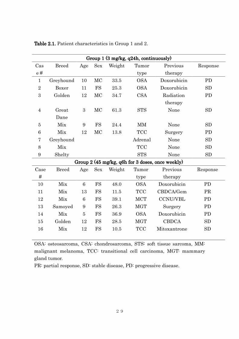
2.4.1 Patient population .............................................................................. 24
2.4.2 Hematological toxicities ..................................................................... 24
2.4.3 Biochemical toxicities ......................................................................... 25
2.4.4 Clinical adverse events ....................................................................... 26
2.4.5 Tumor response .................................................................................. 27
ii
2.4.6 Whole blood concentration measurement .......................................... 27
2.5 Discussion .................................................................................................. 32
3 Biologic Activity of Dihydroartemisinin in Canine Osteosarcoma Cell Lines:
Effects on Cellular Proliferation, Cell Cycle Distribution, Induction of Apoptosis,
and Intracellular Free Radical Contents
3.1 Summary ................................................................................................... 36
3.2 Introduction ............................................................................................... 38
3.3 Materials and methods
3.3.1 Cell culture and cell lines ................................................................... 41
3.3.2 Chemicals ............................................................................................ 41
3.3.3 Assessment of cell viability ................................................................ 42
3.3.4 Nucleosome fragmentation (cell death) ELISA ................................. 43
3.3.5 Immunoblotting .................................................................................. 43
3.3.6 Cell cycle analysis ............................................................................... 44
3.3.7 Measurement of reactive oxygen species ........................................... 45
3.3.8 Statistical analysis ............................................................................. 46
3.3.9 Footnotes ............................................................................................. 46
3.4 Results
3.4.1 Inhibitory effects of dihydroartemisinin on proliferation of canine
osteosarcoma cells .............................................................................. 49
3.4.2 Induction of cell death through apoptosis .......................................... 50
3.4.3 Accumulation of subG0 cells and G2/M cells ....................................... 50
3.4.4 Generation of reactive oxygen species by an iron-dependent
mechanism .......................................................................................... 51
3.5 Discussion .................................................................................................. 64
iii
4 Cytotoxic Effects of Dihydroartemisinin in Canine Histiocytic Sarcoma Cell
Lines: A Relationship Between Intracellular Ferrous Contents and Cytotoxicity
of Dihydroartemisinin
4.1 Summary ................................................................................................... 68
4.2 Introduction ............................................................................................... 70
4.3 Materials and methods
4.3.1 Evaluation of tissue iron contents of canine histiocytic sarcoma ...... 73
4.3.2 Cell culture and cell lines ................................................................... 74
4.3.3 Chemicals ............................................................................................ 75
4.3.4 Assessment of cell viability ................................................................ 76
4.3.5 Detection of apoptosis ......................................................................... 77
4.3.6 Manipulation and measurement of intracellular iron contents ........ 78
4.3.7 Statistical analysis ............................................................................. 79
4.3.8 Footnotes ............................................................................................. 79
4.4 Results
4.4.1 Tissue iron contents in canine histiocytic sarcoma .......................... 84
4.4.2 Effects of intracellular iron concentration on cytotoxic activity of
dihydroartemisinin in canine histiocytic sarcoma cells ..................... 85
4.5 Discussion .................................................................................................. 99
5 General Conclusion .......................................................................................... 106
6 References ........................................................................................................ 108
7 Japanese Summary .......................................................................................... 122
8 Acknowlegements ............................................................................................. 124
1
1. Preface
1.1 History of arteminisin in the treatment of human
Plasmodium infection (malaria)
Artemisinin is a sesquiterpene lactone currently used in the
treatment of malaria. Malaria is a fatal infectious disease caused by four
species of the blood parasite Plasmodium; P. falciparum, P. vivax, P.
malariae, and P. ovale (Dhinga, 2000). Emergence of multidrug-resistant
malaria has been a major threat to public health in Africa and South Asia
since the late 1960s (Mackenzie, 1999; Butler, 1997; Riedley, 2002; Guerin,
2002). Artemisia annua L. (also known as Qinghaosu, or Sweet wormwood) is
a plant belonging to the Asteraceae family native to China, which has long
been used in traditional Chinese medicine (TCM) for the treatment of chills
and fever (Nosten, 1995). In 1967, a program was launched by the Chinese
government to screen for antimalarial principles in the various plants used
in TCM, which eventually led to the discovery of a highly active chemical
extracted from Artemisia annua L in 1972. The chemical is now known as
artemisinin (also known as qinghaosu) (Qinghaosu Antimalaria
Coordinating Research Group, 1979; Jiang, 1982). The parasiticidal effect of
2
artemisinin and its derivatives were impressive; they induced a rapid arrest
of parasite metabolism at concentrations in the low nanomolar range and a
more rapid death of parasites than other antimalarial agents (White, 1994).
Artemisinin and its derivatives (i.e., artemisinin-derived 1,2,4-trioxanes)
cause structural changes in the erythrocyte stage of the parasite by affecting
the membranes of the food vacuole, nucleus, mitochondria, endoplasmic
reticulum, and nucleoplasm, leading to the formation of autophagous
vacuoles and loss of cytoplasm, which kill the parasite (Anon, 1979; Maeno,
1993). Discovery of artemisinin and its derivatives was a milestone in the
treatment of malaria due to its remarkable efficacy in curing
multidrug-resistant malaria (Li, 1998; Klayman, 1985; Ziffer, 1997; Haynes,
1997), although their use is still restricted by cost in developing countries.
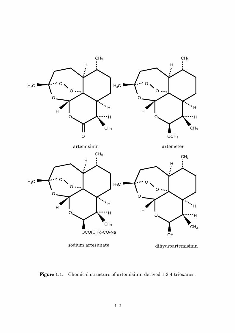
The chemical structure of artemisinin contains a unique
endoperoxide trioxane moiety (the bridge-like structure is specifically called
an endoperoxide bridge, Figure 1.1). Artemisinin is relatively easily
purified by crystallization after extraction from the Artemisia annua L. plant
from which more lipid- or water-soluble derivatives can be semi-synthesized.
However, these endoperoxide-containing trioxanes are extremely difficult to
3
synthesize de novo (Woodrow, 2005). Through a reaction catalyzed by iron,
artemisinin-derived trioxanes are converted to carbon-centered free radicals
(Meshnick, 1991; Meshnick, 1994). These trioxanes are first activated by
intraparasitic heme-iron, which catalyzes the cleavage of the endoperoxide.
The release of heme-iron during hemoglobin digestion within the parasite’s
food vacuole facilitates the cleavage of the endoperoxide moiety by a
Fe(II)-Fenton reaction. Breakage of the endoperoxide bridge results in the
generation of reactive oxygen species (ROS), such as hydroxyl radicals and
superoxide anions (Bergman, 1997; O’Neill, 2004). The parasiticidal effect of
artemisinin-derived trioxanes is thought to be mediated by this production of
free radicals, because artemisinin derivatives that lack the endoperoxide
bridge, a known source of free radicals, do not have antimalarial activity
(Brossi, 1988; Klayman, 1985). Antimalarial activities are enhanced by high
oxygen tension or other free radical generating compounds (Krungkrai, 1987;
Elford, 1987), and free radical scavengers block antimalarial activity
(Krungkrai, 1987; Meshnick, 1989). Although this evidence strongly suggests
free radical generation is involved in the parasiticidal mechanism, the
exceptionally high in vitro activities of artemisinin-derived trioxanes
4
compared to other free radical generating drugs cannot be explained entirely.
More recently, alkylation of specific target proteins, rather than ubiquitous
oxidation, has been suggested as an alternative mechanism for the marked
parasiticidal effect of artemisinin-derived free radicals. The postulated
target proteins include the P. falciparum translationally controlled tumor
protein (TCTP) and the P. falciparum orthologue of the calcium ATPase
(sarcoplasmic endoplasmic reticulum calcium ATPase, SERCA)
(Eckstein-Ludwig, 2003; Asawagahasakda, 1994; Bhisutthibhan, 2001).
More definitive mechanism(s) of action remain to be identified.
1.2 Artemisinin-derived trioxanes in cancer therapy
Recently, artemisinin-derived trioxanes have been found to have
antineoplastic properties (Woerdenbag, 1993; Efferth, 1996; Efferth, 2001;
Singh, 2001; Efferth, 2003; Singh, 2004). The tumoricidal effects are also
thought to be mediated by free radical generation. Since iron is a cofactor in
the synthesis of deoxyriboses, most neoplastic cells overexpress cell surface
transferrin receptors and have greater intracellular iron concentrations than
normal somatic cells (May, 1985). For this reason, artemisinins are
5
selectively cytotoxic for neoplastic cells. Lai et al. (1995), and Singh, et al.
(2001) reported that both human leukemia cells and human breast cancer
cells are more susceptible to the cytotoxicity of arteminisin than normal
human lymphocytes and breast epithelial cells, respectively, by a factor of up
to 100. Several additional studies have demonstrated that artemisinin and
its derivatives have cytotoxic effects against multiple cancer cell lines in
vitro (Lai, 2005; Efferth, 2004; Nam, 2007; Pik, 2006; Efferth, 2002; Zheng,
1994; Beekman, 1998; Posner, 1999) and in vivo (Moore, 1995). A few case
reports have documented the clinical efficacy of artemisinin derivatives in
humans with laryngeal squamous cell carcinoma (Singh, 2002), metastatic
uveal melanoma (Berer, 2005), and pituitary macroadenoma (Singh, 2006).
To our knowledge, no in vitro or in vivo studies of the potential antineoplastic
effects of artemisinin in canine cancer cells have been published.
1.3 Osteosarcoma in dogs and its comparative aspects
Osteosarcoma is the most common primary bone tumor in dogs,
accounting for up to 85% of all malignant neoplasm in the bone (Brodey,
1969; Ling, 1974; Lui, 1977; Brodey, 1963; Brodey, 1959). The estimated

6
incidence of canine osteosarcoma in the United States is approximately 8,000
to 10,000 cases per year (Withrow, 1991; Priester, 1980); however, the true
incidence is probably much higher since many pet animals do not have
histologic confirmation. Osteosarcoma is primarily a disease of large and
giant breed dogs and Greyhounds (Kistler, 1981; Dernell, 2007). Breeds
reported to be at an increased risk include the Saint Bernard, Great Dane,
Irish Setter, Doberman Pinscher, Rottweiler, German Shepherd Dog, and
Golden Retriever (Dernell, 2007). Approximately 75% of canine
osteosarcomas occur in the appendicular skeleton and 25% occur in the axial
skeleton (Brodey, 1969; Heyman, 1992). For appendicular osteosarcoma,
forelimbs are affected twice as often as hindlimbs (Knecht, 1978).
The clinical behavior of canine osteosarcoma is highly aggressive.
Although radiographic evidence of metastases is uncommon, approximately
90% of affected dogs develop metastatic disease (Brodey, 1969; Spodnick,
1992). The median survival time after amputation alone is 134 days, with
extremely low 1- and 2- year survival rates of 11.5% and 2.0%, respectively
(Spodnick, 1992). Thus, although a considerable amount of effort has been
made to develop limb-sparing techniques that improve local tumor control
7
(Vasseur, 1987; Berg, 1992; LaRue, 1989; Thrall, 1990; Withrow, 1993;
Straw, 1996), survival times are still limited by the development of
metastatic disease. Adjuvant systemic chemotherapy inhibits the
progression of metastatic disease and thereby prolongs survival. Numerous
studies have evaluated different types of chemotherapy for canine
osteosarcoma and all showed a survival advantage compared to amputation
alone; however, further improvements in disease-free intervals and overall
survival have not been made (Straw, 1991; Bacci, 1988; Shapiro, 1988;
Thompson, 1992; Kraegel, 1991; Bergman, 1996; Berg, 1995; Mauldin, 1988;
Berg, 1997; Chun, 2000; Kent, 2004; Bailey, 2003; Kirpensteijn, 2002;
Withrow, 1995). The general trend in adjuvant chemotherapy for residual
micrometastatic disease will likely be towards non-cytotoxic
chemotherapeutics, such as molecularly targeted pharmaceuticals, in
combination with conventional cytotoxic drugs, rather than further
investigation of cytotoxic drug combination.
Spontaneously occurring osteosarcoma in the dog has many
similarities to its human counterpart, and has been proposed as a large
animal model for human osteosarcoma (Withrow, 1991). The high annual
8
incidence of canine osteosarcoma (>8,000/year) compared to human
osteosarcoma (1,000/year) is particularly attractive for this purpose. The
clinical behavior of osteosarcoma in human is as aggressive as canine
osteosarcoma; 85-90% of the cases are histologically high-grade (95% in dogs),
and the metastatic rate is 80% within 2 years without chemotherapy (90%
within 1 year in dogs). The most common metastatic sites are lungs, bone,
and soft tissue, which is similar to dogs. As in dogs, improvement in
prognosis with adjuvant chemotherapy is observed; however, the long-term
survival rate, despite aggressive chemotherapy, is approximately 60%.
Further improvement in prognosis with systemic cytotoxic chemotherapy is
unlikely without significantly increasing toxicity. Investigation of
non-cytotoxic chemotherapy and other strategies in canine osteosarcoma
may contribute to the advancement of treatment in human osteosarcoma.
1.4 Altered iron metabolism in canine histiocytic sarcoma
Canine histiocytic sarcoma is a tumor of dendritic cell or macrophage
origin (Affolter, 2002). It is further subclassified into ones of
9
antigen-presenting dendritic cell origin and ones of phagocytic activated
macrophage origin; the latter occasionally present with signs of hemolysis
and thrombocytopenia known as hemophagocytic syndrome. Clinically,
canine histiocytic sarcoma either presents as an isolated lesion or multifocal
lesions. The former is termed localized histiocytic sarcoma, often of dendritic
cell origin without showing the signs of hemophagocytic syndrome until
progressed to advanced stage, and the latter is termed disseminated
histiocytic sarcoma, some of which are of macrophage origin and present
with marked hemophagocytic syndrome at diagnosis.
Many diseased conditions can cause alteration in iron metabolisms.
Ferritin is an iron-binding protein for cells to store iron and is also one of the
acute phase proteins. Normally, it is synthesized by the hepatocytes and cells
of hematopoietic origin (Yoda, 1980). Hyperferritinemia has been
documented in several diseases in human including malignant neoplasia,
hemolytic disorders, and liver disease. In particular, hyperferritinemia has
been reported with malignant histiocytosis, a human counterpart of canine
disseminated histiocytic sarcoma (Esumi, 1989; Ya-You, 1998). Occurrence of
hyperferritinemia has been investigated in dogs and has been associated

10
with liver disease, malignant tumor including histiocytic sarcoma and
lymphoma, immune-mediated hemolytic anemia or any other erythrocyte
destruction, and marked inflammation (Friedrichs, 2010; Nielsen, 2011).
Among these diseases, canine histiocytic sarcoma has been shown to cause
marked hyperferritinemia and serum ferritin concentration is proposed to be
utilized as an early tumor marker for this malignancy (Nielsen, 2011).
Disseminated form of canine histiocytic sarcoma, previously termed as
malignant histiocytosis, is known to cause hemolysis and thrombocytopenia,
with marked similarity with immune-mediated hemolytic anemia from a
clinical standpoint. Together with the documented marked hyperferritinemia,
the hemophagocytic characteristic of this tumor leads to the assumption that
canine histiocytic sarcoma cells contains higher intracellular iron contents,
although this has not been evaluated.
1.5. Aims of the study
In this study, the clinical adverse effects and potential antitumor
effects of orally administered artemisinin were investigated in dogs with
11
spontaneous tumors. To further investigate the mechanism of cytotoxicity,
the in vitro cytotoxic effect of dihydroartemisinin, an active metabolite of
most artemisinin-derivatives, was demonstrated using four canine
osteosarcoma cell lines. Dihydroartemisinin induced
concentration-dependent free radical generation by an iron-dependent
mechanism, resulting in growth inhibition in all four cell lines tested.
Dihydroartemisinin also induced cellular apoptosis and G2/M cell cycle
arrest in a concentration-dependent manner. The correlation between the
cytotoxicity of dihydroartemisinin and intracellular iron concentration was
also investigated in two canine histiocytic sarcoma cell lines. The cytotoxicity
of dihydroartemisinin was enhanced by increasing the cellular iron
concentration and inhibited by chelating iron from the culture media.
12
H
CH3
H H
H
CH3
H3C
O
O
O
O
O
H H
H
CH3
O
O
H
CH3
H3C O
O
OCH3
H H
H
CH3
O
O
H
CH3
H3C O
O
OH
O
H H
H
CH3
O
H
CH3
H3C O
O
OCO(CH2)2CO2Na
Figure 1.1.Figure 1.1.Figure 1.1.Figure 1.1. Chemical structure of artemisinin-derived 1,2,4-trioxanes.
artemisinin artemeter
sodium artesunate dihydroartemisinin
13
2. Phase I Clinical Trial of Oral Artemisinin in Cancer-Bearing
Dogs: Comparison of High-Dose Intermittent and Low-Dose
Continuous Dosing Schedules in Dogs with Naturally
Occurring Tumors
2.1. Summary
To evaluate the clinical toxicity and activity of orally administered
artemisinin in dogs with spontaneous tumors, 24 client-owned dogs were
randomly divided into two groups and received either low-continuous dose (3
mg/kg, q24h) or intermittent-high dose (3 doses of 45 mg/kg, q6h, repeated
once weekly) of artemisinin orally. Treatment was continued for 21 days.
Dogs were evaluated weekly for clinical, hematological, and biochemical
adverse events. Whole blood concentrations of artemisinin and
dihydroartemisinin were measured by liquid chromatography/tandem mass
spectrometry after the first dose of artemisinin in three dogs in each group.
All dogs tolerated oral artemisinin in both groups. The most frequent
adverse event was anorexia, which was observed in 11% of the low dose
group and 29% of the high dose group. Objective tumor response was
observed in one dog with a urinary bladder transitional carcinoma, which
14
lasted for 6 weeks. Blood concentrations of artemisinin and
dihydroartemisinin were below 0.1 µM at all time points, and there was no
difference in blood concentration between the two dosing groups. Oral
artemisinin, both in low- continuous dose and high- intermittent dose, is
well-tolerated in dogs but results in low bioavailability. The parenteral
administration route should be considered for future studies.

15
2.2. Introduction
Artemisinin is a sesquiterpene lactone extracted from a plant
Artemisia annua L., which is used in TCM. It was first identified and
isolated in 1972 in a project to discover new antimalarial drugs from TCM
launched by the Chinese government (Qinghaosu Antimalaria Coordinating
Research Group, 1979; Klayman, 1985), and now is the first line treatment of
malaria in countries in South Asia (Li, 1994; Hien, 1994).
Artemisinin has a unique chemical structure, an endoperoxide
bridge; by a cleavage of the endoperoxide bridge catalyzed by iron,
artemisinin becomes a carbon-centered free radical (Meshnick, 1993; Zhang,
1992). This reactive free radical results in damage to lysosomal membranes,
leading to autodigestion (Bergman, 1997; O’Neill, 2004), and alkylation of
essential proteins of malaria parasites, including Plasmodium falciparum
translationally controlled tumor protein (TCTP) (Asawamahasakda, 1994;
Bhisutthibhan, 2001; Eckstein-Ludwig, 2003).
More recently, artemisinins have been found to have antineoplastic
properties (Woerdenbag, 1993; Efferth, 1996; Efferth, 2001; Singh, 2001;
Efferth, 2003; Singh, 2004). Tumor cells often overexpress transferrin
16
receptors to uptake iron, which is a cofactor of deoxyribose synthesis; thus,
they have higher intracellular iron concentrations than their somatic
counterparts (Aulbert, 1980; Karin, 1981; Reizenstein, 1991; Raaf, 1993;
Shterman, 1991; Castaneda, 1991; Das-Gupta, 1996). The mechanism of
artemisinin’s anticancer activity is also thought to be through generation of
free radicals mediated by intracellular iron molecules. The cellular target of
these free radicals have not been completely identified, but the expression
level of TCTP correlates with sensitivity to artemisinin derivatives,
suggesting that TCTP may be one of the target proteins in anticancer
mechanism (Efferth, 2005); however, other cellular structures, including
mitochondrial membranes and DNA can also be damaged. Alternatively,
some of the antitumor effects of artemisinin may not be due to direct
cytocidal effect, but rather due to indirect effects, such as inhibition of
neoangiogenesis (Zhou, 2007; Ricci, 2010) or modification of the T-regulatory
response (Langroudi, 2010; Noori, 2009).
Several studies have demonstrated that artemisinin and its
derivatives have cytotoxic effects against multiple human cancer cell lines in
vitro and against a rat fibrosarcoma cell line in vivo (Woerdenbag, 1993;
17
Efferth, 1996; Efferth, 2001; Singh, 2001; Efferth, 2003; Singh, 2004; Lai,
1995; Lai, 2005; Efferth, 2004; Nam, 2007; Pik, 2006; Efferth, 2002; Zheng,
1994; Beekman, 1998; Posner, 1999; Moore, 1995). To date, three case
reports have been published in human medicine documenting tumor control
by artesunate in laryngeal squamous cell carcinoma, metastatic uveal
melanoma, and pituitary macroadenoma (Singh, 2002; Berger, 2005; Singh,
2006). In veterinary medicine, there is a case report of a dog with an
appendicular osteosarcoma that had a complete response to oral artemisinin
(Rowley, 2004); however, this report was not published in a peer-reviewed
journal.
Despite long-term use of artemisinin for the treatment of malaria,
there is a paucity of information on the pharmacokinetics of artemisinin and
its derivatives. The dosages and administration schedules of artemisinin
derivatives in both veterinary and human medicine are mostly anecdotal.
Orally administered artemisinin is commonly used in veterinary medicine,
with low-dose (100mg/dog) daily or twice daily administration. In general,
after oral administration, artemisinin and its derivatives are rapidly
absorbed from the gastrointestinal tract, with peak plasma concentration
18
occurring in one hour (Dhingra, 2000). All artemisinin derivatives except
artemisinin per se (plasma metabolite of artemisinin is currently unknown
[Navaratnam, 2000]) are primarily metabolized in the liver to the active
metabolite dihydroartemisinin (DHA), and then eliminated in urine and
feces in a relatively short period, with a plasma half-life ranging from 45
minutes to 11 hours (Dhingra, 2000). Pharmacokinetics of artemisinin is
significantly influenced by individual variation, gender, and concurrent fat
intake (Navaratnam, 2000). There also is a significant decrease (oral and
rectal administration) or increase (intramuscular administration) in the area
under the curve with repeated administrations, which is of great concern
since most pharmacokinetic studies were performed after administering a
single dose. After daily oral administration of artemisinin, the AUC at day 7
is only 24% of that of day 1 in healthy adults (Ashton, 1998). Similar results
were obtained in dogs (Classen, 1999). This suggests that the administration
schedule of artemisinin commonly used in dogs (daily or twice daily oral
administration) may not be clinically effective because most of the
administered artemisinin may not be absorbed.
To our knowledge, no studies investigating the safety and

19
antineoplastic effects of artemisinin in dogs with cancer in a clinical setting
have been published. Therefore, this study was conducted to evaluate the
in vivo effects of oral artemisinin, the most commonly used over-the-counter
artemisinin derivatives, in dogs with spontaneous cancers.

20
2.3. Materials and methods
2.3.1. Animals
Twenty-four client-owned dogs with various spontaneous tumors
were included in the study. The inclusion criteria included; measurable
tumor burden, histopathologic or cytopathologic confirmation of the tumor
type, failure of conventional treatments or the owner’s consent to use
artemisinin in lieu of conventional therapy, expected survival time of >4
weeks, and written owner’s consent. The study protocol was approved by the
Veterinary Teaching Hospital Board. The exclusion criteria included
concurrent severe renal or hepatic disease and concurrent use of therapies
other than analgesics. The use of non-steroidal anti-inflammatory drugs was
permitted only if they were necessary for analgesic purposes, and they had
been initiated >4 weeks prior to enrollment into the current study and the
patient had no measurable tumor response. Two additional healthy dogs
owned by hospital staff were used for a pharmacokinetic analysis of the oral
artemisinin, as insufficient number of the clients agreed to hospitalize their
dogs to obtain multiple blood samples.
21
2.3.2. Artemisinin
Artemisinin with greater than 99% purity was purchased from a
commercial supplier (Holleypharma, Tustin, CA). The drug was available as
either 100- or 50-mg capsules, which were reformulated to produce smaller
capsules when required by the standard dosage used by the study. The
capsules were administered orally in conjunction with fat-containing food
(peanut butter) for pilling purpose and to maximize the intestinal absorption
of the artemisinin.
2.3.3. Treatment schedule
Dogs were randomly assigned to either Group 1 (low-continuous dose;
3 mg/kg, PO, q24h) or Group 2 (high-intermittent dose; 45 mg/kg, PO, q6h for
3 doses, repeated once weekly). Absorption of orally administered
artemisinin can be affected by the fasting status. For the pilling purpose and
to minimize variability of intestinal absorption, artemisinin capsules were
always given with a same amount of fat containing food (i.e., peanut butter).
Dogs were fully evaluated on the day of the enrollment, by means of a
22
complete physical examination, complete blood count (CBC), serum
biochemical profile, urinalysis, and appropriate imaging modality
(radiographs or ultrasonography) when tumor measurement required such
imaging techniques. The dogs were readmitted and physical examination
and gross tumor measurement were repeated once weekly or whenever the
attending clinician judged reassessment was necessary. On week 4, a
complete physical examination, CBC, serum biochemical profile, urinalysis,
and tumor imaging, when necessary, were repeated to make a final
assessment of tumor response and potential adverse effects. The owners
were given a standardized adverse event assessment scheme, and asked to
document any changes/abnormality of their dogs that could potentially be a
drug-related adverse effect. The owner’s record was evaluated by one of the
investigators at each visit, and adverse effects were graded according to the
Veterinary Cooperative Oncology Group Common Terminology for Criteria of
Adverse Event (Veterinary Cooperative Oncology Group, 2004).
2.3.4. Pharmacokinetic study
Blood samples were collected from one dog from Group 1 and three
23
dogs from Group 2 for pharmacokinetic analysis of artemisinin and its
putative metabolite, dihydroartemisinin. Two additional clinically healthy
dogs, owned by hospital staff, were given one dose of 3 mg/kg of artemisinin.
For the dogs receiving conventional doses (one dog in Group 1 and two
healthy volunteers), samples were collected at 5, 10, 20, 35, 60, and 90
minutes and at 2, 4, 6, 9, 12, and 16 hours after drug administration on day 1.
For Group 2, time points for blood collection included immediately before the
first, second, and third doses and at 5, 10, 20, 35, 60, and 90 minutes and at 2,
4, 6, 9, 12 and 16 hours after the third dose was administered on day 1.
Artemisinin derivatives have been shown to bind significantly to red
blood cells (Li, 1998). A modified procedures reported by Naik, et al. (2005)
utilizing liquid chromatography/tandem mass spectrometry (LC/MS/MS)
analytical method with atmospheric pressure chemical ionization (APCI)
methods was used for determination of whole blood concentrations of both
artemisinin and dihydroartemisinin, which was validated to measure
artemisinin and dihydroartemisinin within canine whole blood samples at
the investigators’ laboratory.
24
2.4. Results
2.4.1. Patient population
Twenty-four dogs were included in the study; 12 dogs received
low-continuous dose (Group 1), and 12 received intermittent-high dose
(Group 2). The tumor type included metastatic/unresectable osteosarcoma
(n=7), transitional cell carcinoma of the urinary bladder (n=5), soft tissue
sarcoma (n=3), mammary gland carcinoma (n=2), malignant melanoma (n=2),
and each one of multicentric lymphoma, chondrosarcoma, mast cell tumor,
hemangiosarcoma, and invasive adrenal gland tumor. Most of these dogs had
advanced stage disease, and eight dogs did not complete the planned
treatment course for various reasons; three dogs died or were euthanized
before completion of the study due to tumor progression, two dogs did not
return for recheck for unknown reason, the owner of another dog decided to
start chemotherapy before completing the study, artemisinin treatment was
not given as instructed in one dog, one dog became too aggressive to pill and
oral artemisinin was discontinued. Sixteen dogs (9 dogs in Group 1, 7 dogs in
Group 2; Table 1) completed the protocol and were available for toxicity and
tumor response evaluation.
25
2.4.2. Hematological toxicities
No significant changes in hematocrit (p=0.30), segmented neutrophil
count (p=0.66), or platelet count (p=0.71) were seen. Grade 1 neutropenia
(1.9 x 10⁹/L and 2.8 x 10⁹/L, respectively) was seen in 2 dogs (Dog #1 and #4,
both in the Conventional group); however, the pretreatment neutrophil
counts in these dogs were low (2.8 x 10⁹/L and 3.1 x 10⁹/L, respectively).
These neutrophil counts were interpreted as physiologic values for Dog #1 (a
10 year-old Greyhound). The reason for mild neutropenia in Dog #4 was
unknown, but thought to be unrelated to artemisinin administration. Grade
1 anemia was seen in 2 dogs (Dog #10 and #5). No thrombocytopenia was
observed.
2.4.3. Biochemical toxicities
There were 2 dogs with BUN increase over baseline; in Dog #1, BUN
increased from 20 mg/dL to 27 mg/dL; and in Dog #8, it increased from 16
mg/dL to 33 mg/dL. Neither of these 2 dogs had relevant changes in serum
creatinine concentration over baseline; no dogs had significant increases in

26
serum creatinine concentration. Two dogs had elevations of serum ALT
activity over baseline; Dog #1 had ALT activity increase from 51 IU/L to 58
IU/L; Dog #16 had increase of serum ALT activity from 85 IU/L to 125 IU/L.
Both were considered to be clinically irrelevant.
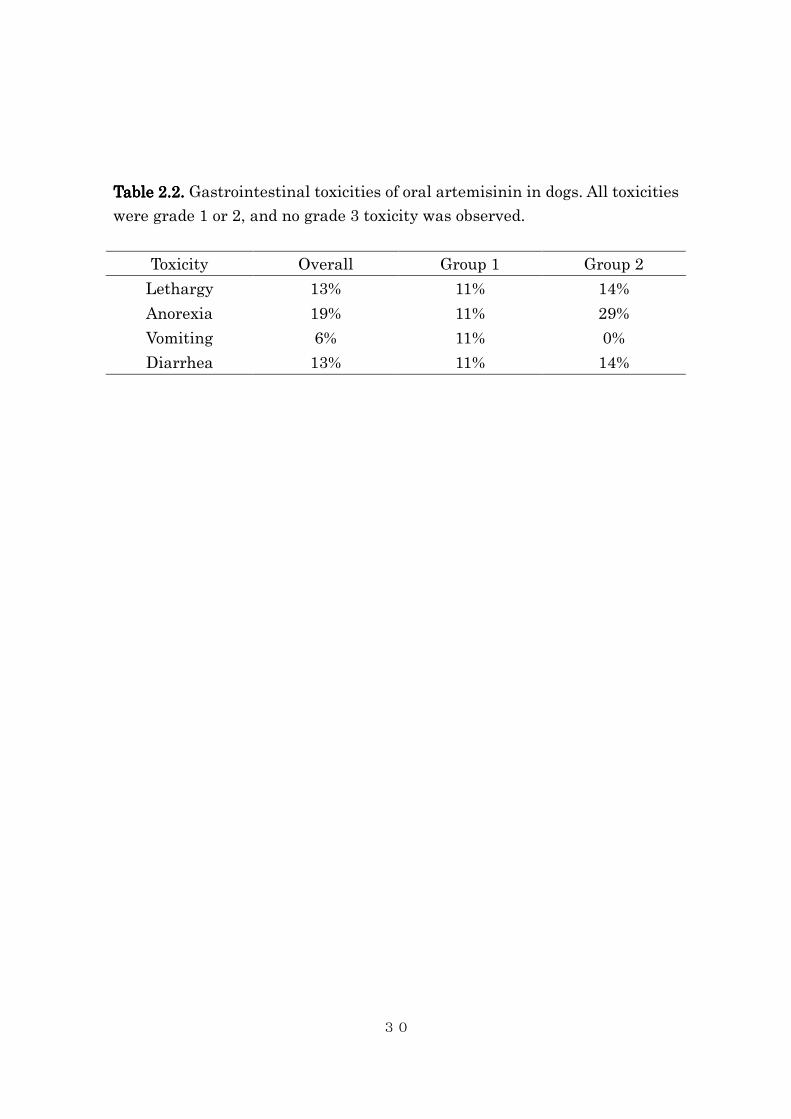
2.4.4. Clinical adverse events
Potential neurotoxicity was seen in 1/16 dogs (6%). This dog (Dog
#13) developed tremors after the first dosing of high-dose artemisinin;
however, the dog was diagnosed with a urinary tract infection by the
referring veterinarian, and the tremors resolved after initiation of antibiotics.
The dog did not have any adverse event after the subsequent doses of
high-dose artemisinin.
Grade 2 lethargy was seen in 2 dogs (Dog #5 and #13). As discussed
above, dog #13 was diagnosed with urinary tract infection, and the lethargy
resolved after initiation of antibiotics.
Anorexia was seen in 3 dogs (Dog #10 [Grade 1], #5 [Grade 1], #13
[Grade 2]). Dog #10 developed Grade 1 anorexia after the first high-dose
artemisinin, but no such adverse event was seen after the subsequent doses.
27
Dog #13 was diagnosed with urinary tract infection, and the anorexia
resolved after initiation of antibiotics. Vomiting was seen in 1 dog (Dog #2,
Grade 1). The dog had an episode of vomiting after the first dose of low-dose
artemisinin, but none after the subsequent doses. Diarrhea was seen in 2
dogs (Dog #5 [Grade1], Dog #13 [Grade 2]). Dog #13 was diagnosed with
urinary tract infection, and the diarrhea resolved after initiation of
antibiotics.
2.4.5. Tumor response
Nine dogs completed the treatment protocol in Group 1 and 7 dogs in
Group 2. In Group 1, 3 dogs had PD and 5 had SD. In Group 2, 1 dog had PR
(TCC, Fig 1a and 1b), 4 had PD, and 2 had SD. In the dog that had PR, the
treatment was continued for 3 additional weeks, and disease progression was
noted 6 weeks from the start of the treatment.
2.4.6. Whole blood concentration measurement
The measured whole blood concentrations of artemisinin and
dihydroartemisinin were less than 0.1 µM at all points after administration
28
of conventional doses of artemisinin. No meaningful pharmacokinetic
analysis was possible due to the extremely low blood concentration of the
agent. This low level of blood concentration was not improved by
administration of high-dose artemisinin at short intervals; no significant
increase in blood concentrations of artemisinin or dihydroartemisinin was
observed with IHD group, suggesting poor and saturable gastrointestinal
absorption of the drug in dogs.
29
Table Table Table Table 2.2.2.2.1.1.1.1. Patient characteristics in Group 1 and 2.
Group 1 (3 Group 1 (3 Group 1 (3 Group 1 (3 mg/kg, q24h, continuously)mg/kg, q24h, continuously)mg/kg, q24h, continuously)mg/kg, q24h, continuously)
Cas
e #
Breed Age Sex Weight Tumor
type
Previous
therapy
Response
1 Greyhound 10 MC 33.5 OSA Doxorubicin PD
2 Boxer 11 FS 25.3 OSA Doxorubicin SD
3 Golden 12 MC 34.7 CSA Radiation
therapy
PD
4 Great
Dane
3 MC 61.3 STS None SD
5 Mix 9 FS 24.4 MM None SD
6 Mix 12 MC 13.8 TCC Surgery PD
7 Greyhound Adrenal None SD
8 Mix TCC None SD
9 Shelty STS None SD
Group 2 (45 mg/kg, q6h for 3 doses, once weekly)Group 2 (45 mg/kg, q6h for 3 doses, once weekly)Group 2 (45 mg/kg, q6h for 3 doses, once weekly)Group 2 (45 mg/kg, q6h for 3 doses, once weekly)
Case
#
Breed Age Sex Weight Tumor
type
Previous
therapy
Response
10 Mix 6 FS 48.0 OSA Doxorubicin PD
11 Mix 13 FS 11.5 TCC CBDCA/Gem PR
12 Mix 6 FS 39.1 MCT CCNU/VBL PD
13 Samoyed 9 FS 26.3 MGT Surgery PD
14 Mix 5 FS 36.9 OSA Doxorubicin PD
15 Golden 12 FS 28.5 MGT CBDCA SD
16 Mix 12 FS 10.5 TCC Mitoxantrone SD
OSA: osteosarcoma, CSA: chondrosarcoma, STS: soft tissue sarcoma, MM:
malignant melanoma, TCC: transitional cell carcinoma, MGT: mammary
gland tumor.
PR: partial response, SD: stable disease, PD: progressive disease.
30
Table Table Table Table 2.2.2.2.2222.... Gastrointestinal toxicities of oral artemisinin in dogs. All toxicities
were grade 1 or 2, and no grade 3 toxicity was observed.
Toxicity Overall Group 1 Group 2
Lethargy 13% 11% 14%
Anorexia 19% 11% 29%
Vomiting 6% 11% 0%
Diarrhea 13% 11% 14%
31
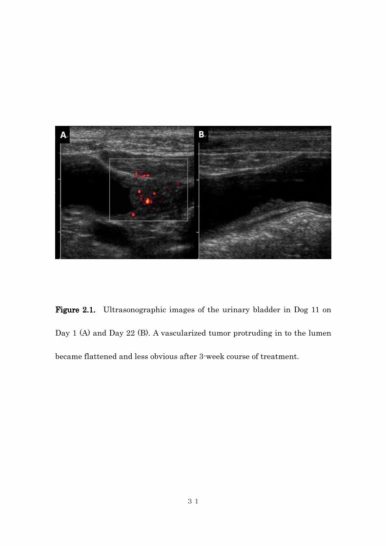
Figure 2.1.Figure 2.1.Figure 2.1.Figure 2.1. Ultrasonographic images of the urinary bladder in Dog 11 on
Day 1 (A) and Day 22 (B). A vascularized tumor protruding in to the lumen
became flattened and less obvious after 3-week course of treatment.
32
2.5. Discussion
During the past two decades, the anti-malarial agent artemisinin and its
derivatives have attracted significant interest as potential novel anticancer
agents (Woerdenbag, 1993; Efferth, 1996; Efferth, 2001; Singh, 2001; Efferth,
2003; Singh, 2004; Lai, 1995; Lai, 2005; Efferth, 2004; Nam, 2007; Pik, 2006;
Efferth, 2002; Zheng, 1994; Beekman, 1998; Posner, 1999; Moore, 1995),
cancer preventatives (Lai, 2006; Disbrow, 2005), multidrug resistance
reversal agents (Mukanganyama, 2002; Reungpatthanaphong, 2002), and
radiosensitizers (Kim, 2006). These artemisinin-derived 1,2,4-trioxanes
exhibit significant activity in the nanomolar to micromolar range against a
variety of human cancer cell lines. The biologic activity in human cancer cell
lines and apparently low toxicity in human malaria patients have stimulated
the use of artemisinin by dog owners to treat various canine malignancies,
particularly osteosarcomas, as the drug is readily available without a
prescription. However, to our knowledge, there are no reports on the safety
and potential activity of artemisinin in dogs with cancer in vivo.
The prevalence of hematological/biochemical toxicity in the current
33
study was low. The Grade 1 anemia seen in 2 dogs were likely secondary to
disease progression (i.e.; anemia of chronic disease) rather than to
artemisinin toxicity. The Grade 1 neutropenia seen in 2 dogs were
interpreted as normal value for these individuals. Similarly, the 2 events of
high serum BUN concentration (both in Group 1) were interpreted as
secondary to disease progression and dehydration, or subclinical
gastrointestinal bleeding, as none of the dogs developed increases in serum
creatinine concentration. An unreported change in diet by the owners to one
with higher protein content may also have accounted for the increases in
BUN in light of normal creatinine concentrations. Two dogs had minimal
increases in the serum ALT activity; these minor changes were thought to be
clinically irrelevant and likely a result of daily fluctuation of ALT values.
However, the possibility of hepatotoxicity associated with high-dose
artemisinin could not be ruled out, as Dog #19 developed Grade 2 ALT
increase.
This study also shows that the neurotoxicity reported in rodents
studies are not common in dogs at the dosing scheme used in this study. The
only questionable neurotoxicity seen in the study population was manifested
34
by tremors in 1 dog; however, the tremors resolved with the treatment of
urinary tract infection. Since the same effect was not observed after the
subsequent three weekly treatments, we believe this was not
treatment-related.
Both low-dose continuous and high-dose intermittent regimens were
well-tolerated. No dogs developed Grade 3 or higher toxicities. Occasional
anorexia and vomiting were seen in small number of dogs, but they were
self-limiting and clinically acceptable.
No durable tumor response was observed in the study; objective
response was observed only in one dog, which lasted for a relatively short
period of time (6 weeks). This lack of tumor response can be explained by the
low blood concentration achieved after oral administration of artemisinin.
The blood concentration of artemisinin was well below the therapeutic target
concentration (10 µM, based on our previously reported in vitro study
[Hosoya, 2008]). The concentration of dihydroartemisinin was even lower.
Furthermore, the blood concentrations of artemisinin and dihydroartemisinn
were similarly low after administration of high-dose artemisinin. These
findings were consistent with the previous study, in which orally
35
administered artemether (600 mg/kg) resulted in less than 1 nM peak
plasma concentration of artemether and less than 0.5 nM peak concentration
of dihydroartemisinin (Classen, 1999). This suggests that the bioavailability
of orally administered artemisinin is poor and gastrointestinal absorption
saturable in dogs, and the conversion of artemisinin to dihydroartemisinin is
incomplete. We are currently performing a more detailed pharmacokinetic
assay of orally administered and parentally administered artemisinin
derivatives in normal dogs.
Although the study showed minimal and acceptable adverse events,
it does not support the use of single agent oral artemisinin in the treatment
of neoplasia in dogs, as the study showed the targeted therapeutic
concentration is not achievable even with high oral dose. Possible ways to
circumvent this problem are use of parental formulation and/or use of more
potent artemisinin-derivatives, such as artesunate, dihydroartemisinin, or
investigational newer generation artemisinin-derivatives.
36
3. Biologic Activity of Dihydroartemisinin in Canine
Osteosarcoma Cell Lines: Effects on Cellular Proliferation,
Cell Cycle Distribution, Induction of Apoptosis, and
Intracellular Free Radical Contents
3.1. Summary
Artemisinin is a chemical extracted from a plant Artemisia annua. It has
recently been discovered to have anti-neoplastic properties. It is anecdotally
being used by veterinarians and pet owners to treat various type of cancer,
expecially osteosarcoma. However, evidence is lacking to support its biologic
activity against canine osteosarcomas. Spontaneously occurring
osteosarcoma in the dog is also an excellent large animal model for human
osteosarcoma. In this study, the in vitro cytotoxic effect of
dihydroartemisinin, a newly emerging potential anti-cancer agent with low
systemic toxicity, was investigated in four canine osteosarcoma cell lines.
Dihydroartemisinin induced concentration-dependent free radical
generation by an iron-dependent mechanism, resulting in growth inhibition
in all four cell lines tested. Dihydroartemisinin also induced cellular
apoptosis and decrease of G0/G1 cell population in a concentration-dependent

37
manner. This study is the first to demonstrate in vitro cytotoxic activity of an
artemisinin derivative in canine osteosarcoma and provides relevant
information in order to design a clinical trial in canine osteosarcoma.
38
3.2. Introduction
Artemisinin is an extract from the Chinese plant quinghao (Artemisia
annua L.) and is used in traditional Chinese medicine for chills and fever
(Nosten, 1995). Since 1972, when its antimalarial properties were discovered,
artemisinin and its semisynthetic derivatives (i.e., arteether, artemether,
artesunate, artelinate, and artenimol [dihydroartemisinin]) have been
studied extensively as anti-malarial agents (Jiang, 1982). These compounds
react with intracellular iron in free heme molecules within malaria-infected
red blood cells and are converted to highly reactive free radicals. Their
anti-malarial activity is due to their generation of reactive oxygen species
(ROS), which are thought to alkylate one or more essential malarial proteins
(Posner, 2000; Meshnick, 1998).
More recently, artemisinins have been found to have antineoplastic
properties (Woerdenbag, 1993; Efferth, 1996; Efferth, 2001; Singh, 2001;
Efferth, 2003; Singh, 2004). Similar to its anti-malarial effects, the
tumoricidal mechanism of artemisinin is secondary to ROS generation. Since
iron is a cofactor in the synthesis of deoxyriboses, most neoplastic cells
39
overexpress cell surface transferrin receptors (TfR) and have greater
intracellular iron concentrations than normal somatic cells (May, 1985). This
may explain the basis of the selective cytotoxicity of artemisinins for
neoplastic cells. For example, it has been shown that human leukemia and
breast cancer cells are more susceptible to the cytotoxic effects of arteminisin
than normal human lymphocytes and breast epithelial cells by a factor of up
to 100 (Singh, 2001; Lai, 1995). Several additional studies have
demonstrated that artemisinin and its derivatives have cytotoxic effects
against multiple human cell lines in vitro and against a rat fibrosarcoma cell
line in vivo (Woerdenbag, 1993; Efferth, 1996; Efferth, 2001; Singh, 2001;
Efferth, 2003; Singh, 2004; Lai, 1995; Lai, 2005; Efferth, 2004; Nam, 2007;
Pik, 2006; Efferth, 2002; Zheng, 1994; Beekman, 1998; Posner, 1999; Moore,
1995). Finally, the potential clinical efficacy of artemisinin derivatives has
been documented in case reports of humans with laryngeal squamous cell
carcinoma, metastatic uveal melanoma, and pituitary macroadenoma (Singh,
2002; Berger, 2005; Singh, 2006). Together, these data suggest that
artemisinin may be a useful agent to treat cancer in the clinical setting.
Given the low toxicity and antineoplastic activity of artemisinin in

40
human cancers and cell lines, and anecdotal reports of efficacy in canine
cancers, especially osteosarcoma, an increasing number of pet owners are
treating their cancer affected dogs with artemisinin, which is readily
accessible as an over-the-counter product. To our knowledge, no studies
investigating the potential antineoplastic effects of artemisinin in canine
cancer cells have been published. Therefore, this study was initiated to
evaluate the in vitro effects of dihydroartemisinin (DHA), the active
metabolite of most artemisinin derivatives, on cell viability, cytotoxicity, and
cell cycle progression in canine osteosarcoma cell lines.
41
3.3. Materials and methods
3.3.1. Cell culture and cell lines
Four canine osteosarcoma cell lines (D17, OSCA2, OSCA16, and
OSCA50) were evaluated (OSCA cell lines were kindly provided by Dr. Jaime
F. Modiano). The D17 cell line was cultured in RPMI-1640a supplemented
with 10% fetal bovine serumb (FBS) and antibiotics (penicillin [100 U/ml]
and streptomycin [0.1 mg/ml])c. The other three cell lines were cultured in
DMEM supplemented with 10% FBS and antibiotics. All cell lines were
maintained in a humidified environment at 37° C under 5% CO2 and 95%
room air.
2.1.2 Chemicals
Dihydroartemisinin (DHA)e was dissolved in 100% dimethylsulfoxide
(DMSO) at the concentration of 100 mM as a stock solution, protected from
light, and kept at -80° C for long-term storage. The stock solution was
further diluted with DMSO prior to use in tissue culture, resulting in a
consistent DMSO concentration of 0.1% volume. DHA solutions were newly

42
prepared from the stock solution for each experiment.
2.1.3 Assessment of Cell Viability
Cells were seeded in 96-well plates at densities of 1,000–2,000 cells per
well in six replicates to a final volume of 100 µL/well. After 24 hours, 100 µL
of medium containing DMSO (control) or different concentrations of DHA
was added to each well to create the final DHA concentrations of 0.1, 1, 5, 10,
50, and 100 µM and the cells were incubated for additional 48 hours. After
treatment, the medium was removed by gentle suction and plates were stored
at -80° C until use. Cell proliferation was assessed using the CyQUANTTM
nucleic acid fluorescence assay kitf, according to the manufacturer’s
instructions. The intensity of fluorescence of the reaction product, after
background subtraction, was measured with excitation at 485 nm and
emission detection at 530 nm on a Spectra Max M2 plate reader.g Cell
proliferation was expressed as a percentage of the control wells: fluorescence
of sample/ fluorescence of control (DMSO only) cells. Experiments were
repeated at least three times.
43
2.1.4 Nucleosome Fragmentation (Cell Death) ELISA
To assess drug-induced apoptotic cell death a Cell Death Detection
ELISA kith was used according to the manufacturer’s instructions. The
assay is based on the quantitative determination of cytoplasmic
histone-associated DNA fragments in the form of mononucleosomes or
oligonucleosomes generated after apoptotic cell death. Briefly, 2.0-5.0 x 10⁵
osteosarcoma cells were cultured in medium in 6-well plates for 24 hours
before treatment. Cells were treated with varying concentrations of DHA (0
to 40 µM) or DMSO vehicle as a control for 24 hours. Cells were collected and
1.0 x 10⁵ cells were counted and used in the ELISA.
2.1.5 Immunoblotting
Osteosarcoma cell lines were treated with varying concentrations of
DHA or DMSO vehicle control and collected after 24 hours of exposure. Cells
were lysed in M-PER protein extraction reagenti unless otherwise stated.
Additionally, HaltTM Protease inhibitorj cocktail mix and phosphatase
inhibitorsk were added to the lysis buffer. Cells were washed with ice-cold
PBS, and resuspended in lysis buffer containing 20 mM Tris-HCl (pH 8.0),
44
137 mM NaCl, 1 mM CaCl2, 10% glycerol, 1% NP40, 0.5% deoxycholate, 0.1%
SDS, 100 µM 4-(2-aminoethyl)-benzenesulfonyl fluoride, leupeptin at 10
µg/ml, and aprotinin at 10 µg/ml. Following protein quantitation,
equivalent amounts of proteins (50-100 µg) from each lysate were resolved on
8% SDS-polyacylamide gelsl and transferred to nitrocellulose membranesm.
Membranes were washed with Tris-buffered saline (pH 7.4) containing 0.05%
Tween 20 (TBST), blocked in 5% skim milk in TBST for 1 hour at room
temperature, and probed with antibodies specific for caspase 3n, or β-actino
all at a dilution of 1:500 in TBST/5% BSA overnight at 4o C. The membranes
were washed and incubated with goat anti-rabbit IgG conjugated to
horseradish peroxidasep at 1:15,000 in 2.5% milk in TBST for 1 hour. Blots
were then developed using ECL reagentq.
2.1.6 Cell Cycle Analysis
For the detection of cell cycle progression and cell death, cell lines
were seeded in 6-well plates at a density of 2.0 x 10⁵ cells/well. Cells were
incubated for 24 hours, after which the medium was replaced with medium
containing DMSO (control) or 1, 10, or 50 µM of DHA. Following 24, 48, or 72
45
hours of drug exposure, cells were collected, washed with PBS, and stored in
70% ethanol. Cell suspensions were stored at 4° C overnight. Fixed cells were
washed in 5 ml of PBS, suspended in 500 µl of PBS containing 100 µg/ml of
RNase A and 50 µg/ml of propidium iodider. Flow cytometric analysis was
performed using a BD FACSCaliburs instrument. Data were analyzed with
Cell Quest Pro softwaret. The accumulation of subG0/G1 cells, an indicator of
DNA fragmentation and apoptosis, was used to quantify cell death.
Experiments were repeated twice.
2.1.7 Measurement of Reactive Oxygen Species
Production of ROS was determined using
6-carboxy-2′,7′-dichlorodihydrofluoresceu and flow cytometric analysis as
described (16, 17). Briefly, D17 cells (5.0 x 10⁵) were seeded in 50-mm cell
culture dishes. Twenty-four hours later, the cells were incubated with PBS
(0.1% volume) or with desferrioxaminev at the concentration of 150 µM. Six
hours later, cells were incubated with 10, 25, 50, 100 µM of DHA or DMSO
(0.1% volume, control) for 12 hours. The cells were then incubated for 1 hour
with 6-carboxy-2′,7′-dichlorodihydrofluoresce at the concentration of 10 µM in

46
RPMI and simultaneously treated with DHA or DMSO (control). The media
was removed, cells were rinsed with PBS, then incubated in fresh RPMI
containing DHA or DMSO for 3 hours. Cells were collected, resuspended in
PBS, and analyzed by flow cytometry. Experiments were repeated twice.
2.1.8 Statistical Analysis
Changes in cell viability, measured by CyQUANTTM assay, and
cytoplasmic nucleosome, measured by ELISA, were compared using one-way
analysis of variance. A pairwise multiple comparison procedure was
performed using the Dunnet test. The proportions of cells in sub-G0/G1, G0/G1,
S, and G2/M phases of the cell cycle in the control and treated groups at
different time points were compared statistically with contingency tables
using the Chi-square test. All statistical analysis was performed using
GraphPad Prism 4.0 softwarew. Statistical significance was established as P≤
0.05.
2.1.9. Footnotes
a RPMI-1640, GIBCO, Invitrogen, Carlsbad, CA

47
b FBS, Gemini, West Sacrament, CA
c Penstrep, GIBCO, Invitrogen, Carlsbad, CA
d DMEM, GIBCO, Invitrogen, Carlsbad, CA
e Dihydroartemisinin, Sigma, St. Louis, MO
f CyQUANTTM nucleic acid fluorescence assay kit, Molecular Probes, Leiden,
the Netherlands
g Spectra Max M2 plate reader, Molecular Devices, Sunnyvale, CA
h Cell Death Detection ELISAd kit, Roche Diagnostics Corporation,
Indianapolis, IN
i M-PER protein extraction reagent, Pierce Biotechnology, Rockford, IL
j HaltTM protease inhibitor, Pierce Biotechnology, Rockford, IL
k Phosphatase inhibitor, Vanadate, NaF
l Gene Mate Express Gels, ISC-Bioexpress, Keysville, UT
m BiotraceTM NT, Pall Corporation, Pensacola, FL
n Anti-caspase 3 antibody, Cell Signaling Technology Inc., Beverly, MA
o Anti-actin antibody, Sigma, Saint Louis, MO
p Goat anti-rabbit IgG conjugated to horseradish peroxidase, Cell Signaling
Technology Inc., Beverly, MA
48
q ECL reagent, Perkin-Elmer, Boston, MA
r Propidium iodide, Sigma-Aldrich, St. Louis, MO
s BD FACSCalibur, Becton Dickinson, San Jose, CA
t Cell Quest Pro software, Becton Dickinson-Biosciences, San Jose, CA
u Aminophenyl fluorescein (APF) and hydroxyphenyl fluorescein (HPF),
Molecular Probes, Eugene, OR
v Desferrioxamine mesylate, Hospira, Lake Forest, IL
w GraphPad Prism 4.0, GraphPad Software Inc., San Diego, CA
49
3.4. Results
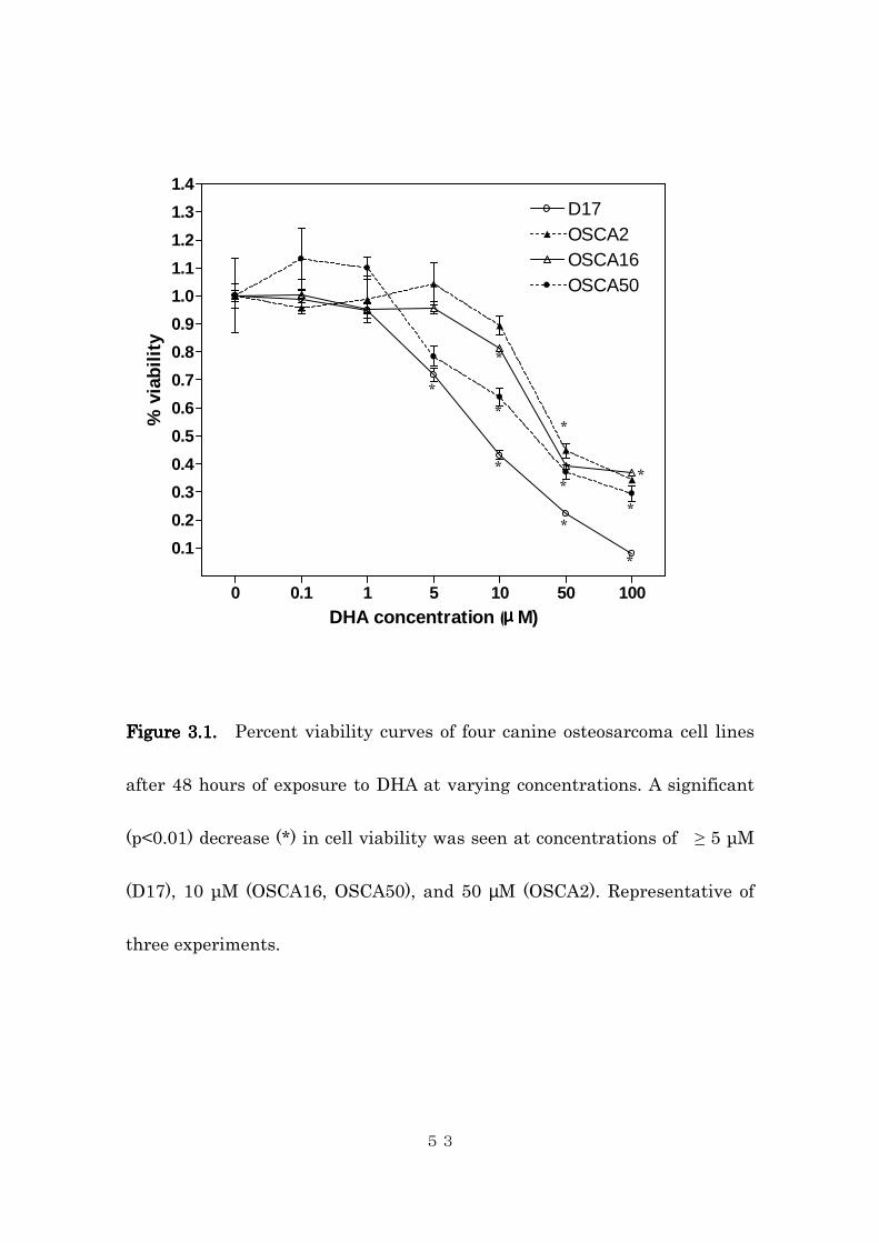
3.4.1. Inhibitory effects of dihydroartemisinin on proliferation of canine
osteosarcoma cells
To test whether DHA can inhibit canine osteosarcoma cell growth, we
determined the growth inhibitory effect of DHA in four canine osteosarcoma
cell lines; D17, OSCA2, OSCA16, and OSCA50. Cells were treated with DHA
at various concentrations for 48 hours and cell viability determined using the
CyQuantTM assay. DHA decreased cell viability in all four canine
osteosarcoma cell lines in a dose-dependent manner (Figure 3.1). A
significant decrease in cell viability was observed at the concentrations of 5,
50, 10, and 10 µM or greater for D17, OSCA2, OSCA16, and OSCA50 cell
lines, respectively. The calculated IC50 (95% CI) for the three cell lines were
8.7, 43.6, 16.8, and 14.8 µM for D17, OSCA2, OSCA16, and OSCA50,
respectively. Microscopic examination revealed cellular fragmentation and
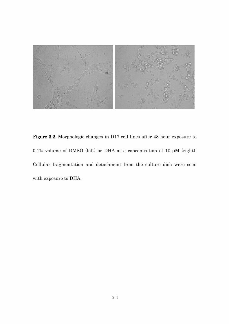
loss of attachment to the cell culture plate in all four cell lines (Figure 3.2).
In contrast, DMSO vehicle-treated cells showed no evidence of cytotoxicity.

50
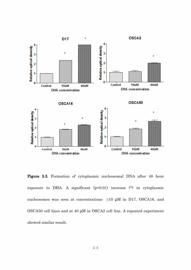
3.4.2. Induction of cell death through apoptosis
The mode of DHA-induced cell death was assessed using an ELISA
based technique for the detection and quantification of cytoplasmic histone
associated DNA fragments in the form of mononucleosomes or
oligonucleosomes. Histone associated DNA fragments are generated during
the process of apoptosis, and are thus represent a quantitative measure of
this process. As demonstrated in Figure 3.3, DHA treatment induced a
dose-dependent increase in free cytoplasmic nucleosome formation. A
significant increase in cytoplasmic nucleosomes was observed at the
concentration of 10 µM in the D17, OSCA16, and OSCA50 cell lines, and at
the concentration of 40 µM in the OSCA2 cell line. This was accompanied by
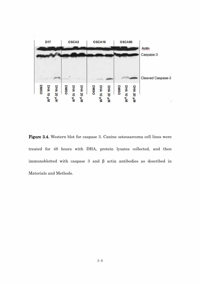
activation (cleavage) of caspase 3 (Figure 3.4). Caspase 3 is one of the effector
caspases, which play key roles in cellular apoptotic pathway. Activation of
caspase 3 is indicative of apoptosis. Taken together, these data indicate the
anti-proliferative effect of DHA was, at least in part, due to apoptosis of
treated cells.
3.4.3. Accumulation of subG0 cells and G2/M cells
51
We also investigated the effect of DHA on cell cycle progression. As
shown in Figure 3.5, 3.6A-D, and Table 3.1, an increased proportion of
subG0/G1 and G2/M phase populations was observed in all four cell lines after
DHA treatment, suggesting G1/S phase specific cytotoxicity and induction of
apoptosis (Figure 3.5, Figure 3.6A-D, Table 3.1).
3.4.4. Generation of reactive oxygen species by an iron-dependent
mechanism
Finally, in order to determine if DHA induced iron-mediated ROS
generation in canine osteosarcoma cells, we measured induction of ROS by
DHA in the D17 canine osteosarcoma cell line (Figure 3.7A-B).
6-carboxy-2′,7′-dichlorodihydrofluoresce is a cell-permeating non-fluorescent
probe that, after oxidation by ROS, exhibits fluorescence which can be
measured using flow cytometric analysis. Thus, in this assay an increase in
fluorescence indicates an increase in the level of ROS. DHA induced a
dose-dependent increase in ROS generation in D17 cells (Figure 3.7A).
Significantly, this increase in ROS was suppressed by pretreatment of cells
with the iron chelator desferrioxamine (Figure 3.7B), consistent with

52
previous studies regarding the role of iron in artemisinin cytotoxicity.
53
0 0.1 1 5 10 50 100
0.1
0.2
0.3
0.4
0.5
0.6
0.7
0.8
0.9
1.0
1.1
1.2
1.3
1.4
D17OSCA2OSCA16OSCA50
DHA concentration (uM)
% v
iab
ilit
y
Figure 3.1. Figure 3.1. Figure 3.1. Figure 3.1. Percent viability curves of four canine osteosarcoma cell lines
after 48 hours of exposure to DHA at varying concentrations. A significant
(p<0.01) decrease (*) in cell viability was seen at concentrations of ≥ 5 µM
(D17), 10 µM (OSCA16, OSCA50), and 50 µM (OSCA2). Representative of
three experiments.
*
*
*
*
*
*
*
*
*
*
µµµµ
54
Figure Figure Figure Figure 3.23.23.23.2. . . . Morphologic changes in D17 cell lines after 48 hour exposure to
0.1% volume of DMSO (left) or DHA at a concentration of 10 µM (right).
Cellular fragmentation and detachment from the culture dish were seen
with exposure to DHA.
55
Figure Figure Figure Figure 3.33.33.33.3.... Formation of cytoplasmic nucleosomal DNA after 48 hour
exposure to DHA. A significant (p<0.01) increase (*) in cytoplasmic
nucleosomes was seen at concentrations ≥10 µM in D17, OSCA16, and
OSCA50 cell lines and at 40 µM in OSCA2 cell line. A repeated experiment
showed similar result.
56
Figure Figure Figure Figure 3.43.43.43.4.... Western blot for caspase 3. Canine osteosarcoma cell lines were
treated for 48 hours with DHA, protein lysates collected, and then
immunoblotted with caspase 3 and β actin antibodies as described in
Materials and Methods.
57
Figure Figure Figure Figure 3.53.53.53.5. . . . Proportion of subG0/G1 cells in canine osteosarcoma cell lines D17
(A), OSCA2 (B), OSCA16 (C), and OSCA50 (D) after incubation with DMSO
(control treatment [white bar]) or various concentrations of
dihydroartemsiinin (1 µM [gray bar], 10 µM [diagonal-striped bar], 50 µM
[black bar]) for 24, 48, or 72 hours. An increase in the subG0/G1 population
was seen at concentrations of ≥ 10 µM in the D17, OSCA16, and OSCA50 cell
lines, and at a concentration of 50 µM in the OSCA2 cell line. A repeated
experiment yielded similar results.
58
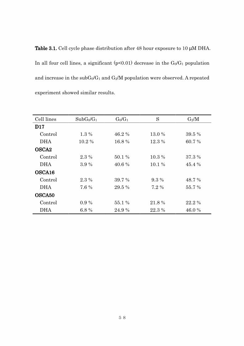
Table Table Table Table 3333.1. .1. .1. .1. Cell cycle phase distribution after 48 hour exposure to 10 µM DHA.
In all four cell lines, a significant (p<0.01) decrease in the G0/G1 population
and increase in the subG0/G1 and G2/M population were observed. A repeated
experiment showed similar results.
Cell lines SubG0/G1 G0/G1 S G2/M
D17D17D17D17
Control
DHA
1.3 %
10.2 %
46.2 %
16.8 %
13.0 %
12.3 %
39.5 %
60.7 %
OSCA2OSCA2OSCA2OSCA2
Control
DHA
2.3 %
3.9 %
50.1 %
40.6 %
10.3 %
10.1 %
37.3 %
45.4 %
OSCA16OSCA16OSCA16OSCA16
Control
DHA
2.3 %
7.6 %
39.7 %
29.5 %
9.3 %
7.2 %
48.7 %
55.7 %
OSCA50OSCA50OSCA50OSCA50
Control
DHA
0.9 %
6.8 %
55.1 %
24.9 %
21.8 %
22.3 %
22.2 %
46.0 %
59
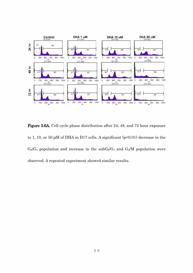
FigureFigureFigureFigure 3333....6A6A6A6A. . . . Cell cycle phase distribution after 24, 48, and 72 hour exposure
to 1, 10, or 50 µM of DHA in D17 cells. A significant (p<0.01) decrease in the
G0/G1 population and increase in the subG0/G1 and G2/M population were
observed. A repeated experiment showed similar results.
60
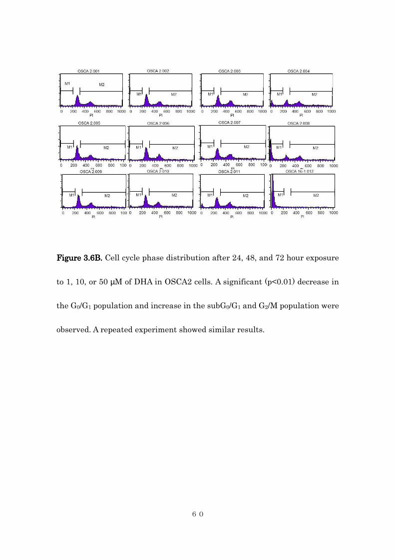
FigureFigureFigureFigure 3333....6B6B6B6B. . . . Cell cycle phase distribution after 24, 48, and 72 hour exposure
to 1, 10, or 50 µM of DHA in OSCA2 cells. A significant (p<0.01) decrease in
the G0/G1 population and increase in the subG0/G1 and G2/M population were
observed. A repeated experiment showed similar results.
61
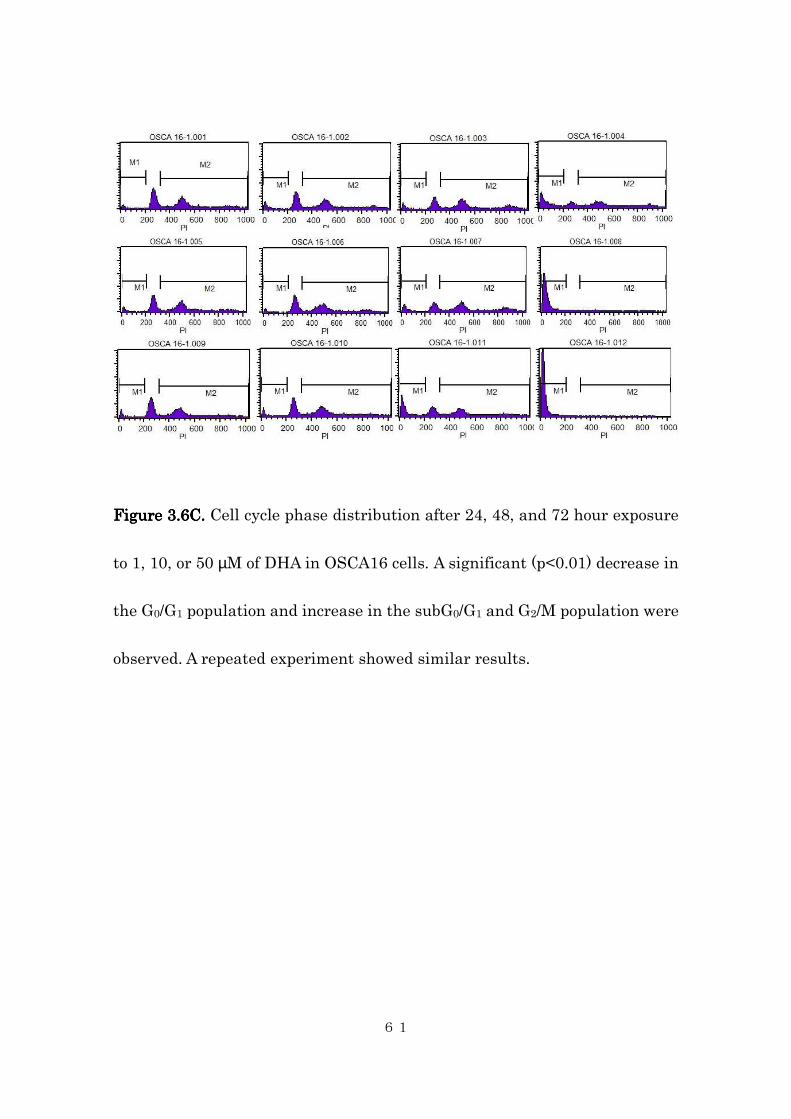
FigureFigureFigureFigure 3333....6C6C6C6C. . . . Cell cycle phase distribution after 24, 48, and 72 hour exposure
to 1, 10, or 50 µM of DHA in OSCA16 cells. A significant (p<0.01) decrease in
the G0/G1 population and increase in the subG0/G1 and G2/M population were
observed. A repeated experiment showed similar results.
62
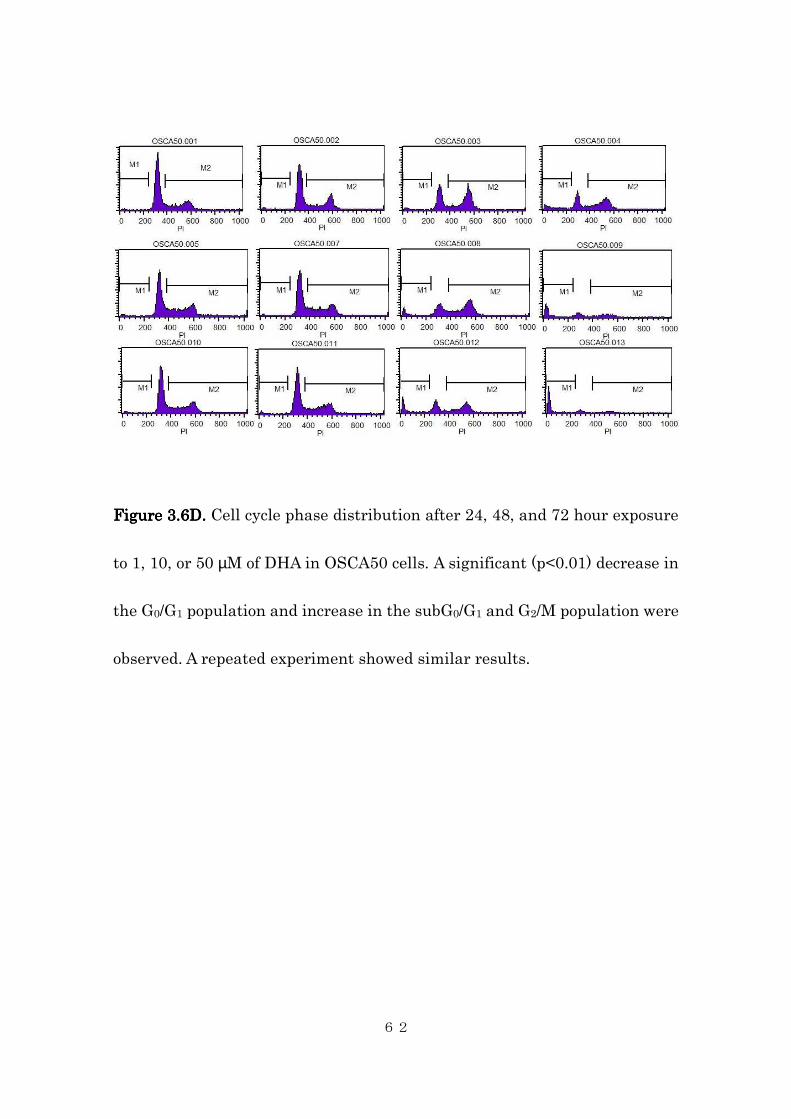
FigureFigureFigureFigure 3333....6D6D6D6D. . . . Cell cycle phase distribution after 24, 48, and 72 hour exposure
to 1, 10, or 50 µM of DHA in OSCA50 cells. A significant (p<0.01) decrease in
the G0/G1 population and increase in the subG0/G1 and G2/M population were
observed. A repeated experiment showed similar results.
63
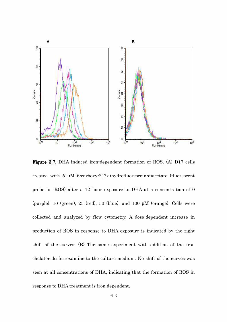
Figure Figure Figure Figure 3.3.3.3.7777.... DHA induced iron-dependent formation of ROS. (A) D17 cells
treated with 5 µM 6-carboxy-2’,7’dihydrofluorescein-diacetate (fluorescent
probe for ROS) after a 12 hour exposure to DHA at a concentration of 0
(purple), 10 (green), 25 (red), 50 (blue), and 100 µM (orange). Cells were
collected and analyzed by flow cytometry. A dose-dependent increase in
production of ROS in response to DHA exposure is indicated by the right
shift of the curves. (B) The same experiment with addition of the iron
chelator desferroxamine to the culture medium. No shift of the curves was
seen at all concentrations of DHA, indicating that the formation of ROS in
response to DHA treatment is iron dependent.
A B

64
3.5. Discussion
During the past two decades, the anti-malarial agent artemisinin and
its derivatives have attracted interest as potential novel anticancer agents,
cancer preventatives, multi-drug resistance reversal agents, and
radiosensitizers. These artemisinin-derived 1,2,4-trioxanes exhibit
significant activity in the nanomolar to micromolar range against a variety
of human cancer cell lines. Their biologic activity in human cancer cell lines
and apparently low toxicity in human malaria patients have stimulated
interest in using artemisinin and its derivatives to treat canine cancers,
particularly osteosarcoma, as the drug is readily available over-the-counter.
However, to our knowledge, no study has evaluated the potential activity of
artemisinin against canine cancer cells, either in vitro or in vivo.
In this study, a dose-dependent decrease in the viability of
osteosarcoma cell lines was demonstrated after exposure to DHA. The IC50
values for these osteosarcoma cell lines ranged from 8.7 to 43.6 µM. These
values are similar to those reported for three artemisinin derivatives
(artesunate, artemether, and arteether) when screened with the National
65
Cancer Institute (NCI) 55 cell line panel (Efferth, 2002). In this panel, the
greatest sensitivity was found in leukemia and colon cancer cell lines (mean
IC50: 1-2 µM of artesunate) and the lowest sensitivity was found in non-small
cell lung cancer cell lines (mean IC50: 26 µM of artesunate) (Efferth, 2002).
Unfortunately, no sarcoma cell lines were tested.
Our study also demonstrated a dose-dependent increase in
cytoplasmic nucleosomes and accumulation of activated caspase 3, consistent
with DHA-induced apoptotic cell death. Furthermore, there was a
dose-dependent increase in the subG0/G1 cell population in treated cells,
further supporting that the mechanism of DHA cell killing is, at least in part,
due to apoptosis. Induction of apoptosis by artemisinin derivatives has been
demonstrated in other studies evaluating human cancer cell lines (Efferth,
1996; Singh, 2004; Nam, 2007).
A decreased proportion of cells in the G0/G1 phase was observed in
DHA treated cells. This is in accordance with previous reports, where the
cytotoxicity of artesunate correlated with the proportion of the cells in G0/G1
phase (Efferth, 2003). The underlying mechanism for this cell cycle
phase-selective cytotoxicity is currently unknown.
66
One of the postulated mechanisms of artemisinin and DHA-induced
cytotoxicity is iron-mediated ROS generation, as is in its anti-malarial
activity. Both antimalarial and tumoricidal activities of artemisinin are
known to be iron-dependent (Disbrow, 2005; Meshnick, 2002). For example,
the in vitro cytotoxicity of DHA in papilloma virus-infected epithelial cells
could be reversed by chelating iron from the culture medium (Disbrow, 2005).
To confirm that generation of ROS occurred in canine osteosarcoma cells, we
measured ROS activity in the D17 canine osteosarcoma cell line treated with
DHA. As expected, dose-dependent generation of ROS by DHA was
observed. ROS generation was completely inhibited by the addition of
desferroxamine, an iron chelating agent, to the culture medium,
demonstrating that ROS generation by DHA is iron-dependent in canine
osteosarcoma cells.
Our data indicate that DHA induced apoptosis of canine osteosarcoma
cell lines requires drug concentrations in the micromolar range. Peak plasma
concentrations after an antimalarial dose of oral DHA (4 mg/kg) in human
malaria patients ranged from 1.9 to 16 µM (median: 4 µM) (Newton, 2002).
These values are lower than the cytotoxic range observed in our study;
67
however, the low toxicity of artemisinins in human patients and the
extremely high LD50 values of artemisinin derivatives reported in a previous
animal study suggests that cytotoxic plasma concentrations might be
achievable in vivo by using higher doses of artemisinin derivatives than
those currently being used empirically in dogs (China Cooperative Research
Group on Qinghaosu and Its Derivatives as Antimalarials, 1982). In
summary, our data demonstrate biologic activity of DHA against canine
osteosarcoma. Further studies to evaluate the safety and efficacy of
artemisinin and its derivatives in dogs with osteosarcoma are warranted.
68
4. Cytotoxic Effects of Dihydroartemisinin in Canine Histiocytic
Sarcoma Cell Lines: A Relationship Between Intracellular
Ferrous Contents and Cytotoxicity of Dihydroartemisinin
4.1. Summary
Artemisinin-derived trioxanes has been shown to exhibit cytotoxic effect
through iron-dependent generation of free radicals, thus making them
potential candidates for cancer therapeutics for neoplasia with high
iron-contents. Canine histiocytic sarcoma is a neoplasia of
macrophage/dendricytic origin. It retains hemophagocytic capability and
causes erythrophagocytosis clinically called hemophagocytic syndrome, and
therefore is suggested to have high iron content. In this study, we have
evaluated cytotoxicity of dihydroartemisinin in two canine histiocytic
sarcoma cell lines in relation to the cellular iron content. Dihydroartemisinin
caused significant decrease in cell viability in both cell lines in a
concentration-dependent manner. The mode of cell death was at least in part
through induction of apoptosis. As predicted, dihydroartemisinin’s
69
cytotoxicity was influenced by the cellular iron content; increasing
intracellular iron content by adding ferrous citrate ammonium or
holotransferrin resulted in enhanced cytotoxicity of dihydroartemisinin and
decreasing cellular iron content by removing iron from the media by
deferroxamine markedly obliterated the drug’s effect. This study is the first
to demonstrate in vitro cytotoxic activity of an artemisinin derivative in
canine histiocytic sarcoma and suggests that neoplasm that contains high
iron load may be appropriate candidate for artemisinin derivatives.
70
4.2. Introduction
Artemisinin-derived trioxanes has been shown to exhibit cytotoxic effect on
various cancer cell lines through generation of free radicals and oxidation of
intracellular targets such as transcriptionally controlled tumor protein
(TCTP). Due to this unique mechanism of action, the cytotoxic effect of
artemisinin-derivatives are generally more pronounced in neoplastic cells
than their somatic counterparts because tumor cells overexpress
transferring receptor to increase uptake of iron to the cytoplasm and
therefore contain higher iron contents than normal cells. However, some
tumor cells are suspected to actively produce iron-binding protein such as
ferritin and may contain increased amount of iron molecules independent of
transferring receptor expression. Hepatocellular carcinoma, lymphoma, and
histiocytic sarcoma all have been known to cause hyperferritinemia;
proposed underlying mechanism for this hyperferritinemia include
erythrocyte destruction, alteration in erythropoiesis, release by tissue
damage, injury to hepatocytes, and production by tumor cells.
Canine histiocytic sarcoma is a highly malignant tumor of phagocytic
71
macrophage or antigen-presenting dendricytic origin (Affolter, 2002). The
disease is rapidly progressive with limited success in therapeutic approach to
intervene the course of disease (Skorupski, 2002; Rassnick, 2011). The
reported response rates to 1-(2-chloroethyl)3-cyclohexyl-1-nitrosourea
(CCNU), the current standard systemic chemotherapeutic agent, range from
29% to 46%, with median progression free interval of as short as 85-96 days
(Skorupski, 2002; Rassnick, 2011). Without effective systemic treatment
options, the overall prognosis of the affected dogs is guarded; even dogs with
localized form of histiocytic sarcoma, the majority eventually develop
disseminated metastatic lesions and sccumb to disease within 6 months of
diagnosis (Affolter, 2002; Craig, 2002; Fidel, 2006; Skorupski, 2009).
One unique aspect of canine histiocytic sarcoma is that the disease is
known to cause marked hyperferritinemia supposedly due to
erythrophagocytosis and production by the tumor cells; however, whether
intracellular iron content is in fact elevated has not been elucidated. We
hypothesized that canine histiocytic sarcoma cells, via direct
erythrophagocytosis or ferritin production or both, contains high iron content
than other histologic types of tumor, and the amount of intracellular iron

72
contents is directly associated with increased cytotoxicity of
artemisinin-derivatives due to enhanced free radical generation. To test
these hypotheses, we first quantified and semiquantified tissue iron contents
by inductively coupled plasma-mass spectrometry (ICP-MS) and Berlin-blue
stain, respectively. The relationship between intracellular iron concentration
and cytotoxicity of dihydroartemisinin, cellular contents of iron molecules
were manipulated and measured by ICP-MS, then anti-proliferative and
apoptosis-inducing effects of dihydroartemisinin were compared between
different conditions with either low, normal, and high cellular iron contents.

73
4.3. Materials and methods
4.3.1. Evaluation of tissue iron contents of canine histiocytic sarcoma
To test whether canine histiocytic sarcoma tissue contains higher
amount of iron molecules compared to other tumor types, tumor tissue
samples were collected from 7 dogs with histologically confirmed histiocytic
sarcoma (Table 4.1) and 9 additional dogs with spontaneously occurring soft
tissue neoplasia other than histiocytic sarcoma (Table 4.2).
For ICP-MS, 0.5 g of tumor tissue samples from each dog were
washed by water and dried at 45 degrees for 12 hours. After adding 5 mL of
65% nitric acida and 1 mL of 30% hydrogenperoxideb to the samples, tissue
samples were decomposed using microwave sample decomposerc. The
processed samples were mess upped to the total of 10 mL by adding distilled
water before used for ICP-MS. Amount of iron contents within each tumor
sample was measured with ICP-MSd. Briefly, the samples were ionized by
collision with argon plasma, and amount of ferrous ion (m/z = 56) was
detected and quantified within the sample.
Since ICP-MS measures the iron contents within the entire sample,
74
regardless of their origin (i.e., tumor cell or red blood cell, etc.),
semiquantification of iron contents within the tumor cells was performed
using Berlin-blue stain. Tissues were formalin-fixed and embedded in
paraffin block and then tissue sections of 4 µm-thickness were prepared
using rotary microtomee. Deparaffinization of the tissue sections were
routinely performed by dipping the samples in xylenef for 5 minutes 3 times,
then decreasing concentrations of ethanol solutionsg. Tissue sections were
then used for Berlin-blue stain. A mixed solution of equal volumes of 2%
potassium ferrocyanideh and 0.7N hydrochloric acidi was prepared
immediately before each experiment. Appropriate amount of this solution
was mounted on the tissue slides and incubated at room temperature for 60
minutes. Samples were washed with distilled water 5 times, and then
counter-stained with 1% Safranin Oj in order to stain the nuclei.
4.3.2. Cell culture and cell lines
Two canine osteosarcoma cell lines (CHS-4 and DH82) were
evaluated. The CHS-4 cell line (Azakami, 2006) was kindly provided by Dr.
Makoto Bonkobara (Japan Veterinary and Life Science University). The

75
DH82 cell line was commercially purchased from European Collection of Cell
Cultures, Salisbury, UK. CHS-4 cell line is derived from canine histiocytic
sarcoma of dendricytic origin, and DH82 is derived from canine histiocytic
sarcoma of phagocytic macrophage origin. Both cell lines were cultured in
Dulbecco’s modified Eagle mediumk (DMEM) supplemented with 10% fetal
bovine seruml (FBS) and antibiotics (penicillinm [100 U/ml] and
streptomycinn [0.1 mg/ml]) with the addition of 12 mM of
2-[4-(2-hydroxyethyl)-1-piperazinyl]ethanesulfonic acid (HEPES)o. Both cell
lines were maintained in a humidified environment at 37° C under 5% CO2
and 95% room air.
4.3.3. Chemicals
Dihydroartemisininp was dissolved in 100% dimethylsulfoxide
(DMSO) at the concentration of 100 mM as a stock solution, protected from
light, and kept at -80° C for long-term storage. The stock solution was
further diluted with DMSO prior to use in tissue culture, resulting in a
consistent DMSO concentration of 0.1% volume. Dihydroartemisinin
solutions were newly prepared from the stock solution for each experiment.
76
Ferric ammonium citratep was dissolved in DMEM in 200 µM
concentration and stored at -80 ℃ until use.
Holotransferrinr was prepared in DMEM at a concentration of 1
mg/mL, and stored at 4 ℃ until use.
Deferoxamine mesylates was dissolved in distilled water at a
concentration of 100 mM and was stored at 4 ℃ until use.
4.3.4. Assessment of cell viability
Cell viability was assessed using tetrazolium assay also known as
MTT assay (Rubinstein, 1990). Cells were seeded in 96-well plates at density
of 1,000 - 2,000 cells per well in triplicates to a final volume of 50 µL/well.
After 24 hours, 50 µL of medium containing DMSO (control) or different
concentrations of dihydroartemisinin was added to each well to create final
dihydroartemisinin concentrations of 1, 5, 10, 25, 50, 75, and 100 µM and the
cells were incubated for 24 additional hours. After treatment, 20 µL of 5
mg/mL 3-(4,5-dimethyl-2--thyazoryl)-2,5-diphenyltetrazoliumbromidet
(MTT) was added and the cells were further incubated at 37 ℃, 5% CO2 for 4
horus to allow for cells to form tetrazolium crystals. After incubation, the
77
medium were gently suctioned, and 50 µL of dissolving solution was added to
each well. Dissolving solution was prepared by mixing 20% sodium dodecyl
sulfateu (SDS) with equal volume of N,N-dimethylformamidev with pH
adjusted to 4.7 by adding 11.5N hydrochloric acid. Optical density at a wave
length of 570 nm (OD570)of each well was measured by microplate readerw.
Cell proliferation was expressed as a percentage of the control wells: OD570
of sample/ OD570 of control (DMSO only) wells. Experiments were repeated
at least three times.
4.3.5. Detection of apoptosis
Number of apoptotic cells were semiquantified by fluorescent
staining using FITC-conjugated annexin V staining kitx according to the
manufacturer’s instruction. Briefly, cells were suspended in binding buffer
containing annexin V and propidium iodide and incubated for 5 minutes in a
dark room. Cells were then fixed with 2% formaldehyde and blocked with
10 % rabbit serum. Cells were stained with fluorescein conjugatey for 15
minutes for observation. Microscopic inspection of the cell membrane and
nuclei was performed using a fluorescein microscopyz.
78
4.3.6. Manipulation and measurement of intracellular iron contents
To increase the intracellular iron contents, 200 µM of ferric
ammonium citrate or 1 mg/mL of holotransferrin was added to the culture
media. Cells were incubated for 24 hours (in case of ferric ammonium citrate)
or for 6 hours (in case of holotransferrin) to allow for iron uptake. In each
condition, the final concentration of DMSO was kept at <0.1% in all
experiments.
To decrease intracellular iron contents, ferrous ion was chelated by
the addition of 50 mM of deferoxamine mesylate to the culture media for 6
hours before each experiment. The final concentration of DMSO was kept at
<0.1% in all experiments.
Measurement of intracellular iron contents were carried out using
ICP-MS. Cells pretreated in above conditions were washed three times with
isotonic phosphate buffer solution (pH 7.4). 5.0 x 10⁵ cells were counted and
cell pellet was created by centrifuging at 200xg for 5 minutes. After removing
the supernatant, 800 µL of 65% nitric acid and 200 µL of 30% hydrogen
peroxide were added and the samples were incubated at room temperature
79
for 24 hours. After incubation, 2% nitric acid was added to make the final
volume of each sample to 8 mL for ICP-MS measurement. The rate of
retrieval of each sample was confirmed by using a substance with known
iron contents (DORM-3aa).
4.3.7. Statistical analysis
Changes in cell viability, measured by tetrazolium assay, were
compared using one-way analysis of variance. A pairwise multiple
comparison procedure was performed using the Dunnet test. All statistical
analysis was performed using GraphPad Prism 4.0 softwareab. Statistical
significance was established as P≤ 0.05.
4.3.8. Footnotes
a Nitric acid, Kanto Chemicals, Tokyo, Japan
b Hydrogen peroxide, Kanto Chemicals, Tokyo, Japan
c SpeedWaveTwo, BERGHOF, Eningen, Germany
d ICP-MS Agilent 7700, Agilent Technologies, Santa Clara, CA
e HM325, MICROM, Waltham, MA

80
f Xylene, Wako Chemicals, Osaka, Japan
g Ethanol, Wako Chemicals, Osaka, Japan
h Potassium ferrocyanide solution, Muto Chemicals, Tokyo, Japan
i Hydrochloric acid, Muto Chemicals, Tokyo, Japan
j 1% Safranin O, Muto Chemicals, Tokyo, Japan
k DMEM, GIBCO BRL, Gaithersburg, NY
l FBS, Nichirei Bioscience, Tokyo, Japan
m Penicillin, Nakarai Tesque, Kyoto, Japan
n Streptomycin, Nakarai Tesque, Kyoto, Japan
o HEPES, Chemical Dojin, Kumamoto, Japan
p Dihydroartemisinin, Sigma-Aldrich Inc., Saint Louis, MO
q Ferric ammonium citrate, Sigma-Aldich Inc., Saint Louis, MO
r Holtransferrin, Sigma-Aldrich Inc., Saint Louis, MO
s Deferoxamine mesylate, Novartis Pharma, Tokyo, Japan
t MTT, Wako Chemicals, Osaka, Japan
u Sodium dodecyl sulfate, Wako Chemicals, Osaka, Japan
v N,N-dimethyl formamide, Wako Chemicals, Osaka, Japan
w Multiskan FC, Thermo Fisher Scientific, Waltham, MA
81
x Annexin V-Biotin Apoptosis Detection Kit, BioVision Inc., Milpitas, CA
y Fluorescein Conjugate, Birmingham, Southan Biotech, AL
z FLoidTM cell imaging station, Life Technologies Japan, Tokyo, Japan
aa DORM-3 Fish protein, Certified Reference Material, National Research
Council of Canada, Ottawa, Canada
ab Graphpad Prism 4.0, GraphPad Software Inc., La Jolla, CA
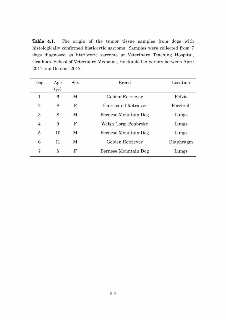
82
Table 4.1Table 4.1Table 4.1Table 4.1.... The origin of the tumor tissue samples from dogs with
histologically confirmed histiocytic sarcoma. Samples were collected from 7
dogs diagnosed as histiocytic sarcoma at Veterinary Teaching Hospital,
Graduate School of Veterinary Medicine, Hokkaido University between April
2011 and October 2012.
Dog Age
(yr)
Sex Breed Location
1 6 M Golden Retriever Pelvis
2 8 F Flat-coated Retriever Forelimb
3 8 M Bernese Mountain Dog Lungs
4 9 F Welsh Corgi Penbroke Lungs
5 10 M Bernese Mountain Dog Lungs
6 11 M Golden Retriever Diaphragm
7 5 F Bernese Mountain Dog Lungs
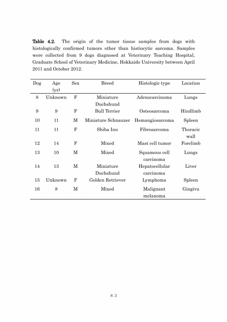
83
Table 4.Table 4.Table 4.Table 4.2222.... The origin of the tumor tissue samples from dogs with
histologically confirmed tumors other than histiocytic sarcoma. Samples
were collected from 9 dogs diagnosed at Veterinary Teaching Hospital,
Graduate School of Veterinary Medicine, Hokkaido University between April
2011 and October 2012.
Dog Age
(yr)
Sex Breed Histologic type Location
8 Unknown F Miniature
Duchshund
Adenocarcinoma Lungs
9 9 F Bull Terrier Osteosarcoma Hindlimb
10 11 M Miniature Schnauzer Hemangiosarcoma Spleen
11 11 F Shiba Inu Fibrosarcoma Thoracic
wall
12 14 F Mixed Mast cell tumor Forelimb
13 10 M Mixed Squamous cell
carcinoma
Lungs
14 13 M Miniature
Duchshund
Hepatocellular
carcinoma
Liver
15 Unknown F Golden Retriever Lymphoma Spleen
16 8 M Mixed Malignant
melanoma
Gingiva
84
4.4. Results
4.4.1. Tissue iron contents in canine histiocytic sarcoma
To test whether canine histiocytic sarcoma and other neoplasm tissue
actually contains high amount of iron contents, direct measurement of tissue
iron contents by ICP-MS was performed. In this method, both protein-bound
and free ionized form of iron contents were measured, as well as
extratumoral iron contents (e.g., iron contents of hemoglobin within red
blood cell in tumor vasculature). The rate of iron retrieval was estimated as
95.8 +/- 6.1%, suggesting a sufficient rate of retrieval for tissue iron content
measurement. The median iron contents of histiocytic sarcoma tissues was
142 µg/g tissue (range: 40 - 700 µg/g tissue) and the median iron contents of
other tumor tissues was 321 µg/g tissue (range: 36 – 1,580 µg/g tissue). There
was no significant difference between the two groups (p=0.21, Figure 4.1).
The two highest values were observed in samples from hemangiosarcoma
(1,581 µg/g tissue) and hepatocellular carcinoma (1,195 µg/g tissue).
The results of Berlin-blue staining were summarized in Table 4. All
histiocytic sarcoma tissues and majority of the other tumor tissue samples

85
contained Berlin-blue-positive cells.
4.4.2. Effects of intracellular iron concentration on cytotoxic activity of
dihydroartemisinin in canine histiocytic sarcoma cells
Dihydroartemisinin showed significant cytotoxicity in both cell
CHS-4 and DH82 cell lines in a concentration-dependent manner (Figure 4.2
and 4.3). The estimated IC50 value for CHS-4 and DH82 cells were 10 µM and
7.6 µM, respectively.
To investigate whether cytotoxicity of dihydroartemisinin correlates
with intracellular iron concentration, cells were incubated in regular DMEM,
with or without addition of deferoxamine, an iron-chelator, ferric ammonium
citrate, or holotransferrin. In each conditions, the amount of intracellular
iron was quantified by ICP-MS. In both CHS-4 and DH82 cell lines,
intracellular iron contents decreased with the addition of deferoxamine and
increased with the addition of ferric ammonium citrate and holotransferrin
(Figure 4.4 and 4.5). Although the difference did not reach statistical
significance, there was a clear tendency for the cellular iron contents to
decrease with pretreatment with deferoxamine and to increase with
86
preincubation with holotransferrin.
These increase or decrease in cellular iron concentration directly
correlated with cytotoxicity of dihydroartemisinin. As shown in Figure 4.6
and 4.7, the sensitivity of both CHS-4 and DH82 cells to dihydroartemisinin
were enhanced by increasing intracellular iron contents by ferric ammonium
citrate, and the effect of dihydroartemisinin was obliterated by chelating iron
from the media by deferroxamine. Similarly, the cytotoxic effect of
dihydroartemisinin was enhanced by pretreatment with holotransferrin.
These changes were more pronounced in DH82 cells, a cell line with stronger
phagocytic capacity.
Dihydroartemisinin treatment also induced cellular apoptosis in a
dose-dependent manner, demonstrated by increased number of
annexin-positive cells as shown in Figure 4.10. Addition of ferric ammonium
citrate and holotransferrin both decreased the required dihydroartemisinin
concentration to induce significant apoptosis. Due to excessive cell death, few
viable cells were observed at the concentration of 100 µM in these conditions.
In contrast, pretreatment with deferoxamine markedly inhibited the effect of
dihydroartemisinin, resulting in no discernible apoptosis at 10 µM, and

87
significant apoptosis was only seen at a concentration of 100 µM.
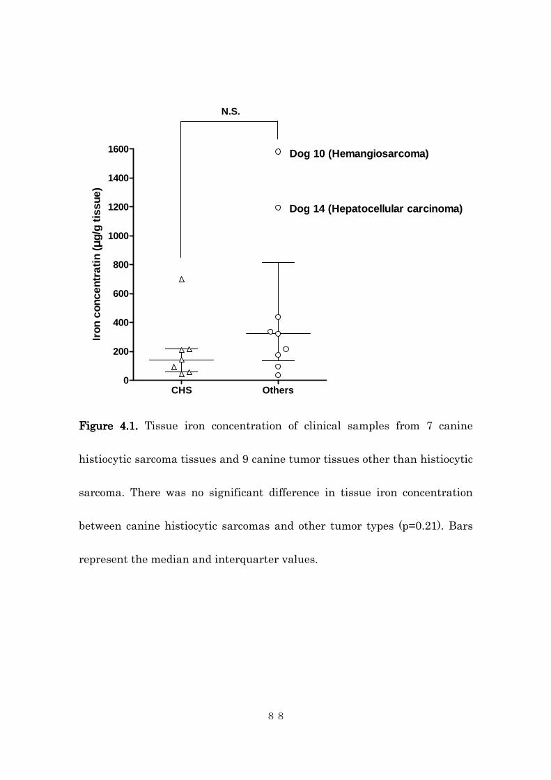
88
CHS Others0
200
400
600
800
1000
1200
1400
1600
N.S.
Dog 10 (Hemangiosarcoma)
Dog 14 (Hepatocellular carcinoma)
Iron
con
cent
rati
n ( µµ µµ
g/g
tiss
ue)
Figure 4.1.Figure 4.1.Figure 4.1.Figure 4.1. Tissue iron concentration of clinical samples from 7 canine
histiocytic sarcoma tissues and 9 canine tumor tissues other than histiocytic
sarcoma. There was no significant difference in tissue iron concentration
between canine histiocytic sarcomas and other tumor types (p=0.21). Bars
represent the median and interquarter values.
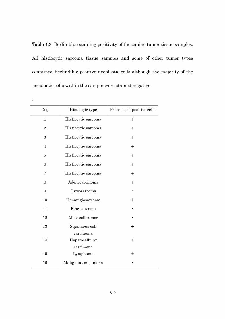
89
Table 4.3.Table 4.3.Table 4.3.Table 4.3. Berlin-blue staining positivity of the canine tumor tissue samples.
All histiocytic sarcoma tissue samples and some of other tumor types
contained Berlin-blue positive neoplastic cells although the majority of the
neoplastic cells within the sample were stained negative
.
Dog Histologic type Presence of positive cells
1 Histiocytic sarcoma +
2 Histiocytic sarcoma +
3 Histiocytic sarcoma +
4 Histiocytic sarcoma +
5 Histiocytic sarcoma +
6 Histiocytic sarcoma +
7 Histiocytic sarcoma +
8 Adenocarcinoma +
9 Osteosarcoma -
10 Hemangiosarcoma +
11 Fibrosarcoma -
12 Mast cell tumor -
13 Squamous cell
carcinoma
+
14 Hepatocellular
carcinoma
+
15 Lymphoma +
16 Malignant melanoma -
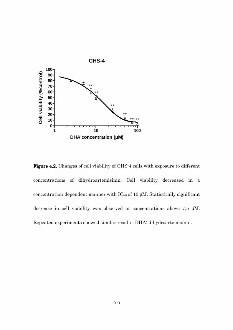
90
CHS-4
1 10 1000
102030405060708090
100
DHA concentration (µµµµM)
Cel
l vi
abil
ity
(%co
ntr
ol)
Figure 4.2.Figure 4.2.Figure 4.2.Figure 4.2. Changes of cell viability of CHS-4 cells with exposure to different
concentrations of dihydroartemisinin. Cell viability decreased in a
concentration-dependent manner with IC50 of 10 µM. Statistically significant
decrease in cell viability was observed at concentrations above 7.5 µM.
Repeated experiments showed similar results. DHA: dihydroartemisinin.
** ** **
**
**
**
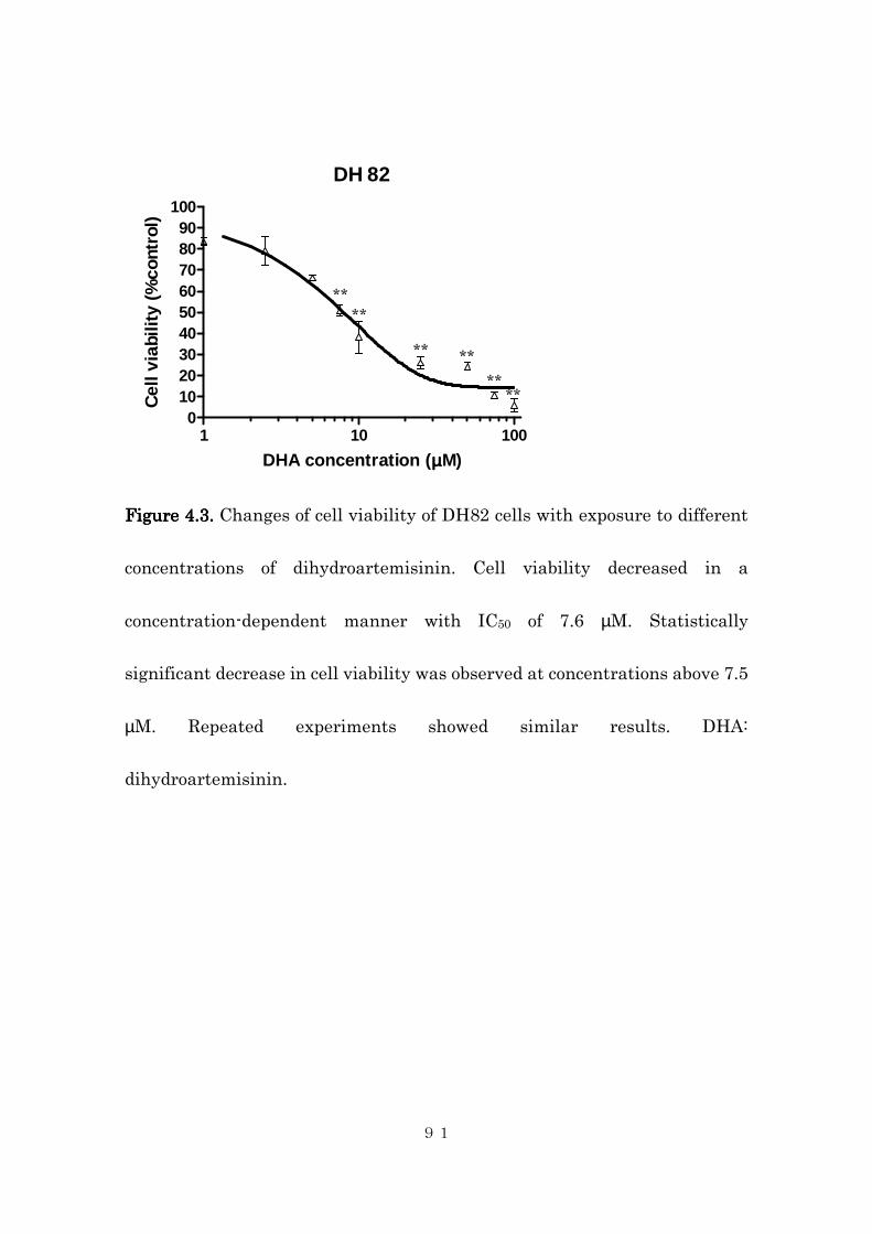
91
DH 82
1 10 1000
102030405060708090
100
DHA concentration (µµµµM)
Cel
l via
bilit
y (%
cont
rol)
Figure 4.3.Figure 4.3.Figure 4.3.Figure 4.3. Changes of cell viability of DH82 cells with exposure to different
concentrations of dihydroartemisinin. Cell viability decreased in a
concentration-dependent manner with IC50 of 7.6 µM. Statistically
significant decrease in cell viability was observed at concentrations above 7.5
µM. Repeated experiments showed similar results. DHA:
dihydroartemisinin.
** **
** **
** **
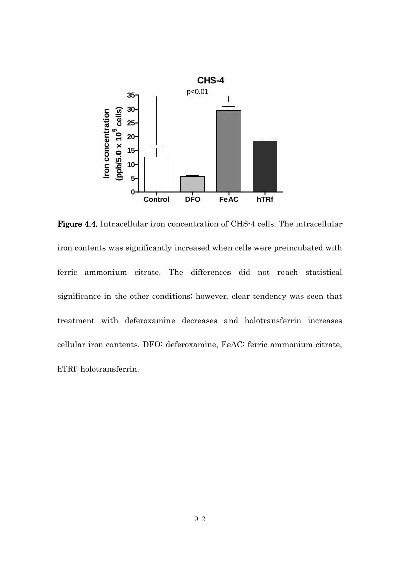
92
CHS-4
Control DFO FeAC hTRf0
5
10
15
20
25
30
35 p<0.01
Iron
con
cent
rati
on
(ppb
/5.0
x 1
05 c
ells
)
Figure 4.4Figure 4.4Figure 4.4Figure 4.4.... Intracellular iron concentration of CHS-4 cells. The intracellular
iron contents was significantly increased when cells were preincubated with
ferric ammonium citrate. The differences did not reach statistical
significance in the other conditions; however, clear tendency was seen that
treatment with deferoxamine decreases and holotransferrin increases
cellular iron contents. DFO: deferoxamine, FeAC: ferric ammonium citrate,
hTRf: holotransferrin.
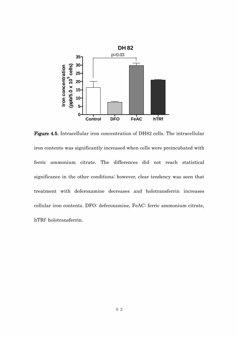
93
DH 82
Control DFO FeAC hTRf0
5
10
15
20
25
30
35 p=0.03
Iron
con
cent
rati
on
(ppb
/5.0
x 1
05 c
ells
)
Figure 4.5Figure 4.5Figure 4.5Figure 4.5.... Intracellular iron concentration of DH82 cells. The intracellular
iron contents was significantly increased when cells were preincubated with
ferric ammonium citrate. The differences did not reach statistical
significance in the other conditions; however, clear tendency was seen that
treatment with deferoxamine decreases and holotransferrin increases
cellular iron contents. DFO: deferoxamine, FeAC: ferric ammonium citrate,
hTRf: holotransferrin.
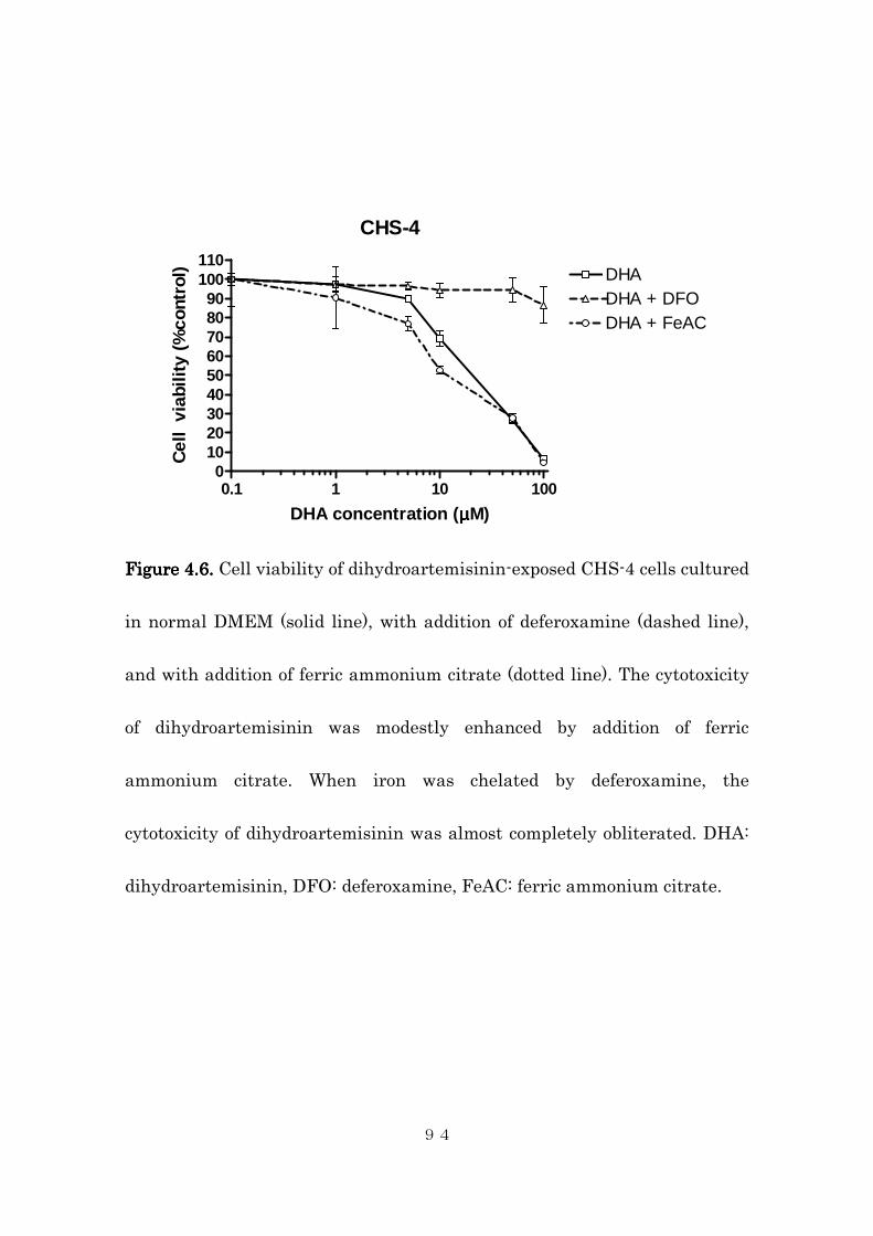
94
CHS-4
0.1 1 10 1000
102030405060708090
100110
DHADHA + DFODHA + FeAC
DHA concentration (µµµµM)
Cel
l vi
abili
ty (
%co
ntro
l)
Figure 4.6.Figure 4.6.Figure 4.6.Figure 4.6. Cell viability of dihydroartemisinin-exposed CHS-4 cells cultured
in normal DMEM (solid line), with addition of deferoxamine (dashed line),
and with addition of ferric ammonium citrate (dotted line). The cytotoxicity
of dihydroartemisinin was modestly enhanced by addition of ferric
ammonium citrate. When iron was chelated by deferoxamine, the
cytotoxicity of dihydroartemisinin was almost completely obliterated. DHA:
dihydroartemisinin, DFO: deferoxamine, FeAC: ferric ammonium citrate.
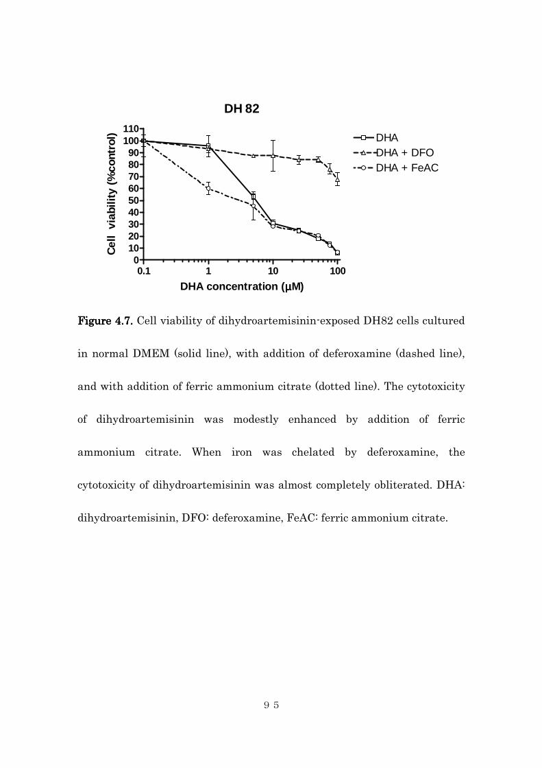
95
DH 82
0.1 1 10 1000
102030405060708090
100110
DHADHA + DFODHA + FeAC
DHA concentration (µµµµM)
Cel
l vi
abili
ty (
%co
ntro
l)
Figure 4.7.Figure 4.7.Figure 4.7.Figure 4.7. Cell viability of dihydroartemisinin-exposed DH82 cells cultured
in normal DMEM (solid line), with addition of deferoxamine (dashed line),
and with addition of ferric ammonium citrate (dotted line). The cytotoxicity
of dihydroartemisinin was modestly enhanced by addition of ferric
ammonium citrate. When iron was chelated by deferoxamine, the
cytotoxicity of dihydroartemisinin was almost completely obliterated. DHA:
dihydroartemisinin, DFO: deferoxamine, FeAC: ferric ammonium citrate.

96
CHS-4
0.1 1 10 1000
102030405060708090
100110
DHADHA + DFODHA + hTRf
DHA concentration (µµµµM)
Cel
l vi
abili
ty (
%co
ntro
l)
Figure 4.8.Figure 4.8.Figure 4.8.Figure 4.8. Cell viability of dihydroartemisinin-exposed CHS-4 cells cultured
in normal DMEM (solid line), with addition of deferoxamine (dashed line),
and with addition of holotransferrin (dotted line). The cytotoxicity of
dihydroartemisinin was markedly enhanced by addition of holotransferrin.
When iron was chelated by deferoxamine, the cytotoxicity of
dihydroartemisinin was almost completely obliterated. DHA:
dihydroartemisinin, DFO: deferoxamine, hTRf: holotransferrin.
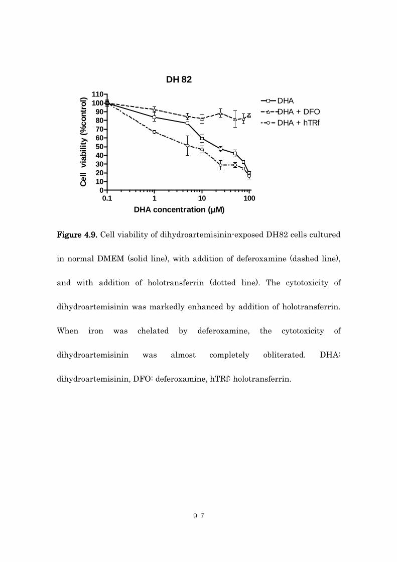
97
DH 82
0.1 1 10 1000
102030405060708090
100110
DHADHA + DFODHA + hTRf
DHA concentration (µµµµM)
Cel
l vi
abili
ty (
%co
ntro
l)
Figure 4.9.Figure 4.9.Figure 4.9.Figure 4.9. Cell viability of dihydroartemisinin-exposed DH82 cells cultured
in normal DMEM (solid line), with addition of deferoxamine (dashed line),
and with addition of holotransferrin (dotted line). The cytotoxicity of
dihydroartemisinin was markedly enhanced by addition of holotransferrin.
When iron was chelated by deferoxamine, the cytotoxicity of
dihydroartemisinin was almost completely obliterated. DHA:
dihydroartemisinin, DFO: deferoxamine, hTRf: holotransferrin.
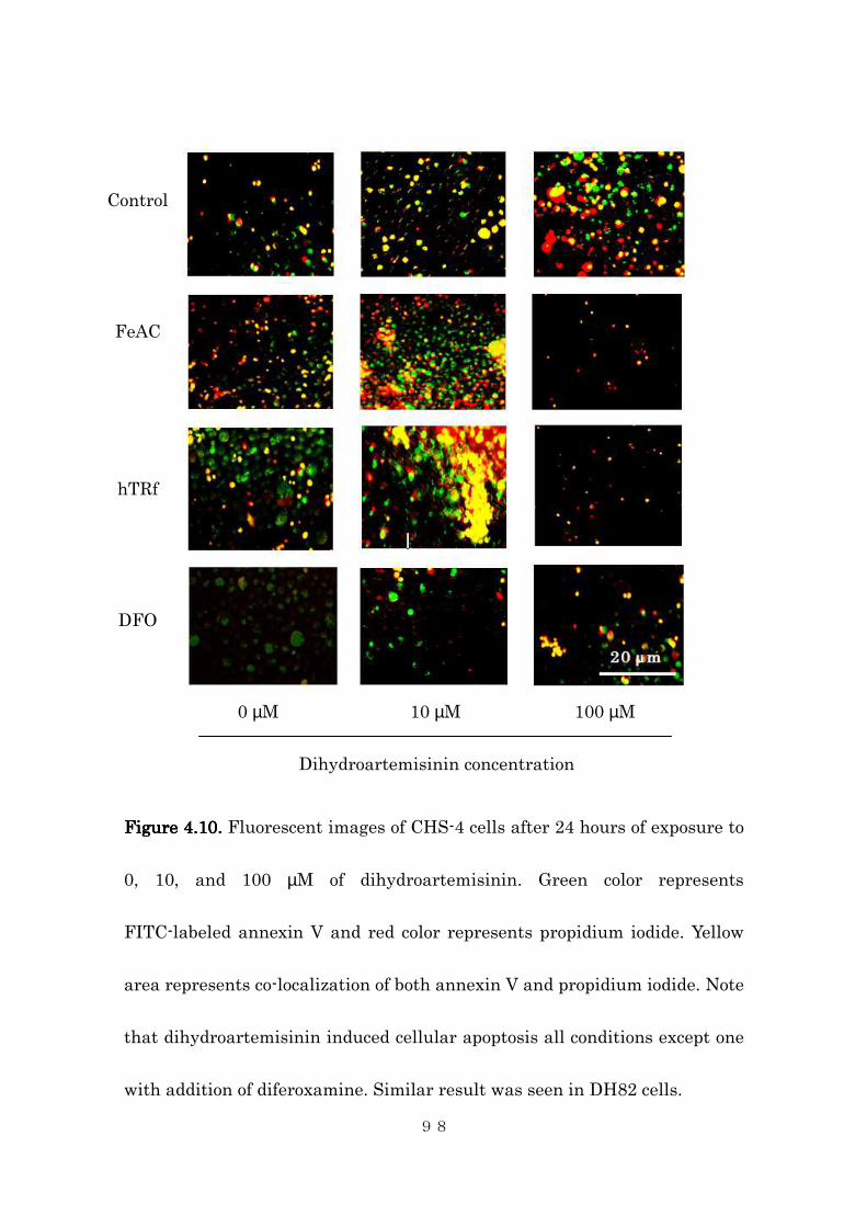
98
Figure 4.10.Figure 4.10.Figure 4.10.Figure 4.10. Fluorescent images of CHS-4 cells after 24 hours of exposure to
0, 10, and 100 µM of dihydroartemisinin. Green color represents
FITC-labeled annexin V and red color represents propidium iodide. Yellow
area represents co-localization of both annexin V and propidium iodide. Note
that dihydroartemisinin induced cellular apoptosis all conditions except one
with addition of diferoxamine. Similar result was seen in DH82 cells.
Control
FeAC
hTRf
DFO
0 µM 10 µM 100 µM
Dihydroartemisinin concentration
99
4.5. Discussion
Hyperferritinemia has long been recognized in several diseased conditions in
humans including tumors of histiocytic origin (Yoda, 1980; Valberg, 1980;
Esumi, 1989; Ya-You, 1998; Tang, 1987; Morita, 1981). Hyperferritinemia
has also been investigated as an early diagnostic marker for canine
histiocytic sarcoma (Friedrichs, 2010; Nielsen, 2011), and has been shown to
exhibit marked hyperferritinemia similar to its human counterpart.
However, whether this hyperferritinemia is caused by direct release from the
tumor cells or through indirect mechanism such as erythrodestruction is
unknown, and actual iron contents within the tumor tissue has not been
elucidated. The result of this study indicated that canine tissue contents of
iron in canine histiocytic sarcoma do not differ significantly from that of
other tumor tissues. This is not surprising given the fact that all malignant
tumor cells should contain increased intracellular iron contents through
overexpression of transferrin receptors because iron is an essential cofactor
for DNA synthesis. Furthermore, some of the evaluated tumor types are
thought to have high iron contents; lymphoma in dogs is known to cause
100
hyperferritinemia and thus could have high tissue iron content (Friedrichs,
2010). Hepatocellular carcinoma, one of the two samples with extremely high
iron content in this study, is a tumor of hepatocyte origin and thus expected
to produce ferritin and have iron stores intracellularly. The other tumor
sample with high iron contents was that of hemangiosarcoma. Unfortunately,
ICP-MS could only evaluate the amount of iron in the entire sample and does
not discriminate iron contents inside the tumor cells and hemoglobin-bound
iron contents within red blood cells. Hemangiosarcoma tissue is primarily
composed of abnormal neoplastic vascular space which is filled with high
number of red blood cells or a large blood clot. Therefore, the reason for the
high tissue iron concentration in our sample could either be due to high red
blood cell contamination or due to true high iron contents within the tumor
cells.
Histological evaluation of tissue sections with Berlin-blue staining
can distinguish intracellular iron contents and other sources; however, this
technique is qualitative rather than quantitative in nature, and the low
sensitivity of this technique precluded detailed assessment of cellular iron
contents. Many of the tumor samples, including all histiocytic sarcoma
101
samples evaluated, contained some Berlin-blue positive cells but majority of
the tumor cells within the same section stained negative for Berlin-blue,
indicating that not all tumor cells consistently have high intracellular iron
load. One possible explanation is that Berlin-blue positive cells represent the
tumor cells which have phagocytosed erythrocytes and those stained
negative represents the tumor cells which have not. Therefore, the tissue
iron concentration of histiocytic sarcoma not accompanied by clinical
findings of hemophagocytic syndrome such as anemia, erythrodestruction, or
thrombocytopenia, may have the similar level of tissue iron content as the
other malignant neoplasia tissue. Unfortunately, none of the histiocytic
sarcoma samples in this study were of phagocytic macrophage origin, and
these dogs did not show clinical changes suggestive of hemophagocytosis.
Tissue iron measurement and histological evaluation of intracellular iron
contents of phagocytic histiocytic sarcoma samples are warranted to further
clarify whether these tumors in fact have high intracellular iron contents
through erythrophagocytosis.
The in vitro assay on canine histiocytic sarcoma cell lines of both
dendritic cell origin and macrophage origin showed clear sensitivity of these
102
two cell lines to cytotoxic effect of dihydroartemisinin. The calculated IC50
values were in low micromolar range, which is relatively low compared to
other reported canine and human tumor cell lines (Efferth, 2002; Hosoya,
2008).
This cytotoxic effect of dihydroartemisinin on canine histiocytic
sarcoma cell lines were further enhanced by supplementation of ferrous iron
to the culture media in a form of ferric ammonium citrate, and abolished by
chelating ferrous iron within the media by pretreating the cells with
deferoxamine mesylate, thus providing a proof of concept that
dihydroartemisinin exhibits cytotoxicity though iron-mediated mechanism.
Experiments in the chapter 3 showed that dihydroartemisinin generates
free-radicals through a mechanism mediated by iron; together, these findings
indicate that dihydroartemisinin damage the cells through iron-mediated
generation of free radicals and amount of intracellular ferrous iron contents
is an important factor that determines the cellular sensitivity to cytoxocity of
dihydroartemisinin. Similar effect was seen when iron was supplemented by
a form of holotransferrin, a form more closely resembles in vivo cellular
environment. These effects were more pronounced in DH82 cells, a cell line
103
of phagocytic macrophage origin, and both treatment with ferric ammonium
citrate and holotransferrin resulted in significant decrease in cell viability at
the concentration as low as 1 µM of dihydroartemisinin. Targeting tumors
with naturally high iron content, artificially increasing intracellular iron
contents, or exploiting cellular mechanism to uptake ferrous iron to increase
the drug uptake, such as holotransferrin-conjugated dihydroartemisinin (Lai,
2005), all seem viable strategies to enhance the cytotoxicity of
dihydroartemisinin for clinical use.
These decreases in cell viability were, at least in part, due to
induction of apoptosis. In a normal culture condition, dihydroartemisinin
caused slight increase in a number of cells undergoing apoptosis at 10 µM
and majority of the cells were undergoing apoptosis at 100 µM. Increasing
cellular iron contents with either ferric ammonium citrate or holotransferrin
also enhanced this effect, resulting in marked induction of apoptosis at 10
µM in both conditions. Increasing the concentration of dihydroartemisinin to
100 µM resulted in near complete cell death and apoptosis could not be
observed microscopically. As predicted, pretreatment with deferoxamine
mesylate inhibited this induction of apoptosis, presumably via prevention of
104
iron-mediated free radical generation, again supporting our hypothesis.
These enhancement or inhibition of cytotoxicity of
dihydroartemisinin correlated well with actual measured intracellular iron
contents. In both cell lines, deferoxamine decreased and ferric ammonium
citrate and holotransferrin increased cellular iron content, although the
difference between the control did not reach statistical significance in
deferoxamine and holotransferrin, suspectedly due to small sample size
precluding detection of true difference (type II error). Despite the fact that
pretreatment with holotransferrin enhanced cytotoxicity to a similar degree
as treatment with ferric ammonium citrate, measured cellular iron content
of holotransferrin-treated cells were somewhat lower than that of ferric
ammonium citrate-treated cells in both cell lines. This is probably
attributable to shorter preincubation time with holotransferrin. Cells were
exposed only for 6 hours before intracellular iron measurement, whereas in
cell viability and apoptotic assays, cells were preincubated for 6 hours and
continued to be exposed to holotransferrin for additional 24 hours. To the
author’s knowledge, this study is the first to actually quantify the changes in
intracellular iron concentration with iron supplementation or chelating in
105
relationship with enhancement or inhibition of cytotoxicity of
dihydroartemisinin.
In conclusion, our study showed that tissues of many canine tumors
including histiocytic sarcoma contains high iron contents, and thus could be
candidates for dihydroartemisinin treatment, although the tissue iron
content of non-phagocytic histiocytic sarcoma did not differ significantly from
other tumor types. Furthermore, both phagocytic and non-phagocytic canine
histiocytic sarcoma cell lines were shown to be susceptible to
dihydroartemisinin with marked enhancement of cytotoxicity by increasing
intracellular iron content. These results suggest that dihydroartemisinin can
be a viable antineoplastic agent for treatment of canine histiocytic sarcoma.
Further investigation toward its clinical use is warranted.

106
5. General Conclusion
These studies demonstrated clinical safety and antitumor activity of
oral artemisinin in spontaneously occurring tumors in dogs. This is the first
study to document the antineoplastic activity in a large animal model.
However, the pharmacokinetic analysis indicated low bioavailability and
incomplete conversion of orally administered artemisinin to active
metabolite, dihydroartemisinin, suggesting that use of more potent
derivatives such as artesunate and dihydroartemisinin and parenteral route
of administration should be considered for future studies.
Antiproliferative and proapoptotic activities of dihydroartemisinin
were also confirmed in vitro using four canine osteosacoma cell lines and two
canine histiocytic sarcoma cell lines. Estimated IC50 values of these cell lines
suggest the use of oral artemisinin with the dose that currently being used or
higher is unlikely to achieve meaningful plasma concentration; however, by
using more potent artemisinin-derivatives such as dihydroartemisinin and
parenteral route of administration, plasma concentrations with antitumor
activity may be achievable especially in tumor types with higher sensitivity
such as canine histiocytic sarcoma.
107
An iron-mediated generation of free radicals, a proposed mechanism
for artemisinins to exhibit cytotoxic activity against cancer cells, was
successfully observed in a canine osteosarcoma cell line in a
concentration-dependent manner, and this generation of free radicals could
be completely prevented by chelating ferrous iron from the media, providing
a proof of concept for this mechanism. Increase and decrease of intracellular
ferrous iron content were associated with increased and decreased
sensitivity to cytotoxic effect of dihydroartemisinin in canine histiocytic
sarcoma cell lines, further confirming that cell damage by generation of
reactive oxygen species was the mechanism of cytotoxicity and this required
presence of ferrous iron within the cells.
Measurement of intratumoral iron contents showed that various
tumor tissues in dogs including histiocytic sarcomas and hepatocellular
carcinoma contains high amount of iron within the tissue, and can be
candidates for the treatment with dihydroartemisinin. These findings lay
basis for further research of artemisinin-derived trioxanes in treatment of
malignant tumors in dogs.
108
6. References
Affolter VK, Moore PF. Localized and disseminated histiocytic sarcoma
of dendritic cell origin in dogs. Vet Pathol. 2002;39:74-83.
Asawamahasakda W, Ittarat I Pu YM, Ziffer H, Meshnick SR. Reaction
of antimalarial endoperoxides with specific parasite proteins.
Antimicrob Agents Chemother. 1994;38:1854-1858.
Ashton M, Hai TN, Sy ND, et al. Artemisinin pharmacokinetics is
time-dependent during repeated oral administration in healthy male
adults. Drug Metabolism Dispostion 1998;26:25-27.
Aulbert E, Disselhoff W, Sorje H, et al. Lysosomal accumulation of
67kDa-transferrin in malignant tumors in relation to their growth
rate. Eur J Cancer 1980;16:1217-1232.
Anon. Chin Med J. 1979;92:811-816.
Azakami D, Bonkobara M, Wahizu T, Iida A, Kondo M, Kato R, Niikura Y,
Iwaki S, Tamahara S, Matsuki N, Ono K. Establishment and
biological characterization of canine histiocytic sarcoma cell lines. J
Vet Med Sci. 2006;68:1343-1346.
Bacci G, Avella M, Picci P. Metastatic patterns in osteosarcoma. Tumori.
1988;74:421-427.
Bailey D, Erb H, Williams L, et al. Carboplatin and doxorubicin
combination chemotherapy for the treatment of appendicular
osteosarcoma in the dog. J Cet Intern Med. 2003;17:199-205.
Beekman AC, Wierenga PK, Woerdenbag HJ, et al. Artemisinin-derived
sesquiterpene lactones as potential antitumour compounds: cytotoxic
action against bone marrow and tumour cells. Planta Med
1998;64:615-619.
109
Berg J, Weinstein MJ, Schelling SH, et al. Treatment of dogs with
osteosarcoma by administration of cisplatin after amputation or
limb-sparing surgery: 22 cases (1987-1990). J Am Vet Med Assoc.
1992;200:2005-2008.
Berg J, Weinstein MJ, Springfield DS, et al. Response of osteosarcoma in
the dog to surgery and chemotherapy with doxorubicin. J Am Vet Med
Assoc. 1995;206:1555-1560.
Berg J, Gebhard MC, Rand WM. Effect of timing on postoperative
chemotherapy on survival of dogs with osteosarcoma. Cancer
1997;79:1343-1350.
Berger TG, Dieckmann D, Efferth T, et al. Artesunate in the treatment of
metastatic uveal melanoma: first experiences. Oncol Rep.
2005;14:1599-1603.
Bergman PJ, MacEwen EG, Kurzman ID, et al. Amputation and
carboplatin for treatment of dogs with osteosarcoma: 48 cases
(1991-1993). J Vet Intern Med. 1996;10:76-81.
Berman PA, Adams PA. Artemisinin enhances hamo-catalyzed oxidation
of lipit membranes. Free Radic Biol Med. 1997;22:1283-1288.
Bhisutthibhan J and Meshnick SR. Immunoprecipitation of H-3
dihydroartemisinin translationally controlled tumor protein (Tctp)
adducts from Plasmodium falciparum-infected erythrocytes by using
anti-Tctp antibodies. Antimicrob Agents Chemother.
2001;45:2397-2399.
Brodey RS, McGrath JT, Reynolds H. A clinical and radiological study of
canine bone neoplasms: part I. J Am Vet Med Assoc. 1959;134:53-71.
Brodey RS, Sauer RM, Medway W. Canine bone neoplasms. J Am Vet
Med Assoc. 1963:143;471-495.
110
Brodey RS, Riser WH. Canine osteosarcoma: a clinicopathological study
of 194 cases. Clin Orthop. 1969;62:54-64.
Brossi A, Venugopalan B, Dominguez GL, Yeh HJC, Flippen-Anderson
JL, Buchs P, Luo XD, Milhous W, and Peters W. J Med Chem.
1988;31:645-650.
Butler D. Time to put malaria control on the global agenda. Nature
1997;386:535-536.
Castaneda VL, Parmley RT, Saldivar VA, et al. Childhood
undifferentiated leukemia with early erythroid markers and c-myb
duplication. Leukemia 1991;5:142-149.
China Cooperative Research Group on Qinghaosu and Its Derivatives as
Antimalarials. Studies on the toxicity of qinghaosu and its
derivatives. J Trad Chin Med. 1982;2:31-38.
Chun R, Kurzman ID, Couto G, et al. Cisplatin and doxorubicin
combination chemotherapy for the treatment of canine osteosarcoma:
preliminary findings. J Vet Intern Med. 2000;14:495-498.
Classen W, Altmann B, Gretener P, et al. Differential effects of orally
versus parenterally administered qunghaosu derivative artemether
in dogs. Exp Toxicol Phathol. 1999;51:517-516.
Das-Gupta A, Patil J, Shah VI. Transferrin receptor expression by blast
cells in acute lymphoblastic leukemia correlates with white cell count
and immunophenotype. Indian J Med Res. 1996;104:226-233.
Dernell WS, Ehrhart NP, Straw RC, Vail DM. Tumors of the skeletal
system. In: Small Animal Clinical Oncology 4th ed. pp540-582. 2007
Saunders St. Louis, Missouri.
Dhingra V, Rao KV, Narasu ML. Current status of artemisinin and its
111
derivatives as antimalarial drugs. Life Sci. 2000;66:279-300.
Disbrow GL, Baege AC, Kierpiec KA, et al. Dihydroartemisinin is
cytotoxic to papillomavirus-expressing epithelial cells in vitro and in
vivo. Cancer Res. 2005;65:10854-10861.
Eckstein-Ludwig U, Webb R, Van Goethem ID, et al. Artemisinins target
the SERCA of Plasmodium falciparum. Nature 2003;424:957-961.
Efferth T, Rucker G, Falkenberg M, et al. Detection of apoptosis in
KG-1a leukemic cells treated with investigational drugs.
Arzneimittelforschung 1996;46:196-200.
Efferth T, Dustan H, Sauerbrey A, et al. The antimalarial artesunate is
also active against cancer. Int J Oncol. 2001;18:767-773.
Efferth T, Olbrich A, Bauer R. mRNA expression profiles for the response
of human tumor cell lines to the antimalarial drugs artesunate,
arteether, and artemether. Biochem Pharmacol. 2002;64:617-623.
Efferth T, Sauerbrey A, Olbrich A, et al. Molecular modes of action of
artesunate in tumor cell lines. Mol Pharmacol. 2003;64:382-394.
Efferth T, Benakis A, Romero MR. Enhancement of cytotoxicity of
artemisinins toward cancer cells by ferrous iron. Free Radical Biol
Med. 2004;37:998-1009.
Efferth T. Mechanistic perspectives for 1,2,4-trioxanes in anti-cancer
therapy. Drug Resistance Updates 2005;8:85-97.
Elford BC, Roberts MF, Phillipson JD, and Wilson RJ. Potentiation of the
antimalarial activity of qinghaosu by methoxylated flavones. Trans R
Soc Trop Med Hyg. 1987;81:434-436.
Esumi N, Ikushima S, Todo S, Imashuku S. Hyperferritinemia in
malignant histiocytosis, virus-associated hemophagocytic syndrome
112
and familial erythrophagocytic lymphohistiocytosis. Acta Paediatr
Scand. 1989;78:268-270.
Fidel J, Schiller I, Hauser B, Jausi Y, Rohrer-Bley C, Roos M, Kaser-Hotz
B. Histiocytic sarcomas in flat-coated retrievers: a summary of 37
cases (November 1998-March 2005). Vet Comp Oncol. 2006;4:63-74.
Friedrichs KR, Thomas C, Plier M, Andrews GA, Chavey PS, Young KM.
Evaluation of serum ferritin as a tumor marker for canine histiocytic
sarcoma. J Vet Intern Med. 2010;24:904-911.
Guerin PJ, Olliaro P, Nosten F, Druilhe P, Laxminarayan R, Binka F,
Haynes RK and Vonwiller SC. From Qinghao, Marvelous herb of
antiquity, to the antimalarial trioxane qinghaosu. Some remarkable
new chemistry. Acc Chem Res. 1997;37:129-143.
Heyman SJ, Diefenderfer DL, Goldschmidt MH, et al. Canine axial
skeletal osteosarcoma: a retrospective study of 116 cases (1986-1989).
Vet Surg. 1992;21:304-310.
Hien TT. An overview of the clinical use of artemisinin and its
derivatives in the treatment of falciparum malaria in Viet Nam.
Trans R Soc Trop Med Hyg. 1994;88(Suppl.1):7-8.
Hosoya K, Murahari S, Laio A, et al. Biological activity of
dihydroarteminisin in canine osteosarcoma cell lines. Am J Vet Res.
2008;69:519-526.
Jiang JB, Li GQ, Guo XB, et al. Antimalarial activity of mefloquine and
qinghaosu. Lancet 1982;2:285-288.
Karin M and Mintz B. Receptor-mediated endocytosis of transferrin in
developmentally totipotent mouse teratocarcinoma stem cells. J Biol
Chem. 1981;256:3245-3252.
Kent MS, Strom A, London CA, et al. Alternating carboplatin and
113
doxorubicin as adjunctive chemotherapy for the treatment of
appendicular osteosarcoma in the dog. J Vet Intern Med.
2004;18:540-544.
Kilama WL, Ford N, and White NJ. Malaria: current status of control,
diagnosis, treatment, and a proposed agenda for research and
development. Lancet Infct Dis. 2002;2:564-573.
Kim SJ, Kim SM, Lee JW, et al. Dihydroartemisinin anhances
radiosensitivity of human glioma cells in vitro. J Cancer Res Clin
Oncol. 2006;132:129-135.
Kirpensteijn J, Teske E, Kik M, et al. Lobaplatin as an adjuvant
chemotherapy to surgery in canine appendicular osteosarcoma: a
phase II evaluation. Anticancer Res. 2002;22:2765-2770.
Kistler KR. Canine osteosarcoma: 1462 cases reviewed to uncover
patterns of height, weight, breed, sex, age and site involvement. Phi
Zeta Awards, University of Pennsylvania, School of Veterinary
Medicine, 1981.
Klayman DL. Qinghaosu (artemisinin): an antimalarial drug from China.
Science 1985;228:1049-1055.
Knecht CD, Priester WA. Musculoskeletal tumors in dogs. J Am Vet Med
Assoc. 1978;172:72-74.
Kraegel SA, Madewell BR, Simonson E. Osteogenic sarcoma and
cisplatin chemotherapy in dogs: 16 cases (1986-1989). J Am Vet Med
Assoc. 1991;199:1057-1059.
Krungkrai SR and Yuthavong Y. The antimalarial action on Plasmodium
falciparum of quinghaosu and artesunate in combination with agents
which modulate oxidant stress. Trans R Soc Trop Med Hyg.
1987;81:710-714.

114
Lai H, Singh NP. Selective cancer cell cytotoxicity from exposure to
dihydroartemisinin and holotransferrin. Cancer Lett. 1995;91:41-46.
Lai H, Sasaki T, Singh NP, Messay A. Effects of arteminisin-tagged
holotransferrin on cancer cells. Life Sci. 2005;76:1267-1279.
Lai H and Singh NP. Oral artemisinin prevents and delays the
development of 7,12-dimethylbenz[a]anthracene (DMBA)-induced
breast cancer in the rat. Cancer Lett. 2006;231:43-48.
Langroudi L, Hassan ZM, Ebtekar M, et al. A comparison of low-dose
cyclophosphamide treatment with artemisinin treatment in reducing
the number of regulatory T cells in murine breast cancer model. Int
Immunopharmacol. 2010;10:1055-1061.
LaRue SM, Withrow SJ, Powers BE, et al. Limb sparing treatment for
osteosarcoma in dogs. J Am Vet Med Assoc. 1989;195:1734-1744.
Li GQ, Guo XB, Fu LC, et al. Clinical trials of artemisinin and its
derivatives in the treatment of malaria in China. Trans R Soc Trop
Med Hyg. 1994;88(Suppl.1):5-6.
Li QG, Peggins JO, Lin AJ, et al. Pharmacology and toxicology of
artelinic acid: preclinical investigations on pharmacokinetics,
metabolism, protein and red blood cell binding, and acute and
anorectic toxicities. Trans R Soc Trop Med Hyg. 1998;92: 332-40.
Li Y and Wu YL. How Chinese scientists discovered qinghaosu
(artemisinin) and its derivatives? What are the future perspectives?
Med Trop (Marseille). 1998;58(Suppl.3):9-12.
Ling GV, Morgan JP, Pool RR. Primary bone tumors in the dog: a
combined clinical, radiological, and histologic approach to early
diagnosis. J Am Vet Med Assoc. 1974;165:55-67.
Lui SK, Dorfman HD, Hurvitz AI, et al. Primary and secondary bone
115
tumors in the dog. J Am Vet Med Assoc. 1977;18:313-326.
Mackenzie D. The comeback killer. New Sci. 1999;163:13-13.
Maeno Y, Toyoshima T, Fujioka H, Ito Y, Meshnick SR, Benakis A,
Milhous WK, and Aikawa M. Am J Trop Med Hyg. 1993;49:485-491.
Mauldin GN, Matus RE, Withrow SJ. Canine osteosarcoma treatment by
amputation versus amputation and adjuvant chemotherapy using
doxorubicin and cisplatin. J Vet Intern Med. 1988;2:177-180.
May WS, Cuatrecasas P. Transferrin receptor: its biological significance.
J Membr Biol. 1985;88:205-215.
May WS, Cuatrecasas P. Transferrin receptor: its biological significance.
J Membr Biol. 1985;88:205-215.
Meshnick SR, Tsang TW, Lin FB, Pan HZ, Chang CN, Kuypers F, Chiu D,
Lubin B. Activated oxygen mediates the antimalarial activity of
qinghaosu. Prog Clin Biol Res. 1989;313;95-104.
Meshnick SR, Thomas A, Ranz A, et al. Artemisinin (qinghaosu): the role
of intracellular hemin in its mechanism of antimalarial action. Mol
Biochem Parasitol. 1991;49:181-189.
Meshnick SR, Yang YZ, Lima V, et al. Iron-dependent free ra dical
generation from the antimalarial agent artemisinin (qinghaosu),
Antimicrob. Agent Chemother. 1993;37:1108-1114.
Meshnick SR. Free radicals and antioxidants. Lancet
1994;344;1441-1442.
Meshnick SR. Artemisinin antimalarials: mechanisms of action and
resistance. Med Trop. 1998;58 (3 Suppl.):13-17.
Meshnick SR. Artemisinin: mechanisms of action, resistance and toxicity.
116
Int J Parasitol. 2002;32:1655-1660.
Moore JC, Lai H, Li JR, et al. Oral administration of dihydroartemisinin
and ferrous sulfate retarded implanted fibrosarcoma growth in the
rat. Cancer Lett. 1995;27:83-87.
Morita R, Yoshii M, Nakajima K. Clinical evaluation of serum ferritin to
iron ratio in malignant diseases. Eur J Nucl Med. 1981;6:331-336.
Mukanganyama S, Widersten M, Naik YS, et al. Inhibition of
glutathione S-transferases by antimalarial drugs possible
implications for circumbenting anticancer drug resistance. Int J
Cancer 2002;97:700-705.
Naik H, Murry DJ, Kirsch LE, et al. Development and validation of a
high-performance liquid chromatography-mass spectroscopy assay
for determination of artesunate and dihydroartemisinin in human
plasma. J Chromatogr B Analyt Technol Biomed Life Sci. 2005;816:
233-42.
Nam W, Tak J, Ryu JK, et al. Effects of artemisinin and its derivatives
on growth inhibition and apoptosis of oral cancer cells. Head Neck
2007;29:335-340.
Navaratnam V, Mansor SM, Sit NW, et al. Pharmacokinetics of
artemisinin-type compounds. Clin Pharmacokinet. 2000;39:255-270.
Newton PN, van Vugt M, Teja-Isavadharm P, et al. Comparison of oral
artesunate and dihydroartemisinin antimalarial bioavailabilities in
acute falciparum malaria. Antimicrob Agents Chemother.
2002;46:1125-1127.
Nielsen LN, McEvoy F, Jessen LR, Kristensen AT. Investigation of a
screening program and the possible identification of biomarkers for
early disseminated histiocytic sarcoma in Bernese Mountain dogs.
Vet Comp Oncol. 2011;10:124-134.

117
Noori S, Taghikhani M, Hassan ZM, et al. Tehranolide could shift the
immune response towards Th1 and modulate the intra-tumor
infiltrated T regulatory cells. Iran J Immunol. 2009;6:216-224.
Nosten F, Price RN. New antimalarials: a risk-benefit analysis. Drug Saf.
1995;12:264-273.
O'Neill PM and Posner GH. A medicinal chemistry perspective on
artemisinin and related endoperoxides. J Med Chem.
2004;47:2945-2964.
Pik IH, Xie S, Shapiro TA, et al. Second generation, orally active,
antimalarial, artemisinin-derived trioxane dimmers with high
stability, efficacy, and anticancer activity. J Med Chem.
2006;49:2731-2734.
Posner GH, Ploypradith P, Parker MH, et al. Antimalarial,
antiproliferative, and antitumor activities of artemisinin-derived
chemically robust, trioxane dimmers. J Med Chem.
1999;42:4275-4280.
Posner GH, Cumming JN, Krasavin M. In: Carbon-centered radicals and
rational design of new antimalarial peroxide drugs. New York:
Biomedical Chemistry 2000;289-309.
Priester WA, ed: The occurrence of tumors in domestic animals. NCI
Monogr No. 54, November 1980.
Qinghaosu Antimalaria Coordinating Reserch Group. Antimalaria
studies on Qinghaosu. Chin Med J (Engl). 1979;92:811-816.
Raaf HN, Jacobsen DW, Savon S, et al. Serum transferrin receptor level
is not altered in invasive adenocarcinoma of the breast. Am J Clin
Pathol. 1993;99:232-237.
118
Rassnick KM, Moore AS, Russell DS, Northrup NC, Kristal O, Bailey DB,
Flory AB, Kiselow MA, Intile JL. Phase II, open-label trial of
single-agent CCNU in dogs with previously untreated histiocytic
sarcoma. J Vet Intern Med. 2010;24:1528-1531.
Reizenstein P. Iron, free radicals and cancer. Med Oncol tumor
Pharmacother. 1991;8:229-233.
Reungpatthanaphong P and Mankhetkorn S. Modulation of multidrug
resistance by artemisinin, artesunate and dihydroartemisinin in
K562/adr and GLC4/adr resistant cell lines. Biol Pharm Bull.
2002;25:1555-1561.
Ricci J, Park J, Chung WY, et al. Concise synthesis and antiangiogenic
activity of artemisinin-glycolipid hybrids on chorioallantoic
membranes. Bioorg Med Chem Lett. 2010;20:6858-6860.
Riedley RG. Medical need, scientific opportunity and drive for
antimalarial drugs. Nature 2002;415:686-693.
Rowley K. Miracles do happen. Artemisinin. cgmagazine. 2004
(Fall):19-23.
Rubinstein, LV, Shoemaker RH, Paull KD, Simon RM, Tosini S, Skehan P,
Scudiero DA, Monks A, Boyd MR. Comparison of in vitro
anticancer-drug-screening data generated with a tetrazolium assay
versus a protein assay against a diverse panel of human tumor cell
lines. J Natl Cancer Inst. 1990;82:1113-1118.
Shapiro W, Fossum TW, Kitchell BE. Use of cisplatin for the treatment of
appendicular osteosarcoma in dogs. J Am Vet Med Assoc.
1988;4:507-511.
Shterman N, Kupfer B, Moroz C. Comparison of transferrin receptors,
iron content and isoferritin profile in normal and malignant human
breast cell lines. Pathobiology 1991;59:19-25.

119
Singh NP and Lai H. Selective toxicity of dihydroartemisinin and
holotransferrin toward human breast cancer cells. Life Sci.
2001;70:49-56.
Singh NP, Cerma KB. Case report of a laryngeal squamous cell
carcinoma treated with artesunate. Archive Onclol. 2002;10:279-280.
Singh NP and Lai H. Artemisinin induces apoptosis in human cancer
cells. Anticancer Res. 2004;24:2277-2280.
Singh NP, Panwar VK. Case report of a pituitary macroadenoma treated
with artemether. Integr Cancer Ther. 2006;5:391-394.
Skorupski KA, Clifford CA, Paoloni MC, Lara-Garcia A, Barber L, Kent
MS, LeBlanc AK, Sabhlok A, Mauldin EA, Shofer FS, Couto CG,
Sorenmo KU. CCNU for the treatment of dogs with histiocytic
sarcoma. J Vet intern Med. 2007;21:121-126.
Skorupski KA, Rodriguez CO, Krick EL, Clifford CA, Ward R, Kent MS.
Long-term survival in dogs with localized histiocytic sarcoma treated
with CCNU as adjuvant to local therapy. Vet Comp Oncol.
2009;7:139-144.
Spodnick GJ, Berg RJ, Rand WM, et al. Prognosis for dogs with
appendicular osteosarcoma treated by amputation alone: 162 cases
(1978-1988). J Am Vet Med Assoc. 1992;200:995-999.
Straw RC, Withrow SJ, Richter SL, et al. Amputation and cisplatin for
treatment of canineosteosarcoma. J Vet Intern Med. 1991;5:205-210.
Straw RC, Withrow SJ. Limb-sparing surgery versus amputation for
dogs with bone tumors. Vet Clin North Am. 1996;26:135-143.
Tang D, Lang Y, Liang Y. Serum ferritin in normal subjects and in some
diseases. Chin Med J. 1987;100:631-635.

120
Thompson JP, Fugent MJ. Evaluation of survival times after limb
amputation, with and without subsequent administration of cisplatin,
for treatment of osteosarcoma in dogs: 30 cases (1979-1990). J Am Vet
Med Assoc. 1992;200:531-533.
Thrall DE, Withrow SJ, Powers BE, et al. Radiotherapy prior to cortical
allograft limb sparing in dogs with osteosarcoma: a dose response
assay. Int J Rad Onc Biol Phys. 1990;18:1354-1357.
Valberg LS. Plasma ferritin concentrations: Their clinical significance
and relevance to patient care. Can Med Assoc J. 1980;122:1240-1248.
Vasseur P. Limb preservation in dogs with primary bone tumors. Vet
Clin North Am. 1987;17:889-993.
Veterinary Cooperative Oncology Group. Veterinary Co-operative
Oncology Group-Common Terminology Criteria for Adverse Events
(VCOG-CTCAE) following chemotherapy or biological antineoplastic
therapy in dogs and cats v1.0. Vet Comp Oncol. 2004;2:195-213.
White NJ. Clinical pharmacokinetics and pharmacodynamics of
artemisinin and derivatives. Trans R Soc Trop Med Hyg.
1994;88(suppl 1):S41-43.
Withrow SJ, Powers BE, Straw RC, Wilkins RM. Comaparative aspects
of osteosarcoma; dog versus man. Clin Orthop Relat Res.
1991;270:159-167.
Withrow SJ, Powers BE, Straw RC, Wilkins RM. Comparative aspects of
osteosarcoma: dog versus man. Clin Orthop. 1991;270:159-68.
Withrow SJ, Thrall DE, Straw RC, et al. Intra-arterial cisplatin with or
without radiation in limbsparing for canine osteosarcoma. Cancer
1993;71:2484-2490.
121
Withrow SJ, Straw RC, Brekke JH, et al. Slow release adjuvant cisplatin
for the treatment of metastatic canine osteosarcoma. Eur J
Musculoskel Res. 1995;4:105-110.
Woerdenbag HJ, Moskal TA, Pras N, et al. Cytotoxicity of
artemisinin-related endoperoxides to Ehrlich ascites tumor cells. J
Nat Prod. 1993;56:849-856.
Woodrow CJ, Haynes RK, Krishna S. Artemisinins. Postgrad Med J.
2005;81:71-78.
Ya-You J, Yan-Fang L, Bo-Yun W, De-Yun Y. An immunocytochemical
study on the distribution of ferritin and other markers in 36 cases of
malignant histiocytosis. Cancer 1998;64:1281-1289.
Yoda Y, Abe T. Acute monocytic leukemia cell isoferritin. Cancer
1980;46:289-292.
Zhang F, Gosser Jr. DK, Meshnick SR. Hemin-catalyzed decomposition of
artemisinin (qinghaosu). Biochem Pharmacol. 1992;43:1805-1809.
Zheng GQ. Cytotoxic terpenoids and flavonoids from Artemisia annua.
Planta Med. 1994;60:54-57.
Zhou HJ, Wang WQ, Wu GD, et al. Artesunate inhibits angiogenesis and
downregulates vascular endothelial growth factor expression in
chronic myeloid leukemia K562 cells. Vascul Pharmacol.
2007;47:131-138.
Ziffer H, Highet RJ, and Klayman DL. Artemisinin: an endoperoxidic
antimalarial from Arteminia annuua L. Prog Chem Org Nat Prod.
1997;72:121-214.
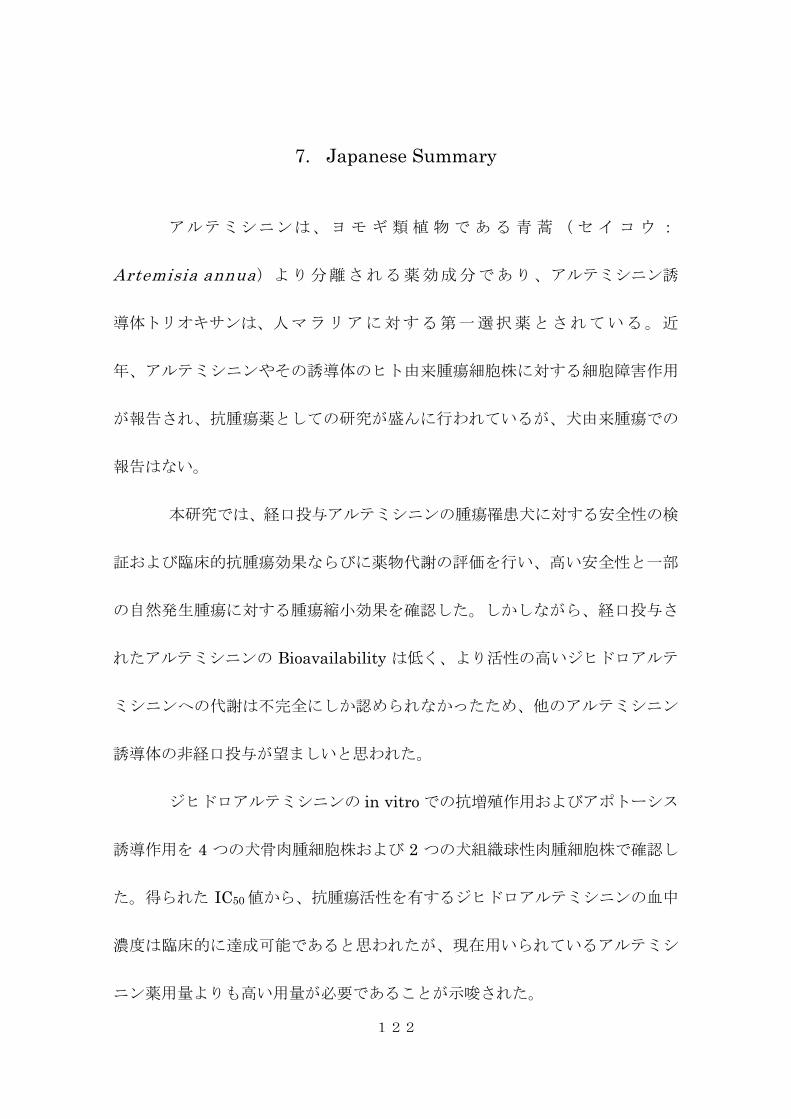
122
7. Japanese Summary
アルテミシニンは、ヨ モ ギ 類 植 物 で あ る 青 蒿 ( セ イ コ ウ :
Artemisia annua)より分離される薬効成分であり、アルテミシニン誘
導体トリオキサンは、人マラリアに対する第一選択薬とされている。近
年、アルテミシニンやその誘導体のヒト由来腫瘍細胞株に対する細胞障害作用
が報告され、抗腫瘍薬としての研究が盛んに行われているが、犬由来腫瘍での
報告はない。
本研究では、経口投与アルテミシニンの腫瘍罹患犬に対する安全性の検
証および臨床的抗腫瘍効果ならびに薬物代謝の評価を行い、高い安全性と一部
の自然発生腫瘍に対する腫瘍縮小効果を確認した。しかしながら、経口投与さ
れたアルテミシニンの Bioavailability は低く、より活性の高いジヒドロアルテ
ミシニンへの代謝は不完全にしか認められなかったため、他のアルテミシニン
誘導体の非経口投与が望ましいと思われた。
ジヒドロアルテミシニンの in vitro での抗増殖作用およびアポトーシス
誘導作用を 4 つの犬骨肉腫細胞株および 2 つの犬組織球性肉腫細胞株で確認し
た。得られた IC50 値から、抗腫瘍活性を有するジヒドロアルテミシニンの血中
濃度は臨床的に達成可能であると思われたが、現在用いられているアルテミシ
ニン薬用量よりも高い用量が必要であることが示唆された。
123
また、ジヒドロアルテミシニンの抗腫瘍効果発現機序と考えられている
活性酸素による細胞障害について、犬骨肉腫細胞株を用いて実際に細胞内活性
酸素量がジヒドロアルテミシニンの濃度依存性に上昇すること、さらに、この
反応が培地内の鉄分をキレートすることで抑制されることを確認した。また、
複数の犬組織球性肉腫細胞株を用いた実験にて、細胞内鉄量を増加または減少
させることにより、ジヒドロアルテミシニンの細胞障害性に対する感受性が細
胞内鉄量に比例して増加または減少することが確認され、上記実験結果と合わ
せて、ジヒドロアルテミシニンンによる細胞障害作用は、鉄を介したフリーラ
ジカルの産生およびそれによる細胞構造の損傷によると考えられた。
また、犬臨床例からの腫瘍サンプルを用いた組織内鉄含有量の測定では、
組織球性肉腫および肝細胞癌組織を含む、複数の腫瘍組織サンプルにて高い鉄
含有量が示された。本研究の結果は今後の犬腫瘍におけるアルテミシニン誘導
体の抗腫瘍活性および臨床応用に関する研究の基礎となるものと考えられる。

124
8. Acknowlegements
We are grateful to Drs. Masahiro Okumura, Satoshi Takagi, and Yuki
Hoshino for helpful discussions. We also thank Ms. Risa Nagata for assisting
a part of experiments. This study was partly supported by a grant from the
American Kennel Club ACORN foundation and Japan Health Science
Foundation and the Ministries of Education, Science, Sports and Culture,
Japan.


![Toxicity of Antineoplastic Druocr.yuhs.ac/UploadZone/Common/[최종본]항… · PPT file · Web view · 2014-05-18Neutropenic febrile event : dose reduction in subsequent cycle,](https://img.pdfslide.tips/doc/110x75/5b017ae17f8b9af1148e32b4/toxicity-of-antineoplastic-ppt-fileweb-view2014-05-18neutropenic.jpg)