Embed Size (px)
Citation preview
CharitéCentrum 12 für Innere Medizin und Dermatologie
Medizinische Klinik mit Schwerpunkt Infektiologie und Pneumologie
Direktor: Professor Dr. med. Norbert Suttorp
Habilitationsschrift
Artemisinin-basierte Therapie der Plasmodium falciparum-Malaria in Afrika und Europa
zur Erlangung der Venia legendi
für das Fach Innere Medizin
vorgelegt dem Fakultätsrat der Medizinischen Fakultät
Charité-Universitätsmedizin Berlin
von
Dr. med. Florian Michael Kurth MSc
aus München
Eingereicht: August 2017
Dekan: Prof. Dr. Axel Radlach Pries
1. Gutachter: Prof. Dr. E. C. Reisinger
2. Gutachter: Prof. Dr. T. Löscher
INHALTSVERZEICHNIS 1 Einleitung ......................................................................................................... 4
1.1 Plasmodien-Entwicklungszyklus ....................................................................... 4
1.2 Klinisches Bild der Malaria ............................................................................... 5
1.2.1 Inkubationszeit ............................................................................................. 6
1.2.2 Unkomplizierte Malaria ................................................................................ 6
1.2.3 Schwere Malaria ........................................................................................... 7
1.3 Epidemiologie ................................................................................................... 9
1.3.1 Aktuelle epidemiologische Entwicklung in Afrika ...................................... 11
1.3.2 Epidemiologie in Europa ............................................................................. 13
1.4 Therapie .......................................................................................................... 14
1.4.1 Medikamentöse Therapie der unkomplizierten Falciparum-Malaria ........ 14
1.4.2 Therapie der Non-falciparum-Malaria ....................................................... 16
1.4.3 Therapie der schweren Malaria.................................................................. 16
2 Eigene Arbeiten .............................................................................................. 19
2.1 Artemisinin-basierte Malariatherapie bei Kindern in Afrika .......................... 19
2.1.1 Pyronaridin-Artesunat Kombinationstherapie zur Behandlung von
Kindern mit unkomplizierter Malaria in Gabun ...................................................... 19
2.1.2 In vitro Aktivität von Pyronaridin gegen Plasmodium falciparum in
Gabun ..................................................................................................................... 29
2.1.3 Pädiatrische Darreichungsformen Artemisinin-basierter
Kombinationstherapien bei Kindern mit Malaria – systematischer Review
und Metaanalyse ..................................................................................................... 36
2.2 Artemisinin-basierte Malariatherapie bei Migranten und
Reiserückkehrern in Europa ........................................................................................ 46
2.2.1 Therapie der schweren Malaria in Europa ................................................. 46
2.2.1.1 Ergebnisse der multizentrischen Beobachtungsstudie des
Europäischen Netzwerkes für Tropen- und Reisemedizin (TropNet) zu
schwerer Malaria in Europa. ............................................................................... 46
2
2.2.1.2 Artesunat versus Chinin bei europäischen Patienten mit
schwerer Malaria ................................................................................................. 59
2.2.2 Hämolyse nach oraler Artemisinin-basierter Kombinationstherapie
bei unkomplizierter Malaria .................................................................................... 64
3 Diskussion....................................................................................................... 71
3.1 Artemisinin-basierte Malariatherapie in Afrika ............................................. 71
3.2 Artemisinin-basierte Malariatherapie in Europa ........................................... 77
4 Zusammenfassung und Ausblick ...................................................................... 82
5 Literatur ......................................................................................................... 84
6 Danksagung .................................................................................................... 93
7 Erklärung ........................................................................................................ 94
3
1 Einleitung
Etwa die Hälfte der Weltbevölkerung - mehr als 3 Milliarden Menschen - lebt in
Malaria-Risikogebieten. Trotz kontinuierlicher Fortschritte während der letzten Dekade
im Bestreben, die Malaria-bedingte Morbidität und Mortalität zu senken, kommt es
jährlich weltweit zu über 200 Millionen Fällen von Malaria (1). Die Zahl der Todesfälle
wird aktuell auf 400.000 bis 700.000 pro Jahr geschätzt (1, 2). Damit ist die Malaria die
bedeutendste parasitäre Erkrankung des Menschen und zählt zusammen mit
Pneumonie, HIV/AIDS, infektiösen Durchfallerkrankungen und Tuberkulose zu den fünf
wichtigsten Infektionskrankheiten (2).
Malaria wird verursacht durch eine Infektion mit Protozoen des Genus Plasmodium,
die durch Stiche der weiblichen Anopheles-Mücke übertragen werden. Plasmodium
falciparum ist für den Großteil der klinischen Fälle in Subsahara-Afrika und für 99% der
Malaria-Todesfälle weltweit verantwortlich. Auch der überwiegende Teil der
importierten Infektionen in Europa und USA wird durch P. falciparum hervorgerufen.
Infektionen mit P. vivax sind vor allem in Südostasien und Südamerika prävalent und
können ebenfalls schwere klinische Verläufe verursachen (3). Infektionen mit P. ovale,
bzw. den sympatrischen Spezies P. ovale curtisi und P. ovale wallikeri (4), und
P. malariae verlaufen hingegen in der Regel deutlich milder (5). In einigen Gegenden
Südostasiens spielt der Erreger der Makaken-Malaria, P. knowlesi, eine Rolle als
Krankheitserreger. Auch er kann beim Menschen schwere klinische Verläufe
verursachen (6, 7).
1.1 Plasmodien-Entwicklungszyklus
Während der Blutmahlzeit des Vektors, der weiblichen Anopheles, gelangen
Sporozoiten aus den Speicheldrüsen der Mücke in die menschliche Haut und von dort
über die Blutbahn in die Leber. Hier invadieren sie Hepatozyten und treten in die erste
asexuelle Vermehrungsphase ein. Während dieser sogenannten exo-erythrozytären
4
Schizogonie, die je nach Plasmodien Spezies zwischen 5,5 und 15 Tagen dauert,
entstehen aus den Sporozoiten Gewebeschizonten, die anschließend rupturieren und
jeweils bis zu 30.000 Merozoiten in die Blutbahn freisetzen (8, 9). Bei P. vivax und
P. ovale kommt es im Rahmen der exo-erythrozytären Schizogonie zur Bildung von
Hypnozoiten, die zunächst in eine Ruhephase eintreten und die Schizogonie erst nach
Wochen oder Monaten fortsetzen. Da die wissenschaftliche Evidenz hierfür gering ist,
wurde dieses Dogma – vor allem für P. ovale - in letzter Zeit mehrfach in Frage gestellt
(5, 10, 11).
Die freigesetzten Merozoiten dringen rasch in zirkulierende Erythrozyten ein, in denen
die zweite asexuelle Vermehrungsphase abläuft. Diese erythrozytäre Schizogonie
dauert pro Zyklus ca. 24 (P. knowlesi), 48 (P. falciparum, P. vivax, P. ovale) oder 72
Stunden (P. malariae), in denen die Erreger die Entwicklung von jungen Ringformen
über Trophozoiten zu Blutschizonten durchlaufen (12). Nach abgeschlossener Reifung
rupturieren die Blutschizonten und setzen je nach Plasmodien Spezies jeweils zwischen
6 und 30 neue Merozoiten in die Blutbahn frei, die anschließend rasch neue
Erythrozyten befallen und den asexuellen erythrozytären Zyklus fortsetzen. Ein kleiner
Teil der Merozoiten entwickelt sich zu sexuellen Formen, den weiblichen und
männlichen Gametozyten. Gelangen diese bei einer erneuten Blutmahlzeit der
weiblichen Anopheles in deren Mitteldarm, kommt es dort zur Befruchtung und
Bildung einer Zygote, die anschließend durch Reduktionsteilung als Ookinete und
Oozyste eine Vielzahl an Sporozoiten hervorbringt. Diese wandern in die
Speicheldrüsen der Mücke um von dort erneut auf den Menschen übertragen zu
werden, wodurch sich der Lebenszyklus schließt (8, 9).
1.2 Klinisches Bild der Malaria
Das klinische Bild der Malaria weist eine enorme Bandbreite auf und reicht von der
asymptomatischen Infektion bis zum hochakuten letalen Verlauf. Die Symptomatik ist
abhängig von Plasmodien-Spezies und -Stamm, Alter, Immunität, genetischer
5
Disposition, Gesundheits- und Ernährungszustand des Patienten sowie dem Einfluss
von Chemoprophylaxe und Therapie.
1.2.1 Inkubationszeit
Während der exo-erythrozytären Schizogonie ist die Infektion mit Plasmodien klinisch
inapparent. Erste Symptome treten in der Regel auf, wenn die Parasitendichte im Blut
50/µl übersteigt. Dies ist meist nach 3 bis 4 erythrozytären Vermehrungszyklen der
Fall. Für P. falciparum, P. vivax und P. ovale ergibt sich aus der Summation von 5,5-8
Tagen Gewebe-Schizogonie und 6-8 Tagen klinisch inapparenter erythrozytärer
Vermehrung eine Inkubationszeit von ca. 11-16 Tagen. Mit zunehmender klinischer
Semi-Immunität steigt die Parasitendichte, die ohne Symptome vom Infizierten
toleriert wird. Zudem ist bei semi-immunen Patienten die Vermehrung der Plasmodien
weniger effizient, sodass sich die Inkubationszeit zusätzlich verlängert. Die
Inkubationszeit bei Monoinfektion mit P. malariae ist durch die langsamere
Vermehrung dieser Spezies ebenfalls deutlich länger (13).
1.2.2 Unkomplizierte Malaria
Die klinische Symptomatik der Malaria ist wenig spezifisch. Fieber ist das häufigste
Symptom, kann jedoch vor allem in der frühen Krankheitsphase und bei Semi-
Immunen fehlen. Weitere häufige Beschwerden sind Abgeschlagenheit, Schwäche,
Kopfschmerzen, Myalgien, Übelkeit, Erbrechen und Diarrhoe. Die klassische klinische
Präsentation mit schubweisem Fieber jeden 2. bzw. 3. Tag kann auf eine Malaria
tertiana (P. ovale oder P. vivax) oder Malaria quartana (P. malariae) hindeuten. Die
Regelmäßigkeit des Fiebers nimmt dabei durch die Synchronisation der
Entwicklungszyklen im Verlauf der Infektion klassischerweise zu. Bei Infektionen mit
P. falciparum bleiben die Fieberschübe meist unregelmäßig. Fieberkrämpfe sind
besonders bei Kindern in Endemiegebieten häufig. In der klinischen Untersuchung sind
im Verlauf der Erkrankung häufig Splenomegalie, Ikterus und klinische Zeichen der
Anämie zu finden (8, 9).
6
1.2.3 Schwere Malaria
Patienten, die aufgrund einer Malaria keine oralen Medikamente zu sich nehmen
können, eine hohe Parasitenlast aufweisen oder Fehlfunktionen von lebenswichtigen
Organen entwickeln, haben ein erhöhtes Risiko an der Infektion zu versterben. Das
Risiko hängt dabei von Alter, Immunität, Anzahl der betroffenen Organe, Ausmaß der
Beeinträchtigung, Komorbiditäten und vom Zeitpunkt einer effektiven Therapie ab
(14). Während schwere bzw. lebensbedrohliche Verläufe bei Infektionen mit P. ovale,
P. malariae und P. vivax selten sind, kommen sie bei Infektionen mit P. falciparum -
abhängig von der Endemizität - regelhaft vor (6, 15). Pathophysiologische
Hauptursache hierfür ist die Eigenschaft von P. falciparum, durch die Zytoadherenz
infizierter Erythrozyten an Gefäßendothelzellen Störungen der Mikrozirkulation
hervorzurufen (16, 17). Für den Parasiten hat diese sogenannte Sequestration den
Vorteil, der für ihn ungünstigen Passage durch die Milz zu entgehen (6, 18).
Es existieren zahlreiche Definitionen und Kriterien zur Klassifikation einer P. falciparum
Infektion als komplizierte oder schwere Malaria. Nach einer in Subsahara-Afrika weit
verbreiteten Definition des Severe Malaria in African Children (SMAC) Netzwerks gilt
beispielsweise jede Malaria mit einer Parasitämie ≥ 5000 Parasiten/µl als schwer,
wenn die klinischen Symptome eine Hospitalisierung rechtfertigen (19). Auch
außerhalb der Endemiegebiete unterscheiden sich die Definitionen abhängig von
nationalen Empfehlungen zum Teil deutlich (20-22). Die Weltgesundheitsorganisation
(WHO) hat seit 1986 verschiedene Definitionen von schwerer Malaria für
epidemiologische und klinische Zwecke erarbeitet, die weit verbreitet und akzeptiert
sind (14, 23, 24). Vor allem bezüglich des Kriteriums der Hyperparasitämie bei
P. falciparum haben sich dabei zuletzt mehrfach relevante Änderungen ergeben. So
galt in den Empfehlungen von 2010 abhängig von der Transmissionsintensität eine
Parasitendichte von >2 % befallene Erythrozyten bzw. 100.000 Parasiten pro µl oder
>5 % befallene Erythrozyten bzw. 250.000 Parasiten pro µl als hyperparasitäm,
während aktuell unabhängig von der Endemizität >10 % befallene Erythrozyten als
Grenze angegeben werden. Die derzeit gültigen Charakteristika sind in der Tabelle
dargestellt (14, 23, 24).
7
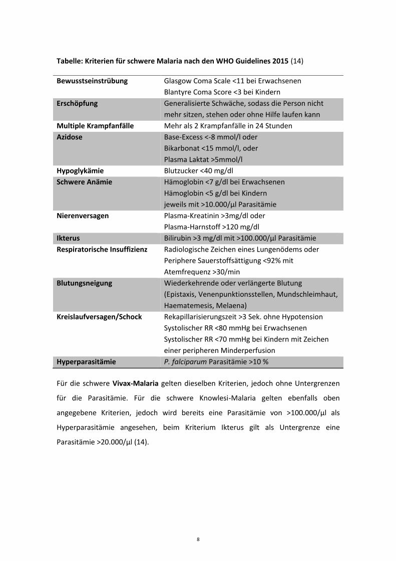
Tabelle: Kriterien für schwere Malaria nach den WHO Guidelines 2015 (14)
Bewusstseinstrübung Glasgow Coma Scale <11 bei Erwachsenen
Blantyre Coma Score <3 bei Kindern
Erschöpfung Generalisierte Schwäche, sodass die Person nicht
mehr sitzen, stehen oder ohne Hilfe laufen kann
Multiple Krampfanfälle Mehr als 2 Krampfanfälle in 24 Stunden
Azidose Base-Excess <-8 mmol/l oder
Bikarbonat <15 mmol/l, oder
Plasma Laktat >5mmol/l
Hypoglykämie Blutzucker <40 mg/dl
Schwere Anämie Hämoglobin <7 g/dl bei Erwachsenen
Hämoglobin <5 g/dl bei Kindern
jeweils mit >10.000/µl Parasitämie
Nierenversagen Plasma-Kreatinin >3mg/dl oder
Plasma-Harnstoff >120 mg/dl
Ikterus Bilirubin >3 mg/dl mit >100.000/µl Parasitämie
Respiratorische Insuffizienz Radiologische Zeichen eines Lungenödems oder
Periphere Sauerstoffsättigung <92% mit
Atemfrequenz >30/min
Blutungsneigung Wiederkehrende oder verlängerte Blutung
(Epistaxis, Venenpunktionsstellen, Mundschleimhaut,
Haematemesis, Melaena)
Kreislaufversagen/Schock Rekapillarisierungszeit >3 Sek. ohne Hypotension
Systolischer RR <80 mmHg bei Erwachsenen
Systolischer RR <70 mmHg bei Kindern mit Zeichen
einer peripheren Minderperfusion
Hyperparasitämie P. falciparum Parasitämie >10 %
Für die schwere Vivax-Malaria gelten dieselben Kriterien, jedoch ohne Untergrenzen
für die Parasitämie. Für die schwere Knowlesi-Malaria gelten ebenfalls oben
angegebene Kriterien, jedoch wird bereits eine Parasitämie von >100.000/µl als
Hyperparasitämie angesehen, beim Kriterium Ikterus gilt als Untergrenze eine
Parasitämie >20.000/µl (14).
8

1.3 Epidemiologie
Die Malaria ist weltweit in den Regionen der tropischen Klimazone verbreitet. Ihr
Vorkommen wird bestimmt von den Lebensbedingungen für Mensch, Parasit und
Vektor. Vor allem für Parasit und Vektor werden diese durch warme
Umgebungstemperaturen begünstigt. Historische Daten vom Beginn des
20. Jahrhunderts, vor der Zeit flächendeckender Malaria-Kontrollmaßnahmen, geben
Hinweise, dass die 60°F (bzw. 15,6°C) Isothermen der Sommer-Monate eine natürliche
Grenze für die Ausbreitung der Malaria darstellen. Die Verbreitung ist oft fokal mit
großen Unterschieden zwischen nahegelegenen Gebieten. P. vivax hat die weiteste
geographische Verbreitung und ist vor allem in Mittel- und Südamerika, Asien und
Ozeanien prävalent. P. falciparum ist die vorherrschende Spezies in den tropischen
Gebieten Afrikas. In Südamerika, der Karibik, Asien und Ozeanien entspricht seine
Prävalenz in etwa der von P. vivax. P. ovale und P. malariae kommen vor allem im
tropischen Afrika vor und sind insgesamt deutlich seltener als die anderen Spezies. Das
Vorkommen von P. knowlesi beschränkt sich auf die Verbreitungsgebiete der Ringel-
und Langschwanz-Makaken in Südostasien (1, 8, 9).
Zum Verständnis der Malaria-Transmission in Abhängigkeit von Mensch, Parasit und
Vektor wird oft die auf den Arbeiten von Ronald Ross und George Macdonald
basierende Formel
𝑅0 =𝑚𝑎2𝑝𝑛
−𝑟 𝑙𝑜𝑔𝑒𝑝
herangezogen, bei der R0 die Basis-Reproduktionsrate, m die Anzahl von Vektoren pro
Mensch, a die Frequenz mit der ein Vektor einen Menschen sticht, p die tägliche
Überlebenswahrscheinlichkeit eines Vektors, n die Dauer der parasitären Reifung im
Vektor (sog. Sporogonie) und r die Heilungsrate beim Menschen darstellt (25). R0 gibt
an, wie viele weitere Personen im Mittel durch eine infektiöse Indexperson angesteckt
werden.
Das Ross-Macdonald Model verdeutlicht den großen Einfluss der Lebensdauer des
Vektors auf die Transmission, da diese abhängig von der Umgebungstemperatur in
9
10.-12. Potenz in die Berechnung eingeht. Es erklärt zudem die hohe Wirksamkeit des
vor allem in Afrika prävalenten Vektors Anopheles gambiae, die auf dessen
Langlebigkeit und Anthropophilie sowie der hohen Vektordichte basiert (8, 9, 25).
Neben R0 wird die entomologische Inokulationsrate (EIR) als Maß für die
Malariatransmission verwendet, definiert als Anzahl infektiöser Moskitostiche pro
Person pro Jahr. Sie variiert zwischen weniger als 1 Stich pro Person und Jahr in den
meisten Gebieten mit instabiler Malariatransmission, beispielsweise in weiten Teilen
Asiens, Zentral- und Südamerikas, und über 1.000 Stichen pro Person und Jahr in
manchen Gebieten mit stabiler Transmission in Subsahara-Afrika (6, 26). Als Maß für
die Malaria-Endemizität wird zudem als Näherungsparameter der Anteil von Kindern
mit tastbarer Milz (sog. Milz-Rate) herangezogen, oder direkt die Infektionsprävalenz
(P. falciparum Parasiten Rate PfPR, Anteil der Kinder mit positiver Parasitämie) bei
Kindern im Alter zwischen 2 und 9 Jahren. Anhand dieser Maßzahlen werde Gebiete als
hypoendemisch (<10 % Milz-Rate bzw. PfPR), mesoendemisch (11-50 % Milz-Rate bzw.
PfPR), hyperendemisch (>50 % Milz-Rate bzw. PfPR) oder holoendemisch (>75 % Milz-
Rate, bzw. >75 % PfPR bei Kindern zwischen 0 und 11 Monaten) klassifiziert (8, 27).
In holoendemischen Gebieten mit stabiler Transmission ist die Malaria Morbidität und
Mortalität besonders während der Kindheit hoch. Kinder unter 5 Jahren tragen dort
die Hauptkrankheitslast und sind von über 80 % der Malaria-bedingten Todesfälle
betroffen. Infektionen in der Jugend und im Erwachsenenalter hingegen verlaufen
durch Zunahme der erworbenen Immunität oft nur noch mit niedriger Parasitämie und
oligo- bzw. asymptomatisch (1, 6, 28). Komplizierte bzw. tödliche Verläufe kommen bei
Erwachsenen in holoendemischen Gebieten in der Regel nicht vor. In Gebieten mit
instabiler Transmission und niedriger Endemizität ist die Ausbildung einer erworbenen
Semi-Immunität verzögert oder fehlt gänzlich, sodass symptomatische Infektionen alle
Altersgruppen betreffen. Veränderungen der Umgebungsbedingungen wie schwere
Regenfälle, Migration oder Flucht, sowie Veränderungen der Malaria-
Bekämpfungsmaßnahmen können in diesen Gebieten zum Auftreten epidemischer
Malaria führen (29). Inwiefern bzw. wie rasch die erworbene Semi-Immunität mit
abnehmender Re-Exposition zurückgeht, z.B. bei Verlassen der Endemiegebiete durch
10
Migration oder Abnahme der EIR durch verbesserte Kontrollmaßnahmen, ist nicht
abschließend geklärt und Gegenstand aktueller Untersuchungen (30).
1.3.1 Aktuelle epidemiologische Entwicklung in Afrika
Über 90% der Malaria-Erkrankungen und -Todesfälle weltweit betreffen die WHO
Region Afrika (1). In der Millenniumserklärung der Vereinten Nationen aus dem Jahr
2000 wurde das Ziel deklariert, bis 2015 die Ausbreitung von Malaria zum Stillstand zu
bringen und eine Trendwende bzgl. der Inzidenz zu bewirken (20). In dieser Zeit hat
sich die Bereitstellung von finanziellen Fördermitteln zur Malariabekämpfung weltweit
in etwa verzwanzigfacht (1, 27). Die drei wesentlichen Säulen der Malaria-
Kontrollprogramme sind dabei:
1. Insektizid-behandelte Moskitonetze (ITNs)
2. Innenraum-Besprühung mit Insektiziden (IRS)
3. Frühe Diagnostik und rasche medikamentöse Therapie.
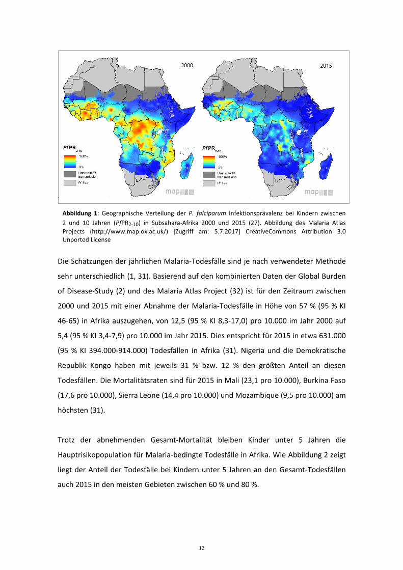
Schätzungen aus dem Malaria Atlas Project zufolge hat die P. falciparum
Infektionsinzidenz in den afrikanischen Endemiegebieten 2000-2015 um insgesamt
40% abgenommen, von 312 (95 % KI 253-427) auf 192 (95 % KI 135-265) pro 1.000
Personen pro Jahr (27). Dies entspricht für 2015 etwa 187 (95 % KI 132-259) Millionen
klinischen Malariafällen in Afrika. Die P. falciparum Infektionsprävalenz bei Kindern
zwischen 2 und 10 Jahren (PfPR2-10) als Maßzahl für die Endemizität hat sich seit der
Jahrtausendwende in etwa halbiert, von 33 % (95 % KI 31-35) im Jahr 2000 auf 16 %
(95 % KI 14-19) im Jahr 2015. Der Anteil der afrikanischen Gebiete, die als holo- oder
hyperendemisch klassifiziert werden, hat damit von 11,6 % bzw. 21,5 % im Jahr 2000
auf 1,4 % bzw. 7,9 % im Jahr 2015 abgenommen, wohingegen sich der Anteil der
hypoendemischen Gebiete im selben Zeitraum von 27,0 % auf 49,7 % fast verdoppelt
hat (27). Abbildung 1 zeigt eine graphische Darstellung der Verteilung der PfPR2-10 in
Afrika für die Jahre 2000 und 2015.
11
.
Abbildung 1: Geographische Verteilung der P. falciparum Infektionsprävalenz bei Kindern zwischen
2 und 10 Jahren (PfPR2-10) in Subsahara-Afrika 2000 und 2015 (27). Abbildung des Malaria Atlas
Projects (http://www.map.ox.ac.uk/) [Zugriff am: 5.7.2017] CreativeCommons Attribution 3.0 Unported License
Die Schätzungen der jährlichen Malaria-Todesfälle sind je nach verwendeter Methode
sehr unterschiedlich (1, 31). Basierend auf den kombinierten Daten der Global Burden
of Disease-Study (2) und des Malaria Atlas Project (32) ist für den Zeitraum zwischen
2000 und 2015 mit einer Abnahme der Malaria-Todesfälle in Höhe von 57 % (95 % KI
46-65) in Afrika auszugehen, von 12,5 (95 % KI 8,3-17,0) pro 10.000 im Jahr 2000 auf
5,4 (95 % KI 3,4-7,9) pro 10.000 im Jahr 2015. Dies entspricht für 2015 in etwa 631.000
(95 % KI 394.000-914.000) Todesfällen in Afrika (31). Nigeria und die Demokratische
Republik Kongo haben mit jeweils 31 % bzw. 12 % den größten Anteil an diesen
Todesfällen. Die Mortalitätsraten sind für 2015 in Mali (23,1 pro 10.000), Burkina Faso
(17,6 pro 10.000), Sierra Leone (14,4 pro 10.000) und Mozambique (9,5 pro 10.000) am
höchsten (31).
Trotz der abnehmenden Gesamt-Mortalität bleiben Kinder unter 5 Jahren die
Hauptrisikopopulation für Malaria-bedingte Todesfälle in Afrika. Wie Abbildung 2 zeigt
liegt der Anteil der Todesfälle bei Kindern unter 5 Jahren an den Gesamt-Todesfällen
auch 2015 in den meisten Gebieten zwischen 60 % und 80 %.
12
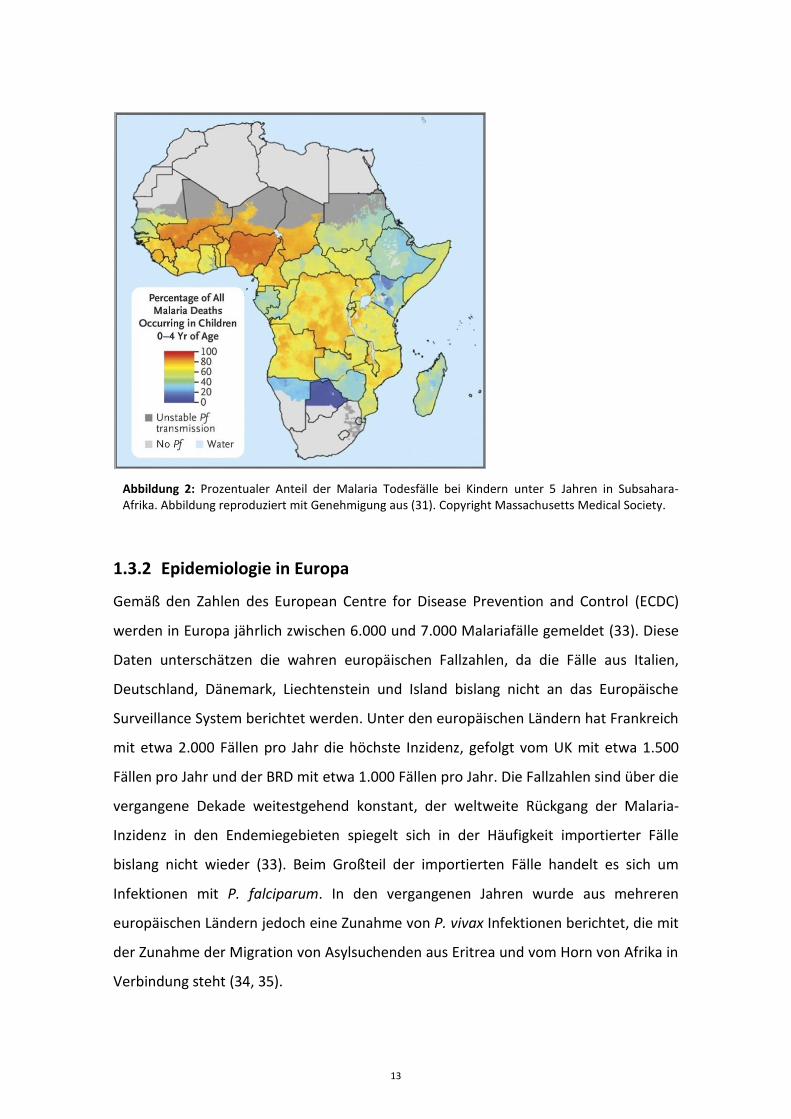
Abbildung 2: Prozentualer Anteil der Malaria Todesfälle bei Kindern unter 5 Jahren in Subsahara-Afrika. Abbildung reproduziert mit Genehmigung aus (31). Copyright Massachusetts Medical Society.
1.3.2 Epidemiologie in Europa
Gemäß den Zahlen des European Centre for Disease Prevention and Control (ECDC)
werden in Europa jährlich zwischen 6.000 und 7.000 Malariafälle gemeldet (33). Diese
Daten unterschätzen die wahren europäischen Fallzahlen, da die Fälle aus Italien,
Deutschland, Dänemark, Liechtenstein und Island bislang nicht an das Europäische
Surveillance System berichtet werden. Unter den europäischen Ländern hat Frankreich
mit etwa 2.000 Fällen pro Jahr die höchste Inzidenz, gefolgt vom UK mit etwa 1.500
Fällen pro Jahr und der BRD mit etwa 1.000 Fällen pro Jahr. Die Fallzahlen sind über die
vergangene Dekade weitestgehend konstant, der weltweite Rückgang der Malaria-
Inzidenz in den Endemiegebieten spiegelt sich in der Häufigkeit importierter Fälle
bislang nicht wieder (33). Beim Großteil der importierten Fälle handelt es sich um
Infektionen mit P. falciparum. In den vergangenen Jahren wurde aus mehreren
europäischen Ländern jedoch eine Zunahme von P. vivax Infektionen berichtet, die mit
der Zunahme der Migration von Asylsuchenden aus Eritrea und vom Horn von Afrika in
Verbindung steht (34, 35).
13
Mit Ausnahme weniger Berichte von autochthonen P. vivax Infektionen aus
Griechenland und anderen südeuropäischen Ländern werden die in Europa
auftretenden Fälle durch Reisen in Malaria-Endemiegebiete importiert (36). Betroffen
sind im Wesentlichen zwei unterschiedliche Patientengruppen:
1. Patienten, die außerhalb von Endemiegebieten geboren wurden und aus
touristischen oder beruflichen Gründen in Endemiegebiete reisen, und
2. Reisende mit Migrationshintergrund, die zum Besuch Ihrer
Familienangehörigen und Freunde in ihre ehemalige Heimat in den
Endemiegebieten zurückkehren (sog. „visiting friends and relatives“, VFR).
Während die Patienten in der ersten Gruppe in der Regel noch nie zuvor mit Malaria in
Kontakt gekommen sind, verbleibt bei der zweiten Gruppe durch die Malaria-
Exposition vor der Migration und durch regelmäßige Reisen in Endemiegebiete oft ein
gewisses Maß an Semi-Immunität (22).
1.4 Therapie
Die rasche Behandlung mit einem wirksamen Chemotherapeutikum ist der wichtigste
Bestandteil der Malaria-Therapie. Grundsätzlich kommt bei der unkomplizierten
Malaria in der Regel eine orale Behandlung zum Einsatz, während die Therapie der
schweren Malaria in der Regel parenteral erfolgt.
1.4.1 Medikamentöse Therapie der unkomplizierten Falciparum-Malaria
Die Artemisinin-basierte Kombinationstherapie (ACT) ist derzeit die empfohlene
Erstlinien-Behandlung der unkomplizierten Falciparum-Malaria in allen Malaria
Endemiegebieten (14). Das Grundprinzip der simultanen Anwendung von zwei
Wirkstoffen mit unterschiedlichen Wirkmechanismen basiert dabei - wie in der
Therapie von Tuberkulose, HIV und Tumorerkrankungen – auf drei Überlegungen:
Erstens wird durch den Angriff von zwei unterschiedlichen biologischen Zielstrukturen
die Wirksamkeit erhöht. Zweitens kann durch die Kombination zweier Wirkstoffe die
Therapiedauer gegenüber einer Monotherapie verkürzt werden. Drittens wird die
14
Entstehung von resistenten Plasmodien-Stämmen verlangsamt. Dies beruht auf der
Tatsache, dass die Wahrscheinlichkeit für das Auftreten eines Parasiten mit
gleichzeitiger Resistenz gegen zwei Wirkstoffe dem Produkt der Wahrscheinlichkeiten
des Auftretens der Einzelresistenzen entspricht, somit also um ein Vielfaches geringer
ist als bei Monotherapie. Die zwei Wirkstoffe schützen sich also gegenseitig gegen
Resistenzen (37, 38).
Fünf verschiedene ACTs werden derzeit von der WHO zur Therapie der
unkomplizierten Falciparum-Malaria empfohlen: Artemether + Lumefantrin (A+L),
Artesunat + Amodiaquin (A+AQ), Artesunat + Mefloquin (A+MQ), Artesunat +
Sulfadoxin-Pyrimethamin (A+SP) und Dihydroartemisinin + Piperaquin (DHA+P) (14). In
all diesen ACTs wird ein kurzwirksames Artemisinin-Derivat mit einem langwirksamen
Partner-Wirkstoff kombiniert und über 3 Tage oral verabreicht. ACTs sollten
vorzugsweise als sogenannte „fixed dose combinations“ verwendet werden, bei denen
beide Wirkstoffe in derselben Tablette kombiniert sind (14, 39). Fixed dose
Kombinationen sind mittlerweile für alle ACTs mit Ausnahme von A+SP verfügbar. ACTs
gelten im Allgemeinen als sicher und gut verträglich und haben in zahlreichen Studien
das von der WHO angestrebte Ziel einer PCR-korrigierten Heilungsrate >95 % an Tag 28
bewiesen (40).
Während sich die verschiedenen Artemisinin-Derivate der einzelnen ACTs nur
geringfügig (in ihrer Lipophilie bzw. Hydrophilie) unterscheiden, bestehen bei den
pharmakokinetischen und pharmakodynamischen Eigenschaften der Partner-
Medikamente beträchtliche Unterschiede. So liegen die terminalen Eleminations-
Halbwertszeiten von Lumefantrin und Sulfadoxin-Pyrimethamin jeweils im Bereich von
3-7 Tagen, während die von Mefloquin und Piperaquin im Bereich von etwa 3-4
Wochen liegen (41-44). Die Partnermedikamente Sulfadoxin-Pyrimethamin,
Amodiaquin, Mefloquin und Piperaquin wurden und werden zum Teil weiterhin als
eigenständige Monotherapien verwendet, mit der Gefahr einer rascheren
Resistenzentwicklung. Lumefantrin hingegen fand bislang nie als Monotherapeutikum
Verwendung (14).
15
1.4.2 Therapie der Non-falciparum-Malaria
Während Chloroquin in der Therapie der Falciparum-Malaria aufgrund zunehmender
Resistenzen weltweit seit Ende des 20. Jahrhunderts kontinuierlich an Bedeutung
verloren hat, galt es für die Non-falciparum-Malaria bis zuletzt weiterhin als
Medikament der Wahl. Chloroquin-Resistenzen für P. vivax wurden erstmals 1989 in
Papua Neuguinea beschrieben und kommen mittlerweile in nahezu allen P. vivax-
Endemiegebieten vor (45, 46).
Für P. ovale, P. malariae und P. knowlesi ist die Datenlage aufgrund der niedrigeren
Prävalenz dünn. Diese Plasmodien gelten generell als Chloroquin-sensibel, obgleich für
P. malariae aus Sumatra erste Chloroquin-resistente Stämme berichtet wurden (47,
48). ACTs scheinen in der Behandlung von Non-falciparum-Spezies generell gut
wirksam zu sein (5, 49, 50), sodass die WHO aktuell in Gebieten mit bekannter
Chloroquin-Resistenz von P. vivax generell den Einsatz von ACTs zur Behandlung der
Non-falciparum-Malaria empfiehlt (14). In Gebieten ohne bekannter Chloroquin-
Resistenz von P. vivax werden weiterhin ACTs oder Chloroquin gleichermaßen
empfohlen. Aufgrund der Häufigkeit von P. falciparum und P. vivax Koinfektionen bzw.
von falscher Spezies-Diagnose wurde diese Empfehlung zuletzt häufig kontrovers
diskutiert (6, 51). Zur Prävention von Rückfällen durch Hypnozoiten wird bei P. vivax
und P. ovale zusätzlich zur Akuttherapie eine 14-tägige Behandlung mit Primaquin
empfohlen (14).
1.4.3 Therapie der schweren Malaria
Die schwere Malaria ist ein medizinischer Notfall und erfordert eine rasche Therapie-
Einleitung sowie intensive Überwachung und Betreuung. Von besonderer Bedeutung
ist die schnellstmögliche Gabe eines wirksamen Malariamedikamentes. Wie mehrere
große randomisiert-kontrollierte Studien (RCTs) in afrikanischen und asiatischen
Endemiegebieten in den letzten Jahren gezeigt haben, ist die parenterale (intravenöse,
i.v. oder intramuskuläre, i.m.) Therapie mit Artesunat der seit vielen Jahrzehnten
etablierten Standardtherapie mit i.v. Chinin überlegen. So konnte bei erwachsenen
16
Patienten in Südostasien durch Artesunate eine Reduktion der Sterblichkeit um 35 %
gegenüber Chinin gezeigt werden, die Sterblichkeit bei Kindern in Afrika verringerte
sich um 22,5 % (9, 52, 53). Die parenterale Therapie mit Artesunat erfolgt am ersten
Tag in 12-stündigem Abstand und anschließend alle 24 Stunden, bis der Patient orale
Medikamente einnehmen kann. Die 24-stündige Gabe einer höheren Artesunat-Dosis
(4 mg/kg) von Beginn an war in einer Studie bei afrikanischen Kindern dem
konventionellen Schema mit Standarddosis (2,4 mg/kg) gleichwertig (19, 54). Für
Kinder mit einem Körpergewicht <20 kg wurde in der aktuellen WHO-Leitlinie die
empfohlene Dosis auf 3 mg/kg erhöht (55). Falls Artesunat nicht verfügbar ist, wird
eine intramuskuläre Therapie mit Artemether empfohlen, das jedoch weniger rasch
wirkt als Artesunat (56). Chinin sollte nach den aktuellen Empfehlungen der WHO bei
schwerer Malaria nur noch verwendet werden, wenn keine Artemisinin-Derivate
verfügbar sind (14). Für Patienten mit schwerer Malaria in ländlichen Gebieten, bei
denen der Beginn einer parenteralen Therapie z.B. durch Transport mehrere Stunden
verzögert wird und die keine oralen Medikamente einnehmen können, kann eine
einmalige rektale Artesunat-Gabe die Prognose verbessern (57). Im Anschluss an eine
parenterale Therapie sollte bei allen Patienten eine 3-tägige Standard-ACT zur
vollständigen Elimination verbleibender Parasiten erfolgen.
Supportive Therapie-Maßnahmen haben einen unterschiedlichen Stellenwert.
Während sich Glukokortikoide, Mannitol-Infusionen und eine prophylaktische
Antikonvulsiva-Gabe als wirkungslos bzw. nachteilig erwiesen haben, ist die
Empfehlung des frühzeitigen Einsatzes von Hämofiltration bzw. Hämodialyse bei
Nierenversagen und metabolischer Azidose unumstritten (9).
Die Transfusion von Erythrozytenkonzentraten wird in der Regel bei
Hämoglobinkonzentrationen unter 7 g/dl empfohlen, in afrikanischen Gebieten mit
hoher Prävalenz von chronischer Anämie abhängig von der Verfügbarkeit von
Blutkonserven bei Werten unter 5 g/dl. Bei nicht-semiimmunen erwachsenen
Patienten sollten vor allem kardiale und pulmonale Komorbiditäten bei der
Entscheidung für oder gegen eine Transfusion mit in Betracht gezogen werden.
Hypoglykämien stellen eine häufige Komplikation bei schwerer Malaria dar, vor allem
17
unter Chinin-Therapie. Die Blutzuckerwerte sollten daher regelmäßig überwacht und
ggf. durch Glukoseinfusionen stabilisiert werden.
Eine besondere Herausforderung stellt die Volumen-Supplementation dar. Eine
Standard-Bolusgabe von i.v. Flüssigkeit mit oder ohne Albumin erhöht bei febrilen
afrikanischen Kindern mit Hypotonie bei Malaria die Mortalität (58). Azidose und
Nierenfunktion können durch liberale i.v. Volumen-Supplementation oftmals nur
wenig beeinflusst werden, da sie meist eher durch Sequestration und daraus
resultierender Mikrozirkulationsstörung bedingt sind, als durch Hypovolämie.
Demgegenüber ist die Gefahr des Lungenödems bei liberaler Volumen-
Supplementation deutlich erhöht (59).
18

2 Eigene Arbeiten
2.1 Artemisinin-basierte Malariatherapie bei Kindern in Afrika
2.1.1 Pyronaridin-Artesunat Kombinationstherapie zur Behandlung von Kindern mit unkomplizierter Malaria in Gabun
Ramharter M, Kurth F, Schreier AC, Nemeth J, Glasenapp I, Bélard S, Schlie M, Kammer
J, Koumba PK, Cisse B, Mordmüller B, Lell B, Issifou S, Oeuvray C, Fleckenstein L,
Kremsner PG. Fixed-dose pyronaridine-artesunate combination for treatment of
uncomplicated falciparum malaria in pediatric patients in Gabon. Journal of Infectious
Diseases, 09/2008, 911-9, 198
Kleinkinder in Subsahara-Afrika sind bezüglich Morbidität und Mortalität weltweit am
schwersten von Malaria betroffen. Sie sind daher die Hauptzielgruppe bei der
Entwicklung neuer Malariamedikamente. In dieser Phase II Studie wurden 60
gabunische Kinder im Alter von 2-14 Jahren in 4 Behandlungsgruppen über drei Tage
mit unterschiedlichen Dosierungen (6:2, 9:3 und 12:4 mg/kg) einer Pyronaridin-
Artesunat Kombinationstherapie behandelt. Neben drei Tabletten-Koformulierungen
wurde eine spezielle pädiatrische Granulen-Darreichungsform in der Dosierung 9:3
mg/kg untersucht. Primäre Studienendpunkte waren Verträglichkeit, Sicherheit und
Pharmakokinetik. Wirksamkeit war als sekundärer Endpunkt definiert.
Verträglichkeit und Sicherheit wurden in allen Dosisgruppen als gut bewertet. In der
pharmakokinetischen Analyse zeigte sich eine lineare Beziehung zwischen Dosis und
Blut-/Plasma-Konzentrationen. Die Bioverfügbarkeit der pädiatrischen Granulen-
Darreichungsform war vergleichbar zur entsprechenden Tabletten-Koformulierung. Die
PCR-korrigierte Heilungsrate an Tag 28 in der Per-Protocol-Analyse war in allen 4
Behandlungsgruppen 100%.
Aufgrund dieser Phase II Ergebnisse wurde eine Weiterentwicklung der Pyronaridin-
Artesunat Kombinationstherapie sowohl für die Tabletten-Koformulierung als auch für
die pädiatrische Granulen-Koformulierung empfohlen.
19

https://doi.org/10.1086/591096
20
2.1.2 In vitro-Aktivität von Pyronaridin gegen Plasmodium falciparum in Gabun
Kurth F, Pongratz P, Bélard S, Mordmüller B, Kremsner PG, Ramharter M. In vitro
activity of pyronaridine against Plasmodium falciparum and comparative evaluation of
anti-malarial drug susceptibility assays. Malaria Journal, 2009, 79-85, 8
Ex vivo Testungen von Antimalaria-Wirkstoffen liefern wichtige Informationen über die
Sensitivität von Plasmodium-Isolaten gegenüber bekannten und neuen
Malariamedikamenten. In dieser Arbeit wurden die in vitro-Aktivitäten von
Pyronaridin, Artesunat, Chloroquin und Chinin an P. falciparum Isolaten von
Malariapatienten in Lambaréné, Gabun, getestet. Für Pyronaridin geschah dies in
diesem Gebiet erstmalig, parallel zur klinischen Entwicklung der Wirkstoffkombination
Pyronaridin-Artesunat, die sich damals in der Phase II befand. Für die anderen
Medikamente gab es bereits mehrere Voruntersuchungen aus Gabun. Methodisch
wurden parallel der seit langem von der WHO etablierte Mikroskopie-basierte
Schizonten-Maturations-Inhibition-Assay sowie der ELISA-basierte HRP-2 Assay
durchgeführt und miteinander verglichen.
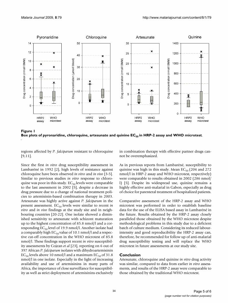
Pyronaridin zeigte eine hohe in vitro Aktivität gegen P. falciparum mit einer Cut-off-
Konzentration im geometrischen Mittel von 9,3 nmol/l (95 % KI 5,2-13,9) und
50 %-effektiven Konzentrationen von 1,9 nmol/l (95 % KI 1,4-2,5) im WHO Test und 2,0
nmol/l (95 % KI 1,6-2,6) im HRP-2 Assay. Bei einem Isolat bestand eine verminderte in
vitro Sensitivität gegenüber Artesunat. Die Resistenz-Niveaus von Chloroquin und
Chinin waren denen der Voruntersuchungen vergleichbar. Die Ergebnisse der
Testungen im WHO- und HRP-2 Assay zeigten eine gute Übereinstimmung.
29
BioMed Central
Page 1 of 6(page number not for citation purposes)
Malaria Journal
Open AccessResearchIn vitro activity of pyronaridine against Plasmodium falciparum and comparative evaluation of anti-malarial drug susceptibility assaysFlorian Kurth*1,2,3, Peter Pongratz1,2, Sabine Bélard1,2,4, Benjamin Mordmüller1,2, Peter G Kremsner1,2 and Michael Ramharter1,5
Address: 1Medical Research Unit, Albert Schweitzer Hospital, Lambaréné, Gabon, 2Institute for Tropical Medicine, Department of Parasitology, University of Tübingen, Tübingen, Germany, 3Department of Paediatrics, University Hospital Carl Gustav Carus Dresden, Dresden, Germany, 4Department of Pediatrics and Adolescent Medicine, University Freiburg, Freiburg, Germany and 5Department of Medicine I, Division of Infectious Diseases and Tropical Medicine, Medical University of Vienna, Vienna, Austria
Email: Florian Kurth* - [email protected]; Peter Pongratz - [email protected]; Sabine Bélard - [email protected]; Benjamin Mordmüller - [email protected]; Peter G Kremsner - [email protected]; Michael Ramharter - [email protected]
* Corresponding author
AbstractBackground: Pyronaridine, a Mannich base anti-malarial with high efficacy against drug resistantPlasmodium falciparum, is currently evaluated as a fixed dose combination with artesunate for thetreatment of uncomplicated malaria. In this study, the in vitro activity of pyronaridine against clinicalisolates of P. falciparum from Lambaréné, Gabon, was assessed in order to obtain baseline data onits activity prior to its future use in routine therapy. Moreover, follow-up assessment on the in vitroactivity of chloroquine, artesunate and quinine was performed.
Methods: In vitro response of field isolates of P. falciparum to pyronaridine, chloroquine, artesunateand quinine was assessed using the traditional WHO microtest. In addition, the histidine-richprotein 2 (HRP-2) assay was performed and evaluated for its future implementation for follow-upof drug susceptibility testing.
Results: Pyronaridine exhibited a high in vitro activity against P. falciparum, with a geometric meancut-off concentration of 9.3 nmol/l. Fifty percent effective concentrations were 1.9 nmol/l and 2.0nmol/l in the WHO microtest and HRP-2 assay, respectively. Results matched closely in vivo findingsfrom a recent clinical trial on pyronaridine-artesunate treatment. One isolate showed diminishedsensitivity to artesunate. For chloroquine and quinine resistance levels were comparable to priorstudies from Lambaréné. Results from the novel HRP-2 assay corresponded well to those obtainedby the WHO microtest.
Conclusion: Pyronaridine is highly active in chloroquine-resistant parasites and seems a promisingpartner drug for artemisinin-based combination therapy in Africa.
BackgroundMalaria continues to be a major cause of morbidity andmortality in sub-Saharan Africa, particularly in young
children. Early detection and effective chemotherapyremain the cornerstones in its control [1]. The rapid devel-opment and spread of anti-malarial drug resistance has
Published: 23 April 2009
Malaria Journal 2009, 8:79 doi:10.1186/1475-2875-8-79
Received: 18 November 2008Accepted: 23 April 2009
This article is available from: http://www.malariajournal.com/content/8/1/79
© 2009 Kurth et al; licensee BioMed Central Ltd. This is an Open Access article distributed under the terms of the Creative Commons Attribution License (http://creativecommons.org/licenses/by/2.0), which permits unrestricted use, distribution, and reproduction in any medium, provided the original work is properly cited.
30
Malaria Journal 2009, 8:79 http://www.malariajournal.com/content/8/1/79
Page 2 of 6(page number not for citation purposes)
made surveillance of drug sensitivity a high priority issue.In addition to assessing the activity of common anti-malarials against Plasmodium falciparum in routine sur-veys, the evaluation of new compounds against field iso-lates is of major importance for drug development.
In Lambaréné, Gabon, routine anti-malarial drug suscep-tibility monitoring has been performed since 1992 [2-5].So far the WHO microtest – one of the longest used andbest validated assays for the assessment of in vitro drugsensitivity under field conditions – has been used for thispurpose [6]. Meanwhile, novel methods in drug suscepti-bility testing have been developed, such as the histidine-rich protein II (HRP-2) assay [7]. This method, based onHRP-2 measurement in an enzyme-linked immunosorb-ent assay (ELISA), is equally simple to implement, butconsiderably less labour intensive compared with theWHO microtest. Due to these advantages, the WHOmicrotest will be replaced for standard drug susceptibilitymonitoring in Lambaréné in the future by the novel HRP-2 assay. Previous findings from laboratory adapted clonesand from different geographical regions suggest thatresults obtained by the HRP-2 assay be comparable withthose obtained by the WHO microtest [7].
The anti-malarial agent pyronaridine is a Mannich basederivative of mepacrine, one of the earliest synthetic anti-malarials [8]. It is currently evaluated as a fixed dose com-bination with artesunate for the treatment of uncompli-cated falciparum and vivax malaria in adult and paediatricpatients [9]. Its anti-plasmodial activity involves interfer-ence with the glutathione-dependent detoxification ofhaem and targeting of β-haematin formation [10].Reports from paediatric patients in Africa showed thatpyronaridine is effective against chloroquine resistantstrains of P. falciparum in vivo [9,11], yet data from South-East Asia indicate the potential for rapid development ofresistance against pyronaridine, when used as mono-therapy [12].
The present study aimed to assess the susceptibility ofclinical P. falciparum field isolates from Lambaréné to pyr-onaridine in order to obtain baseline data on the activityof this for Africa yet novel anti-malarial drug prior to itswidespread use in routine therapy. In addition the studywas designed to assess the potential for the novel HRP-2assay to replace the standard WHO microtest.
MethodsStudy area and patientsThe study was carried out at the Medical Research Unit ofthe Albert Schweitzer Hospital in Lambaréné, Gabon, in aregion of stable, hyperendemic P. falciparum malariatransmission [13,14]. Patients attending the outpatientclinic between March and October 2006 were asked to
participate in the study if they met the following inclusioncriteria: P. falciparum monoinfection with 1,000–100,000asexual parasites per μl blood, no schizontaemia, no signsor symptoms of severe malaria, and no history of intake ofanti-malarial drugs in the preceding month. Informationabout age, sex, and duration of fever was collected on aquestionnaire. Informed consent was obtained from par-ticipants or their legal representatives. The study wasapproved by the Ethics Committee of the InternationalFoundation for the Albert Schweitzer Hospital in Lam-baréné.
In vitro drug sensitivity assaysTwo different methods of drug sensitivity testing weredeployed in order to assess the susceptibility of fresh fieldisolates of P. falciparum to pyronaridine, artesunate, chlo-roquine and quinine. First, the standard World HealthOrganization in vitro microtest was used similar to previ-ous reports, measuring drug-dependent inhibition of sch-izont maturation (SMI) within 24 hours by microscopicassessment [2-5]. Briefly, two millilitres of venous bloodwere mixed with complete parasite culture medium(RPMI 1640, 200 μM hypoxanthine, 25 mM Hepes, 0.5%albumax, 2 mM l-glutamine) to a final concentration of5% blood medium mixture (BMM). Ninety-six-well testplates were pre-dosed in ascending quantities of drugs,dosing each plate with all respective drugs for one isolate.Final drug concentrations were 0.5–365.9 nmol/l BMMfor pyronaridine (Mr: 910.04), 0.1–85.8 nmol/l BMM forartesunate (Mr: 384.425), 0.8–51.2 μmol/l blood forchloroquine (Mr: 515.867) and 55–3567 nmol/l BMMfor quinine (Mr: 785.06). Artesunate, chloroquine andquinine were dissolved in 70% ethanol, pyronaridine wasresuspended in distilled water. In accordance to the proto-cols distributed by the World Health Organization drugconcentrations for chloroquine are expressed as related toblood due to their considerable accumulation in erythro-cytes [6].
Fifty μl BMM were transferred into scheduled wells andincubated at 37.5°C in candle jars. After 24 hours para-sites were harvested and Giemsa-stained thick blood filmswere prepared. The number of mature schizonts wasmicroscopically counted against 200 asexual parasites ineach well. Tests were considered successful if at least 10%schizont maturation was observed in the drug-free con-trol-well.
In addition, the HRP-2 assay was performed according tothe published standard operational procedures [7]. Twoml of venous blood were mixed with parasite medium toa concentration of 3% BMM. Test plates were pre-dosed tothe same final concentrations as in the WHO test andincubated for 72 hours at 37°C in candle jars. To test forsuccessful in vitro parasite-growth, a thick blood smear of
31
Malaria Journal 2009, 8:79 http://www.malariajournal.com/content/8/1/79
Page 3 of 6(page number not for citation purposes)
one control-well was performed after 26 h. One non-treated 26 h sample was frozen to calculate backgroundHRP-2 production. Parasite culture was judged successfulif at least 10% parasites matured to schizonts at the 26-hour time point. After 72 h plates were freeze-thawedtwice. Parasite growth, calculated from HRP-2 levels, wasmeasured with an enzyme linked immunosorbent assay atan absorbent maximum of 450 nm.
Statistical analysisNon-linear regression analysis with 4-parameter fits oflog-concentration/response curves was used to determineindividual inhibitory concentrations of the respective iso-lates. All regressions were checked manually. Cut off con-centrations were calculated as geometric mean of thelowest individual concentrations with no mature schizontamong 200 parasites in the WHO microtest. Nonparamet-ric analysis was used for concentration data that was notnormally distributed. A two-tailed Mann-Whitney-U-Testwas performed in order to test for difference between thetwo drug sensitivity assays. All tests were performed at atwo sided significance level of α = 0.05.
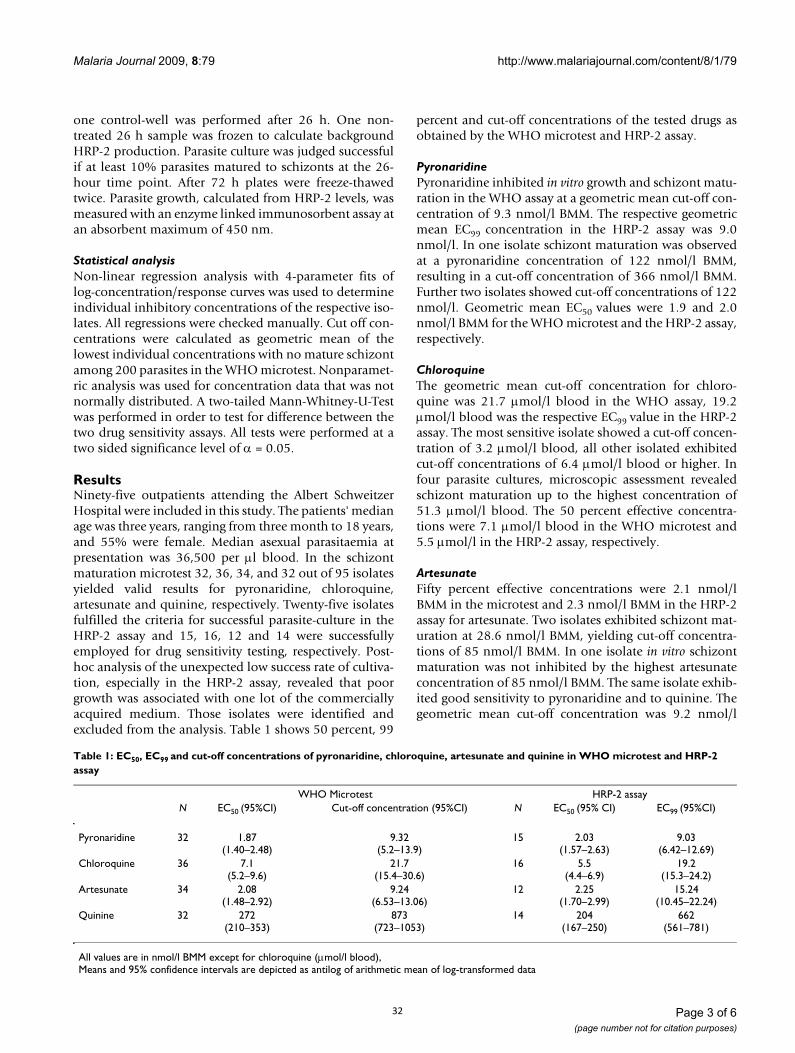
ResultsNinety-five outpatients attending the Albert SchweitzerHospital were included in this study. The patients' medianage was three years, ranging from three month to 18 years,and 55% were female. Median asexual parasitaemia atpresentation was 36,500 per μl blood. In the schizontmaturation microtest 32, 36, 34, and 32 out of 95 isolatesyielded valid results for pyronaridine, chloroquine,artesunate and quinine, respectively. Twenty-five isolatesfulfilled the criteria for successful parasite-culture in theHRP-2 assay and 15, 16, 12 and 14 were successfullyemployed for drug sensitivity testing, respectively. Post-hoc analysis of the unexpected low success rate of cultiva-tion, especially in the HRP-2 assay, revealed that poorgrowth was associated with one lot of the commerciallyacquired medium. Those isolates were identified andexcluded from the analysis. Table 1 shows 50 percent, 99
percent and cut-off concentrations of the tested drugs asobtained by the WHO microtest and HRP-2 assay.
PyronaridinePyronaridine inhibited in vitro growth and schizont matu-ration in the WHO assay at a geometric mean cut-off con-centration of 9.3 nmol/l BMM. The respective geometricmean EC99 concentration in the HRP-2 assay was 9.0nmol/l. In one isolate schizont maturation was observedat a pyronaridine concentration of 122 nmol/l BMM,resulting in a cut-off concentration of 366 nmol/l BMM.Further two isolates showed cut-off concentrations of 122nmol/l. Geometric mean EC50 values were 1.9 and 2.0nmol/l BMM for the WHO microtest and the HRP-2 assay,respectively.
ChloroquineThe geometric mean cut-off concentration for chloro-quine was 21.7 μmol/l blood in the WHO assay, 19.2μmol/l blood was the respective EC99 value in the HRP-2assay. The most sensitive isolate showed a cut-off concen-tration of 3.2 μmol/l blood, all other isolated exhibitedcut-off concentrations of 6.4 μmol/l blood or higher. Infour parasite cultures, microscopic assessment revealedschizont maturation up to the highest concentration of51.3 μmol/l blood. The 50 percent effective concentra-tions were 7.1 μmol/l blood in the WHO microtest and5.5 μmol/l in the HRP-2 assay, respectively.
ArtesunateFifty percent effective concentrations were 2.1 nmol/lBMM in the microtest and 2.3 nmol/l BMM in the HRP-2assay for artesunate. Two isolates exhibited schizont mat-uration at 28.6 nmol/l BMM, yielding cut-off concentra-tions of 85 nmol/l BMM. In one isolate in vitro schizontmaturation was not inhibited by the highest artesunateconcentration of 85 nmol/l BMM. The same isolate exhib-ited good sensitivity to pyronaridine and to quinine. Thegeometric mean cut-off concentration was 9.2 nmol/l
Table 1: EC50, EC99 and cut-off concentrations of pyronaridine, chloroquine, artesunate and quinine in WHO microtest and HRP-2 assay
WHO Microtest HRP-2 assayN EC50 (95%CI) Cut-off concentration (95%CI) N EC50 (95% CI) EC99 (95%CI)
Pyronaridine 32 1.87(1.40–2.48)
9.32(5.2–13.9)
15 2.03(1.57–2.63)
9.03(6.42–12.69)
Chloroquine 36 7.1(5.2–9.6)
21.7(15.4–30.6)
16 5.5(4.4–6.9)
19.2(15.3–24.2)
Artesunate 34 2.08(1.48–2.92)
9.24(6.53–13.06)
12 2.25(1.70–2.99)
15.24(10.45–22.24)
Quinine 32 272(210–353)
873(723–1053)
14 204(167–250)
662(561–781)
All values are in nmol/l BMM except for chloroquine (μmol/l blood),Means and 95% confidence intervals are depicted as antilog of arithmetic mean of log-transformed data
32
Malaria Journal 2009, 8:79 http://www.malariajournal.com/content/8/1/79
Page 4 of 6(page number not for citation purposes)
BMM. The HRP-2 assay showed a mean EC99 level of 15.2nmol/l BMM.
QuinineAll isolates were susceptible to quinine. Highest cut-offconcentrations were at 1783 nmol/l BMM, thus wellbelow the threshold of resistance (5120 nmol/l BMM).The geometric mean cut-off concentration was 873 nmol/l, the EC99 in the HRP-2 assay was 662 nmol/l BMM. TheEC50 values in the WHO microtest and in the HRP-2 assaywere 272 and 204 nmol/l BMM, respectively.
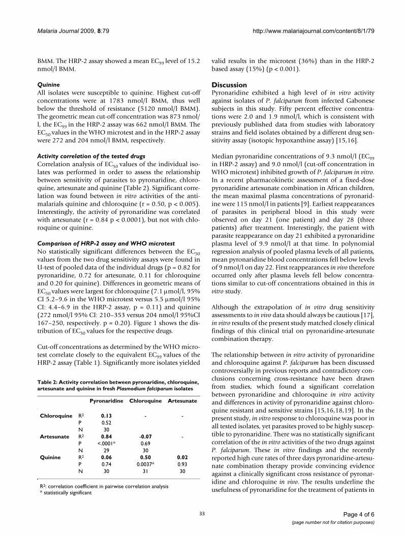
Activity correlation of the tested drugsCorrelation analysis of EC50 values of the individual iso-lates was performed in order to assess the relationshipbetween sensitivity of parasites to pyronaridine, chloro-quine, artesunate and quinine (Table 2). Significant corre-lation was found between in vitro activities of the anti-malarials quinine and chloroquine (r = 0.50, p < 0.005).Interestingly, the activity of pyronaridine was correlatedwith artesunate (r = 0.84 p < 0.0001), but not with chlo-roquine or quinine.
Comparison of HRP-2 assay and WHO microtestNo statistically significant differences between the EC50values from the two drug sensitivity assays were found inU-test of pooled data of the individual drugs (p = 0.82 forpyronaridine, 0.72 for artesunate, 0.11 for chloroquineand 0.20 for quinine). Differences in geometric means ofEC50 values were largest for chloroquine (7.1 μmol/l, 95%CI 5.2–9.6 in the WHO microtest versus 5.5 μmol/l 95%CI: 4.4–6.9 in the HRP-2 assay, p = 0.11) and quinine(272 nmol/l 95% CI: 210–353 versus 204 nmol/l 95%CI167–250, respectively. p = 0.20). Figure 1 shows the dis-tribution of EC50 values for the respective drugs.
Cut-off concentrations as determined by the WHO micro-test correlate closely to the equivalent EC99 values of theHRP-2 assay (Table 1). Significantly more isolates yielded
valid results in the microtest (36%) than in the HRP-2based assay (15%) (p < 0.001).
DiscussionPyronaridine exhibited a high level of in vitro activityagainst isolates of P. falciparum from infected Gabonesesubjects in this study. Fifty percent effective concentra-tions were 2.0 and 1.9 nmol/l, which is consistent withpreviously published data from studies with laboratorystrains and field isolates obtained by a different drug sen-sitivity assay (isotopic hypoxanthine assay) [15,16].
Median pyronaridine concentrations of 9.3 nmol/l (EC99in HRP-2 assay) and 9.0 nmol/l (cut-off concentration inWHO microtest) inhibited growth of P. falciparum in vitro.In a recent pharmacokinetic assessment of a fixed-dosepyronaridine artesunate combination in African children,the mean maximal plasma concentrations of pyronarid-ine were 115 nmol/l in patients [9]. Earliest reappearancesof parasites in peripheral blood in this study wereobserved on day 21 (one patient) and day 28 (threepatients) after treatment. Interestingly, the patient withparasite reappearance on day 21 exhibited a pyronaridineplasma level of 9.9 nmol/l at that time. In polynomialregression analysis of pooled plasma levels of all patients,mean pyronaridine blood concentrations fell below levelsof 9 nmol/l on day 22. First reappearances in vivo thereforeoccurred only after plasma levels fell below concentra-tions similar to cut-off concentrations obtained in this invitro study.
Although the extrapolation of in vitro drug sensitivityassessments to in vivo data should always be cautious [17],in vitro results of the present study matched closely clinicalfindings of this clinical trial on pyronaridine-artesunatecombination therapy.
The relationship between in vitro activity of pyronaridineand chloroquine against P. falciparum has been discussedcontroversially in previous reports and contradictory con-clusions concerning cross-resistance have been drawnfrom studies, which found a significant correlationbetween pyronaridine and chloroquine in vitro activityand differences in activity of pyronaridine against chloro-quine resistant and sensitive strains [15,16,18,19]. In thepresent study, in vitro response to chloroquine was poor inall tested isolates, yet parasites proved to be highly suscep-tible to pyronaridine. There was no statistically significantcorrelation of the in vitro activities of the two drugs againstP. falciparum. These in vitro findings and the recentlyreported high cure rates of three days pyronaridine-artesu-nate combination therapy provide convincing evidenceagainst a clinically significant cross resistance of pyronar-idine and chloroquine in vivo. The results underline theusefulness of pyronaridine for the treatment of patients in
Table 2: Activity correlation between pyronaridine, chloroquine, artesunate and quinine in fresh Plasmodium falciparum isolates
Pyronaridine Chloroquine Artesunate
Chloroquine R2 0.13 - -P 0.52N 30
Artesunate R2 0.84 -0.07 -P <.0001* 0.69N 29 30
Quinine R2 0.06 0.50 0.02P 0.74 0.0037* 0.93N 30 31 30
R2: correlation coefficient in pairwise correlation analysis* statistically significant
33
Malaria Journal 2009, 8:79 http://www.malariajournal.com/content/8/1/79
Page 5 of 6(page number not for citation purposes)
regions affected by P. falciparum resistant to chloroquine[9,11].
Since the first in vitro drug susceptibility assessment inLambaréné in 1992 [2], high levels of resistance againstchloroquine have been observed in vitro and in vivo [3-5].Similar to previous studies in vitro response to chloro-quine was poor in this study. EC50 levels were comparableto the last assessment in 2002 [5], despite a decrease indrug pressure due to a change of national treatment poli-cies to artemisinin-based combination therapy in 2003.Artesunate was highly active against P. falciparum in thepresent assessment. EC50 levels were similar to recent invitro and in vivo findings at the study site and in neigh-bouring countries [20-22]. One isolate showed a dimin-ished sensitivity to artesunate with schizont maturationup to the highest concentration of 85.8 nmol/l and a cor-responding EC50 level of 19.9 nmol/l. Another isolate hada comparably high EC50 value of 10.1 nmol/l and a respec-tive cut-off concentration in the WHO microtest of 85.8nmol/l. These findings support recent in vitro susceptibil-ity assessments by Cojean et al [23], reporting on 6 out of397 African P. falciparum isolates with dihydroartemisininEC50 levels above 10 nmol/l and a maximum EC50 of 31.8nmol/l in one isolate. Especially in the light of increasingavailability and use of artemisinins in many parts ofAfrica, the importance of close surveillance for susceptibil-ity as well as strict deployment of artemisinins exclusively
in combination therapy with effective partner drugs can-not be overemphasized.
As in previous reports from Lambaréné, susceptibility toquinine was high in this study. Mean EC50 (204 and 272nmol/l in HRP-2 assay and WHO microtest, respectively)were comparable to results obtained in 2002 (286 nmol/l) [5]. Despite its widespread use, quinine remains ahighly effective anti-malarial in Gabon, especially as drugof choice for parenteral treatment of hospitalized patients.
Comparative assessment of the HRP-2 assay and WHOmicrotest was performed in order to establish baselinedata for the use of the ELISA based assay in Lambaréné inthe future. Results obtained by the HRP-2 assay closelyparalleled those obtained by the WHO microtest despitemethodological problems in this study due to a deficientbatch of culture medium. Considering its reduced labour-intensity and good reproducibility the HRP-2 assay can,therefore, be recommended for follow up of anti-malarialdrug susceptibility testing and will replace the WHOmicrotest in future assessments at our study site.
ConclusionArtesunate, chloroquine and quinine in vitro drug activitywas similar, compared to data from earlier in vitro assess-ments, and results of the HRP-2 assay were comparable tothose obtained by the traditional WHO microtest.
Box plots of pyronaridine, chloroquine, artesunate and quinine EC50 in HRP-2 assay and WHO microtestFigure 1Box plots of pyronaridine, chloroquine, artesunate and quinine EC50 in HRP-2 assay and WHO microtest.
34
Publish with BioMed Central and every scientist can read your work free of charge
"BioMed Central will be the most significant development for disseminating the results of biomedical research in our lifetime."
Sir Paul Nurse, Cancer Research UK
Your research papers will be:
available free of charge to the entire biomedical community
peer reviewed and published immediately upon acceptance
cited in PubMed and archived on PubMed Central
yours — you keep the copyright
Submit your manuscript here:http://www.biomedcentral.com/info/publishing_adv.asp
BioMedcentral
Malaria Journal 2009, 8:79 http://www.malariajournal.com/content/8/1/79
Page 6 of 6(page number not for citation purposes)
The study demonstrated high anti-malarial activity of pyr-onaridine against fresh field isolates of P. falciparum andcorresponded well to recent findings of pyronaridine anti-malarial activity in vivo. Pyronaridine is recommended forfurther clinical development in combination therapy andcontinued in vitro drug activity monitoring.
Competing interestsThe authors declare that they have no competing interests.
Authors' contributionsFK contributed to the conception and design of the study,performed parasite cultivation and microscopic assess-ment, analysed the data and wrote the manuscript. PP per-formed parasite cultivation and HRP-2 immunoassay. SBgathered field isolates from patients and performed para-site cultivation. BM contributed to performance of immu-noassay and analysis of data, PGK revised the manuscriptand supervised the research group, MR conceived anddesigned the study, contributed to analysis of data, per-formed second reading for microscopic assessments inWHO microtest and drafted and revised the manuscript.All authors read and approved the final version of themanuscript.
AcknowledgementsWe thankfully acknowledge the participations of our patients in the Albert Schweitzer Hospital in Lambaréné. This study was supported by a grant of the independent Karl Landsteiner Gesellschaft, Austria.
References1. Kremsner PG, Krishna S: Antimalarial combinations. Lancet
2004, 364:285-294.2. Winkler S, Brandts C, Wernsdorfer WH, Graninger W, Bienzle U,
Kremsner PG: Drug sensitivity of Plasmodium falciparum inGabon. Activity correlations between various antimalarials.Trop Med Parasitol 1994, 45:214-218.
3. Philipps J, Radloff PD, Wernsdorfer W, Kremsner PG: Follow-up ofthe susceptibility of Plasmodium falciparum to antimalarialsin Gabon. Am J Trop Med Hyg 1998, 58:612-618.
4. Borrmann S, Binder RK, Adegnika AA, Missinou MA, Issifou S, Ram-harter M, Wernsdorfer WH, Kremsner PG: Reassessment of theresistance of Plasmodium falciparum to chloroquine inGabon: implications for the validity of tests in vitro vs. invivo. Trans R Soc Trop Med Hyg 2002, 96:660-663.
5. Ramharter M, Wernsdorfer WH, Kremsner PG: In vitro activity ofquinolines against Plasmodium falciparum in Gabon. Acta Trop2004, 90:55-60.
6. In vitro test (mark II) for the assessment of the response ofPlasmodium falciparum to chloroquine, mefloquine, qui-nine, sulfadoxine/pyrimethamine and amodiaquine. WHOdocument CTD/MAL/97.20 Rev 2 2001 [http://www.who.int/drugresistance/malaria/en/markiii.pdf]
7. Noedl H, Wernsdorfer WH, Miller RS, Wongsrichanalai C: Histi-dine-rich protein II: a novel approach to malaria drug sensi-tivity testing. Antimicrob Agents Chemother 2002, 46:1658-1664.
8. Chang C, Lin-Hua T, Jantanavivat C: Studies on a new antimalar-ial compound: pyronaridine. Trans R Soc Trop Med Hyg 1992,86:7-10.
9. Ramharter M, Kurth F, Schreier AC, Nemeth J, Glasenapp IV, BelardS, Schlie M, Kammer J, Koumba PK, Cisse B, Mordmuller B, Lell B, Issi-fou S, Oeuvray C, Fleckenstein L, Kremsner PG: Fixed-dose pyro-naridine-artesunate combination for treatment ofuncomplicated falciparum malaria in pediatric patients inGabon. J Infect Dis 2008, 198:911-919.
10. Auparakkitanon S, Chapoomram S, Kuaha K, Chirachariyavej T,Wilairat P: Targeting of hematin by the antimalarial pyronar-idine. Antimicrob Agents Chemother 2006, 50:2197-2200.
11. Ringwald P, Bickii J, Basco LK: Efficacy of oral pyronaridine forthe treatment of acute uncomplicated falciparum malaria inAfrican children. Clin Infect Dis 1998, 26:946-953.
12. Yang HL, Liu DQ, Yang YM, Huang KG, Dong Y, Yang PF, Liao MZ,Zhang CY: In vitro sensitivity of Plasmodium falciparum toeight antimalarials in China-Myanmar and China-Lao PDRborder areas. Southeast Asian J Trop Med Public Health 1997,28:460-464.
13. Ramharter M, Adegnika AA, Agnandji ST, Matsiegui PB, Grobusch MP,Winkler S, Graninger W, Krishna S, Yazdanbakhsh M, Mordmüller B,Lell B, Missinou MA, Mavoungou E, Issifou S, Kremsner PG: Historyand perspectives of medical research at the Albert Sch-weitzer Hospital in Lambaréné, Gabon. Wien Klin Wochenschr2007, 119:8-12.
14. Wildling E, Winkler S, Kremsner PG, Brandts C, Jenne L, Wernsdor-fer WH: Malaria epidemiology in the province of MoyenOgoov, Gabon. Trop Med Parasitol 1995, 46:77-82.
15. Pradines B, Mabika Mamfoumbi M, Parzy D, Owono Medang M, Leb-eau C, Mourou Mbina JR, Doury JC, Kombila M: In vitro suscepti-bility of African isolates of Plasmodium falciparum fromGabon to pyronaridine. Am J Trop Med Hyg 1999, 60:105-108.
16. Vivas L, Rattray L, Stewart L, Bongard E, Robinson BL, Peters W,Croft SL: Anti-malarial efficacy of pyronaridine and artesu-nate in combination in vitro and in vivo. Acta Trop 2008,105:222-228.
17. Ekland EH, Fidock DA: In vitro evaluations of antimalarial drugsand their relevance to clinical outcomes. Int J Parasitol 2008,38:743-747.
18. Pradines B, Tall A, Parzy D, Spiegel A, Fusai T, Hienne R, Trape JF,Doury JC: In-vitro activity of pyronaridine and amodiaquineagainst African isolates (Senegal) of Plasmodium falciparum incomparison with standard antimalarial agents. J AntimicrobChemother 1998, 42:333-339.
19. Elueze EI, Croft SL, Warhurst DC: Activity of pyronaridine andmepacrine against twelve strains of Plasmodium falciparum invitro. J Antimicrob Chemother 1996, 37:511-518.
20. Pradines B, Hovette P, Fusai T, Atanda HL, Baret E, Cheval P, MosnierJ, Callec A, Cren J, Amalvict R, Gardair JP, Rogier C: Prevalence ofin vitro resistance to eleven standard or new antimalarialdrugs among Plasmodium falciparum isolates from Pointe-Noire, Republic of the Congo. J Clin Microbiol 2006,44:2404-2408.
21. Ramharter M, Kurth FM, Belard S, Bouyou-Akotet MK, MamfoumbiMM, Agnandji ST, Missinou MA, Adegnika AA, Issifou S, Cambon N,Heidecker JL, Kombila M, Kremsner PG: Pharmacokinetics of twopaediatric artesunate mefloquine drug formulations in thetreatment of uncomplicated falciparum malaria in Gabon. JAntimicrob Chemother 2007, 60:1091-1096.
22. Ramharter M, Noedl H, Thimasarn K, Wiedermann G, WernsdorferG, Wernsdorfer WH: In vitro activity of tafenoquine alone andin combination with artemisinin against Plasmodium falci-parum. Am J Trop Med Hyg 2002, 67:39-43.
23. Cojean S, Hubert V, Le Bras J, Durand R: Resistance to dihydroar-temisinin. Emerg Infect Dis 2006, 12:1798-1799.
35
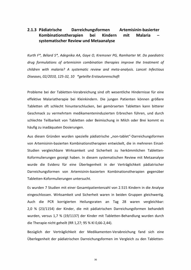
2.1.3 Pädiatrische Darreichungsformen Artemisinin-basierter Kombinationstherapien bei Kindern mit Malaria – systematischer Review und Metaanalyse
Kurth F*, Bélard S*, Adegnika AA, Gaye O, Kremsner PG, Ramharter M. Do paediatric
drug formulations of artemisinin combination therapies improve the treatment of
children with malaria? A systematic review and meta-analysis. Lancet Infectious
Diseases, 02/2010, 125-32, 10 *geteilte Erstautorenschaft
Probleme bei der Tabletten-Verabreichung sind oft wesentliche Hindernisse für eine
effektive Malariatherapie bei Kleinkindern. Die jungen Patienten können größere
Tabletten oft schlecht hinunterschlucken, bei gemörserten Tabletten kann bitterer
Geschmack zu vermehrtem medikamenteninduzierten Erbrechen führen, und durch
schlechte Teilbarkeit von Tabletten oder Beimischung in Milch oder Brei kommt es
häufig zu inadäquaten Dosierungen.
Aus diesen Gründen wurden spezielle pädiatrische „non-tablet“-Darreichungsformen
von Artemisinin-basierten Kombinationstherapien entwickelt, die in mehreren Einzel-
Studien vergleichbare Wirksamkeit und Sicherheit zu herkömmlichen Tabletten-
Koformulierungen gezeigt haben. In diesem systematischen Review mit Metaanalyse
wurde die Evidenz für eine Überlegenheit in der Verträglichkeit pädiatrischer
Darreichungsformen von Artemisinin-basierten Kombinationstherapien gegenüber
Tabletten-Koformulierungen untersucht.
Es wurden 7 Studien mit einer Gesamtpatientenzahl von 2.515 Kindern in die Analyse
eingeschlossen. Wirksamkeit und Sicherheit waren in beiden Gruppen gleichwertig.
Auch die PCR korrigierten Heilungsraten an Tag 28 waren vergleichbar:
2,0 % (23/1154) der Kinder, die mit pädiatrischen Darreichungsformen behandelt
wurden, versus 1,7 % (19/1137) der Kinder mit Tabletten-Behandlung wurden durch
die Therapie nicht geheilt (RR 1,27; 95 % KI 0,66-2,44).
Bezüglich der Verträglichkeit der Medikamenten-Verabreichung fand sich eine
Überlegenheit der pädiatrischen Darreichungsformen im Vergleich zu den Tabletten-
36
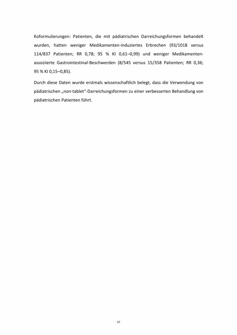
Koformulierungen: Patienten, die mit pädiatrischen Darreichungsformen behandelt
wurden, hatten weniger Medikamenten-induziertes Erbrechen (93/1018 versus
114/837 Patienten; RR 0,78; 95 % KI 0,61–0,99) und weniger Medikamenten-
assoziierte Gastrointestinal-Beschwerden (8/545 versus 15/358 Patienten; RR 0,36;
95 % KI 0,15–0,85).
Durch diese Daten wurde erstmals wissenschaftlich belegt, dass die Verwendung von
pädiatrischen „non-tablet“-Darreichungsformen zu einer verbesserten Behandlung von
pädiatrischen Patienten führt.
37
https://doi.org/10.1016/S1473-3099(09)70327-5
38
2.2 Artemisinin-basierte Malariatherapie bei Migranten und Reiserückkehrern in Europa
2.2.1 Therapie der schweren Malaria in Europa
2.2.1.1 Ergebnisse der multizentrischen Beobachtungsstudie des Europäischen Netzwerkes für Tropen- und Reisemedizin (TropNet) zu schwerer Malaria in Europa.
Kurth F, Develoux M, Mechain M, Malvy D, Clerinx J, Antinori S, Gjørup IE, Gascon J,
Mørch K, Nicastri E, Ramharter M, Bartoloni A, Visser L, Rolling T, Zanger P, Calleri G,
Salas-Coronas J, Nielsen H, Just-Nübling G, Neumayr A, Hachfeld A, Schmid ML,
Antonini P, Lingscheid T, Kern P, Kapaun A, Saraiva da Cunha J, Pongratz P, Soriano-
Arandes A, Schunk M, Suttorp N, Hatz C, Zoller T. Severe malaria in Europe – An eight-
year multi-centre observational study. Malaria Journal, 2017, 57-68, 16
Da europäische Reisende und Migranten in der Regel keinen oder nur wenig Schutz
gegen die Malaria durch Semi-Immunität besitzen, ist das Risiko eine schwere Malaria
zu entwickeln für sie deutlich höher als für Patienten in Endemiegebieten.
In dieser Beobachtungsstudie des Europäischen Netzwerkes für Tropen- und
Reisemedizin (TropNet) wurden zwischen 2006 und 2014 epidemiologische Daten,
Behandlungsmodalitäten und Behandlungsergebnisse von 185 Patienten mit schwerer
Malaria (gemäß den WHO-Kriterien von 2006) aus 12 europäischen Ländern erfasst.
Drei Patienten verstarben, die Überlebensrate an Tag 28 betrug 98,4 %. Während des
Studienzeitraumes zeigte sich ein kontinuierlicher Anstieg des Anteils von Patienten,
die mit i.v. Artesunat behandelt wurden, von 27 % im Jahr 2006 auf 60 % im Jahr 2013.
Die medikamentösen Behandlungsregime waren auffallend divers, in den 28
Studienzentren kamen 56 verschiedene Kombinationen von parenteralen und oralen
Malariamedikamenten zur Anwendung. Respiratorisches Versagen mit der
Notwendigkeit einer mechanischen Beatmung war assoziiert mit dem Risiko zu
versterben (13 % versus 0 %; p=0,001). Patienten im Alter von ≥60 Jahren hatten ein
statistisch signifikant höheres Risiko für akutes Nierenversagen (36 % versus 17 %;
46
p=0,04) und zerebrale Malaria (54 % versus 20 %; p=0,001) als jüngere Patienten. Bei
27 % (19/70) der Patienten, die mit i.v. Artesunat behandelt wurden, kam es zu
verzögerten hämolytischen Reaktionen in den Wochen nach der Therapie.
47
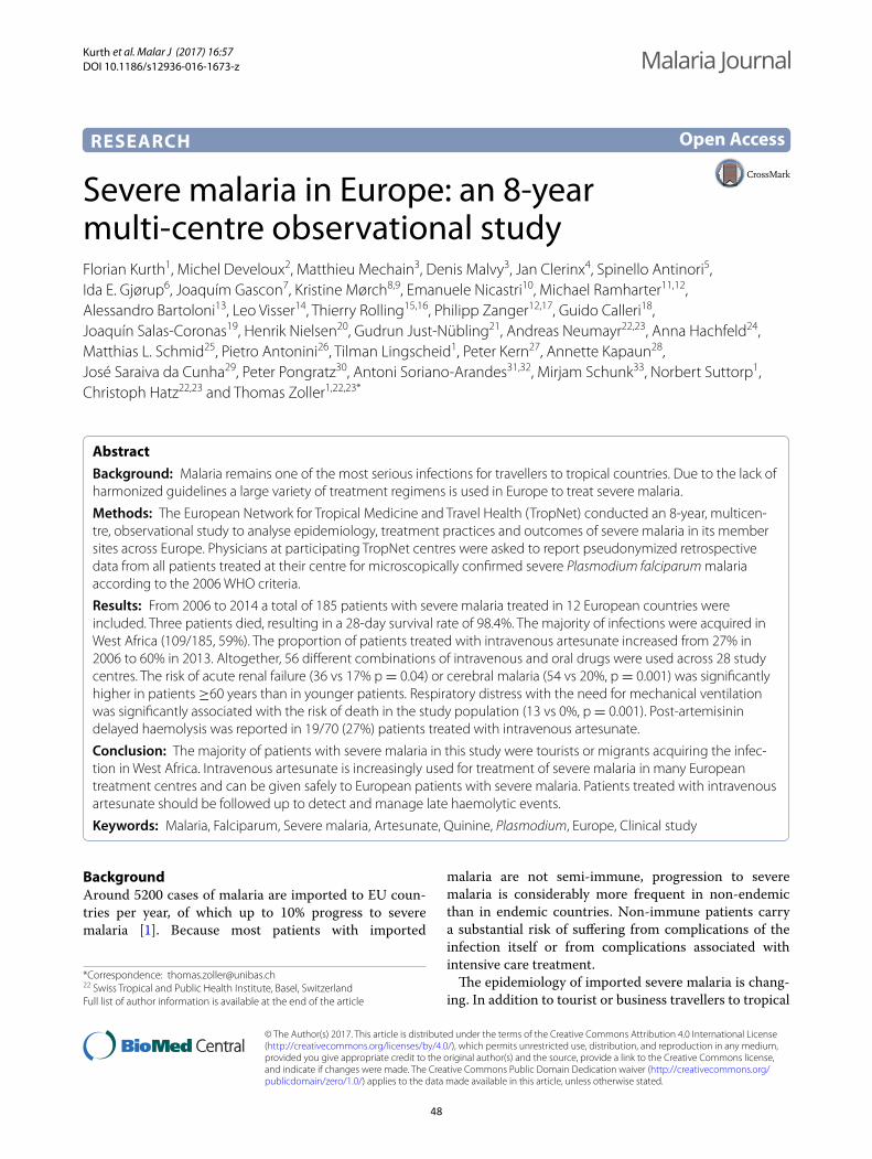
Kurth et al. Malar J (2017) 16:57 DOI 10.1186/s12936-016-1673-z
RESEARCH
Severe malaria in Europe: an 8-year multi-centre observational studyFlorian Kurth1, Michel Develoux2, Matthieu Mechain3, Denis Malvy3, Jan Clerinx4, Spinello Antinori5, Ida E. Gjørup6, Joaquím Gascon7, Kristine Mørch8,9, Emanuele Nicastri10, Michael Ramharter11,12, Alessandro Bartoloni13, Leo Visser14, Thierry Rolling15,16, Philipp Zanger12,17, Guido Calleri18, Joaquín Salas‑Coronas19, Henrik Nielsen20, Gudrun Just‑Nübling21, Andreas Neumayr22,23, Anna Hachfeld24, Matthias L. Schmid25, Pietro Antonini26, Tilman Lingscheid1, Peter Kern27, Annette Kapaun28, José Saraiva da Cunha29, Peter Pongratz30, Antoni Soriano‑Arandes31,32, Mirjam Schunk33, Norbert Suttorp1, Christoph Hatz22,23 and Thomas Zoller1,22,23*
Abstract
Background: Malaria remains one of the most serious infections for travellers to tropical countries. Due to the lack of harmonized guidelines a large variety of treatment regimens is used in Europe to treat severe malaria.
Methods: The European Network for Tropical Medicine and Travel Health (TropNet) conducted an 8‑year, multicen‑tre, observational study to analyse epidemiology, treatment practices and outcomes of severe malaria in its member sites across Europe. Physicians at participating TropNet centres were asked to report pseudonymized retrospective data from all patients treated at their centre for microscopically confirmed severe Plasmodium falciparum malaria according to the 2006 WHO criteria.
Results: From 2006 to 2014 a total of 185 patients with severe malaria treated in 12 European countries were included. Three patients died, resulting in a 28‑day survival rate of 98.4%. The majority of infections were acquired in West Africa (109/185, 59%). The proportion of patients treated with intravenous artesunate increased from 27% in 2006 to 60% in 2013. Altogether, 56 different combinations of intravenous and oral drugs were used across 28 study centres. The risk of acute renal failure (36 vs 17% p = 0.04) or cerebral malaria (54 vs 20%, p = 0.001) was significantly higher in patients ≥60 years than in younger patients. Respiratory distress with the need for mechanical ventilation was significantly associated with the risk of death in the study population (13 vs 0%, p = 0.001). Post‑artemisinin delayed haemolysis was reported in 19/70 (27%) patients treated with intravenous artesunate.
Conclusion: The majority of patients with severe malaria in this study were tourists or migrants acquiring the infec‑tion in West Africa. Intravenous artesunate is increasingly used for treatment of severe malaria in many European treatment centres and can be given safely to European patients with severe malaria. Patients treated with intravenous artesunate should be followed up to detect and manage late haemolytic events.
Keywords: Malaria, Falciparum, Severe malaria, Artesunate, Quinine, Plasmodium, Europe, Clinical study
© The Author(s) 2017. This article is distributed under the terms of the Creative Commons Attribution 4.0 International License (http://creativecommons.org/licenses/by/4.0/), which permits unrestricted use, distribution, and reproduction in any medium, provided you give appropriate credit to the original author(s) and the source, provide a link to the Creative Commons license, and indicate if changes were made. The Creative Commons Public Domain Dedication waiver (http://creativecommons.org/publicdomain/zero/1.0/) applies to the data made available in this article, unless otherwise stated.
BackgroundAround 5200 cases of malaria are imported to EU coun-tries per year, of which up to 10% progress to severe malaria [1]. Because most patients with imported
malaria are not semi-immune, progression to severe malaria is considerably more frequent in non-endemic than in endemic countries. Non-immune patients carry a substantial risk of suffering from complications of the infection itself or from complications associated with intensive care treatment.
The epidemiology of imported severe malaria is chang-ing. In addition to tourist or business travellers to tropical
Open Access
Malaria Journal
*Correspondence: [email protected] 22 Swiss Tropical and Public Health Institute, Basel, SwitzerlandFull list of author information is available at the end of the article
48
Page 2 of 11Kurth et al. Malar J (2017) 16:57
regions, migrants visiting friends and relatives (VFR) in their previous home country are increasingly affected [2, 3]. This population is less likely to seek pre-travel advice and to take anti-malarial prophylaxis [4, 5]. Most migrants are not aware of the waning of semi-immunity against malaria when they travel to their home countries.
Guidelines to define and to treat severe malaria have undergone major changes in the past decade at inter-national and national levels. Criteria for the definition of severe malaria were amended by the World Health Organization (WHO) in 2006, 2010 and 2015, par-ticularly with regard to the definition of hyperparasi-taemia [6], and numerous classification and treatment recommendations still exist across European countries. Another challenge is the transition from intravenous qui-nine to intravenous artesunate as first-line treatment for severe malaria. Despite its superior potential to save lives and shorten duration of hospital and intensive care unit (ICU) treatment [7–10], many difficulties with regard to registration, availability and quality of artesunate have to be overcome before it will be easily available and widely used outside of specialist referral centres for tropical medicine in Europe. The pathophysiology of late haemo-lytic reactions occurring 2–6 weeks after treatment is not fully understood and harmonized guidelines for follow-up care of patients receiving this drug in Europe need to be developed [11, 12].
Data on epidemiology and treatment of imported severe malaria across Europe, together with a ‘road map’ towards drug approval of intravenous artesunate for the treatment of severe malaria in non-endemic countries are needed to eventually improve and harmonize treatment recommendations. Data are however available only from national cohorts and case registries. The European Net-work for Tropical Medicine and Travel Health (TropNet) [13] conducted an 8-year, multi-centre, observational study to analyse epidemiology, treatment practices and outcome of severe malaria in its member sites across 12 European countries.
MethodsThis multicentre observational study was performed among member sites of TropNet. All patients with microscopically confirmed severe falciparum malaria, according to the 2006 WHO criteria treated at one of the participating TropNet centres between 2006 and 2014, were eligible. Physicians at participating TropNet centres were asked to report pseudonymized retrospective data on demographic characteristics, medical and travel his-tory, clinical presentation, anti-malarial drug regimen, supportive treatment, parasitaemia before and under treatment, complications under treatment, adverse drug reactions, outcome and follow-up during 28 days from
all patients treated for severe malaria at their centre. The treatment remained the responsibility of the treating physician. Parasite clearance time was defined as time to the first thick blood smear without evidence of asex-ual parasites after initiation of anti-malarial treatment. For the analysis of this dataset, post-artemisinin delayed haemolysis (PADH) was defined as a decrease in haemo-globin together with signs of haemolysis (elevated LDH) after completion of anti-malarial treatment and complete parasite clearance. Electronic case report forms were used for data entry and transmission to the coordinating centre at Charité University Hospital, Berlin, where data were transferred into a database and checked manually for plausibility.
The primary objective of this study was to assess clini-cal presentation, treatment and outcome in patients with imported severe malaria. Descriptive statistics was performed on sociodemographic, medical, treatment, and outcome data. Mann–Whitney U test (two groups, continuous data), Fisher exact test (two groups, categori-cal data), or Kruskal–Wallis test (>two groups continu-ous data) at a two-sided significance level of α = 0.05 were used for comparative analysis. Analysis of variance (ANOVA) for continuous data and Pearson Chi squared test for categorical data were used to test the distribution of demographical characteristics of patients over time. Data are displayed as median (±interquartile range). Sta-tistical analysis was performed using JMP (JMP 7.0, SAS Institute Inc, NC, USA).
The study was approved by the Ethics Committee of Charité University Hospital, Berlin. Ethical clearance for transfer of retrospective pseudonymized patient data was sought at participating Tropnet centres according to local regulations.
ResultsFrom 2006 to 2014, 190 patients with severe falciparum malaria were reported by the participating 28 TropNet centres from 12 European countries. After excluding three cases without documented criteria for severe malaria and two cases with double reporting, 185 data-sets were available for analysis. The number of patients per centre ranged from one to 31 patients. The number of reported patients per country is shown in Table 1.
Demography, place of infection and anti‑malarial prophylaxisDemographic data are shown in Table 2. The propor-tion of female patients was comparatively small (29%). The majority of patients (106/185, 57%) were of Euro-pean origin without history of migration. Tourism was the main purpose of travel in Europeans (54/106, 51%), whereas VFRs were the predominant purposes of travel
49
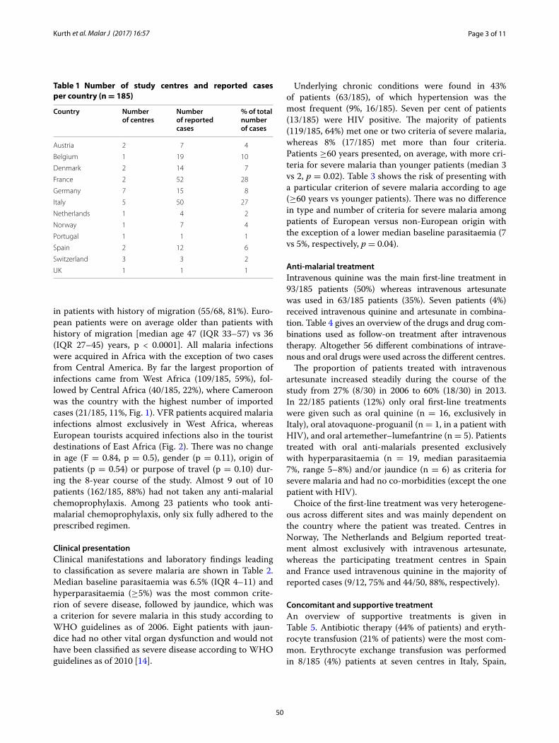
Page 3 of 11Kurth et al. Malar J (2017) 16:57
in patients with history of migration (55/68, 81%). Euro-pean patients were on average older than patients with history of migration [median age 47 (IQR 33–57) vs 36 (IQR 27–45) years, p < 0.0001]. All malaria infections were acquired in Africa with the exception of two cases from Central America. By far the largest proportion of infections came from West Africa (109/185, 59%), fol-lowed by Central Africa (40/185, 22%), where Cameroon was the country with the highest number of imported cases (21/185, 11%, Fig. 1). VFR patients acquired malaria infections almost exclusively in West Africa, whereas European tourists acquired infections also in the tourist destinations of East Africa (Fig. 2). There was no change in age (F = 0.84, p = 0.5), gender (p = 0.11), origin of patients (p = 0.54) or purpose of travel (p = 0.10) dur-ing the 8-year course of the study. Almost 9 out of 10 patients (162/185, 88%) had not taken any anti-malarial chemoprophylaxis. Among 23 patients who took anti-malarial chemoprophylaxis, only six fully adhered to the prescribed regimen.
Clinical presentationClinical manifestations and laboratory findings leading to classification as severe malaria are shown in Table 2. Median baseline parasitaemia was 6.5% (IQR 4–11) and hyperparasitaemia (≥5%) was the most common crite-rion of severe disease, followed by jaundice, which was a criterion for severe malaria in this study according to WHO guidelines as of 2006. Eight patients with jaun-dice had no other vital organ dysfunction and would not have been classified as severe disease according to WHO guidelines as of 2010 [14].
Underlying chronic conditions were found in 43% of patients (63/185), of which hypertension was the most frequent (9%, 16/185). Seven per cent of patients (13/185) were HIV positive. The majority of patients (119/185, 64%) met one or two criteria of severe malaria, whereas 8% (17/185) met more than four criteria. Patients ≥60 years presented, on average, with more cri-teria for severe malaria than younger patients (median 3 vs 2, p = 0.02). Table 3 shows the risk of presenting with a particular criterion of severe malaria according to age (≥60 years vs younger patients). There was no difference in type and number of criteria for severe malaria among patients of European versus non-European origin with the exception of a lower median baseline parasitaemia (7 vs 5%, respectively, p = 0.04).
Anti‑malarial treatmentIntravenous quinine was the main first-line treatment in 93/185 patients (50%) whereas intravenous artesunate was used in 63/185 patients (35%). Seven patients (4%) received intravenous quinine and artesunate in combina-tion. Table 4 gives an overview of the drugs and drug com-binations used as follow-on treatment after intravenous therapy. Altogether 56 different combinations of intrave-nous and oral drugs were used across the different centres.
The proportion of patients treated with intravenous artesunate increased steadily during the course of the study from 27% (8/30) in 2006 to 60% (18/30) in 2013. In 22/185 patients (12%) only oral first-line treatments were given such as oral quinine (n = 16, exclusively in Italy), oral atovaquone-proguanil (n = 1, in a patient with HIV), and oral artemether–lumefantrine (n = 5). Patients treated with oral anti-malarials presented exclusively with hyperparasitaemia (n = 19, median parasitaemia 7%, range 5–8%) and/or jaundice (n = 6) as criteria for severe malaria and had no co-morbidities (except the one patient with HIV).
Choice of the first-line treatment was very heterogene-ous across different sites and was mainly dependent on the country where the patient was treated. Centres in Norway, The Netherlands and Belgium reported treat-ment almost exclusively with intravenous artesunate, whereas the participating treatment centres in Spain and France used intravenous quinine in the majority of reported cases (9/12, 75% and 44/50, 88%, respectively).
Concomitant and supportive treatmentAn overview of supportive treatments is given in Table 5. Antibiotic therapy (44% of patients) and eryth-rocyte transfusion (21% of patients) were the most com-mon. Erythrocyte exchange transfusion was performed in 8/185 (4%) patients at seven centres in Italy, Spain,
Table 1 Number of study centres and reported cases per country (n = 185)
Country Number of centres
Number of reported cases
% of total number of cases
Austria 2 7 4
Belgium 1 19 10
Denmark 2 14 7
France 2 52 28
Germany 7 15 8
Italy 5 50 27
Netherlands 1 4 2
Norway 1 7 4
Portugal 1 1 1
Spain 2 12 6
Switzerland 3 3 2
UK 1 1 1
50
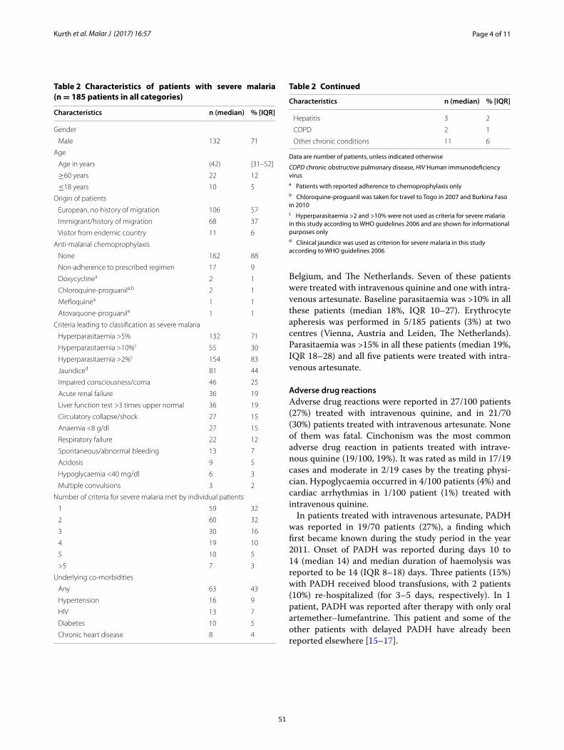
Page 4 of 11Kurth et al. Malar J (2017) 16:57
Belgium, and The Netherlands. Seven of these patients were treated with intravenous quinine and one with intra-venous artesunate. Baseline parasitaemia was >10% in all these patients (median 18%, IQR 10–27). Erythrocyte apheresis was performed in 5/185 patients (3%) at two centres (Vienna, Austria and Leiden, The Netherlands). Parasitaemia was >15% in all these patients (median 19%, IQR 18–28) and all five patients were treated with intra-venous artesunate.
Adverse drug reactionsAdverse drug reactions were reported in 27/100 patients (27%) treated with intravenous quinine, and in 21/70 (30%) patients treated with intravenous artesunate. None of them was fatal. Cinchonism was the most common adverse drug reaction in patients treated with intrave-nous quinine (19/100, 19%). It was rated as mild in 17/19 cases and moderate in 2/19 cases by the treating physi-cian. Hypoglycaemia occurred in 4/100 patients (4%) and cardiac arrhythmias in 1/100 patient (1%) treated with intravenous quinine.
In patients treated with intravenous artesunate, PADH was reported in 19/70 patients (27%), a finding which first became known during the study period in the year 2011. Onset of PADH was reported during days 10 to 14 (median 14) and median duration of haemolysis was reported to be 14 (IQR 8–18) days. Three patients (15%) with PADH received blood transfusions, with 2 patients (10%) re-hospitalized (for 3–5 days, respectively). In 1 patient, PADH was reported after therapy with only oral artemether–lumefantrine. This patient and some of the other patients with delayed PADH have already been reported elsewhere [15–17].
Table 2 Characteristics of patients with severe malaria (n = 185 patients in all categories)
Data are number of patients, unless indicated otherwise
COPD chronic obstructive pulmonary disease, HIV Human immunodeficiency virusa Patients with reported adherence to chemoprophylaxis onlyb Chloroquine-proguanil was taken for travel to Togo in 2007 and Burkina Faso in 2010c Hyperparasitaemia >2 and >10% were not used as criteria for severe malaria in this study according to WHO guidelines 2006 and are shown for informational purposes onlyd Clinical jaundice was used as criterion for severe malaria in this study according to WHO guidelines 2006
Characteristics n (median) % [IQR]
Gender
Male 132 71
Age
Age in years (42) [31–52]
≥60 years 22 12
≤18 years 10 5
Origin of patients
European, no history of migration 106 57
Immigrant/history of migration 68 37
Visitor from endemic country 11 6
Anti‑malarial chemoprophylaxis
None 162 88
Non‑adherence to prescribed regimen 17 9
Doxycyclinea 2 1
Chloroquine‑proguanila,b 2 1
Mefloquinea 1 1
Atovaquone‑proguanila 1 1
Criteria leading to classification as severe malaria
Hyperparasitaemia >5% 132 71
Hyperparasitaemia >10%c 55 30
Hyperparasitaemia >2%c 154 83
Jaundiced 81 44
Impaired consciousness/coma 46 25
Acute renal failure 36 19
Liver function test >3 times upper normal 36 19
Circulatory collapse/shock 27 15
Anaemia <8 g/dl 27 15
Respiratory failure 22 12
Spontaneous/abnormal bleeding 13 7
Acidosis 9 5
Hypoglycaemia <40 mg/dl 6 3
Multiple convulsions 3 2
Number of criteria for severe malaria met by individual patients
1 59 32
2 60 32
3 30 16
4 19 10
5 10 5
>5 7 3
Underlying co‑morbidities
Any 63 43
Hypertension 16 9
HIV 13 7
Diabetes 10 5
Chronic heart disease 8 4
Characteristics n (median) % [IQR]
Hepatitis 3 2
COPD 2 1
Other chronic conditions 11 6
Table 2 Continued
51
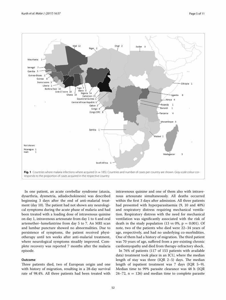
Page 5 of 11Kurth et al. Malar J (2017) 16:57
In one patient, an acute cerebellar syndrome (ataxia, dysarthria, dysmetria, adiadochokinesis) was described beginning 3 days after the end of anti-malarial treat-ment (day 10). The patient had not shown any neurologi-cal symptoms during the acute phase of malaria and had been treated with a loading dose of intravenous quinine on day 1, intravenous artesunate from day 1 to 4 and oral artemether–lumefantrine from day 5 to 7. An MRI scan and lumbar puncture showed no abnormalities. Due to persistence of symptoms, the patient received physi-otherapy until ten weeks after anti-malarial treatment, where neurological symptoms steadily improved. Com-plete recovery was reported 7 months after the malaria episode.
OutcomeThree patients died, two of European origin and one with history of migration, resulting in a 28-day survival rate of 98.4%. All three patients had been treated with
intravenous quinine and one of them also with intrave-nous artesunate simultaneously. All deaths occurred within the first 3 days after admission. All three patients had presented with hyperparasitaemia (9, 10 and 40%) and respiratory distress requiring mechanical ventila-tion. Respiratory distress with the need for mechanical ventilation was significantly associated with the risk of death in the study population (13 vs 0%, p = 0.001). Of note, two of the patients who died were 22–34 years of age, respectively, and had no underlying co-morbidities. One of them had a history of migration. The third patient was 70 years of age, suffered from a pre-existing chronic cardiomyopathy and died from therapy-refractory shock.
In 76% of patients (117 of 153 patients with available data) treatment took place in an ICU, where the median length of stay was three (IQR 2–5) days. The median length of inpatient treatment was 7 days (IQR 5–9). Median time to 99% parasite clearance was 48 h (IQR 24–72, n = 126) and median time to complete parasite
Fig. 1 Countries where malaria infections where acquired (n = 185). Countries and number of cases per country are shown. Gray‑scale colour cor‑responds to the proportion of cases acquired in the respective country
52
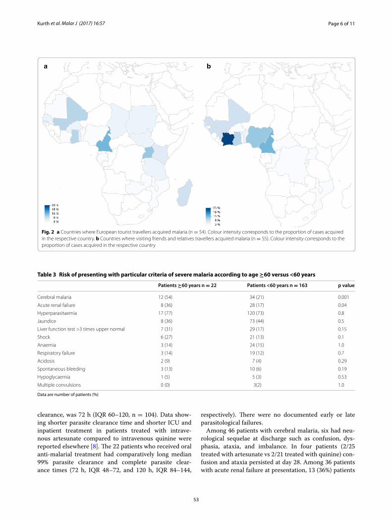
Page 6 of 11Kurth et al. Malar J (2017) 16:57
clearance, was 72 h (IQR 60–120, n = 104). Data show-ing shorter parasite clearance time and shorter ICU and inpatient treatment in patients treated with intrave-nous artesunate compared to intravenous quinine were reported elsewhere [8]. The 22 patients who received oral anti-malarial treatment had comparatively long median 99% parasite clearance and complete parasite clear-ance times (72 h, IQR 48–72, and 120 h, IQR 84–144,
respectively). There were no documented early or late parasitological failures.
Among 46 patients with cerebral malaria, six had neu-rological sequelae at discharge such as confusion, dys-phasia, ataxia, and imbalance. In four patients (2/25 treated with artesunate vs 2/21 treated with quinine) con-fusion and ataxia persisted at day 28. Among 36 patients with acute renal failure at presentation, 13 (36%) patients
Fig. 2 a Countries where European tourist travellers acquired malaria (n = 54). Colour intensity corresponds to the proportion of cases acquired in the respective country. b Countries where visiting friends and relatives travellers acquired malaria (n = 55). Colour intensity corresponds to the proportion of cases acquired in the respective country
Table 3 Risk of presenting with particular criteria of severe malaria according to age ≥60 versus <60 years
Data are number of patients (%)
Patients ≥60 years n = 22 Patients <60 years n = 163 p value
Cerebral malaria 12 (54) 34 (21) 0.001
Acute renal failure 8 (36) 28 (17) 0.04
Hyperparasitaemia 17 (77) 120 (73) 0.8
Jaundice 8 (36) 73 (44) 0.5
Liver function test >3 times upper normal 7 (31) 29 (17) 0.15
Shock 6 (27) 21 (13) 0.1
Anaemia 3 (14) 24 (15) 1.0
Respiratory failure 3 (14) 19 (12) 0.7
Acidosis 2 (9) 7 (4) 0.29
Spontaneous bleeding 3 (13) 10 (6) 0.19
Hypoglycaemia 1 (5) 5 (3) 0.53
Multiple convulsions 0 (0) 3(2) 1.0
53
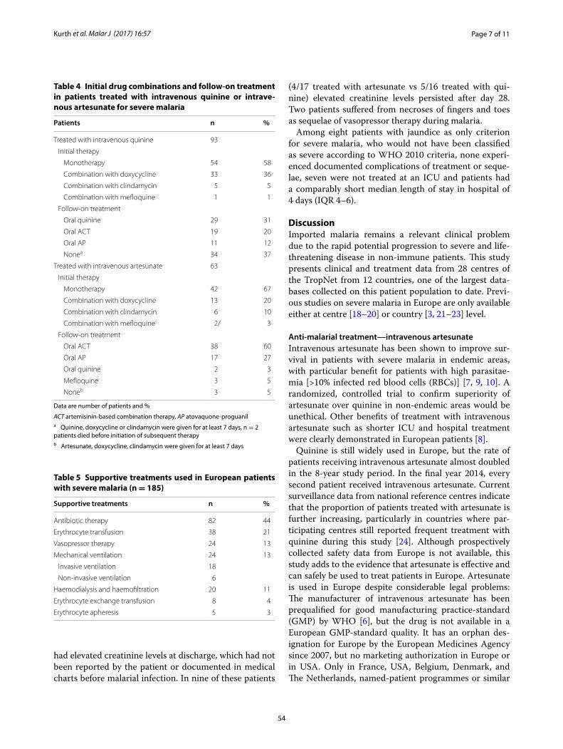
Page 7 of 11Kurth et al. Malar J (2017) 16:57
had elevated creatinine levels at discharge, which had not been reported by the patient or documented in medical charts before malarial infection. In nine of these patients
(4/17 treated with artesunate vs 5/16 treated with qui-nine) elevated creatinine levels persisted after day 28. Two patients suffered from necroses of fingers and toes as sequelae of vasopressor therapy during malaria.
Among eight patients with jaundice as only criterion for severe malaria, who would not have been classified as severe according to WHO 2010 criteria, none experi-enced documented complications of treatment or seque-lae, seven were not treated at an ICU and patients had a comparably short median length of stay in hospital of 4 days (IQR 4–6).
DiscussionImported malaria remains a relevant clinical problem due to the rapid potential progression to severe and life-threatening disease in non-immune patients. This study presents clinical and treatment data from 28 centres of the TropNet from 12 countries, one of the largest data-bases collected on this patient population to date. Previ-ous studies on severe malaria in Europe are only available either at centre [18–20] or country [3, 21–23] level.
Anti‑malarial treatment—intravenous artesunateIntravenous artesunate has been shown to improve sur-vival in patients with severe malaria in endemic areas, with particular benefit for patients with high parasitae-mia [>10% infected red blood cells (RBCs)] [7, 9, 10]. A randomized, controlled trial to confirm superiority of artesunate over quinine in non-endemic areas would be unethical. Other benefits of treatment with intravenous artesunate such as shorter ICU and hospital treatment were clearly demonstrated in European patients [8].
Quinine is still widely used in Europe, but the rate of patients receiving intravenous artesunate almost doubled in the 8-year study period. In the final year 2014, every second patient received intravenous artesunate. Current surveillance data from national reference centres indicate that the proportion of patients treated with artesunate is further increasing, particularly in countries where par-ticipating centres still reported frequent treatment with quinine during this study [24]. Although prospectively collected safety data from Europe is not available, this study adds to the evidence that artesunate is effective and can safely be used to treat patients in Europe. Artesunate is used in Europe despite considerable legal problems: The manufacturer of intravenous artesunate has been prequalified for good manufacturing practice-standard (GMP) by WHO [6], but the drug is not available in a European GMP-standard quality. It has an orphan des-ignation for Europe by the European Medicines Agency since 2007, but no marketing authorization in Europe or in USA. Only in France, USA, Belgium, Denmark, and The Netherlands, named-patient programmes or similar
Table 4 Initial drug combinations and follow-on treatment in patients treated with intravenous quinine or intrave-nous artesunate for severe malaria
Data are number of patients and %
ACT artemisinin-based combination therapy, AP atovaquone-proguanila Quinine, doxycycline or clindamycin were given for at least 7 days, n = 2 patients died before initiation of subsequent therapyb Artesunate, doxycycline, clindamycin were given for at least 7 days
Patients n %
Treated with intravenous quinine 93
Initial therapy
Monotherapy 54 58
Combination with doxycycline 33 36
Combination with clindamycin 5 5
Combination with mefloquine 1 1
Follow‑on treatment
Oral quinine 29 31
Oral ACT 19 20
Oral AP 11 12
Nonea 34 37
Treated with intravenous artesunate 63
Initial therapy
Monotherapy 42 67
Combination with doxycycline 13 20
Combination with clindamycin 6 10
Combination with mefloquine 2/ 3
Follow‑on treatment
Oral ACT 38 60
Oral AP 17 27
Oral quinine 2 3
Mefloquine 3 5
Noneb 3 5
Table 5 Supportive treatments used in European patients with severe malaria (n = 185)
Supportive treatments n %
Antibiotic therapy 82 44
Erythrocyte transfusion 38 21
Vasopressor therapy 24 13
Mechanical ventilation 24 13
Invasive ventilation 18
Non‑invasive ventilation 6
Haemodialysis and haemofiltration 20 11
Erythrocyte exchange transfusion 8 4
Erythrocyte apheresis 5 3
54
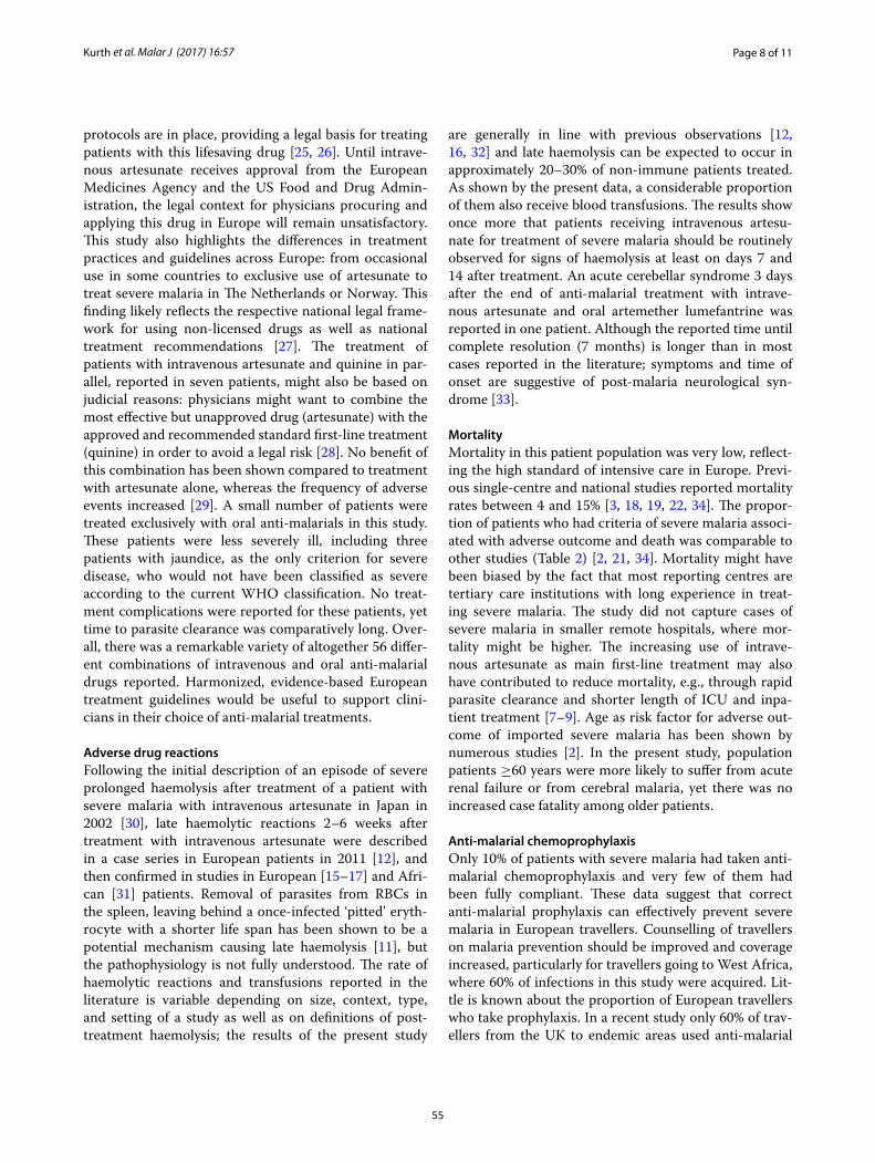
Page 8 of 11Kurth et al. Malar J (2017) 16:57
protocols are in place, providing a legal basis for treating patients with this lifesaving drug [25, 26]. Until intrave-nous artesunate receives approval from the European Medicines Agency and the US Food and Drug Admin-istration, the legal context for physicians procuring and applying this drug in Europe will remain unsatisfactory. This study also highlights the differences in treatment practices and guidelines across Europe: from occasional use in some countries to exclusive use of artesunate to treat severe malaria in The Netherlands or Norway. This finding likely reflects the respective national legal frame-work for using non-licensed drugs as well as national treatment recommendations [27]. The treatment of patients with intravenous artesunate and quinine in par-allel, reported in seven patients, might also be based on judicial reasons: physicians might want to combine the most effective but unapproved drug (artesunate) with the approved and recommended standard first-line treatment (quinine) in order to avoid a legal risk [28]. No benefit of this combination has been shown compared to treatment with artesunate alone, whereas the frequency of adverse events increased [29]. A small number of patients were treated exclusively with oral anti-malarials in this study. These patients were less severely ill, including three patients with jaundice, as the only criterion for severe disease, who would not have been classified as severe according to the current WHO classification. No treat-ment complications were reported for these patients, yet time to parasite clearance was comparatively long. Over-all, there was a remarkable variety of altogether 56 differ-ent combinations of intravenous and oral anti-malarial drugs reported. Harmonized, evidence-based European treatment guidelines would be useful to support clini-cians in their choice of anti-malarial treatments.
Adverse drug reactionsFollowing the initial description of an episode of severe prolonged haemolysis after treatment of a patient with severe malaria with intravenous artesunate in Japan in 2002 [30], late haemolytic reactions 2–6 weeks after treatment with intravenous artesunate were described in a case series in European patients in 2011 [12], and then confirmed in studies in European [15–17] and Afri-can [31] patients. Removal of parasites from RBCs in the spleen, leaving behind a once-infected ‘pitted’ eryth-rocyte with a shorter life span has been shown to be a potential mechanism causing late haemolysis [11], but the pathophysiology is not fully understood. The rate of haemolytic reactions and transfusions reported in the literature is variable depending on size, context, type, and setting of a study as well as on definitions of post-treatment haemolysis; the results of the present study
are generally in line with previous observations [12, 16, 32] and late haemolysis can be expected to occur in approximately 20–30% of non-immune patients treated. As shown by the present data, a considerable proportion of them also receive blood transfusions. The results show once more that patients receiving intravenous artesu-nate for treatment of severe malaria should be routinely observed for signs of haemolysis at least on days 7 and 14 after treatment. An acute cerebellar syndrome 3 days after the end of anti-malarial treatment with intrave-nous artesunate and oral artemether lumefantrine was reported in one patient. Although the reported time until complete resolution (7 months) is longer than in most cases reported in the literature; symptoms and time of onset are suggestive of post-malaria neurological syn-drome [33].
MortalityMortality in this patient population was very low, reflect-ing the high standard of intensive care in Europe. Previ-ous single-centre and national studies reported mortality rates between 4 and 15% [3, 18, 19, 22, 34]. The propor-tion of patients who had criteria of severe malaria associ-ated with adverse outcome and death was comparable to other studies (Table 2) [2, 21, 34]. Mortality might have been biased by the fact that most reporting centres are tertiary care institutions with long experience in treat-ing severe malaria. The study did not capture cases of severe malaria in smaller remote hospitals, where mor-tality might be higher. The increasing use of intrave-nous artesunate as main first-line treatment may also have contributed to reduce mortality, e.g., through rapid parasite clearance and shorter length of ICU and inpa-tient treatment [7–9]. Age as risk factor for adverse out-come of imported severe malaria has been shown by numerous studies [2]. In the present study, population patients ≥60 years were more likely to suffer from acute renal failure or from cerebral malaria, yet there was no increased case fatality among older patients.
Anti‑malarial chemoprophylaxisOnly 10% of patients with severe malaria had taken anti-malarial chemoprophylaxis and very few of them had been fully compliant. These data suggest that correct anti-malarial prophylaxis can effectively prevent severe malaria in European travellers. Counselling of travellers on malaria prevention should be improved and coverage increased, particularly for travellers going to West Africa, where 60% of infections in this study were acquired. Lit-tle is known about the proportion of European travellers who take prophylaxis. In a recent study only 60% of trav-ellers from the UK to endemic areas used anti-malarial
55
Page 9 of 11Kurth et al. Malar J (2017) 16:57
chemoprophylaxis [35]. VFR travellers are a large trav-eller population to Africa with different perceptions of malaria and its prevention [4, 5]. This may also influ-ence the longer delay between onset of symptoms of malaria and presentation in hospital. The fact that 37% of patients in the present study had a history of migration clearly demonstrates the risk of these patients to suffer from severe malaria. Moreover, one of the patients who died was a 34 years old, otherwise healthy patient with history of migration. She presented with hyperparasitae-mia, acute renal failure, jaundice, and respiratory failure. There was altogether no difference in symptoms and clin-ical presentation between patients with or without his-tory of migration, suggesting a waning of semi-immunity in migrants who left endemic areas [36].
Supportive treatmentsSupportive treatments such as exchange transfusions and erythrocyte apheresis are a matter of controversy and their use is guided by national or local practices. Euro-pean single-centre studies recently failed to demonstrate improved parasite clearance through whole blood or erythrocyte exchange, compared to patients treated with quinine or artesunate alone [37, 38]. Only 4% of patients received exchange transfusions in this study. It was per-formed in only 7 out of 28 participating centres, mainly in patients treated with intravenous quinine. Likewise, only five patients in two centres received automated erythrocyte apheresis. With the increased use of intrave-nous artesunate and its potential to rapidly reduce high parasite loads it needs to be determined whether par-ticular patient populations might still benefit from these adjunctive treatments [39].
LimitationsThis observational study has inherent limitations. As patient information was collected retrospectively in Trop- Net centres and not all patients treated may have been reported equally, selective under-reporting, e.g., for patients who died, may have occurred. Bias in reporting data on PADH must be assumed as the condition was not known at the beginning of the study and no universally accepted clinical definition exists to date. All TropNet centres are referral centres for tropical medicine and patient composition as well as treatment data may not fully reflect treatment practices and outcomes in non-referral hospitals. Moreover, data reported by the par-ticipating treatment centres may not always fully reflect treatment practices in the respective countries. For patients with elevated creatinine levels at the end of fol-low-up, it cannot be ruled out that unknown or undoc-umented elevation of creatinine had existed before the episode of severe malaria.
ConclusionThe data show that the majority of patients with severe malaria in Europe are tourists or migrants acquir-ing infection in West Africa. Intravenous artesunate is increasingly used for treatment of severe malaria; it is the most effective drug and can be safely given to European patients with severe malaria. There is need for harmoni-zation of guidelines for the treatment of severe malaria in Europe. Patients treated with intravenous artesunate should be followed up to detect late haemolytic events.
Authors’ contributionsTZ designed the study; TZ and FK performed acquisition, entry, cleaning and analysis of data, wrote the manuscript; MD, MM, DM, JC, SA, IEG, JG, KM, EN, MR, AB, LV, TR, PZ, GC, JSC, HN, GJN, AN, AH, MLS, PA, TL, PK, AK, JSdC, PP, ASA, MS, NS, and CH treated patients, gathered data in study centres, contributed to and corrected the manuscript. All authors read and approved the final manuscript.
Author details1 Medizinische Klinik mit Schwerpunkt Infektiologie und Pneumologie, Charité Universitätsmedizin Berlin, Berlin, Germany. 2 Hôpital Saint‑Antoine, Paris, France. 3 Section Tropical Medicine and Clinical International Health, Division of Infectious and Tropical Diseases, Department of Medicine, University Hospital Centre, Bordeaux, France. 4 Institute of Tropical Medicine, Antwerp, Belgium. 5 Department of Biomedical and Clinical Sciences L.Sacco, University of Milano, Milan, Italy. 6 Infectious Diseases Unit, Herlev University Hospital, Copenhagen, Denmark. 7 ISGlobal, Barcelona Center for International Health Research. (CRESIB), Hospital Clínic‑Universitat de Barcelona, Barcelona, Spain. 8 Department of Medicine, National Centre for Tropical Infectious Diseases, Haukeland University Hospital, Bergen, Norway. 9 Department of Clinical Science, University of Bergen, Bergen, Norway. 10 National Institute of Infec‑tious Diseases Lazzaro Spallanzani, Rome, Italy. 11 Department of Medicine I, Division of Infectious Diseases and Tropical Medicine, Medical University of Vienna, Vienna, Austria. 12 Institut für Tropenmedizin, Universität Tübingen, Tübingen, Germany. 13 SOD Malattie Infettive e Tropicali, Azienda Ospedaliero Universitaria Careggi, Florence, Italy. 14 Department of Infectious Diseases, Leiden University Medical Centre, Leiden, The Netherlands. 15 Section Tropical Medicine, Department of Internal Medicine I, University Medical Center Hamburg‑Eppendorf, Hamburg, Germany. 16 Department of Clinical Research, Bernhard Nocht Institute for Tropical Medicine, Hamburg, Germany. 17 Institute of Public Health, University of Heidelberg, Heidelberg, Germany. 18 Travel Medicine Unit, Department of Infectious Diseases, Amedeo di Savoia Hospital‑ASLTO2, Turin, Italy. 19 Tropical Medicine Unit, Hospital de Poniente, El Ejido, Spain. 20 Department of Infectious Diseases, Aalborg University Hospital, Aalborg, Denmark. 21 Department of Internal Medicine II, Section Infectious Diseases and Tropical Medicine, University Hospital Frankfurt/Main, Frankfurt/Main, Germany. 22 Swiss Tropical and Public Health Institute, Basel, Switzerland. 23 University of Basel, Basel, Switzerland. 24 Department of Infectious Diseases, Bern University Hospital and University of Bern, Bern, Switzerland. 25 Depart‑ment of Infection & Tropical Medicine, Royal Victoria Infirmary, Newcastle upon Tyne, UK. 26 Clinica Luganese, Lugano, Switzerland. 27 Comprehensive Infectious Diseases Center, Department of Internal Medicine III, Ulm University Hospital, Ulm, Germany. 28 Section Clinical Tropical Medicine, Department of Infectious Diseases, University Hospital Heidelberg, Heidelberg, Germany. 29 Centro Hospitalar e Universitário de Coimbra, Coimbra, Portugal. 30 Division of Tropical Medicine and Infectious Diseases, Center of Internal Medicine II, University of Rostock, Rostock, Germany. 31 Hospital Universitari Joan XXIII, Tarragona, Spain. 32 Hospital Universitari Vall d’Hebron, Barcelona, Spain. 33 Division of Infectious Diseases and Tropical Medicine, Medical Center of the University of Munich (LMU), Munich, Germany.
AcknowledgementsNone.
Competing interestsThe authors declare that they have no competing interests.
56
Page 10 of 11Kurth et al. Malar J (2017) 16:57
Availability of dataThe datasets analysed during the current study are available from the cor‑responding author on reasonable request.
Ethical approvalThe study was approved by the Ethics Committee of Charité University Hospital, Berlin (AE1/334/14). Ethical clearance for transfer of retrospective, pseudonymized patient data was sought at participating Tropnet centres according to local regulations.
FundingFK is supported by Charité clinical Scientist Programme, funded by Charité Universitätsmedizin Berlin and the Berlin Institute of Health.
Received: 3 October 2016 Accepted: 30 December 2016
References 1. ECDC. Annual epidemiological report 2014—emerging and vector‑borne
diseases. Stockholm: ECDC; 2014. 2. Muhlberger N, Jelinek T, Behrens RH, Gjorup I, Coulaud JP, Clerinx J, et al.
Age as a risk factor for severe manifestations and fatal outcome of falcipa‑rum malaria in European patients: observations from TropNetEurop and SIMPID surveillance data. Clin Infect Dis. 2003;36:990–5.
3. Checkley AM, Smith A, Smith V, Blaze M, Bradley D, Chiodini PL, et al. Risk factors for mortality from imported falciparum malaria in the United Kingdom over 20 years: an observational study. BMJ. 2012;344:e2116.
4. van Genderen PJ, van Thiel PP, Mulder PG, Overbosch D, Dutch Schiphol Airport Study Group. Trends in the knowledge, attitudes and practices of travel risk groups towards prevention of malaria: results from the Dutch Schiphol Airport Survey. Malar J. 2012;11:179.
5. Wieten RW, Harting J, Biemond PM, Grobusch MP, van Vugt M. Towards improved uptake of malaria chemoprophylaxis among West African travellers: identification of behavioural determinants. Malar J. 2013;12:360.
6. WHO. Guidelines for the treatment of malaria—3rd edition. Geneva: World Health Organization; 2015.
7. Dondorp A, Nosten F, Stepniewska K, Day N, White N, South East Asian Quinine Artesunate Malaria Trial Group. Artesunate versus quinine for treatment of severe falciparum malaria: a randomised trial. Lancet. 2005;366:717–25.
8. Kurth F, Develoux M, Mechain M, Clerinx J, Antinori S, Gjorup IE, et al. Intravenous artesunate reduces parasite clearance time, duration of intensive care, and hospital treatment in patients with severe malaria in Europe: the TropNet severe malaria study. Clin Infect Dis. 2015;61:1441–4.
9. Sinclair D, Donegan S, Isba R, Lalloo DG. Artesunate versus quinine for treating severe malaria. Cochrane Database Syst Rev. 2012;6:CD005967.
10. Dondorp AM, Fanello CI, Hendriksen IC, Gomes E, Seni A, Chhaganlal KD, et al. Artesunate versus quinine in the treatment of severe falciparum malaria in African children (AQUAMAT): an open‑label, randomised trial. Lancet. 2010;376:1647–57.
11. Jaureguiberry S, Ndour PA, Roussel C, Ader F, Safeukui I, Nguyen M, et al. Postartesunate delayed hemolysis is a predictable event related to the lifesaving effect of artemisinins. Blood. 2014;124:167–75.
12. Zoller T, Junghanss T, Kapaun A, Gjorup I, Richter J, Hugo‑Persson M, et al. Intravenous artesunate for severe malaria in travelers, Europe. Emerg Infect Dis. 2011;17:771–7.
13. European Network for Tropical Medicine and Travel health. http://www.tropnet.eu.
14. WHO. Guidelines for the treatment of malaria—2nd edition. Geneva: World Health Organization; 2010.
15. De Nardo P, Oliva A, Giancola ML, Ghirga P, Mencarini P, Bibas M, et al. Haemolytic anaemia after oral artemether‑lumefantrine treatment in a patient affected by severe imported falciparum malaria. Infection. 2013;41:863–5.
16. Kreeftmeijer‑Vegter AR, van Genderen PJ, Visser LG, Bierman WF, Clerinx J, van Veldhuizen CK, et al. Treatment outcome of intravenous artesunate in patients with severe malaria in the Netherlands and Belgium. Malar J. 2012;11:102.
17. Rolling T, Wichmann D, Schmiedel S, Burchard GD, Kluge S, Cramer JP. Artesunate versus quinine in the treatment of severe imported malaria: comparative analysis of adverse events focussing on delayed haemolysis. Malar J. 2013;12:241.
18. Marks ME, Armstrong M, Suvari MM, Batson S, Whitty CJ, Chiodini PL, Bell‑inghan G, Doherty JF. Severe imported falciparum malaria among adults requiring intensive care: a retrospective study at the hospital for tropical diseases. London. BMC Infect Dis. 2013;13:118.
19. Santos LC, Abreu CF, Xerinda SM, Tavares M, Lucas R, Sarmento AC. Severe imported malaria in an intensive care unit: a review of 59 cases. Malar J. 2012;11:96.
20. Bruneel F, Hocqueloux L, Alberti C, Wolff M, Chevret S, Bedos JP, et al. The clinical spectrum of severe imported falciparum malaria in the intensive care unit: report of 188 cases in adults. Am J Respir Crit Care Med. 2003;167:684–9.
21. Bruneel F, Tubach F, Corne P, Megarbane B, Mira JP, Peytel E, et al. Severe imported falciparum malaria: a cohort study in 400 critically ill adults. PLoS ONE. 2010;5:e13236.
22. Legros F, Bouchaud O, Ancelle T, Arnaud A, Cojean S, Le Bras J, et al. Risk factors for imported fatal Plasmodium falciparum malaria, France, 1996–2003. Emerg Infect Dis. 2007;13:883–8.
23. Bruneel F, Tubach F, Mira JP, Houze S, Gibot S, Huisse MG, et al. Imported falciparum malaria in adults: host‑ and parasite‑related factors associated with severity. The French prospective multicenter PALUREA cohort study. Intensive Care Med. 2016;42:1588–96.
24. Rapport annuel d’activité, Année d’exercice 2014. Centre de national de référence du Paludisme de France; 2015.
25. Kreeftmeijer‑Vegter AR, van Veldhuizen CK, de Vries PJ. Roll out of intrave‑neous artesunate under named patient programmes in the Netherlands, Belgium and France. Orphanet J Rare Dis. 2013;8:150.
26. Twomey PS, Smith BL, McDermott C, Novitt‑Moreno A, McCarthy W, Kachur SP, et al. Intravenous artesunate for the treatment of severe and complicated malaria in the United States: clinical use under an investiga‑tional new drug protocol. Ann Intern Med. 2015;163:498–506.
27. Morch K, Myrvang B. Treatment of malaria in Norway. Tidsskr Nor Laege‑foren. 2012;132:664–7.
28. Bartoloni A, Tomasoni L, Bartalesi F, Galli L, Sani S, Veloci S, et al. Combined intravenous treatment with artesunate and quinine for severe malaria in Italy. Am J Trop Med Hyg. 2010;83:274–6.
29. Newton PN, Chierakul W, Ruangveerayuth R, Silamut K, Teerapong P, Krudsood S, et al. A comparison of artesunate alone with combined artesunate and quinine in the parenteral treatment of acute falciparum malaria. Trans R Soc Trop Med Hyg. 2001;95:519–23.
30. Itoda I, Yasunami T, Kikuchi K, Yamaura H, Totsuka K, Yoshinaga K, et al. Severe falciparum malaria with prolonged hemolytic anemia after suc‑cessful treatment with intravenous artesunate (in Japanese). Kansen‑shogaku Zasshi. 2002;76:600–3.
31. Rolling T, Agbenyega T, Issifou S, Adegnika AA, Sylverken J, Spahlinger D, et al. Delayed hemolysis after treatment with parenteral artesunate in African children with severe malaria–a double‑center prospective study. J Infect Dis. 2014;209:1921–8.
32. Jaureguiberry S, Thellier M, Ndour PA, Ader F, Roussel C, Sonneville R, et al. Delayed‑onset hemolytic anemia in patients with travel‑associated severe malaria treated with artesunate, France, 2011–2013. Emerg Infect Dis. 2015;21:804–12.
33. Nguyen TH, Day NP, Ly VC, Waller D, Mai NT, Bethell DB, Tran TH, White NJ. Post‑malaria neurological syndrome. Lancet. 1996;348:917–21.
34. Kurth F, Zoller T, et al. Reply to Jaureguiberry et al. Clin Infect Dis. 2016;62:271.
35. Behrens RH, Alexander N. Malaria knowledge and utilization of chemo‑prophylaxis in the UK population and in UK passengers departing to malaria‑endemic areas. Malar J. 2013;12:461.
36. Moncunill G, Mayor A, Bardaji A, Puyol L, Nhabomba A, Barrios D, et al. Cytokine profiling in immigrants with clinical malaria after extended periods of interrupted exposure to Plasmodium falciparum. PLoS ONE. 2013;8:e73360.
37. Kreeftmeijer‑Vegter AR, Melo Mde M, de Vries PJ, Koelewijn R, van Hel‑lemond JJ, van Genderen PJ. Manual blood exchange transfusion does not significantly contribute to parasite clearance in artesunate‑treated individuals with imported severe Plasmodium falciparum malaria. Malar J. 2013;12:115.
57
Page 11 of 11Kurth et al. Malar J (2017) 16:57
38. Calvo‑Cano A, Gomez‑Junyent J, Lozano M, Castro P, Cid J, Nicolas JM, et al. The role of red blood cell exchange for severe imported malaria in the artesunate era: a retrospective cohort study in a referral centre. Malar J. 2016;15:216.
39. Auer‑Hackenberg L, Staudinger T, Bojic A, Locker G, Leitner GC, Graninger W, et al. Automated red blood cell exchange as an adjunctive treatment for severe Plasmodium falciparum malaria at the Vienna General Hospital in Austria: a retrospective cohort study. Malar J. 2012;11:158.
58
2.2.1.2 Artesunat versus Chinin bei europäischen Patienten mit schwerer Malaria
Kurth F, Develoux M, Mechain M, Clerinx J, Antinori S, Gjørup IE, Gascon J, Mørch K,
Nicastri E, Ramharter M, Bartoloni A, Visser L, Rolling T, Zanger P, Calleri G, Salas-
Coronas J, Nielsen H, Just-Nübling G, Neumayr A, Hachfeld A, Schmid ML, Antonini P,
Pongratz P, Kern P, Saraiva da Cunha J, Soriano-Arandes A, Schunk M, Suttorp N, Hatz
C, Zoller T; TropNet Severe Malaria Investigator Group. Intravenous Artesunate
Reduces Parasite Clearance Time, Duration of Intensive Care, and Hospital Treatment in
Patients With Severe Malaria in Europe: The TropNet Severe Malaria Study. Clinical
Infectious Diseases, 11/2015, 1441-4, 61
In mehreren prospektiven Studien bei Patienten mit schwerer Malaria in Asien und
Afrika konnte eine klare Überlegenheit für die Therapie mit i.v. Artesunat gegenüber
i.v. Chinin gezeigt werden. Für Patienten außerhalb von Malaria-Endemiegebieten
existieren demgegenüber sehr wenige Daten zur Therapie mit i.v. Artesunat.
In dieser Analyse wurden die Daten von 151 europäischen Patienten mit schwerer
Malaria und i.v. Artesunat- oder i.v. Chinin-Behandlung aus der TropNet-
Beobachtungsstudie zur schweren Malaria verglichen. Patienten mit i.v. Artesunat-
Behandlung zeigten eine schnellere Parasiten-Clearance als Patienten mit i.v. Chinin-
Behandlung (median 72 versus 96 Stunden; p=0,005), benötigten eine um einen Tag
kürzere Behandlung auf der Intensivstation (median 2 versus 3 Tage; p<0,05) und
konnten einen Tag früher aus der Klinik entlassen werden (median 6 versus 7 Tage;
p<0,01). In einer multifaktoriellen Varianzanalyse (F=16; df=4; p<0,0001) war die
Behandlung mit i.v. Artesunat gegenüber i.v. Chinin ein unabhängiger Faktor für eine
kürzere intensivmedizinische Behandlung (F=6,1; p=0,01).
Dies ist die erste multizentrische Studie, die eine Überlegenheit der Behandlung mit i.v.
Artesunat gegenüber i.v. Chinin für Patienten außerhalb von Endemiegebieten belegt.
59

https://doi.org/10.1093/cid/civ575
60
2.2.2 Hämolyse nach oraler Artemisinin-basierter Kombinationstherapie bei unkomplizierter Malaria
Kurth F, Lingscheid T, Steiner F, Stegemann M, Bélard S, Menner N, Pongratz P, Kim J,
Bernuth H, Mayer B, Damm G, Seehofer D, Salama A, Suttorp N, Zoller T. Hemolysis
after Oral Artemisinin Combination Therapy for Uncomplicated Plasmodium falciparum
Malaria. Emerging Infectious Diseases, 08/2016, 1381-6, 22
Die Überlegenheit Artemisinin-basierter Therapieregime gegenüber Artemisinin-freien
Behandlungen wurde in Endemiegebieten sowohl für die unkomplizierte als auch für
die schwere Malaria belegt. Bei 20-30% der nicht-semiimmunen Patienten mit
schwerer Malaria und i.v. Artesunat-Behandlung wurden hämolytische Spätreaktionen
2-6 Wochen nach Therapie beschrieben. Diese sind in der Regel selbstlimitierend,
erfordern aber eine zusätzliche Nachverfolgung der Patienten und in einigen Fällen
Bluttransfusionen und stationäre Überwachung.
In der Haemo-Art Studie wird die Hypothese untersucht, dass hämolytische
Spätreaktionen auch nach unkomplizierter Malaria und oraler Artemisinin-basierter
Therapie auftreten. Von 20 Patienten, die im Rahmen einer ersten Interim-Analyse
ausgewertet wurden, erfüllten 8 Patienten die Kriterien einer verzögerten Hämolyse
2 Wochen nach Behandlung (erniedrigtes Haptoglobin, erhöhte LDH). Bei 5 Patienten
bestanden die Hämolysezeichen noch 1 Monat nach Behandlung. Die Analyse der Hb-
Werte im Beobachtungszeitraum 2 Wochen nach Therapie zeigte für Patienten mit
Hämolyse einen medianen Hb-Abfall von 1,3 g/dl (IQR 0,3-2,0 g/dl), während Patienten
ohne Hämolyse einen Hb-Anstieg von 0,3 g/dl (IQR -0,1-0,7 g/dl; p=0,002) aufwiesen.
Die Ergebnisse zeigen, dass bei nicht-semiimmunen Patienten auch nach
unkomplizierter Malaria verzögerte hämolytischen Reaktionen und Hämoglobinverlust
in den Wochen nach Therapie auftreten.
64
Episodesofdelayedhemolysis2–6weeksaftertreatmentofseveremalariawithintravenousartesunatehavebeende-scribed.Weperformedaprospectiveobservationalstudyofpatientswithuncomplicatedmalariatoinvestigatewhetherposttreatmenthemolysisalsooccursafteroralartemisinin-based combination therapy. Eight of 20 patients with un-complicatedmalariawhoweregivenoralartemisinin-basedcombination therapy met the definition of posttreatmenthemolysis(lowhaptoglobinlevelandincreasedlactatede-hydrogenaselevelonday14).Fivepatientshadhemolysispersisting for1month.Patientswithposttreatmenthemo-lysishadamediandecreaseinhemoglobinlevelof1.3g/dL (interquartile range 0.3–2.0 g/dL) in the posttreatmentperiod, andpatientswithout posttreatment hemolysis hadamedian increaseof0.3g/dL (IQR–0.1 to0.7g/dL;p=0.002).Thesefindings indicateaneed for increasedvigi-lance forhemolyticevents inmalariapatients,particularlythosewithpredisposingfactorsforanemia.
Artemisinin-based drugs are the mainstay of current an-timalarial treatment and play a key role in the World
Health Organization (WHO) global strategy to reduce ma-laria illness and death caused by malaria. These drugs act rapidly against Plasmodium spp. and are usually well toler-ated. Artemisinin-based combination therapies (ACTs) are the recommended first-line treatment for uncomplicated malaria in most countries (1).
Episodes of delayed hemolysis 2–6 weeks after treat-ment for severe malaria with intravenous artesunate have been observed in non–malaria-immune patients in Europe (2). This phenomenon, recently referred to as postartemis-inin-delayed hemolysis (PADH) (3,4), has been confirmed
in other nonimmune patients (4,5) and in children in Africa (6). Approximately 20%–30% of nonimmune patients giv-en intravenous artesunate show signs of PADH that vary in intensity and duration (5,7). Hemolysis is usually self-lim-iting, but patients need to be actively followed up because transfusion of erythrocytes and rehospitalization might be necessary (2,5).
The pathophysiology of hemolysis after artemisinin therapy is not fully understood. Once-infected erythrocytes that have been cleared of parasites in the spleen have a shorter life span and play a role. Patients with higher con-centrations of once-infected erythrocytes after artemisinin treatment are at higher risk for PADH (4). However, other features of posttreatment hemolysis, such as prolonged he-molytic reactions over several weeks (2), are not explained by this mechanism. Several reports suggest involvement of a drug-dependent autoimmune hemolysis mechanism (8), but systematic investigations have not been performed in most published cases (2,9). Given the key role of artemisi-nins in malaria treatment, WHO calls for prospective clini-cal studies and further research to improve the understand-ing of delayed hemolysis after artemisinin therapy (10).
Only 2 single cases and 2 patients in a recent analysis of surveillance data in the United States have been reported with signs of delayed hemolysis after oral ACT treatment (11–13). We hypothesize that delayed hemolysis occurs not only after intravenous treatment for severe malaria but also in a substantial number of patients given oral ACTs for uncomplicated malaria. Because delayed hemolysis has not been captured by safety studies on ACTs, we assume that delayed hemolysis after oral ACTs is less pronounced and occurs to a subclinical degree.
We conducted a study of patients with uncomplicated Plasmodium falciparum malaria to investigate the clinical, laboratory, and immunohematologic features of hemolysis and anemia during and after antimalarial treatment. This ar-ticle presents data for patients investigated during the first 12 months of this ongoing study.
Hemolysis after Oral Artemisinin Combination
Therapy for Uncomplicated Plasmodium falciparum Malaria
Florian Kurth, Tilman Lingscheid, Florian Steiner, Miriam S. Stegemann, Sabine Bélard, Nikolai Menner, Peter Pongratz, Johanna Kim, Horst von Bernuth, Beate Mayer,
Georg Damm, Daniel Seehofer, Abdulgabar Salama, Norbert Suttorp, Thomas Zoller
EmergingInfectiousDiseases•www.cdc.gov/eid•Vol.22,No.8,August2016 1381
Authoraffiliations:Charité–UniversitätsmedizinBerlin,Berlin, Germany(F.Kurth,T.Lingscheid,F.Steiner,M.S.Stegemann, S.Bélard,N.Menner,P.Pongratz,J.Kim,H.vonBernuth, B.Mayer,G.Damm,D.Seehofer,A.Salama,N.Suttorp, T.Zoller);SwissTropicalandPublicHealthInstitute,Basel, Switzerland(T.Zoller)
DOI:http://dx.doi.org/10.3201/eid2208.151905
65
RESEARCH
MethodsThis prospective observational study was conducted at the University Hospital of Charité–Universitätsmedizin Berlin (Berlin, Germany). The study protocol was approved by the ethical committee of Charité–Universitätsmedizin Berlin and is registered at the WHO International Clinical Trials Registry Platform (DRKS00007104). All laboratory analy-ses and parasitologic examinations were performed in ac-credited laboratories at Charité-Universitätsmedizin, Berlin.
All patients who sought treatment at the hospital and were found to have microscopically confirmed uncompli-cated P. falciparum malaria were included in the study af-ter written informed consent was obtained. Patients were excluded if they had received antimalarial treatment (ex-cluding prophylaxis) within 12 weeks before inclusion; had medical conditions that potentially cause hemolysis (e.g., glucose-6-phosphate dehydrogenase deficiency, he-moglobinopathy, mechanical heart valve, lymphoprolifer-ative disease); or were taking medication that potentially causes hemolysis.
Patients were seen for study visits at admission before treatment (day 0). After the first treatment, they were seen again after the last treatment dose on day 3, on day 7 (range day 6–day 10), and on day 14 (range day 14–day 20). If symptoms or signs of hemolysis were detected, patients were seen on day 30 (range day 27–day 31) and thereafter if clinically indicated.
Study visits included obtaining a medical history and conducting a physical examination. Laboratory investiga-tions were parasitologic (thick and thin blood smears), he-matologic (differential blood count), and biochemical (hap-toglobin, lactate dehydrogenase [LDH], C-reactive protein, potassium, and sodium levels and renal and liver function tests) examinations; screening for glucose-6-phosphate de-hydrogenase deficiency; and immunohematologic exami-nations (direct and indirect antiglobulin test, including test-ing with enzyme-treated erythrocytes).
This analysis evaluated data for all patients given oral ACTs during the first 12 months of the study. The primary objective was to assess the proportion of pa-tients with posttreatment hemolysis, which was defined as a low haptoglobin level (<0.3 g/L) and an LDH level above the age-dependent upper normal level 14 days af-ter treatment. Secondary objectives were to compare in patients with posttreatment hemolysis and those without it possible risk factors (age, ethnicity, sex, initial parasit-emia) and the course of anemia (hemoglobin [Hb] level, reticulocyte production index) during treatment, after treatment, and overall. Hemolysis with a loss of Hb >1.5 g/dL during days 3–14 was classified as uncompensated hemolysis; hemolysis without a decrease in Hb level or a decrease <1.5 g/dL during days 3–14 was classified as compensated hemolysis.
Differences between patients with and without signs of posttreatment hemolysis and between patients with compensated and uncompensated hemolysis were ana-lyzed by using the Mann-Whitney U test for continuous data and the Fisher exact test for binary data at a 2-sided significance level of α = 0.05. Data are presented as median and interquartile range (IQR). Statistical analysis was per-formed by using JMP version 7.0 (SAS Institute Inc., Cary, NC, USA).
The sample size of the ongoing study was calculated to detect an incidence of posttreatment hemolysis of 20% with a 95% CI, ± 7.5% precision, and 15% lost to follow-up. This calculation resulted in a sample size of 130 pa-tients. This study evaluated 27 patients, which represented 21% of the intended total sample size. Because we could find no published prospective data for hemolysis after oral ACT treatment, we decided to communicate the findings of this interim analysis before completion of the study.
ResultsDuring May 2014–April 2015, a total of 27 patients with uncomplicated P. falciparum malaria and a standard 3-day treatment course of oral ACT were included in the study. All malaria infections had been acquired in Africa, and none of the patients had taken antimalarial prophylax-is. Six patients did not complete all necessary follow-up visits, and 1 patient was excluded because of sickle cell disease. Twenty patients with >4 study visits until day 20 were available for this interim analysis; of these patients, 3 were children (Table). All patients showed rapid clinical improvement with clearance of peripheral asexual parasit-emia no later than 72 hours after initiation of treatment. There were no treatment failures.
The criteria for posttreatment hemolysis (haptoglobin and LDH levels) on day 14 were met by 8 (40%) of 20 patients. An additional 2 patients showed signs of in vitro hemolysis on day 14 (increased LDH and potassium levels but haptoglobins level within reference ranges). The LDH values of these patients were excluded from further analy-sis and these patients were classified as patients without hemolysis. Patient characteristics showed no differences between those with and without hemolysis, with the excep-tion of slightly higher Hb levels on day 0 and day 3 in pa-tients with posttreatment hemolysis (Table).
After treatment (during days 3–14), patients with posttreatment hemolysis showed a decrease in Hb level (median change -1.3 g/dL, IQR -2.0 to -0.3), and patients without posttreatment hemolysis showed an increase in Hb level (median change 0.3 g/dL, IQR -0.1 to 0.7; p = 0.002) (Figure 1, panel C). During treatment (during days 0 3), patients with posttreatment hemolysis showed a tendency toward a smaller decrease in Hb level (median change -0.15 g/dL, IQR -0.6 to 0.6) than did patients without
1382 EmergingInfectiousDiseases•www.cdc.gov/eid•Vol.22,No.8,August2016
66
HemolysisafterArtemisininTherapyforMalaria
posttreatment hemolysis (median change -0.5 g/dL, IQR -1.0 to -0.3; p = 0.07) (Figure 1, panel B). Overall (during days 0 14), patients with posttreatment hemolysis showed a larger decrease in Hb level (median change -1.35 g/dL, IQR -2.1 to -0.3) than did patients without posttreatment hemolysis (median change -0.45 g/dL, IQR -0.8 to 0.4; p = 0.03) (Figure 1, panel A).
Analysis of the course of anemia in the 8 patients with posttreatment hemolysis showed that a decrease in Hb lev-el during days 3–14 occurred in only 4 patients (uncom-pensated hemolysis). The other 4 patients with hemolysis maintained stable Hb levels during days 3–14 (compen-sated hemolysis) (Figure 2, Panel C). Consistent with this observation, we found that patients with compensated post-treatment hemolysis showed a higher reticulocyte produc-tion index on day 14 than did patients with uncompensated posttreatment hemolysis or without hemolysis on day 14.
No differences were observed in median LDH levels and initial parasitemia between patients with compensated or uncompensated posttreatment hemolysis (Table). All pa-tients with compensated posttreatment hemolysis were of African ethnicity, and all patients with uncompensated posttreatment hemolysis were Caucasian.
Five patients with posttreatment hemolysis (4 patients with uncompensated hemolysis and 1 patient with compen-sated hemolysis) were followed-up until day 30. All of these patients had persistent low haptoglobin levels (<0.3 g/L) on day 30, and 2 patients still had LDH levels above the age-dependent upper reference level on day 30. Exemplary cas-es of a patient without hemolysis (Figure 3, panel A), a pa-tient with compensated hemolysis (Figure 3, panel B), and a patient with uncompensated hemolysis (Figure 3, panel C) show the course of laboratory values over time. The patient with compensated hemolysis had a low haptoglobin level
EmergingInfectiousDiseases•www.cdc.gov/eid•Vol.22,No.8,August2016 1383
Table. Baselinecharacteristicsandfollow-uplaboratorydataforpatientswithuncomplicatedPlasmodium falciparum malariawhoweregivenACT*
Characteristic All,n=20
Withoutposttreatment
hemolysis,n=12
Withposttreatment hemolysis, n=8 pvalue
Withcompensatedposttreatment hemolysis,n=4
Withuncompensatedposttreatment hemolysis,n=4 pvalue
Baseline Age,y 35(26–40) 31(17–40) 38(30–43) 0.18 32(22–42) 40(27–46) 0.15 Children 3/20(15.0) 3/12(25.0) 0/8(0) 0.24 0/4(0) 0(0) Africanethnicity 13/20(65.0) 9/12(75.0) 4/8(50.0) 0.35 4/4(100.0) 0/4(0) 0.001 Femalesex 9/20(45.0) 7/12(58.3) 2/8(25.0) 0.19 2/4(50.0) 0/4(0) 0.42 Treatment withARM/LUM
5/20(25.0) 4/12(33.3) 1/8(12.5) 0.60 0/4(0) 1/4(25.0) 1.0
Treatmentwith DHA/PPQ
15/20(75.0) 8/12(75.0) 7/8(87.5) 0.60 4/4(100.0) 3/4(75.0) 1.0
Parasitemia† 0.4(0.2–1.1) 0.3(0.1–0.9) 0.9(0.4–1.4) 0.12 1.15(0.4–1.9) 0.8(0.2–1.1) 0.40 Hbd0 12.5
(11.1–14.0) 11.3
(10.5–13.5) 13.1
(12.5–14.1) 0.11 12.7
(12.4–13.8) 13.7
(12.6–14.6) 0.30
Laboratoryfollow-up‡ Hbd3 12.2
(10.6–13.6) 11.1
(9.7–12.7) 13.2
(12.2–14.3) 0.02 12.8
(11.9–13.4) 14.2
(12.5–14.6) 0.15
Hbd7 12.1 (11.1–13.0)
11.7 (10.5–12.5)
12.5 (11.9–12.6)
0.33 12.6 (11.8–13.1)
12.7 (11.5–12.9)
0.66
Hbd14 12.0 (10.9–12.6)
11.7 (10.5–12.6)
12.2 (11.9–12.6)
0.33 12.5 (12.2–12.8)
11.9 (10.5–12.5)
0.11
ΔHbd0–d3 0.4 (0.8to0.0)
0.5 (1.0to0.3)
0.1 (0.6to0.6)
0.07 0.4 (1.1to0.7)
0.1 (0.2to0.6)
0.40
ΔHbd3–d7 0.0 (0.8to0.5)
0.3 (0.3–0.8)
0.8 (1.5to0.1)
0.007 0.3 (0.6to0.2)
1.5 (1.7to1.0)
NA
ΔHbd7–d14 0.1 (0.5to0.5)
0.3 (0.1–0.5)
0.4 (0.9to0.1)
0.04 0.0 (0.4to+0.4)
0.8 (1.2to0.3)
NA
ΔHbd3–d14 0.0 (0.7to0.5)
0.3 (0.1to0.7)
1.3 (2.0to0.3)
0.002 0.3 (0.6to0.3)
1.9 (2.6to1.9)
NA
ΔHbd0–d14 0.7 (1.1to0.1)
0.4 (0.8to0.4)
1.3 (2.1to0.3)
0.03 0.4 (1.3to0.2)
1.9 (2.8to1.3)
NA
LDHd7,U/L 250(225–331) 244(222–274) 327(229–407) 0.16 329(211–495) 327(248–381) 0.77 LDHd14,U/L 256(210–283) 210(197–247) 280(256–365) 0.006 273(255–394) 298(234–365) 1.0 RPId3 0.5(0.3–0.7) 0.4(0.2–0.7) 0.7(0.4–0.7) 0.22 0.7(0.4–0.7) 0.6(0.5–0.7) 1.0 RPId7 1.4(0.9–1.6) 1.4(0.9–1.5) 1.3(0.8–2.0) 0.66 1.7(1.2–2.7) 1.0(0.4–1.8) 0.24 RPId14 1.4(1.0–1.8) 1.1(1.0–1.6) 1.9(1.4–2.6) 0.015 2.5(1.9–2.9) 1.5(1.1–1.9) 0.04 *Valuesaremedian(interquartilerange)orn/N(%).ACT,artemisinin-basedcombinationtherapy;ARM,artemether;d,day;Δ, periodbetweenindicateddays;DHA,dihydroartemisinin;Hb,hemoglobin;LDH,lactatedehydrogenase;LUM,lumefantrine;NA,notapplicable;PPQ,piperaquine;RPI,reticulocyteproductionindex. †Percentageoferythrocytesinfected. ‡Hblevelsareingrams/deciliter.
67
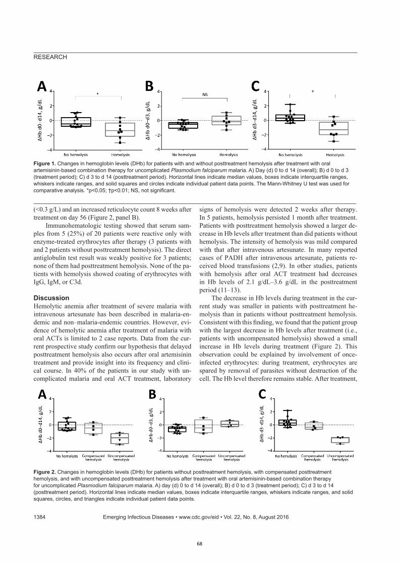
RESEARCH
(<0.3 g/L) and an increased reticulocyte count 8 weeks after treatment on day 56 (Figure 2, panel B).
Immunohematologic testing showed that serum sam-ples from 5 (25%) of 20 patients were reactive only with enzyme-treated erythrocytes after therapy (3 patients with and 2 patients without posttreatment hemolysis). The direct antiglobulin test result was weakly positive for 3 patients; none of them had posttreatment hemolysis. None of the pa-tients with hemolysis showed coating of erythrocytes with IgG, IgM, or C3d.
DiscussionHemolytic anemia after treatment of severe malaria with intravenous artesunate has been described in malaria-en-demic and non–malaria-endemic countries. However, evi-dence of hemolytic anemia after treatment of malaria with oral ACTs is limited to 2 case reports. Data from the cur-rent prospective study confirm our hypothesis that delayed posttreatment hemolysis also occurs after oral artemisinin treatment and provide insight into its frequency and clini-cal course. In 40% of the patients in our study with un-complicated malaria and oral ACT treatment, laboratory
signs of hemolysis were detected 2 weeks after therapy. In 5 patients, hemolysis persisted 1 month after treatment. Patients with posttreatment hemolysis showed a larger de-crease in Hb levels after treatment than did patients without hemolysis. The intensity of hemolysis was mild compared with that after intravenous artesunate. In many reported cases of PADH after intravenous artesunate, patients re-ceived blood transfusions (2,9). In other studies, patients with hemolysis after oral ACT treatment had decreases in Hb levels of 2.1 g/dL–3.6 g/dL in the posttreatment period (11–13).
The decrease in Hb levels during treatment in the cur-rent study was smaller in patients with posttreatment he-molysis than in patients without posttreatment hemolysis. Consistent with this finding, we found that the patient group with the largest decrease in Hb levels after treatment (i.e., patients with uncompensated hemolysis) showed a small increase in Hb levels during treatment (Figure 2). This observation could be explained by involvement of once-infected erythrocytes: during treatment, erythrocytes are spared by removal of parasites without destruction of the cell. The Hb level therefore remains stable. After treatment,
1384 EmergingInfectiousDiseases•www.cdc.gov/eid•Vol.22,No.8,August2016
Figure 1.Changesinhemoglobinlevels(DHb)forpatientswithandwithoutposttreatmenthemolysisaftertreatmentwithoralartemisinin-basedcombinationtherapyforuncomplicatedPlasmodium falciparummalaria.A)Day(d)0tod14(overall);B)d0tod3(treatmentperiod);C)d3tod14(posttreatmentperiod).Horizontallinesindicatemedianvalues,boxesindicateinterquartileranges,whiskersindicateranges,andsolidsquaresandcirclesindicateindividualpatientdatapoints.TheMann-WhitneyUtestwasusedforcomparativeanalysis.*p<0.05;†p<0.01;NS,notsignificant.
Figure 2.Changesinhemoglobinlevels(DHb)forpatientswithoutposttreatmenthemolysis,withcompensatedposttreatmenthemolysis,andwithuncompensatedposttreatmenthemolysisaftertreatmentwithoralartemisinin-basedcombinationtherapyforuncomplicatedPlasmodium falciparummalaria.A)day(d)0tod14(overall);B)d0tod3(treatmentperiod);C)d3tod14(posttreatmentperiod).Horizontallinesindicatemedianvalues,boxesindicateinterquartileranges,whiskersindicateranges,andsolidsquares,circles,andtrianglesindicateindividualpatientdatapoints.
68
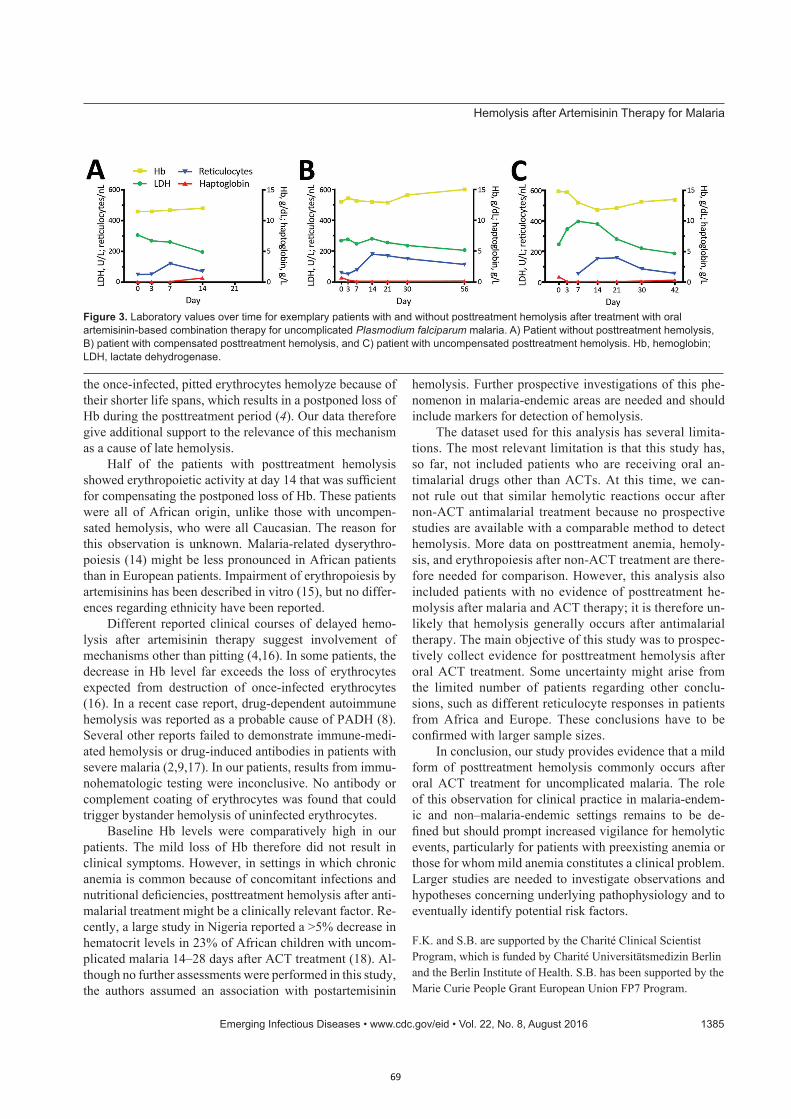
HemolysisafterArtemisininTherapyforMalaria
the once-infected, pitted erythrocytes hemolyze because of their shorter life spans, which results in a postponed loss of Hb during the posttreatment period (4). Our data therefore give additional support to the relevance of this mechanism as a cause of late hemolysis.
Half of the patients with posttreatment hemolysis showed erythropoietic activity at day 14 that was sufficient for compensating the postponed loss of Hb. These patients were all of African origin, unlike those with uncompen-sated hemolysis, who were all Caucasian. The reason for this observation is unknown. Malaria-related dyserythro-poiesis (14) might be less pronounced in African patients than in European patients. Impairment of erythropoiesis by artemisinins has been described in vitro (15), but no differ-ences regarding ethnicity have been reported.
Different reported clinical courses of delayed hemo-lysis after artemisinin therapy suggest involvement of mechanisms other than pitting (4,16). In some patients, the decrease in Hb level far exceeds the loss of erythrocytes expected from destruction of once-infected erythrocytes (16). In a recent case report, drug-dependent autoimmune hemolysis was reported as a probable cause of PADH (8). Several other reports failed to demonstrate immune-medi-ated hemolysis or drug-induced antibodies in patients with severe malaria (2,9,17). In our patients, results from immu-nohematologic testing were inconclusive. No antibody or complement coating of erythrocytes was found that could trigger bystander hemolysis of uninfected erythrocytes.
Baseline Hb levels were comparatively high in our patients. The mild loss of Hb therefore did not result in clinical symptoms. However, in settings in which chronic anemia is common because of concomitant infections and nutritional deficiencies, posttreatment hemolysis after anti-malarial treatment might be a clinically relevant factor. Re-cently, a large study in Nigeria reported a >5% decrease in hematocrit levels in 23% of African children with uncom-plicated malaria 14–28 days after ACT treatment (18). Al-though no further assessments were performed in this study, the authors assumed an association with postartemisinin
hemolysis. Further prospective investigations of this phe-nomenon in malaria-endemic areas are needed and should include markers for detection of hemolysis.
The dataset used for this analysis has several limita-tions. The most relevant limitation is that this study has, so far, not included patients who are receiving oral an-timalarial drugs other than ACTs. At this time, we can-not rule out that similar hemolytic reactions occur after non-ACT antimalarial treatment because no prospective studies are available with a comparable method to detect hemolysis. More data on posttreatment anemia, hemoly-sis, and erythropoiesis after non-ACT treatment are there-fore needed for comparison. However, this analysis also included patients with no evidence of posttreatment he-molysis after malaria and ACT therapy; it is therefore un-likely that hemolysis generally occurs after antimalarial therapy. The main objective of this study was to prospec-tively collect evidence for posttreatment hemolysis after oral ACT treatment. Some uncertainty might arise from the limited number of patients regarding other conclu-sions, such as different reticulocyte responses in patients from Africa and Europe. These conclusions have to be confirmed with larger sample sizes.
In conclusion, our study provides evidence that a mild form of posttreatment hemolysis commonly occurs after oral ACT treatment for uncomplicated malaria. The role of this observation for clinical practice in malaria-endem-ic and non–malaria-endemic settings remains to be de-fined but should prompt increased vigilance for hemolytic events, particularly for patients with preexisting anemia or those for whom mild anemia constitutes a clinical problem. Larger studies are needed to investigate observations and hypotheses concerning underlying pathophysiology and to eventually identify potential risk factors.
F.K. and S.B. are supported by the Charité Clinical Scientist Program, which is funded by Charité Universitätsmedizin Berlin and the Berlin Institute of Health. S.B. has been supported by the Marie Curie People Grant European Union FP7 Program.
EmergingInfectiousDiseases•www.cdc.gov/eid•Vol.22,No.8,August2016 1385
Figure 3.Laboratoryvaluesovertimeforexemplarypatientswithandwithoutposttreatmenthemolysisaftertreatmentwithoralartemisinin-basedcombinationtherapyforuncomplicatedPlasmodium falciparummalaria.A)Patientwithoutposttreatmenthemolysis,B)patientwithcompensatedposttreatmenthemolysis,andC)patientwithuncompensatedposttreatmenthemolysis.Hb,hemoglobin;LDH,lactatedehydrogenase.
69
RESEARCH
Dr. Kurth is a physician and scientist in the Department of Infectious Diseases and Respiratory Medicine at Charité Medical University, Berlin, Germany, and in the Infectious Disease Control Group at the Centre de Recherches Medicales de Lambaréné, Lambaréné, Gabon. His primary research interests are pathophysiology of malaria and artemisinin treatment.
References 1. World Health Organization. Guidelines for the treatment of
malaria. 3rd ed. [cited 2016 Apr 15]. http://www.who.int/malaria/publications/atoz/9789241549127/en/
2. Zoller T, Junghanss T, Kapaun A, Gjorup I, Richter J, Hugo-Persson M, et al. Intravenous artesunate for severe malaria in travelers, Europe. Emerg Infect Dis. 2011;17:771–7. http://dx.doi.org/10.3201/eid1705.101229
3. Arguin PM. Case definition: postartemisinin delayed hemolysis. Blood. 2014;124:157–8. http://dx.doi.org/10.1182/blood-2014-06-578922
4. Jauréguiberry S, Ndour PA, Roussel C, Ader F, Safeukui I, Nguyen M, et al. Postartesunate delayed hemolysis is a predictable event related to the lifesaving effect of artemisinins. Blood. 2014;124:167–75. http://dx.doi.org/10.1182/blood-2014-02-555953
5. Jauréguiberry S, Thellier M, Ndour PA, Ader F, Roussel C, Sonneville R, et al. Delayed-onset hemolytic anemia in patients with travel-associated severe malaria treated with artesunate, France, 2011–2013. Emerg Infect Dis. 2015;21:804–12. http://dx.doi.org/10.3201/eid2105.141171
6. Rolling T, Agbenyega T, Issifou S, Adegnika AA, Sylverken J, Spahlinger D, et al. Delayed hemolysis after treatment with parenteral artesunate in African children with severe malaria: a double-center prospective study. J Infect Dis. 2014;209:1921–8. http://dx.doi.org/10.1093/infdis/jit841
7. Rehman K, Lotsch F, Kremsner PG, Ramharter M. Haemolysis associated with the treatment of malaria with artemisinin derivatives: a systematic review of current evidence. International journal of infectious diseases. Int J Infect Dis. 2014;29:268–73. http://dx.doi.org/10.1016/j.ijid.2014.09.007
8. Raffray L, Receveur MC, Beguet M, Lauroua P, Pistone T, Malvy D. Severe delayed autoimmune haemolytic anaemia following artesunate administration in severe malaria: a case report. Malar J. 2014;13:398. http://dx.doi.org/10.1186/1475-2875-13-398
9. Kreeftmeijer-Vegter AR, van Genderen PJ, Visser LG, Bierman WF, Clerinx J, van Veldhuizen CK, et al. Treatment outcome of intravenous artesunate in patients with severe malaria in the
Netherlands and Belgium. Malar J. 2012;11:102. http://dx.doi.org/10.1186/1475-2875-11-102
10. World Health Organization. WHO/GMP information note on delayed haemolytic anaemia following treatment with artesunate, 2013 [cited 2016 Mar 9]. http://www.who.int/malaria/publications/atoz/who_note_delayed_haemolytic_anaemia_oct13.pdf
11. Corpolongo A, De Nardo P, Ghirga P, Gentilotti E, Bellagamba R, Tommasi C, et al. Haemolytic anaemia in an HIV-infected patient with severe falciparum malaria after treatment with oral artemether-lumefantrine. Malar J. 2012;11:91. http://dx.doi.org/10.1186/1475-2875-11-91
12. De Nardo P, Oliva A, Giancola ML, Ghirga P, Mencarini P, Bibas M, et al. Haemolytic anaemia after oral artemether- lumefantrine treatment in a patient affected by severe imported falciparum malaria. Infection. 2013;41:863–5. http://dx.doi.org/10.1007/s15010-013-0451-x
13. Gray AM, Arguin PM, Hamed K. Surveillance for the safety and effectiveness of artemether-lumefantrine in patients with uncomplicated Plasmodium falciparum malaria in the USA: a descriptive analysis. Malar J. 2015;14:349.
14. Haldar K, Mohandas N. Malaria, erythrocytic infection, and anemia. Hematology Am Soc Hematol Educ Program. 2009:87–93. http://dx.doi.org/10.1182/asheducation-2009.1.87
15. Finaurini S, Basilico N, Corbett Y, D’Alessandro S, Parapini S, Olliaro P, et al. Dihydroartemisinin inhibits the human ery-throid cell differentiation by altering the cell cycle. Toxicology. 2012;300:57–66. http://dx.doi.org/10.1016/j.tox.2012.05.024
16. Plewes K, Haider MS, Kingston HW, Yeo TW, Ghose A, Hossain MA, et al. Severe falciparum malaria treated with artesunate complicated by delayed onset haemolysis and acute kidney injury. Malar J. 2015;14:246. http://dx.doi.org/10.1186/s12936-015-0760-x
17. Rolling T, Wichmann D, Schmiedel S, Burchard GD, Kluge S, Cramer JP. Artesunate versus quinine in the treatment of severe imported malaria: comparative analysis of adverse events focussing on delayed haemolysis. Malar J. 2013;12:241. http://dx.doi.org/10.1186/1475-2875-12-241
18. Sowunmi A, Akano K, Ayede AI, Ntadom G, Fatunmbi B, Aderoyeje T, et al. Temporal changes in haematocrit following artemisinin-based combination treatments of uncomplicated falciparum malaria in children. BMC Infect Dis. 2015;15:454. http://dx.doi.org/10.1186/s12879-015-1219-y
Address for correspondence: Florian Kurth, Department of Infectious Diseases and Respiratory Medicine, Charité Campus, Virchow Klinikum, Südring 2, Augustenburger Platz 1, 13353 Berlin, Germany; email: [email protected]
1386 EmergingInfectiousDiseases•www.cdc.gov/eid•Vol.22,No.8,August2016
70
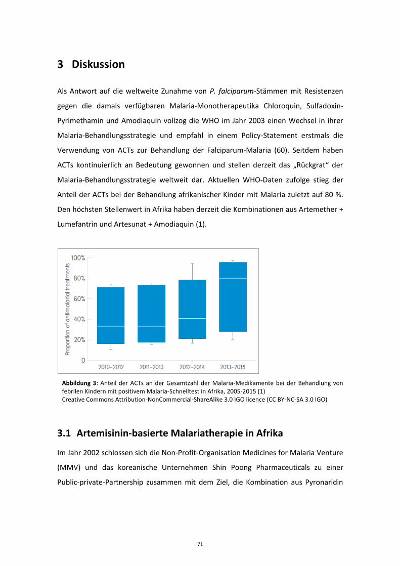
3 Diskussion
Als Antwort auf die weltweite Zunahme von P. falciparum-Stämmen mit Resistenzen
gegen die damals verfügbaren Malaria-Monotherapeutika Chloroquin, Sulfadoxin-
Pyrimethamin und Amodiaquin vollzog die WHO im Jahr 2003 einen Wechsel in ihrer
Malaria-Behandlungsstrategie und empfahl in einem Policy-Statement erstmals die
Verwendung von ACTs zur Behandlung der Falciparum-Malaria (60). Seitdem haben
ACTs kontinuierlich an Bedeutung gewonnen und stellen derzeit das „Rückgrat“ der
Malaria-Behandlungsstrategie weltweit dar. Aktuellen WHO-Daten zufolge stieg der
Anteil der ACTs bei der Behandlung afrikanischer Kinder mit Malaria zuletzt auf 80 %.
Den höchsten Stellenwert in Afrika haben derzeit die Kombinationen aus Artemether +
Lumefantrin und Artesunat + Amodiaquin (1).
Abbildung 3: Anteil der ACTs an der Gesamtzahl der Malaria-Medikamente bei der Behandlung von febrilen Kindern mit positivem Malaria-Schnelltest in Afrika, 2005-2015 (1) Creative Commons Attribution-NonCommercial-ShareAlike 3.0 IGO licence (CC BY-NC-SA 3.0 IGO)
3.1 Artemisinin-basierte Malariatherapie in Afrika
Im Jahr 2002 schlossen sich die Non-Profit-Organisation Medicines for Malaria Venture
(MMV) und das koreanische Unternehmen Shin Poong Pharmaceuticals zu einer
Public-private-Partnership zusammen mit dem Ziel, die Kombination aus Pyronaridin
71
und Artesunat (P+A) als neue fixed-dose ACT zur Behandlung der Falciparum- und
Vivax-Malaria zu testen und zur Zulassung zu bringen (61).
In der 2006 in Lambaréné an 60 Kindern durchgeführten Phase II Studie zeigten alle
verwendeten Dosierungen von P+A (6:2, 9:3, und 12:4 mg/kg) eine gute Wirksamkeit,
Verträglichkeit und Sicherheit. Insbesondere ergab sich kein Anhalt für eine Dosis-
abhängige Zunahme von unerwünschten Arzneimittelwirkungen. Die
pharmakokinetische Analyse zeigte eine lineare Dosis-Plasmaspiegel-Beziehung für die
unterschiedlichen Dosierungen (62).
Auf der Basis dieser Ergebnisse und den Daten aus einer weiteren bislang
unveröffentlichten Phase II Studie bei Erwachsenen konnte die 3 Tages P+A-
Kombinationstherapie anschließend in mehreren Phase III Studien doppelblind
randomisiert gegen A+L (63, 64) und A+MQ (65) an insgesamt 1.953 Patienten mit
Falciparum-Malaria weiter getestet werden. In allen Studien zeigte sich eine
Wirksamkeit deutlich oberhalb der von der WHO geforderten PCR-korrigierten
Mindest-Heilungsrate von 95% an Tag 28 (66). Nach einer zusätzlichen Phase IIIb/IV
Studie in West-Afrika, in der Sicherheitsbedenken bezüglich der Hepatotoxizität von
P+A bei wiederholter Anwendung ausgeräumt werden konnten, wurde P+A 2015 von
der European Medicines Agency (EMA) in Kollaboration mit der WHO für die
Verwendung außerhalb der EU zugelassen (sog. Artikel 58 Regulation) (67, 68).
Angesichts der Verfügbarkeit von 5 bereits entwickelten und zugelassenen ACTs - im
Jahr 2006 waren es noch 4 – stellt sich die Frage nach der Sinnhaftigkeit der
Entwicklung einer weiteren ACT. Im Falle der P+A-Kombinationstherapie waren die
vorrangigen Gründe hierfür das einfache Dosierungs-Schema (3 Dosen bei P+A
gegenüber 6 Dosen bei A+L), die Unabhängigkeit der Pyronaridin-Bioverfügbarkeit von
gleichzeitiger Nahrungsaufnahme (im Gegensatz zu den lipophilen Wirkstoffen
Lumefantrin oder Piperaquin), die lange Lagerbarkeit von 2 Jahren und der günstige
angestrebte Preis (unter 1$ für ein Erwachsenen-Dreitagesregimen, erzielt durch
niedrige Produktionskosten für Pyronaridin und durch die Entwicklung im Rahmen des
72
Public-private-Partnerships) (61). Aus heutiger Sicht kommt ein weiterer wesentlicher
Punkt hinzu: Durch die Abnahme der Sensitivität von P. falciparum gegenüber den
Artemisinin-derivaten in der Mekong-Subregion steigt in diesen Gebieten der
Selektionsdruck für Resistenzen gegen die Partner-Medikamente (69). Gleichzeitig ist
bei Artemisinin-resistenten Plasmodien eine effektive medikamentöse Elimination
durch die Partner-Medikamente im Hinblick auf die individuelle Heilung und die
Unterbindung einer Transmission dieser Plasmodien-Stämme besonders wichtig (13,
14). Auch in Afrika hat die Wirksamkeit einiger ACT Partner-Medikamente mit
zunehmender Verwendung in den letzten Jahren kontinuierlich abgenommen. Für A+L
wurden in mehreren Studien bei Kindern aus Nord-Angola PCR-korrigierte
Heilungsraten an Tag 28 von unter 90% berichtet. In den Plasmodien-Isolaten der
Patienten mit Rekrudeszenzen nach A+L Therapie fanden sich gehäuft molekulare
Marker, die mit einer Resistenz gegenüber Lumefantrin in Verbindung stehen (70, 71).
In dieser Situation kann die Verfügbarkeit von unterschiedlichen ACT-Partner-
Medikamenten von entscheidendem Vorteil sein, besonders wenn diese einen
gegenläufigen Selektions-Druck auf P. falciparum ausüben. Untersuchungen der
Polymorphismen im P. falciparum multidrug resistance gen 1 (pfmdr1) zeigen, dass
durch die Behandlung mit A+L die Polymorphismen N86 und D1246 selektioniert
werden, die mit einer verminderten Lumefantrin-Sensitivität assoziiert sind. Eine
Behandlung mit DHA+P scheint hingegen die entgegengesetzten Polymorphismen 86Y
und 1246Y zu selektionieren (72-74). Diese Daten legen nahe, dass eine
Behandlungspolitik mit alternierendem oder parallelem Einsatz verschiedener ACTs
der Entstehung von Resistenzen vorbeugen könnte (75). Für derartige
Rotationsprinzipien ist die Verfügbarkeit von P+A als einer weiteren ACT mit
unterschiedlichem Kombinationspartner von Vorteil.
Parallel zur klinischen Entwicklung von P+A in Phase II und Phase III wurde in
Lambaréné 2006 eine in vitro Sensitivitäts-Testung von P. falciparum-Feldisolaten
gegenüber Pyronaridin durchgeführt (76). In vitro Testungen von
Malariamedikamenten erfüllen im Allgemeinen zwei Ziele: 1. die Wirksamkeit neuer
73
Wirkstoffe an bekannten Plasmodium-Laborstämmen oder -Feldisolaten zu prüfen (in
Ergänzung zur Testung in klinischen Studien), 2. durch regelmäßige Testungen von
Feldisolaten die Resistenz-Entwicklung in Endemiegebieten zu überwachen. Im Fall von
Pyronaridin erschien eine konsequente Sensitivitätstestung vor allem wichtig, weil sich
unter der Verwendung von Pyronaridin als Monotherapie in China in den 1980er
Jahren rasch Resistenzen entwickelt hatten (77). Zudem gab es Anhalt für
Kreuzresistenzen mit Amodiaquin und Chloroquin (78, 79).
Die in der Studie ermittelten EC-50 Werte für Pyronaridin von 1,9 nmol/l (95 % KI 1,4-
2,5) im WHO Test und 2.0 nmol/l (95 % KI 1,6-2,6) im HRP-2 Assay waren vergleichbar
zu den vorbeschriebenen Daten von Laborstämmen (80) und anderen afrikanischen
P. falciparum-Feldisolaten (78, 79). Alle getesteten Feldisolate wiesen eine schlechte
Sensitivität gegenüber Chloroquin auf. Hinweise für eine Kreuz-Sensitivität zwischen
Pyronaridin und Chloroquin ergaben sich im Gegensatz zu früheren Berichten nicht
(78, 79).
Der logistische und finanzielle Aufwand für ex-vivo Sensitivitätsuntersuchungen von
P. falciparum ist gegenüber der Testung in klinischen Studien deutlich geringer. In
kurzer Zeit kann eine große Anzahl von Feldisolaten getestet werden, auch können
Wirkstoffe unabhängig von variablen patientenbezogenen Faktoren wie Compliance
bei der Medikamenteneinnahme, Semi-Immunität oder Pharmakokinetik beurteilt
werden. Andererseits sind die in vitro ermittelten Werte aufgrund eben dieser
Faktoren in der Regel nur sehr eingeschränkt auf die Situation in vivo übertragbar (81).
In der aktuellen Arbeit lag die Cut-off-Konzentration im geometrischen Mittel für
Pyronaridin im WHO-Test bei 9,3 nmol/l (95 % KI 5,2-13,9), die korrespondierende
99 %-effektive Konzentration im HRP-2 Assay war 9,0 nmol/l (95 % KI 6,4-12,7). Im
Vergleich dazu lagen die bei den Patienten in der klinischen Phase II Studie bestimmen
mittlere maximale Plasmakonzentrationen von Pyronaridin bei 115 nmol/l, also mehr
als 10-fach über der ermittelten in vitro Cut-off-Konzentration (62). Die frühesten
Rekrudeszenzen in der klinischen Studie traten an Tag 21 und Tag 28 auf, wobei der
erste Patient mit Rekrudeszenz an Tag 21 an diesem Tag eine Pyronaridin
Plasmakonzentration von 9,9 nmol/l aufwies. In einer Regressionsanalyse der
74
gepoolten Pyronaridin Plasmakonzentrationen aller Patienten aus der klinischen Studie
fielen diese im Mittel an Tag 22 unter die Schwelle von 9 nmol/l. Die in vitro
ermittelten Pyronaridin Cut-off-Werte korrespondierten also in diesem Fall
ausnehmend gut mit den Pyronaridin Plasmakonzentrationen zum Zeitpunkt der
Rekrudeszenzen in der klinischen Studie (62, 76).
Im Rahmen der klinischen Testung von P+A wurde - erstmalig für eine ACT - von Beginn
des Entwicklungsprogrammes an eine spezielle pädiatrische Darreichungsform in Form
von Geschmacks-maskierten Granulen mitentwickelt. Ziel dieser „non-tablet“
Darreichungsformen ist es, die Medikamentenverabreichung, Dosierbarkeit und
Verträglichkeit bei kleinen Kindern, der Haupt-Patientengruppe in Endemiegebieten,
zu verbessern (62, 82).
Für kleine Kinder ist es oft schwierig, Tabletten im Ganzen zu schlucken. Probleme bei
der Medikamenten-Verabreichung und arzneimittelinduziertes Erbrechen sind bei
afrikanischen Kindern mit Malaria die häufigsten Gründe für eine Hospitalisierung,
auch ohne dass eine schwere Malaria besteht (83, 84). Die weit verbreitete Praxis,
Tabletten zu mörsern oder in kleine Stücke zu zermahlen, kann zu Wirkstoffverlust und
Dosierungsfehlern führen, auch kann der bittere Geschmack vieler Malaria-
Medikamente dadurch noch verstärkt werden (85, 86). Gerade bei der Therapie der
unkomplizierten Malaria mit ACTs, bei denen Fieber und Krankheitsgefühl oft schon
nach der ersten oder zweiten Medikamenten-Einnahme rasch verschwinden, können
die Probleme bei der Medikamentengabe zu einer schlechteren Compliance führen,
sodass beispielsweise die Medikamente am dritten Behandlungstag nicht mehr
eingenommen werden. Dies wiederum gefährdet die Wirksamkeit der Medikamente
und fördert die Entstehung von Resistenzen (85).
Im Rahmen von klinischen Studien, in denen die Medikamente meist unter Aufsicht
verabreicht und bei Erbrechen konsequent erneut gegeben werden, sind diese
Probleme oft nicht sichtbar. Erst in Wirksamkeitsstudien unter realen Feldbedingungen
(engl. effectiveness-trials) ohne Überwachung der Medikamenten-Verabreichung
zeigen sich oft deutlich schlechtere Heilungsraten. So lag die PCR-korrigierte
75
Heilungsrate für A+AQ an Tag 28 bei nicht-überwachter Medikamentengabe bei
Kindern mit unkomplizierter Malaria in Gabun bei nur 63% (95 % KI 45-77%) und somit
23 % unter der Heilungsrate in der Vergleichsgruppe mit überwachter
Medikamentengabe (86 %, 95 % KI 69-95 %) (87). Bei Kindern unter 5 Jahren sind die
Heilungsraten bei nicht-überwachter ACT nochmals geringer als bei älteren Patienten
(88), was auf die besonderen Probleme bei der Therapie in dieser Patientengruppe
hindeutet.
Die P+A-Granulen zeigten in der Phase II Studie bezüglich Sicherheit, Verträglichkeit
und Bioverfügbarkeit vergleichbare Ergebnisse wie bei entsprechender Tabletten-
Dosierung (62). Auch die neu-entwickelte pädiatrische Darreichungsform von A+L, in
Form von Kirschgeschmacks-maskierten dispergierbaren Tabletten, zeigte in einer
Nicht-Unterlegenheitsstudie an 899 afrikanischen Kindern mit Malaria vergleichbar
gute Heilungsraten wie die entsprechende Tabletten-Darreichungsform (89). Die
ursprüngliche Rationale für die Entwicklung pädiatrischer Darreichungsformen, eine
Verbesserung der Medikamenten-Verabreichung, konnte jedoch in keiner der
Einzelstudien belegt werden.
In einem systematischen Review konnten 2009 insgesamt sieben klinische Studien
identifiziert werden, in denen pädiatrische „non-tablet“ ACTs mit konventionellen
ACTs in Tablettenform verglichen wurden (84). Wie in den Einzelstudien zeigte sich
auch in der Meta-Analyse der sieben Studien eine vergleichbare Wirksamkeit und
Sicherheit von pädiatrischen und konventionellen Darreichungsformen. Bei der
Analyse der Verträglichkeit der Medikamentenverabreichung konnte jedoch ein klarer
Vorteil für die pädiatrischen Darreichungsformen festgestellt werden: Die Häufigkeit
von arzneimittelinduziertem Erbrechen war bei den Patienten, die mit „non-tablet“
Darreichungsformen behandelt wurden, 4,5 % niedriger als bei Patienten in der
Vergleichsgruppe (Relatives Risiko 0,78; 95 % KI 0,61-0,99). Auch das Risiko für
unerwünschte gastrointestinale Arzneimittelwirkungen war in der „non-tablet“ Gruppe
statistisch signifikant verringert. Auf Grundlage dieser Daten müsste man
durchschnittlich 22 Patienten mit pädiatrischen ACTs anstelle von konventionellen
76
Tabletten behandeln, um eine Episode von arzneimittelinduziertem Erbrechen zu
verhindern. Einschränkend muss gesagt werden, dass in dieser Meta-Analyse auch
nicht-randomisierte Studien eingeschlossen wurden, sowie Studien die pädiatrische
ACTs mit nicht-wirkstoffgleichen konventionellen ACTs verglichen, sodass die
Unterschiede in der Verträglichkeit außer durch die Darreichungsform auch durch die
unterschiedlichen Wirkstoffe mitbeeinflusst sein kann. Allerdings war die
Überlegenheit der „non-tablet“ ACTs konsistent für alle untersuchten pädiatrischen
ACTs, für alle geographischen Studienregionen in Afrika und Asien sowie für
randomisierte und nicht-randomisierte Studien.
Pädiatrische ACTs finden mittlerweile in Afrika breite Anwendung. In einer Umfrage
unter 71 Angehörigen von Gesundheitsberufen in sieben afrikanischen Ländern im Jahr
2011 gaben 83 % der Befragten an, pädiatrische Darreichungsformen als
Erstlinientherapie bei jungen Kindern mit Malaria zu verwenden (90). In der aktuellen
dritten Auflage der WHO Leitlinie zur Malariatherapie werden pädiatrische ACTs
erstmals klar als bevorzugte Behandlungsoption für Kinder mit unkomplizierter Malaria
empfohlen (14). Ob es durch pädiatrische ACTs tatsächlich zu einer verbesserten
Heilungsrate sowie Verringerung von Behandlungs-Komplikationen und
Hospitalisierungen bei nicht-überwachter Therapie kommt, sollte abschließend in
randomisiert-kontrollierten effectiveness-trials geprüft werden.
3.2 Artemisinin-basierte Malariatherapie in Europa
Die importierte Malaria in Nicht-Endemiegebieten wie Europa unterscheidet sich in
vielen Aspekten deutlich von der Malaria in Endemiegebieten. Wie die Auswertung der
8-Jahres Beobachtungsdaten der TropNet-Studie zur schweren Malaria in Europa zeigt,
haben sowohl europäische Touristen als auch VFRs ein erhebliches Risiko, an einer
schweren Malaria zu erkranken (22, 91). Ursache hierfür ist das Fehlen (bei Touristen)
bzw. der Verlust (bei VFRs) der Semi-Immunität gegenüber Erreger und Krankheit. Das
Risiko für kaukasische Touristen ist dabei höher als für VFRs, was auf ein gewisses Maß
an Rest-Schutz bei den VFRs hindeutet (92). Auch genetische Faktoren spielen
möglicherweise eine Rolle (93).
77
Die Altersstruktur der Patienten mit importierter Malaria (medianes Alter in der
Tropnet Studie 42 Jahre) unterscheidet sich ebenfalls deutlich von der in
Endemiegebieten, wo überwiegend junge Kinder betroffen sind (53). Durch das höhere
Alter liegen bei Patienten mit importierter Malaria häufig Komorbiditäten wie
Hypertonie oder Diabetes mellitus vor. Die aktuelle Arbeit lässt zudem darauf
schließen, dass aufgrund des höheren Alters Komplikationen wie akutes
Nierenversagen, und ZNS-Symptome häufiger auftreten (91). Diese Ergebnisse stehen
in Einklang mit früheren Studien bei europäischen Reisenden mit importierter Malaria
(94).
Eine Hyperparasitämie ≥5 % tritt bei europäischen Patienten in der aktuellen TropNet-
Studie ebenfalls deutlich häufiger auf als bei Patienten mit schwerer Malaria in
Endemiegebieten, bei denen Azidose, Koma und Anämie die häufigsten Merkmale
einer schweren Infektion sind (52, 53). Dies ist relevant, da in den randomisiert
kontrollierten Studien zum Vergleich von i.v. Artesunat und i.v. Chinin in Afrika und
Asien die Subgruppen der Patienten mit Hyperparasitämie besonders von einer
Artesunat-Therapie profitiert hatten (OR für letalen Verlauf unter Artesunat-Therapie
für Patienten mit Hyperparasitämie >10 % in Afrika 0,67; 95 % KI 0,47-0,97 gegenüber
0,84; 95 % KI 0,66-1,06 für Patienten ohne Hyperparasitämie bzw. 0,75;
95 % KI 0,63-0,90 in der Gesamtpopulation (53)). Diese Überlegenheit wird vor allem
auf die rasche Parasiten-Elimination der Artemisinine zurückgeführt, die mit der
vergleichsweise guten Wirksamkeit gegen jüngere Plasmodium-Stadien in
Zusammenhang steht (13, 95, 96).
Gerade in Europa wurde der Übergang zu Artesunat als Erstlinientherapie der
komplizierten Malaria jedoch deutlich verlangsamt, da wenige Behandlungsdaten von
Patienten in Nicht-Endemiegebieten vorliegen, und da in Europa kein zugelassenes,
GMP-konform produziertes Artesunat-Präparat auf dem Mark erhältlich ist. Die
TropNet-Daten belegen, dass der Anteil der Patienten mit schwerer Malaria in Europa,
die primär mit Artesunat therapiert wurden, in den Jahren zwischen 2006 und 2014
kontinuierlich von 27 % auf 60 % zunahm. Zudem zeigen sie, dass auch europäische
78
Patienten mit schwerer Malaria zweifelsfrei von einer Behandlung mit Artesunat
gegenüber Chinin profitieren: zwar ließ sich unter anderem aufgrund der niedrigen
Letalitätsrate in der europäischen Population (1,6 %) kein Überlebensvorteil für die
Therapie mit Artesunat beweisen. Die Patienten mit Artesunat-Therapie boten jedoch
eine raschere Parasiten-Clearance als die Patienten mit Chinin-Therapie, benötigten in
der Folge eine um einen Tag kürzere intensivmedizinische Behandlung und konnten
einen Tag früher aus der Klinik entlassen werden. Auch in der TropNet-Studie
profitierten die Patienten mit Hyperparasitämie ≥5 % deutlicher von einer Artesunat-
Therapie als Patienten ohne Hyperparasitämie (97). Auch wenn es sich hier um
retrospektive Daten handelt, können die Ergebnisse dieser multizentrischen Studie in
Zukunft helfen, die Entscheidungsfindung für eine Artesunat-Therapie für Kliniker zu
erleichtern, zumal prospektive randomisierte Studien in Europa angesichts der
bestehenden Evidenz aus Asien und Afrika nicht mehr durchführbar sein werden.
Die aktuelle Arbeit belegt auch die Sicherheit und gute Verträglichkeit der Artesunat-
Therapie bei europäischen Patienten. Ähnlich wie in den Studien in Afrika und Asien
traten keine unerwarteten schwerwiegenden unerwünschten Ereignisse mit Bezug zur
Artesunat-Therapie auf. Besondere Aufmerksamkeit verdient allerdings das Phänomen
der verzögerten Hämolyse nach Artemisinin-Gabe (engl. post-artemisinin delayed
haemolysis – PADH), das in der aktuellen Arbeit bei 27 % der Patienten mit
Artemisinin-Therapie beobachtet wurde. Hierbei kommt es in den Wochen nach der
Therapie und nach vollständiger Elimination der Plasmodien aus dem Blut zu einem
verzögerten Hb-Abfall mit Hämolysezeichen (LDH Erhöhung, Erniedrigung des freien
Haptoglobins). PADH wurde erstmals 2002 bei einem nicht-semiimmunen Patienten in
Japan nach i.v. Artesunat Therapie beschrieben und 2011 in einer Fallserie bei nicht-
semiimmunen Patienten in Europa (98). Seitdem wurde das Phänomen in mehreren
Zentren in Europa (99-103), den USA (104) und 2014 auch bei Kindern in Afrika
beobachtet (105). Die Häufigkeit der PADH bei nicht-semiimmunen Patienten war
dabei in etwa gleich zur Inzidenz der PADH in der aktuellen TropNet-Studie (106). Bei
den Patienten in Afrika trat PADH seltener auf (7% Inzidenz bei Kindern in Afrika), ein
Kind in der afrikanischen Beobachtungsstudie bot im Rahmen der PADH jedoch einen
79
Hb-Abfall auf 2,8 g/dl (105). Die pathophysiologischen Hintergründe der PADH sind
derzeit noch nicht vollständig aufgeklärt. In einer Studie bei Reiserückkehrern in
Frankreich wurde ein Zusammenhang zwischen dem Auftreten von PADH und einer
erhöhten Anzahl von ehemals mit Plasmodien infizierten Erythrozyten (engl. once
infected erythrocytes, oi-E) beschrieben (101). Die Konzentration dieser Zellen im Blut
der Behandelten ist nach Artemisinin-Therapie vergleichsweise höher als nach
Therapie mit anderen Malaria-Medikamenten. Die Autoren postulieren, dass PADH
durch eine verkürzte Lebensspanne und Zerstörung dieser oi-E in den Wochen nach
Therapie verursacht wird (101).
Die aktuelle Beobachtungsstudie bei Reiserückkehrern an der Charité zeigt, dass es
auch nach unkomplizierter Malaria und oraler Artemisinin-basierter Therapie zu
verzögerten hämolytischen Reaktionen bei den Behandelten kommt. Bei 40 % der
Patienten nach unkomplizierter Malaria und oraler ACT bestanden an Tag 14 nach
Therapie Hämolysezeichen (LDH-Erhöhung, Erniedrigung des freien Haptoglobins). Bei
einem Großteil dieser Patienten wurden die Hämolysezeichen auch noch 1 Monat nach
Therapie festgestellt. Zu einem Hb-Abfall nach Therapie kam es allerdings nur bei der
Hälfte der Patienten mit Hämolyse, bei den übrigen Patienten wurde der Hb-Verlust
durch die gesteigerte Erythropoese nach Malaria vollständig kompensiert. Auffallend
ist dabei die Tatsache, dass alle Patienten mit unkompensierter Hämolyse kaukasisch
waren, alle Patienten mit kompensierter Hämolyse hingegen afrikanisch. Dies könnte
auf eine unterschiedliche Beeinträchtigung der kompensatorischen Erythropoese in
Abhängigkeit von Semi-Immunität bzw. Herkunft hindeuten (107). Die Patienten mit
verzögerter Hämolyse und Hb-Abfall nach der Therapie hatten während der Therapie
weniger Hb-Abfall als die Patienten ohne verzögerte Hämolyse. Dies deutet auf die
Rolle der oi-E hin, durch die ein Hb-Verlust während der Infektion und Therapie
vermieden und in die Phase nach der akuten Infektion verlagert werden könnte. Für
Patienten, bei denen der Hb-Verlust in der Phase nach der Behandlung wie
beschrieben bereits durch gesteigerte Erythropoese kompensiert wird, könnte die
Verzögerung des Hb-Verlustes also von Vorteil sein. Andererseits bieten die Daten
Anhalt dafür, dass der verzögerte Hb-Verlust nach Therapie den Hb-Verlust durch den
80
Abbau von oi-Es übersteigt, sodass die verzögerte Hämolyse insgesamt zu einem
vermehrten Hb-Verlust führen könnte (107).
Klinische Hämolyse-Zeichen bzw. eine relevante Beeinträchtigung durch Anämie
fanden sich in der aktuelle Arbeit bei keinem der Patienten. Dennoch lassen die Daten
vermuten, dass verzögerte hämolytische Reaktionen nach oraler ACT bei
unkomplizierter Malaria insgesamt zur Malaria-assoziierten Anämie beitragen.
Relevanz hat dies möglicherweise für Patienten in Endemiegebieten, die aufgrund
höherer Malaria-Infektionsraten, Ko-Infektionen und Mangelernährung vermehrt
unter chronischer Anämie leiden (108). Die Bedeutung von verzögerten hämolytischen
Reaktionen nach Artemisinin-basierter Malariatherapie sollte daher in Malaria-
Endemiegebieten bei Patienten mit höherer Prävalenz von chronischer Anämie weiter
untersucht werden.
81
4 Zusammenfassung und Ausblick
Die Malaria zählt nach wie vor zu den wichtigsten Infektionskrankheiten. Eine rasche
und effektive medikamentöse Therapie ist von entscheidender Bedeutung. ACTs sind
die empfohlene Erstlinien-Therapie bei unkomplizierter Malaria in allen Malaria-
Endemiegebieten. Bei schwerer Malaria führt die parenterale Artesunat-Therapie zu
einem Überlebensvorteil gegenüber Chinin. Insgesamt haben die Artemisinine in den
letzten 15 Jahren einen entscheidenden Beitrag zur Reduktion der Malaria-bedingten
Morbidität und Mortalität weltweit geleistet.
Seit 2002 wird die Kombination aus Pyronaridin und Artesunat als neue ACT klinisch
getestet. In der Phase II Studie zeigte P+A eine gute Wirksamkeit, Verträglichkeit und
Sicherheit. Auch in den in vitro Testungen von P. falciparum-Feldisolaten war
Pyronaridin gut wirksam. Nach mehreren Phase III Studien wurde P+A 2015 von der
EMA zugelassen. Eine Besonderheit von P+A ist die Verfügbarkeit einer pädiatrischen
non-Tablet Darreichungsform. Durch diese Darreichungsformen kann Medikamenten-
induziertes Erbrechen bei Kindern mit Malaria reduziert werden.
In Europa betrifft die Malaria Touristen und ehemalige Migranten, die zu ihren
Familienangehörigen in Endemiegebiete reisen. Durch die fehlende bzw. abnehmende
Semi-Immunität haben diese Patienten ein deutlich erhöhtes Risiko an einer schweren
Malaria zu erkranken. Die Etablierung von i.v. Artesunat zur Therapie der schweren
Malaria war in Europa ob des Fehlens eines GMP-konform produzierten Artesunat-
Präparates verlangsamt. Die TropNet Studie zur schweren Malaria in Europa belegt,
dass auch europäische Patienten zweifelsfrei von einer Artesunat-Therapie gegenüber
Chinin profitieren. Parenterales Artesunat ist bei europäischen Patienten mit Malaria
sicher und gut verträglich, führt jedoch bei einem Teil der Patienten zu verzögerten
hämolytischen Reaktionen in den Wochen nach Therapie. In einer Studie mit Malaria-
Patienten in Berlin wurde gezeigt, dass verzögerte hämolytischen Reaktionen in
milderer Form auch bei Patienten mit unkomplizierter Malaria nach oraler ACT
vorkommen.
82
Seit einigen Jahren ist die Möglichkeit einer weltweiten Malaria-Eradikation
zunehmend in den Fokus der Forschung gerückt. Nach einer mehr als 30 Jahre
dauernden Entwicklung wurde 2015 mit RTS,S der erste Impfstoff gegen Plasmodium
falciparum zugelassen. Aufgrund der geringen Wirksamkeit vor allem bei Kindern unter
drei Monaten, wird dieser jedoch derzeit von der WHO nicht empfohlen. Daten aus
Pilot-Implementierungsprogrammen werden in den kommenden Jahren zeigen,
inwiefern RTS,S einen Beitrag zur Malaria Elimination leisten kann. Durch den raschen
Wissenszuwachs in der biomedizinischen Forschung werden zukünftig neue
Generationen von verbesserten Impfstoffen, beispielsweise attenuierte Sporozoiten-
Vakzine, an Bedeutung gewinnen.
Durch den Rückgang der Transmissions-Intensität in vielen Malaria-Endemiegebieten
und der damit verbundenen Abnahme der Semi-Immunität ändert sich die klinische
Präsentation und Altersverteilung der Patienten mit Malaria in diesen Gebieten.
Zusätzliche epidemiologische und immunologische Erkenntnisse werden in Zukunft
notwendig sein, um die Malaria-Kontrollprogramme für diese Veränderungen zu
rüsten. Immunologische Untersuchungen von nicht-semiimmunen Patienten wie
Touristen oder Migranten können hierzu einen Beitrag leisten.
Die Entstehung von P. falciparum-Stämmen mit reduzierter Artemisinin-Sensitivität im
Westen Kambodschas und in der thailändisch-myanmarischen Grenzregion stellt
derzeit die größte Bedrohung für die Malaria-Bekämpfung dar. Bei Parasiten-Stämmen
mit zusätzlicher Resistenz gegen die Kombinations-Partnermedikamente kommt es zu
hohen Raten von ACT-Therapieversagen. Neben der rigorosen Bekämpfung einer
Verbreitung Artemisinin-resistenter Plasmodien wird die klinische Entwicklung neuer
Substanzklassen wie Spiroindolone und Imidazolopiperazine in Zukunft eine
entscheidende Rolle spielen.
83
5 Literatur
1. World Health Organization. World Malaria Report 2016. Geneva, 2016.
2. Global Burden of Disease contributors. Mortality Causes of Death Collaborators: Global, regional, and national life expectancy, all-cause mortality, and cause-specific mortality for 249 causes of death, 1980-2015: a systematic analysis for the Global Burden of Disease Study 2015. Lancet. 2016;388(10053):1459-544.
3. Tjitra E, Anstey NM, Sugiarto P, Warikar N, Kenangalem E, Karyana M, et al. Multidrug-resistant Plasmodium vivax associated with severe and fatal malaria: a prospective study in Papua, Indonesia. PLoS Med. 2008;5(6):e128.
4. Sutherland CJ, Tanomsing N, Nolder D, Oguike M, Jennison C, Pukrittayakamee S, et al. Two nonrecombining sympatric forms of the human malaria parasite Plasmodium ovale occur globally. J Infect Dis. 2010;201(10):1544-50.
5. Groger M, Fischer HS, Veletzky L, Lalremruata A, Ramharter M. A systematic review of the clinical presentation, treatment and relapse characteristics of human Plasmodium ovale malaria. Malar J. 2017;16(1):112.
6. White NJ, Pukrittayakamee S, Hien TT, Faiz MA, Mokuolu OA, Dondorp AM. Malaria. Lancet. 2014;383(9918):723-35.
7. Singh B, Kim Sung L, Matusop A, Radhakrishnan A, Shamsul SS, Cox-Singh J, et al. A large focus of naturally acquired Plasmodium knowlesi infections in human beings. Lancet. 2004;363(9414):1017-24.
8. Warrel DA, Gilles HM. Essential Malariology. 4th Edition ed. London: Wiley; 2002.
9. White NJ, Breman JG. Malaria. In: Suttorp N, Möckel M, Siegmund B, Dietel M, editors. Harrisons Innere Medizin. Berlin: ABW Wissenschaftsverlag; 2016. p. 1674-92.
10. Richter J, Franken G, Mehlhorn H, Labisch A, Haussinger D. What is the evidence for the existence of Plasmodium ovale hypnozoites? Parasitol Res. 2010;107(6):1285-90.
11. Markus MB. Do hypnozoites cause relapse in malaria? Trends Parasitol. 2015;31(6):239-45.
12. Trager W, Jensen JB. Human malaria parasites in continuous culture. Science. 1976;193(4254):673-5.
13. White NJ. Malaria parasite clearance. Malar J. 2017;16(1):88.
84
14. World Health Organization. Guidelines for the treatment of malaria - 3rd edition. Geneva 2015.
15. Douglas NM, Lampah DA, Kenangalem E, Simpson JA, Poespoprodjo JR, Sugiarto P, et al. Major burden of severe anemia from non-falciparum malaria species in Southern Papua: a hospital-based surveillance study. PLoS Med. 2013;10(12):e1001575; discussion e.
16. Miller LH, Good MF, Milon G. Malaria pathogenesis. Science. 1994;264(5167):1878-83.
17. Rosenthal PJ. Artesunate for the treatment of severe falciparum malaria. N Engl J Med. 2008;358(17):1829-36.
18. Magowan C, Wollish W, Anderson L, Leech J. Cytoadherence by Plasmodium falciparum-infected erythrocytes is correlated with the expression of a family of variable proteins on infected erythrocytes. J Exp Med. 1988;168(4):1307-20.
19. Kremsner PG, Adegnika AA, Hounkpatin AB, Zinsou JF, Taylor TE, Chimalizeni Y, et al. Intramuscular Artesunate for Severe Malaria in African Children: A Multicenter Randomized Controlled Trial. PLoS Med. 2016;13(1):e1001938.
20. Deutsche Gesellschaft für Tropenmedizin und Internationale Gesundheit. Leitlinie: Diagnostik und therapie der Malaria [Verfügbar unter http://www.awmf.org/leitlinien/detail/ll/042-001.html. Zugriff am 5.4.2017]
21. Rapport annuel d'activité du Centre national de référende du Paludisme 2015, France [Verfügbar unter https://ssl.voozanoo.net/palu/rapport.activites.2014.pdf. Zugriff am 1.4.2017]
22. Argy N, Kendjo E, Auge-Courtoi C, Cojean S, Clain J, Houze P, et al. Influence of host factors and parasite biomass on the severity of imported Plasmodium falciparum malaria. PLoS One. 2017;12(4):e0175328.
23. World Health Organization. Guidelines for the treatment of malaria - 2nd edition. Geneva 2010.
24. World Health Organization. Guidelines for the treatment of malaria. Geneva 2006.
25. MacDonald G. The Epidemiology and Control of Malaria. London: Oxford University Press; 1957.
26. Gething PW, Patil AP, Smith DL, Guerra CA, Elyazar IR, Johnston GL, et al. A new world malaria map: Plasmodium falciparum endemicity in 2010. Malar J. 2011;10:378.
85
27. Bhatt S, Weiss DJ, Cameron E, Bisanzio D, Mappin B, Dalrymple U, et al. The effect of malaria control on Plasmodium falciparum in Africa between 2000 and 2015. Nature. 2015;526(7572):207-11.
28. Gupta S, Snow RW, Donnelly CA, Marsh K, Newbold C. Immunity to non-cerebral severe malaria is acquired after one or two infections. Nat Med. 1999;5(3):340-3.
29. Alonso D, Bouma MJ, Pascual M. Epidemic malaria and warmer temperatures in recent decades in an East African highland. Proc Biol Sci. 2011;278(1712):1661-9.
30. McCutchan TF, Grim KC, Li J, Weiss W, Rathore D, Sullivan M, et al. Measuring the effects of an ever-changing environment on malaria control. Infect Immun. 2004;72(4):2248-53.
31. Gething PW, Casey DC, Weiss DJ, Bisanzio D, Bhatt S, Cameron E, et al. Mapping Plasmodium falciparum Mortality in Africa between 1990 and 2015. N Engl J Med. 2016;375(25):2435-45.
32. The Malaria Atlas Project [Verfügbar unter: http://www.map.ox.ac.uk/ Zugriff am 5.5.2017].
33. European Centre for Disease Prevention and Control. Annual Epidemiological Report 2016 – Malaria. Stockholm, 2016. [Verfügbar unter: http://ecdc.europa.eu/en/healthtopics/malaria/Pages/Annual-epidemiological-report-2016.aspx. Zugriff am 1.5.2017]
34. de Gier B, Suryapranata FS, Croughs M, van Genderen PJ, Keuter M, Visser LG, et al. Increase in imported malaria in the Netherlands in asylum seekers and VFR travellers. Malar J. 2017;16(1):60.
35. Roggelin L, Tappe D, Noack B, Addo MM, Tannich E, Rothe C. Sharp increase of imported Plasmodium vivax malaria seen in migrants from Eritrea in Hamburg, Germany. Malar J. 2016;15:325.
36. Danis K, Baka A, Lenglet A, Van Bortel W, Terzaki I, Tseroni M, et al. Autochthonous Plasmodium vivax malaria in Greece, 2011. Euro Surveill. 2011;16(42).
37. Kremsner PG, Krishna S. Antimalarial combinations. Lancet. 2004;364(9430):285-94.
38. White NJ, Nosten F, Looareesuwan S, Watkins WM, Marsh K, Snow RW, et al. Averting a malaria disaster. Lancet. 1999;353(9168):1965-7.
39. Gerstl S, Namagana A, Palacios L, Mweshi F, Aprile S, Lima A. High adherence to malaria treatment: promising results of an adherence study in South Kivu, Democratic Republic of the Congo. Malar J. 2015;14:414.
86
40. Sinclair D, Zani B, Donegan S, Olliaro P, Garner P. Artemisinin-based combination therapy for treating uncomplicated malaria. Cochrane Database Syst Rev. 2009(3):CD007483.
41. Karbwang J, White NJ. Clinical pharmacokinetics of mefloquine. Clin Pharmacokinet. 1990;19(4):264-79.
42. Chotsiri P, Wattanakul T, Hoglund R, Hanboonkunupakarn B, Pukrittayakamee S, Blessborn D, et al. Population pharmacokinetics and electrocardiographic effects of dihydroartemisinin-piperaquine in healthy volunteers. Br J Clin Pharmacol. 2017.
43. White NJ, van Vugt M, Ezzet F. Clinical pharmacokinetics and pharmacodynamics and pharmacodynamics of artemether-lumefantrine. Clin Pharmacokinet. 1999;37(2):105-25.
44. Barnes KI, Little F, Smith PJ, Evans A, Watkins WM, White NJ. Sulfadoxine-pyrimethamine pharmacokinetics in malaria: pediatric dosing implications. Clin Pharmacol Ther. 2006;80(6):582-96.
45. Price RN, von Seidlein L, Valecha N, Nosten F, Baird JK, White NJ. Global extent of chloroquine-resistant Plasmodium vivax: a systematic review and meta-analysis. Lancet Infect Dis. 2014;14(10):982-91.
46. Rieckmann KH, Davis DR, Hutton DC. Plasmodium vivax resistance to chloroquine? Lancet. 1989;2(8673):1183-4.
47. Maguire JD, Sumawinata IW, Masbar S, Laksana B, Prodjodipuro P, Susanti I, et al. Chloroquine-resistant Plasmodium malariae in south Sumatra, Indonesia. Lancet. 2002;360(9326):58-60.
48. Siswantoro H, Russell B, Ratcliff A, Prasetyorini B, Chalfein F, Marfurt J, et al. In vivo and in vitro efficacy of chloroquine against Plasmodium malariae and P. ovale in Papua, Indonesia. Antimicrob Agents Chemother. 2011;55(1):197-202.
49. Mombo-Ngoma G, Kleine C, Basra A, Wurbel H, Diop DA, Capan M, et al. Prospective evaluation of artemether-lumefantrine for the treatment of non-falciparum and mixed-species malaria in Gabon. Malar J. 2012;11:120.
50. Visser BJ, Wieten RW, Kroon D, Nagel IM, Belard S, van Vugt M, et al. Efficacy and safety of artemisinin combination therapy (ACT) for non-falciparum malaria: a systematic review. Malar J. 2014;13:463.
51. Douglas NM, Anstey NM, Angus BJ, Nosten F, Price RN. Artemisinin combination therapy for vivax malaria. Lancet Infect Dis. 2010;10(6):405-16.
52. Dondorp A, Nosten F, Stepniewska K, Day N, White N, South East Asian Quinine Artesunate Malaria Trial Group. Artesunate versus quinine for treatment of severe falciparum malaria: a randomised trial. Lancet. 2005;366(9487):717-25.
87
53. Dondorp AM, Fanello CI, Hendriksen IC, Gomes E, Seni A, Chhaganlal KD, et al. Artesunate versus quinine in the treatment of severe falciparum malaria in African children (AQUAMAT): an open-label, randomised trial. Lancet. 2010;376(9753):1647-57.
54. Kremsner PG, Taylor T, Issifou S, Kombila M, Chimalizeni Y, Kawaza K, et al. A simplified intravenous artesunate regimen for severe malaria. J Infect Dis. 2012;205(2):312-9.
55. Zaloumis SG, Tarning J, Krishna S, Price RN, White NJ, Davis TM, et al. Population Pharmacokinetics of Intravenous Artesunate: A Pooled Analysis of Individual Data From Patients With Severe Malaria. CPT Pharmacometrics Syst Pharmacol. 2014;3(11):1-9.
56. Tran TH, Day NP, Nguyen HP, Nguyen TH, Tran TH, Pham PL, et al. A controlled trial of artemether or quinine in Vietnamese adults with severe falciparum malaria. N Engl J Med. 1996;335(2):76-83.
57. Gomes MF, Faiz MA, Gyapong JO, Warsame M, Agbenyega T, Babiker A, et al. Pre-referral rectal artesunate to prevent death and disability in severe malaria: a placebo-controlled trial. Lancet. 2009;373(9663):557-66.
58. Maitland K, Kiguli S, Opoka RO, Engoru C, Olupot-Olupot P, Akech SO, et al. Mortality after fluid bolus in African children with severe infection. N Engl J Med. 2011;364(26):2483-95.
59. Hanson J, Anstey NM, Bihari D, White NJ, Day NP, Dondorp AM. The fluid management of adults with severe malaria. Crit Care. 2014;18(6):642.
60. Position of WHO's Roll Back Malaria Department on malaria treatment policy 2003 [Verfügbar unter: http://www.who.int/malaria/publications/atoz/who_apt_position/en/. Zugriff am 3.3.2017]
61. Kurth F, Belard S, Basra A, Ramharter M. Pyronaridine-artesunate combination therapy for the treatment of malaria. Curr Opin Infect Dis. 2011;24(6):564-9.
62. Ramharter M, Kurth F, Schreier AC, Nemeth J, Glasenapp I, Belard S, et al. Fixed-dose pyronaridine-artesunate combination for treatment of uncomplicated falciparum malaria in pediatric patients in Gabon. J Infect Dis. 2008;198(6):911-9.
63. Tshefu AK, Gaye O, Kayentao K, Thompson R, Bhatt KM, Sesay SS, et al. Efficacy and safety of a fixed-dose oral combination of pyronaridine-artesunate compared with artemether-lumefantrine in children and adults with uncomplicated Plasmodium falciparum malaria: a randomised non-inferiority trial. Lancet. 2010;375(9724):1457-67.
88
64. Kayentao K, Doumbo OK, Penali LK, Offianan AT, Bhatt KM, Kimani J, et al. Pyronaridine-artesunate granules versus artemether-lumefantrine crushed tablets in children with Plasmodium falciparum malaria: a randomized controlled trial. Malar J. 2012;11:364.
65. Rueangweerayut R, Phyo AP, Uthaisin C, Poravuth Y, Binh TQ, Tinto H, et al. Pyronaridine-artesunate versus mefloquine plus artesunate for malaria. N Engl J Med. 2012;366(14):1298-309.
66. Bukirwa H, Unnikrishnan B, Kramer CV, Sinclair D, Nair S, Tharyan P. Artesunate plus pyronaridine for treating uncomplicated Plasmodium falciparum malaria. Cochrane Database Syst Rev. 2014(3):CD006404.
67. Sagara I, Beavogui AH, Zongo I, Soulama I, Borghini-Fuhrer I, Fofana B, et al. Safety and efficacy of re-treatments with pyronaridine-artesunate in African patients with malaria: a substudy of the WANECAM randomised trial. Lancet Infect Dis. 2016;16(2):189-98.
68. Belard S, Kurth F. Pyronaridine-artesunate retreatment for malaria. Lancet Infect Dis. 2016;16(2):136-7.
69. Imwong M, Suwannasin K, Kunasol C, Sutawong K, Mayxay M, Rekol H, et al. The spread of artemisinin-resistant Plasmodium falciparum in the Greater Mekong subregion: a molecular epidemiology observational study. Lancet Infect Dis. 2017;17(5):491-7.
70. Plucinski MM, Dimbu PR, Macaia AP, Ferreira CM, Samutondo C, Quivinja J, et al. Efficacy of artemether-lumefantrine, artesunate-amodiaquine, and dihydroartemisinin-piperaquine for treatment of uncomplicated Plasmodium falciparum malaria in Angola, 2015. Malar J. 2017;16(1):62.
71. Plucinski MM, Talundzic E, Morton L, Dimbu PR, Macaia AP, Fortes F, et al. Efficacy of artemether-lumefantrine and dihydroartemisinin-piperaquine for treatment of uncomplicated malaria in children in Zaire and Uige Provinces, angola. Antimicrob Agents Chemother. 2015;59(1):437-43.
72. Tumwebaze P, Conrad MD, Walakira A, LeClair N, Byaruhanga O, Nakazibwe C, et al. Impact of antimalarial treatment and chemoprevention on the drug sensitivity of malaria parasites isolated from ugandan children. Antimicrob Agents Chemother. 2015;59(6):3018-30.
73. Conrad MD, LeClair N, Arinaitwe E, Wanzira H, Kakuru A, Bigira V, et al. Comparative impacts over 5 years of artemisinin-based combination therapies on Plasmodium falciparum polymorphisms that modulate drug sensitivity in Ugandan children. J Infect Dis. 2014;210(3):344-53.
89
74. Taylor AR, Flegg JA, Holmes CC, Guerin PJ, Sibley CH, Conrad MD, et al. Artemether-Lumefantrine and Dihydroartemisinin-Piperaquine Exert Inverse Selective Pressure on Plasmodium Falciparum Drug Sensitivity-Associated Haplotypes in Uganda. Open Forum Infect Dis. 2017;4(1):ofw229.
75. Taylor SM, Juliano JJ. Artemisinin combination therapies and malaria parasite drug resistance: the game is afoot. J Infect Dis. 2014;210(3):335-7.
76. Kurth F, Pongratz P, Belard S, Mordmuller B, Kremsner PG, Ramharter M. In vitro activity of pyronaridine against Plasmodium falciparum and comparative evaluation of anti-malarial drug susceptibility assays. Malar J. 2009;8:79.
77. Yang HL, Liu DQ, Yang YM, Huang KG, Dong Y, Yang PF, et al. In vitro sensitivity of Plasmodium falciparum to eight antimalarials in China-Myanmar and China-Lao PDR border areas. Southeast Asian J Trop Med Public Health. 1997;28(3):460-4.
78. Pradines B, Mabika Mamfoumbi M, Parzy D, Owono Medang M, Lebeau C, Mourou Mbina JR, et al. In vitro susceptibility of African isolates of Plasmodium falciparum from Gabon to pyronaridine. Am J Trop Med Hyg. 1999;60(1):105-8.
79. Pradines B, Tall A, Parzy D, Spiegel A, Fusai T, Hienne R, et al. In-vitro activity of pyronaridine and amodiaquine against African isolates (Senegal) of Plasmodium falciparum in comparison with standard antimalarial agents. J Antimicrob Chemother. 1998;42(3):333-9.
80. Vivas L, Rattray L, Stewart L, Bongard E, Robinson BL, Peters W, et al. Anti-malarial efficacy of pyronaridine and artesunate in combination in vitro and in vivo. Acta Trop. 2008;105(3):222-8.
81. Ekland EH, Fidock DA. In vitro evaluations of antimalarial drugs and their relevance to clinical outcomes. Int J Parasitol. 2008;38(7):743-7.
82. Premji ZG. Coartem: the journey to the clinic. Malar J. 2009;8 Suppl 1:S3.
83. Creek D, Bigira V, Arinaitwe E, Wanzira H, Kakuru A, Tappero J, et al. Increased risk of early vomiting among infants and young children treated with dihydroartemisinin-piperaquine compared with artemether-lumefantrine for uncomplicated malaria. Am J Trop Med Hyg. 2010;83(4):873-5.
84. Kurth F, Belard S, Adegnika AA, Gaye O, Kremsner PG, Ramharter M. Do paediatric drug formulations of artemisinin combination therapies improve the treatment of children with malaria? A systematic review and meta-analysis. Lancet Infect Dis. 2010;10(2):125-32.
85. Abdulla S, Sagara I. Dispersible formulation of artemether/lumefantrine: specifically developed for infants and young children. Malar J. 2009;8 Suppl 1:S7.
90
86. Teklehaimanot A, Teklehaimanot HD. Alternative form of artemether-lumefantrine for infants. Lancet. 2008;372(9652):1786-7.
87. Oyakhirome S, Potschke M, Schwarz NG, Dornemann J, Laengin M, Salazar CO, et al. Artesunate--amodiaquine combination therapy for falciparum malaria in young Gabonese children. Malar J. 2007;6:29.
88. Sondo P, Derra K, Diallo-Nakanabo S, Tarnagda Z, Zampa O, Kazienga A, et al. Effectiveness and safety of artemether-lumefantrine versus artesunate-amodiaquine for unsupervised treatment of uncomplicated falciparum malaria in patients of all age groups in Nanoro, Burkina Faso: a randomized open label trial. Malar J. 2015;14:325.
89. Abdulla S, Sagara I, Borrmann S, D'Alessandro U, Gonzalez R, Hamel M, et al. Efficacy and safety of artemether-lumefantrine dispersible tablets compared with crushed commercial tablets in African infants and children with uncomplicated malaria: a randomised, single-blind, multicentre trial. Lancet. 2008;372(9652):1819-27.
90. Agnandji ST, Kurth F, Fernandes JF, Soulanoudjingar SS, Abossolo BP, Mombo-Ngoma G, et al. The use of paediatric artemisinin combinations in sub-Saharan Africa: a snapshot questionnaire survey of health care personnel. Malar J. 2011;10:365.
91. Kurth F, Develoux M, Mechain M, Malvy D, Clerinx J, Antinori S, et al. Severe malaria in Europe: an 8-year multi-centre observational study. Malar J. 2017;16(1):57.
92. Phillips A, Bassett P, Zeki S, Newman S, Pasvol G. Risk factors for severe disease in adults with falciparum malaria. Clin Infect Dis. 2009;48(7):871-8.
93. Mackinnon MJ, Mwangi TW, Snow RW, Marsh K, Williams TN. Heritability of malaria in Africa. PLoS Med. 2005;2(12):e340.
94. Muhlberger N, Jelinek T, Behrens RH, Gjorup I, Coulaud JP, Clerinx J, et al. Age as a risk factor for severe manifestations and fatal outcome of falciparum malaria in European patients: observations from TropNetEurop and SIMPID Surveillance Data. Clin Infect Dis. 2003;36(8):990-5.
95. Klonis N, Xie SC, McCaw JM, Crespo-Ortiz MP, Zaloumis SG, Simpson JA, et al. Altered temporal response of malaria parasites determines differential sensitivity to artemisinin. Proc Natl Acad Sci U S A. 2013;110(13):5157-62.
96. White NJ. Qinghaosu (artemisinin): the price of success. Science. 2008;320(5874):330-4.
97. Kurth F, Develoux M, Mechain M, Clerinx J, Antinori S, Gjorup IE, et al. Intravenous Artesunate Reduces Parasite Clearance Time, Duration of Intensive Care, and Hospital Treatment in Patients With Severe Malaria in Europe: The TropNet Severe Malaria Study. Clin Infect Dis. 2015;61(9):1441-4.
91
98. Zoller T, Junghanss T, Kapaun A, Gjorup I, Richter J, Hugo-Persson M, et al. Intravenous artesunate for severe malaria in travelers, Europe. Emerging infectious diseases. 2011;17(5):771-7.
99. Corpolongo A, De Nardo P, Ghirga P, Gentilotti E, Bellagamba R, Tommasi C, et al. Haemolytic anaemia in an HIV-infected patient with severe falciparum malaria after treatment with oral artemether-lumefantrine. Malaria journal. 2012;11:91.
100. De Nardo P, Oliva A, Giancola ML, Ghirga P, Mencarini P, Bibas M, et al. Haemolytic anaemia after oral artemether-lumefantrine treatment in a patient affected by severe imported falciparum malaria. Infection. 2013;41(4):863-5.
101. Jaureguiberry S, Ndour PA, Roussel C, Ader F, Safeukui I, Nguyen M, et al. Postartesunate delayed hemolysis is a predictable event related to the lifesaving effect of artemisinins. Blood. 2014;124(2):167-75.
102. Kreeftmeijer-Vegter AR, van Genderen PJ, Visser LG, Bierman WF, Clerinx J, van Veldhuizen CK, et al. Treatment outcome of intravenous artesunate in patients with severe malaria in the Netherlands and Belgium. Malaria journal. 2012;11:102.
103. Rolling T, Wichmann D, Schmiedel S, Burchard GD, Kluge S, Cramer JP. Artesunate versus quinine in the treatment of severe imported malaria: comparative analysis of adverse events focussing on delayed haemolysis. Malaria journal. 2013;12:241.
104. Paczkowski MM, Landman KL, Arguin PM, Centers for Disease C, Prevention. Update on cases of delayed hemolysis after parenteral artesunate therapy for malaria - United States, 2008 and 2013. MMWR Morb Mortal Wkly Rep. 2014;63(34):753-5.
105. Rolling T, Agbenyega T, Issifou S, Adegnika AA, Sylverken J, Spahlinger D, et al. Delayed hemolysis after treatment with parenteral artesunate in African children with severe malaria--a double-center prospective study. The Journal of infectious diseases. 2014;209(12):1921-8.
106. Rehman K, Lotsch F, Kremsner PG, Ramharter M. Haemolysis associated with the treatment of malaria with artemisinin derivatives: a systematic review of current evidence. International journal of infectious diseases : IJID : official publication of the International Society for Infectious Diseases. 2014;29:268-73.
107. Kurth F, Lingscheid T, Steiner F, Stegemann MS, Belard S, Menner N, et al. Hemolysis after Oral Artemisinin Combination Therapy for Uncomplicated Plasmodium falciparum Malaria. Emerg Infect Dis. 2016;22(8):1381-6.
108. Bustinduy AL, Parraga IM, Thomas CL, Mungai PL, Mutuku F, Muchiri EM, et al. Impact of polyparasitic infections on anemia and undernutrition among Kenyan children living in a Schistosoma haematobium-endemic area. Am J Trop Med Hyg. 2013;88(3):433-40.
92
6 Danksagung Von den akademischen und klinischen Lehrern, Mentoren, Freunden, Kollegen, Doktoranden, Mitarbeitern und Studienpatienten, die zur Entstehung dieser Arbeit beigetragen haben, können leider nur einige Wenige hier erwähnt werden: Mein Dank gilt Prof. Dr. N. Suttorp, Direktor der Klinik m. S. Infektiologie und Pneumologie, Charité Universitätsmedizin Berlin, der mich während der letzten Jahre als Mentor begleitet hat, und mir insbesondere ermöglichte, mein wissenschaftliches Interesse auch abseits des Forschungsschwerpunkts unserer Klinik weiterzuverfolgen. Danken möchte ich auch Prof. Peter Kremsner, wissenschaftlicher Direktor des Centre de Recherches Médicales Lambaréné, Gabun, der meine Neugierde für klinische Malariaforschung weckte und mich bei meinen Projekten in Gabun stets uneingeschränkt unterstützt hat. Prof. Michael Ramharter danke ich von Herzen für Rückhalt, Rat und Freundschaft während der letzten 12 Jahre, vom ersten Kontakt mit Plasmodium falciparum über die Unterstützung und wesentlichen Gedanken bei nahezu allen wissenschaftlichen Projekten bis hin zur Korrektur dieser Arbeit. Auch Dr. Sabine Bélard möchte ich für die unzähligen wertvollen Diskussionen und für die gute Zusammenarbeit ab der ersten Stunde von Herzen danken. Dr. Thomas Zoller gilt mein besonderer Dank für seine Unterstützung und die gute Zusammenarbeit bei den Projekten an der Charité und im Rahmen von TropNet. Auch den vielen Netzwerk-Kooperationspartnern von TropNet-Europe möchte ich herzlich für die Zusammenarbeit während der letzten Jahre danken. Prof. Leif Sander danke ich für die enge Zusammenarbeit, die vielen konstruktiven Diskussionen und seine Unterstützung während der letzten Jahre. Auch Prof. Martin Witzenrath möchte ich für Rat und Unterstützung während der vergangenen Jahre herzlich danken. Darüber hinaus gilt mein herzlicher Dank den vielen Kolleginnen und Kollegen aus der Klinik m. S. Infektiologie und Pneumologie, die hier namentlich nicht erwähnt werden können und die meine Arbeit in den vergangen Jahren auf unterschiedlichste Weise unterstützt haben. Von ganzem Herzen möchte ich meiner Familie danken: Meinen Eltern, die mich in jeder Hinsicht liebevoll und bedingungslos unterstützen und ohne deren Rückhalt kein Stück dieser Arbeit möglich gewesen wäre. Meiner Frau Lena für ihr Verständnis, für ihre praktische Unterstützung und für die Rückzugsmöglichkeit in die private Welt. Meinem Sohn Leo danke ich für sein sonniges Gemüt und seine Lebensfreude, die mir die Hoffnung und Zuversicht geben, mit der jedes Hindernis überwunden werden kann.
93
7 Erklärung
gemäß § 4 Abs. 3 (k) der HabOMed der Charité
Hiermit erkläre ich, dass
- weder früher noch gleichzeitig ein Habilitationsverfahren durchgeführt oder
angemeldet wurde,
- die vorgelegte Habilitationsschrift ohne fremde Hilfe verfasst, die beschriebenen
Ergebnisse selbst gewonnen sowie die verwendeten Hilfsmittel, die Zusammenarbeit
mit anderen Wissenschaftlern/Wissenschaftlerinnen und mit technischen Hilfskräften
sowie die verwendete Literatur vollständig in der Habilitationsschrift angegeben
wurden,
- mir die geltende Habilitationsordnung bekannt ist.
Ich erkläre ferner, dass mir die Satzung der Charité – Universitätsmedizin Berlin zur
Sicherung Guter Wissenschaftlicher Praxis bekannt ist und ich mich zur Einhaltung
dieser Satzung verpflichte.
…………………………………. ……………………………………………
Datum Unterschrift
94