Embed Size (px)
Citation preview

International Journal of
Molecular Sciences
Review
Extracellular Vesicles Deliver Host and Virus RNAand Regulate Innate Immune Response
Takahisa Kouwaki 1, Masaaki Okamoto 1, Hirotake Tsukamoto 1, Yoshimi Fukushima 1
and Hiroyuki Oshiumi 1,2,*1 Department of Immunology, Graduate School of Medical Sciences, Faculty of Life Sciences,
Kumamoto University, 1-1-1 Honjo, Chuo-ku, Kumamoto 860-8556, Japan;[email protected] (T.K.); [email protected] (M.O.);[email protected] (H.T.); [email protected] (Y.F.)
2 Japan Science and Technology Agency, PRESTO, 1-1-1 Honjo, Chuo-ku, Kumamoto 860-8556, Japan* Correspondence: [email protected]; Tel.: +81-96-373-5134
Academic Editors: Thomas Ritter, Matthew Griffin and Aideen RyanReceived: 17 February 2017; Accepted: 16 March 2017; Published: 20 March 2017
Abstract: The innate immune system plays a crucial role in controlling viral infection. Patternrecognition receptors (PRRs), such as Toll-like receptors and RIG-I-like receptors, sense viralcomponents called pathogen-associated molecular patterns (PAMPs) and trigger signals to induceinnate immune responses. Extracellular vesicles (EVs), including exosomes and microvesicles,deliver functional RNA and mediate intercellular communications. Recent studies have revealedthat EVs released from virus-infected cells deliver viral RNA to dendritic cells and macrophages,thereby activating PRRs in recipient cells, which results in the expression of type I interferon andpro-inflammatory cytokines. On the other hand, EVs transfer not only viral RNA but also hostmicroRNAs to recipient cells. Recently, infection of hepatocytes with hepatitis B virus (HBV) wasshown to affect microRNA levels in EVs released from virus-infected cells, leading to attenuationof host innate immune response. This suggests that the virus utilizes the EVs and host microRNAsto counteract the antiviral innate immune responses. In this review, we summarize recent findingsrelated to the role of EVs in antiviral innate immune responses.
Keywords: innate immunity; microRNA; virus; extracellular vesicles
1. Introduction
Exosomes are released from multivesicular bodies (MVBs) and deliver functional RNAs,such as mRNA and microRNA (miRNA), to other cells, and thus exosomes mediate intercellularcommunications [1]. In contrast to exosomes, microvesicles are released from plasma membrane,and it has been shown that microvesicles also deliver functional RNAs and mediate intercellularcommunications [2]. Recent studies have revealed important roles of these extracellular vesicles(EVs) in controlling antiviral innate immune responses. In the innate immune system, viral RNAsare recognized by pattern recognition receptors (PRRs), such as Toll-like receptors (TLRs) andRIG-I-like receptors (RLRs) [3,4]. In endosomes, viral double-stranded RNA (dsRNA) is recognizedby TLR3 [4–7], whereas single-stranded RNA (ssRNA) is recognized by TLR7 and TLR8 [4,8,9].In contrast, cytoplasmic viral dsRNAs are sensed by RLRs, such as RIG-I and MDA5, with accessoryfactors including LGP2 and other cytoplasmic helicases [3,10,11]. Activation of RLRs is regulated byK63-linked polyubiquitination and phosphorylation [12]. For instance, TRIM25 and Riplet ubiquitinligases mediate the K63-linked polyubiquitination of RIG-I N- and C-terminal regions, which areessential for the production of type I interferon (IFN) [13–15]. Other accessory factors are also involvedin the activation of RLRs [16–19]. Activation of these adaptor proteins leads to the production of type I
Int. J. Mol. Sci. 2017, 18, 666; doi:10.3390/ijms18030666 www.mdpi.com/journal/ijms
Int. J. Mol. Sci. 2017, 18, 666 2 of 12
IFN and pro-inflammatory cytokines. PRRs recognize viral DNA as well as viral RNA. TLR9 sensesthe non-methylated CpG DNA within the endosome [4,20], and cytoplasmic double-stranded DNA(dsDNA) is recognized by a DNA sensor, cyclic-GMP-AMP synthase (cGAS) [21–23].
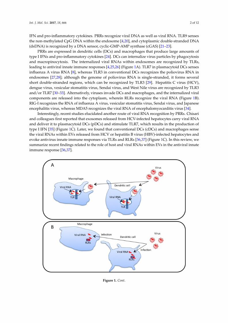
PRRs are expressed in dendritic cells (DCs) and macrophages that produce large amounts oftype I IFNs and pro-inflammatory cytokines [24]. DCs can internalize virus particles by phagocytosisand macropinocytosis. The internalized viral RNAs within endosomes are recognized by TLRs,leading to antiviral innate immune responses [4,25,26] (Figure 1A). TLR7 in plasmacytoid DCs sensesinfluenza A virus RNA [8], whereas TLR3 in conventional DCs recognizes the poliovirus RNA inendosomes [27,28]; although the genome of poliovirus RNA is single-stranded, it forms severalshort double-stranded regions, which can be recognized by TLR3 [29]. Hepatitis C virus (HCV),dengue virus, vesicular stomatitis virus, Sendai virus, and West Nile virus are recognized by TLR3and/or TLR7 [30–33]. Alternatively, viruses invade DCs and macrophages, and the internalized viralcomponents are released into the cytoplasm, wherein RLRs recognize the viral RNA (Figure 1B).RIG-I recognizes the RNA of influenza A virus, vesicular stomatitis virus, Sendai virus, and Japaneseencephalitis virus, whereas MDA5 recognizes the viral RNA of encephalomyocarditis virus [34].
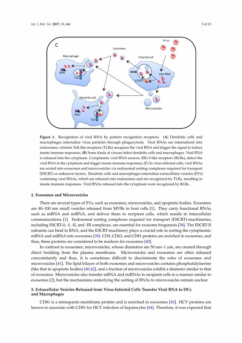
Interestingly, recent studies elucidated another route of viral RNA recognition by PRRs. Chisariand colleagues first reported that exosomes released from HCV-infected hepatocytes carry viral RNAand deliver it to plasmacytoid DCs (pDCs) and stimulate TLR7, which results in the production oftype I IFN [35] (Figure 1C). Later, we found that conventional DCs (cDCs) and macrophages sensethe viral RNAs within EVs released from HCV or hepatitis B virus (HBV)-infected hepatocytes andevoke antivirus innate immune responses via TLRs and RLRs [36,37] (Figure 1C). In this review, wesummarize recent findings related to the role of host and viral RNAs within EVs in the antiviral innateimmune response [36,37].
Int. J. Mol. Sci. 2017, 18, 666 2 of 11
production of type I IFN and pro-inflammatory cytokines. PRRs recognize viral DNA as well as viral RNA. TLR9 senses the non-methylated CpG DNA within the endosome [4,20], and cytoplasmic double-stranded DNA (dsDNA) is recognized by a DNA sensor, cyclic-GMP-AMP synthase (cGAS) [21–23].
PRRs are expressed in dendritic cells (DCs) and macrophages that produce large amounts of type I IFNs and pro-inflammatory cytokines [24]. DCs can internalize virus particles by phagocytosis and macropinocytosis. The internalized viral RNAs within endosomes are recognized by TLRs, leading to antiviral innate immune responses [4,25,26] (Figure 1A). TLR7 in plasmacytoid DCs senses influenza A virus RNA [8], whereas TLR3 in conventional DCs recognizes the poliovirus RNA in endosomes [27,28]; although the genome of poliovirus RNA is single-stranded, it forms several short double-stranded regions, which can be recognized by TLR3 [29]. Hepatitis C virus (HCV), dengue virus, vesicular stomatitis virus, Sendai virus, and West Nile virus are recognized by TLR3 and/or TLR7 [30–33]. Alternatively, viruses invade DCs and macrophages, and the internalized viral components are released into the cytoplasm, wherein RLRs recognize the viral RNA (Figure 1B). RIG-I recognizes the RNA of influenza A virus, vesicular stomatitis virus, Sendai virus, and Japanese encephalitis virus, whereas MDA5 recognizes the viral RNA of encephalomyocarditis virus [34].
Interestingly, recent studies elucidated another route of viral RNA recognition by PRRs. Chisari and colleagues first reported that exosomes released from HCV-infected hepatocytes carry viral RNA and deliver it to plasmacytoid DCs (pDCs) and stimulate TLR7, which results in the production of type I IFN [35] (Figure 1C). Later, we found that conventional DCs (cDCs) and macrophages sense the viral RNAs within EVs released from HCV or hepatitis B virus (HBV)-infected hepatocytes and evoke antivirus innate immune responses via TLRs and RLRs [36,37] (Figure 1C). In this review, we summarize recent findings related to the role of host and viral RNAs within EVs in the antiviral innate immune response [36,37].
Figure 1. Cont.
Int. J. Mol. Sci. 2017, 18, 666 3 of 12
Int. J. Mol. Sci. 2017, 18, 666 3 of 11
Figure 1. Recognition of viral RNA by pattern recognition receptors. (A) Dendritic cells and macrophages internalize virus particles through phagocytosis. Viral RNAs are internalized into endosomes, wherein Toll-like receptors (TLRs) recognize the viral RNA and trigger the signal to induce innate immune responses; (B) Some kinds of viruses infect dendritic cells and macrophages. Viral RNA is released into the cytoplasm. Cytoplasmic viral RNA sensors, RIG-I-like receptors (RLRs), detect the viral RNA in the cytoplasm and trigger innate immune responses; (C) In virus-infected cells, viral RNAs are sorted into exosomes and microvesicles via endosomal sorting complexes required for transport (ESCRT) or unknown factors. Dendritic cells and macrophages internalize extracellular vesicles (EVs) containing viral RNAs, which are released into endosomes and are recognized by TLRs, resulting in innate immune responses. Viral RNAs released into the cytoplasm were recognized by RLRs.
2. Exosomes and Microvesicles
There are several types of EVs, such as exosomes, microvesicles, and apoptotic bodies. Exosomes are 40–100 nm small vesicles released from MVBs in host cells [1]. They carry functional RNAs such as mRNA and miRNA, and deliver them to recipient cells, which results in intercellular communications [1]. Endosomal sorting complexes required for transport (ESCRT)-machineries, including ESCRT-0, -I, -II, and -III complexes, are essential for exosome biogenesis [38]. The ESCRT-II subunits can bind to RNA, and the ESCRT-machinery plays a crucial role in sorting the cytoplasmic mRNA and miRNA into exosomes [39]. CD9, CD63, and CD81 proteins are enriched in exosomes, and thus, these proteins are considered to be markers for exosomes [40].
In contrast to exosomes, microvesicles, whose diameters are 50 nm–1 μm, are created through direct budding from the plasma membrane. Microvesicles and exosomes are often released concomitantly and thus, it is sometimes difficult to discriminate the roles of exosomes and microvesicles [41]. The lipid bilayer of both exosomes and microvesicles contains phosphatidylserine (like that in apoptotic bodies) [40,42], and a fraction of microvesicles exhibit a diameter similar to that of exosomes. Microvesicles also transfer mRNA and miRNAs to recipient cells in a manner similar to exosomes [2], but the mechanisms underlying the sorting of RNAs to microvesicles remain unclear.
3. Extracellular Vesicles Released from Virus-Infected Cells Transfer Viral RNA to DCs and Macrophages
CD81 is a tetraspanin membrane protein and is enriched in exosomes [43]. HCV proteins are known to associate with CD81 for HCV infection of hepatocytes [44]. Therefore, it was expected that HCV would associate with exosomes via CD81. Indeed, it was reported that HCV protein and RNA were associated with exosomes that were captured from the plasma of HCV-infected patients [45].
Figure 1. Recognition of viral RNA by pattern recognition receptors. (A) Dendritic cells andmacrophages internalize virus particles through phagocytosis. Viral RNAs are internalized intoendosomes, wherein Toll-like receptors (TLRs) recognize the viral RNA and trigger the signal to induceinnate immune responses; (B) Some kinds of viruses infect dendritic cells and macrophages. Viral RNAis released into the cytoplasm. Cytoplasmic viral RNA sensors, RIG-I-like receptors (RLRs), detect theviral RNA in the cytoplasm and trigger innate immune responses; (C) In virus-infected cells, viral RNAsare sorted into exosomes and microvesicles via endosomal sorting complexes required for transport(ESCRT) or unknown factors. Dendritic cells and macrophages internalize extracellular vesicles (EVs)containing viral RNAs, which are released into endosomes and are recognized by TLRs, resulting ininnate immune responses. Viral RNAs released into the cytoplasm were recognized by RLRs.
2. Exosomes and Microvesicles
There are several types of EVs, such as exosomes, microvesicles, and apoptotic bodies. Exosomesare 40–100 nm small vesicles released from MVBs in host cells [1]. They carry functional RNAssuch as mRNA and miRNA, and deliver them to recipient cells, which results in intercellularcommunications [1]. Endosomal sorting complexes required for transport (ESCRT)-machineries,including ESCRT-0, -I, -II, and -III complexes, are essential for exosome biogenesis [38]. The ESCRT-IIsubunits can bind to RNA, and the ESCRT-machinery plays a crucial role in sorting the cytoplasmicmRNA and miRNA into exosomes [39]. CD9, CD63, and CD81 proteins are enriched in exosomes, andthus, these proteins are considered to be markers for exosomes [40].
In contrast to exosomes, microvesicles, whose diameters are 50 nm–1 µm, are created throughdirect budding from the plasma membrane. Microvesicles and exosomes are often releasedconcomitantly and thus, it is sometimes difficult to discriminate the roles of exosomes andmicrovesicles [41]. The lipid bilayer of both exosomes and microvesicles contains phosphatidylserine(like that in apoptotic bodies) [40,42], and a fraction of microvesicles exhibit a diameter similar to thatof exosomes. Microvesicles also transfer mRNA and miRNAs to recipient cells in a manner similar toexosomes [2], but the mechanisms underlying the sorting of RNAs to microvesicles remain unclear.
3. Extracellular Vesicles Released from Virus-Infected Cells Transfer Viral RNA to DCsand Macrophages
CD81 is a tetraspanin membrane protein and is enriched in exosomes [43]. HCV proteins areknown to associate with CD81 for HCV infection of hepatocytes [44]. Therefore, it was expected that
Int. J. Mol. Sci. 2017, 18, 666 4 of 12
HCV would associate with exosomes via CD81. Indeed, it was reported that HCV protein and RNAwere associated with exosomes that were captured from the plasma of HCV-infected patients [45].However, Dreux et al. reported that HCV RNA was packaged within exosomes that were released fromHCV-infected hepatocytes [35]. They also showed that exosomes carrying HCV RNA activate pDCs,resulting in type I IFN production [35]. Moreover, knockdown of ESCRT-I or -III complex componentsin HCV-infected cells attenuated the exosome-mediated type I IFN production by pDCs [35]. Theseobservations indicate that HCV RNA packaged within exosomes was delivered to pDCs, whichrecognized the HCV RNA via TLR7, leading to type I IFN production. Later, we observed that EVsreleased from hepatocytes with HCV replicons delivered viral RNA to CD8α+ DCs, a subset of cDCs,leading to TLR3-mediated type I and type III IFN production [37].
EV-mediated transfer of HBV RNA has also been reported. HBV is a hepadnavirus and is amajor cause of hepatocellular carcinoma. Recently, we found that EVs released from HBV-infectedhepatocytes contain viral RNA, which is transferred to macrophages [36], resulting in the expressionof NKG2D ligands, such as ULBP1 and ULBP2. The expression of NKG2D ligands in macrophagesis known to induce NK cell-mediated IFN-γ production [46,47], and we observed that intravenousinfection with HBV induced hepatic IFN-γ expression in an animal model [36].
The ability to transfer viral RNA to DCs and macrophages via EVs is not specific to vesiclesreleased from hepatocytes. Epstein-Barr virus (EBV) has a strong B cell tropism and has evolved alatency strategy to avoid immune detection. Exosomes released from EBV-infected lymphoblastoidcell lines activate the innate immune response in primary DCs [48]. An EBV transcript, EBER1 RNAwith 5′ triphosphate, was transferred to DCs by exosomes, thereby inducing interferon inducible geneexpression in DCs [48].
Packaging of viral RNA within exosomes was also detected in other studies. The exosomesreleased from Vero cells infected with Rift Valley Fever Virus also contained viral genomic RNA, andtheir components were delivered to recipient cells [49]. Moreover, HIV-infected T lymphocytesand macrophages constitutively produce exosomes containing high amounts of trans-activationresponse element (TAR) RNA, a viral transcript, which was delivered to macrophages and induced theproduction of pro-inflammatory cytokines in macrophages [50]. An accumulating body of evidencedemonstrates that EVs released from virus-infected cells transfer viral RNAs to recipient cells includingDCs and macrophages, thereby inducing innate immune responses.
4. Roles of MicroRNAs within Exosomes for Regulation of Innate Immune Responses
It has been reported that miRNAs within exosomes regulate innate immune responses.Stimulation with lipopolysaccharide (LPS), which is a ligand of TLR4, increases miR-155 expressionlevels [51]. Exosomes containing miR-155 are released from bone-marrow-derived DCs (BM-DCs)and are transferred to recipient BM-DCs [52]. The transferred miR-155 increased the inflammatoryresponse in recipient BM-DCs [52].
Although the innate immune system is the first line of defense against virus infection, manyviruses have evolved to escape the host innate immune response in order to infect host cells [12].Recently, we found that HBV attenuates the innate immune response via the miRNA within exosomes(Figure 2). The X protein of HBV plays a key role in the molecular pathogenesis of HBV-relatedhepatocellular carcinoma (HCC) [53,54]. Expression of the X protein of HBV induces miR-21 expressionin HCC cell lines [55]. IL-12 is a heterodimeric cytokine, which consists of p35 and p40 subunits,and is produced by DCs and macrophages [56]. The miR-21 targets the 3′ UTR of IL-12p35, resultingin the reduction of the target protein levels [57]. Interestingly, we found that the expression of theHBV proteins in HCC cell lines increased the exosomal miR-21 levels [36]. Moreover, we found thatexosomal miR-29 level was also increased by the expression of HBV proteins in HCC cell lines [36].miR-29 targets IL-12p40 [58], which is another subunit of IL-12. Therefore, the expression of HBVproteins in HCC cell lines increased exosomal miR21 and miR-29 levels, which target each subunit ofIL-12. IL-12 is well known to activate natural killer (NK) cells [59], and NK cells play a crucial role in

Int. J. Mol. Sci. 2017, 18, 666 5 of 12
suppressing HBV proliferation [60]. When macrophages were incubated with the exosomes releasedfrom HBV-infected cells and were then activated by TLR ligands, IL-12 production was reduced byexosomes released from HBV-infected cells compared to those released from mock-infected cells. Theseobservations indicate that HBV proteins increase the exosomal miR-21 and miR-29 levels to attenuatethe IL-12 production from macrophages and to counteract host innate immune responses.
Int. J. Mol. Sci. 2017, 18, 666 5 of 11
These observations indicate that HBV proteins increase the exosomal miR-21 and miR-29 levels to attenuate the IL-12 production from macrophages and to counteract host innate immune responses.
Figure 2. MicroRNAs within the extracellular vesicles regulate the innate immune response. (A) miRNAs are sorted into exosomes via the ESCRT complex, hnRNPA2B1, and other proteins. It has been reported that viral infection affects the microRNA levels in EVs. Dendritic cells and macrophages uptake miRNA-containing EVs. Internalized miRNAs are released into the cytoplasm and reduce the target mRNA expression or translation; (B) There are several pathways by which cytoplasmic miRNAs are sorted into exosomes. hnRNPs and Ago2 proteins are involved in the miRNA sorting, and post-translational modification of miRNA or proteins associating with miRNAs affect the sorting processes (see main text). MVB, multivesicular bodies. The proteins required for sorting of uridylated miRNA remain unclear.
5. Sorting of miRNA into Exosomes
ESCRT machineries are reported to be capable of sorting HCV RNA into exosomes, whereas the mechanisms of sorting of other virus RNAs into exosomes have not been elucidated. In contrast, recent studies have shed light on the mechanisms of miRNA sorting into exosomes (Figure 2). The hnRNPA2B1 protein is a ubiquitously expressed RNA-binding protein, and binds to a short motif within miRNA, which is called the EXOmotifs [61]. miRNAs with EXOmotifs are preferentially sorted into exosomes, and miRNAs without the motif remain in the cytoplasm in primary T lymphocyte [61]. In addition, sumoylation of hnRNPA2B1 promotes the sorting of miRNAs with EXOmotif into
Figure 2. MicroRNAs within the extracellular vesicles regulate the innate immune response.(A) miRNAs are sorted into exosomes via the ESCRT complex, hnRNPA2B1, and other proteins.It has been reported that viral infection affects the microRNA levels in EVs. Dendritic cells andmacrophages uptake miRNA-containing EVs. Internalized miRNAs are released into the cytoplasmand reduce the target mRNA expression or translation; (B) There are several pathways by whichcytoplasmic miRNAs are sorted into exosomes. hnRNPs and Ago2 proteins are involved in the miRNAsorting, and post-translational modification of miRNA or proteins associating with miRNAs affect thesorting processes (see main text). MVB, multivesicular bodies. The proteins required for sorting ofuridylated miRNA remain unclear.
5. Sorting of miRNA into Exosomes
ESCRT machineries are reported to be capable of sorting HCV RNA into exosomes, whereasthe mechanisms of sorting of other virus RNAs into exosomes have not been elucidated. In contrast,

Int. J. Mol. Sci. 2017, 18, 666 6 of 12
recent studies have shed light on the mechanisms of miRNA sorting into exosomes (Figure 2).The hnRNPA2B1 protein is a ubiquitously expressed RNA-binding protein, and binds to a short motifwithin miRNA, which is called the EXOmotifs [61]. miRNAs with EXOmotifs are preferentially sortedinto exosomes, and miRNAs without the motif remain in the cytoplasm in primary T lymphocyte [61].In addition, sumoylation of hnRNPA2B1 promotes the sorting of miRNAs with EXOmotif intoexosomes [61]. In hepatocytes, miRNAs with another motif, hEXO motif, were sorted into exosomesvia a hnRNP, hnRNP-Q, also called SYNCRIP [62]. Another study has shown that the Y-box protein 1is involved in miR-223 sorting to exosomes in a cell-free system [63]. Post-translational modificationis also involved in miRNA sorting. For example, uridylation of 3′ ends of miRNAs promotes theirsorting to exosomes in B cells [64].
The Ago2 protein binds to miRNA, and the Ago2-miRNA complexes are released from cells asfree proteins [65]; however, there are studies showing that Ago2 is packaged into exosomes along withmiRNA [66,67]. Interestingly, it was shown that MEK-ERK activation inhibits the sorting of Ago2 andAgo2-miRNA complex into exosomes [68]. Activation of TLRs affects ERK signaling [69]. Consideringthat HBV infection is detected by TLRs [36], it is possible that the increase in miR-21 and miR-29alevels in exosomes after HBV infection could be regulated by the activation of TLRs.
6. Uptake of Exosomes
Exosomes are efficiently internalized through phagocytosis [70], and macrophages and DCsinternalize exosomes very efficiently [70,71]. Therefore, host and viral RNA within exosomes areexpected to be efficiently transferred to DCs and macrophages compared to other types of cells.The proteins involved in the uptake of exosomes by DCs and macrophages have been reported.Treatment with cytochalasin D reduced exosome uptake [71], suggesting the requirement of actinpolymerization. In addition, blocking antibodies against αv (CD51) and β3 (CD61) integrins, LFA-1(CD11a), CD54 (ICAM-1), CD9, and CD81 reduced the uptake of exosomes [1,71]. Moreover, the solubleanalog of phosphatidylserine also reduced exosome uptake [71], suggesting that the phosphatidylserine on exosomes is recognized by recipient DCs. The integrin αvβ3 is expressed in macrophagesand binds MFG-E8, which is a soluble factor associating with phosphatidyl serine [72,73]. CD9 andCD81 are enriched in the membrane of the exosomes [38], and bind the αvβ3 integrin [74]. CD54(ICAM-1) is a ligand of LFA-1 (CD11a) [75]. LFA-1 (CD11a) is expressed in DCs, and it has been shownthat activated T cells bind to DC-derived EVs through LFA-1 [76]. In addition, the expression of α4integrin promotes the uptakes of EVs by lymph node stromal cells [77], and macrophages requiregalectin-5 expression on EVs released from erythrocytes [78]. Considering that blocking these proteinswith antibodies only partially reduced the uptake, it is expected that the recipient cells internalize EVsthrough several redundant uptake pathways.
The fusion of the exosome membrane with the membrane of recipient dendritic cells or cancercells has been demonstrated using assays with the lipophilic dye R18, which has been used to monitorthe fusion of enveloped viruses and lipid vesicles. The R18 dye is a self-quenching fluorescent lipidprobe at its high concentration. When exosomes labeled with self-quenched R18 were incubatedwith recipient DCs, the fluorescence immediately increased, suggesting the fusion of the exosomemembrane with the recipient DC membrane [79]. This exosome membrane fusion with recipient cellswas also observed in the case of cancer cells [1].
The release of the luminal content of exosomes into the cytosol of DCs has also been reported.When the exosomes containing the luciferin were incubated with DCs expressing luciferase, emissionof light was observed at 10 min after incubation [79]. Moreover, miR-148a and miR-451 within theexosomes were internalized into the recipient DCs and they down-regulated their target [79].
These facts are consistent with the notion that miRNAs within EVs released by viral-infectedcells are transferred into the cytoplasm of recipient cells and that they regulate the innate immuneresponse. In the case of HBV infection, the expression of HBV proteins increased the miR-21 andmiR-29 levels in EVs, which are transferred into macrophages [36], whereas the role of cell surface
Int. J. Mol. Sci. 2017, 18, 666 7 of 12
proteins involved in the uptake of EVs has not been fully elucidated. Further studies are required touncover the underlying mechanisms.
7. Perspectives
Recent studies have elucidated the crucial roles of RNA within EVs on antiviral innate immuneresponses, as described above. Therefore, several researchers have tried to use the miRNA within EVsas new biomarkers or to utilize EVs with RNAs as novel drugs. Several studies have already shownthat exosomal miRNAs can be used as biomarkers of cancer diagnosis [80,81]. Considering that viralinfection changes the miRNA levels in EVs released form virus-infected cells, the miRNA levels in EVscan be used as a biomarker for the diagnosis of infection. EVs are enriched in human serum, and serumexosomal miR-122 is significantly increased by liver injury and inflammation caused by virus infection,and thus it is postulated that miR-122 serves as a biomarker of liver damage and inflammation [82].
The loop-mediated isothermal amplification (LAMP) method can detect specific DNA or RNAwithin samples by incubating the samples for 15–50 min at 65 ◦C. The LAMP method does not requirea thermal cycler, but only requires a simple incubator [83]. miRNAs can be detected by a modifiedLAMP method [84]. Because EVs are enriched in human blood or human serum samples, it is expectedthat the miRNAs within serum EVs can be easily detected by using modified LAMP methods and canalso be used for the diagnosis of infectious disease in a short period and at low cost in hospitals or inepidemiological studies.
In addition, because of the effect of miRNAs within EVs on DCs and macrophages, it has beenpostulated that EVs with miRNA can be used for the treatment of infectious diseases or for developinga vaccine. For instance, miR-21 and miR-29 reduce the production of IL-12, which is known to promoteTh1 differentiation, and thus it is expected that EVs with miR-21 and miR-29 could modulate Th1versus Th2 response patterns and activate cytotoxic T lymphocytes (CTL) and NK cells. This might beuseful to attenuate cytotoxicity against HBV- or HCV-infected hepatocytes by CTL or NK cells and toincrease the production of neutralizing antibodies by Th2 response. Moreover, miR-155 is shown toaugment innate immune responses, and thus EVs with miR-155 are expected to augment the adjuvanteffects of vaccines, thereby improving the efficacy of vaccines. Further studies of EV-mediated immuneregulation will help to establish new strategies for the treatment and prevention of infectious diseaseusing EVs with miRNA.
Acknowledgments: This work was supported in part by Grants-in-Aid from the Ministry of Education, Science,and Technology (MEXT) and the Ministry of Health, Labor, and Welfare of Japan (MHLW), and also supported byGrant-in-Aid from the Japan Agency for Medical Research and Development (AMED), PRESTO JST, MochidaMemorial foundation, Japan Diabetes Foundation, Takeda Science Foundation, the Kao Foundation for Arts andSciences, the Japanese Association for Complement Research, and Daiichi Sankyo Foundation of Life Science.
Author Contributions: Takahisa Kouwaki, Masaaki Okamoto, Hirotake Tsukamoto, Yoshimi Fukushima andHiroyuki Oshiumi contributed to the conception and writing of the manuscript.
Conflicts of Interest: The authors declare no conflict of interest.
References
1. Colombo, M.; Raposo, G.; Thery, C. Biogenesis, secretion, and intercellular interactions of exosomes andother extracellular vesicles. Annu. Rev. Cell Dev. Biol. 2014, 30, 255–289. [CrossRef] [PubMed]
2. Muralidharan-Chari, V.; Clancy, J.W.; Sedgwick, A.; D’Souza-Schorey, C. Microvesicles: Mediators ofextracellular communication during cancer progression. J. Cell Sci. 2010, 123, 1603–1611. [CrossRef][PubMed]
3. Loo, Y.M.; Gale, M., Jr. Immune signaling by RIG-I-like receptors. Immunity 2011, 34, 680–692. [CrossRef][PubMed]
4. Kawai, T.; Akira, S. Toll-like receptors and their crosstalk with other innate receptors in infection andimmunity. Immunity 2011, 34, 637–650. [CrossRef] [PubMed]
Int. J. Mol. Sci. 2017, 18, 666 8 of 12
5. Johnsen, I.B.; Nguyen, T.T.; Ringdal, M.; Tryggestad, A.M.; Bakke, O.; Lien, E.; Espevik, T.; Anthonsen, M.W.Toll-like receptor 3 associates with C-SRC tyrosine kinase on endosomes to initiate antiviral signaling.EMBO J. 2006, 25, 3335–3346. [CrossRef] [PubMed]
6. Matsumoto, M.; Funami, K.; Tanabe, M.; Oshiumi, H.; Shingai, M.; Seto, Y.; Yamamoto, A.; Seya, T. Subcellularlocalization of toll-like receptor 3 in human dendritic cells. J. Immunol. 2003, 171, 3154–3162. [CrossRef][PubMed]
7. Alexopoulou, L.; Holt, A.C.; Medzhitov, R.; Flavell, R.A. Recognition of double-stranded RNA and activationof NF-κB by toll-like receptor 3. Nature 2001, 413, 732–738. [CrossRef] [PubMed]
8. Diebold, S.S.; Kaisho, T.; Hemmi, H.; Akira, S.; Reis e Sousa, C. Innate antiviral responses by means ofTLR7-mediated recognition of single-stranded RNA. Science 2004, 303, 1529–1531. [CrossRef] [PubMed]
9. Hemmi, H.; Kaisho, T.; Takeuchi, O.; Sato, S.; Sanjo, H.; Hoshino, K.; Horiuchi, T.; Tomizawa, H.; Takeda, K.;Akira, S. Small anti-viral compounds activate immune cells via the TLR7 MYD88-dependent signalingpathway. Nat. Immunol. 2002, 3, 196–200. [CrossRef] [PubMed]
10. Yoneyama, M.; Kikuchi, M.; Matsumoto, K.; Imaizumi, T.; Miyagishi, M.; Taira, K.; Foy, E.; Loo, Y.M.;Gale, M., Jr.; Akira, S.; et al. Shared and unique functions of the Dexd/H-box helicases RIG-I, MDA5, ANDLGP2 in antiviral innate immunity. J. Immunol. 2005, 175, 2851–2858. [CrossRef] [PubMed]
11. Yoneyama, M.; Kikuchi, M.; Natsukawa, T.; Shinobu, N.; Imaizumi, T.; Miyagishi, M.; Taira, K.; Akira, S.;Fujita, T. The RNA helicase RIG-I has an essential function in double-stranded RNA-induced innate antiviralresponses. Nat. Immunol. 2004, 5, 730–737. [CrossRef] [PubMed]
12. Chan, Y.K.; Gack, M.U. Viral evasion of intracellular DNA and RNA sensing. Nat. Rev. Microbiol. 2016, 14,360–373. [CrossRef] [PubMed]
13. Oshiumi, H.; Miyashita, M.; Matsumoto, M.; Seya, T. A distinct role of riplet-mediated K63-Linkedpolyubiquitination of the RIG-I repressor domain in human antiviral innate immune responses. PLoS Pathog.2013, 9, e1003533. [CrossRef] [PubMed]
14. Gack, M.U.; Shin, Y.C.; Joo, C.H.; Urano, T.; Liang, C.; Sun, L.; Takeuchi, O.; Akira, S.; Chen, Z.; Inoue, S.; et al.TRIM25 RING-finger E3 ubiquitin ligase is essential for RIG-I-mediated antiviral activity. Nature 2007, 446,916–920. [CrossRef] [PubMed]
15. Oshiumi, H.; Miyashita, M.; Inoue, N.; Okabe, M.; Matsumoto, M.; Seya, T. The ubiquitin ligase riplet isessential for RIG-I-dependent innate immune responses to RNA virus infection. Cell Host Microbe 2010, 8,496–509. [CrossRef] [PubMed]
16. Oshiumi, H.; Mifsud, E.J.; Daito, T. Links between recognition and degradation of cytoplasmic viral RNA ininnate immune response. Rev. Med. Virol. 2016, 26, 90–101. [CrossRef] [PubMed]
17. Oshiumi, H.; Kouwaki, T.; Seya, T. Accessory factors of cytoplasmic viral RNA sensors required for antiviralinnate immune response. Front. Immunol. 2016, 7, 200. [CrossRef] [PubMed]
18. Takashima, K.; Oshiumi, H.; Takaki, H.; Matsumoto, M.; Seya, T. Riok3-mediated phosphorylation ofMDA5 interferes with its assembly and attenuates the innate immune response. Cell Rep. 2015, 11, 192–200.[CrossRef] [PubMed]
19. Oshiumi, H.; Miyashita, M.; Okamoto, M.; Morioka, Y.; Okabe, M.; Matsumoto, M.; Seya, T. Ddx60 is involvedin RIG-I-dependent and independent antiviral responses, and its function is attenuated by virus-inducedegfr activation. Cell Rep. 2015, 11, 1193–1207. [CrossRef] [PubMed]
20. Hemmi, H.; Takeuchi, O.; Kawai, T.; Kaisho, T.; Sato, S.; Sanjo, H.; Matsumoto, M.; Hoshino, K.; Wagner, H.;Takeda, K.; et al. A toll-like receptor recognizes bacterial DNA. Nature 2000, 408, 740–745. [PubMed]
21. Wu, J.; Sun, L.; Chen, X.; Du, F.; Shi, H.; Chen, C.; Chen, Z.J. Cyclic GMP-AMP is an endogenous secondmessenger in innate immune signaling by cytosolic DNA. Science 2013, 339, 826–830. [CrossRef] [PubMed]
22. Sun, L.; Wu, J.; Du, F.; Chen, X.; Chen, Z.J. Cyclic GMP-AMP synthase is a cytosolic DNA sensor that activatesthe type I interferon pathway. Science 2013, 339, 786–791. [CrossRef] [PubMed]
23. Li, X.D.; Wu, J.; Gao, D.; Wang, H.; Sun, L.; Chen, Z.J. Pivotal roles of CGAS-CGAMP signaling in antiviraldefense and immune adjuvant effects. Science 2013, 341, 1390–1394. [CrossRef] [PubMed]
24. Honda, K.; Takaoka, A.; Taniguchi, T. Type I interferon gene induction by the interferon regulatory factorfamily of transcription factors. Immunity 2006, 25, 349–360. [CrossRef] [PubMed]
25. Banchereau, J.; Steinman, R.M. Dendritic cells and the control of immunity. Nature 1998, 392, 245–252.[CrossRef] [PubMed]
Int. J. Mol. Sci. 2017, 18, 666 9 of 12
26. Sallusto, F.; Cella, M.; Danieli, C.; Lanzavecchia, A. Dendritic cells use macropinocytosis and the mannosereceptor to concentrate macromolecules in the major histocompatibility complex class II compartment:Downregulation by cytokines and bacterial products. J. Exp. Med. 1995, 182, 389–400. [CrossRef] [PubMed]
27. Abe, Y.; Fujii, K.; Nagata, N.; Takeuchi, O.; Akira, S.; Oshiumi, H.; Matsumoto, M.; Seya, T.; Koike, S. Thetoll-like receptor 3-mediated antiviral response is important for protection against poliovirus infection inpoliovirus receptor transgenic mice. J. Virol. 2012, 86, 185–194. [CrossRef] [PubMed]
28. Oshiumi, H.; Okamoto, M.; Fujii, K.; Kawanishi, T.; Matsumoto, M.; Koike, S.; Seya, T. The TLR3/TICAM-1pathway is mandatory for innate immune responses to poliovirus infection. J. Immunol. 2011, 187, 5320–5327.[CrossRef] [PubMed]
29. Tatematsu, M.; Nishikawa, F.; Seya, T.; Matsumoto, M. Toll-like receptor 3 recognizes incomplete stemstructures in single-stranded viral RNA. Nat. Commun. 2013, 4, 1833. [CrossRef] [PubMed]
30. Ebihara, T.; Shingai, M.; Matsumoto, M.; Wakita, T.; Seya, T. Hepatitis c virus-infected hepatocytesextrinsically modulate dendritic cell maturation to activate T cells and natural killer cells. Hepatology2008, 48, 48–58. [CrossRef] [PubMed]
31. Wang, J.P.; Liu, P.; Latz, E.; Golenbock, D.T.; Finberg, R.W.; Libraty, D.H. Flavivirus activation of plasmacytoiddendritic cells delineates key elements of TLR7 signaling beyond endosomal recognition. J. Immunol. 2006,177, 7114–7121. [CrossRef] [PubMed]
32. Lund, J.M.; Alexopoulou, L.; Sato, A.; Karow, M.; Adams, N.C.; Gale, N.W.; Iwasaki, A.; Flavell, R.A.Recognition of single-stranded RNA viruses by toll-like receptor 7. Proc. Natl. Acad. Sci. USA 2004, 101,5598–5603. [CrossRef] [PubMed]
33. Welte, T.; Reagan, K.; Fang, H.; Machain-Williams, C.; Zheng, X.; Mendell, N.; Chang, G.J.; Wu, P.; Blair, C.D.;Wang, T. Toll-like receptor 7-induced immune response to cutaneous west nile virus infection. J. Gen. Virol.2009, 90, 2660–2668. [CrossRef] [PubMed]
34. Kato, H.; Takeuchi, O.; Sato, S.; Yoneyama, M.; Yamamoto, M.; Matsui, K.; Uematsu, S.; Jung, A.; Kawai, T.;Ishii, K.J.; et al. Differential roles of MDA5 and RIG-I helicases in the recognition of RNA viruses. Nature2006, 441, 101–105. [CrossRef] [PubMed]
35. Dreux, M.; Garaigorta, U.; Boyd, B.; Decembre, E.; Chung, J.; Whitten-Bauer, C.; Wieland, S.; Chisari, F.V.Short-range exosomal transfer of viral RNA from infected cells to plasmacytoid dendritic cells triggers innateimmunity. Cell Host Microbe 2012, 12, 558–570. [CrossRef] [PubMed]
36. Kouwaki, M.; Fukushima, Y.; Daito, T.; Sanada, T.; Yamamoto, N.; Mifsud, E.J.; Leong, C.R.;Tsukiyama-Kohara, K.; Kohara, M.; Matsumoto, M.; et al. Extracellular vesicles including exosomes regulateinnate immune responses to hepatitis b virus infection. Front. Immunol. 2016, 7. [CrossRef] [PubMed]
37. Okamoto, M.; Oshiumi, H.; Azuma, M.; Kato, N.; Matsumoto, M.; Seya, T. IPS-1 is essential for type III IFNproduction by hepatocytes and dendritic cells in response to hepatitis c virus infection. J. Immunol. 2014, 192,2770–2777. [CrossRef] [PubMed]
38. Simons, M.; Raposo, G. Exosomes—Vesicular carriers for intercellular communication. Curr. Opin. Cell Biol.2009, 21, 575–581. [CrossRef] [PubMed]
39. Irion, U.; St. Johnston, D. Bicoid RNA localization requires specific binding of an endosomal sorting complex.Nature 2007, 445, 554–558. [CrossRef] [PubMed]
40. Hulsmans, M.; Holvoet, P. MicroRNA-containing microvesicles regulating inflammation in association withatherosclerotic disease. Cardiovasc. Res. 2013, 100, 7–18. [CrossRef] [PubMed]
41. Heijnen, H.F.; Schiel, A.E.; Fijnheer, R.; Geuze, H.J.; Sixma, J.J. Activated platelets release two typesof membrane vesicles: Microvesicles by surface shedding and exosomes derived from exocytosis ofmultivesicular bodies and α-granules. Blood 1999, 94, 3791–3799. [PubMed]
42. Thery, C.; Ostrowski, M.; Segura, E. Membrane vesicles as conveyors of immune responses. Nat. Rev.Immunol. 2009, 9, 581–593. [CrossRef] [PubMed]
43. Escola, J.M.; Kleijmeer, M.J.; Stoorvogel, W.; Griffith, J.M.; Yoshie, O.; Geuze, H.J. Selective enrichment oftetraspan proteins on the internal vesicles of multivesicular endosomes and on exosomes secreted by humanB-lymphocytes. J. Biol. Chem. 1998, 273, 20121–20127. [CrossRef] [PubMed]
44. Hsu, M.; Zhang, J.; Flint, M.; Logvinoff, C.; Cheng-Mayer, C.; Rice, C.M.; McKeating, J.A. Hepatitis cvirus glycoproteins mediate pH-dependent cell entry of pseudotyped retroviral particles. Proc. Natl. Acad.Sci. USA 2003, 100, 7271–7276. [CrossRef] [PubMed]
Int. J. Mol. Sci. 2017, 18, 666 10 of 12
45. Masciopinto, F.; Giovani, C.; Campagnoli, S.; Galli-Stampino, L.; Colombatto, P.; Brunetto, M.; Yen, T.S.;Houghton, M.; Pileri, P.; Abrignani, S. Association of hepatitis C virus envelope proteins with exosomes.Eur. J. Immunol. 2004, 34, 2834–2842. [CrossRef] [PubMed]
46. Kloss, M.; Decker, P.; Baltz, K.M.; Baessler, T.; Jung, G.; Rammensee, H.G.; Steinle, A.; Krusch, M.; Salih, H.R.Interaction of monocytes with NK cells upon toll-like receptor-induced expression of the NKG2D ligandmica. J. Immunol. 2008, 181, 6711–6719. [CrossRef] [PubMed]
47. Ebihara, T.; Masuda, H.; Akazawa, T.; Shingai, M.; Kikuta, H.; Ariga, T.; Matsumoto, M.; Seya, T. Induction ofNKG2D ligands on human dendritic cells by TLR ligand stimulation and RNA virus infection. Int. Immunol.2007, 19, 1145–1155. [CrossRef] [PubMed]
48. Baglio, S.R.; van Eijndhoven, M.A.; Koppers-Lalic, D.; Berenguer, J.; Lougheed, S.M.; Gibbs, S.; Leveille, N.;Rinkel, R.N.; Hopmans, E.S.; Swaminathan, S.; et al. Sensing of latent EBV infection through exosomaltransfer of 5′pppRNA. Proc. Natl. Acad. Sci. USA 2016, 113, E587–E596. [CrossRef] [PubMed]
49. Ahsan, N.A.; Sampey, G.C.; Lepene, B.; Akpamagbo, Y.; Barclay, R.A.; Iordanskiy, S.; Hakami, R.M.;Kashanchi, F. Presence of viral RNA and proteins in exosomes from cellular clones resistant to RIFT valleyfever virus infection. Front. Microbiol. 2016, 7, 139. [CrossRef] [PubMed]
50. Sampey, G.C.; Saifuddin, M.; Schwab, A.; Barclay, R.; Punya, S.; Chung, M.C.; Hakami, R.M.; Zadeh, M.A.;Lepene, B.; Klase, Z.A.; et al. Exosomes from HIV-1-infected cells stimulate production of pro-inflammatorycytokines through trans-activating response (TAR) RNA. J. Biol. Chem. 2016, 291, 1251–1266. [CrossRef][PubMed]
51. O’Connell, R.M.; Rao, D.S.; Chaudhuri, A.A.; Boldin, M.P.; Taganov, K.D.; Nicoll, J.; Paquette, R.L.;Baltimore, D. Sustained expression of microRNA-155 in hematopoietic stem cells causes a myeloproliferativedisorder. J. Exp. Med. 2008, 205, 585–594. [CrossRef] [PubMed]
52. Alexander, M.; Hu, R.; Runtsch, M.C.; Kagele, D.A.; Mosbruger, T.L.; Tolmachova, T.; Seabra, M.C.;Round, J.L.; Ward, D.M.; O’Connell, R.M. Exosome-delivered microRNAs modulate the inflammatoryresponse to endotoxin. Nat. Commun. 2015, 6, 7321. [CrossRef] [PubMed]
53. Wang, C.; Yang, W.; Yan, H.X.; Luo, T.; Zhang, J.; Tang, L.; Wu, F.Q.; Zhang, H.L.; Yu, L.X.;Zheng, L.Y.; et al. Hepatitis B virus x (HBx) induces tumorigenicity of hepatic progenitor cells in3,5-diethoxycarbonyl-1,4-dihydrocollidine-treated HBx transgenic mice. Hepatology 2012, 55, 108–120.[CrossRef] [PubMed]
54. Brechot, C. Pathogenesis of hepatitis B virus-related hepatocellular carcinoma: Old and new paradigms.Gastroenterology 2004, 127, S56–S61. [CrossRef] [PubMed]
55. Qiu, X.; Dong, S.; Qiao, F.; Lu, S.; Song, Y.; Lao, Y.; Li, Y.; Zeng, T.; Hu, J.; Zhang, L.; et al. HBx-mediatedmiR-21 upregulation represses tumor-suppressor function of PDCD4 in hepatocellular carcinoma. Oncogene2013, 32, 3296–3305. [CrossRef] [PubMed]
56. Hunter, C.A. New il-12-family members: Il-23 and Il-27, cytokines with divergent functions. Nat. Rev.Immunol. 2005, 5, 521–531. [CrossRef] [PubMed]
57. Lu, T.X.; Munitz, A.; Rothenberg, M.E. MicroRNA-21 is up-regulated in allergic airway inflammation andregulates Il-12P35 expression. J. Immunol. 2009, 182, 4994–5002. [CrossRef] [PubMed]
58. Brain, O.; Owens, B.M.; Pichulik, T.; Allan, P.; Khatamzas, E.; Leslie, A.; Steevels, T.; Sharma, S.; Mayer, A.;Catuneanu, A.M.; et al. The intracellular sensor NOD2 induces microRNA-29 expression in human dendriticcells to limit Il-23 release. Immunity 2013, 39, 521–536. [CrossRef] [PubMed]
59. Andrews, D.M.; Scalzo, A.A.; Yokoyama, W.M.; Smyth, M.J.; Degli-Esposti, M.A. Functional interactionsbetween dendritic cells and NK cells during viral infection. Nat. Immunol. 2003, 4, 175–181. [CrossRef][PubMed]
60. Maini, M.K.; Peppa, D. Nk cells: A double-edged sword in chronic hepatitis B virus infection. Front. Immunol.2013, 4, 57. [CrossRef] [PubMed]
61. Villarroya-Beltri, C.; Gutierrez-Vazquez, C.; Sanchez-Cabo, F.; Perez-Hernandez, D.; Vazquez, J.;Martin-Cofreces, N.; Martinez-Herrera, D.J.; Pascual-Montano, A.; Mittelbrunn, M.; Sanchez-Madrid, F.Sumoylated HNRNPA2B1 controls the sorting of miRNAs into exosomes through binding to specific motifs.Nat. Commun. 2013, 4, 2980. [CrossRef] [PubMed]
62. Santangelo, L.; Giurato, G.; Cicchini, C.; Montaldo, C.; Mancone, C.; Tarallo, R.; Battistelli, C.; Alonzi, T.;Weisz, A.; Tripodi, M. The RNA-binding protein syncrip is a component of the hepatocyte exosomalmachinery controlling microRNA sorting. Cell Rep. 2016, 17, 799–808. [CrossRef] [PubMed]
Int. J. Mol. Sci. 2017, 18, 666 11 of 12
63. Shurtleff, M.J.; Temoche-Diaz, M.M.; Karfilis, K.V.; Ri, S.; Schekman, R. Y-box protein 1 is required to sortmicroRNAs into exosomes in cells and in a cell-free reaction. Elife 2016, 5, e19276. [CrossRef] [PubMed]
64. Koppers-Lalic, D.; Hackenberg, M.; Bijnsdorp, I.V.; van Eijndhoven, M.A.; Sadek, P.; Sie, D.; Zini, N.;Middeldorp, J.M.; Ylstra, B.; de Menezes, R.X.; et al. Nontemplated nucleotide additions distinguish thesmall RNA composition in cells from exosomes. Cell Rep. 2014, 8, 1649–1658. [CrossRef] [PubMed]
65. Arroyo, J.D.; Chevillet, J.R.; Kroh, E.M.; Ruf, I.K.; Pritchard, C.C.; Gibson, D.F.; Mitchell, P.S.; Bennett, C.F.;Pogosova-Agadjanyan, E.L.; Stirewalt, D.L.; et al. Argonaute2 complexes carry a population of circulatingmicroRNAs independent of vesicles in human plasma. Proc. Natl. Acad. Sci. USA 2011, 108, 5003–5008.[CrossRef] [PubMed]
66. Melo, S.A.; Sugimoto, H.; O’Connell, J.T.; Kato, N.; Villanueva, A.; Vidal, A.; Qiu, L.; Vitkin, E.; Perelman, L.T.;Melo, C.A.; et al. Cancer exosomes perform cell-independent microRNA biogenesis and promotetumorigenesis. Cancer Cell 2014, 26, 707–721. [CrossRef] [PubMed]
67. Squadrito, M.L.; Baer, C.; Burdet, F.; Maderna, C.; Gilfillan, G.D.; Lyle, R.; Ibberson, M.; de Palma, M.Endogenous RNAs modulate microRNA sorting to exosomes and transfer to acceptor cells. Cell Rep. 2014, 8,1432–1446. [CrossRef] [PubMed]
68. McKenzie, A.J.; Hoshino, D.; Hong, N.H.; Cha, D.J.; Franklin, J.L.; Coffey, R.J.; Patton, J.G.; Weaver, A.M.KRAS-MEK signaling controls Ago2 sorting into exosomes. Cell Rep. 2016, 15, 978–987. [CrossRef] [PubMed]
69. Saraiva, M.; O’Garra, A. The regulation of IL-10 production by immune cells. Nat. Rev. Immunol. 2010, 10,170–181. [CrossRef] [PubMed]
70. Feng, D.; Zhao, W.L.; Ye, Y.Y.; Bai, X.C.; Liu, R.Q.; Chang, L.F.; Zhou, Q.; Sui, S.F. Cellular internalization ofexosomes occurs through phagocytosis. Traffic 2010, 11, 675–687. [CrossRef] [PubMed]
71. Morelli, A.E.; Larregina, A.T.; Shufesky, W.J.; Sullivan, M.L.; Stolz, D.B.; Papworth, G.D.; Zahorchak, A.F.;Logar, A.J.; Wang, Z.; Watkins, S.C.; et al. Endocytosis, intracellular sorting, and processing of exosomes bydendritic cells. Blood 2004, 104, 3257–3266. [CrossRef] [PubMed]
72. Hanayama, R.; Tanaka, M.; Miwa, K.; Shinohara, A.; Iwamatsu, A.; Nagata, S. Identification of a factor thatlinks apoptotic cells to phagocytes. Nature 2002, 417, 182–187. [CrossRef] [PubMed]
73. Aziz, M.; Jacob, A.; Matsuda, A.; Wang, P. Review: Milk fat globule-EGF factor 8 expression, function andplausible signal transduction in resolving inflammation. Apop. Int. J. Program. Cell Death 2011, 16, 1077–1086.[CrossRef] [PubMed]
74. Yu, J.; Lee, C.Y.; Changou, C.A.; Cedano-Prieto, D.M.; Takada, Y.K.; Takada, Y. The CD9, CD81, and CD151EC2 domains bind to the classical RGD-binding site of integrin αvβ3. Biochem. J. 2016. [CrossRef] [PubMed]
75. Marlin, S.D.; Springer, T.A. Purified intercellular adhesion molecule-1 (ICAM-1) is a ligand for lymphocytefunction-associated antigen 1 (LFA-1). Cell 1987, 51, 813–819. [CrossRef]
76. Nolte-’t Hoen, E.N.; Buschow, S.I.; Anderton, S.M.; Stoorvogel, W.; Wauben, M.H. Activated t cells recruitexosomes secreted by dendritic cells via LFA-1. Blood 2009, 113, 1977–1981. [CrossRef] [PubMed]
77. Rana, S.; Yue, S.; Stadel, D.; Zoller, M. Toward tailored exosomes: The exosomal tetraspanin web contributesto target cell selection. Int. J. Biochem. Cell Biol. 2012, 44, 1574–1584. [CrossRef] [PubMed]
78. Barres, C.; Blanc, L.; Bette-Bobillo, P.; Andre, S.; Mamoun, R.; Gabius, H.J.; Vidal, M. Galectin-5 is boundonto the surface of rat reticulocyte exosomes and modulates vesicle uptake by macrophages. Blood 2010, 115,696–705. [CrossRef] [PubMed]
79. Montecalvo, A.; Larregina, A.T.; Shufesky, W.J.; Stolz, D.B.; Sullivan, M.L.; Karlsson, J.M.; Baty, C.J.;Gibson, G.A.; Erdos, G.; Wang, Z.; et al. Mechanism of transfer of functional microRNAs between mousedendritic cells via exosomes. Blood 2012, 119, 756–766. [CrossRef] [PubMed]
80. Kosaka, N.; Iguchi, H.; Ochiya, T. Circulating microRNA in body fluid: A new potential biomarker for cancerdiagnosis and prognosis. Cancer Sci. 2010, 101, 2087–2092. [CrossRef] [PubMed]
81. Taylor, D.D.; Gercel-Taylor, C. MicroRNA signatures of tumor-derived exosomes as diagnostic biomarkers ofovarian cancer. Gynecol. Oncol. 2008, 110, 13–21. [CrossRef] [PubMed]
82. Bala, S.; Petrasek, J.; Mundkur, S.; Catalano, D.; Levin, I.; Ward, J.; Alao, H.; Kodys, K.; Szabo, G. CirculatingmicroRNAs in exosomes indicate hepatocyte injury and inflammation in alcoholic, drug-induced, andinflammatory liver diseases. Hepatology 2012, 56, 1946–1957. [CrossRef] [PubMed]
Int. J. Mol. Sci. 2017, 18, 666 12 of 12
83. Notomi, T.; Okayama, H.; Masubuchi, H.; Yonekawa, T.; Watanabe, K.; Amino, N.; Hase, T. Loop-mediatedisothermal amplification of DNA. Nucleic Acids Res. 2000, 28, E63. [CrossRef] [PubMed]
84. Li, C.; Li, Z.; Jia, H.; Yan, J. One-step ultrasensitive detection of microRNAs with loop-mediated isothermalamplification (LAMP). Chem. Commun. 2011, 47, 2595–2597. [CrossRef] [PubMed]
© 2017 by the authors. Licensee MDPI, Basel, Switzerland. This article is an open accessarticle distributed under the terms and conditions of the Creative Commons Attribution(CC BY) license (http://creativecommons.org/licenses/by/4.0/).