Embed Size (px)
Citation preview

Research article
342 TheJournalofClinicalInvestigation http://www.jci.org Volume 121 Number 1 January 2011
Fbxw7 regulates lipid metabolism and cell fate decisions in the mouse liver
Ichiro Onoyama,1,2 Atsushi Suzuki,3,4 Akinobu Matsumoto,1,2 Kengo Tomita,5 Hideki Katagiri,6 Yuichi Oike,4,7 Keiko Nakayama,2,8 and Keiichi I. Nakayama1,2
1Department of Molecular and Cellular Biology, Medical Institute of Bioregulation, Kyushu University, Higashi-ku, Fukuoka, Fukuoka, Japan. 2CREST, Japan Science and Technology Agency, Kawaguchi, Saitama, Japan. 3Division of Organogenesis and Regeneration, Medical Institute of Bioregulation,
Kyushu University, Higashi-ku, Fukuoka, Fukuoka, Japan. 4PRESTO, Japan Science and Technology Agency, Kawaguchi, Saitama, Japan. 5Division of Gastroenterology and Hepatology, Department of Internal Medicine, National Defense Medical College, Tokorozawa, Saitama, Japan.
6Division of Advanced Therapeutics for Metabolic Diseases, Center for Translational and Advanced Animal Research, Tohoku University Graduate School of Medicine, Sendai, Japan. 7Department of Molecular Genetics, Graduate School of Medical Sciences, Kumamoto University, Kumamoto, Japan. 8Division of Developmental Genetics, Center for Translational and Advanced Animal Research,
Tohoku University Graduate School of Medicine, Sendai, Japan.
E3ubiquitinligasecomplexesoftheSCFtypeconsistofring-box1(Rbx1),cullin1(Cul1),S-phasekinase-associ-atedprotein1(Skp1),andamemberoftheF-boxfamilyofproteins.TheidentityoftheF-boxproteindeterminesthesubstratespecificityofthecomplex.TheF-boxfamilymemberF-box–andWDrepeatdomain–containing7(Fbxw7;alsoknownasFbw7,SEL-10,hCdc4,andhAgo)targetsfordegradationproteinswithwide-rangingfunctions,anduncoveringitsinvivorolehasbeendifficult,becauseFbxw7–/–embryosdieinutero.UsingtwodifferentCre-loxPsystems(Mx1-CreandAlb-Cre),wegeneratedmicewithliver-specificnullmutationsofFbxw7.HepaticablationofFbxw7resultedinhepatomegalyandsteatohepatitis,withmassivedepositionoftriglyceride,aphenotypesimilartothatobservedinhumanswithnonalcoholicsteatohepatitis.BothcellproliferationandtheabundanceofFbxw7substrateswereincreasedintheFbxw7-deficientliver.Long-termFbxw7deficiencyresultedinmarkedproliferationofthebiliarysystemandthedevelopmentofhamartomas.Fbxw7deficiencyalsoskewedthedifferentiationofliverstemcellstowardthecholangiocytelineageratherthanthehepatocytelineageinvitro.ThisbiaswascorrectedbyadditionallossoftheNotchcofactorRBP-J,suggestingthatNotchaccumulationtrig-geredtheabnormalproliferationofthebiliarysystem.Together,ourresultssuggestthatFbxw7playskeyroles,regulatinglipogenesisandcellproliferationanddifferentiationintheliver.
IntroductionThe abundance of cellular proteins is regulated in a coordinated manner at the levels of their synthesis and degradation. In par-ticular, intracellular proteolysis is thought to be subject to highly specific regulation. The ubiquitin-proteasome system is respon-sible for such specific degradation of proteins, with ubiquitylation playing the regulatory role in this process. Ubiquitylation of tar-get proteins is mediated by the sequential action of 3 enzymes: a ubiquitin-activating enzyme (E1), a ubiquitin-conjugating enzyme (E2), and a ubiquitin ligase (E3). The ubiquitylated substrates are then selectively recognized and degraded by the 26S proteasome (1). Uncontrolled proteolysis is implicated in dysregulation of cell proliferation and aberrant cell differentiation and is thought to underlie many human malignancies (2).
F-box proteins determine the substrate specificity of the SCF-type E3 complex, which consists of the RING-finger protein ring-box 1 (Rbx1; also known as Roc1 and Hrt1), the scaffold protein cullin 1 (Cul1), and the adaptor protein S-phase kinase-associated protein 1 (Skp1) in addition to an F-box protein (2–4). F-box– and WD repeat domain–containing 7 (Fbxw7; also known as Fbw7, SEL-10, hCdc4, and hAgo) is a member of the F-box protein family that was ini-tially identified as a negative regulator of LIN-12–mediated (Notch-mediated) signaling in Caenorhabditis elegans by genetic analysis (5, 6). Fbxw7 also interacts with Notch family proteins and promotes their ubiquitin-dependent turnover in mammalian cells (5, 7, 8).
Furthermore, it targets for degradation various mammalian pro-teins that control cell cycle progression (2, 4), including cyclin E (9–11), c-Myc (12, 13), and c-Jun (14, 15), as well as other proteins that do not contribute directly to cell cycle control, such as SREBPs (16–18), mammalian target of rapamycin (mTOR) (19), and PPAR-γ coactivator–1α (PGC-1α) (20).
Given its ability to promote degradation of cyclin E, c-Myc, c-Jun, and Notch, all of which are products of proto-oncogenes, Fbxw7 was expected to function as an oncosuppressor protein. Indeed, mutations in the Fbxw7 gene have been detected in many types of human malignancy, including cholangiocarcinoma and T cell acute lymphoblastic leukemia as well as pancreatic, gastric, colorectal, prostate, and endometrial cancer (21–31). The study of Fbxw7 is thus important not only from the point of view of basic biology but also from the medical standpoint.
To analyze the functions of Fbxw7 in vivo, we and others have generated Fbxw7-deficient mice. However, Fbxw7–/– embryos were found to die in utero at E10.5, manifesting marked abnormalities in vascular development as a result of dysregulation of Notch sig-naling (32, 33). To avoid this early embryonic mortality, we have established mice in which Fbxw7 is conditionally disrupted in T cells (34) or in hematopoietic stem cells (35), and we have also examined the effects of Fbxw7 ablation in mouse embryonic fibro-blasts (36). The loss of Fbxw7 in immature T cells results in the failure of these cells to exit the cell cycle, leading to thymic hyper-plasia and the subsequent development of lymphoma. Among known targets of Fbxw7, only c-Myc and Notch accumulated in the Fbxw7-deficient thymocytes, and c-Myc accumulation was found
Conflictofinterest: The authors have declared that no conflict of interest exists.
Citationforthisarticle: J Clin Invest. 2010;121(1):342–354. doi:10.1172/JCI40725.
research article
TheJournalofClinicalInvestigation http://www.jci.org Volume 121 Number 1 January 2011 343
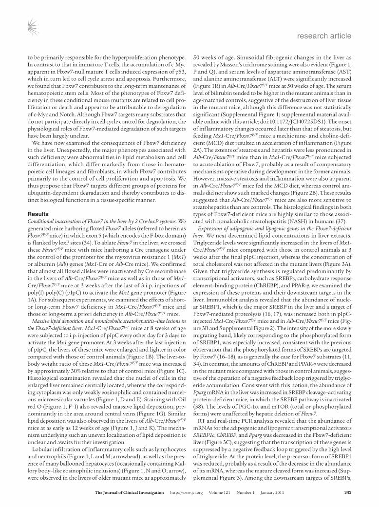
to be primarily responsible for the hyperproliferation phenotype. In contrast to that in immature T cells, the accumulation of c-Myc apparent in Fbxw7-null mature T cells induced expression of p53, which in turn led to cell cycle arrest and apoptosis. Furthermore, we found that Fbxw7 contributes to the long-term maintenance of hematopoietic stem cells. Most of the phenotypes of Fbxw7 defi-ciency in these conditional mouse mutants are related to cell pro-liferation or death and appear to be attributable to deregulation of c-Myc and Notch. Although Fbxw7 targets many substrates that do not participate directly in cell cycle control for degradation, the physiological roles of Fbxw7-mediated degradation of such targets have been largely unclear.
We have now examined the consequences of Fbxw7 deficiency in the liver. Unexpectedly, the major phenotypes associated with such deficiency were abnormalities in lipid metabolism and cell differentiation, which differ markedly from those in hemato-poietic cell lineages and fibroblasts, in which Fbxw7 contributes primarily to the control of cell proliferation and apoptosis. We thus propose that Fbxw7 targets different groups of proteins for ubiquitin-dependent degradation and thereby contributes to dis-tinct biological functions in a tissue-specific manner.
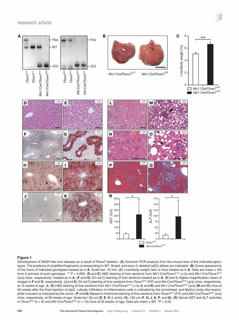
ResultsConditional inactivation of Fbxw7 in the liver by 2 Cre-loxP systems. We generated mice harboring floxed Fbxw7 alleles (referred to herein as Fbxw7F/F mice) in which exon 5 (which encodes the F-box domain) is flanked by loxP sites (34). To ablate Fbxw7 in the liver, we crossed these Fbxw7F/F mice with mice harboring a Cre transgene under the control of the promoter for the myxovirus resistance 1 (Mx1) or albumin (Alb) genes (Mx1-Cre or Alb-Cre mice). We confirmed that almost all floxed alleles were inactivated by Cre recombinase in the livers of Alb-Cre/Fbxw7F/F mice as well as in those of Mx1-Cre/Fbxw7F/F mice at 3 weeks after the last of 3 i.p. injections of poly(I)-poly(C) (pIpC) to activate the Mx1 gene promoter (Figure 1A). For subsequent experiments, we examined the effects of short- or long-term Fbxw7 deficiency in Mx1-Cre/Fbxw7F/F mice and those of long-term a priori deficiency in Alb-Cre/Fbxw7F/F mice.
Massive lipid deposition and nonalcoholic steatohepatitis–like lesions in the Fbxw7-deficient liver. Mx1-Cre/Fbxw7F/F mice at 8 weeks of age were subjected to i.p. injection of pIpC every other day for 3 days to activate the Mx1 gene promoter. At 3 weeks after the last injection of pIpC, the livers of these mice were enlarged and lighter in color compared with those of control animals (Figure 1B). The liver-to-body weight ratio of these Mx1-Cre/Fbxw7F/F mice was increased by approximately 30% relative to that of control mice (Figure 1C). Histological examination revealed that the nuclei of cells in the enlarged liver remained centrally located, whereas the correspond-ing cytoplasm was only weakly eosinophilic and contained numer-ous microvesicular vacuoles (Figure 1, D and E). Staining with Oil red O (Figure 1, F–I) also revealed massive lipid deposition, pre-dominantly in the area around central veins (Figure 1G). Similar lipid deposition was also observed in the livers of Alb-Cre/Fbxw7F/F mice at as early as 12 weeks of age (Figure 1, J and K). The mecha-nism underlying such an uneven localization of lipid deposition is unclear and awaits further investigation.
Lobular infiltration of inflammatory cells such as lymphocytes and neutrophils (Figure 1, L and M; arrowhead), as well as the pres-ence of many ballooned hepatocytes (occasionally containing Mal-lory body–like eosinophilic inclusions) (Figure 1, N and O; arrow), were observed in the livers of older mutant mice at approximately
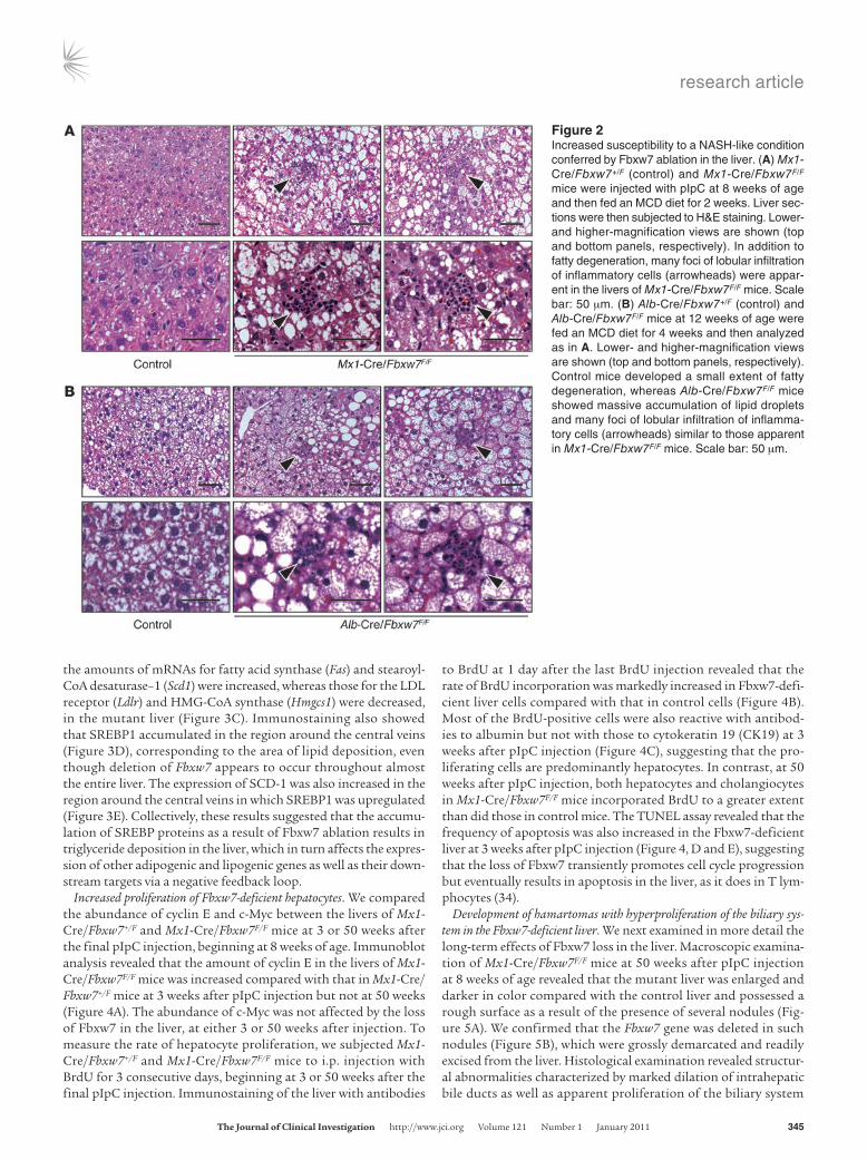
50 weeks of age. Sinusoidal fibrogenic changes in the liver as revealed by Masson’s trichrome staining were also evident (Figure 1, P and Q), and serum levels of aspartate aminotransferase (AST) and alanine aminotransferase (ALT) were significantly increased (Figure 1R) in Alb-Cre/Fbxw7F/F mice at 50 weeks of age. The serum level of bilirubin tended to be higher in the mutant animals than in age-matched controls, suggestive of the destruction of liver tissue in the mutant mice, although this difference was not statistically significant (Supplemental Figure 1; supplemental material avail-able online with this article; doi:10.1172/JCI40725DS1). The onset of inflammatory changes occurred later than that of steatosis, but feeding Mx1-Cre/Fbxw7F/F mice a methionine- and choline-defi-cient (MCD) diet resulted in acceleration of inflammation (Figure 2A). The extents of steatosis and hepatitis were less pronounced in Alb-Cre/Fbxw7F/F mice than in Mx1-Cre/Fbxw7F/F mice subjected to acute ablation of Fbxw7, probably as a result of compensatory mechanisms operative during development in the former animals. However, massive steatosis and inflammation were also apparent in Alb-Cre/Fbxw7F/F mice fed the MCD diet, whereas control ani-mals did not show such marked changes (Figure 2B). These results suggested that Alb-Cre/Fbxw7F/F mice are also more sensitive to steatohepatitis than are controls. The histological findings in both types of Fbxw7-deficient mice are highly similar to those associ-ated with nonalcoholic steatohepatitis (NASH) in humans (37).
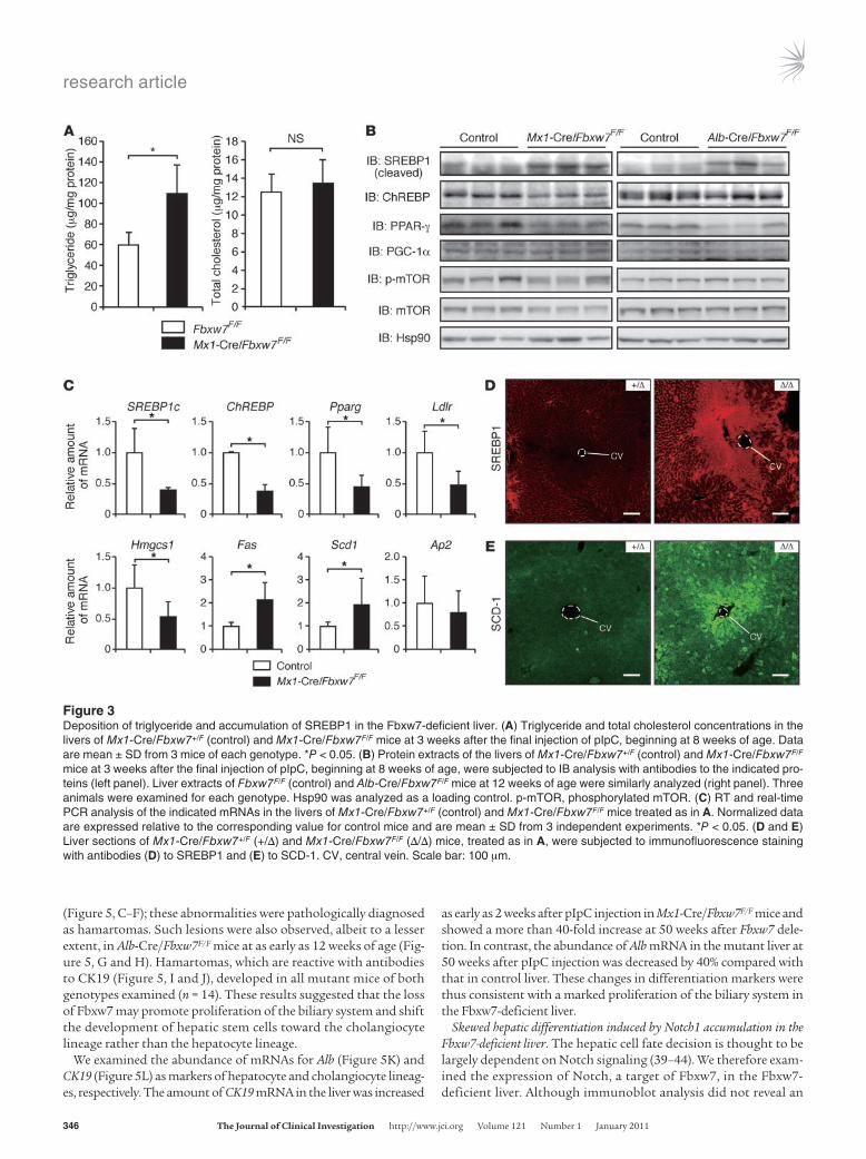
Expression of adipogenic and lipogenic genes in the Fbxw7-deficient liver. We next determined lipid concentrations in liver extracts. Triglyceride levels were significantly increased in the livers of Mx1-Cre/Fbxw7F/F mice compared with those in control animals at 3 weeks after the final pIpC injection, whereas the concentration of total cholesterol was not affected in the mutant livers (Figure 3A). Given that triglyceride synthesis is regulated predominantly by transcriptional activators, such as SREBPs, carbohydrate response element–binding protein (ChREBP), and PPAR-γ, we examined the expression of these proteins and their downstream targets in the liver. Immunoblot analysis revealed that the abundance of nucle-ar SREBP1, which is the major SREBP in the liver and a target of Fbxw7-mediated proteolysis (16, 17), was increased both in pIpC-injected Mx1-Cre/Fbxw7F/F mice and in Alb-Cre/Fbxw7F/F mice (Fig-ure 3B and Supplemental Figure 2). The intensity of the more slowly migrating band, likely corresponding to the phosphorylated form of SREBP1, was especially increased, consistent with the previous observation that the phosphorylated forms of SREBPs are targeted by Fbxw7 (16–18), as is generally the case for Fbxw7 substrates (11, 34). In contrast, the amounts of ChREBP and PPAR-γ were decreased in the mutant mice compared with those in control animals, sugges-tive of the operation of a negative feedback loop triggered by triglyc-eride accumulation. Consistent with this notion, the abundance of Pparg mRNA in the liver was increased in SREBP cleavage–activating protein–deficient mice, in which the SREBP pathway is inactivated (38). The levels of PGC-1α and mTOR (total or phosphorylated forms) were unaffected by hepatic deletion of Fbxw7.
RT and real-time PCR analysis revealed that the abundance of mRNAs for the adipogenic and lipogenic transcriptional activators SREBP1c, ChREBP, and Pparg was decreased in the Fbxw7-deficient liver (Figure 3C), suggesting that the transcription of these genes is suppressed by a negative feedback loop triggered by the high level of triglyceride. At the protein level, the precursor form of SREBP1 was reduced, probably as a result of the decrease in the abundance of its mRNA, whereas the mature cleaved form was increased (Sup-plemental Figure 3). Among the downstream targets of SREBPs,
research article
344 TheJournalofClinicalInvestigation http://www.jci.org Volume 121 Number 1 January 2011
Figure 1Development of NASH-like liver disease as a result of Fbxw7 deletion. (A) Genomic PCR analysis from the mouse liver of the indicated geno-types. The positions of amplified fragments corresponding to WT, floxed, and exon 5–deleted (ΔE5) alleles are indicated. (B) Gross appearance of the livers of indicated genotypes treated as in A. Scale bar: 10 mm. (C) Liver/body weight ratio of mice treated as in A. Data are mean ± SD from 5 animals of each genotype. ***P < 0.005. (D and E) H&E staining of liver sections from Mx1-Cre/Fbxw7+/F (+/Δ) and Mx1-Cre/Fbxw7F/F (Δ/Δ) mice, respectively, treated as in A. (F and G) Oil red O staining of liver sections treated as in A. (H and I) Higher-magnification views of images in F and G, respectively. (J and K) Oil red O staining of liver sections from Fbxw7F/F (F/F) and Alb-Cre/Fbxw7F/F (Δ/Δ) mice, respectively, at 12 weeks of age. (L–O) H&E staining of liver sections from Mx1-Cre/Fbxw7+/F (+/Δ) (L and N) and Mx1-Cre/Fbxw7F/F (Δ/Δ) (M and O) mice at 50 weeks after the final injection of pIpC. Lobular infiltration of inflammatory cells is indicated by the arrowhead, and Mallory body–like eosino-philic inclusion is indicated by the arrow. (P and Q) Masson’s trichrome staining of liver sections from Fbxw7F/F (F/F) and Alb-Cre/Fbxw7F/F (Δ/Δ) mice, respectively, at 50 weeks of age. Scale bar: 50 μm (D, E, H, I, and L–O); 100 μm (F, G, J, K, P, and Q). (R) Serum AST and ALT activities in Fbxw7F/F (n = 6) and Alb-Cre/Fbxw7F/F (n = 10) mice at 50 weeks of age. Data are mean ± SD. *P < 0.05.
research article
TheJournalofClinicalInvestigation http://www.jci.org Volume 121 Number 1 January 2011 345
the amounts of mRNAs for fatty acid synthase (Fas) and stearoyl-CoA desaturase–1 (Scd1) were increased, whereas those for the LDL receptor (Ldlr) and HMG-CoA synthase (Hmgcs1) were decreased, in the mutant liver (Figure 3C). Immunostaining also showed that SREBP1 accumulated in the region around the central veins (Figure 3D), corresponding to the area of lipid deposition, even though deletion of Fbxw7 appears to occur throughout almost the entire liver. The expression of SCD-1 was also increased in the region around the central veins in which SREBP1 was upregulated (Figure 3E). Collectively, these results suggested that the accumu-lation of SREBP proteins as a result of Fbxw7 ablation results in triglyceride deposition in the liver, which in turn affects the expres-sion of other adipogenic and lipogenic genes as well as their down-stream targets via a negative feedback loop.
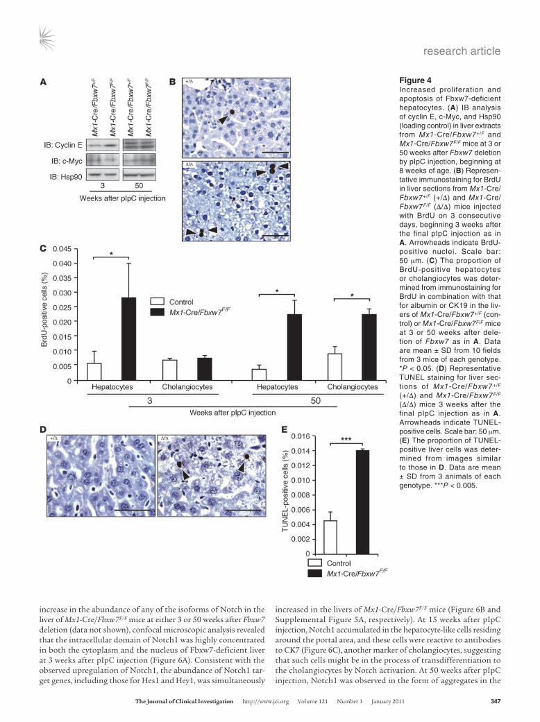
Increased proliferation of Fbxw7-deficient hepatocytes. We compared the abundance of cyclin E and c-Myc between the livers of Mx1-Cre/Fbxw7+/F and Mx1-Cre/Fbxw7F/F mice at 3 or 50 weeks after the final pIpC injection, beginning at 8 weeks of age. Immunoblot analysis revealed that the amount of cyclin E in the livers of Mx1-Cre/Fbxw7F/F mice was increased compared with that in Mx1-Cre/Fbxw7+/F mice at 3 weeks after pIpC injection but not at 50 weeks (Figure 4A). The abundance of c-Myc was not affected by the loss of Fbxw7 in the liver, at either 3 or 50 weeks after injection. To measure the rate of hepatocyte proliferation, we subjected Mx1-Cre/Fbxw7+/F and Mx1-Cre/Fbxw7F/F mice to i.p. injection with BrdU for 3 consecutive days, beginning at 3 or 50 weeks after the final pIpC injection. Immunostaining of the liver with antibodies
to BrdU at 1 day after the last BrdU injection revealed that the rate of BrdU incorporation was markedly increased in Fbxw7-defi-cient liver cells compared with that in control cells (Figure 4B). Most of the BrdU-positive cells were also reactive with antibod-ies to albumin but not with those to cytokeratin 19 (CK19) at 3 weeks after pIpC injection (Figure 4C), suggesting that the pro-liferating cells are predominantly hepatocytes. In contrast, at 50 weeks after pIpC injection, both hepatocytes and cholangiocytes in Mx1-Cre/Fbxw7F/F mice incorporated BrdU to a greater extent than did those in control mice. The TUNEL assay revealed that the frequency of apoptosis was also increased in the Fbxw7-deficient liver at 3 weeks after pIpC injection (Figure 4, D and E), suggesting that the loss of Fbxw7 transiently promotes cell cycle progression but eventually results in apoptosis in the liver, as it does in T lym-phocytes (34).
Development of hamartomas with hyperproliferation of the biliary sys-tem in the Fbxw7-deficient liver. We next examined in more detail the long-term effects of Fbxw7 loss in the liver. Macroscopic examina-tion of Mx1-Cre/Fbxw7F/F mice at 50 weeks after pIpC injection at 8 weeks of age revealed that the mutant liver was enlarged and darker in color compared with the control liver and possessed a rough surface as a result of the presence of several nodules (Fig-ure 5A). We confirmed that the Fbxw7 gene was deleted in such nodules (Figure 5B), which were grossly demarcated and readily excised from the liver. Histological examination revealed structur-al abnormalities characterized by marked dilation of intrahepatic bile ducts as well as apparent proliferation of the biliary system
Figure 2Increased susceptibility to a NASH-like condition conferred by Fbxw7 ablation in the liver. (A) Mx1-Cre/Fbxw7+/F (control) and Mx1-Cre/Fbxw7F/F mice were injected with pIpC at 8 weeks of age and then fed an MCD diet for 2 weeks. Liver sec-tions were then subjected to H&E staining. Lower- and higher-magnification views are shown (top and bottom panels, respectively). In addition to fatty degeneration, many foci of lobular infiltration of inflammatory cells (arrowheads) were appar-ent in the livers of Mx1-Cre/Fbxw7F/F mice. Scale bar: 50 μm. (B) Alb-Cre/Fbxw7+/F (control) and Alb-Cre/Fbxw7F/F mice at 12 weeks of age were fed an MCD diet for 4 weeks and then analyzed as in A. Lower- and higher-magnification views are shown (top and bottom panels, respectively). Control mice developed a small extent of fatty degeneration, whereas Alb-Cre/Fbxw7F/F mice showed massive accumulation of lipid droplets and many foci of lobular infiltration of inflamma-tory cells (arrowheads) similar to those apparent in Mx1-Cre/Fbxw7F/F mice. Scale bar: 50 μm.
research article
346 TheJournalofClinicalInvestigation http://www.jci.org Volume 121 Number 1 January 2011
(Figure 5, C–F); these abnormalities were pathologically diagnosed as hamartomas. Such lesions were also observed, albeit to a lesser extent, in Alb-Cre/Fbxw7F/F mice at as early as 12 weeks of age (Fig-ure 5, G and H). Hamartomas, which are reactive with antibodies to CK19 (Figure 5, I and J), developed in all mutant mice of both genotypes examined (n = 14). These results suggested that the loss of Fbxw7 may promote proliferation of the biliary system and shift the development of hepatic stem cells toward the cholangiocyte lineage rather than the hepatocyte lineage.
We examined the abundance of mRNAs for Alb (Figure 5K) and CK19 (Figure 5L) as markers of hepatocyte and cholangiocyte lineag-es, respectively. The amount of CK19 mRNA in the liver was increased
as early as 2 weeks after pIpC injection in Mx1-Cre/Fbxw7F/F mice and showed a more than 40-fold increase at 50 weeks after Fbxw7 dele-tion. In contrast, the abundance of Alb mRNA in the mutant liver at 50 weeks after pIpC injection was decreased by 40% compared with that in control liver. These changes in differentiation markers were thus consistent with a marked proliferation of the biliary system in the Fbxw7-deficient liver.
Skewed hepatic differentiation induced by Notch1 accumulation in the Fbxw7-deficient liver. The hepatic cell fate decision is thought to be largely dependent on Notch signaling (39–44). We therefore exam-ined the expression of Notch, a target of Fbxw7, in the Fbxw7-deficient liver. Although immunoblot analysis did not reveal an
Figure 3Deposition of triglyceride and accumulation of SREBP1 in the Fbxw7-deficient liver. (A) Triglyceride and total cholesterol concentrations in the livers of Mx1-Cre/Fbxw7+/F (control) and Mx1-Cre/Fbxw7F/F mice at 3 weeks after the final injection of pIpC, beginning at 8 weeks of age. Data are mean ± SD from 3 mice of each genotype. *P < 0.05. (B) Protein extracts of the livers of Mx1-Cre/Fbxw7+/F (control) and Mx1-Cre/Fbxw7F/F mice at 3 weeks after the final injection of pIpC, beginning at 8 weeks of age, were subjected to IB analysis with antibodies to the indicated pro-teins (left panel). Liver extracts of Fbxw7F/F (control) and Alb-Cre/Fbxw7F/F mice at 12 weeks of age were similarly analyzed (right panel). Three animals were examined for each genotype. Hsp90 was analyzed as a loading control. p-mTOR, phosphorylated mTOR. (C) RT and real-time PCR analysis of the indicated mRNAs in the livers of Mx1-Cre/Fbxw7+/F (control) and Mx1-Cre/Fbxw7F/F mice treated as in A. Normalized data are expressed relative to the corresponding value for control mice and are mean ± SD from 3 independent experiments. *P < 0.05. (D and E) Liver sections of Mx1-Cre/Fbxw7+/F (+/Δ) and Mx1-Cre/Fbxw7F/F (Δ/Δ) mice, treated as in A, were subjected to immunofluorescence staining with antibodies (D) to SREBP1 and (E) to SCD-1. CV, central vein. Scale bar: 100 μm.
research article
TheJournalofClinicalInvestigation http://www.jci.org Volume 121 Number 1 January 2011 347
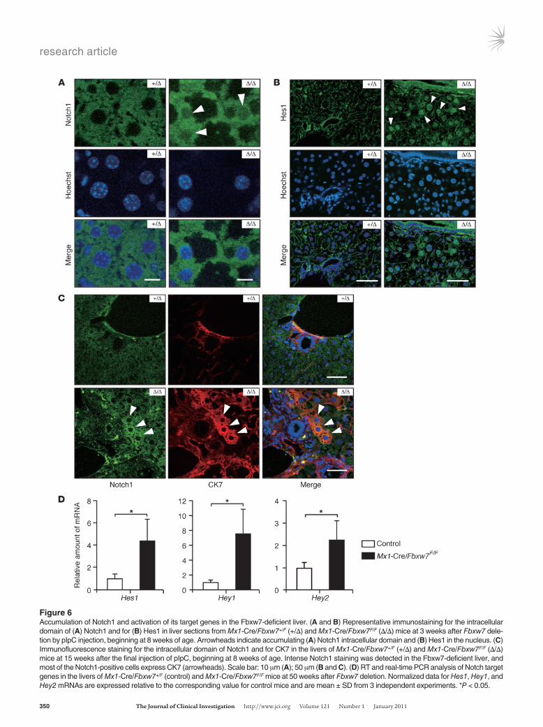
increase in the abundance of any of the isoforms of Notch in the liver of Mx1-Cre/Fbxw7F/F mice at either 3 or 50 weeks after Fbxw7 deletion (data not shown), confocal microscopic analysis revealed that the intracellular domain of Notch1 was highly concentrated in both the cytoplasm and the nucleus of Fbxw7-deficient liver at 3 weeks after pIpC injection (Figure 6A). Consistent with the observed upregulation of Notch1, the abundance of Notch1 tar-get genes, including those for Hes1 and Hey1, was simultaneously
increased in the livers of Mx1-Cre/Fbxw7F/F mice (Figure 6B and Supplemental Figure 5A, respectively). At 15 weeks after pIpC injection, Notch1 accumulated in the hepatocyte-like cells residing around the portal area, and these cells were reactive to antibodies to CK7 (Figure 6C), another marker of cholangiocytes, suggesting that such cells might be in the process of transdifferentiation to the cholangiocytes by Notch activation. At 50 weeks after pIpC injection, Notch1 was observed in the form of aggregates in the
Figure 4Increased proliferation and apoptosis of Fbxw7-deficient hepatocytes. (A) IB analysis of cyclin E, c-Myc, and Hsp90 (loading control) in liver extracts from Mx1-Cre/Fbxw7+/F and Mx1-Cre/Fbxw7F/F mice at 3 or 50 weeks after Fbxw7 deletion by pIpC injection, beginning at 8 weeks of age. (B) Represen-tative immunostaining for BrdU in liver sections from Mx1-Cre/Fbxw7+/F (+/Δ) and Mx1-Cre/Fbxw7F/F (Δ/Δ) mice injected with BrdU on 3 consecutive days, beginning 3 weeks after the final pIpC injection as in A. Arrowheads indicate BrdU-positive nuclei. Scale bar: 50 μm. (C) The proportion of BrdU-positive hepatocytes or cholangiocytes was deter-mined from immunostaining for BrdU in combination with that for albumin or CK19 in the liv-ers of Mx1-Cre/Fbxw7+/F (con-trol) or Mx1-Cre/Fbxw7F/F mice at 3 or 50 weeks after dele-tion of Fbxw7 as in A. Data are mean ± SD from 10 fields from 3 mice of each genotype. *P < 0.05. (D) Representative TUNEL staining for liver sec-tions of Mx1-Cre/Fbxw7+/F (+/Δ) and Mx1-Cre/Fbxw7F/F (Δ/Δ) mice 3 weeks after the final pIpC injection as in A. Arrowheads indicate TUNEL-positive cells. Scale bar: 50 μm. (E) The proportion of TUNEL-positive liver cells was deter-mined from images similar to those in D. Data are mean ± SD from 3 animals of each genotype. ***P < 0.005.

research article
348 TheJournalofClinicalInvestigation http://www.jci.org Volume 121 Number 1 January 2011
Figure 5Hamartoma development as a result of long-term ablation of Fbxw7 in the liver. (A) Gross appearance of the livers of Mx1-Cre/Fbxw7+/F and Mx1-Cre/Fbxw7F/F mice at 50 weeks after the final injection of pIpC, beginning at 8 weeks of age. Scale bar: 10 mm. (B) PCR analysis of genomic DNA from the dilated bile ducts excised from hamartomas in the livers of 2 Mx1-Cre/Fbxw7F/F mice. Genomic DNA from control mice was also analyzed. (C–F) H&E staining of liver sections from a Mx1-Cre/Fbxw7+/F (+/Δ) mouse (C) and from a Mx1-Cre/Fbxw7F/F (Δ/Δ) mouse (D–F) that developed hamartoma after Fbxw7 deletion as in A. (G and H) H&E staining of liver sections from Fbxw7F/F (F/F) and Alb-Cre/Fbxw7F/F (Δ/Δ) mice, respectively, at 12 weeks of age. Arrowheads indicate malformation of the ductal plate. Scale bar: 50 μm (F); 100 μm (E, G, and H); 200 μm (C and D). (I and J) Immunofluorescence staining for CK19 in the livers of Mx1-Cre/Fbxw7+/F (+/Δ) and Mx1-Cre/Fbxw7F/F (Δ/Δ) mice at 50 weeks after the final injection of pIpC, beginning at 8 weeks of age. The dashed line indicates the outer boundary of portal vein. PV, portal vein; BD, bile duct. Scale bar: 25 μm. (K and L) RT and real-time PCR analysis of Alb and CK19 mRNAs, respectively, in the livers of Mx1-Cre/Fbxw7+/F (control) and Mx1-Cre/Fbxw7F/F mice at 1, 2, or 50 weeks after deletion of Fbxw7 as in A. Normalized data are expressed relative to the corresponding value for control mice. Data are mean ± SD from 3 independent experiments. *P < 0.05.

research article
TheJournalofClinicalInvestigation http://www.jci.org Volume 121 Number 1 January 2011 349
cytoplasm or the nucleus (Supplemental Figure 4A). The increase in Hes1 or Hey1 was not detected by immunostaining analysis at this period (Supplemental Figure 4B and Supplemental Figure 5B), but the abundance of mRNAs for Hes1, Hey1, and Hey2 was increased in the livers of Mx1-Cre/Fbxw7F/F mice at 50 weeks after pIpC injection (Figure 6D). Neither Notch2, mutations in the gene in which mutations result in Alagille disease, nor Notch3 or Notch4, the expression of both of which is increased in hepatocel-lular carcinoma, were detected by immunofluorescence analysis in the livers of either control or Mx1-Cre/Fbxw7F/F mice (data not shown). However, neither the expression of Notch ligands, such as Dll-1 and Jagged-1, nor that of the Notch cofactor RBP-J in the liver appeared to be affected by the loss of Fbxw7 (Supplemental Figure 6). We also examined the expression of TSC1 and TSC2, given that the loss of function of either TSC1 or TSC2 is known to result in the development of hamartoma in humans. However, no difference in expression of TSC1 or TSC2 was found between Fbxw7-deficient and control mice (Supplemental Figure 6).
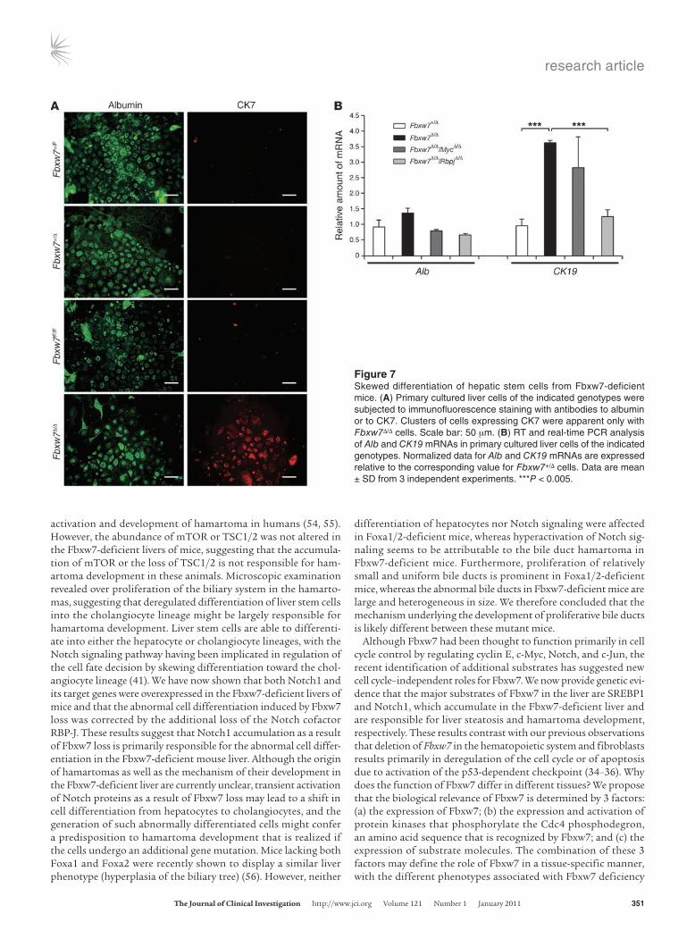
To investigate whether the skewed developmental orientation toward the cholangiocyte lineage apparent in the Fbxw7-defi-cient liver is dependent on Notch1 accumulation, we examined the differentiation of hepatic stem cells in culture (45). A frac-tion containing hepatic stem cells was prepared from the livers of Fbxw7+/F and Fbxw7F/F embryos and was then infected with a retrovirus encoding Cre recombinase or with the empty virus alone to generate Fbxw7+/F, Fbxw7+/Δ, Fbxw7F/F, and Fbxw7Δ/Δ cells in the presence of HGF and EGF. Immunofluorescence analysis revealed that most of the Fbxw7+/F, Fbxw7+/Δ, and Fbxw7F/F cells differentiated into the hepatocyte lineage, characterized by albu-min expression, with only a small subset of cells differentiating into the cholangiocyte lineage (Figure 7A). In contrast, the per-centage of Fbxw7Δ/Δ cells that differentiated into the cholangio-cyte lineage, characterized by expression of CK7, was markedly increased compared with that for cells of the control genotypes. To confirm these results in a quantitative manner, we performed RT and real-time PCR analysis of Alb and CK19 mRNAs. Con-sistent with the immunofluorescence data, the amount of CK19 mRNA was significantly increased in Fbxw7Δ/Δ cells compared with that in Fbxw7+/Δ cells, whereas the abundance of Alb mRNA did not differ between the 2 genotypes (Figure 7B).
Notch signaling is implicated in the differentiation of liver stem cells into the cholangiocyte lineage. Indeed, immunofluorescence analysis revealed that Notch1 accumulated in Fbxw7Δ/Δ cells to a greater extent than in Fbxw7+/Δ cells (Supplemental Figure 7). We therefore examined whether additional ablation of the Notch cofactor RBP-J might correct the abnormal development of Fbxw7-deficient liver stem cells. We generated Fbxw7-deficient hepatic stem cells with additional deletion of either Rbpj or Myc genes and examined the level of CK19 mRNA. The abundance of CK19 mRNA was increased in Fbxw7Δ/ΔMycΔ/Δ cells but not in Fbxw7Δ/ΔRbpjΔ/Δ cells (Figure 7B). These results indicate that the skewed developmental orientation of hepatic stem cells to the cholangiocyte lineage is dependent on Notch1 accumulation induced by the loss of Fbxw7.
DiscussionGiven that the substrates of Fbxw7 include key proteins that con-tribute to diverse biological processes, including the cell cycle, cell differentiation, and apoptosis, and that the binding of Fbxw7 to its substrates depends on their phosphorylation, the function
of this protein is likely complex. Although much attention has focused on the relation between the accumulation of cyclin E due to loss of Fbxw7 function and tumorigenesis, Notch degradation by Fbxw7 is critical during embryogenesis, suggesting that Fbxw7 functions in development- and tissue-dependent manners. To pro-vide insight into the physiological and pathological relevance of Fbxw7, we have induced conditional inactivation of Fbxw7 in sev-eral mouse tissues. Our previous studies have shown that ablation of Fbxw7 in hematopoietic cells or fibroblasts results in abnormali-ties that are mainly related to the cell cycle and apoptosis. We now show that liver-specific ablation of Fbxw7 induced fatty liver and abnormal cell differentiation, likely as a result of the accumula-tion of SREBPs and Notch1, respectively, as well as promoted cell proliferation (Figure 8).
We generated 2 types of mice with liver-specific deficiency of Fbxw7 with the use of the Mx1 or Alb gene promoters to drive Cre expression. The phenotypes of Alb-Cre/Fbxw7F/F mice are milder than those induced by acute ablation of Fbxw7 in Mx1-Cre/Fbxw7F/F mice, probably because of the operation of compen-satory mechanisms during development in the former animals. In Mx1-Cre/Fbxw7F/F mice, it would be expected for Fbxw7 to be deleted in cells and tissues other than the liver, such as hemato-poietic cells. To exclude the possibility that ablation of Fbxw7 in hematopoietic cell lineages might be responsible for steatohepa-titis, we have generated Lck-Cre/Fbxw7F/F and CD4-Cre/Fbxw7F/F mice (in both of which Fbxw7 deletion occurs in T cells), CD19-Cre/Fbxw7F/F mice (Fbxw7 deletion occurs in B cells), and LysM-Cre/Fbxw7F/F mice (Fbxw7 deletion occurs in myeloid cells). None of these animals showed either fatty liver or hepatic inflammation (data not shown). Furthermore, Alb-Cre/Fbxw7F/F mice manifested pronounced hepatic infiltration of inflammatory cells when they were fed an MCD diet, confirming that the steatohepatitis induced by Fbxw7 deletion is attributable to an effect that is intrinsic to the liver.
Nonalcoholic fatty liver disease (NAFLD) is a growing health concern, due to its rapidly increasing prevalence worldwide. NASH is a progressive form of NAFLD that has the potential to develop into hepatocellular carcinoma. We now show that mice with liver-specific ablation of Fbxw7 developed clinicopathologic features similar to those of NAFLD or NASH in humans, including triglyc-eride deposition around central veins, pericellular fibrosis, infiltra-tion of inflammatory mononuclear cells, and the appearance of Mallory bodies in the liver as well as increases in the serum levels of ALT and AST. However, these animals were not found to devel-op hepatocellular carcinoma. Genetic mouse models for human NASH have been established by functional deletion of leptin (46) or its receptor (47), phosphatase and tensin homolog (PTEN) (48), NEMO (also known as IKK-γ) (49), interleukin-1 receptor α (50), galectin-3 (51), or retinoic acid receptor α (52). Mice transgenic for SREBP1c also manifest pronounced NASH (53). SREBP1c is degraded in an Fbxw7-dependent manner (16), and we have now shown that it accumulated in the Fbxw7-deficient liver. These findings thus suggest that an Fbxw7-SREBP1 axis plays a key phys-iological role in the regulation of lipid metabolism in the liver as well as a pathological role in the development of NASH.
Whereas steatosis develops in the acute phase of liver-specific Fbxw7 deficiency, hamartoma develops in the chronic phase. Fbxw7 targets mTOR for degradation (19). The TSC complex, consisting of TSC1 (hamartin) and TSC2 (tuberin), is the major negative regulator of mTOR, and its genetic loss results in mTOR
research article
350 TheJournalofClinicalInvestigation http://www.jci.org Volume 121 Number 1 January 2011
Figure 6Accumulation of Notch1 and activation of its target genes in the Fbxw7-deficient liver. (A and B) Representative immunostaining for the intracellular domain of (A) Notch1 and for (B) Hes1 in liver sections from Mx1-Cre/Fbxw7+/F (+/Δ) and Mx1-Cre/Fbxw7F/F (Δ/Δ) mice at 3 weeks after Fbxw7 dele-tion by pIpC injection, beginning at 8 weeks of age. Arrowheads indicate accumulating (A) Notch1 intracellular domain and (B) Hes1 in the nucleus. (C) Immunofluorescence staining for the intracellular domain of Notch1 and for CK7 in the livers of Mx1-Cre/Fbxw7+/F (+/Δ) and Mx1-Cre/Fbxw7F/F (Δ/Δ) mice at 15 weeks after the final injection of pIpC, beginning at 8 weeks of age. Intense Notch1 staining was detected in the Fbxw7-deficient liver, and most of the Notch1-positive cells express CK7 (arrowheads). Scale bar: 10 μm (A); 50 μm (B and C). (D) RT and real-time PCR analysis of Notch target genes in the livers of Mx1-Cre/Fbxw7+/F (control) and Mx1-Cre/Fbxw7F/F mice at 50 weeks after Fbxw7 deletion. Normalized data for Hes1, Hey1, and Hey2 mRNAs are expressed relative to the corresponding value for control mice and are mean ± SD from 3 independent experiments. *P < 0.05.
research article
TheJournalofClinicalInvestigation http://www.jci.org Volume 121 Number 1 January 2011 351
activation and development of hamartoma in humans (54, 55). However, the abundance of mTOR or TSC1/2 was not altered in the Fbxw7-deficient livers of mice, suggesting that the accumula-tion of mTOR or the loss of TSC1/2 is not responsible for ham-artoma development in these animals. Microscopic examination revealed over proliferation of the biliary system in the hamarto-mas, suggesting that deregulated differentiation of liver stem cells into the cholangiocyte lineage might be largely responsible for hamartoma development. Liver stem cells are able to differenti-ate into either the hepatocyte or cholangiocyte lineages, with the Notch signaling pathway having been implicated in regulation of the cell fate decision by skewing differentiation toward the chol-angiocyte lineage (41). We have now shown that both Notch1 and its target genes were overexpressed in the Fbxw7-deficient livers of mice and that the abnormal cell differentiation induced by Fbxw7 loss was corrected by the additional loss of the Notch cofactor RBP-J. These results suggest that Notch1 accumulation as a result of Fbxw7 loss is primarily responsible for the abnormal cell differ-entiation in the Fbxw7-deficient mouse liver. Although the origin of hamartomas as well as the mechanism of their development in the Fbxw7-deficient liver are currently unclear, transient activation of Notch proteins as a result of Fbxw7 loss may lead to a shift in cell differentiation from hepatocytes to cholangiocytes, and the generation of such abnormally differentiated cells might confer a predisposition to hamartoma development that is realized if the cells undergo an additional gene mutation. Mice lacking both Foxa1 and Foxa2 were recently shown to display a similar liver phenotype (hyperplasia of the biliary tree) (56). However, neither
differentiation of hepatocytes nor Notch signaling were affected in Foxa1/2-deficient mice, whereas hyperactivation of Notch sig-naling seems to be attributable to the bile duct hamartoma in Fbxw7-deficient mice. Furthermore, proliferation of relatively small and uniform bile ducts is prominent in Foxa1/2-deficient mice, whereas the abnormal bile ducts in Fbxw7-deficient mice are large and heterogeneous in size. We therefore concluded that the mechanism underlying the development of proliferative bile ducts is likely different between these mutant mice.
Although Fbxw7 had been thought to function primarily in cell cycle control by regulating cyclin E, c-Myc, Notch, and c-Jun, the recent identification of additional substrates has suggested new cell cycle–independent roles for Fbxw7. We now provide genetic evi-dence that the major substrates of Fbxw7 in the liver are SREBP1 and Notch1, which accumulate in the Fbxw7-deficient liver and are responsible for liver steatosis and hamartoma development, respectively. These results contrast with our previous observations that deletion of Fbxw7 in the hematopoietic system and fibroblasts results primarily in deregulation of the cell cycle or of apoptosis due to activation of the p53-dependent checkpoint (34–36). Why does the function of Fbxw7 differ in different tissues? We propose that the biological relevance of Fbxw7 is determined by 3 factors: (a) the expression of Fbxw7; (b) the expression and activation of protein kinases that phosphorylate the Cdc4 phosphodegron, an amino acid sequence that is recognized by Fbxw7; and (c) the expression of substrate molecules. The combination of these 3 factors may define the role of Fbxw7 in a tissue-specific manner, with the different phenotypes associated with Fbxw7 deficiency
Figure 7Skewed differentiation of hepatic stem cells from Fbxw7-deficient mice. (A) Primary cultured liver cells of the indicated genotypes were subjected to immunofluorescence staining with antibodies to albumin or to CK7. Clusters of cells expressing CK7 were apparent only with Fbxw7Δ/Δ cells. Scale bar: 50 μm. (B) RT and real-time PCR analysis of Alb and CK19 mRNAs in primary cultured liver cells of the indicated genotypes. Normalized data for Alb and CK19 mRNAs are expressed relative to the corresponding value for Fbxw7+/Δ cells. Data are mean ± SD from 3 independent experiments. ***P < 0.005.

research article
352 TheJournalofClinicalInvestigation http://www.jci.org Volume 121 Number 1 January 2011
being attributable to different expression patterns of Fbxw7, its substrates, and kinases that phosphorylate each substrate.
MethodsGeneration of conditional knockout mice. Mice homozygous for the floxed Fbxw7 allele (Fbxw7F/F mice) (34) were crossed with Mx1-Cre transgenic mice (57) provided by K. Rajewsky (Harvard Medical School, Boston, Mas-sachusetts, USA) or Alb-Cre transgenic mice (58) purchased from The Jackson Laboratory. Expression of Cre recombinase in the resulting off-spring of the former cross was induced by i.p. injection of 500 μg pIpC (GE Healthcare Biosciences) on 3 alternate days. Deletion of exon 5 of the floxed Fbxw7 allele was confirmed by PCR analysis of genomic DNA as previously described (34). Fbxw7F/F mice were also crossed with RbpjF/F mice (59) provided by T. Honjo (Kyoto University, Kyoto, Japan) or MycF/F mice (60) provided by I.M. de Alborán (National Center for Biotechnology, Madrid, Spain). The experimental protocols were approved by the Institu-tional Animal Care and Use Committee of Kyushu University.
Histological and biochemical analysis. Liver tissue was fixed with 4% paraformaldehyde in PBS, embedded in paraffin, and stained with H&E or Masson’s trichrome solution. Some sections were stained with Oil red O (Nakalai Tesque) according to standard procedures, in order to examine the extent of lipid accumulation in hepatocytes. Serum levels of AST and ALT were measured with a standard clinical autoanalyzer.
Dietary model of NASH. Mice were fed with an MCD diet (Funabashi Farm) for the indicated periods (see the legend for Figure 2) and analyzed.
Measurement of triglyceride and total cholesterol levels in the liver. Frozen liver tissue was homogenized, and triglyceride and total cholesterol were extracted from the homogenate with chloroform/methanol (2:1, vol/vol),
dried, and resuspended in 2-propanol. The amounts of triglyceride and total cholesterol in the extract were measured with the use of Lipidos liq-uid and Cholescolor liquid kits (Toyobo), respectively.
Immunoblot analysis. Total protein extracts were prepared from liver with RIPA buffer. The extracts (30 μg) were subjected to immunoblot analysis as described previously (61) with antibodies to cyclin E (M-20), to c-Myc (N-262), to ChREBP (P-13), or to PPAR-γ (E-8), all of which were obtained from Santa Cruz Biotechnology Inc.; with antibodies to Ser2448-phosphorylated or total (7C10) forms of mTOR (Cell Signaling Technol-ogy); with antibodies to SREBP1 (2A4, NeoMarkers); or with antibodies to PGC-1α (Chemicon). As a control, each membrane was stripped and then probed with antibodies to Hsp90 (BD Transduction Laboratories).
RT and real-time PCR analysis. Total RNA was extracted from liver using the guanidinium thiocyanate–phenol-chloroform method, purified, and subjected (1 μg) to RT with random hexanucleotide primers (ReverTra Ace α, Toyobo). The resulting cDNA was subjected to real-time PCR in a reaction mixture that contained 1× SYBR Green PCR Master Mix (Applied Biosystems) and 200 nM of gene-specific primers. Assays were performed in triplicate with an ABI Prism 7700 Sequence Detector (Applied Biosystems). The PCR protocol comprised 40 cycles of incubation at 60°C for 30 seconds and 95°C for 5 seconds. The sequences of the PCR primers (sense and antisense, respectively) were 5′-TGCTCCCAGCTGCAGGC-3′ and 5′-GCCCGGTAGCTCTGGGTGTA-3′ for Fas, 5′-TGGGTTGGCTGCTTGTG-3′ and 5′-GCGTGGGCAGGATGAAG-3′ for Scd1, 5′-CTGCCGACCTGATGAATTCC-3′ and 5′-TAGGGCCATCACACT-GTGTC-3′ for Ldlr, 5′-GCTCTCCATACAGTGCTACC-3′ and 5′-GAGTGAAA-GATCATGAAGCC-3′ for Hmgcs1, 5′-AGAGATGCCATCTCCAGCCTC-3′ and 5′-CTTGGTCTTAGGGTCTTCAGG-3′ for ChREBP, 5′-CTGTGAAGTTCAAT-GCACTGGAA-3′ and 5′-CCTCGATGGGCTTCACGTT-3′ for Pparg, 5′-CAT-GGATTGCACATTTGAAG-3′ and 5′-CCTGTGTCCCCTGTCTCA-3′ for SREBP1c, 5′-TCCTGTGCTGCAGCCTTTCTCA-3′ and 5′-CCAGGTTCCCA-CAAAGGCATCA-3′ for fatty acid–binding protein 4, 5′-GTCCTACA-GATTGACAATGC-3′ and 5′-CACGCTCTGGATCTGTGACAG-3′ for CK19, 5′-CATGACACCATGCCTGCTGAT-3′ and 5′-CTCTGATCTTCAG-GAAGTGTAC-3′ for Alb, 5′-CATTCCAAGCTAGAGAAGGCAG-3′ and 5′-TATTTCCCCAACACGCTCG-3′ for Hes1, 5′-AAAATGCTGCACACTG-CAGG-3′ and 5′-CGAGTCCTTCAATGATGCTCAG-3′ for Hey1, 5′-AAAC-GACCTCCGAAAGCGA-3′ and 5′-CGGTGAATTGGACCTCATCACT-3′ for Hey2, and 5′-GGAACATAGCCGTAAACTGC-3′ and 5′-TCACTGTGCCT-GAACTTACC-3′ for β-tubulin. Reactions for β-tubulin mRNA were performed concurrently on the same plate as those for the test mRNAs, and results were normalized by the corresponding amount of β-tubulin mRNA.
BrdU incorporation in vivo. Mice were injected with BrdU (1 mg, i.p.) on 3 consecutive days. The liver was removed 24 hours after the third injec-tion of BrdU, and BrdU incorporation was examined with an In Situ BrdU Detection Kit (BD Biosciences). BrdU-positive cells were counted in 10 dif-ferent fields at high (×400) magnification, and the percentage of BrdU-positive cells was calculated.
Immunofluorescence microscopy. Liver tissue was fixed with 4% para-formaldehyde in PBS and sectioned at a thickness of 40 μm with a vibra-tome. Sections were then immunostained with antibodies to the intracellular domain of Notch1 or to SCD-1 (both from Cell Signaling Technology), to Hes1 (AB5702, Millipore), to SREBP1 (2A4, NeoMarkers), to albumin (Bio-genesis), to CK19 (45), or to CK7 (MAB3226, Chemicon). Immune complex-es were detected with Alexa Fluor 488– or Alexa Fluor 546–conjugated goat antibodies to mouse or rabbit IgG (Invitrogen). Cultured liver cells were also subjected to immunostaining, as described previously (45), with the antibod-ies to albumin and to CK7. For confocal microscopic analysis, we used Zeiss LSM 510 META Confocal Microscope (Carl Zeiss MicroImaging).
TUNEL assay. The TUNEL assay was performed as described previously (62). In brief, paraffin-embedded sections of liver were treated with H2O2,
Figure 8A model for Fbxw7 functions in vivo. Fbxw7 mediates ubiquitin-depen-dent degradation of substrates in different functional categories. For example, Fbxw7 controls cell proliferation by targeting c-Myc, cyclin E, and c-Jun for degradation. However, it also regulates lipid metabo-lism and cell differentiation by targeting SREBP and Notch proteins, respectively. Major phenotypes associated with Fbxw7 deficiency in different tissues are shown in red. Ub, ubiquitin.
research article
TheJournalofClinicalInvestigation http://www.jci.org Volume 121 Number 1 January 2011 353
permeabilized for 15 minutes at 37°C with proteinase K (20 μg/ml, Sigma-Aldrich), and then incubated for 1 hour at 37°C with a reaction mixture containing terminal deoxynucleotidyl transferase (Invitrogen) and biotinylated dUTP (Boehringer Ingelheim). Labeled DNA was visual-ized with an ABC Kit (Vector Laboratories) and diaminobenzidine.
Primary culture of fetal hepatocytes. For the preparation of a single-cell sus-pension, the livers of mice at E13.5 were dissociated in culture medium (DMEM supplemented with 10% FBS, γ-insulin [1 μg/ml, Wako], 0.1 μM dexamethasone [Sigma-Aldrich], 10 mM nicotinamide [Sigma-Aldrich], 2 mM l-glutamine [Gibco BRL], 50 μM β-mercaptoethanol [Sigma-Aldrich], 5 mM HEPES [Wako], and penicillin-streptomycin [Gibco BRL]) by repeated passage of the tissue through the mouth of a pipette. Human recombinant HGF (50 ng/ml, Sigma-Aldrich) and EGF (20 ng/ml, Sigma-Aldrich) were added to the cells at 24 hours after culture initiation. Cells were seeded at a density of 1 × 10⁶ cells per well in 6-well plates for infection with retroviruses as described below (45).
Gene deletion in cultured cells by retroviral infection. cDNA encoding Cre recombinase was subcloned into the retroviral vector pMX-puro provided by T. Kitamura (University of Tokyo, Tokyo, Japan), and the resulting construct was introduced into Plat E packaging cells (63) with the use of the FuGENE6 reagent (Roche). The resulting culture supernatants con-taining the recombinant ecotropic retrovirus were harvested and incu-bated for 24 hours in the presence of Polybrene (2 μg/ml; Sigma-Aldrich) with proliferating liver cells harboring floxed alleles of Fbxw7, Rbpj, or Myc. The cells were cultured for an additional 24 hours in virus-free med-
ium, subjected to selection in medium containing puromycin (3 μg/ml), cultured for 96 hours in puromycin-free medium, and then harvested.
Statistics. Data are presented as mean ± SD and were analyzed using 2-tailed Student’s t test. A P value of less than 0.05 was considered sta-tistically significant.
AcknowledgmentsWe thank T. Honjo for Rbpj floxed mice; I.M. de Alborán for Myc floxed mice; K. Rajewsky for Mx1-Cre transgenic mice; T. Kitamura for pMX-puro; S. Aishima, Y. Nishihara, M. Sakamoto, and R. Irie for discussion; N. Kitajima, Y. Yamada, and K. Takeda for technical assistance; members of our laboratories for comments on the manu-script; and A. Ohta and M. Kimura for help in the preparation of the manuscript. This work was supported in part by a grant from the Ministry of Education, Culture, Sports, Science, and Technology of Japan and by a research grant from the Takeda Science Foundation.
Received for publication August 6, 2009, and accepted in revised form September 29, 2010.
Address correspondence to: Keiichi I. Nakayama, Department of Molecular and Cellular Biology, Medical Institute of Bioregulation, Kyushu University, 3-1-1 Maidashi, Higashi-ku, Fukuoka, Fukuoka 812-8582, Japan. Phone: 81.92.642.6815; Fax: 81.92.642.6819; E-mail: [email protected].
1. Hershko A, Ciechanover A. The ubiquitin system. Annu Rev Biochem. 1998;67:425–479.
2. Nakayama KI, Nakayama K. Ubiquitin ligases: cell-cycle control and cancer. Nat Rev Cancer. 2006; 6(5):369–381.
3. Frescas D, Pagano M. Deregulated proteolysis by the F-box proteins SKP2 and beta-TrCP: tipping the scales of cancer. Nat Rev Cancer. 2008;8(6):438–449.
4. Welcker M, Clurman BE. FBW7 ubiquitin ligase: a tumour suppressor at the crossroads of cell divi-sion, groWTh and differentiation. Nat Rev Cancer. 2008;8(2):83–93.
5. Hubbard EJ, Wu G, Kitajewski J, Greenwald I. sel-10, a negative regulator of lin-12 activity in Caenorhab-ditis elegans, encodes a member of the CDC4 family of proteins. Genes Dev. 1997;11(23):3182–3193.
6. Sundaram M, Greenwald I. Suppressors of a lin-12 hypomorph define genes that interact with both lin-12 and glp-1 in Caenorhabditis elegans. Genetics. 1993;135(3):765–783.
7. Gupta-Rossi N, et al. Functional interaction between SEL-10, an F-box protein, and the nuclear form of activated Notch1 receptor. J Biol Chem. 2001;276(37):34371–34378.
8. Oberg C, Li J, Pauley A, Wolf E, Gurney M, Lendahl U. The Notch intracellular domain is ubiquitinated and negatively regulated by the mammalian Sel-10 homolog. J Biol Chem. 2001;276(38):35847–35853.
9. Koepp DM, et al. Phosphorylation-dependent ubiquitination of cyclin E by the SCFFbw7 ubiquitin ligase. Science. 2001;294(5540):173–177.
10. Moberg KH, Bell DW, Wahrer DC, Haber DA, Hari-haran IK. Archipelago regulates Cyclin E levels in Drosophila and is mutated in human cancer cell lines. Nature. 2001;413(6853):311–316.
11. Strohmaier H, Spruck CH, Kaiser P, Won KA, Sang-felt O, Reed SI. Human F-box protein hCdc4 targets cyclin E for proteolysis and is mutated in a breast cancer cell line. Nature. 2001;413(6853):316–322.
12. Yada M, et al. Phosphorylation-dependent degra-dation of c-Myc is mediated by the F-box protein Fbw7. EMBO J. 2004;23(10):2116–2125.
13. Welcker M, et al. The Fbw7 tumor suppressor regu-lates glycogen synthase kinase 3 phosphorylation-dependent c-Myc protein degradation. Proc Natl
Acad Sci U S A. 2004;101(24):9085–9090. 14. Nateri AS, Riera-Sans L, Da Costa C, Behrens A. The
ubiquitin ligase SCFFbw7 antagonizes apoptotic JNK signaling. Science. 2004;303(5662):1374–1378.
15. Wei W, Jin J, Schlisio S, Harper JW, Kaelin WG Jr. The v-Jun point mutation allows c-Jun to escape GSK3-dependent recognition and destruction by the Fbw7 ubiquitin ligase. Cancer Cell. 2005;8(1):25–33.
16. Sundqvist A, et al. Control of lipid metabolism by phosphorylation-dependent degradation of the SREBP family of transcription factors by SCFFbw7. Cell Metab. 2005;1(6):379–391.
17. Punga T, Bengoechea-Alonso MT, Ericsson J. Phosphorylation and ubiquitination of the tran-scription factor sterol regulatory element-binding protein-1 in response to DNA binding. J Biol Chem. 2006;281(35):25278–25286.
18. Bengoechea-Alonso MT, Ericsson J. A phosphory-lation cascade controls the degradation of active SREBP1. J Biol Chem. 2009;284(9):5885–5895.
19. Mao JH, et al. FBXW7 targets mTOR for degrada-tion and cooperates with PTEN in tumor suppres-sion. Science. 2008;321(5895):1499–1502.
20. Olson BL, et al. SCFCdc4 acts antagonistically to the PGC-1alpha transcriptional coactivator by target-ing it for ubiquitin-mediated proteolysis. Genes Dev. 2008;22(2):252–264.
21. Maser RS, et al. Chromosomally unstable mouse tumours have genomic alterations similar to diverse human cancers. Nature. 2007;447(7147):966–971.
22. Lee JW, et al. Mutational analysis of the hCDC4 gene in gastric carcinomas. Eur J Cancer. 2006; 42(14):2369–2373.
23. Kemp Z, et al. CDC4 mutations occur in a sub-set of colorectal cancers but are not predicted to cause loss of function and are not associated with chromosomal instability. Cancer Res. 2005; 65(24):11361–11366.
24. Hubalek MM, et al. Cyclin E dysregulation and chromosomal instability in endometrial cancer. Oncogene. 2004;23(23):4187–4192.
25. Koh MS, Ittmann M, Kadmon D, Thompson TC, Leach FS. CDC4 gene expression as potential bio-marker for targeted therapy in prostate cancer. Can-cer Biol Ther. 2006;5(1):78–83.
26. Calhoun ES, et al. BRAF and FBXW7 (CDC4, FBW7, AGO, SEL10) mutations in distinct subsets of pancreatic cancer: potential therapeutic targets. Am J Pathol. 2003;163(4):1255–1260.
27. Akhoondi S, et al. FBXW7/hCDC4 is a general tumor suppressor in human cancer. Cancer Res. 2007;67(19):9006–9012.
28. Song JH, Schnittke N, Zaat A, Walsh CS, Miller CW. FBXW7 mutation in adult T-cell and B-cell acute lymphocytic leukemias. Leuk Res. 2008; 32(11):1751–1755.
29. Thompson BJ, et al. The SCFFBW7 ubiquitin ligase complex as a tumor suppressor in T cell leukemia. J Exp Med. 2007;204(8):1825–1835.
30. O’Neil J, et al. FBW7 mutations in leukemic cells mediate NOTCH pathway activation and resis-tance to gamma-secretase inhibitors. J Exp Med. 2007;204(8):1813–1824.
31. Malyukova A, et al. The tumor suppressor gene hCDC4 is frequently mutated in human T-cell acute lymphoblastic leukemia with functional consequences for Notch signaling. Cancer Res. 2007;67(12):5611–5616.
32. Tsunematsu R, et al. Mouse Fbw7/Sel-10/Cdc4 is required for notch degradation during vascular development. J Biol Chem. 2004;279(10):9417–9423.
33. Tetzlaff MT, et al. Defective cardiovascular develop-ment and elevated cyclin E and Notch proteins in mice lacking the Fbw7 F-box protein. Proc Natl Acad Sci U S A. 2004;101(10):3338–3345.
34. Onoyama I, et al. Conditional inactivation of Fbxw7 impairs cell-cycle exit during T cell differ-entiation and results in lymphomatogenesis. J Exp Med. 2007;204(12):2875–2888.
35. Matsuoka S, et al. Fbxw7 acts as a critical fail-safe against premature loss of hematopoietic stem cells and development of T-ALL. Genes Dev. 2008;22(8):986–991.
36. Ishikawa Y, Onoyama I, Nakayama KI, Nakayama K. Notch-dependent cell cycle arrest and apoptosis in mouse embryonic fibroblasts lacking Fbxw7. Oncogene. 2008;27(47):6164–6174.
37. Matteoni CA, Younossi ZM, Gramlich T, Boparai N, Liu YC, McCullough AJ. Nonalcoholic fatty liver disease: a spectrum of clinical and pathological
research article
354 TheJournalofClinicalInvestigation http://www.jci.org Volume 121 Number 1 January 2011
severity. Gastroenterology. 1999;116(6):1413–1419. 38. Horton JD, et al. Combined analysis of oligonucle-
otide microarray data from transgenic and knock-out mice identifies direct SREBP target genes. Proc Natl Acad Sci U S A. 2003;100(21):12027–12032.
39. Kim BJ, Fulton AB. The genetics and ocular find-ings of Alagille syndrome. Semin Ophthalmol. 2007;22(4):205–210.
40. Loomes KM, et al. Bile duct proliferation in liver-specific Jag1 conditional knockout mice: effects of gene dosage. Hepatology. 2007;45(2):323–330.
41. Nishikawa Y, et al. Transdifferentiation of mature rat hepatocytes into bile duct-like cells in vitro. Am J Pathol. 2005;166(4):1077–1088.
42. McCright B, Lozier J, Gridley T. A mouse model of Alagille syndrome: Notch2 as a genetic modi-fier of Jag1 haploinsufficiency. Development. 2002;129(4):1075–1082.
43. McDaniell R, et al. NOTCH2 mutations cause Ala-gille syndrome, a heterogeneous disorder of the notch signaling pathway. Am J Hum Genet. 2006; 79(1):169–173.
44. Kodama Y, Hijikata M, Kageyama R, Shimotohno K, Chiba T. The role of notch signaling in the devel-opment of intrahepatic bile ducts. Gastroenterology. 2004;127(6):1775–1786.
45. Suzuki A, et al. Flow-cytometric separation and enrichment of hepatic progenitor cells in the develop-ing mouse liver. Hepatology. 2000;32(6):1230–1239.
46. Leclercq IA, Farrell GC, Schriemer R, Robert-son GR. Leptin is essential for the hepatic fibro-
genic response to chronic liver injury. J Hepatol. 2002;37(2):206–213.
47. Sahai A, et al. Obese and diabetic db/db mice develop marked liver fibrosis in a model of nonalcoholic ste-atohepatitis: role of short-form leptin receptors and osteopontin. Am J Physiol Gastrointest Liver Physiol. 2004;287(5):G1035–G1043.
48. Horie Y, et al. Hepatocyte-specific Pten deficiency results in steatohepatitis and hepatocellular carci-nomas. J Clin Invest. 2004;113(12):1774–1783.
49. Luedde T, et al. Deletion of NEMO/IKKgamma in liver parenchymal cells causes steatohepatitis and hepatocellular carcinoma. Cancer Cell. 2007; 11(2):119–132.
50. Isoda K, et al. Deficiency of interleukin-1 receptor antagonist deteriorates fatty liver and cholesterol metabolism in hypercholesterolemic mice. J Biol Chem. 2005;280(8):7002–7009.
51. Nakanishi Y, et al. Nonalcoholic steatohepatitis and hepatocellular carcinoma in galectin-3 knock-out mice. Hepatol Res. 2008;38(12):1241–1251.
52. Yanagitani A, et al. Retinoic acid receptor alpha dominant negative form causes steatohepatitis and liver tumors in transgenic mice. Hepatology. 2004;40(2):366–375.
53. Nakayama H, et al. Transgenic mice expressing nuclear sterol regulatory element-binding protein 1c in adipose tissue exhibit liver histology simi-lar to nonalcoholic steatohepatitis. Metabolism. 2007;56(4):470–475.
54. Consortium ECTS. Identification and characteriza-
tion of the tuberous sclerosis gene on chromosome 16. Cell. 1993;75(7):1305–1315.
55. van Slegtenhorst M, et al. Identification of the tuberous sclerosis gene TSC1 on chromosome 9q34. Science. 1997;277(5327):805–808.
56. Li Z, White P, Tuteja G, Rubins N, Sackett S, Kaest-ner KH. Foxa1 and Foxa2 regulate bile duct develop-ment in mice. J Clin Invest. 2009;119(6):1537–1545.
57. Kuhn R, Schwenk F, Aguet M, Rajewsky K. Inducible gene targeting in mice. Science. 1995; 269(5229):1427–1429.
58. Postic C, Magnuson MA. DNA excision in liver by an albumin-Cre transgene occurs progressively with age. Genesis. 2000;26(2):149–150.
59. Tanigaki K, et al. Regulation of alphabeta/gam-madelta T cell lineage commitment and periph-eral T cell responses by Notch/RBP-J signaling. Immunity. 2004;20(5):611–622.
60. de Alboran IM, et al. Analysis of C-MYC function in normal cells via conditional gene-targeted muta-tion. Immunity. 2001;14(1):45–55.
61. Kamura T, et al. Cytoplasmic ubiquitin ligase KPC regulates proteolysis of p27Kip1 at G1 phase. Nat Cell Biol. 2004;6(12):1229–1235.
62. Nishiyama M, Nakayama K, Tsunematsu R, Tsuki-yama T, Kikuchi A, Nakayama KI. Early embryonic death in mice lacking the beta-catenin-binding pro-tein Duplin. Mol Cell Biol. 2004;24(19):8386–8394.
63. Morita S, Kojima T, Kitamura T. Plat-E: an efficient and stable system for transient packaging of retro-viruses. Gene Ther. 2000;7(12):1063–1066.