Embed Size (px)
Citation preview

2016/12/3
1
日本ALS協会 IBCグラント研究報告会2016年11月6日,東京
グリア細胞を標的としたALSの治療法開発と分子病態解明
山中 宏二名古屋大学 環境医学研究所 病態神経科学分野
1
Take home message:神経細胞の維持には、神経周囲の環境の改善も重要である。
“Non-cell autonomous” neuron death.
非細胞自律性の神経細胞死
神経周囲の環境(ご近所)は重要である!
神経周囲のグリア、非神経細胞
2

2016/12/3
2
Outline
1. 動物モデルを用いたALSの病態研究:神経周囲環境(グリア細胞)の重要性に気づくに至る道のり
1) 遺伝性ALS2) ALSマウス3) 細胞群特異的なALSの病態解明:
「非細胞自律性」の神経変性:コンセプトの確立
2. グリア細胞からみたALSの病態研究、神経炎症制御1)ALSにおける神経炎症(ミクログリア、アストロサイト)2)アストロサイトにおける治療標的分子:TGF-β1
3. 運動神経の変性メカニズムについて
4.神経変性疾患の病態解明に向けて:今後の展望
3
Lou Gehrig (1903-1941)
Dr. Stephen Hawking(1942- )Cajal, 1899
Upper motor neurons
Lower motor neurons
1.大脳皮質(upper)および脊髄(lower)運動ニューロンの選択的細胞死をきたす神経変性疾患
2.晩発性(50-70歳代),発症後1-5年以内に呼吸筋麻痺により人工呼吸器が不可欠
ALS (Amyotrophic Lateral Sclerosis):筋萎縮性側索硬化症
3.Denervation(脱神経)による骨格筋の萎縮や麻痺4.Prevalence: 6 / 100,000 population.
本邦では約9,000人, 新規発症数:約2,000人/年5.大半は孤発性,約10%は遺伝性6.神経変性の原因は不明
An Atlas of Clinical Neurology Provided by Prof. I. Nakano 4

2016/12/3
3
Genetics of ALS (2016)Notation (OMIM) Chromosome Gene Inheritance/onset
ALS1 21q22 SOD1 (1993) AD / Adult ALS2 2q33 ALS2 (Alsin) (2001) AR / Infantile ALS3 18q21 ? AD / AdultALS4 9q34 senetaxin(helicase, 2004) AD / Juvenile ALS5 15q21 Spatacsin (SPG11, 2010) AR / Juvenile ALS6 16q12 FUS/TLS (2009) AD / AdultALS7 20p13 ? AD / AdultALS8 20q13.3 VAPB (2004) AD / AdultALS9 14q11 Angiogenin (2006) AD / AdultALS10 1p36.22 TDP-43 (2008) AD / Adult ALS11 6q21 FIG4 (2009) AR / Progressive LMN 2p13 Dynactin p150 subunit (2003) AD / adult, slow progressionALS12 10p14-15 Optineurin (OPTN) (2010) AR, AD / adult onsetALS13 12q24 Ataxin-2 (SCA2, 2010) AD / Susceptible gene (27-39Q)ALS14 9p13 VCP (2010) AD / adultALS15 Xp11 UBQLN2 (2011) XD / adult with or without FTDFTD-ALS1 9p21 C9Orf72 (2011) AD / adultALS16 9p13.3 Sigma1R (2011) AR / Juvenile ALS17 3p11.2 CHMP2B (2010) AD?ALS18 17p13.2 Profilin1 (2012) ADALS19 2q34 ErbB4 (2013) AD / adultALS20 12q13 HNRNPA1 (2013) ADALS21 5q31 Matrin-3 (2014) ADFTD-ALS2 22q11.23 CHCHD10(2014) AD?ALS22 2q35 TUBA4A (2014) AD? / with or without FTDFTD-ALS3 5q35.3 SQSTM1(p62, 2013) AD?FTD-ALS4 12q14.2 TBK1(2015) AD?, variable phenotype? 5
原因遺伝子からみたALSの原因は多岐にわたる。Genetics revealed ALS is a multifactorial disease
Modified from Chen, Mol Neurodegeneration (2013)
SOD1VCP
UBQLN2OPTN
TARDBPSQSTM1CHMP2B
FIG4TBK1
TARDBPFUS
SETXHNRNPA1
ANGC9orf72MATR3
SOD1ALSIN
SOD1CHCHD10TARDBPSIGMAR1
SOD1SIGMAR1
VAPBVCP
SOD1DCTNSPG
TUBA4APFN1
SOD1PGRNERBB4
タンパク質代謝異常
RNA代謝異常
酸化ストレス
ミトコンドリア異常
ER stress 神経炎症栄養因子
軸索異常細胞骨格
運動神経変性
6

2016/12/3
4
7
SOD1変異モデル動物によるALS研究
-グリア病態への気づき-
SOD1優性変異は 機能喪失(loss of function)ではなく, 酵素活性と無関係な毒性を獲得して(gain of toxicity)神経変性を来たす.
“Loss of function” 仮説に反するevidence
1.Null mutationの報告はない.2.酵素活性が保たれているALS変異が
かなり存在する.3.SOD1-/- マウスは神経変性を来さない.
SOD1G93A mouse
“Gain of toxicities” 仮説を支持するevidence
4. 変異SOD1トランスジェニックマウス、ラットはその酵素活性の有無にかかわらず進行性の運動神経変性を引き起こす.Gurney et al (1994)Wong et al (1995)Bruijn et al (1997)Nagai et al (2001)Howland et al(2002)Jonsson et al (2004)and many reports
SOD1 (Cu/Zn Superoxide dismutase)
1. 活性酸素( O2- ) の除去に関わる酵素
2. 153アミノ酸からなり,銅,亜鉛を配位、homodimerとして存在
3. 遺伝性ALSの約20%はSOD1の優性変異による(1993)(大半はミスセンス変異)(>140 mutations known).
SOD1 homodimer
glutathione peroxidasecatalase
SOD-Cu2+ SOD-Cu1+
H2O2+O2
H2O2 H+ + 2O2-
8

2016/12/3
5
ALS マウス(変異 SOD1トランスジェニックマウス)
後肢の麻痺のため速く走れない。平均寿命 約5ヶ月(SOD1G93A)
野生型 (15 ヶ月齢) ALS マウス (4.5 ヶ月齢)
SOD1G93A (Gurney, 1994)SOD1G85R (Bruijn, 1997)SOD1G127X (Jonsson, 2004)loxSOD1G37R (Boillee, Yamanaka, 2006)
9
Ilieva et al. J. Cell. Biol., 2009
変異SOD1タンパク質による神経毒性メカニズム:多くの学説
10
ミトコンドリア機能異常
軸索輸送の傷害
プロテアソーム機能不全
小胞体ストレス グリア炎症SOD1
mutation
Mutant SOD1

2016/12/3
6
11
SOD1遺伝子変異の発見から数年間, 運動神経死のメカニズム解明とその抑止に関する研究が中心であった。
運動神経の細胞死カスケードを遮断すると、寿命はある程度延長したが、モデルマウスは運動麻痺で死亡した。
他に何か重要なファクターが隠れていないか ? 11
vehicle-treated littermates, representing a life-span prolonged by 22% (Fig. 2, A through C).zVAD-fmk–mediated neuroprotection wasdose-dependent because mice treated with alower dose (100 !g/20 g body weight/28 days)survived 11% longer than vehicle-treated mice.However, this protection did not reach statisti-cal significance. Motor strength and coordina-tion, as evaluated by Rotarod performance,were significantly improved in zVAD-fmk–treated mice (Fig. 2, D through F).
A hallmark of ALS in humans, as well as inmSOD1G93A transgenic mice, is a progressiveloss of spinal motor neurons (2, 5, 6). To eval-uate the effect of zVAD-fmk on motor neuronloss, we compared the numbers of cervical andlumbar motor neurons in both zVAD-fmk– andvehicle-treated mSOD1G93A mice at 110 days ofage (26). At this stage, vehicle-treated mice areat the end stage of the disease. zVAD-fmk–treated mice had a significantly greater numberof motor neurons at the cervical level as com-pared with vehicle-treated mice (Fig. 3A). Atthe lumbar level, zVAD-fmk–treated mice alsohad a greater number of motor neurons; howev-er, this did not reach statistical significance (Fig.3B). The greater protection from motor neuronloss at the cervical level likely represents azVAD-fmk concentration effect. BecausezVAD-fmk is delivered to the cerebral ventricle,the concentration reaching cervical motor neu-rons is higher than in the lumbar area, demon-strating a concentration-dependent effect of
zVAD-fmk neuroprotection. Degeneration ofphrenic nerve axons was also significantly in-hibited in zVAD-fmk–treated mice (Fig. 3C).zVAD-fmk extends survival of mSOD1 mice byinhibiting motor neuron cell death.
Given that caspase-1 is activated in the spi-nal cords of mSOD1G93A mice, we evaluatedwhether zVAD-fmk inhibits caspase-1 activity(22). Detection of mature interleukin 1–" (IL-1") has been used as a sensitive and specificmarker of caspase-1 activation (11–14, 27). Ma-ture IL-1" levels were 2.4-fold higher in spinalcord samples of 100-day-old mSOD1G93A micewhen compared with age-matched wild-type lit-termates, indicating caspase-1 activation (28).zVAD-fmk treatment resulted in a 37% reduc-tion of caspase-1 activity in the spinal cords ofmSOD1G93A mice (Fig. 3D). We next evaluatedwhether caspase-1 activation is also detectedin the spinal cords of humans with ALS. Wedemonstrated an 81.5% elevation of caspase-1activity in the spinal cord of humans withALS when compared with normal controls(Fig. 3E). These results further validate thismouse as a relevant disease model. Becausemature IL-1" plays a functional role in neu-ronal cell death, zVAD-fmk–mediated neuro-protection in mSOD1 mice is likely mediatedin part by inhibiting activation of this cytokine(19, 29, 30).
Because increased caspase-1 and caspase-3activity in transgenic mSOD1G93A mice hasbeen demonstrated, we investigated whether
these caspases might also be regulated at thetranscription level (20, 22). Using reverse tran-scriptase–polymerase chain reaction (RT-PCR),we quantified caspase-1 and caspase-3 mRNAexpression in transgenic mSOD1G93A mice andevaluated the effect of caspase inhibition ontheir expression (31). Beginning at 70 days ofage, caspase-1 mRNA levels began to increase,peaking at 3.2-fold above wild-type levels at 90days (Fig. 4, A and B). Caspase-3 mRNA ele-vation began at 90 days of age and peaked at110 days with levels 2.6-fold above those in thewild-type mice (Fig. 4, C and D). Caspase-1 andcaspase-3 mRNA levels were significantly re-duced in zVAD-fmk–treated mice by 27 and34%, respectively, as compared with vehicle-treated mSOD1 littermates.
zVAD-fmk is an enzymatic caspase inhibi-tor. However, decreased caspase mRNA expres-sion levels mediated by zVAD-fmk are consis-tent with a detrimental role of intracellular andextracellular diffusible factors resulting fromcaspase activation (19, 30). Caspase inhibitiondecreases the production of diffusible factorssuch as mature IL-1" and free radicals (11–14).In addition, blocking extracellular binding ofendogenously produced IL-1" inhibits celldeath, suggesting a proapoptotic role of extra-cellular caspase downstream mediators (19, 30).Furthermore, direct injection of IL-1" into therat brain induces neuronal apoptosis (32). As inhuman neurodegeneration, cell loss in mSOD1mice is not synchronized—it occurs over a pro-
Fig. 2. Cumulativeprobability of onset ofRotarod deficits (A) andsurvival (B) in mSOD1mice. Survival was sig-nificantly prolongedand the onset of Ro-tarod deficit was signif-icantly delayed inmSOD1 mice treatedwith zVAD-fmk whencompared with vehicle-treated transgenic lit-termates. Solid line,zVAD-fmk; dashed line,vehicle. (C) Table of on-set of motor deficit andmortality. Motor func-tion was tested withthe Rotarod at 5 (D), 15(E), and 20 rpm (F).Testing was terminatedeither when the micefell from the rod or at 7min if the mouse re-mained on the rod.Mice treated withzVAD-fmk performedsignificantly better thanvehicle-treated mice(*P # 0.05; vehicle, n$ 12 mice; zVAD-fmk, n $ 7 mice).Square, zVAD-fmk;circle, vehicle.
R E P O R T S
www.sciencemag.org SCIENCE VOL 288 14 APRIL 2000 337
Li et al. Science (2000)
We propose the following model for CTXphage secretion. CTX!’s homolog of f1 pI,Zot, a presumed inner membrane protein (21),interacts with a multimer of the outer mem-brane protein EpsD and thereby induces open-ing of this outer membrane channel, throughwhich the phage is released. Additional inter-actions between Zot, EpsD, and phage coatproteins are also likely; interactions betweenCTX phage-encoded proteins and proteins ofthe eps apparatus other than EpsD are probablynot required. It is not known whether a singleEpsD multimer can interact simultaneouslywith components of both secretory pathways orwhether the phage and protein secretory pro-cesses compete for access to the outer mem-brane channel.
Phage exploitation of a host secretin has notbeen demonstrated previously; however, analy-ses of the genomes of additional filamentousphages suggest that reliance upon a chromo-some-encoded secretin may be a common strat-egy for phage secretion. Within the GenBankdatabase, there are at least five filamentousphages other than CTX!—fs1, Vf12, Vf33,Cf1c, and Pf1—that do not appear to encode apIV homolog and thus may rely upon a chro-mosomal protein instead. All these phages in-fect bacterial species that contain type II secre-tion systems. In contrast, the filamentous co-liphages that encode a phage-specific secretininfect a host that generally does not produce asecretory apparatus (22). Thus, coliphages mayhave been constrained during their evolution torely upon a phage-encoded secretin. Alterna-tively, phage-encoded secretins may grant ac-cess to a broader range of host species orconfer some other evolutionary advantage.
It is somewhat surprising that EpsD canmediate both CTX! and CT secretion. Mostsecretins are unable to function within heterol-ogous systems, even systems composed of verysimilar proteins with highly related substrates(9, 23). Furthermore, the two secretory process-es to which EpsD contributes are markedly dif-ferent. Phage export releases a cytoplasmicDNA molecule coated with inner membrane–derived coat proteins, whereas type II secretionsystems export only free, periplasmic proteins.Nonetheless, in V. cholerae, these two disparateclasses of secretion substrates both appear topass through an outer membrane pore composedof EpsD. The convergence of phage and proteinsecretion pathways may be a clue that structur-ally similar periplasmic complexes are assem-bled during each process. Indirect evidence insupport of this hypothesis has already been pro-vided by the findings that both pathways bearsimilarities, at either the sequence or structurallevel, to type IV pilus assembly (2). Our findingthat a filamentous phage and a type II secretionapparatus use the same secretin provides addi-tional evidence that the two export systems havea common evolutionary origin and suggests thatthey may still maintain mechanistic similarities.
References and Notes1. B. B. Finlay and S. Falkow, Microbiol. Mol. Biol. Rev.61, 136 (1997).
2. M. Russel, J. Mol. Biol. 279, 485 (1998).3. P. J. Christie, J. Bacteriol. 179, 3085 (1997).4. R. Binet, S. Letoffe, J. M. Ghigo, P. Delepelaire, C.
Wandersman, Gene 192, 7 (1997).5. K. J. Fullner and J. J. Mekalanos, Infect. Immun. 67,
1393 (1999).6. M. K. Waldor and J. J. Mekalanos, Science 272, 1910
(1996).7. D. K. Marciano, M. Russel, S. M. Simon, Science 284,
1516 (1999).8. M. Russel, Trends Microbiol. 3, 223 (1995).9. !!!! , J. Mol. Biol. 231, 689 (1993).
10. We searched the V. cholerae database with the In-stitute for Genome Research’s BLAST Search Enginefor Unfinished Microbial Genomes (www.tigr.org/cgi-bin/BlastSearch/blast.cgi?).
11. M. Sandkvist et al., J. Bacteriol. 179, 6994 (1997).12. J. B. Kaper, J. G. Morris Jr., M. M. Levine, Clin. Micro-biol. Rev. 8, 48 (1995).
13. The targeting vector is described by A. Ali et al.,Infect. Immun. 68, 1967 (2000).
14. B. M. Davis, E. H. Lawson, M. Sandkvist, M. K. Waldor,H. H. Kimsey, unpublished data.
15. G. D. Pearson, A. Woods, S. L. Chiang, J. J. Mekalanos,Proc. Natl. Acad. Sci. U.S.A. 90, 3750 (1993).
16. C. C. Hase and R. A. Finkelstein, J. Bacteriol. 173,3311 (1991).
17. EpsD (base pairs 1159 to 3245 of the eps genecluster: GenBank accession number L33796) was
cloned into the CmR, isopropyl-"-D-thiogalactopyr-anoside (IPTG)–inducible plasmid pGZ119HE (24).
18. M. Judson and J. J. Mekalanos, unpublished data.19. pMS43 and pMS42 are equivalent to pMS27 and
pMS12 (25), except that they are CmR.20. pCTX-Kn is a more stable replicon than pCTX-Ap; this
probably accounts for the consistently higher phagetiters from strains containing pCTX-Kn. Transductionassays were performed as described in Table 1.
21. E. V. Koonin, FEBS Lett. 312, 3 (1992).22. A. P. Pugsley and O. Francetic, Cell Mol. Life Sci. 54,
347 (1998).23. M. Lindeberg, G. P. Salmond, A. Collmer, Mol. Micro-biol. 20, 175 (1996).
24. M. Lessl et al., J. Bacteriol. 174, 2493 (1992).25. M. Sandkvist, M. Bagdasarian, S. P. Howard, V. J.
DiRita, EMBO J. 14, 1664 (1995).26. M. Sandkvist, L. P. Hough, M. M. Bagdasarian, M.
Bagdasarian, J. Bacteriol. 181, 3129 (1999).27. We thank M. Russel, A. Camilli, A. L. Sonenshein, and
Waldor lab colleagues for helpful suggestions andcritical reading of the manuscript and A. Kane and theNew England Medical Center GRASP Center for prep-aration of plates and media. S.S. thanks G. Morris andJ. Johnson for research support. This work was sup-ported by NIH grant AI-42347 to M.K.W. M.K.W. is aPew Scholar in the Biomedical Sciences. M.S. wassupported by funds from the American Red Cross. S.S.was supported by a University of Maryland Intramu-ral Grant.
23 November 1999; accepted 25 February 2000
Functional Role of Caspase-1and Caspase-3 in an ALSTransgenic Mouse Model
Mingwei Li,1 Victor O. Ona,1 Christelle Guegan,2 Minghua Chen,1
Vernice Jackson-Lewis,2 L. John Andrews,1 Adam J. Olszewski,1
Philip E. Stieg,1 Jean-Pyo. Lee,4 Serge Przedborski,2,3
Robert M. Friedlander1*
Mutations in the copper/zinc superoxide dismutase (SOD1) geneproduce an animalmodel of familial amyotrophic lateral sclerosis (ALS), a fatal neurodegenerativedisorder. To test a new therapeutic strategy for ALS, we examined the effect ofcaspase inhibition in transgenic mice expressing mutant human SOD1 with asubstitution of glycine to alanine in position 93 (mSOD1G93A). Intracerebroven-tricular administration of zVAD-fmk, a broad caspase inhibitor, delays disease onsetandmortality. Moreover, zVAD-fmk inhibits caspase-1 activity aswell as caspase-1and caspase-3mRNAup-regulation, providing evidence for a non–cell-autonomouspathway regulating caspase expression. Caspases play an instrumental role inneurodegeneration in transgenic mSOD1G93A mice, which suggests that caspaseinhibition may have a protective role in ALS.
ALS is a neurodegenerative disorder involv-ing motor neuron loss in the brain, brainstem,and spinal cord and resulting in progressiveparalysis. ALS is universally fatal, with anaverage mortality of 5 years after onset (1).
Familial ALS accounts for 10 to 20% of allcases; the remaining cases are sporadic. Bothforms of the disease have indistinguishableclinical and histopathological features (2).Mutations of the SOD1 (mSOD1) gene havebeen identified in some cases of familial ALS(3, 4). Transgenic mice have been generatedexpressing different mSOD1 genes identifiedin ALS patients (5, 6). Like humans withALS, these mice develop an adult-onset pro-gressive motor deterioration universally lead-ing to early death and have been used asmodels for the disease (5, 7). Although themechanisms leading to motor neuron degen-
1Neuroapoptosis Laboratory and Neurosurgical Ser-vice, Department of Surgery, Brigham and Women’sHospital, Harvard Medical School, Boston, MA 02115,USA. 2Department of Neurology, 3Department of Pa-thology, Columbia University, New York, NY 10032,USA. 4Department of Medicine, University of Chicago,Chicago, IL 60637, USA.
*To whom correspondence should be addressed. E-mail: [email protected]
R E P O R T S
www.sciencemag.org SCIENCE VOL 288 14 APRIL 2000 335
on F
ebru
ary
18, 2
016
Dow
nloa
ded
from
on
Febr
uary
18,
201
6D
ownl
oade
d fro
m o
n Fe
brua
ry 1
8, 2
016
Dow
nloa
ded
from
on
Febr
uary
18,
201
6D
ownl
oade
d fro
m o
n Fe
brua
ry 1
8, 2
016
Dow
nloa
ded
from
神経細胞
アストロサイト
ミクログリア
オリゴデンドロサイト
血管系
免疫系
内分泌系
神経外臓器
多くの優性遺伝性神経変性疾患の原因遺伝子産物はすべての細胞に発現しているが、神経以外の細胞の役割はわかっていなかった。
12
神経の中だけでなく外の環境にも
着目してみよう。

2016/12/3
7
“Cell autonomous” neuron death.
細胞自律性の神経細胞死
神経周囲の環境は関係ない!血筋が重要
変異遺伝子をもつ運動神経
これまで,神経変性疾患における神経細胞死の原因は神経細胞の病変のみに起因する(Cell autonomous)という考えが優位であった .
神経細胞死の機序を説明する2つの病態仮説
“Non-cell autonomous” neuron death.
非細胞自律性の神経細胞死
神経周囲の環境は重要!
変異遺伝子をもつグリア、非神経細胞
キメラマウスは 100% SOD1G37R 発現運動ニューロン(and oligodendrocyte)と様々な割合の その他の細胞群 (SOD1G37Rを発現しないOlig–/–)からなる.
SOD1G37R 胚(Can make all cell types)
Olig–/Olig–胚(Can NOT make motor neurons**)
**Zhou and Anderson, Cell (2002)
Olig–/–::SOD1G37R chimera
細胞自律性仮説を検証する:100% SOD1G37R 由来の運動ニューロンをもつ
キメラマウスは作成可能か?
14

2016/12/3
8
キメラマウス #1 (239 day)
SOD1G37R (180 day: 終末期)
Yamanaka et al, PNAS (2008)
生存期間
020406080
100
100 150 200 250Age (days)
Surv
ival
(%)
(n=4)*(n=33)
SOD1G37R Olig–/–::SOD1G37R
Free from disease for 50-80 days after mean onset time of SOD1G37R
Yamanaka et al. PNAS (2008)
Olig‒/‒:: SOD1G37R マウスは長期生存し,ALS発症を免れる:「非細胞自律性」機序の証明
15
変異SOD1毒性はNon-cell autonomous (非細胞自律性)である(運動神経以外の細胞を正常にすると、ALSの
病態は改善した。)
骨格筋
シュワン細胞
シナプス間隙のグルタミン酸
運動ニューロン
Protein aggregates
Mitochondrialdysfunction
Axonal transport defect
Astrogliosis
血管系(内皮)
神経筋接合部
活性化ミクログリア
Mutant SOD1Mutant SOD1
Mutant SOD1
Mutant SOD1
Mutant SOD1
Mutant SOD1
それでは、変異 SOD1 毒性はどの細胞群に起因するのか?
どの非神経細胞を正常化すれば運動ニューロンは保護できるか?さらに,ALS発症とその進行に関与する細胞群はどれか?
オリゴデンドロサイト
16

2016/12/3
9
グリア細胞:”神経膠細胞”Glia = Glue : ニューロンの隙間をうめる“のり”(膠:にかわ) 神経変性疾患ではこれまで“脇役”と考えられてきた
u脳に固有の自然免疫細胞u脳内の異常を検知して移動し,病原体や死細胞を貪食
ミクログリア(Microglia)
ミクログリア/ニューロンu神経細胞より多数存在uニューロン間の神経伝達を調節u神経栄養因子を分泌
アストロサイト/ニューロン
アストロサイト(Astrocyte)
17
グリア細胞(ミクログリア、アストロサイト)の異常1. ミクログリア, アストロサイトの活性化はALS 患者病巣や mutant SOD1マウスの病
巣(ニューロン変性に先立ってみられる)でみられる.(Engelhardt & Appel, 1990; Hall et al., 1998; Turner et al., 2004)
これまでALSをはじめとした神経変性疾患におけるグリア細胞の活性化は神経細胞死に伴う二次的な現象、脇役と考えられていた。
神経変性疾患における共通所見
Pre-onset Symptomatic
SOD1G37R
GFAP Mac-2 SMI-32 100μm
18
2. グリア細胞から, 炎症性サイトカインや細胞傷害性因子の異常放出が変異SOD1 マウスやALS患者でみられる.TNFα, IL1β, MCP1, NO, superoxide,….

2016/12/3
10
特定の細胞群から変異SOD1を除去できる新しいALSモデルマウスの開発
Islet1-Cre, VAchT-Cre
Creマウス
GFAP-Cre
CD11b-Cre
Creタンパク質の発現する細胞群
運動ニューロン(運動神経細胞)
アストロサイト
骨格筋
ミクログリア
MCK-Cre
P0-Cre シュワン細胞
loxP loxP
ヒト SOD1 遺伝子 (12kb)
G37R変異
ATG TAA
(コドン37 GGA AGA)
LoxSOD1G37R トランスジェニックマウス Creマウス(細胞群特異的にCre酵素を発現)
Cell-type specificpromoter Crex
細胞群ごとに,変異SOD1をノックダウンできるモデルを開発して,個体レベルで細胞群ごとの毒性や病態を理解する. 19
グリア細胞から変異型 SOD1を除去すると疾患進行を著明に遅延する
Boillee & Yamanaka et al. Science (2006), Yamanaka et al. Nat Neurosci (2008)
疾患進行の遅延
+60 days
Survival
Cre–: 376 ± 26Cre+: 436 ± 38
200 300 400 500Days
020406080
100
Surv
ival
(%)
アストロサイト特異的ノックダウン
GFAP-Cre x LoxSOD1G37R
Cre+ (n=12)Cre– (n=17)Cre+ (n=12)Cre– (n=7)
Surv
ival
(%) 100
020406080
Days200 300 400 500
+99 days
Survival
ミクログリア (macrophage)特異的ノックダウン
CD11b-Cre x LoxSOD1G37R
疾患進行の遅延
Cre–: 269 ± 17Cre+: 369 ± 85
Cre+ (n=12)Cre– (n=21)
Onset
Una
ffect
ed (%
)
020406080
100Cre–: 244 ± 22Cre+: 294 ± 41
200 300 400 500
発症時期の遅延
運動ニューロン特異的ノックダウン
VAChT-Cre x LoxSOD1G37R
Days
+50 days
グリア細胞における変異SOD1毒性は疾患進行を加速する.神経変性疾患におけるグリア細胞の役割は、脇役ではなかった.
20

2016/12/3
11
High glutamate at synapse
Neuromuscular junction
Protein aggregates
Mitochondrialdysfunction
Axonal transport defect
ミクログリア(進行)
Boillee (2006)Wang (2009)
運動神経(発症)
Boillee, Yamanaka (2006, 2008)Wang (2009)
Schwann cells(no beneficial effect)Lobsiger et al. (2009)
Beneficial: Wang (2012)
Muscles(no effect)Miller et al
(2006)
Endotherial cells of vasculature
No effect, BBB damaged Zhong et al (2009)
アストロサイト(進行)
Yamanaka (2008)Wang (2011)
オリゴデンドロサイト(発症 & 進行)
Kang et al. (2013)
21
グリア細胞(ミクログリア、アストロサイト、オリゴデンドロサイト)は疾患進行を遅延させる治療標的細胞といえる.
Summary 1: ALSは運動ニューロンとグリアの疾患である
ALSの発症と進行時期は,異なる細胞群により規定され (運動神経とグリア), Non-cell autonomous (非細胞自律性)の神経変性を来す.
グリア幹細胞(アストロサイト)の移植により,ALSモデル動物の生存期間は延長した
アストロサイト前駆細胞(rat glial restricted precursor:幹細胞の一種)をALSラット頚髄 (SOD1G93A rat) に移植すると生存期間が延長した。155 days 172 days.
Lepore et al. Nat Neurosci (2008)
ラット グリア幹細胞(アストロサイト)の移植 (2008, Lepore et al.)
ヒトiPS細胞をグリア前駆細胞に分化させてからALSマウス腰髄に移植すると生存期間が延長した。
150 days 162 days.
Kondo et al. Stem Cell Rep (2014)
Stem Cell ReportsReport
Focal Transplantation of Human iPSC-Derived Glial-Rich Neural ProgenitorsImproves Lifespan of ALS Mice
Takayuki Kondo,1,2,3 Misato Funayama,1,3 Kayoko Tsukita,1,3 Akitsu Hotta,1,3,4,5 Akimasa Yasuda,6
Satoshi Nori,6 Shinjiro Kaneko,6,7 Masaya Nakamura,6 Ryosuke Takahashi,2 Hideyuki Okano,8
Shinya Yamanaka,1,9 and Haruhisa Inoue1,3,*1Center for iPS Cell Research and Application (CiRA), Kyoto University, Kyoto 606-8507, Japan2Department of Neurology, Graduate School of Medicine, Kyoto University, Kyoto 606-8507, Japan3CREST, JST, Saitama 332-0012, Japan4PRESTO, JST, Saitama 332-0012, Japan5iCeMS, Kyoto University, Kyoto 606-8507, Japan6Department of Orthopedic Surgery, School of Medicine, Keio University, Tokyo 160-8582, Japan7Department of Orthopaedic Surgery, National Hospital Organization, Murayama Medical Center, Tokyo 208-0011, Japan8Department of Physiology, School of Medicine, Keio University, Tokyo 160-8582, Japan9Gladstone Institute of Cardiovascular Disease, San Francisco, CA 94158, USA*Correspondence: [email protected]://dx.doi.org/10.1016/j.stemcr.2014.05.017This is an open access article under the CC BY license (http://creativecommons.org/licenses/by/3.0/).
SUMMARY
Transplantation of glial-rich neural progenitors has been demonstrated to attenuatemotor neuron degeneration and disease progression
in rodent models of mutant superoxide dismutase 1 (SOD1)-mediated amyotrophic lateral sclerosis (ALS). However, translation of these
results into a clinical setting requires a renewable human cell source. Here, we derived glial-richneural progenitors fromhuman iPSCs and
transplanted them into the lumbar spinal cord of ALSmousemodels. The transplanted cells differentiated into astrocytes, and the treated
mouse group showed prolonged lifespan. Our data suggest a potential therapeutic mechanism via activation of AKT signal. The results
demonstrated the efficacy of cell therapy for ALS by the use of human iPSCs as cell source.
INTRODUCTION
Amyotrophic lateral sclerosis (ALS) is a disorder of motorneurons (MNs) that is characterized by their relatively rapiddegeneration, resulting in progressive muscle weaknessand respiratory failure (Bruijn et al., 2004). Approximately90%–95% of ALS cases are sporadic in nature, with 20% ofthe remaining familial cases linked to various point muta-tions in the Cu/Zn superoxide dismutase 1 (SOD1) gene.Transgenic mice and rats carrying ALS-associated mutanthuman SOD1 genes (mSOD1) recapitulate many featuresof the human disease (Gurney et al., 1994).Despite the relative selectivity of MN loss in ALS, studies
in mSOD1 rodent and tissue culture models show non-neuronal (glial) cell involvement in the disease process(Boillee et al., 2006; Yamanaka et al., 2008). Astrocytes inparticular are hypothesized to play a role in both mSOD1and sporadic forms of ALS (Haidet-Phillips et al., 2011;Howland et al., 2002; Papadeas et al., 2011). Regardless ofwhether astrocyte dysfunction is a cause of the disease ora consequence of neuronal death, altered astrocyte physi-ology results in further susceptibility to MN loss (Boilleeet al., 2006). Targeted enrichment of normal astrocytes inmSOD1 rat spinal cord via intraspinal transplantation ofrodent glial-restricted progenitors promoted focal MNprotection, delayed decline in respiratory function, andextended disease progression (Xu et al., 2011).
Various kinds of cells have been investigated for trans-plantation studies (Corti et al., 2004; Garbuzova-Daviset al., 2008; Iwanami et al., 2005; Piccini et al., 1999).Neuronal cells are probably the most relevant cell type forALS treatment, but such cells suffer from a limited supply,ethical issues, and/or invasive harvest fromhuman donors.On the other hand, human induced pluripotent stem cells(hiPSCs) can be obtained from a donor less invasively andcan be expanded indefinitely in vitro. In this context,here we established a differentiation protocol of glial-richneural progenitors (GRNPs) from hiPSCs and investigatedthe potential of hiPSC-derived glial-rich neural progenitors(hiPSC-GRNPs) as a cell source for intraspinal transplanta-tion therapy of ALS.
RESULTS
Cell Resource Establishment for TransplantationAs a cell resource, we selected human iPSC line ‘‘201B7clone,’’ which had been previously evaluated as possessinglow tumorigenicity after transplantation therapy (Kobaya-shi et al., 2012; Nori et al., 2011) To distinguish thetransplanted cells from host cells, we introduced a piggyBacvector, which stably expresses GFP gene under the controlof the ubiquitous EF1a promoter, into hiPSCs and observedcontinuous GFP fluorescence even after neural-lineage
242 Stem Cell Reports j Vol. 3 j 242–249 j August 12, 2014 j ª2014 The Authors
ヒト iPS細胞由来のグリア幹細胞の移植(2014, Kondo et al.)
グリア細胞(アストロサイト)の移植や健全化などグリアを標的としたALS治療の可能性を示している 22

2016/12/3
12
Toxicities of iPS-derived astrocytes carrying TDP-43M337V
Next, we plated HB9:GFP-transfected iPSC-derived MNs fromboth genotypes on a monolayer of either mutant or CTRL iPSC-derived astrocytes and compared their survival by real-time lon-gitudinal microscopy as described (11). We first tested the effectof CTRL and mutant astrocyte background on the survival of WTMN cultures. The cumulative risk of death of WT MNs culturedon mutant astrocytes was not significantly different from that ofWT MNs cultured on WT astrocytes, suggesting that mutantastrocytes are not toxic to WT MNs (Fig. 5D).
We next determined the survival of mutant M337V MNs inastrocyte coculture experiments. First, we confirmed that mutantMNs cultured in isolation demonstrated greater cell-autonomousvulnerability than CTRL MNs, as previously shown (Fig. 5E)(11). Using real-time survival analysis, we found that mutantastrocytes did not exert non-cell-autonomous toxic effects, evenon MNs carrying the M337V TDP-43 mutation (Fig. 5F). In-deed, when cocultured with astrocytes of either genotype, thepreviously observed cell-autonomous vulnerability of isolatedmutant MNs was no longer evident. These results suggest that,independent of the genetic background of glia, astrocyte co-culture rescues the survival difference associated with the TDP-43 M337V mutation in motor neuronal cultures and that iPSC-derived TDP-43 mutant astrocytes do not exert an in vitro toxiceffect on neurons.
DiscussionThe present study describes a platform to study the glial com-ponent of TDP-43 proteinopathies and its effect on coculturedneurons. We efficiently generated near-homogenous populationsof astrocytes from both CTRL and mutant iPSC lines andcocultured astrocytes and MNs from different genetic back-grounds to study potential non-cell-autonomous contributions toALS pathology.We exploited developmental progliogenic signaling pathways
to generate enriched and scalable astroglial progenitors. Propa-gating NPCs with LIF and EGF, but without FGF2, promotedglial commitment and expansion and resulted in efficient APCfate specification within 6 wk (33–36). A number of methodolo-gies derive astroglia from pluripotent cells with varying levels offunction and purity (37–40). The protocol described here achievesfunctional astrocyte differentiation without serum supplementa-tion, presenting a more chemically defined and comparativelyfaster alternative to other methods (37). The resulting astroglialpopulations have low numbers of neuronal cells (<2%), enablingthe study of astroglial survival, function, and biochemistry innear-homogenous populations. This homogeneity has a clearbenefit over methodologies that yield glial populations withgreater neuronal differentiation (38) or those that require longerperiods of time. Highly enriched astrocyte populations can also begenerated by prolonged culture (3–9 mo) of neural progenitorswith gradual temporal loss of neurogenic potential, similar to theneurogenic–gliogenic switch during development (39, 40).M337V-expressing astrocytes displayed cytoplasmic mis-
localization of TDP-43 with elevated levels of soluble TDP-43protein, which was not due to an increase in TARDBP mRNA.There was no increase in detergent-resistant TDP-43 in M337Vmutant astrocytes, but their survival was significantly reducedunder basal conditions. The effects on survival were replicated inCTRL iPSC-derived astrocytes transiently transfected withM337V mutant TDP-43. These features are comparable to ear-lier findings in isolated MN cultures (12) and, together with theincreased stability of mutant TDP-43 reported in isogenic stablecell lines (41), suggest that the M337V mutation affects TDP-43
Fig. 3. Characterization of TDP-43 in iPSC-derived astrocytes.(A) qRT-PCR expression analysis of iPSC-derived astrocytes showsno significant difference (P > 0.05 for all pairs; unpaired t test) inmRNA levels of TARDBP, HDAC6, S100β, or GFAP. Results arepresented as expression fold increase normalized toM337V-1. (B)Representative Western blot from soluble TDP-43 analysis ofM337V-1, -2, and CTRL-1 astrocytes. (C) Semiquantitative analy-ses of band signal intensitywas accomplished by using ImageJ onthree independent extractions (M337V-1 mean = 2.0 ± 0.4, SEM;M337V-2 mean = 1.9 ± 0.4, SEM; CTRL-1 mean = 1.1 ± 0.3, SEM).**P = 0.003 (repeated measures ANOVA). (D) Representativeconfocal images of TDP-43/GFAP immunolabeling used for den-sitometric analysis. (E) Densitometric quantification of TDP-43localization by confocal microscopy showing a significant in-crease of cytoplasmic TDP-43 in M337V astrocytes; results arepresented asdensitometric signal fold increase normalized to theCTRL (n = 4 independent experiments). ***P < 0.001.
Fig. 4. Ectopic TDP-43 expression partially reproduces the iPSC phenotype.(A) Representative images of cytoplasmic and nuclear-only TDP43:mApple-transfected astrocytes showing the criteria for inclusion of astrocytes in ei-ther cytoplasmic (Upper) or nuclear (Lower). (B) Analysis of exogenous WTTDP-43 localization on stratified relative expression groups. The localizationof WT TDP-43 was significantly related to relative expression levels [nuclearvs. cytoplasmic TDP-43: A, not significant (n.s.); B, n.s.; C, n.s.; D, P < 0.05;E, P < 0.01; F, P < 0.001). Data are pooled from three independent experi-ments. (C) Analysis of exogenous M337 TDP-43 localization on stratified rel-ative expression groups. M337V TDP-43 remained localized mainly to thecytoplasm regardless of expression level (nuclear vs. cytoplasmic TDP-43: A–F,P < 0.001). Data are pooled from three independent experiments. (D) Com-parison of ectopic TDP-43 localization in astrocytes from the lowest relativeexpression level groups (group A in B and C). At low expression levels, 73.6 ±3.3% of astrocytes from the TDP43M337V:mApple group presented mainly orexclusively cytoplasmic localization of the protein, compared with 44.5 ±1.6% in the TDP43WT:mApple. **P < 0.01; ***P < 0.001 (one-way ANOVA).
4 of 6 | www.pnas.org/cgi/doi/10.1073/pnas.1300398110 Serio et al.
protein levels via a posttranslational mechanism. It is also ofinterest that in the astrocyte populations analyzed, TDP-43 doesnot seem to negatively autoregulate its own mRNA, as reportedin established stable lines (42). Mutant astrocytes, unlike mutantMNs derived from the same lines, do not show increased levelsof detergent-resistant TDP-43 levels. The reason for this findingis unclear but suggests different cell-specific processing of pro-tein accumulation. Along with accumulation of soluble TDP-43,mutant astrocytes displayed inherent mislocalization of cyto-plasmic TDP-43. Increased cytoplasmic TDP-43 levels were notaccompanied by a corresponding depletion of nuclear TDP-43,further supporting the idea that the additional TDP-43 is due toincreased stability and/or slower clearance. Interestingly, the loss ofTDP-43 nuclear localization has been linked to neuronal de-generation in TDP-43 proteinopathies in animal models (43) andcellular systems (44, 45). The absence of TDP-43 nuclear clearancein M337V iPSC-derived MNs (11) and astrocyte cultures suggeststhat loss of nuclear TDP-43 is a later event in the disease process.
Survival analysis of M337V iPSC-derived astrocytes revealeda significantly greater risk of death compared with WT astrocytesunder basal conditions. This result represents a unique report ofcell-autonomous astrocyte toxicity in a mutant iPSC line. Thesefindings are consistent with the idea that—at least initially or inpart—reactive astrogliosis observed in ALS might not be simplya response to neuronal injury but a consequence of direct mu-tation-mediated astrocyte toxicity. Treatment of mutant andCTRL astrocytes with pan-caspase inhibitor resulted in a three tofourfold improvement in cell survival on both genetic back-grounds. However, when QVD-treated M337V and CTRLastrocytes were compared directly, they still had a significantdifference in their survival, as measured by Cox proportionalhazards analysis. This result suggests that the difference in survivalbetween M337V and CTRL astrocytes can be attributed to cas-pase-independent mechanisms. Indirect support for this possibilityis based on the absence of TDP-43 aggregates or significant in-crease of TDP-43 cleavage products in mutant astrocytes, both ofwhich are associated with caspase activity (46, 47). Althoughfurther studies are required to gain a better understanding of themechanism of M337V TDP-43 toxicity on astrocytes, the ectopicM337V and WT TDP-43 expression studies on isogenic WTastrocytes reported here demonstrate that the M337V mutationconfers an inherent bias toward cytoplasmic localization. In turn,the observation that cytoplasmic localization is associated with anincreased risk of death offers an interesting correlation betweenthe increased cytoplasmic TDP-43 of iPSC-derived astrocytes andtheir lower survival, compared with CTRLs. Importantly, theseexperiments involved isogenic human astrocytes, so the differ-ences in mislocalization and toxicity can be attributed specificallyto the presence of a disease-associated mutation in TDP-43, andnot to potential influences of differences in genetic background.Recent in vitro and in vivo models convincingly demonstrated
that astrocytes mediate, at least in part, disease progression inSOD1 fALS models (1, 8, 9, 31, 32). This finding, in turn, led tothe concept of non-cell-autonomous neurodegeneration. How-ever, SOD1 fALS cases, unlike the great majority of sporadicALS (sALS) and fALS cases, do not show TDP-43 pathology andare not therefore classified as TDP-43 proteinopathies (26).Understanding whether the TDP-43 mutation carrying astrocyteshave a comparable neurotoxic effect is thus of great interest. Ourprevious iPSC-based study showed that the M337V mutationimpairs survival in isolated MNs cultured under basal conditions(11). In this study, we used the same longitudinal microscopy–based approach to examine the survival of MNs coculturedwith either M337V or CTRL astrocytes. We detected no sig-nificant difference in survival of CTRL MNs plated on mutantastrocytes and equivalent MNs cocultured with CTRL astrocytes.Importantly, we show, using the same longitudinal microscopy-basedmethodology, that murine glia overexpressing hSOD1G93A are toxicto human iPSC-derived MNs, confirming the ability of this ex-perimental system to address cellular autonomy in iPSC-basedmodels as has previously been shown in other xeno-cultures (1, 8).Notwithstanding the necessity of further validation of our
results in independent human iPSC lines, our results suggesta potential difference in the consequence of astroglial–neuronalinteraction, at least when modeled under basal conditions in vitro,between SOD1 and TDP-43 fALS. Interestingly, astrocytes de-rived from postmortem sALS and SOD1MT fALS patient materialhave a non-cell-autonomous toxic effect on mouse ES cell-derivedMNs, extending the concept of toxicity mediated by reactiveastroglia to include sporadic MN disease (9). Reactive astrogliosisis defined by a number of phenotypic changes in astrocytes (7), andthe extent of mechanistic overlap between the SOD1MT
fALS and sALS astroglia-mediated MN toxicity remains to beindependently confirmed. It is also important to note that non-cell-autonomous toxicity of human sporadic or, indeed, SOD1MT
iPSC-derived astrocytes has, to date, not been reported. Onepotential explanation is that in vitro differentiation of astrocytesfrom human iPSCs, as in our study, in the absence of degeneratingneurons does not capture some of the as-yet-uncharacterizedmutation-specific or more general reactive properties of in vivoastroglia. Nevertheless, the platform described here is ideal to
Fig. 5. Survival analysis on iPSC-derived astrocytes and MN-astrocyte cocul-tures. (A) Real-time survival analysis of M337V iPSC-derived astrocytes andCTRLs. Mutant astrocytes showed increased cumulative risk of death associ-ated withM337V TDP-43 under basal conditions (HR = 2.5, P = 2 × 10⁻¹⁶; CTRLtaken as baseline; ***P < 0.001). (B) Real-time survival analysis of M337V andCTRL iPSC-derived astrocytes in the presence of 10 μMQVD-oph. The inhibitionof caspase activation reduced the risk of death in M337V and CTRL astrocytes(with CTRL+vehicle as a reference, in M337V+vehicle, HR = 2.61, P = 8.13 × 10⁻¹²; M337V+QVD, HR = 0.72, P = 0.0145; CTRL+QVD, HR = 0.33, P = 5.07 × 10⁻¹¹;***P< 0.001). (C) Comparison between cumulative HR ofM337V astrocytes inQVD- and vehicle-treated groups. The error bars represent 95% confidenceintervals. (D) Real-time survival analysis of WT iPSC-derived MNs plated oneither M337V or CTRL iPSC-derived astrocytes. Mutant astrocytes were nottoxic to coculturedWTMNs (withWTMNs onWT astrocytes as a reference, inWT MNs on M337V astrocytes, HR 0.98; P = 0.68). (E) Real-time survivalanalysis of MNs cultured in isolation (n = 3), demonstrating cell-autonomoustoxicity of mutant TDP43 (with WT as a reference, in M337V, HR = 2.02; P =8.91 × 10⁻⁷). (F) Longitudinal analysis of M337V MNs plated on either WT orM337V astrocytes. Both WT and M337V astrocytes improved the survival ofM337V MNs. M337 astrocytes had no detectable toxicity to cocultured MNs.All graphs represent pooled data from three independent experiments.
Serio et al. PNAS Early Edition | 5 of 6
MED
ICALSC
IENCE
S
Mutant astrocytes die faster.
Serio et al. PNAS (2013)
23
Astrocytes derived from postmortem spinal cord of ALS patients are toxic to ES-derived motor neurons.
Haidet-Phillips, Nat Biotech (2011)
Inflammatory genes are upregulated in sALS astrocytes: chemokines, etc.
TDP-43変異ALSや孤発性ALSにおいてもアストロサイトの異常や毒性がみられる
Outline
1. 動物モデルを用いたALSの病態研究:神経周囲環境(グリア細胞)の重要性に気づくに至る道のり
1) 遺伝性ALS2) ALSマウス3) 細胞群特異的なALSの病態解明:
「非細胞自律性」の神経変性:コンセプトの確立
2. グリア細胞からみたALSの病態研究、神経炎症制御1)ALSにおける神経炎症(ミクログリア、アストロサイト)2)アストロサイトにおける治療標的分子:TGF-β1
3. 運動神経の変性メカニズムについて
4.神経変性疾患の病態解明に向けて:今後の展望
24
2016/12/3
13
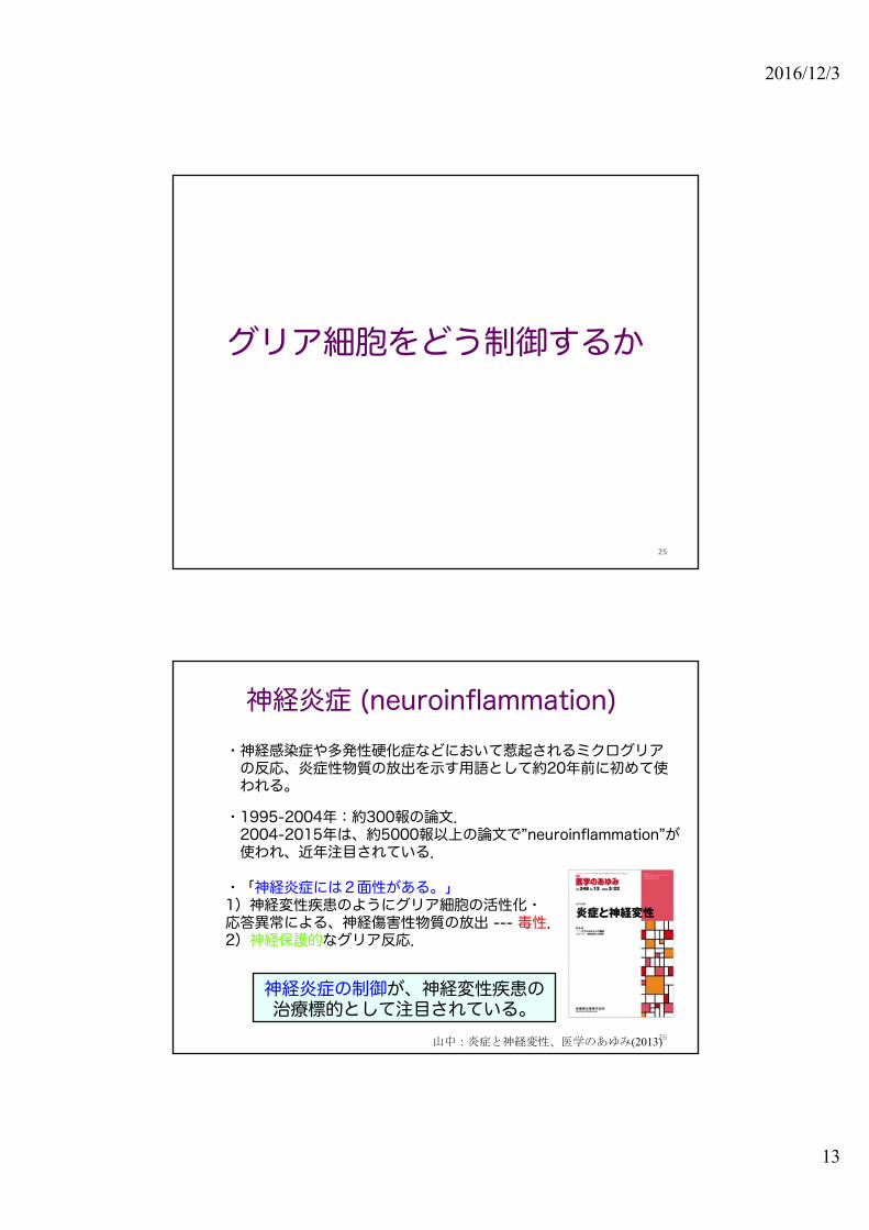
グリア細胞をどう制御するか
25
26
神経炎症 (neuroinflammation)・神経感染症や多発性硬化症などにおいて惹起されるミクログリアの反応、炎症性物質の放出を示す用語として約20年前に初めて使われる。
・1995-2004年:約300報の論文.2004-2015年は、約5000報以上の論文で”neuroinflammation”が使われ、近年注目されている.
・「神経炎症には2面性がある。」1)神経変性疾患のようにグリア細胞の活性化・応答異常による、神経傷害性物質の放出 --- 毒性.2)神経保護的なグリア反応.
神経炎症の制御が、神経変性疾患の治療標的として注目されている。
山中:炎症と神経変性、医学のあゆみ(2013)
2016/12/3
14
27
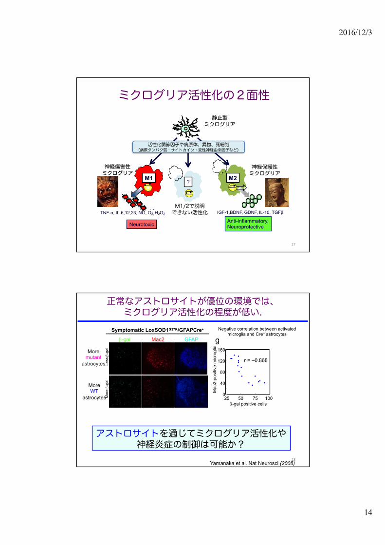
ミクログリア活性化の2面性
?
M1/2で説明できない活性化
静止型ミクログリア
M1 M2
TNF-α, IL-6,12,23, NO, O2, H2O2. -
IGF-1,BDNF, GDNF, IL-10, TGFβ
Neurotoxic Anti-inflammatory,Neuroprotective
神経傷害性ミクログリア
神経保護性ミクログリア
活性化調節因子や病原体、異物、死細胞(病原タンパク質・サイトカイン・変性神経由来因子など)
正常なアストロサイトが優位の環境では、ミクログリア活性化の程度が低い.
g
0
40
80
120
160
25 50 75 100b-gal positive cells
Mac
2-po
sitiv
e m
icro
glia
r = –0.868
Symptomatic LoxSOD1G37R/GFAPCre+
b-gal Mac2 GFAP
Mor
e b-
gal
Less
b-g
al
100μm
アストロサイトを通じてミクログリア活性化や神経炎症の制御は可能か?
Yamanaka et al. Nat Neurosci (2008)
Negative correlation between activated microglia and Cre+ astrocytes
Moremutant
astrocytes
MoreWT
astrocytes
28

2016/12/3
15
ALSにおける神経炎症:beneficial and detrimental
構成細胞群アストロサイトミクログリアT細胞 など
神経傷害性因子炎症性サイトカインTNF-α,IL-1β, etc.
酸化ストレスROS,NO, etc.
グルタミン酸による興奮毒性
神経保護性因子神経栄養因子IGF-I,GDNF,BDNF, etc.抗炎症性サイトカインTGF-β,IL-10
神経炎症活性化アストロサイト
変性運動ニューロン
骨格筋
IFN-γ
神経傷害性因子
神経保護性因子
IL-4
GLT-1↓神経傷害因子
神経栄養因子
M2神経保護性
M1神経傷害性
浸潤T細胞
活性化ミクログリア
炎症反応の増強
M1/M2の制御
グルタミン酸毒性の増強神経保護能の低下
ALSの疾患進行を制御する細胞群
アストロサイト由来の因子は、ミクログリアやT細胞が関与する神経保護環境を制御しうるか? 29
?
ALSのアストロサイトからどのような因子が放出されているか?
30
既報告の分子神経栄養因子:GDNF, BDNF, ...毒性因子:ROS, NO, D-serine, ...炎症性サイトカイン:IFN-g, PGD2,... ケモカイン...
2016/12/3
16
Houi et al. (2002)
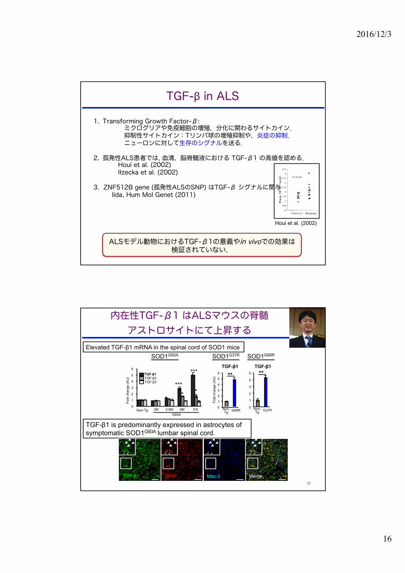
1. Transforming Growth Factor-β: ミクログリアや免疫細胞の増殖,分化に関わるサイトカイン.抑制性サイトカイン:Tリンパ球の増殖抑制や,炎症の抑制.ニューロンに対して生存のシグナルを送る.
2. 孤発性ALS患者では, 血清,脳脊髄液における TGF-β1 の高値を認める.Houi et al. (2002)Iłzecka et al. (2002)
3. ZNF512B gene (孤発性ALSのSNP) はTGF-β シグナルに関与Iida, Hum Mol Genet (2011)
ALSモデル動物におけるTGF-β1の意義やin vivoでの効果は検証されていない.
TGF-β in ALS
内在性TGF-β1 はALSマウスの脊髄アストロサイトにて上昇する
TGF-β1 is predominantly expressed in astrocytes of symptomatic SOD1G93A lumbar spinal cord.
SOD1G93A SOD1G37R SOD1G85R
TGF-β1
0
1
2
3
4
5
Non-Tg G37R0123456
Non-Tg G85R
**
Fold
cha
nge
(AU
)
**
Non-Tg G37RNon-
Tg G85R
Fold
chan
ge (A
U)
0
1
2
3
4
5
6
WT-5M G93A-2M G93A-3.5M G93A-4M G93A-ES
TGF-β1TGF-β2TGF-β3
Non-Tg 2M 4M3.5M ESG93A
***
**** *
TGF-β1
TGF-β1 GFAP Mac-2 Merge
Elevated TGF-β1 mRNA in the spinal cord of SOD1 mice
32

2016/12/3
17
TGF-β1 は、孤発性ALSの脊髄アストロサイトでも増加する
TGF-β1/GFAP/DAPI
sALS-179
CTRL-170 CTRL-239
sALS-260
TGF-β1 is upregulated in astrocytes of sporadic ALS spinal cord
sALS
control
Endo et al. Cell Reports (2015)33
1. Whether neurodegeneration and inflammation are affected when the level of TGF-β1 is altered in astrocytes of ALS mice.
ALSマウスのアストロサイトで、TGF-β1 の量を変えたら病態と神経炎症は変化するか?
2. Whether the level of TGF-β1 is linked to the survival time of ALS mice.TGF-β1 の量は、ALSマウスの生存期間と相関するか?
解決すべき課題Questions to be addressed
34

2016/12/3
18
アストロサイトで TGF-β1 量をさらに増やすとSOD1G93A マウスの進行が加速
生存期間の短縮
SOD1G93A x GFAP-TGFβ1 交配実験
150Age (days)
Surv
ival
(%)
200100
100
50
0
G93A (N=36)
G93A/TGFβ1 (N=33)
-10.2 days(p<0.001)
60
80
100
120
Age(
days
)
G93A G93A/TGFβ1
発症時期は不変
0
10
20
30
40
Early stage Late stage
Dur
atio
n (d
ays)
Early stage
Late stage
*G93AG93A/TGFβ1
罹病期間(進行後期)の短縮
アストロサイト特異的にTGF-β1を
過剰産生するSOD1G93Aマウス
Wyss-Coray (1995)
GFAP-TGFβ1 mice
GFAP-TGF-β1xSOD1G93A
SOD1G93A/TGF-β1 mice
抑制性サイトカインで炎症をおさえることは有益とは限らない
神経炎症はどうなっているか ?
36

2016/12/3
19
アストロサイト由来TGF-β1 はミクログリアを不活性化し、神経栄養因子の産生を抑制する.
Mac-2 250μm
Mac
-2+
cells
/ A
H
*
0
100
200
300
400
G93A G93A/TGFβ1
G93A G93A/TGFβ1
G93A G93A/TGFβ1
Loss of Mac-2+activated microglia
nTg G93A G93A/TGFβ1 mR
NA
fold
cha
nge
0
5
10
15
Non-Tg G93A G93A/TGFβ1
IGF-I**
0
10
20
30
Non-Tg G93A G93A/TGFβ1
CD68*
ミクログリア活性低下
nTg G93A G93A/TGFβ1
神経栄養因子低下
GDNF
G93
A/T
GFβ
1G
93A
IGF-I / Iba1 CD68 / Iba1
Ventral horn
0
1
2
3
Non-Tg G93A G93A/TGFβ1
*
nTg G93A G93A/TGFβ1
37
アストロサイト由来TGF-βは、疾患進行の悪化因子か?
38

2016/12/3
20
0.5
1
1.5
120 140 160 180 200
IFN-
γ / I
L-4
(AU)
Survival (生存期間:日)
2
3
4
120 140 160 180 200
TGF-
β1 m
RNA
Survival (生存期間:日)
TGF-β1
0.5
1
1.5
2 3 4
IFN-
γ / I
L-4
(AU)
r=-0.8194p =0.0003N =14
r=0.7833p =0.0172N =10
r=-0.8368p =0.0083N =10
内在性TGF-β1量 IFN-γ / IL-4 positive
correlation
生存期間
negative correlation
TGF-β1 は疾患進行のバイオマーカー候補の可能性あり
内在性TGF-β1量はSOD1G93Aマウスの生存期間と負に相関する
TGF-β1 vs survival IFN-γ / IL-4 vs survival IFN-γ / IL-4 vs TGF-β1
39
アストロサイト特異的に変異SOD1を除去するとTGF-β1量は減少する.
Activated astrocytes
loxSODG37RloxSODG37R/GFAP-Cre+
Slow disease progression
TGF-β1 ↓
mSOD1 mSOD1
SODG93A/ TGF-β1
TGF-β1 ↑
Fast disease progression
アストロサイト由来TGF-β1は,疾患進行を負に制御する因子である.
Fold
cha
nge
(AU
)
01234567
B6-12M G37R CGLNon-Tg Cre- Cre+
loxSODG37R
**TGF-β1 mRNA
Cre –
Cre +
TGF-β1 GFAP Merge
A B
0.5
1
1.5
120 140 160 180 200
IFN
-γ/IL
-4 (A
U)
Survival (days)
2
3
4
120 140 160 180 200
TGF-β1 mRNA
Survival (days)
C
TGF-β1
0.5
1
1.5
2 3 4
IFN
-γ/IL
-4 (A
U)
r = -0.8194p = 0.0003
r = -0.8368p = 0.0083
r = 0.7833p = 0.0172
Figure 8, Endo et al.
D E
Fold
cha
nge
(AU
)
01234567
B6-12M G37R CGLNon-Tg GFAP-Cre-
GFAP-Cre+
loxSODG37R
**TGF-β1
0
0.2
0.4
0.6
0.8
1
1.2
Cre- Cre+
TGF-β1
*
Fold
cha
nge
of
mea
n flu
ores
cenc
e in
tens
ity
GFAP-Cre-
GFAP-Cre+
loxSODG37R
F
Activated astrocytes
mSOD1
loxSODG37R/GFAP-Cre-
loxSODG37R/GFAP-Cre+
Slowing disease progression
loxS
OD
G37
R/
GFA
P-C
re-
loxS
OD
G37
R/
GFA
P-C
re+
TGF-β1 GFAP Merge GTGF-β1 fluorescence
Endo et al. (2015)40

2016/12/3
21
Endo et al. Cell Reports 11 :592-604 (2015).
l TGF-β1 はALSマウスと孤発性ALSのアストロサイトで増加.
l アストロサイト特異的にTGF-β1を増加させると疾患進行が加速する.
l アストロサイト由来TGF-β1は、ミクログリアなどによる神経保護的反応を減弱させる.
l TGF-β シグナル阻害剤の発症後投与は、ALSマウスの生存期間を延長する.
アストロサイト由来 TGF-β はALSの病態悪化因子である
Muscle atrophy
IFN-γ IL-4
Degeneratedmotor neuron
ExcessTGF-β1
IFN-γ dominantenvironment
deactivatedMicroglia
Neurotrophic factor
(IGF-I, BDNF)
ActivatedAstrocytes
Accelerated disease progression
Deterioration of neuroprotective
environment
Proinflammatorycytokine
signal ?
Therapeutictarget
細胞群特異的な TGF-β1 シグナル経路への介入,つまりアストロサイトでの過剰なTGF-β1 の抑制は
新たなALSの進行抑制の治療標的となりえる.
TGF-β シグナル阻害剤の発症後投与により、ALSマウスの生存期間が延長した.
120
140
160
180
Vehicle SB-431542
Survival
Age
(day
s)
*+9.1d
Treatment from early symptomatic phase (120d)
+ 9.1 day (18% extension after onset)
Survival
Age
(day
s)
140
160
180
Vehicle SB-431542
p=0.0633
Treatment from late symptomatic phase (140d)
+ 4.5 day
Vehicle SB-431542
pSmad
2Mac-2
GFAP
IGF-I
200μm

2016/12/3
22
43
TGF-β シグナル阻害剤による実験的治療:プロトコールの最適化に向けて
1. 投与時期について:早期投与してよいか?
2.投与量について:投与頻度、量は最適か?
3.代用薬はないか?
Low dose protocol: 5μg/g, 2 times/week, Start: 124 daysSurvival
* + 9.4 days
p = 0.0287
120
140
160
180
DMSO SB-431542
投与プロトコールの最適化に向けて
50 μg/g/週(週5回、10 μg/g) から 10 μg/g/週(週2回、5 μg/g)へ
TGF-β シグナル阻害剤による実験的治療:プロトコールの最適化に向けて
投与量や頻度を減らすことができた。→ 副作用を考慮しても、少ない投与量で有効であることは意味がある。
Endo, unpublished

2016/12/3
23
45
TGF-β シグナル阻害による治療開発にむけて
1.TGF-β 阻害剤:抗がん剤として、海外で臨床治験Phase II (肝細胞がん、脳腫瘍ほか)
2.既存薬による代用:drug repositioningの可能性
神経変性疾患の発症と進行のメカニズム解明にむけて
疾患進行のメカニズム:
グリア細胞病態の解明炎症、免疫系の関与.
どうして発症するのか?
運動神経に起こる病的変化の解明
FUS / TLSTDP-43
RNA代謝異常(TDP-43,FUS)
タンパク質品質管理異常、オルガネラ異常
Microglia-Lymphocyte interactionControlling neuroinflammationThe role of astrocytes, NG2 glia

2016/12/3
24
Outline
1. 動物モデルを用いたALSの病態研究:神経周囲環境(グリア細胞)の重要性に気づくに至る道のり
1) 遺伝性ALS2) ALSマウス3) 細胞群特異的なALSの病態解明:
「非細胞自律性」の神経変性:コンセプトの確立
2. グリア細胞からみたALSの病態研究、神経炎症制御1)ALSにおける神経炎症(ミクログリア、アストロサイト)2)アストロサイトにおける治療標的分子:TGF-β1
3. 運動神経の変性メカニズムについてオルガネラ(ミトコンドリア・小胞体)の変調
4.神経変性疾患の病態解明に向けて:今後の展望
47
48
運動神経は最大の神経細胞
軸索は最大1メートル
軸索の長さゆえ、物質を輸送するために多大なエネルギーが必要→ ミトコンドリアなどオルガネラは重要。
直径は、
100ミクロン

2016/12/3
25
SIGMAR1遺伝子のp.L95fs 変異をALS16の新規変異として同定
Genotype・ c.283dupC (p.L95fs) was detected by whole exome sequencing・ Homozygosis was a result of parental uniparental disomy for Chr. 9
Clinical features・ progressive muscle weakness from 5 years old・ both upper & lower motor involvement with slow progression・ well consistent with the features of ALS16
The family is followed by our collaborators,Dr. Mancias & Dr. Ilieva (Texus, USA)
50
MAM: mitochondria-associated membrane・シャペロン分⼦やIP3受容体など、神経の恒常性維持に重要な分⼦が集積.・主にミトコンドリアへのCa2+バッファリングに重要.
(Hayashi & Su Trends Cell Biol. 2009)・コレステロール合成(Vance J. Biol. Chem. 1998)やオートファジーの
隔離膜形成(Hamasaki et al. Nature 2013)などの機能もある.
・Sigma1受容体:MAMに局在する膜貫通型シャペロン様オーファン受容体
→ MAM異常で変性疾患におけるERストレスやミトコンドリア障害を統⼀的に説明できるか.
Sigma1受容体 (ALS16) は小胞体・ミトコンドリア膜間領域(MAM)に局在する

2016/12/3
26
Both E102Q and L95fs Sig1R variants were1) unstable because of constitutive degradation via proteasome2) incapable to bind to IP3R3
Sig1R 変異タンパク質は不安定で、IP3R3と結合できない
cycloheximide chase assay proteasome or lysosomal inhibition
Protein stability in Neuro2a (N2a) cells Co-IP of IP3R3with Sig1R-FLAG
(N2a cells)
MAMの崩壊はSOD1-ALS (ALS1) とSig1R-ALS(ALS16)に共通した病態である
MAM, a contacting surface of ER to mitochondria, was reduced in both SOD1G85R and Sig1R deficient mouse motor neurons.
Watanabe et al. EMBO Molecular Medicine, in pressCollaboration with Drs. Kiyama and Tamada
* p <0.05
タンパク質、RNA代謝異常のみならずオルガネラ異常(ミトコンドリア, 小胞体)も
発症の重要な鍵といえる

2016/12/3
27
vent
ral h
orn
oflu
mba
r spi
nal c
ord
SOD1G93A (onset, 3 months)
PRE-084 treatedsaline treated
SOD1G93A mice were administered 0.25 mg/kg PRE-084 in 3 times/week.
The administration was started from 1 months, and ended in 3 months.
- Sig1R selective agonist (Ki = 2.2 nM)- extends survival time of mutant SOD1 transgenic mice
PRE-084
Sigma1受容体のアゴニストをSOD1マウスに投与するとMAMからのSigma1受容体の喪失、MAM異常を部分的に抑止できる
神経保護治療の可能性
54
MAM異常と神経変性メカニズム
MAMの異常:ALS (SOD1, SigmaR1, TDP-43, FUS, VAPB, 孤発性):アルツハイマー病:パーキンソン病
Watanabe et al. EMBO Molecular Medicine, in press
MAM
エネルギー(ATP)産生 エネルギー(ATP)産生不全神経細胞死

2016/12/3
28
Defect in RNA, protein quality control
Mitochondria-ERdysfunction
アストロサイト
神経筋接合部
ミクログリア
ALSの細胞群特異的な病態解明
保護性?
傷害性?
ミクログリアの分子病態1.免疫系との連関とその意義自然免疫 (TRIF)、獲得免疫2. ミクログリアの活性化調節:神経保護性の活性化機序
アストロサイトの分子病態1.TGF-βによる神経炎症制御:
最適化2. 異常活性化したアストロサイト
の排除機構
運動ニューロンの分子病態1. TDP-43 タンパク質の異常化機序2. タンパク質、RNA代謝異常による神経変性機序の解明
3. ミトコンドリア・ER連関と神経変性(SOD1, TDP-43, Sigma1R)
運動ニューロン
proteasomeinhibition
ニューロン・グリアそれぞれの病態を理解するこ
とが,疾患の発症・進行を制御するうえで重要である。
55
クロストーク
Take home message:1. 神経細胞の維持には、周囲の細胞環境の改善も重要である。
“Non-cell autonomous” neuron death.
非細胞自律性の神経細胞死
56
神経細胞だけの病態解明から神経周囲の環境が重要である
という発想転換
「非自律性」の神経変性機序が示された疾患
ALSアルツハイマー病
パーキンソン病と類縁疾患ポリグルタミン病
など
2016/12/3
29
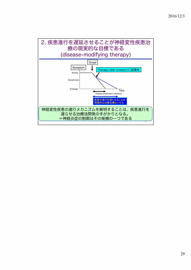
Therapy(after onset)とくに孤発性
Onset
Normal
Endstage
Symptom
TimeDisease progression (duration)
疾患の進行を遅らせることは現実的な治療目標といえる
Symptomatic
神経変性疾患の進行メカニズムを解明することは、疾患進行を遅らせる治療法開発の手がかりとなる。=神経炎症の制御はその候補の一つである
2. 疾患進行を遅延させることが神経変性疾患治療の現実的な目標である
(disease-modifying therapy)
57