Embed Size (px)
Citation preview
7Kawasaki Medical Journal 41(1):7-12,2015 doi:10.11482/KMJ-E41(1)7
Fibrosarcomatous variant of dermatofibrosarcoma protuberans on the right cheek: A case report
Shogo EBISUDANI, Kiichi INAGAWA, Fumiaki NAGASHIMA, Ikuko OSUGI,
Tomomi KIMURA, Takashi HARADA, Miori TERAMOTO, Masateru INAI
Department of Plastic and Reconstructive Surgery, Kawasaki Medical School,577 Matsushima, Kurashiki, 701-0192, Japan
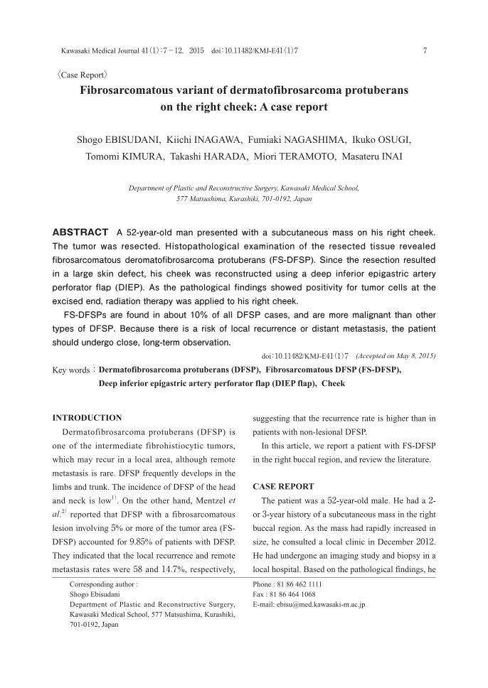
ABSTRACT A 52-year-old man presented with a subcutaneous mass on his right cheek. The tumor was resected. Histopathological examination of the resected tissue revealed fibrosarcomatous deromatofibrosarcoma protuberans (FS-DFSP). Since the resection resulted in a large skin defect, his cheek was reconstructed using a deep inferior epigastric artery perforator flap (DIEP). As the pathological findings showed positivity for tumor cells at the excised end, radiation therapy was applied to his right cheek. FS-DFSPs are found in about 10% of all DFSP cases, and are more malignant than other types of DFSP. Because there is a risk of local recurrence or distant metastasis, the patient should undergo close, long-term observation. doi:10.11482/KMJ-E41(1)7 (Accepted on May 8, 2015)
Key words: Dermatofibrosarcoma protuberans (DFSP), Fibrosarcomatous DFSP (FS-DFSP), Deep inferior epigastric artery perforator flap (DIEP flap), Cheek
INTRODUCTION Dermatofibrosarcoma protuberans (DFSP) is
one of the intermediate fibrohistiocytic tumors,
which may recur in a local area, although remote
metastasis is rare. DFSP frequently develops in the
limbs and trunk. The incidence of DFSP of the head
and neck is low1). On the other hand, Mentzel et al.2) reported that DFSP with a fibrosarcomatous
lesion involving 5% or more of the tumor area (FS-
DFSP) accounted for 9.85% of patients with DFSP.
They indicated that the local recurrence and remote
metastasis rates were 58 and 14.7%, respectively,
suggesting that the recurrence rate is higher than in
patients with non-lesional DFSP.
In this article, we report a patient with FS-DFSP
in the right buccal region, and review the literature.
CASE REPORT The patient was a 52-year-old male. He had a 2- or 3-year history of a subcutaneous mass in the right
buccal region. As the mass had rapidly increased in
size, he consulted a local clinic in December 2012. He had undergone an imaging study and biopsy in a
local hospital. Based on the pathological findings, he
Corresponding author : Shogo EbisudaniDepartment of Plastic and Reconstructive Surgery, Kawasaki Medical School, 577 Matsushima, Kurashiki, 701-0192, Japan
Phone : 81 86 462 1111Fax : 81 86 464 1068E-mail: [email protected]
〈Case Report〉

8 Kawasaki Medical Journal
was referred to our clinic for a suspected malignant
soft tissue tumor, and the patient was referred to our
department. On the initial consultation, an elastic-
hard subcutaneous mass measuring approximately
5 cm was noted in the right buccal region (Fig. 1).
There was no assimilation between the mass
and skin. There were no dermal changes. The
mobility of the mass was favorable. There was
no assimilation with the inferior base. Computed
tomography (CT) and Magnetic resonance imaging
Fig. 1. Photograph on the initial consultationA subcutaneous mass was noted (red round mark).
Fig. 2. Contrast-enhanced cephalic MR imagesa. T1-weighted image, b. T2-weighted image, c. Contrast-enhanced imageThe T1-weighted image shows a low-signal-intensity mass, and the T2-weighted image shows a high-signal-intensity mass. After contrast enhancement, marked enhancement effects were observed.
a b
c

9Ebisudani S, et al. : Fibrosarcomatous variant of dermatofibrosarcoma protuberans
(MRI) were performed. T1-weighted images showed
a low signal intensity in the right buccal region, and
T2-weighted images showed a high signal intensity.
After contrast enhancement, marked enhancement
effects were observed (Fig. 2). Under a tentative
diagnosis of a malignant soft tissue tumor, such as
fibrosarcoma, resection was performed in January
2013. Resection was conducted, with a 2-cm margin
lateral to the tumor. At a site adjacent to the lower
eyelid, a 1-cm margin was established (Fig. 3a).
Resection involving the cheekbone periosteum and
large/small cheek muscles was performed, and the
infraorbital nerve was amputated. It was possible
to preserve the parotid duct (Fig. 3b). The site of
the soft tissue defect was covered with artificial
dermis. We planned to perform reconstruction after
confirming the results of pathological examination.
As histopathological findings, the proliferation of
spindle cells with oval nuclei was noted, showing
Fig. 3a. Intraoperative photographThe extent of resection was established, with lateral and lower eyelid margins of 2 and 1 cm, respectively.b. After tumorectomyResection involving the cheekbone periosteum was performed.c. After reconstruction of the right cheek with a flapIn the right buccal region, reconstruction was performed using a DIEP flap.d. Condition 26 months after surgeryThe buccal shape was favorable. There has been no relapse.
a b
c d

10 Kawasaki Medical Journal
a storiform pattern. The border of the tumor was
unclear, and adipose tissue infiltration was observed.
Immunostaining showed a diffuse CD34-positive
reaction, suggesting DFSP. However, the cellular
density was high at the tumor center to margin. A
herringbone pattern was partially noted. Based on
these findings, the central lesion was considered
to be a fibrosarcomatous change of DFSP (Fig. 4), leading to a diagnosis of FS-DFSP. The margin
of the resected specimen at the inferior base was
partially positive for tumor cells, whereas its lateral
margin was negative.
In February 2013, reconstruction of the right
cheek was performed. As the margin of the resected
specimen at the inferior base was partially positive
for tumor cells, the cheekbone surface was cut, and
buccal reconstruction was conducted using a deep
inferior epigastric artery perforator flap (DIEP flap).
A flap was collected from the right abdomen, and
anastomosed to the right facial artery/vein (Fig. 3c).
The postoperative course was favorable. Complete
graft survival was achieved. As postoperative
adjuvant therapy, radiotherapy was applied to the
right cheek at a total radiation dose of 60 Gy from
April 2013. There has been no relapse in the right buccal
region during the 26-month postoperative follow-
up. As the flap was slightly over-sized, defatting
was performed twice. Although irradiation-related
pigmentation was observed in the right buccal
region, apparent discomfort reduced. The results
were cosmetically satisfactory (Fig. 3d). In the
future, follow-up will be continued, considering
local relapse and remote metastasis.
DISCUSSION DFSP was reported by Darier et al.3) in 1924 and
by Hoffman4) in 1925. Remote metastasis is rare, but
the tumor border is unclear, and local relapse may
occur5). As a rule, resection is performed. However,
Goldblum et al.1) reported that the recurrence rate
was 20% when establishing a surgical margin of
3 cm, whereas it was 41% when establishing that
of 2 cm or less. DFSP frequently develops in the
limbs and trunk. However, when it develops in
the head and neck region, as demonstrated in the
present patient, the surgical margin is insufficient
in many cases. Especially in cases involving the
face, the extent of resection is cosmetically and
functionally limited, increasing the local recurrence
rate6). In the present case, the lateral space was
sufficient, and a 2-cm margin was established, but,
Fig. 4. Histopathological photograph (magnification×20)a. HE stainingThe proliferation of spindle cells was noted, showing a storiform pattern.b. Immunostaining at the site of FS (anti-CD34 antibody)At the site of FS, a diffusely positive reaction was also observed.
ba

11Ebisudani S, et al. : Fibrosarcomatous variant of dermatofibrosarcoma protuberans
on the cephalic side, a 1-cm margin was established
to preserve the lower eyelid. Concerning inferior
base margins, many studies have reported resection
involving the fascia. In the present case, the tumor
was adjacent to the cheekbone body, and resection
involving the periosteum was performed. On
pathological diagnosis, the peripheral margins were
negative for tumor cells, but a portion of the inferior
zygomatico-orbital margin was positive for them.
Therefore, additional resection was conducted on
buccal reconstruction. Thus, the tumor may have
been completely resected. However, this patient
was pathologically diagnosed with FS-DFSP.
Mentzel et al.2) reported that the local recurrence
and remote metastasis rates in FS-DFSP patients
with FS changes involving 5% or more of the
tumor area were higher than in those with non-
lesional DFSP. Furthermore, Goldblum et al.7) performed extended resection (3 cm or larger) in
18 patients with FS-DFSP, and indicated that there
was no remote metastasis. These results suggest
the necessity of extended resection in patients with
FS-DFSP. Postoperative adjuvant therapy may be
necessary. William et al.8) reported that radiotherapy
was useful for preventing relapse in patients whose
surgical margin was positive for tumor cells or those
in whom the cheekbone was adjacent to the tumor.
We also selected adjuvant radiotherapy. Previous
studies reported surgical margins of DFSP, but a
consensus has not been reached. They should be
decided in accordance with individual patients. In
many patients with FS-DFSP, relapse is reportedly
detected within 1 to 3 years after surgery. However,
according to a study, relapse was detected after long-
term follow-up2). In the future, it may be important
to continue strict follow-up over a long period,
considering local relapse and remote metastasis.
In the present case, tumorectomy involving
the buccal periosteum was performed, requiring
reconstruction with a flap. Although reconstruction
with skin grafting has also been selected for patients
in whom the periosteum is preserved, reconstruction
with a flap may be cosmetically appropriate. Various
studies have presented flap choices. However, we
selected a DIEP flap. This is a perforating branch
flap in which the deep inferior epigastric artery is
used as a vascular pedicle, as reported by Koshima
et al.9) in 1989. It has been applied in various
fields10). The reasons why this flap was selected
included: (1) it is possible to harvest a flap in a
supine position, and it is unnecessary to change the
position during surgery, (2) the abdominal rectus
muscle can be preserved, and a thin flap can be
collected, (3) the site of flap harvest is discreet,
and complications, such as abdominal wall hernia,
may not occur, and (4) the anastomosed blood
vessel length can be sufficiently maintained, and
the vascular diameter is appropriate for vascular
anastomosis. As a limitation, color matching with
the face is unfavorable due to its abdominal origin.
However, radiotherapy is performed after surgery,
and it may be impossible to avoid flap pigmentation.
As a result, pigmentation remained in the right
buccal region, but the results were morphologically
and cosmetically favorable.
FundingNone
Conflict of interestNone
REFERENCES1)Goldblum JR, Folpe AL, Weiss SW, Enzinger FM:
Enzinger and Weiss s soft tissue tumors (6th ed).
Philadelphia, USA, Elsevier/Saunders, 2014, pp387-4002)Mentzel T, Beham A, Katenkamp D, Dei Tos AP,
Fletcher CD: Fibrosarcomatous ("high-grade")
dermatofibrosarcoma protuberans. Am J Surg Pathol 22: 576-587, 1998
3)Darier J, Ferrand M: Dermatofibromes progressifs et
récidivants ou fibrosarcomes de la peau. Ann Dermatol
Syph 5: 545-562, 19244)Hoffmann E: Uber das Knollentreibende fibrosarkoma

12 Kawasaki Medical Journal
der haut. Dermat Ztschr 43: 1-28, 19255)Bowne WB, Antonescu CR, Leung DH, Katz SC,
Hawkins WG, Woodruff JM, Brennan MF, Lewis JJ :
Dermatofibrosarcoma protuberans: A clinicopathologic
analysis of patients treated and followed at a single
institution. Cancer. 88: 2711-2720. 20006)Tom WD, Hybarger CP, Rasgon BM: Dermatofibrosarcoma
protuberans of the head and neck: Treatment with Mohs
surgery using inverted horizontal paraffin sections.
Laryngoscope. 113: 1289-1293, 20037)Goldblum JR, Reith JD, Weiss SW: Sarcomas arising
in dermatofibrosarcoma protuberans: A reappraisal of
biologic behavior in eighteen cases treated by wide local
excision with extended clinical follow up. Am J Surg
Pathol. 24: 1125-1130, 20008)Mendenhall WM, Zlotecki RA, Scarborough MT:
Dermatofibrosarcoma protuberans. Cancer. 101: 2503-2508, 2004
9)Koshima I, Soeda S: Inferior epigastric artery skin flaps
without rectus abdominis muscle. Br J Plast Surg. 42: 645-648, 1989
10)Allen RJ, Treece P: Deep inferior epigastric perforator
flap for breast reconstruction. Ann Plast Surg. 32: 32-38, 1994