Embed Size (px)
Citation preview
197
CZECH MYCOL. 60(2): 197–212, 2008
Gerronema nemorale (Basidiomycota, Agaricomycetes):
anatomic-morphological, cultivational, enzymatic
and molecular characteristics and its first records
in the Republic of Korea
VLADIMÍR ANTONÍN1, RHIM RYOO
2 and HYEON-DONG SHIN2
1 Moravian Museum, Department of Botany, Zelný trh 6, CZ-659 37 Brno, Czech [email protected]
2 Korea University, Division of Environmental Science and Ecological Engineering, Seoul 136-701,Republic of Korea
[email protected], [email protected]
Antonín V., Ryoo R. and Shin H. D. (2008): Gerronema nemorale (Basidiomycota,Agaricomycetes): anatomic-morphological, cultivational, enzymatic and molecu-lar characteristics and its first records in the Republic of Korea. – Czech Mycol.60(2): 197–212.
The basidiomycetous agaric Gerronema nemorale Har. Takah. was collected at several localities inthe Republic of Korea (South Korea). A macro- and microscopic description and cultivational charac-teristics are given. Also physiological studies were performed. These included measuring ofextracellular ligninolytic enzymes and monitoring of protein and glucose concentration in media. Itsplacement in the genus Gerronema in the recent sense is proven using molecular methods.
Key words: Gerronema, Republic of Korea, cultural characteristics, ITS, ligninolytic enzymes,decolorisation test, protein assay.
Antonín V., Ryoo R. a Shin H. D. (2008): Gerronema nemorale (Basidiomycota,Agaricomycetes): anatomicko-morfologická, enzymatická a molekulární charak-teristika a její první nálezy v Korejské republice. – Czech Mycol. 60(2): 197–212.
Lupenatá houba Gerronema nemorale Har. Takah. byla nalezena na několika lokalitách v Korejskérepublice (Jižní Korea). Autoři uveřejňují její podrobný makroskopický a mikroskopický popis, cha-rakteristiku v kultuře a enzymatickou, proteinovou a glukozovou charakteristiku. Na základě moleku-lárních metod je potvrzeno její zařazení do rodu Gerronema v jeho současném pojetí.
INTRODUCTION
In July 2007, the authors made several joint field excursions in the northernand central parts of the Republic of Korea (South Korea). The main task was tocollect material of marasmioid and gymnopoid fungi for a joint research project.At several excursions, an interesting omphalioid basidiomycetous fungus was col-

lected; it was collected also later by the Korean co-authors. Its microscopic stud-ies showed that it represents Gerronema nemorale Har. Takah., a fungus recentlydescribed as new to science from Honshu, Japan (Takahashi 2000). These collec-tions represent the first published records in the Republic of Korea.
MATERIALS AND METHODS
M a c r o s c o p i c a n d m i c r o s c o p i c s t u d i e s . The macroscopic description is based on freshbasidiocarps of three collections made by the first author. Microscopic features are described fromdried material mounted in H2O, KOH, Melzer’s reagent and Congo Red using an Olympus BX-50 lightmicroscope with a magnification of 1000×. For basidiospores, the factors E (quotient of length andwidth in any one spore) and Q (mean of E-values) are used. Authors of fungal names are cited accord-ing to Kirk and Ansell (1992), herbarium acronyms follow Holmgren and Holmgren (1998).
C u l t u r a l s t u d i e s . The cultures of Gerronema nemorale were obtained from basidiospores offresh basidiocarps. These isolates were incubated at 23 °C in the dark in both Petri dishes (90 × 18 mm)containing PDA (potato dextrose agar, Difco) and test tubes containing PDB (potato dextrose broth,Difco). Isolates were preserved for future reference in KACC (Korean Agricultural Culture Collection)with the following accession numbers: KACC 43598 (from specimens preserved in BRNM 709773,Antonín 07.64), KACC 43599 (BRNM 709772, Antonín 07.100) and KACC 43600 (collection R. RyooKG161). Cultures from the collections of Antonín 07.140 and KG137 were not successful. The culturemat was described using the terminology by Nobles (1965), Stalpers (1978) and Desjardin (1990). Mi-croscopic features of the isolates were observed with an Olympus BX-51 light microscope and an Axioimager AI DIC-light microscope with a magnification of 1000×.
D N A e x t r a c t i o n . DNA extraction was carried out according to the method proposed by Lee andTaylor (1990). Genomic DNAs were extracted using the mycelium of KACC 43599 and KACC 43600 andthe basidiocarp of KG137. Small pieces of tissue were used to fill one-third of 1.5 ml Eppendorf microcen-trifuge tubes. 500 μl of lysis buffer (0.5 M Tris-HCl, 0.5 M EDTA, 3 % SDS, and 1 % 2-mercaptoethanol) wasadded to each tubes before incubation for 3 hrs at 67 °C. 500 μl of buffer (phenol : chloroform : isoamyl al-cohol ratio 25 : 24 : 1) was added to the DNA solutions. The samples were centrifuged at 14,000 rpm for 25min. at 25 °C. 300 μl of aqueous phase containing the DNAs was transferred to new tubes. Each tube wassupplemented with 10 μl of 3 M sodium acetate and 162 μl of isopropanol. The Eppendorf tubes contain-ing the DNA solutions were centrifuged at 14,000 rpm for 15 min. at 4 °C. Supernatants were poured offand the pellets were centrifuged at 14,000 rpm for 5 min. at 4 °C. The DNA pellets were washed with cold70 % ethanol, dried in a vacuum drier for 1 hr at 50 °C and resuspended in 50 μl of TE buffer (10 mM Tris-HCl and 0.1 mM EDTA). RNase was added to each sample and incubated at 37 °C for 1 hr.
P C R a m p l i f i c a t i o n . PCR amplification was performed according to the method described byGardes and Bruns (1993). The forward primer ITS1-F and reverse primer ITS4-B were used for selectiveamplification of the complete ITS region of rDNA. PCRs were conducted in 50 μl of solutions containing 1.2μl of template DNA mixture, 5 μl of 10x buffer (0.5 M KCl, 0.1 M Tris-HCl; pH 8.0, 0.1 % Triton X-100, 15 mMMgCl2), 1 μl of 2.5 mM dNTP, 0.4 μl of 100 μM primer ITS1-F, 0.4 μl of 100 μM primer ITS4-B, 0.4 μl of Taqpolymerase (5 unit/μl), and 36.6 μl of distilled water. Temperature cycling was performed with denaturationfor 30 s at 94 °C, annealing for 30 s at 55 °C, and extension for 1 min. at 72 °C. Thirty-five cycles were runwith the first denaturation and last extension times extended to 5 min. at 72 °C. Purified DNAs were directlysequenced in an automatic sequencer (ABI Prism TM 377 DNA Sequencer) with the primers ITS1-F andITS4-B for ITS rDNA.
P h y l o g e n e t i c a n a l y s i s . Sequences were edited with the DNASTAR computer package,whereby alignment of the sequences was performed using the CLUSTAL_X program (Thompson et al.1997). Bayesian analysis was carried out using the MRBAYES computer program, version 3.1 (Ronquist
198
CZECH MYCOL. 60(2): 197–212, 2008
and Huelsenbeck 2003). The general time reversible model (GTR) was employed with gamma-distrib-uted substitution rates for ITS alignment. Markov chains were run for 106 generations, saving a tree ev-ery 100th generation. Out of these, the first 1,000 trees were discarded. MRBAYES was used to computea 50 % majority rule consensus of the remaining trees to obtain estimates for posterior probabilities ofthe groups. Branch lengths were computed as the mean values for the sampled trees.
S p o t t e s t s a n d d y e d e c o l o r i s a t i o n t e s t s . Spot tests and dye decolorisation tests werecarried out according to Stalpers (1978), Desjardin (1990) and Okino et al. (2000). The spot test was per-formed using an α-naphthol solution for laccase, a ρ-cresol solution for tyrosinase, and a pyrogallol solu-tion for peroxidases. The reactions for laccase and peroxidases were conducted on advancing zones ofmarginal hyphae; the reaction for tyrosinase was performed on aerial mycelium of the culture mat.
The dye decolorisation test was performed using PDA with 0.2 g Congo Red of azo dyes, 0.5 gRBBR (Remazol Brilliant Blue R) of polymeric dyes, and 0.5 g methylene blue of heterocyclic dyes per400 ml solution (Glenn and Gold 1983). Inoculum plugs of the KACC 43589, KACC 43590, KACC 43600isolates were cut using a cork borer (4 mm diameter) and transferred to Petri dishes (90 × 18 mm) con-taining 20 ml PDA mixed with each dye compound. Triplicates of each isolate were incubated for 6weeks at 25 °C in the dark.
L i g n i n o l y t i c e n z y m e a c t i v i t y a s s a y. Ligninolytic enzyme activity was analysed usingKirk’s media (Kirk et al. 1978) and PDB. Kirk’s media are composed of 1 ml of mineral solution per 1 l ofbasic media. The mineral solution contained 1.5 g of nitriliotriacetate, 3.0 g of MgSO4 · 7H2O, 0.5 g ofMnSO4 · H2O, 1.0 g of NaCl, 0.1 g of FeSO4 · H2O, 0.1 g of CoCl2 · 6H2O, 0.1 g of ZnSO4 · H2O, 0.01 g of CuSO4
· 7H2O, 0.01 g of AlK(SO4)2 · 12H2O, 0.01 g of H2BO3 and 0.01 g of Na2MoO4 · 2H2O per 1 l of distilled water.The basic media consisted of 2 g of KH2PO4, 0.5 g of MgSO4, 0.1 g of CaCl2, C-source (10 g of glucose)and N-sources (2.4 mM N of NH4NO3 and L-asparagine) per 1 l of distilled water. Erlenmeyer flasks (100ml) containing 10 ml of Kirk’s media or PDB were inoculated with five agar plugs (4 mm diameter). Cul-tures were incubated at 25 °C in the dark under stationary conditions. These were cultivated for 12, 60,108, 156, 204, 252, and 300 hrs and the cultures were filtered through Whatman filter paper (No. 42).
Absorption spectra were recorded with a UV-Spectrophotometer (model: PowerWaveXS, Bio-Tek)for the activity of laccase, LiP (lignin peroxidases; E.C. 1.11.1.14) and MnP (manganese-dependentperoxidases), known as lignin degradation enzymes (Vares et al. 1995). Laccase activity tests were car-ried out using 33 μl of 5 mM ABTS [2,2-azinobis-(3-ethylbenzthiazoline-6-sulphonate] as substrate and250 μl of 0.1 M sodium acetate buffer (pH 4.6). LiP activity tests were carried out using 25 μl of 2 mMveratryl (3,4-dimethoxybenzyl) alcohol as substrate and 25 μl of 0.4 mM hydrogen peroxide in 225 μl of50 mM sodium tartrate buffer (pH 4.0). MnP activity tests were performed using 10 μl of 1 mM guaiacolas substrate, 10 μl of 1 mM MnSO4 · H2O, 10 μl of 1 mM hydrogen peroxide in 200 μl of 0.5 M sodiumtartrate buffer (pH 3.0). Each kinetic measurement was determined in 420 nm (molar extinction coeffi-cient ε = 36,000/Mcm) for laccase, 310 nm (ε = 9,300/Mcm) for LiP, and 525 nm (ε = 121,000/Mcm) forMnP. The enzyme activity was calculated using the following expression:
Enzyme activity (Unit/ml) = (ΔA × ΔV × 106) / (ε × Δt × ΔS)
(ΔA: absorbance difference value, ΔV: total volume, Δt: reaction time in min., ε: molar extinctioncoefficient, ΔS: material volume).
Protein concentrations were measured using protein assay reagent (Bio-Rad), according to themethod described by Bradford (1976). Bovine serum albumin (Bio-Rad) was the standard protein used.Reducing glucose and pH (pH meter: Model 750P, Istek) were simultaneously measured in Kirk’s me-dia. Glucose remaining in the media was measured according to the method by Miller (1958) using thefollowing reagents: 10 g of 3-5-dinitrosalicylic acid, 2 g of phenol, 0.5 g of sodium sulphite, 200 g of po-tassium sodium tartrate, and 500 ml of 2 % NaOH buffer per 1 l of distilled water. Each absorbance wasanalysed at 595 nm and 575 nm with the same UV-Spectrophotometer.
199
ANTONÍN V., RYOO R. AND SHIN H.-D.: GERRONEMA NEMORALE IN THE REPUBLIC OF KOREA
RESULTS
Gerronema nemorale Har. Takah., Mycoscience 41: 16. 2000.
D e s c r i p t i o n o f c o l l e c t e d b a s i d i o c a r p s
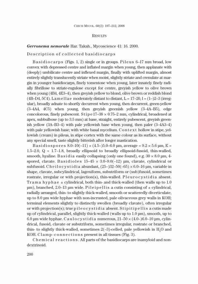
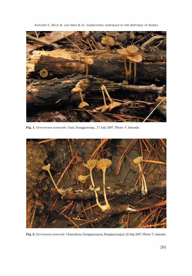
B a s i d i o c a r p s (Figs. 1, 2) single or in groups. P i l e u s 6–17 mm broad, lowconvex with depressed centre and inflated margin when young, then applanate with(deeply) umbilicate centre and inflexed margin, finally with uplifted margin, almostentirely slightly translucently striate when moist, slightly striate and crenulate at mar-gin in younger basidiocarps, finely tomentose when young, later innately finely radi-ally fibrillose to striate-rugulose except for centre, greyish yellow to olive brownwhen young (4B4, 4E3–4), then greyish yellow to blond, olive brown or reddish blond(4B–D4, 5C4). L a m e l l a e moderately distant to distant, L = 17–20, l = (1–)2–3 (irreg-ular), broadly adnate to shortly decurrent when young, then decurrent, green-yellow(3–4A4, 4C5) when young, then greyish greenish yellow (3–4A–B5), edgeconcolorous, finely pubescent. S t i p e 17–38 × 0.75–2 mm, cylindrical, broadened atapex, subbulbose (up to 3.5 mm) at base, straight, entirely pubescent, greyish green-ish yellow (3A–B3–4) with pale yellowish base when young, then paler (3–4A3–4)with pale yellowish base; with white basal mycelium. C o n t e x t hollow in stipe, yel-lowish (cream) in pileus, in stipe cortex with the same colour as its surface, withoutany special smell, taste slightly bitterish after longer mastication.
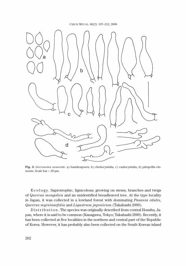
B a s i d i o s p o r e s 8.0–10(–11) × (4.5–)5.0–6.0 μm, average = 9.2 × 5.6 μm, E =1.5–2.0, Q = 1.7–1.8, broadly ellipsoid to broadly ellipsoid-fusoid, thin-walled,smooth, hyaline. B a s i d i a easily collapsing (only one found), e.g. 30 × 8.0 μm, 4-spored, clavate. B a s i d i o l e s 15–45 × 3.0–9.0(–12) μm, clavate, cylindrical orsubfusoid. C h e i l o c y s t i d i a abundant, (25–)32–50(–65) × 6.0–16 μm, variable inshape, clavate, subcylindrical, lageniform, subutriform or (sub)fusoid, sometimesrostrate, irregular or with projection(s), thin-walled. P l e u r o c y s t i d i a absent.Tr a m a h y p h a e ± cylindrical, both thin- and thick-walled (then walls up to 1.0μm), branched, 2.0–13 μm wide. P i l e i p e l l i s a cutis consisting of ± cylindrical,radially arranged, thin- to slightly thick-walled, smooth or scatteredly diverticulate,up to 8.0 μm wide hyphae with non-incrusted, pale olivaceous grey walls in KOH;terminal elements slightly to distinctly swollen (broadly clavate), often irregularor with projection(s); true p i l e o c y s t i d i a absent. S t i p i t i p e l l i s a cutis madeup of cylindrical, parallel, slightly thick-walled (walls up to 1.0 μm), smooth, up to6.0 μm wide hyphae. C a u l o c y s t i d i a numerous, 21–50 × (4.0–)6.0–10 μm, cylin-drical, fusoid, clavate or subutriform, sometimes irregular, rostrate or branched,thin- to slightly thick-walled, sometimes 2(–3)-celled, pale yellowish in H2O andKOH. C l a m p - c o n n e c t i o n s present in all tissues (Fig. 3).
C h e m i c a l r e a c t i o n s . All parts of the basidiocarps are inamyloid and non-dextrinoid.
200
CZECH MYCOL. 60(2): 197–212, 2008
201
ANTONÍN V., RYOO R. AND SHIN H.-D.: GERRONEMA NEMORALE IN THE REPUBLIC OF KOREA
Fig. 1. Gerronema nemorale. Guri, Donggureung , 17 July 2007. Photo: V. Antonín.
Fig. 2. Gerronema nemorale. Chuncheon, Dongsan-myon, Bongmyeong-ri, 22 July 2007. Photo: V. Antonín.
E c o l o g y. Saprotrophic, lignicolous; growing on stems, branches and twigsof Quercus mongolica and an unidentified broadleaved tree. At the type localityin Japan, it was collected in a lowland forest with dominating Pasania edulis,
Quercus myrsinaefolia and Ligustrum japonicum (Takahashi 2000).D i s t r i b u t i o n . The species was originally described from central Honshu, Ja-
pan, where it is said to be common (Kanagawa, Tokyo; Takahashi 2000). Recently, ithas been collected at five localities in the northern and central part of the Republicof Korea. However, it has probably also been collected on the South Korean island
202
CZECH MYCOL. 60(2): 197–212, 2008
Fig. 3. Gerronema nemorale. a) basidiospores, b) cheilocystidia, c) caulocystidia, d) pileipellis ele-ments. Scale bar = 20 μm.
of Jeju (Kim et al. 2005, see discussion below). It seems to be rather common inthis country. It is probably also widely distributed in the East-Asian region.
C o l l e c t i o n s e x a m i n e d . Republic of Korea: Guri, Donggureung (East Nine Royal Grave), 17July 2007 leg. V. Antonín 07.64 (BRNM 709773). – Chuncheon, Dongsan-myon, Bongmyeong-ri, Experi-mental Forest of Kangwon National University, 22 July 2007 leg. V. Antonín (07.100), R. Ryoo and H. D.Shin (BRNM 709772). – Hongcheon, Bukbang-myon, Seongdong-ri, 27 July 2007 leg. V. Antonín (07.140),R. Ryoo and H. D. Shin (BRNM 709771). – Hongcheon, Bukbang-myon, Seongdong-ri, 20 Aug. 2007 leg. R.Ryoo (KG 137). – Muju, Deogyusan National Park, Cheon-yeon falls, 24 Aug. 2007 leg. R. Ryoo (KG 161).
Cultural characteristics
N o b l e s C o d e : 2. 3. 9. 32. 36. 39. 44. 45. 54.S t a l p e r s C o d e : 1. 3. 8. 13. 25. (30). 31. 38. 39. 45. 52. 89.M a c r o m o r p h o l o g i c a l c h a r a c t e r i s t i c s . On PDA (n=3), diameter
12–15 mm in one week, 80–85 mm in 4–5 weeks; culture mat tightly interwoven,initially felty, pale yellow or (pale) cream colour; advancing zone submerged andtranslucently yellow or dark yellow; plug densely felty and yellowish white; re-verse coloration more or less dark yellow; odour not distinctive (Fig. 4a).
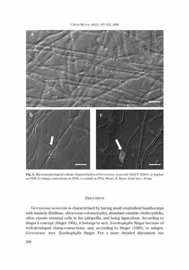
M i c r o m o r p h o l o g i c a l c h a r a c t e r i s t i c s . On PDB (n=3), hyphae2.1–2.9 μm wide, thin-walled, differentiated, straight and more or less irregular inoutline, rarely branched, hyaline (Fig. 5a). Clamp-connections 3.7–4.1 × 2.1–2.5μm, the clamp making an angle of about 90° with the hypha (Fig. 5b). Crystalsscattered on PDA (Fig. 5c).
Phylogenetic characteristics
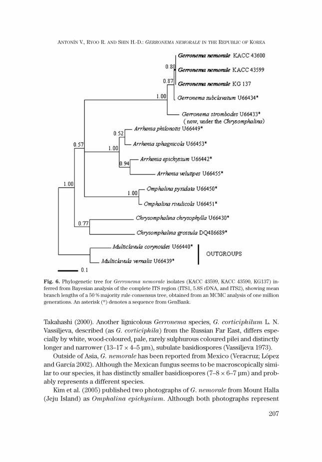
Three isolates of G. nemorale (KACC 43599, KACC 43600 and KG137) were com-pared with sequences within the omphalioid species. Except for G. nemorale, all se-quences were obtained from GenBank (Tab. 1). These clades showed a high probabil-ity value (1.00 in all branches) in the MCMC tree (Fig. 6), in which all sequences ofG. nemorale formed a monophyletic group among which no sequence difference isfound. These sequences formed the Gerronema group with G. subclavatum andG. strombodes, strongly supported by a high probability value of 1.00. G. strombodes
was reallocated to Chrysomphalina strombodes by Clémençon (1982). However thisspecies was significantly distant from the Chrysomphalina clade including C. chryso-
phylla and C. grossula. Species of the genus Omphalina formed a sister clade withGerronema distantly separated from the Arrhenia clade. The Chrysomphalina cladewas distant from both of them and formed an unrelated genus.
We obtained a sequence of Megacollybia platyphylla from GenBank (accordingto Moncalvo et al. 2002), and tried to re-analyse the phylogenetic relationship withour fungi. Unfortunately, this sequence is more closely related with the Gerronema
group than the omphalioid group (results not shown). Therefore, we thinkMegacollybia platyphylla is not appropriate as an outgroup. Based on a study by
203
ANTONÍN V., RYOO R. AND SHIN H.-D.: GERRONEMA NEMORALE IN THE REPUBLIC OF KOREA

Redhead (2002), we selected two sequences, Multiclavula corynoides and M. ver-
nalis, as the outgroup. However, the position of the main groups (Arrhenia,
Omphalina, Chrysomphalia and Gerronema) is consistent in both analyses.
Spot tests and dye decolorisation tests
The results of spots test using α-naphthol solution and a pyrogallol solutionwere positive, but the test using ρ-cresol solution was negative. Evaluation of pos-itive results showing colour changes was made 30 min after treating solutions:purple for laccase and yellowish-brown for peroxidases (Fig. 4b).
The colour of media containing Congo Red was changed from red to purple ordark red after 10 days of incubation, and the colour of media containing RBBRwas changed from blue to orange after 10 days of incubation for the dyedecolorisation test (Figs. 4c, d). However the media containing methylene bluewere not obtained from decolorisation test results, because methylene blue didnot support mycelial growth.
Ligninolytic enzyme activity
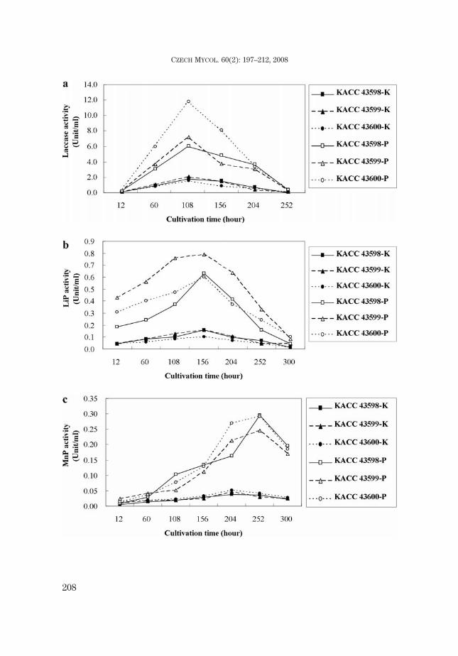
The ligninolytic enzyme activity of G. nemorale showed similar curves onKirk’s media and PDB for laccase, LiP and MnP, but it showed a higher peak onPDB than on Kirk’s media. All samples of laccase activity reached a maximumyield after 108 hrs, whereas the maximum yield of LiP activity was shown after156 hrs. On the other hand, the maximum point of MnP activity was reached after204 hrs on Kirk's media and 252 hrs on PDB (Figs. 7a-c).
204
CZECH MYCOL. 60(2): 197–212, 2008
Tab. 1. Strains of GenBank accession numbers and DNA source used for this research.
Taxon GenBank accession no. DNA Source
Arrhenia epichysium U66442 Lutzoni, 1997 as Omphalina
Arrhenia philonotis U66449 Lutzoni, 1997 as Omphalina
Arrhenia sphagnicola U66453 Lutzoni, 1997 as Omphalina
Arrhenia velutipes U66455 Lutzoni, 1997 as Omphalina
Chrysomphalina chrysophylla U66430 Lutzoni, 1997
Chrysomphalina grossula DQ486689 Matheny, 2006
Chrysomphalina strombodes U66433 Lutzoni, 1997 as Gerronema
Gerronema subclavatum U66434 Lutzoni, 1997
Omphalina pyxidata U66450 Lutzoni, 1997
Omphalina rivulicola U66451 Lutzoni, 1997
Megacollybia platyphylla AF498289 Matheny, 2006
Megacollybia platyphylla DQ 249275 Matheny, 2006
Multiclavula corynoides U66440 Lutzoni, 1997
Multiclavula vernalis U66439 Lutzoni, 1997

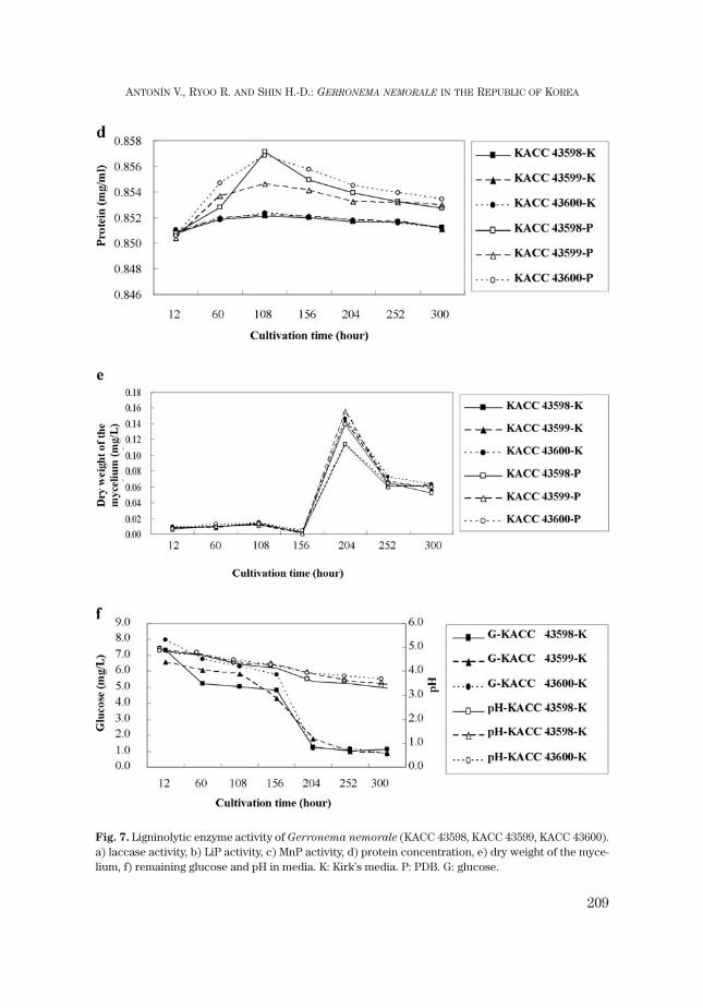
Total protein concentration of G. nemorale showed a similar curve as laccaseactivity (Fig. 7d). In all cases, protein concentration showed a peak after 108 hrs.The peaks of PDB showed a more or less abundant protein concentration in con-trast to Kirk’s media. It was a rising curve similar to the protein sigmoid curve be-tween 12 hrs and 156 hrs. However the dry weight showed a rapid increase after156 hrs and had its maximum after 204 hrs (Fig. 7e). Glucose was rapidly reducedafter 156 hrs, and pH gradually decreased (Fig. 7f). This was an inverse trend be-tween dry weight of the mycelium and glucose remained in the media.
205
ANTONÍN V., RYOO R. AND SHIN H.-D.: GERRONEMA NEMORALE IN THE REPUBLIC OF KOREA
Fig. 4. Macromorphological culture characteristics of Gerronema nemorale (KACC 43600). a) on PDAafter 5 weeks at 25 °C in the dark, b) spot test: on PDA after 3 weeks at 25 °C in the dark, c) on PDA con-taining Congo Red after 3 weeks at 25 °C in the dark, d) on PDA containing RBBR after 3 weeks at 25 °Cin the dark. Photo: R. Ryoo.
DISCUSSION
Gerronema nemorale is characterised by having small omphalioid basidiocarpswith innately fibrillose, olivaceous coloured pilei, abundant variable cheilocystidia,often clavate terminal cells in the pileipellis, and being lignicolous. According toSinger´s concept (Singer 1964), it belongs to sect. Xanthophylla Singer because ofwell-developed clamp-connections, and, according to Singer (1986), to subgen.Gerronema, sect. Xanthophylla Singer. For a more detailed discussion, see
206
CZECH MYCOL. 60(2): 197–212, 2008
Fig. 5. Micromorphological culture characteristics of Gerronema nemorale (KACC 43599). a) hyphaeon PDB, b) clamp-connections on PDB, c) crystal on PDA. Photo: R. Ryoo. Scale bar = 10 μm.
Takahashi (2000). Another lignicolous Gerronema species, G. corticiphilum L. N.Vassiljeva, described (as G. corticiphila) from the Russian Far East, differs espe-cially by white, wood-coloured, pale, rarely sulphurous coloured pilei and distinctlylonger and narrower (13–17 × 4–5 μm), subulate basidiospores (Vassiljeva 1973).
Outside of Asia, G. nemorale has been reported from Mexico (Veracruz; Lópezand García 2002). Although the Mexican fungus seems to be macroscopically simi-lar to our species, it has distinctly smaller basidiospores (7–8 × 6–7 μm) and prob-ably represents a different species.
Kim et al. (2005) published two photographs of G. nemorale from Mount Halla(Jeju Island) as Omphalina epichysium. Although both photographs represent
207
ANTONÍN V., RYOO R. AND SHIN H.-D.: GERRONEMA NEMORALE IN THE REPUBLIC OF KOREA
Fig. 6. Phylogenetic tree for Gerronema nemorale isolates (KACC 43599, KACC 43590, KG137) in-ferred from Bayesian analysis of the complete ITS region (ITS1, 5.8S rDNA, and ITS2), showing meanbranch lengths of a 50 % majority rule consensus tree, obtained from an MCMC analysis of one milliongenerations. An asterisk (*) denotes a sequence from GenBank.
208
CZECH MYCOL. 60(2): 197–212, 2008
209
ANTONÍN V., RYOO R. AND SHIN H.-D.: GERRONEMA NEMORALE IN THE REPUBLIC OF KOREA
Fig. 7. Ligninolytic enzyme activity of Gerronema nemorale (KACC 43598, KACC 43599, KACC 43600).a) laccase activity, b) LiP activity, c) MnP activity, d) protein concentration, e) dry weight of the myce-lium, f) remaining glucose and pH in media. K: Kirk’s media. P: PDB. G: glucose.
our Gerronema species, the mentioned size of the basidiospores (6.0–8.2 × 3.5–4.8μm) is distinctly smaller with values around the lower limit of the basidiosporevariability of O. epichysium. Omphalina epichysium (Pers.: Fr.) Quél. isa lignicolous species with a dark grey-brown to ash-coloured pileus and stipe,(7–)8–9(–10) × (3.5–)4–4.5(–6) μm large basidiospores which grows on decayingwood (stems, trunks) of conifers (Bon 1997).
The genus Gerronema in the sense of Singer (1964, 1986), however, ispolyphyletic (Moncalvo et al. 2002). Norvell et al. (1994) restricted the genusGerronema to lignicolous species with thin-walled basidiospores and typicalsarcodimitic tissue. In this concept, Gerronema is monophyletic and belongs tothe /hydropoid clade together with Hydropus s. str., Megacollybia, Clitocybula
and Porotheleum fimbriatum (Moncalvo et al. 2002). Matheny et al. (2006) alsoincluded Hydnopolyporus fimbriatus, Henningsomyces candidus and someMycena species (M. auricoma, M. amabilissima and M. aurantiidisca) in the/hydropoid clade which belongs to the large marasmioid clade. However, they didnot include any Gerronema species in their studies.
Having a sarcodimitic tissue structure, G. nemorale suits in the currently re-stricted concept of the genus Gerronema.
Cultivational characteristics were used for the identification and classificationof Aphyllophorales (Nobles 1965, Stalpers 1986), and later successfully applied toAgaricales (Desjardin 1990). A group with omphalioid basidiocarps includesOmphalina (Omphalia), Xeromphalina, Chrysomphalina, Arrhenia, Gerronema,Rickenella and many species of Clitocybe. The cultivational characteristics ofG. nemorale are more or less similar to those of Xeromphalina campanella (No-bles 1965, as Omphalia campanella) but differentiated by the hyphal structure andthe growth rate. On the other hand, the cultivational characteristics of G. nemorale
are similar to those of Collybia radicata (= Xerula radicata) and Collybia
velutipes (= Flammulina velutipes) according to the species codes described byNobles (1965). This is supported by the phylogenetic data of omphalioid genera ob-tained by molecular analyses (Fig. 6). The Gerronema and Omphalina species rep-resent the monophyletic group of two sister clades, which is different from othergroups. G. nemorale forms the Gerronema group along with G. subclavatum and“Chrysomphalina” strombodes, which is quite distant from Arrhenia and trueChrysomphalina. The Chrysomphalina strombodes sequence used in this studycorresponds to the concept of Norvell et al. (1994) and Redhead et al. (2002). Thiswas proven through the molecular analyses by Lutzoni (1997). According to them,Chrysomphalina strombodes is restricted to an American species belonging to thegenus Gerronema, so its correct name should be Gerronema strombodes (Berk. &Mont.) Clémençon, whereas the correct name for the European taxon usuallynamed Chrysomphalina (Gerronema) strombodes should be Gerronema xantho-
phyllum (Bres.) Norvell, Redhead & Ammirati.
210
CZECH MYCOL. 60(2): 197–212, 2008
In addition, one of the ecological characteristics of the genus Gerronema is thelignicolous habitat (Norvell et al. 1994). G. nemorale inhabits the part of sapwoodunder the bark. The ligninolytic enzymes laccase, LiP and MnP were required foreffective degradation of the lignin within the woody cell wall (Schmidt 2006).Moreover, although the combination of MnP and laccase was usually studied inwhite-rot fungi, the combination of LiP and MnP was researched only in 40 % ofthe white-rot fungi (Schmidt 2006). Therefore it is inspiring to see the results ofmeasuring the ligninolytic enzyme activity of G. nemorale. Laccase, LiP and MnPwere detected in G. nemorale as extracellular enzymes. This study supports thatG. nemorale belongs to lignicolous basidiomycetes.
ACKNOWLEDGEMENTS
The study trip to the Republic of Korea and the studies of the collected material by the first author wassupported by the Grant Agency of the Czech Republic (No. 206/07/J003). Other authors were supported bythe Korea Research Foundation Grant funded by the Korea Government (KRF-2006-F00001). The authorswish to thank Michal Tomšovský (Brno, Czech Republic) for valuable comments and discussion.
REFERENCES
BON M. (1997): Les clitocybes, omphales et ressemblants. – Doc. Mycol. Mém. hors Sér. 4: 1–181.BRADFORD M. M. (1976): A rapid and sensitive method for the quantitation of microgram quantities of
protein utilizing the principle of protein-dye binding. – Anal. Biochem. 72(1): 248–254.CLÉMENÇON H. (1982): Kompendium der Blätterpilze. Europäische omphalinoide Tricholomataceae. –
Z. Mykol. 48(2): 195–237.DESJARDIN D. E. (1990): Culture morphology of Marasmius species. – Sydowia 42: 17–87.GARDES M. and BRUNS T. D. (1993): ITS primers with enhanced specificity for basidiomycetes–applica-
tion to the identification of mycorrhizae and rusts. – Mol. Ecol. 2: 113–118.GEYER C. J. (1991): Markov Chain Monte Carlo maximum likelihood. – In: Keramidas E. M. (ed.), Com-
puting science and statistics, Proceedings of the 23rd symposium on the interface, p. 156–163, In-terface Foundation, Virginia.
GLENN J. K. and GOLD M. H. (1983): Decolorization of several polymeric dyes by the lignin-degradingBasidiomycete Phanerochaete chrysosporium. – Appl. Environ. Microbiol. 45(6): 1741–1747.
HOLMGREN P. K. and HOLMGREN N. H. (1998 [continuously updated]): Index Herbariorum: A global di-rectory of public herbaria and associated staff. New York Botanical Garden's Virtual Herbarium. –http://s_Hlt208296624w_Hlt208296624eetgum.nybg.org/ih/
KIM Y. S., SEOK S. J., KIM W. K., WON H. Y., LEE G. H., HYEON G. H., KIM B. C., KIM J. S., YANG Y. T. and KIM
S. H. (2005): The mushrooms of Halla mountain. – 317 p. Jeju-do Agricultural Research and Exten-sion Services.
KIRK P. M. and ANSELL A. E. (1992): Authors of fungal names. − 95 p. Kew.KIRK T. K., SCHULTZ E., CONNORS W. J., LORENZ L. F. and ZEIKUS J. G. (1978): Influence of culture param-
eters on lignin metabolism by Phanerochaete chrysosporium. – Arch. Microbiol. 117: 277–285.
211
ANTONÍN V., RYOO R. AND SHIN H.-D.: GERRONEMA NEMORALE IN THE REPUBLIC OF KOREA

LEE S. B. and TAYLOR J. W. (1990): Isolation of DNA from fungal mycelia and single spores. – In: Innis M.A., Gelfand D. H., Sninsky J. J. and White T. J. (eds.), PCR protocols: A guide to methods and appli-cations, p. 282–287, San Diego.
LOPÉZ A. R. and GARCÍA J. A. (2002): Gerronema nemorale. – Funga Veracruzana 72. –http://www.scribd.com/doc/41172/FUNGA-VERACRUZANA-Num72-Gerronema-nemorale.[accessed 1 July 2008]
LUTZONI F. M. (1997): Phylogeny of lichen-and non-lichen-forming omphalinoid mushrooms and theutility of testing for combinability among multiple data sets. – Syst. Biol. 46(3): 373–406.
MATHENY P. B., CURTIS J. M., HOFSTETTER V., AIME C., MONCALVO J.-M., GE Z.-W., SLOT J. C., AMMIRATI J.F., BARONI T. J., BOUGHER N. K., HUGHES K. W., LODGE J., KERRIGAN R. W., SEIDL M. T., AANEN D. K.,DENITIS M., DANIELE G. M., DESJARDIN D. E., KROPP B. R., NORVELL L. L., PARKER A., VELLINGA E.C., VILGALYS R. and HIBBETT D. S. (2006): Major clades of Agaricales: a multilocus phylogeneticoverview. – Mycologia 98(6): 982–995.
MILLER G. L. (1958): Use of dinitrosalicylic acid reagent for determination of reducing sugar. – Anal.Biochem. 1: 426–506.
MONCALVO J.-M., LUTZONI F. M., REHNER S. A., JOHNSON J. and VILGALYS R. (2000): Phylogenetic rela-tionships of agaric fungi based on nuclear large subunit ribosomal DNA sequences. – Syst. Biol.49(2): 278–305.
MONCALVO J.-M., VILGALYS R., REDHEAD S. A., JOHNSON J. E., JAMES T. Y., AIME C., HOFSTETTER V.,VERDUIN S. J. W., LARSSON E., BARONI T. J., THORN R. G., JACOBSSON S., CLÉMENÇON H. and MILLER
O. K. JR. (2002): One hundred and seventeen clades of euagarics. – Mol. Phylogenet. Evol. 23:357–400.
NOBLES M. K. (1965): Identification of cultures of wood-inhabiting Hymenomycetes. – Can. J. Bot. 43:1097–1139.
NORVELL L. L., REDHEAD S. A. and AMMIRATI J. F. (1994): Omphalina sensu lato in North America 1–2. 1:Omphalina wynniae and the genus Chrysomphalina, 2: Omphalina sensu Bigelow. – Mycotaxon50: 379–407.
OKINO L. K., MACHADO K. M. G., FABRIS C. and BONONI V. L. R. (2000): Ligninolytic activity of tropicalrainforest basidiomycetes. – World J. Microbiol. Biotechnol. 16: 889–893.
REDHEAD S. A., LUTZONI F., MONCALVO J.-M. and VILGALYS R. (2002): Phylogeny of agarics: partial sys-tematics solutions for core omphalinoid genera in the Agaricales (euagarics). – Mycotaxon 83:19–57.
RONQUIST F. and HUELSENBECK J. P. (2003): MRBAYES 3: Bayesian phylogenetic inference under mixedmolds. – Bioinformatics 19: 1572–1574.
SCHMIDT O. (2006): Wood and tree fungi: biology, damage, protection and use. – 334 p. Heidelberg.SINGER R. (1964): Die Gattung Gerronema. – Nova Hedwigia 7: 53–92.SINGER R. (1986): The Agaricales in modern taxonomy. – 981 p. Koenigstein.STALPERS J. A. (1978): Identification of cultures of wood-inhabiting fungi in pure culture. – Stud. Mycol.
16: 1–248.TAKAHASHI H. (2000): Two new species and one new variety of Agaricales from central Honshu, Ja-
pan. – Mycoscience 41: 15–23.THOMPSON J. D., GIBSON T. J., PLEWNIAK F., JEANMOUGIN F. and HIGGINS D. G. (1997): The CLUSTAL_X
windows interface: flexible strategies for multiple sequence alignment aided by quality analysistools. – Nucleic Acids Res. 25(24): 4876–4882.
VARES T., KALSI M. and HATAKKA A. (1995): Lignin peroxidases, manganese peroxidases and otherligninolytic enzymes produced by Phlebia radiata during solid-state fermentation of wheatstraw. – Appl. Environ. Microbiol. 61: 3515–3520.
VASSILJEVA L. N. (1973): Agarikovyje shljapochnye griby (por. Agaricales) Primorskogo kraya. – 328 p.Leningrad.
212
CZECH MYCOL. 60(2): 197–212, 2008