Embed Size (px)
Citation preview
An Imaging Guide for the Busy Physician Care to Care’s Brief Reference to Efficient Medical Imaging
Michael Komarow MD, JD Chief Medical Officer
Care to Care An Imaging Guide for the Busy Physician: Brief Reference to Efficient Medical Imaging
Copyright © 2010 Care to Care. All rights reserved. 2
Preface………………………………………………………………………………………………….. 3
Abdominal Pain…………………………………………………………………………………………... 4
Generalized Abdominal Pain………………………………………………………………………………… 5
Epigastric or Upper Abdominal Pain…………………………………………………………………………... 6
Right Upper Quadrant Pain………………………………………………………………………………….. 6
Abdominal Pain, Left Upper Quadrant……………………………………….………………………………... 7
Right Lower Quadrant Pain………………………………………………………………………………….. 8
Left Lower Quadrant Pain…………………………………………………………………………………... 9
Chest Pain in Patients Not Seen in ER……………………………………………………………………... 10
Headache………………………………………………………………………………………………. 11
Low Back Pain………………………………………………………………………………………….. 13
Joint Pain………………………………………………………………………………………………. 15
Seizures………………………………………………………………………………………………... 16
Vascular Disease 16
Imaging of Cerebrovascular Disease…………………………….…………………………………………....17
Carotid Disease……………………………………………………………………………………… 17
Stroke…………………………………………..…………………………………………………... 17
Cardiovascular Disease…………………………………………………………………………………… 18
Peripheral Arterial Disease………………………………………………………………………………… 19
Cough……………………………………………………………………………..……………………..18
Obstetrical Ultrasound…………………………………………………………………………………….. 21
Care to Care An Imaging Guide for the Busy Physician: Brief Reference to Efficient Medical Imaging
Copyright © 2010 Care to Care. All rights reserved. 3
PREFACE
“The amount of information contained in an imaging study is enormous, especially with
computed axial tomography and magnetic resonance imaging technologies. This increase in
information may be essential for the care of patients, or it may be a distraction in the diagnostic
process. Imaging techniques are rapidly evolving, so it is essential to work closely with your
radiologist to select the appropriate imaging studies to answer your questions.1”
The most common complaints arise most commonly, and cause the most over utilization of medical imaging. It is our purpose here to provide a quick guide to appropriate use of imaging examinations to foster prompt and accurate diagnosis. Imaging examinations, from the simplest x-ray to the most complex fused PET-MRI can offer essential diagnostic information, but all have limits. No one benefits if an imaging exam does not reduce the degree of diagnostic uncertainty faced by the treating physician but radiation exposure, expenses, and the risks of false positive results all accumulate. Despite considerable increases in imaging efficiency, radiation dosage from CT remains relatively high, and with it the risk of inducing malignancy. It is poor medicine to expose a patient to any risk without assessment of the potential benefit.
About 15% of the ionizing radiation exposure to the general public comes from artificial sources, and almost all of this exposure is due to medical radiation, largely from diagnostic procedures. Of the approximately 3 mSv annual global per caput effective dose estimated for the year 2000, 2.4 mSv is from natural background and 0.4 mSv from diagnostic medical exams. Diagnostic and therapeutic radiation was used in patients as early as 1896. Since then, continual improvements in diagnostic imaging and radiotherapy as well as the aging of our population have led to greater use of medical radiation. Temporal trends indicate that worldwide population exposure from medical radiation is increasing. In the United States, there has been a steady rise in the use of diagnostic radiologic procedures, especially x rays. Radiotherapy also has increased so that today about 40% of cancer patients receive some treatment with radiation. Epidemiologic data on medically irradiated populations are an important complement to the atomic-bomb survivors' studies. Significant improvement in cancer treatment over the last few decades has resulted in longer survival and a growing number of radiation-related second cancers. Following high-dose radiotherapy for malignant diseases, elevated risks of a variety of radiation-related second cancers have been
1 LeBlond RF, Brown DD, DeGowin RL, "Chapter 17. Principles of Diagnostic Testing" (Chapter). LeBlond RF,
Brown DD, DeGowin RL: DeGowin's Diagnostic Examination, 9th Edition: www.accessmedicine.com/content.aspx?aID=3658178.
Care to Care An Imaging Guide for the Busy Physician: Brief Reference to Efficient Medical Imaging
Copyright © 2010 Care to Care. All rights reserved. 4
observed. Risks have been particularly high following treatment for childhood cancer. Radiation treatment for benign disease was relatively common from the 1940's to the 1960's. While these treatments generally were effective, some resulted in enhanced cancer risks. As more was learned about radiation-associated cancer risks and new treatments became available, the use of radiotherapy for benign disease has declined. At moderate doses, such as those used to treat benign diseases, radiation-related cancers occur in or near the radiation field. Cancers of the thyroid, salivary gland, central nervous system, skin, and breast as well as leukemia have been associated with radiotherapy for tinea capitis, enlarged tonsils or thymus gland, other benign conditions of the head and neck, or benign breast diseases. Because doses from diagnostic examinations typically are low, they are difficult to study using epidemiologic methods, unless multiple examinations are performed. An excess risk of breast cancer has been reported among women with tuberculosis who had multiple chest fluoroscopies as well as among scoliosis patients who had frequent diagnostic x rays during late childhood and adolescence. Dental and medical diagnostic x rays performed many years ago, when doses were presumed to be high, also have been linked to increased cancer risks. The carcinogenic effects of diagnostic and therapeutic radionuclides are less well characterized. High risks of liver cancer and leukemia have been demonstrated following thorotrast injections, and patients treated with radium appear to have an elevated risk of bone sarcomas and possibly cancers of the breast, liver, kidney, thyroid, and bladder. 2
Proper choice of which exam to use, and when to use it is therefore crucial. On the pages that follow we have attempted to give a guide to the proper use of imaging in a variety of non-emergent clinical circumstances. Readers’ comments and suggestions are welcomed and should be sent to me at [email protected].
Abdominal Pain
Paying no heed to the sages who in creating CPT codes divided the abdomen into two parts (abdomen and pelvis) nature recognizes no such border. For our purposes we will consider the abdomen in five parts and just for the sake of being ornery also as the whole. The complex anatomy and multiplicity of structures within the abdomen make such divisions clinically useful, even if there is considerable overlap. In the charts below are listed common (and not so common) causes of abdominal pain and suggestions concerning whether CT or MR are useful in evaluating these particular diagnoses. This presupposes that the history, physical and prior exams have led to the consideration of one of the diagnoses as being the likely source of the patient’s pain. Subjecting everyone with abdominal pain to a CT is not only cost ineffective it is a misapplication of a great deal of radiation. Judicious use of imaging can, however, swiftly lead to appropriate care. Please use what follows as a guide, to be combined with all the other clinical data you compile, and your best judgment. 2 Health Phys. 2003 Jul;85(1):47-59.
Care to Care An Imaging Guide for the Busy Physician: Brief Reference to Efficient Medical Imaging
Copyright © 2010 Care to Care. All rights reserved. 5
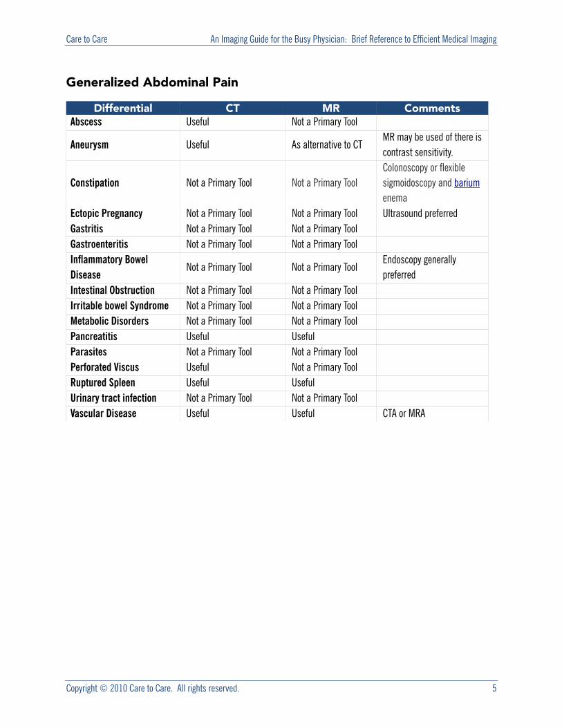
Generalized Abdominal Pain
Differential CT MR CommentsAbscess Useful Not a Primary Tool
Aneurysm Useful As alternative to CT MR may be used of there is contrast sensitivity.
Constipation Not a Primary Tool Not a Primary Tool Colonoscopy or flexible sigmoidoscopy and barium enema
Ectopic Pregnancy Not a Primary Tool Not a Primary Tool Ultrasound preferred Gastritis Not a Primary Tool Not a Primary Tool Gastroenteritis Not a Primary Tool Not a Primary Tool Inflammatory Bowel Disease
Not a Primary Tool Not a Primary Tool Endoscopy generally preferred
Intestinal Obstruction Not a Primary Tool Not a Primary Tool Irritable bowel Syndrome Not a Primary Tool Not a Primary Tool Metabolic Disorders Not a Primary Tool Not a Primary Tool Pancreatitis Useful Useful Parasites Not a Primary Tool Not a Primary Tool Perforated Viscus Useful Not a Primary Tool Ruptured Spleen Useful Useful Urinary tract infection Not a Primary Tool Not a Primary Tool Vascular Disease Useful Useful CTA or MRA
Care to Care An Imaging Guide for the Busy Physician: Brief Reference to Efficient Medical Imaging
Copyright © 2010 Care to Care. All rights reserved. 6
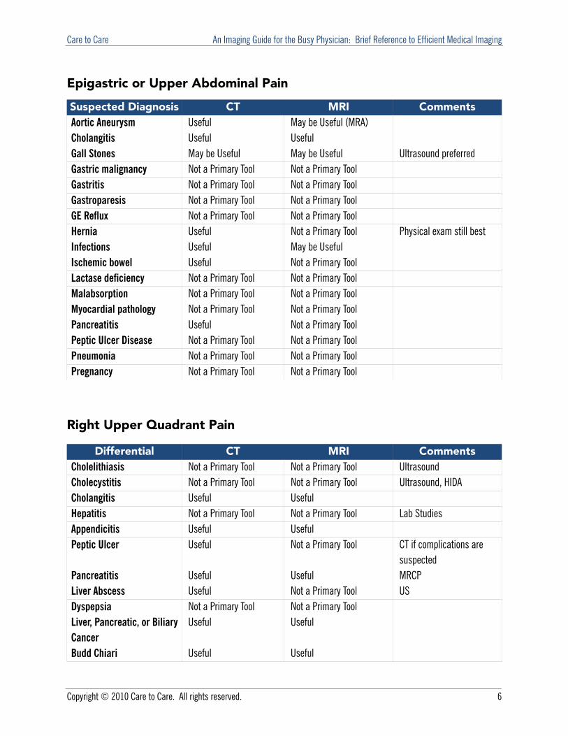
Epigastric or Upper Abdominal Pain
Suspected Diagnosis CT MRI CommentsAortic Aneurysm Useful May be Useful (MRA) Cholangitis Useful Useful Gall Stones May be Useful May be Useful Ultrasound preferred Gastric malignancy Not a Primary Tool Not a Primary Tool Gastritis Not a Primary Tool Not a Primary Tool Gastroparesis Not a Primary Tool Not a Primary Tool GE Reflux Not a Primary Tool Not a Primary Tool Hernia Useful Not a Primary Tool Physical exam still best Infections Useful May be Useful Ischemic bowel Useful Not a Primary Tool Lactase deficiency Not a Primary Tool Not a Primary Tool Malabsorption Not a Primary Tool Not a Primary Tool Myocardial pathology Not a Primary Tool Not a Primary Tool Pancreatitis Useful Not a Primary Tool Peptic Ulcer Disease Not a Primary Tool Not a Primary Tool Pneumonia Not a Primary Tool Not a Primary Tool Pregnancy Not a Primary Tool Not a Primary Tool
Right Upper Quadrant Pain
Differential CT MRI CommentsCholelithiasis Not a Primary Tool Not a Primary Tool Ultrasound Cholecystitis Not a Primary Tool Not a Primary Tool Ultrasound, HIDA Cholangitis Useful Useful Hepatitis Not a Primary Tool Not a Primary Tool Lab Studies Appendicitis Useful Useful Peptic Ulcer Useful Not a Primary Tool CT if complications are
suspected Pancreatitis Useful Useful MRCP Liver Abscess Useful Not a Primary Tool US Dyspepsia Not a Primary Tool Not a Primary Tool Liver, Pancreatic, or Biliary Cancer
Useful Useful
Budd Chiari Useful Useful
Care to Care An Imaging Guide for the Busy Physician: Brief Reference to Efficient Medical Imaging
Copyright © 2010 Care to Care. All rights reserved. 7
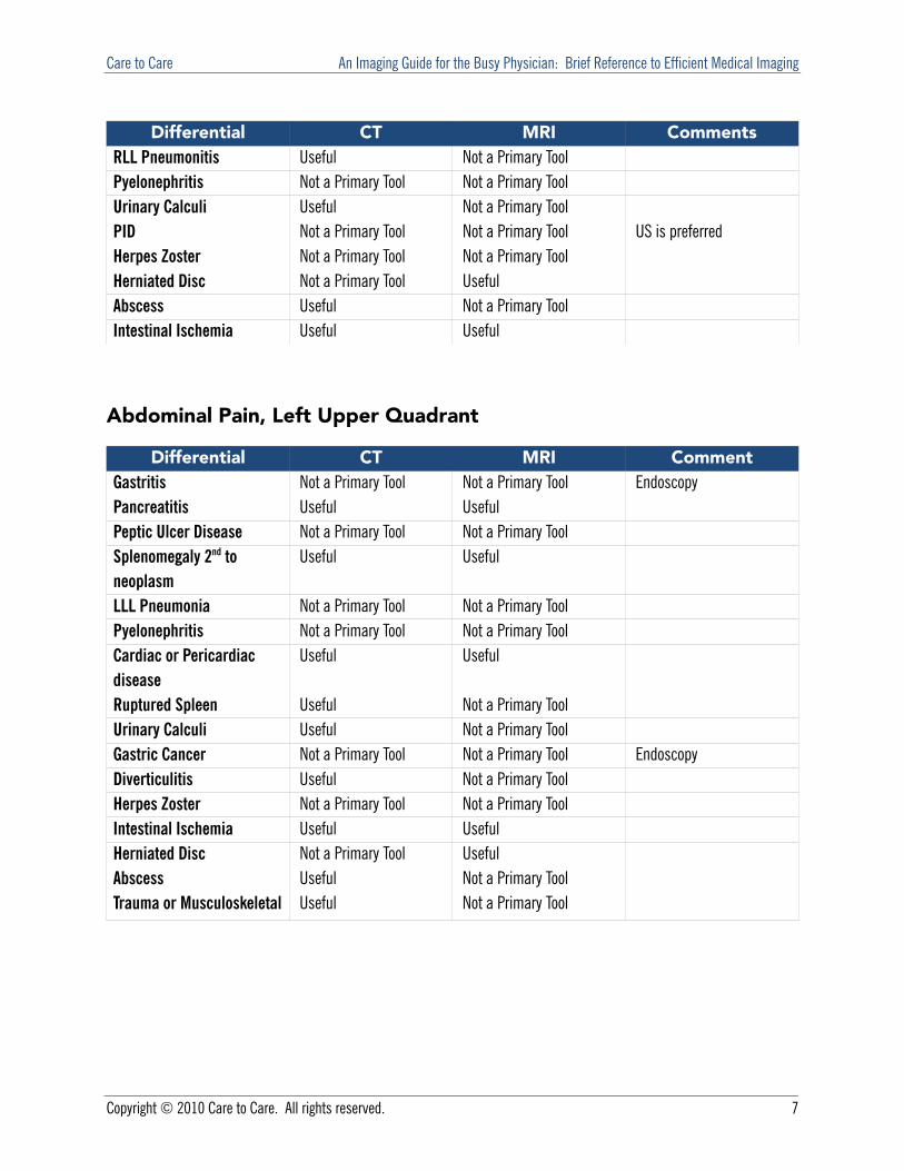
Differential CT MRI CommentsRLL Pneumonitis Useful Not a Primary Tool Pyelonephritis Not a Primary Tool Not a Primary Tool Urinary Calculi Useful Not a Primary Tool PID Not a Primary Tool Not a Primary Tool US is preferred Herpes Zoster Not a Primary Tool Not a Primary Tool Herniated Disc Not a Primary Tool Useful Abscess Useful Not a Primary Tool Intestinal Ischemia Useful Useful
Abdominal Pain, Left Upper Quadrant
Differential CT MRI CommentGastritis Not a Primary Tool Not a Primary Tool Endoscopy Pancreatitis Useful Useful Peptic Ulcer Disease Not a Primary Tool Not a Primary Tool Splenomegaly 2nd to neoplasm
Useful Useful
LLL Pneumonia Not a Primary Tool Not a Primary Tool Pyelonephritis Not a Primary Tool Not a Primary Tool Cardiac or Pericardiac disease
Useful Useful
Ruptured Spleen Useful Not a Primary Tool Urinary Calculi Useful Not a Primary Tool Gastric Cancer Not a Primary Tool Not a Primary Tool Endoscopy Diverticulitis Useful Not a Primary Tool Herpes Zoster Not a Primary Tool Not a Primary Tool Intestinal Ischemia Useful Useful Herniated Disc Not a Primary Tool Useful Abscess Useful Not a Primary Tool Trauma or Musculoskeletal Useful Not a Primary Tool
Care to Care An Imaging Guide for the Busy Physician: Brief Reference to Efficient Medical Imaging
Copyright © 2010 Care to Care. All rights reserved. 8
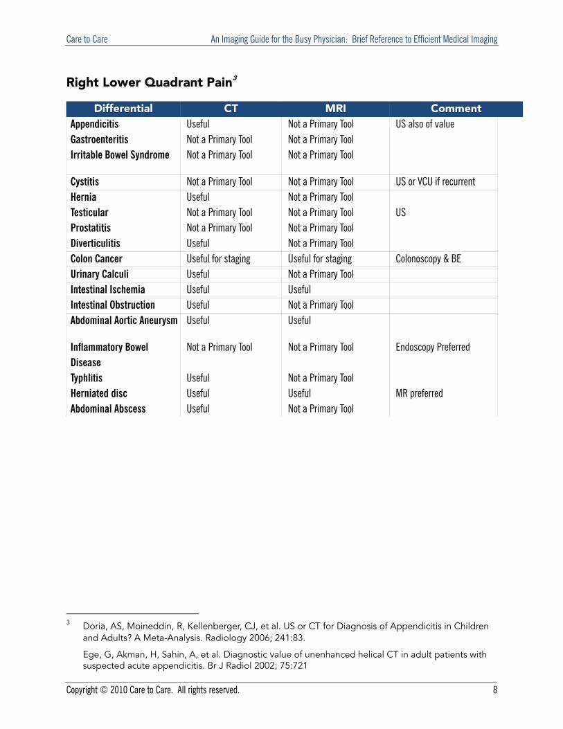
Right Lower Quadrant Pain3
Differential CT MRI CommentAppendicitis Useful Not a Primary Tool US also of value Gastroenteritis Not a Primary Tool Not a Primary Tool Irritable Bowel Syndrome Not a Primary Tool Not a Primary Tool
Cystitis Not a Primary Tool Not a Primary Tool US or VCU if recurrent Hernia Useful Not a Primary Tool Testicular Not a Primary Tool Not a Primary Tool US Prostatitis Not a Primary Tool Not a Primary Tool Diverticulitis Useful Not a Primary Tool Colon Cancer Useful for staging Useful for staging Colonoscopy & BE Urinary Calculi Useful Not a Primary Tool Intestinal Ischemia Useful Useful Intestinal Obstruction Useful Not a Primary Tool Abdominal Aortic Aneurysm Useful Useful
Inflammatory Bowel Disease
Not a Primary Tool Not a Primary Tool Endoscopy Preferred
Typhlitis Useful Not a Primary Tool Herniated disc Useful Useful MR preferred Abdominal Abscess Useful Not a Primary Tool
3 Doria, AS, Moineddin, R, Kellenberger, CJ, et al. US or CT for Diagnosis of Appendicitis in Children
and Adults? A Meta-Analysis. Radiology 2006; 241:83.
Ege, G, Akman, H, Sahin, A, et al. Diagnostic value of unenhanced helical CT in adult patients with suspected acute appendicitis. Br J Radiol 2002; 75:721
Care to Care An Imaging Guide for the Busy Physician: Brief Reference to Efficient Medical Imaging
Copyright © 2010 Care to Care. All rights reserved. 9
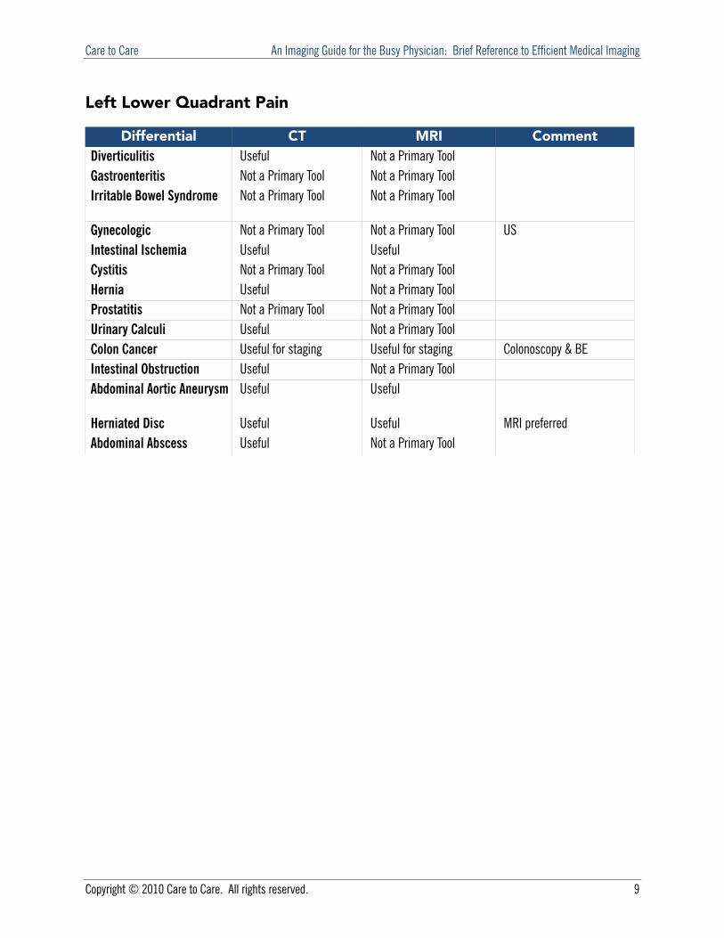
Left Lower Quadrant Pain
Differential CT MRI CommentDiverticulitis Useful Not a Primary Tool Gastroenteritis Not a Primary Tool Not a Primary Tool Irritable Bowel Syndrome Not a Primary Tool Not a Primary Tool
Gynecologic Not a Primary Tool Not a Primary Tool US Intestinal Ischemia Useful Useful Cystitis Not a Primary Tool Not a Primary Tool Hernia Useful Not a Primary Tool Prostatitis Not a Primary Tool Not a Primary Tool Urinary Calculi Useful Not a Primary Tool Colon Cancer Useful for staging Useful for staging Colonoscopy & BE Intestinal Obstruction Useful Not a Primary Tool Abdominal Aortic Aneurysm Useful Useful
Herniated Disc Useful Useful MRI preferred Abdominal Abscess Useful Not a Primary Tool
Care to Care An Imaging Guide for the Busy Physician: Brief Reference to Efficient Medical Imaging
Copyright © 2010 Care to Care. All rights reserved. 10
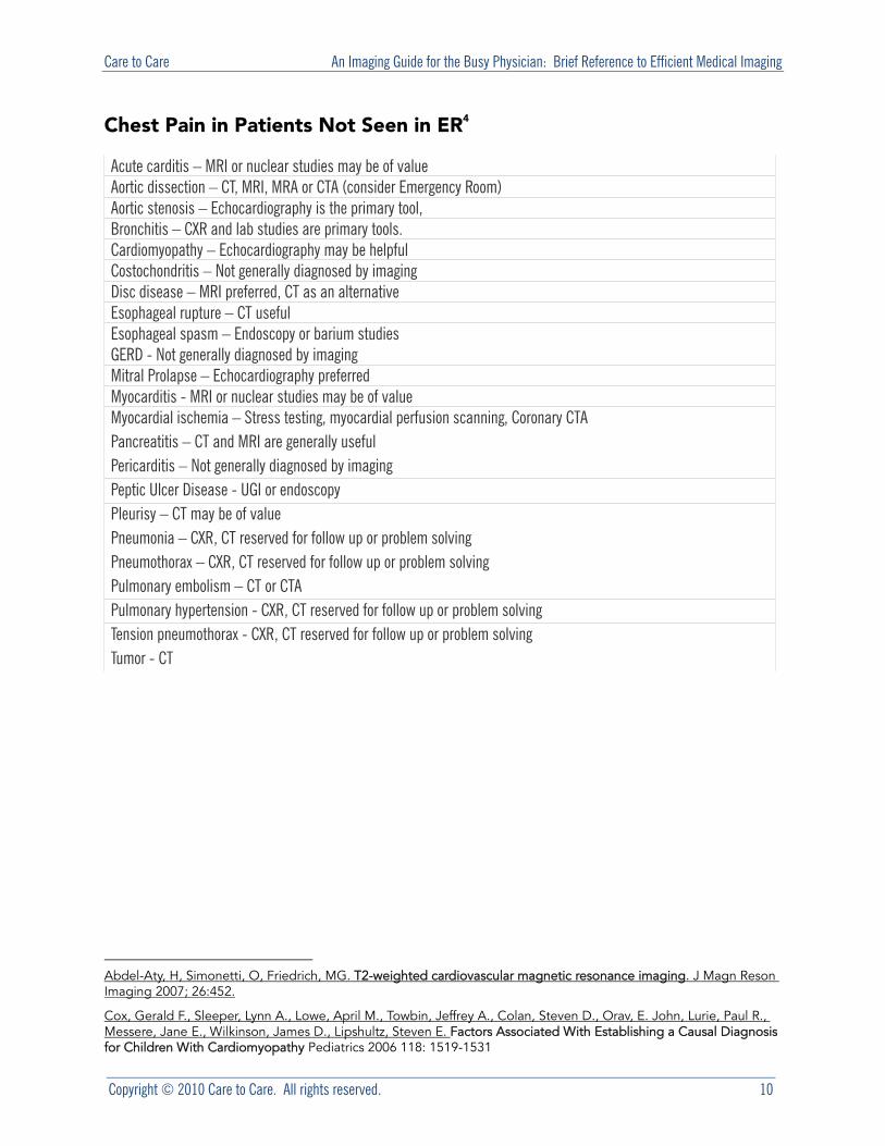
Chest Pain in Patients Not Seen in ER4
Acute carditis – MRI or nuclear studies may be of value Aortic dissection – CT, MRI, MRA or CTA (consider Emergency Room) Aortic stenosis – Echocardiography is the primary tool, Bronchitis – CXR and lab studies are primary tools. Cardiomyopathy – Echocardiography may be helpful Costochondritis – Not generally diagnosed by imaging Disc disease – MRI preferred, CT as an alternative Esophageal rupture – CT useful Esophageal spasm – Endoscopy or barium studies GERD - Not generally diagnosed by imaging Mitral Prolapse – Echocardiography preferred Myocarditis - MRI or nuclear studies may be of value Myocardial ischemia – Stress testing, myocardial perfusion scanning, Coronary CTA Pancreatitis – CT and MRI are generally useful Pericarditis – Not generally diagnosed by imaging Peptic Ulcer Disease - UGI or endoscopy Pleurisy – CT may be of value Pneumonia – CXR, CT reserved for follow up or problem solving Pneumothorax – CXR, CT reserved for follow up or problem solving Pulmonary embolism – CT or CTA Pulmonary hypertension - CXR, CT reserved for follow up or problem solving Tension pneumothorax - CXR, CT reserved for follow up or problem solving Tumor - CT
Abdel-Aty, H, Simonetti, O, Friedrich, MG. T2-weighted cardiovascular magnetic resonance imaging. J Magn Reson Imaging 2007; 26:452.
Cox, Gerald F., Sleeper, Lynn A., Lowe, April M., Towbin, Jeffrey A., Colan, Steven D., Orav, E. John, Lurie, Paul R., Messere, Jane E., Wilkinson, James D., Lipshultz, Steven E. Factors Associated With Establishing a Causal Diagnosis for Children With Cardiomyopathy Pediatrics 2006 118: 1519-1531

Care to Care An Imaging Guide for the Busy Physician: Brief Reference to Efficient Medical Imaging
Copyright © 2010 Care to Care. All rights reserved. 10
Headache
Imaging has a notoriously low yield in the work up of headache when there are no other neurologic complaints or findings. MRI or CT should be used judiciously when there are clinical findings that suggest a secondary, treatable cause. The differential diagnosis is virtually endless, including at least the following:
Arterial dissection (carotid or vertebral) Arteriovenous malformation Brain abscess Brain tumor Caffeine, alcohol, or drug withdrawal Carbon monoxide poisoning Cerebral ischemia Cervical arthritis Cough headache (primary or secondary) Cluster headache Dental abscess Depression Glaucoma Head injury Hypertension Meningitis Migraine Neurocysticercosis Otitis media Post-lumbar puncture Preeclampsia Pseudotumor cerebri Refractive error Sinusitis Subarachnoid hemorrhage Subdural hemorrhage Temporal (giant cell) arteritis Temporomandibular joint syndrome Tension headache Venous sinus thrombosis (intracranial venous thrombosis) Somatoform disorder (eg, somatization) Trigeminal neuralgia Glossopharyngeal neuralgia Facial, tooth, or jaw pain
With so extensive a differential the temptation to search for a cause can be overwhelming, but the fact is that CT or
Care to Care An Imaging Guide for the Busy Physician: Brief Reference to Efficient Medical Imaging
Copyright © 2010 Care to Care. All rights reserved. 12
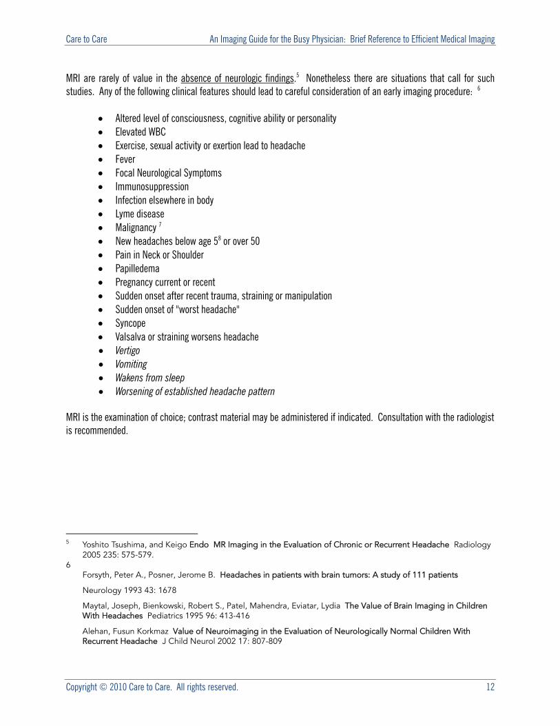
MRI are rarely of value in the absence of neurologic findings.5 Nonetheless there are situations that call for such studies. Any of the following clinical features should lead to careful consideration of an early imaging procedure: 6
Altered level of consciousness, cognitive ability or personality Elevated WBC Exercise, sexual activity or exertion lead to headache Fever Focal Neurological Symptoms Immunosuppression Infection elsewhere in body Lyme disease Malignancy 7 New headaches below age 58 or over 50 Pain in Neck or Shoulder Papilledema Pregnancy current or recent Sudden onset after recent trauma, straining or manipulation Sudden onset of "worst headache" Syncope Valsalva or straining worsens headache Vertigo Vomiting Wakens from sleep Worsening of established headache pattern
MRI is the examination of choice; contrast material may be administered if indicated. Consultation with the radiologist is recommended.
5 Yoshito Tsushima, and Keigo Endo MR Imaging in the Evaluation of Chronic or Recurrent Headache Radiology
2005 235: 575-579. 6
Forsyth, Peter A., Posner, Jerome B. Headaches in patients with brain tumors: A study of 111 patients
Neurology 1993 43: 1678
Maytal, Joseph, Bienkowski, Robert S., Patel, Mahendra, Eviatar, Lydia The Value of Brain Imaging in Children With Headaches Pediatrics 1995 96: 413-416
Alehan, Fusun Korkmaz Value of Neuroimaging in the Evaluation of Neurologically Normal Children With Recurrent Headache J Child Neurol 2002 17: 807-809
Care to Care An Imaging Guide for the Busy Physician: Brief Reference to Efficient Medical Imaging
Copyright © 2010 Care to Care. All rights reserved. 13
Low Back Pain
Back pain is perhaps the most universal affliction this side of aging; and among the most common causes of medical office visits. Large numbers of CT and MRI exams are ordered to investigate LBP, and very few yield useful information or contribute to patient well-being. 9 Worse, there is evidence that early imaging with CT or MRI may lead to unnecessary surgery, with resultant failure to improve and morbidity. 10 Significant numbers of asymptomatic patients have findings on MRI that, in a symptomatic individual, may be taken to be indications for surgery, but which are in fact not contributing to the patient’s discomfort. 11
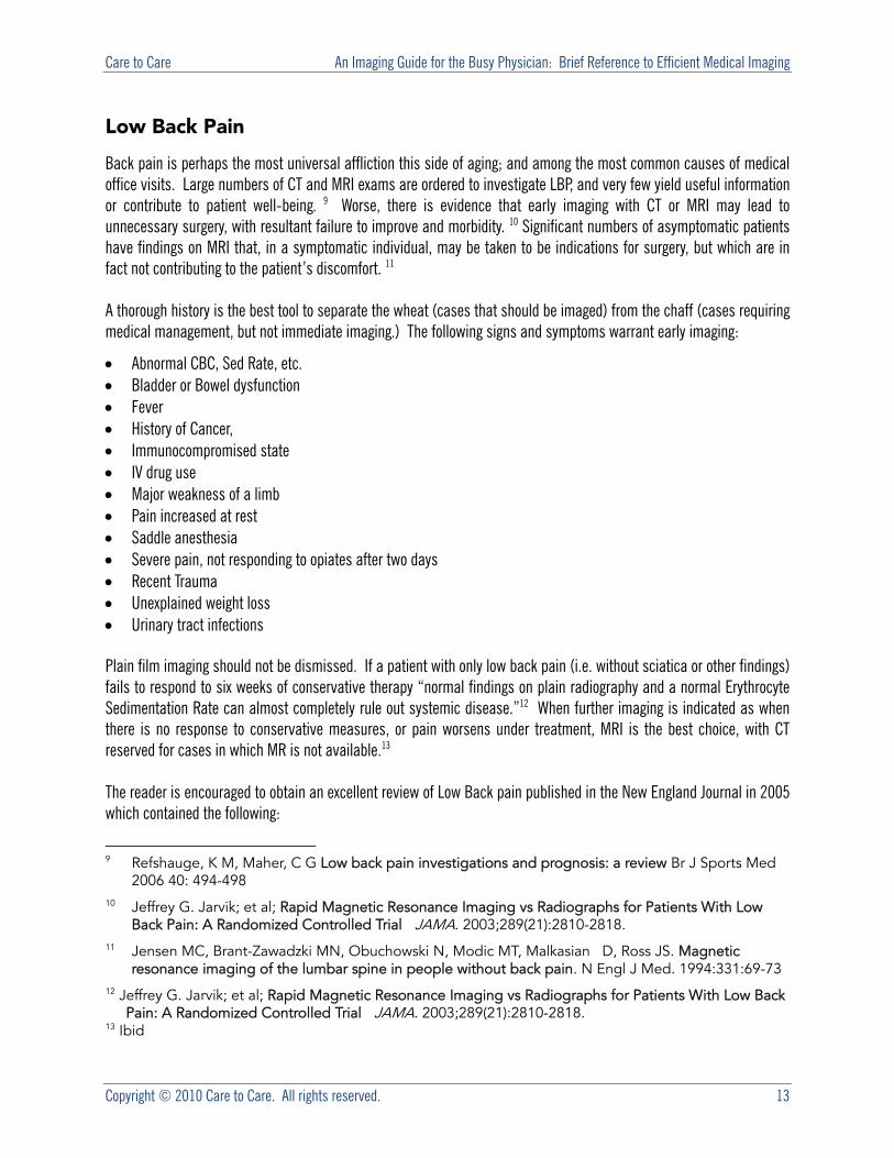
A thorough history is the best tool to separate the wheat (cases that should be imaged) from the chaff (cases requiring medical management, but not immediate imaging.) The following signs and symptoms warrant early imaging:
Abnormal CBC, Sed Rate, etc. Bladder or Bowel dysfunction Fever History of Cancer, Immunocompromised state IV drug use Major weakness of a limb Pain increased at rest Saddle anesthesia Severe pain, not responding to opiates after two days Recent Trauma Unexplained weight loss Urinary tract infections
Plain film imaging should not be dismissed. If a patient with only low back pain (i.e. without sciatica or other findings) fails to respond to six weeks of conservative therapy “normal findings on plain radiography and a normal Erythrocyte Sedimentation Rate can almost completely rule out systemic disease.”12 When further imaging is indicated as when there is no response to conservative measures, or pain worsens under treatment, MRI is the best choice, with CT reserved for cases in which MR is not available.13
The reader is encouraged to obtain an excellent review of Low Back pain published in the New England Journal in 2005 which contained the following:
9 Refshauge, K M, Maher, C G Low back pain investigations and prognosis: a review Br J Sports Med
2006 40: 494-498 10 Jeffrey G. Jarvik; et al; Rapid Magnetic Resonance Imaging vs Radiographs for Patients With Low
Back Pain: A Randomized Controlled Trial JAMA. 2003;289(21):2810-2818. 11 Jensen MC, Brant-Zawadzki MN, Obuchowski N, Modic MT, Malkasian D, Ross JS. Magnetic
resonance imaging of the lumbar spine in people without back pain. N Engl J Med. 1994:331:69-73 12 Jeffrey G. Jarvik; et al; Rapid Magnetic Resonance Imaging vs Radiographs for Patients With Low Back
Pain: A Randomized Controlled Trial JAMA. 2003;289(21):2810-2818. 13 Ibid
Care to Care An Imaging Guide for the Busy Physician: Brief Reference to Efficient Medical Imaging
Copyright © 2010 Care to Care. All rights reserved. 14
MRI or radiography early in the course of an episode of low back pain do not improve clinical outcomes or reduce costs of care.14 MRI is best used to rule out the possibility of impending
neurologic injury, infection, or tumors. Appropriate candidates for MRI include patients with low back pain who have associated neurologic symptoms or signs; associated systemic symptoms; risk factors for cancer, infection, or occult fractures; or persistent pain in the absence of neurologic signs or symptoms after four to eight weeks. Patients should understand that the reason for imaging is to rule out these serious conditions, and that common degenerative findings are expected. Ill-considered attempts to make a diagnosis on the basis of imaging studies may reinforce the suspicion of serious disease, magnify the
importance of nonspecific findings, and label patients with spurious diagnoses.
Carragee, Eugene J Persistent Low Back Pain N Engl J Med 2005 352: 1891-1898
14 Gilbert FJ, Grant AM, Gillan MG, et al. Low back pain: influence of early MR imaging or CT on
treatment and outcome -- multicenter randomized trial. Radiology 2004;231:343-351.
Care to Care An Imaging Guide for the Busy Physician: Brief Reference to Efficient Medical Imaging
Copyright © 2010 Care to Care. All rights reserved. 15
Joint Pain
Another common source of office visits to primary care physicians, joint pain may account for up to 5% of such visits.15 Not all joint pain is due to arthritis. Pain in and around joints can be caused by a dizzying variety of conditions, and a careful history and physical examination is, once again, the single best tool in directing the physician’s course of investigation. Is the joint swollen, erythematous, or deformed? Is local musculature atrophied? Is the joint tender, is range of motion limited? These are all signs that joint pain, arthralgia, is likely due to joint inflammation, arthritis. The role of laboratory studies in determining the nature of an arthritic process, or of a systemic disease causing joint pain remains paramount. Plain film radiography plays a critical role in evaluating joints, and should be used first whenever arthritis is suspected or is being evaluated. Ultrasound is inexpensive, readily available and free of ionizing radiation. It should not be overlooked when evaluating soft tissue sources of joint pain; in experienced hands is essentially equal to MRI in evaluating the rotator cuff. 16
15 Palmer, Trish, Toombs, James D. Managing Joint Pain in Primary Care J Am Board Fam Pract 2004 17: S32-42 16 Al-Shawi, A., Badge, R., Bunker, T. The detection of full thickness rotator cuff tears using ultrasound J Bone Joint
Surg Br 2008 90-B: 889-892
Care to Care An Imaging Guide for the Busy Physician: Brief Reference to Efficient Medical Imaging
Copyright © 2010 Care to Care. All rights reserved. 16
Seizures
From the clinician’s point of view classification of the type of seizure disorder being evaluated is the first step. Generalized seizures always involve the loss of consciousness and may be subclassified as tonic (rigid,) clonic (characterized by repeated jerking movements without rigidity,) myoclonic (the most common,) atonic (flaccid,) and absence (petit mal.) Partial seizures may be simple (without loss of consciousness,) or complex (with some impairment of consciousness,) and may progress to a generalized seizure. 17
As always a careful history and physical examination will be crucial in first determining that a seizure disorder is, in fact present, and in classifying the disorder. In children there may be stigmata of a number of diseases that lead to epilepsy, in adults physical findings of significance are less common. Lab studies and EEG are important data gathering tools, but imaging is of primary importance. 18 Unless unavailable the study of choice in the initial evaluation of new or altered seizures is an MRI performed both without and with contrast. In a retrospective study of children less than two years of age Petrou cast some doubt on the necessity of contrast, indicated it to be essential to diagnosis in about 8% of cases.19 CT should be reserved for emergency situations in children and when MRI is unavailable or contraindicated.20
When surgical planning is the reason for imaging a multimodality approach is recommended, PET imaging has come to be the first choice. 21 22 23
Vascular Disease
17 Catlett Christina L, "Chapter 232. Seizures and Status Epilepticus in Adults" (Chapter). Tintinalli JE, Kelen GD,
Stapczynski JS, Ma OJ, Cline DM: Tintinalli's Emergency Medicine: A Comprehensive Study Guide, 6th Edition: http://www.accessmedicine.com/content.aspx?aID=608041.
18 Krumholz, A., Wiebe, S., Gronseth, G., Shinnar, S., Levisohn, P., Ting, T., Hopp, J., Shafer, P., Morris, H., Seiden, L., Barkley, G., French, J. Practice Parameter: Evaluating an apparent unprovoked first seizure in adults (an evidence-based review): Report of the Quality Standards Subcommittee of the American Academy of Neurology and the American Epilepsy Society Neurology 2007 69: 1996-2007
19 Petrou, Myria, Foerster, Bradley, Maly, Pavel V., Eldevik, Odd P., Leber, Steven, Sundgren, Pia C.
Added Utility of Gadolinium in the Magnetic Resonance Imaging (MRI) Workup of Seizures in Children Younger Than 2 Years
20 Ropper AH, Samuels MA, "Chapter 16. Epilepsy and Other Seizure Disorders" (Chapter). Ropper AH, Samuels MA: Adams and Victor's Principles of Neurology, 9th Edition: http://www.accessmedicine.com/content.aspx?aID=3632229.
21 Elson L. So Role of Neuroimaging in the Management of Seizure Disorders Mayo Clin Proc. November 2002 77(11):1251-1264;
22 Terence J. O'Brien, Ken Miles, Robert Ware, Mark J. Cook, David S. Binns, and Rodney J. Hicks J Nucl Med 49: 931-937; The Cost-Effective Use of 18F-FDG PET in the Presurgical Evaluation of Medically Refractory Focal Epilepsy First published on May 15, 2008; 10.2967/jnumed.107.048207
23 Adrien Didelot , Philippe Ryvlin , Amélie Lothe , Isabelle Merlet , Alexander Hammers , and François Mauguière PET imaging of brain 5-HT1A receptors in the preoperative evaluation of temporal lobe epilepsy Brain 131: 2751-2764.
Care to Care An Imaging Guide for the Busy Physician: Brief Reference to Efficient Medical Imaging
Copyright © 2010 Care to Care. All rights reserved. 17
It is beyond the scope of these brief guidelines to delve deeply into the diagnosis of the various forms of vascular disease. For the sake of clarity, and brevity, we will cover cerebral, coronary, and peripheral vascular disease imaging.
Imaging of Cerebrovascular Disease 24
Stroke represents the major cause of neurologic hospitalization and disability. About 750,000 Americans suffer strokes annually, and 150,000 die. In recent years advances in imaging have led to a new and increased role in the diagnosis and treatment of cerebrovascular disease. No longer is imaging useful only to confirm and measure infracts, or to determine if there is corotid stenosis. Imaging can now distinguish brain tissue that is hypoxic but salvageable, and thus guide therapy to reduce long term impairment. Carotid Disease Carotid Disease can be effectively treated by endarterectomy or angioplasty and stenting.25 Screening asymptomatic individuals without known risk factors for carotid stenosis, however, is not recommended, indeed screening may cause more strokes than it prevents.26 Screening individuals with a history of risk factors such as prior myocardial infarction, bruit, or peripheral vascular disease with Doppler US may be marginally effective.27 When imaging is called for there are several choices available. Carotid Doppler US is reported to be about 90% sensitive and 85% specific for detecting significant stenosis. 28 But Doppler US is very operator sensitive, may over estimate stenoses, and is difficult in elderly patients with tortuous vessels. Magnetic Resonance and Computed Tomographic angiography are nearing gold standard acceptance. Compared to catheter angiography they have the advantages of being less invasive and permitting 3D viewing. The choice depends on local capabilities to a large extent. Stroke Stroke imaging serves several purposes. First it allows definition of whether one is dealing with a hemorrhagic (15%) or ischemic (85%) event. Second it confirms the diagnosis and describes the extent of damage. CT is the imaging modality of choice in the acute phase. 29 Follow up exams, especially after the administration of aggressive therapies, is appropriate. 30
24 Ropper AH, Samuels MA, "Chapter 34. Cerebrovascular Diseases" (Chapter). Ropper AH, Samuels MA: Adams
and Victor's Principles of Neurology, 9th Edition:
25 North American Symptomatic Carotid Endarterectomy Trial Collaborators. Beneficial effect of carotid endarterectomy in symptomatic patients with high-grade carotid stenosis. N Engl J Med 1991; 325:445-453.
26 Whitty, C J M, Sudlow, C L M, Warlow, C P Investigating individual subjects and screening populations for asymptomatic carotid stenosis can be harmful J Neurol Neurosurg Psychiatry 1998 64: 619-623
27 Colin P. Derdeyn, and William J. Powers Cost-Effectiveness of Screening for Asymptomatic Carotid Atherosclerotic Disease Stroke 27: 1944-1950
28 Wardlaw JM; Chappell FM; Best JJ; Wartolowska K; Berry E Non-invasive imaging compared with intra-arterial angiography in the diagnosis of symptomatic carotid stenosis: a meta-analysis. Lancet. 2006 May 6;367(9521):1503-12.
29 Smith Wade S, English Joey D, Johnston S. C, "Chapter 364. Cerebrovascular Diseases" (Chapter). Fauci AS, Braunwald E, Kasper DL, Hauser SL, Longo DL, Jameson JL, Loscalzo J: Harrison's Principles of Internal Medicine, 17th Edition: http://www.accessmedicine.com/content.aspx?aID=2905494.
30 Ertl-Wagner, Birgit, Brandt, Tobias, Seifart, Christina, Forsting, Michael Diagnostic and Therapeutic Consequences of Repeat Brain Imaging and Follow-up Vascular Imaging in Stroke Patients AJNR Am J Neuroradiol 1999 20: 37-42
Care to Care An Imaging Guide for the Busy Physician: Brief Reference to Efficient Medical Imaging
Copyright © 2010 Care to Care. All rights reserved. 18
Cardiovascular Disease
Let’s first look at the simplest of the modern cardiovascular screens, Calcium Scoring. Even after years of research and advocacy on both sides of the issue there remains doubt about the value of CT screening of asymptomatic individuals for calcification in the coronary arteries. 31 It is not a recommended tool. Myocardial Perfusion Imaging is among the most frequently ordered examinations in the United States and perhaps among the most over-ordered. There are a large number of appropriate indications for this sensitive test, but simply having a heart is not among them. In general MPI should be used when one of the following are met: 32
Assessment prior to non-cardiac surgery Any High Risk procedure Intermediate Risk AND ONE Arrhythmia Diabetes Framingham risk percentage is >10% History of CHF History of CVA Mitral or other valvular disease
Assessment within eight weeks after treatment of a cardiac condition Atrial Fibrillation Congestive Heart Failure ONE New onset Recurrent Diabetes Framingham Risk >10% Ischemia at time of prior treatment (MI, unstable angina, chest pain syndrome) Condition not remedied Syncope or Near Syncope
Known Coronary Artery Disease Changed chest pain CHF - annual exam Stable or asymptomatic - allowed every two years
No prior CAD diagnosis Abnormal stress test
“Leontiev, Oleg, Dubinsky, Theodore J.CT-Based Calcium Scoring to Screen for Coronary Artery Disease: Why Aren't We There Yet? Am. J. Roentgenol. 2007 189: 1061-1063
“Brindis RG, Douglas PS, Hendel RC, et al, ACCF/ASNC appropriateness criteria for single-photon emission computed tomography myocardial perfusion imaging (SPECT MPI): a report of the American College of Cardiology Foundation Quality Strategic Directions Committee Appropriateness Criteria Working Group and the American Society of Nuclear Cardiology. J Am Coll Cardiol 2005; 46:1587- 1605
Fleisher LA, Beckman JA, Brown KA, et al, ACC/AHA 2007 guidelines on perioperative cardiovascular evaluation and care for non cardiac surgery: a report of the American College of Cardiology/American Heart Association Task Force on Practice Guidelines, J Am Coll cardiol, 2007; 50:159-241.
Care to Care An Imaging Guide for the Busy Physician: Brief Reference to Efficient Medical Imaging
Copyright © 2010 Care to Care. All rights reserved. 19
Diabetes Framingham risk percentage >10% Patient on Digoxin or similar medication Patient unable to perform stress test Uninterpretable EKG Ventricular tachycardia In most other cases less expensive and radiation free examinations (e.g. EKG stress testing or echocardiography) should be sufficient. When in doubt consultation with a radiologist or cardiologist is advised.
Peripheral Arterial Disease
Peripheral arterial disease is another common complaint that leads to imaging procedures. Diminished or absent peripheral pulses, claudication, early fatigue of distal muscles are among the prominent symptoms. Doppler US studies represent an alternative to imaging in detecting and quantifying flow, and are readily available, noninvasive, and free of any risk of contrast reaction. A sensitive assessment can be had by measuring the systolic blood pressure at the ankle compared with the arm.33 The ankle-brachial index (ABI) is calculated by dividing the ankle pressure by the arm pressure. (Under ideal circumstances BP should be measured using a Doppler device, but auscultation is generally sufficient.)34 Patients with an ankle brachial index well below one require further investigation only if repair is contemplated, CTA or MRA are preferred.35 If repair is not contemplated there is little reason to obtain any further imaging.36 Cough, Subacute and Chronic 37
The evaluation of cough in the adult is among the most frequently encountered reasons for visits to primary care physicians. Acute cough is, by definition, present less than three weeks and is most commonly due to an acute respiratory tract infection. Other considerations include an acute exacerbation of underlying chronic pulmonary disease, pneumonia, and pulmonary embolism. 38 Cough present for more than three weeks is either subacute (three
“Osamu Takahashi , Takuro Shimbo , Mahbubur Rahman , Rahman Musa , Wataru Kurokawa , Takeshi Yoshinaka , and Tsuguya Fukui Validation of the auscultatory method for diagnosing peripheral arterial disease Family Practice Advance Access published on February 1, 2006, DOI 10.1093/fampra/cmi034. Fam. Pract. 23: 10-14.
“GAL Carmo , A Mandil , BR Nascimento , BD Arantes , JC Bittencourt , EB Falqueto , and AL Ribeiro Can we measure the ankle–brachial index using only a stethoscope? A pilot study Family Practice Advance Access published on February 1, 2009, DOI 10.1093/fampra/cmn086. Fam. Pract. 26: 22-26.
Ouwendijk, Rody, et al Multicenter Randomized Controlled Trial of the Costs and Effects of Noninvasive Diagnostic Imaging in Patients with Peripheral Arterial Disease: The DIPAD Trial Am. J. Roentgenol. 2008 190: 1349-1357
“Rody Ouwendijk, Marianne de Vries, Peter M. T. Pattynama, Marc R. H. M. van Sambeek, Michiel W. de Haan, Theo Stijnen, Jos M. A. van Engelshoven, and M. G. Myriam Hunink Imaging Peripheral Arterial Disease: A Randomized Controlled Trial Comparing Contrast-enhanced MR Angiography and Multi–Detector Row CT Angiography Radiology 2005 236: 1094-1103;
37 Richard S. Irwin et al, Diagnosis and Management of Cough: ACCP Evidence-Based Clinical Practice Guidelines Chest January 2006 129:24S; doi:10.1378/chest.129.1_suppl.24S
38 Irwin, Richard S., Madison, J. Mark The Diagnosis and Treatment of Cough N Engl J Med 2000 343: 1715-1721

Care to Care An Imaging Guide for the Busy Physician: Brief Reference to Efficient Medical Imaging
Copyright © 2010 Care to Care. All rights reserved. 20
to eight weeks) or chronic (more than eight weeks.) The starting point in the evaluation of a patient with subacute or chronic cough is, as always, the history and physical. The goal here is characterize the cough by duration and nature, to eliminate serious life threatening disease, and to determine if there is history of smoking or Angiotensin Converting Enzyme (ACE) inhibitor use. Cessation of smoking and ACE use should also be accomplished before any further evaluation is undertaken. It is appropriate to obtain a chest x-ray at initial evaluation, and comparison with prior films is advisable if possible. Should the CXR indicate a potential cause for the cough a course of appropriate therapy should be administered. If that fails to provide relief, or if the CXR does not demonstrate a potential cause, the following stepwise approach is recommended by the American College of Chest Physicians. The most common causes of chronic cough are Upper Airway Cough Syndrome (UACS), Asthma, Gastro-Esophageal Reflux Disorder (GERD), and ACE inhibitor use. None of these can be confidently diagnosed by means of medical imaging, and empirical treatment is therefore recommended to rule these out before advanced imaging techniques are employed. UACS, which includes Post Nasal Drip (PND), is the preferred term as it is more inclusive than PND. It is a common cause of chronic cough. When no other cause for cough is apparent it is worthwhile to attempt a therapeutic trial of decongestants and antihistamines prior to launching a more extensive work up. Asthma is probably the next most common cause of chronic cough in adults, and the most common cause in children. It should be suspected in atopic patients, those with a personal or family history of asthma, or a cough characterized by wheezing, dyspnea or occurrence after administration of a beta blocker. Again, a positive response to a therapeutic trial represents strong evidence that the cause of the cough has been found. If asthma is not found testing for Non Asthmatic Eosinophilic Bronchitis should follow. Gastroesophageal reflux is the next most likely cause, and a treatment for it should be tried before any further imaging is contemplated. If all of these trials fail to diagnose the cause of the cough referral to a cough specialist may be indicated. It is at this point that further imaging may also be helpful, CT of the chest should be considered.
Care to Care An Imaging Guide for the Busy Physician: Brief Reference to Efficient Medical Imaging
Copyright © 2010 Care to Care. All rights reserved. 21
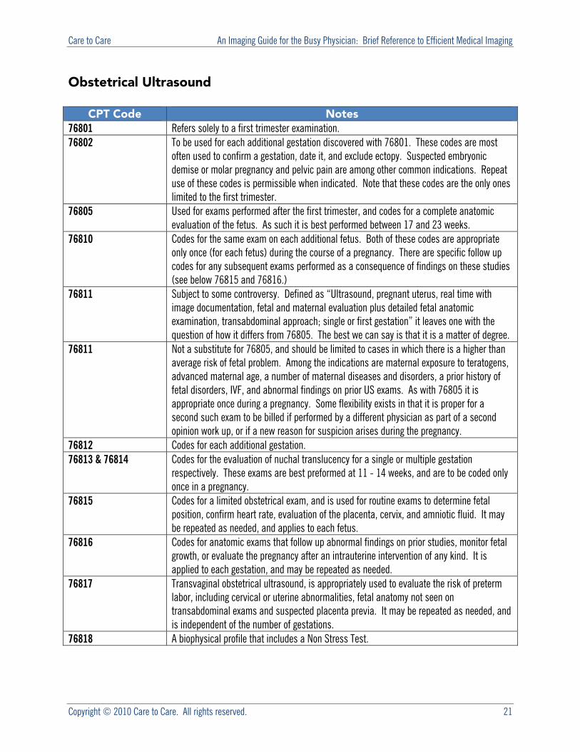
Obstetrical Ultrasound
CPT Code Notes76801 Refers solely to a first trimester examination. 76802 To be used for each additional gestation discovered with 76801. These codes are most
often used to confirm a gestation, date it, and exclude ectopy. Suspected embryonic demise or molar pregnancy and pelvic pain are among other common indications. Repeat use of these codes is permissible when indicated. Note that these codes are the only ones limited to the first trimester.
76805 Used for exams performed after the first trimester, and codes for a complete anatomic evaluation of the fetus. As such it is best performed between 17 and 23 weeks.
76810 Codes for the same exam on each additional fetus. Both of these codes are appropriate only once (for each fetus) during the course of a pregnancy. There are specific follow up codes for any subsequent exams performed as a consequence of findings on these studies (see below 76815 and 76816.)
76811 Subject to some controversy. Defined as “Ultrasound, pregnant uterus, real time with image documentation, fetal and maternal evaluation plus detailed fetal anatomic examination, transabdominal approach; single or first gestation” it leaves one with the question of how it differs from 76805. The best we can say is that it is a matter of degree.
76811 Not a substitute for 76805, and should be limited to cases in which there is a higher than average risk of fetal problem. Among the indications are maternal exposure to teratogens, advanced maternal age, a number of maternal diseases and disorders, a prior history of fetal disorders, IVF, and abnormal findings on prior US exams. As with 76805 it is appropriate once during a pregnancy. Some flexibility exists in that it is proper for a second such exam to be billed if performed by a different physician as part of a second opinion work up, or if a new reason for suspicion arises during the pregnancy.
76812 Codes for each additional gestation. 76813 & 76814 Codes for the evaluation of nuchal translucency for a single or multiple gestation
respectively. These exams are best preformed at 11 - 14 weeks, and are to be coded only once in a pregnancy.
76815 Codes for a limited obstetrical exam, and is used for routine exams to determine fetal position, confirm heart rate, evaluation of the placenta, cervix, and amniotic fluid. It may be repeated as needed, and applies to each fetus.
76816
Codes for anatomic exams that follow up abnormal findings on prior studies, monitor fetal growth, or evaluate the pregnancy after an intrauterine intervention of any kind. It is applied to each gestation, and may be repeated as needed.
76817
Transvaginal obstetrical ultrasound, is appropriately used to evaluate the risk of preterm labor, including cervical or uterine abnormalities, fetal anatomy not seen on transabdominal exams and suspected placenta previa. It may be repeated as needed, and is independent of the number of gestations.
76818 A biophysical profile that includes a Non Stress Test.
Care to Care An Imaging Guide for the Busy Physician: Brief Reference to Efficient Medical Imaging
Copyright © 2010 Care to Care. All rights reserved. 22
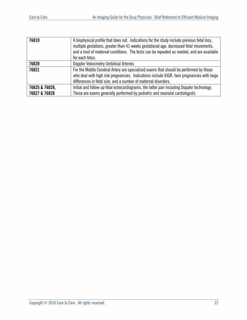
76819
A biophysical profile that does not. Indications for the study include previous fetal loss, multiple gestations, greater than 41 weeks gestational age, decreased fetal movements, and a host of maternal conditions. The tests can be repeated as needed, and are available for each fetus.
76820 Doppler Velocimetry Umbilical Arteries 76821
For the Middle Cerebral Artery are specialized exams that should be performed by those who deal with high risk pregnancies. Indications include IUGR, twin pregnancies with large differences in fetal size, and a number of maternal disorders.
76825 & 76826, 76827 & 76828
Initial and follow up fetal echocardiograms, the latter pair including Doppler technology. These are exams generally performed by pediatric and neonatal cardiologists.