Embed Size (px)
Citation preview

Biomedical Instrumentation
BME 523 (Fall 2009) Final Project Report
Deep Brain Stimulation for Movement Disorder Diseases
by
HAO SU
Submitted to Prof. Yitzhak Mendelson
Date of Submission: December 18, 2009
Worcester Polytechnic Institute
Worcester, MA

BME 523 (Fall 2009) Final Project Report, Hao Su
2
Table of Contents
1. Movement Disorder Diseases Overview ....................................................................................... 4
2. Deep Brain Stimulation ................................................................................................................. 6
2.1 Motivation of Deep Brain Stimulation from Historical Perspective ...................................... 7
2.2 Physiology of Basal Ganglia .................................................................................................... 7
2.3 Physiology of Targeting Sites ................................................................................................. 8
2.4 Medtronic Activa System and Working Principle ................................................................ 10
2.5 Electrical Characteristics of Electrodes and Stimulator ....................................................... 12
2.6 Patient Selection Criteria ..................................................................................................... 13
3. Surgical Procedures ..................................................................................................................... 14
3.1 Framed DBS Lead Placement ............................................................................................... 14
3.1.1 Framed DBS Lead Placement Workflow .................................................................. 14
3.1.2 Framed DBS Tool: Stereotactic Frame ..................................................................... 15
3.1.3 Framed DBS Tool: Neuroguide ................................................................................. 16
3.1.4 Case Study of Framed DBS Lead Placement ............................................................ 17
3.2 Frameless DBS Lead Placement ........................................................................................... 18
3.2.1 Lead Placement Workflow ....................................................................................... 18
3.2.2 Frameless DBS Tool: Neuromate® Robot, Sensors and Drilling Tools ..................... 19
3.2.3 Frameless DBS Tool: Medtronic NexFrame and NexDrive ....................................... 20
3.2.4 Johns Hopkins Neuromate Robot Optical Tracking System ..................................... 20
3.2.5 Robot Assisted Surgery Paradigm ............................................................................ 22
4. Discussion .................................................................................................................................... 25
4.1 Future Trends and Technology for MDD ............................................................................. 25
4.2 Robotic Assisted Interventional MRI Surgery ...................................................................... 26
Reference: ............................................................................................................................................ 28

BME 523 (Fall 2009) Final Project Report, Hao Su
3
Abstract: Movement Disorder Disease (MDD) is one general type of diseases that severely impair human motor ability and reduce the body controllability. Over millions of people in North America are suffering from these diseases and Parkinson's disease (PD) is one of the most typical and common among them. PD is the second most prevalent neurodegenerative disease, affecting over 500,000 people in America and about 4-5% of people over 85.
Deep brain stimulation (DBS) is the surgical implantation of a brain pacemaker to generate electrical impulses and stimulate specific parts of the brain. DBS is an effective, FDA approved treatment of PD. From the long term surgical outcome, DBS has been proven to be one of the most remarkable therapeutic procedure for a diverse array of movement disorder diseases and some physiological syndromes, including PD, dystonia, essential tremor and chronic pain etc.
This report is organized as follows. In the first section, this report would briefly introduce the background information on MDD, its typical syndrome and its engineering implication.
The second section would perceive DBS application from an historical background. Tough the underlying mechanism of DBS is still not clear, and we would present some research hypothesis and the working principle of DBS in practice. The physiology analysis and stimulation targets would be explained. On the other hand, even though DBS has demonstrated good surgical outcome from a 5 year clinician observation, it is not the always first option for PD patients and there is strict patient selection criteria.
The third section focuses on the surgical procedure and categorizes them into two different approaches: the frame based and frameless stereotactic surgery. The DBS lead placement workflow and some commercially available medical apparatus and the working principles would be introduced. A comparison between the two approaches and some related technical issues would be presented.
The last section illustrates the operation cost and articulates the surgical benefits, complications and risks. Some state of the art technology, including robot assisted interventional MRI technique would be presented here also.
This report aims to make brief introduction to MDD with an emphasis on PD and present the basic concepts of DBS and illustrate the practical surgical procedures.

BME 523 (Fall 2009) Final Project Report, Hao Su
4
1. Movement Disorder Diseases Overview MDD is one general type of diseases that severely impair human motor skills and affect the body controllability. Over millions of people in North America are suffering from these diseases and PD is one of the most typical and common among them. PD is the second most prevalent neurodegenerative disease, affecting over 500,000 people in America and about 4-5% of people over 85.
Figure 1: PD by Sir William Richard Gowers from a Manual of Diseases of the Nervous System in 1886
Movement disorders basically stem from the dysfunction of some specific parts of brain and lead to movement speed slowness (typical syndrome like dyskinesia), abnormal frequency limb tremor, body unbalance or postural instability. Parkinson’s disease, dystonia, essential tremor and Tourette's syndrome are some of the most popular diseases. This section would make a brief introduction to the first representative diseases.
Parkinson’s disease, because of its popularity in the elderly, is well known by the public and also the one getting most of the research efforts worldwide. Thought the underlying pathogen is still not well studied, the most famous hypothesis conjectures that the cell inactivity of basal ganglia and substantial nigra could not produce sufficient dopamine which is essential for the stimulation of the motor cortex. About 89% - 99% of the patients suffer from muscle rigidity or muscle stiffness which usually cause body fatigue and muscle pain and 69% -100% patients have significant tremor and the syndrome is more obvious in still position. Approximately 40% of PD patients reported severe depression which can be partially cured by drugs or psychological treatment. Figure 1 shows a pictorial illustration of PD by Sir William Gowers in 1886.
Currently, PD disease is benchmarked by the well-known Hoehn and Yahr scale method which describes the progress levels from 0 to 5 stages.

BME 523 (Fall 2009) Final Project Report, Hao Su
5
1) Symptoms on one side of the body only. 2) Symptoms on both sides of the body. No impairment of balance. 3) Balance impairment. Mild to moderate disease. Physically independent. 4) Severe disability, but still able to walk or stand unassisted. 5) Needing a wheelchair or bedridden unless assisted.
National Institute of Neurological Disorders and Stroke Center defines dystonia as a neurological movement disorder in which sustained muscle contractions cause twisting and repetitive movements or abnormal postures.
Figure 2: A dystonia patient (image courtesy of Medtronic)
Dystonia patient population is not as many as PD, but its syndrome is even more severe. Muscle contractions and abnormal postures are the major symptoms. The symptom progresses gradually and the initial symptom might be mild like handwriting fatigue, but after some short period, patients would have great difficulty to control body movement and body/limb twisting and withering would happen in most patients. In the Duration of the illness, continuous pain and muscle spasms would haunt the patients for most times. Figure 2 shows a dystonia patient illustrating the abnormal posture.

BME 523 (Fall 2009) Final Project Report, Hao Su
6
2. Deep Brain Stimulation
Deep Brain Stimulation is the surgical implantation of a brain pacemaker to generate electrical impulses and stimulate specific parts of the brain. The target diseases of DBS include the Parkinson’s disease, dystonia, Tourette's syndrome, etc. and some physiological symptoms like chronic depression. The debut of DBS is a resultant technological breakthrough originating from several advances: the further development of modern imaging modalities, including X-ray computed tomography (CT) and magnetic resonance imaging (MRI) and modern medical navigation technology, an in-depth understanding of brain electrical mechanism and some related innovations of medical instrumentation.
Figure 3: Artistic schematic of deep brain stimulation
Figure 3 shows the artistic schematic of DBS and the major components. DBS employs a brain pacemaker implanted under the collarbone to generate electrical impulses that would be delivered to specific sites of the brain through an array of electrodes which is connected with the pulse generator with connective wires embedded inside the body and behind the ears.
The list below shows the major electrical characteristics during DBS.
1) Voltage (0-7 Volts) 2) Pulse width (65-450 msec) 3) Frequency (130-180 Hz) 4) Lead location (4 leads, each 1.5 mm apart)

BME 523 (Fall 2009) Final Project Report, Hao Su
7
The further details of this technique would be explained in the following parts, including the historical perspective on DBS, physiology of basal ganglia, Medtronic’s commercially available Activa system for DBS and the underlying working principle of some important apparatus.
2.1 Motivation of Deep Brain Stimulation from Historical Perspective DBS is the state of the art technology to cure Parkinson’s disease, but it is not the first option for most PD patients. The current gold standard is based on medication - levodopa. Moreover, it might not be obvious to perform invasive surgery to stimulate the brain with electrical signals for PD treatment. The motivation and effectiveness of DBS can be observed from a historical perspective.
According to the introductory article [1], 1940s-1950s witnessed the neonatal development of surgical procedures pallidotomy which create lesions in deep parts of the brain to control symptoms of tremor and rigidity without the aid of modern imaging modalities. But the invention of levodopa in 1960s dramatically deemphasized the research on this surgical approach and medication treatment dominated for 30 years.
Like any other medicine, levodopa is not the panacea. After long term ingestion, many of the PD patients experience the “wearing-off” problem that they cannot benefit from individual dose. A large majority of the patients also develop drug-induced, involuntary writhing and twisting movements, known as dyskinesias [1]. On the other hand, medicine resistance happens and motor fluctuations, postural instability and freezing affect patients on a daily basis. All of these issues engender new development of treatment alternatives.
Major clinician breakthrough happened in the early 1990s – a French doctor Alim-Louis Benabid found that instead of performing the “traditional” thalamus lesion, he was able to inhibit tremor by simple electrical stimulation to the same target site. He also conjectured that continuous electrical stimulation would provide effective treatment of PD. The subsequent surgery based on this mechanism gained better surgical outcome (more effective and lower operative risk) than the lesion approach.
In the United States, Food and Drug Administration (FDA) approved DBS as the treatment for one-side-brain essential tremor (ET) in 1997. Parkinson’s disease therapy based on DBS was approved by FDA in 2002 and dystonia approval happened in the next year. Up to date, over 35,000 patients are performed worldwide and more and more patients are benefiting from this technology.
2.2 Physiology of Basal Ganglia The basal ganglia are a group of nuclei deep inside the brain that in charge of motor control and learning. According to the neuroscience tutorial from Washington University, "brake theory" describes the function of basal ganglia in an intuitive manner. “To sit still, you
BME 523 (Fall 2009) Final Project Report, Hao Su
8
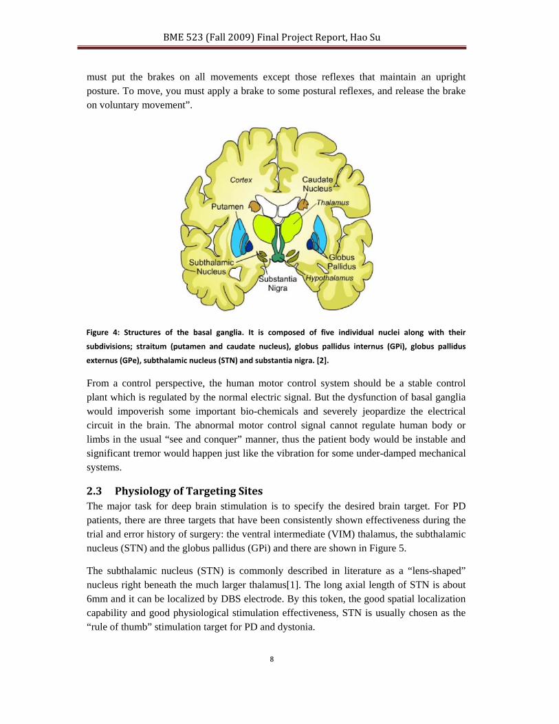
must put the brakes on all movements except those reflexes that maintain an upright posture. To move, you must apply a brake to some postural reflexes, and release the brake on voluntary movement”.
Figure 4: Structures of the basal ganglia. It is composed of five individual nuclei along with their
subdivisions; straitum (putamen and caudate nucleus), globus pallidus internus (GPi), globus pallidus
externus (GPe), subthalamic nucleus (STN) and substantia nigra. [2].
From a control perspective, the human motor control system should be a stable control plant which is regulated by the normal electric signal. But the dysfunction of basal ganglia would impoverish some important bio-chemicals and severely jeopardize the electrical circuit in the brain. The abnormal motor control signal cannot regulate human body or limbs in the usual “see and conquer” manner, thus the patient body would be instable and significant tremor would happen just like the vibration for some under-damped mechanical systems.
2.3 Physiology of Targeting Sites The major task for deep brain stimulation is to specify the desired brain target. For PD patients, there are three targets that have been consistently shown effectiveness during the trial and error history of surgery: the ventral intermediate (VIM) thalamus, the subthalamic nucleus (STN) and the globus pallidus (GPi) and there are shown in Figure 5.
The subthalamic nucleus (STN) is commonly described in literature as a “lens-shaped” nucleus right beneath the much larger thalamus[1]. The long axial length of STN is about 6mm and it can be localized by DBS electrode. By this token, the good spatial localization capability and good physiological stimulation effectiveness, STN is usually chosen as the “rule of thumb” stimulation target for PD and dystonia.

BME 523 (Fall 2009) Final Project Report, Hao Su
9
As literature [1] indicates, the STN can be identified with high accuracy in terms of mapping techniques and a DBS electrode can be placed right through it. The electrical circuits of STN connect with globus pallidus which makes it have significant anti-parkinsonian effects. It is also very noteworthy to know that this stimulation is effective not only for tremor, but slowness, rigidity, dyskinesias, speech, handwriting and dystonia.
The clinician outcome of STN stimulation is significant. A major percentage of PD patients are less reliable on or even get rid of anti-parkinsonian medication after STN stimulation as indicted by Ford [1]. From this perspective, the effectiveness of STN stimulation is comparable with those of medicines like levodopa, but do not surpass the best result of medication treatment.
Figure 5: Three major PD brain targets (left to right: STN, GPi and Vim Thalamus)
The globus pallidus (GPi) is some dense nerve tissue that locates in the center of the basal ganglia region and it was traditionally used as the site of the pallidotomy surgery as noted by [1]. Physiologically, comparing with STN, the globus pallidus is a larger and more complex structure with a more complicated circuitry. Globus pallidus stimulation has salient beneficial anti-parkinsonian effects and it is suitable for treating PD and Dystonia. The center image of Figure 5 shows that GPi has larger volume than STN and its complex structure.
Ventral intermediate thalamus stimulation is effective for Essential Tremor treatment. The electrical functionality of thalamus is similar to relay station for a variety of human motor controls [1]. We do not present much information for thalamus stimulation since it is less popular than the aforementioned two sites, and the interested reads can refer to [1]for further details.
Table 1: Brain sites for DBS and the corresponding effects and outcome
Deep Brain Structure Effects and Outcome Thalamus • Deep brain electrode in VIM thalamus
• Effective treatment for tremor on the opposite body side
Subthalamic • Bilateral thalamic stimulation can be accomplished without significant adverse effects

BME 523 (Fall 2009) Final Project Report, Hao Su
10
Nucleus (STN) • Deep brain electrode in subthalamic nucleus (STN)
• Effective treatment for tremor, slowness, rigidity, dystonia and dyskinesia on the
opposite body side
• Bilateral procedures well tolerated
• Usually allows people with PD to decrease medications
Globus
Pallidus (GPi) • Deep brain electrode in globus pallidus (GPi)
• Effective treatment for tremor, slowness, rigidity,
dystonia and dyskinesia on the opposite body side
• Bilateral procedures well tolerated Table 1 summarized the aforementioned brain sites for DBS and the corresponding effects and outcome.
2.4 Medtronic Activa System and Working Principle Medtronic is the major company that strives to working on research, development and commercialization of DBS equipment. It successfully provides one-stop DBS solution as shown in Figure 6: The one‐stop shop DBS solution provided by Medtronic.
Medtronic provides a variety of systematic instrumentations for DBS and as shown in the official website, it includes:
StimPilot™ System: This is the world’s first integrated platform designed specifically for DBS. It provides the capability of pre-surgical planning and intra-operative target verification in one elegant and easy-to-use solution.
Kinetra® Neurostimulator and DBS™ Lead, Access® Therapy Controller: These are a series of stimulator systems and the details would be presented in the next section. As the system of choice for treatment of Parkinson’s disease, it’s designed to provide patients with optimal therapy.
Activa® DBS Therapy Consultant: This is the medical services provided by Medtronic. They are able to offer single-source expertise for all DBS practice and procedure requirements. The distinct feature of this service is that one professional can addresses business, training, technical and procedure guidance needs.

BME 523 (Fall 2009) Final Project Report, Hao Su
11
Figure 6: The one‐stop shop DBS solution provided by Medtronic
StimPilot™ Single Procedure Kit: For safety issues, these are all pre-sterile single-procedure-use products for DBS, packaged together in one system.
Figure 7: Medtronic DBS stimulator and electrodes
NEXFRAME® System: This novel mechanism design can offer high precision stereotactic frame with the advantage of image-guided surgery. The major advantage of this system is that it simplifies the DBS procedure and offers enhanced patient comfort.
The next section would enumerate some of the most important devices and illustrate the functionality by real medical case studies.

BME 523 (Fall 2009) Final Project Report, Hao Su
12
2.5 Electrical Characteristics of Electrodes and Stimulator This section presents the details electrical property of electrodes and its mechanical/electrical characteristics. Figure 8 shows a schematic of four segment electrode.
Figure 8: The schematic of four segment electrode[3]
As shown in the figures above, this system is composed of three subsystems [3]:
• An implanted pulse generator (IPG) that generates the electric pulses
• An implanted cable that have a galvanic connection between the IPG and the stimulation electrode
• A stimulation electrode composed of several contacts insulated from each other, which delivers the electric pulses to the surrounding tissues
The system can be used in several configurations as shown in the figure below:
• Monopolar stimulation: there is one active contact among the four segments

BME 523 (Fall 2009) Final Project Report, Hao Su
13
Figure 9: Deep brain stimulation implantable system
• Multi-polar balanced stimulation: more than one contacts are active and the currents going out of the contact summation is zero
• Multi-polar unbalanced stimulation: several contacts are active, the sum currents going out of the contact summation is not zero and the difference have to go back to the IPG itself
2.6 Patient Selection Criteria From the official website of Medtronic, it states that the following requirements should be met for appropriate DBS treatment:
1) Patients should have symptoms of advanced, levodopa responsive Parkinson's disease that are not adequately controlled by medications; and
2) Patients should be suitable candidates for stereotactic neurosurgery.
Table 2: Good and poor DBS candidate criteria (courtesy of [1])
Good DBS Candidate Poor DBS Candidate
Typical PD with tremor Atypical parkinsonism
Good response to individual levodopa Poor response to levodopa
Dyskinesias Memory problems
Wearing‐off spells Severe depression or anxiety
Good general health Severe medical problems
On the other hand, before the Medtronic DBS Parkinson's Control System is implanted to the body, the following criteria should be met:
1) Symptom suppression by test stimulation should be demonstrated in the operating room; and

BME 523 (Fall 2009) Final Project Report, Hao Su
14
2) Symptom suppression should occur at less than 3 volts, and with minimal side effects such as visual effects and speech difficulties.
The table below summarizes the Good and poor DBS candidate criteria and it essentially proposes two major concern in the patient selection: the patients should have some benign response to medicine treatment for individual levodopa and should have good general heath. These two criteria are not trivially met in PD patients and pose significant challenge for PD treatments.
3. Surgical Procedures
3.1 Framed DBS Lead Placement
3.1.1 Framed DBS Lead Placement Workflow
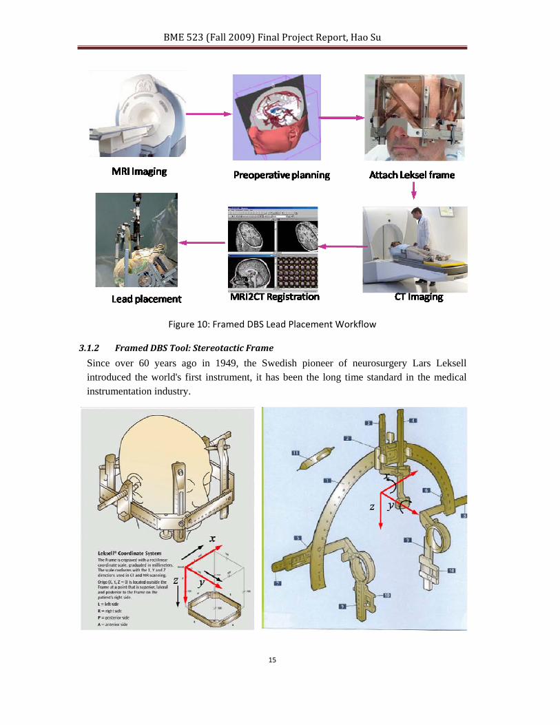
This section presents the major workflow of a typical DBS lead placement procedure based on framed approach. As shown in Figure 10.
• Acquire MR images some days before the surgery • Perform preoperative planning • Attach fiducial frame on day of surgery • Acquire CT images with the fiducial frame bolted to head • Register plan from preoperative MR to CT • Use stereotactic frame system to align the lead guide cannula and insert.
To guarantee high targeting accuracy, microelectrode recording (MER) and/or imaging may be used to confirm or adjust lead placement.
BME 523 (Fall 2009) Final Project Report, Hao Su
15
Figure 10: Framed DBS Lead Placement Workflow
3.1.2 Framed DBS Tool: Stereotactic Frame
Since over 60 years ago in 1949, the Swedish pioneer of neurosurgery Lars Leksell introduced the world's first instrument, it has been the long time standard in the medical instrumentation industry.
BME 523 (Fall 2009) Final Project Report, Hao Su
16
Figure 11: Leksell® Coordinate Frame (left) and stereotactic arc (right)
One of the most famous devices used in this kind surgery might be the distinguished Leksell frame and the arc mechanism. Leksell stereotactic mechanism is the most widely used frame in the world, in particular to neurosurgery. The system is based on the remote center motion theory and the system is composed of the Cartesian coordinate frame and a semi-circular arc structure. This is widely used Gamma Knife® Surgery system. From the figure below, we can see that the frame base itself provides 3 degree-of-freedom (DOF) motion accuracy in the Cartesian space.
As we can see from the figure, the arc provides one revolute joint and one semi-prismatic joint and thus facilitates 2 DOF rotational motions and targeting accuracy.
3.1.3 Framed DBS Tool: Neuroguide
Renishaw is a world leader in engineering technologies, and Renishaw also supplies products for applications as diverse as motion control, automation and medical devices. It is famous for breeding the world No. 1 image-guided neurosurgical robot Neuromate in medical device community.
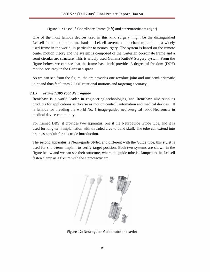
For framed DBS, it provides two apparatus: one it the Neuroguide Guide tube, and it is used for long term implantation with threaded area to bond skull. The tube can extend into brain as conduit for electrode introduction.
The second apparatus is Neuroguide Stylet, and different with the Guide tube, this stylet is used for short-term implant to verify target position. Both two systems are shown in the figure below and we can see their structure, where the guide tube is clamped to the Leksell fasten clamp as a fixture with the stereotactic arc.
Figure 12: Neuroguide Guide tube and stylet

BME 523 (Fall 2009) Final Project Report, Hao Su
17
3.1.4 Case Study of Framed DBS Lead Placement
Now we are in the position to investigate a real world problem and we would briefly present the surgical result from one medical journal and most of these details are from therein. Patel et. Al. [4] presented a magnetic resonance imaging-directed stereotactic system using implantable guide tubes for targeting deep brain nuclei in functional neurosurgery. They took advantage of MRI visualization capability to visualize the deep brain nuclei.
Figure 13: The surgical workflow: A, a probe is inserted through the stop and guide tube to the brain target. B, split guide is unclamped from the lower carriage. C, hub of the guide tube is fixed within the burr hole with acrylic cement. [4]
As shown in the above three subfigure, a guide tube is inserted into one customized split guide (Elekta Instrument, AB). This guide as shown in the previous section is strictly fixed with the lower carriage. After this fixture, the probe is then advanced to the target through the stop. The split guide is then released from the carriage and the halves mechanism is removed.
Acrylic cement is placed into the burr hole to fill the gap therein. The hub of the guide tube is seated in the acrylic cement. Once set, the cement secures the guide tube in place in the above figure. The probe is then removed after these procedures.

BME 523 (Fall 2009) Final Project Report, Hao Su
18
Figure 14: (Left) Probe is replaced with a stylet which is advanced to the right length such that its distal end projects beyond the guide tube into the target. (Right) The stylet is replaced with a DBS lead. The lead is bent through 90 degrees to fit into the groove and fasten to the cranium. [4]
3.2 Frameless DBS Lead Placement
3.2.1 Lead Placement Workflow
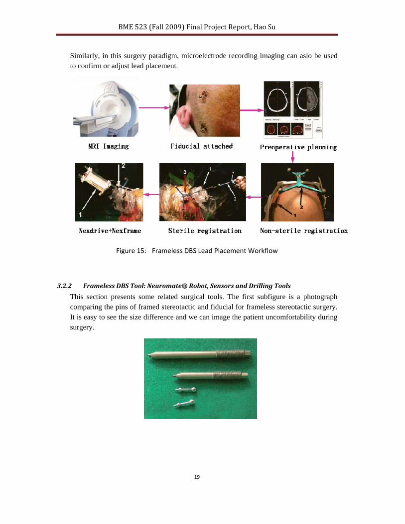
This section presents the major workflow of a typical DBS lead placement procedure based on frameless approach. As shown in Figure 15. Some parts are the same as the framed proceeds, but there are still major differences.
These procedures are based on the journal article [5] and the subfigures are from the same reference.
• Acquire MR images some days before the surgery • Placement of the fiducials under local anesthetic • Perform CT and MRI registration and surgical planning in software • Non-sterile registration. As shown in the subfigure, the nomenclatures are:
1-Fiducial, 2-Reference • Sterile registration. The nomenclatures are: 1-Trajectory guide platform
(NexFrame), 2-Passive planar probe, 3-Passive spinal reference frame. Usually , after this step, the Guideframe-DT (arrow place) is attached to the NexFrameTower3
• Nexdrive in conjunction with NexFrame 1-Microdrive, 2- Multi-lumen adapter
BME 523 (Fall 2009) Final Project Report, Hao Su
19
Similarly, in this surgery paradigm, microelectrode recording imaging can aslo be used to confirm or adjust lead placement.
Figure 15: Frameless DBS Lead Placement Workflow
3.2.2 Frameless DBS Tool: Neuromate® Robot, Sensors and Drilling Tools
This section presents some related surgical tools. The first subfigure is a photograph comparing the pins of framed stereotactic and fiducial for frameless stereotactic surgery. It is easy to see the size difference and we can image the patient uncomfortability during surgery.

BME 523 (Fall 2009) Final Project Report, Hao Su
20
Figure 16: Surgical instrumentation in frameless system [6]
The NeuroMate is made up with 5 revolute joints, each mobilized by a separate, high precision servo. The joint values are read by encoders with high.
3.2.3 Frameless DBS Tool: Medtronic NexFrame and NexDrive
Figure 17: ElectrodeFX (b)NexDrive Micropositioner
3.2.4 Johns Hopkins Neuromate Robot Optical Tracking System
The JHU system is composed of two major mechatronic systems, the NeuroMate robot and the StealthStation navigation system as shown below.

BME 523 (Fall 2009) Final Project Report, Hao Su
21
Figure 18: JHU robotic system
The neurosurgical system uses several different coordinate systems. Homogenous coordinate transformations is used to compute the position and orientation of spatial point in other frames once the intermediate transformations are known.. [6]
Figure 19: Optical tracking system

BME 523 (Fall 2009) Final Project Report, Hao Su
22
Figure 20: pivot calibration
int k kpivotPo R cutterTip T= ⋅ +
intk kR cutterTip pivotPo T⋅ − = −
intk k
cutterTipR I T
pivotPo
⎡ ⎤ ⎡ ⎤⎡ ⎤⎢ ⎥ ⎢ ⎥− = −⎢ ⎥⎢ ⎥ ⎢ ⎥⎣ ⎦⎢ ⎥ ⎢ ⎥⎣ ⎦ ⎣ ⎦
M M M
M M M The above shows a pivot calibration result.
3.2.5 Robot Assisted Surgery Paradigm

BME 523 (Fall 2009) Final Project Report, Hao Su
23
Figure 21: Traditonal classification of robot assisted surgical systems: A, supervisory controlled system. B, telesurgical system. C, shared control system. [7]
The figure above shows three robot paradigms and in DBS, it is advised to used the third one to guarantee safety. Figure 22 shows a DBS programming device and a Stryker face mask for image registration.

BME 523 (Fall 2009) Final Project Report, Hao Su
24
Figure 22: Stimulator programming device and head mounted mask
3.2.6 Error Compensation
To overcome potential sources of error in lead placement, often a number of microelectrode recordings (MER) are performed (4-5) which adds significant length to the case as well as potential morbidity. Even the frameless robotic technique is not immune to brain shift. According to JHU medical school website, MER is claimed to be the most precise method of
localizing the surgical site. MER is a technique for recording activity from single neurons, as well
as microstimulation with a custom designed microelectrode.
Figure 23: Representative MERs in various nuclei along a trajectory through the STN. The drawing is a parasagittal section adapted from the Schaltenbrand and Wahren human brain
atlas. Each trace is 1 second long [9]

BME 523 (Fall 2009) Final Project Report, Hao Su
25
MER is used to further define the stimulation target. Because not all brains are the same, the
information obtained from MER gives a more accurate target for final DBS placement. But the
challenges and difficulty of MER lies in the fact this would increase mortality and heath risk. The
new trend is to take advantage of miniaturized MER or use robot to perform the task.
4. Discussion
4.1 Future Trends and Technology for MDD In [10], the authors discussed most recent method for placement of DBS electrodes using
interventional MRI (iMRI) and reported the accuracy with this new technique. The tools used in the
surgery room are good summery for the aforementioned methods and apparatus. It is also the most
up-to-date medical article on MRI and DBS.
Table 3: Equipments used for DBS in [10]
This article presented the initial clinical effectiveness of iMRI and the corresponding challenges in
a consecutive series of STN DBS implants to treat Parkinson disease. Table 3 shows the related
equipments and with a major from Medtronic. Figure 25 is a series of images showing the detailed
surgical procedures. This surgery has great clinical and technical implication and it is good starting
point for engineers to contribute to this new frontier with robotics and mechatronics knowledge.

BME 523 (Fall 2009) Final Project Report, Hao Su
26
Figure 24: Intraoperative photographs demonstrating surgical draping and trajectory guides. A: Patient’s head is shown at the back of the MR bore. B: Trajectory guides with
alignment stems. C: Trajectory guides with multilumen insert and peel‐away sheath prior to advancing the sheath into the brain. [10]
4.2 Robotic Assisted Interventional MRI Surgery The MRI based medical diagnosis and treatment paradigm capitalizes on the novel benefits and capabilities created by the combination of high sensitivity for detecting tumors, high spatial resolution and high-fidelity soft tissue contrast [11]. This makes it an ideal modality for guiding and monitoring medical procedures, such as needle biopsy and low-dose-rate permanent brachytherapy. Though with so many appealing merits, the magnetic and electrical fields in MRI environment presents significant challenges for mechatronic instrumentation design. Generally, the development of sensors for applications in MR environments requires careful consideration of safety and electromagnetic compatibility constraints.
Figure 25: A CAD model of the robotic DBS cannula guide used for simulating and optimizing the workspace [8]

BME 523 (Fall 2009) Final Project Report, Hao Su
27
In [8], Cole and Fischer described a new robotic system that uses remote center motion mechanism and ultrasound motor to drive online MRI compatible robots. Some initial tests were performed at UMass Medical School recently. There is very insignificant image artifact during robot motion and poses new research potential for iMRI in the near future.
We believe that the medical imaging and engineering techniques collaboration would make great contribution to the DBS filed and produce more medical instrumentations with higher accuracy and shorter surgery time. A more in-depth exploration and review of this field would be impetrative for the future study.

BME 523 (Fall 2009) Final Project Report, Hao Su
28
Reference:
[1] B. Ford, "Deep Brain Stimulation Parkinson's Disease," Parkinson's Disease Foundation,
2008.
[2] N. Bratt, "Deep Brain Stimulation as a Therapy for Parkinson's Disease," Journal of Student
Medical Sciences, 2009.
[3] G. Walckiers, "Bio‐Electromagnetic Model of Deep Brain Stimulation," Ecole Polytechnique
Fédérale de Lausanne, 2009.
[4] N. Patel, P. Plaha, and S. Gill, "Magnetic resonance imaging‐directed method for functional
neurosurgery using implantable guide tubes," Neurosurgery, 2007.
[5] T. Srikijvilaikul and R. Bhidayasiri, "Frameless stereotactic for deep brain stimulation
placement: operative technique.," J Med Assoc Thai, 2008.
[6] T. Haidegger, "Improving the Accuracy and Safety of a Robotic System for Neurosurgery,"
Master thesis for BME degree, 2008.
[7] Nathoo N, C. MC, V. MA, and B. GH., "In touch with robotics: neurosurgery for the future,"
Neurosurgery, 2005
[8] Cole G, Pilitsis J, Fischer GS, Design of a Robotic System for MRI‐Guided Deep Brain Stimulation
Electrode Placement, International Conference on Robotics and Automation ‐ ICRA 2009,
Kobe, Japan, pp 4450‐4456, May 2009
[9] STARR Philip A. ; CHRISTINE Chadwick W. ; THEODOSOPOULOS Philip V. ; LINDSEY Nadja ;
BYRD Deborah ; MOSLEY Anthony ; MARKS William J. ;"Implantation of deep brain
stimulators into the subthalamic nucleus: technical approach and magnetic resonance
imaging‐verified lead locations"
[10] Starr PA, Martin AJ, Ostrem JL, Talke P, Levesque N, Larson PS. "Subthalamic nucleus deep
brain stimulator placement using high‐field interventional magnetic resonance imaging and
a skull‐mounted aiming device: technique and application accuracy".
[11] Hao Su, Gregory Fischer, "A 3‐Axis Optical Force/Torque Sensor for Prostate Needle
Placement in Magnetic Resonance Imaging Environments", IEEE International Conference on
Technologies for Practical Robot Applications, Boston, MA, 2009