Embed Size (px)
Citation preview

INTERNATIONAL JOURNAL OF QUANTUM CHEMISTRY, VOL. 47, 231-238 (1993)
Helical Region of the Potential Energy Surface of a -Aminoisobutyric Acid: A Theoretical Study
CARLOS ALE- AND JUAN J. PEREZ Departament d'Enginyeria Quimica, Escola Tkcnica Superior d'Enginyers
Industrials, Universitat Politecnica de Catalunya, Avinguda Diagonal 647, Spain
Abstract
The helical region of the potential energy surface of blocked a-aminoisobutyric acid (Aib) dipeptide has been studied by using ah initio and semiempirical quantum mechanical methods, as well as force-field- derived methods. Depending on the method, an a-helix or a 3lo-helix is found to be the energy minimum. The conformations obtained from computations performed at the ah initio quantum mechanical level, as well as by using the AMBER force field, are in excellent agreement with X-ray data. Semiempirical results display some important differences with regard to experimental data. On the other hand, the CVFF force field predicts no energy minimum in the helical region of the Aib potential energy surface. 0 1993 John Wiley & Sons, Inc.
Introduction
The a-aminoisobutyric acid (Aib), also named a-methylalanine (MeA), is an a,a- dialkyl amino acid commonly found in peptides produced by microbial sources. Some of these peptides, like chlamydocin, exhibit antibiotic properties [ 11, whereas others like methicin, antiamoebin, emerimicin, and zervamicin produce voltage-gated ion channels in lipid membranes [2].
A detailed analysis of the crystal structures of peptides containing Aib residues have confirmed a helical preference in their conformation [3-51. NMR experiments have also demonstrated the feasibility of C5 (p = -180°, CC, = 1 8 0 O ) and C7 (p = 80", I) =
80") conformations, which are characteristic of the 20 commonly found amino acids, but these conformations are favored in respect to the helical conformation only in apolar solvents IS]. The replacement of the hydrogen of the C" atom of alanine by a methyl group in Aib produces severe restrictions on the conformational freedom. The Ramachandran plot for the Aib shows that the allowable p and I) angles occur only in two very narrow regions, located on the neighborhood of the points (-57", -47") and (57", 47"), respectively [6-91. This explains the pronounced tendency of the Aib residue to favor helical structures and makes this amino acid to be considered as a useful building block for de now protein design [lo].
The prediction of the accessible conformations of polypeptides containing Aib units has been a challenge for theoretical methods, since a delicate balance between the different contributions to the interatomic interactions may favor, depending on the en- vironmental factors, different types of helices. Consequently, the prediction of the pre- ferred conformations of Aib oligomers is a particularly interesting problem per se. In an
0 1993 John Wiley & Sons, Inc. CCC 0020-7608/93/03023 1-08

232 ALEMAN AND PEREZ
early molecular mechanical study of poly-Aib, Prasad and Sasisekharan [ll] using both the Buckingham and Kitaigorodsky potential functions, predicted the 3lo-helix and the a-helix to be the most favorable structures, the latter being more stable by around 3 kcal mol-' residue-'. In a more recent study, Patterson et al. [8] studied oligomers of Aib up to three units by using the EcEPP force field [12]. The results indicated that the minimum-energy conformations correspond to a 3lo-helix. Nevertheless, the preference of this helix was only achieved by imposing an asymmetry on the C" atom. Without this constraint, the a-helix appeared to be more stable. Barone et al. [13] investigated the conformational behavior of Aib by ab initio and empirical methods. They found that the relative stability of the C,, C7, and helical conformations strongly depends on the force-field parametrization when molecular mechanical calculations are performed. Moreover, they found that the three structures are isoenergetic when MO- SCF calculations are performed using an STO-3G basis set. In their calculations, bond lengths were kept fixed and only bond and dihedral angles were optimized. These results seem to be in contradiction with those reported some years before by Peters and Peters [9], who found that the helical conformation is the only accessible at the same level of computation. Hodkin et al. [14], using the united atom parametrization of the AMBER force field [15], characterized three different types of helices: a 310-
helix, an a-helix, and a 3.610 helix. Pointed out in this study was the tendency of oligomers to form 3'0-helices in environments of low dielectric constant and chains with a small number of residues. On the other hand, large number of residues and high dielectric constants tend to favor a-helical conformations. More recently, the present authors [16], using the all-atom parametrization of the AMBER force field [ 171, found in addition to the 3lo-helix and a-helix conformations a new energy minimum corresponding to an o-helix, although clearly unfavored with respect to the other two helices. Furthermore, our study reveals a higher stability of the 3lo-helix when a dimer is considered, whereas for isolated chains, the a-helix seems to be more stable depending on the length of the chain and the dielectric constant.
Examination of the preceding paragraph suggests a high influence of the method of calculation on the characterization of the Aib conformational space. Thus, molecular mechanical calculations seem to provide different results depending on the force- field parametrization, as Barone et al. [13] pointed out. Moreover, available quantum mechanical calculations have been performed using only a minimal basis set. In this paper, we present a wide study of the conformational preferences of Aib in the helical region of the Ramachandran map using quantum mechanical, semiempirical, and ab initio methods and molecular mechanics methods. The results permit a critical evaluation of the different methods in order to characterize the low-energy helical conformation. In addition, we examine the optimized geometrical parameters with the objective of predicting the conformational behavior of Aib.
Methods
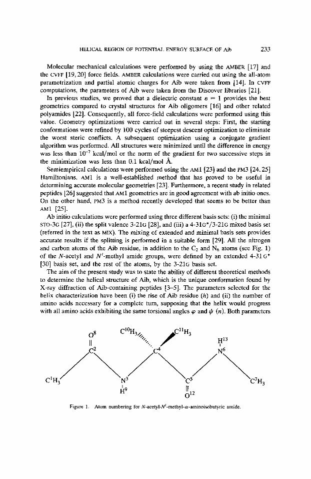
The N-acetyl-N'-methyl-a -aminoisobutyric amide (AAibN) was selected as a model molecule to study the helical trends observed in systems containing monomeric units of Aib (Fig. 1). The internal coordinates were defined as suggested by Pulay et al. [MI.
HELICAL REGION OF POTENTIAL ENERGY SURFACE OF Aib 233
Molecular mechanical calculations were performed by using the AMBER [17] and the CVFF [19,20] force fields. AMBER calculations were carried out using the all-atom parametrization and partial atomic charges for Aib were taken from [14]. In CVFF computations, the parameters of Aib were taken from the Discover libraries [21].
In previous studies, we proved that a dielectric constant E = 1 provides the best geometries compared to crystal structures for Aib oligomers [16] and other related pol yamides [22]. Consequently, all force-field calculations were performed using this value. Geometry optimizations were carried out in several steps: First, the starting conformations were refined by 100 cycles of steepest descent optimization to eliminate the worst steric conflicts. A subsequent optimization using a conjugate gradient algorithm was performed. All structures were minimized until the difference in energy was less than kcal/mol or the norm of the gradient for two successive steps in the minimization was less than 0.1 kcal/mol A.
Semiempirical calculations were performed using the AM^ [23] and the PM3 [24,25] Hamiltonians. AM^ is a well-established method that has proved to be useful in determining accurate molecular geometries [23]. Furthermore, a recent study in related peptides [26] suggested that AM^ geometries are in good agreement with ab initio ones. On the other hand, PM3 is a method recently developed that seems to be better than AM^ [25].
Ab initio calculations were performed using three different basis sets: (i) the minimal STO-3G [27], (ii) the split valence 3-216 [28], and (iii) a 4 - 3 1 ~ * / 3 - 2 1 ~ mixed basis set (referred in the text as MIX). The mixing of extended and minimal basis sets provides accurate results if the splitting is performed in a suitable form [29]. All the nitrogen and carbon atoms of the Aib residue, in addition to the Cz and Ng atoms (see Fig. 1) of the N-acetyl and "-methyl amide groups, were defined by an extended 4-31G* [30] basis set, and the rest of the atoms, by the 3 - 2 1 ~ basis set.
The aim of the present study was to state the ability of different theoretical methods to determine the helical structure of Aib, which is the unique conformation found by X-ray diffraction of Aib-containing peptides [3-51. The parameters selected for the helix characterization have been (i) the rise of Aib residue (h) and (ii) the number of amino acids necessary for a complete turn, supposing that the helix would progress with all amino acids exhibiting the same torsional angles rp and $ (n). Both parameters
Figure 1. Atom numbering for N-acetyl-N'-methyl-a-aminoisobutyric amide.

234 A L E M h AND PEREZ
are calculated from the backbone dihedral angles p and + [31]. Although several intermediate classes of helices could be defined, we find it more consistent to consider that each structure belongs to one of the three different prototype structures: the 310- helix characterized by the parameters n = 3 and h = 2; the a-helix with n = 3.6 and h = 1.5, and the o-helix (or 413) with n = 4 and h = 1.2 [32].
Molecular mechanical computations were performed with AMBER 3.0 Rev. A [33] and DISCOVER [21] programs. Semiempirical calculations were carried out using the MOPAC 5.0 program [34]. Finally, ab initio calculations were performed with the HONDO 7.0 package [35]. All calculations were carried out on an IBM 3090 at the “Centre de Supercomputaci6 de Catalunya” (CESCA).
Results and Discussion
A. Localization and Classification of the Low-energy Helical Conformations
Internal coordinates and helical characteristic parameters of the minimum-energy helical structures found by the different methods are shown in Table I. Minima for the mirror images of these conformations (+p, ++) are not listed. They have the same energies as those listed.
When empirical calculations were performed with the AMBER force field, a unique minimum was found with dihedral angles p = -49.5 and (6. = -41.1. This corre- sponds to an a-helix with some deviations in h and n with respect to the prototype structure. Thus, when an isolated residue of Aib is considered, the 310 -and o-helix found in Aib oligomers [16] are not obtained.
In sharp contrast, when CVFF calculations are performed on AAibN, no helical structure is obtained. Thus, varying the dihedral angles p and + in the helical region uniformly and, subsequently, minimizing the energy according to the procedure described in the Methods section provided a C7 (p = 60.5, + = -71.7) conformation in all cases.
Regarding the semiempirical Hamiltonians, both AM^ and PM3 predict the existence of a helical structure, although with different p and + values. Thus, the conformation yielded by AM^ (p = -51.8, JI = -31.7) corresponds to a slightly deviated 310- helix as their h and n values indicate. On the other hand, PM3 provides a structure (p = -60.2, + = -44.3) whose helical characteristic parameters correspond to a standard a-helix.
Helical structures are also found by ab initio calculations with all of the three basis sets used. The MIX and 3-21G predict a well-defined a-helix (p, fi =
-53.9, -47.8, and - 53.6, -47.9, respectively) with the h and n parameters very close to the standard values. On the other hand, when the STO-3G basis set is used, we obtain a very distorted 3lo-helix (p = -62.1 and + = -25.0) as the energy minimum.
Ab initio and semiempirical methods are, in general, quite consistent with each other. Thus, they agree in predicting a helical structure on the potential energy surface of AAibN, although the position of the minimum is located at different positions depending on the method or basis set used. Whereas the STO-3G results display the

HELICAL REGION OF POTENTIAL ENERGY SURFACE OF Aib 235
TAEILE I. Helical structures of AAibN localized as the energy minimum by different empirical, semiem- pirical, and ab initio procedures (bond distances are in A, angles in degrees); helical parameters h and n
are shown for comparison of the different structures.
AMBER AM^ PM3 STO-36 3-216 MIXa
C1N2 C2N3 N3C4 c4c5 C5N6 N6C7 C4C10 C4Cll C208 C5012 N3H9 N6H13
( ClC2N3 (C2N3C4 ( N3C4C5(7) (C4C5N6 (C5N6C7 ( c1oc4c11 (u)
$ rp
0 1
0 2 h n
1.523 1.338 1.463 1.538 1.334 1.452 1.535 1.537 1.226 1.227 1.008 1.006
116.3 125.1 111.6 116.8 122.5 107.6
-49.5 -41.1 - 178.9 - 177.5
1.7 3.3
1.511 1.336 1.456 1.558 1.380 1.432 1.539 1.541 1.246 1.247 0.993 0.993
116.9 124.1 114.6 118.9 121.2 108.3
-51.8 -31.7 - 166.3 - 177.4
1.8 3.1
1.510 1.543 1.430 1.445 1.502 1.487 1.544 1.570 1.425 1.449 1.471 1.480 1.534 1.554 1.538 1.556 1.221 1.217 1.220 1.219 0.998 1.027 0.996 1.029
114.0 112.9 122.7 117.8 112.7 109.8 117.7 118.3 120.4 119.9 108.1 108.9
-60.2 -62.1 -44.3 -25.0 - 160.0 173.4
167.9 -170.7 1.5 1.8 3.6 3.2
1.517 1.520 1.363 1.366 1.467 1.465 1.536 1.547 1.359 1.364 1.460 1.456 1.542 1.539 1.536 1.533 1.216 1.190 1.219 1.190 0.997 0.999 1 .Ooo 1.001
114.5 113.9 122.0 121.9 111.8 112.0 120.3 119.3 132.0 132.6 109.6 109.6
-53.9 -53.6 -47.8 -47.9 175.7 175.7
-173.8 - 173.5 1.6 1.6 3.5 3.5
a 4-31~*/3-216 mixed basis set (see text).
large deviations in respect to the MIX basis set, 3-216 results are very similar. On the other hand, semiempirical calculations provide smaller deviations, especially PM3.
The AMBER force field provides excellent results compared with MIX, whereas no helical structure was characterized using CVFF. Shortcomings of the CVFF force field were also observed by Bohm and Brode in alanine and glycine dipeptides [36], where commonly predicted local minima by ab initio and other force field methods were not found.
B. Analysis of the Geometrical Parameters
The geometrical parameters obtained with the MIX basis set are in excellent agreement with those found for the Aib residue in published crystal structures [3743], although, of course, slight deviations are observed depending on the peptide sequence. Thus, the conformation obtained with the MIX basis set has been taken as the point of reference in order to make the comparison between the results provided by the different methods.

236 ALE- AND PEREZ
TABLE 11. Absolutea and relativeb (in parentheses) r.m.s. deviations (MIX'-X) of bond lengths, bond angles, and dihedrals for X = AMBER, AM1, PM3, sTO-3~3, and 3-216.
Bond distances 0.020 0.026 0.032 0.040 0.011 (1.5) (1.9) (2.3) (2.9) (0.7)
Bond angles 4.6 5.0 5.1 5.6 0.5 (3.9) (4.2) (4.3) (4.7) (0.4)
Dihedral angles 5.2 12.3 15.7 12.3 0.2 (3.9) (9.2) (11.8) (9.3) (0.2)
aBond length in A; bond and dihedral angles in degrees.
'4-316*/3-21~ mixed basis set (see text). %.
Table I1 shows the absolute and relative r.m.s. deviations (relative to the values obtained with the MIX basis set) for bond lengths, bond angles, and dihedral angles of the AAibN structures found with the different methods. All methods perform well for bond lengths, the largest deviations being 0.040 and 0.032 A, corresponding to relative deviations of 2.3 and 2.9% for STO-36 and PM3, respectively. Similar results were obtained for bond angles, where all the methods provide a deviation of about 5", with the exception of the 3-216 basis set, which predicts an r.m.s. of only 0.5". AM1, PM3, and STO-3G provide a large r.m.s. deviation for dihedral angles. Thus, their deviations range between 9 and 12%. It is interesting to note the drastic deviation from the planarity of the peptidic bond provided by the PM3 method. In sharp contrast, the AMBER force field and ab initio 3-21G calculations predict a relative deviation below 4%. Both AMBER and 3-216 calculations provide consistently the best results with respect to the MIX ones for all the different geometrical parameters considered. This fact suggests the reliability of these procedures for the description of the potential energy surface of the Aib residue. Indeed, the structure of poly (Aib) provided by electron-diffraction experiments [44] was accurately reproduced by the authors using the AMBER force field [16].
Another interesting point is the dependence of the conformational behavior of a ,a - dialkylated peptides around the bond angles 7 and u (see Fig. 1 and Table I). Previous systematic studies seem to suggest some general rules between these angles and the residue conformation [13,45-471. Specifically, small values of 7 favor the a-helical structure, whereas small values of u favor the 310 conformation. The results of Table I cannot support these trends. For instance, the prototype a-helix yielded by the PM3 method exhibits the lowest u value of the different methods investigated. In contrast, the lowest value of T corresponds to the distorted STO-36, which is a 3m-helical structure.
A common feature found in the conformations provided by all the methods considered is the nonplanar distortion of the peptide bonds 01 and 0 2 . Indeed, this observation agrees with the X-ray-determined crystal structures with Aib residues, where the average nonplanar distortion ranges around 3-6" [11,38-47]. Although PM3

HELICAL REGION OF POTENTIAL ENERGY SURFACE OF Aib 237
and AMI provides excessive large deviations, they could be considered as artifacts of the methods.
Conclusions
In this paper, we presented a detailed theoretical study of the helical conformational preferences of model Aib dipeptide. Our results include empirical, semiempirical, and ab initio computations and are a useful test of the performance of various empirical and semiempirical methods. Our principal conclusions are as follow:
AMBER and 3-216 optimized structures are in excellent agreement with the a- helix conformation obtained from the MIX basis set, which is very close to the X-ray observed conformation in Aib-containing polypeptides. On the other hand, there are significant differences in the helical structure predicted by PM3 and the structure predicted by AM^. Thus, whereas PM3 localizes an a-helix as the energy minimum, AM^ provides a 3lo-helix. Moreover, the analysis of the geometrical parameters indicate that both methods display important structural differences in respect to MIX results. Finally, we found that the CVFF force field failed to locate this experimental minimum.
Acknowledgments
We would like to thank Dr. F.J. Luque for helpful discussions. C.A. gratefully acknowledges the support of the Departament d’Ensenyament de la Generalitat de Catanlunya. We thank “Centre de Supercomputacid de Catalunya” (CESCA) for the computational facilities.
Bibliography
[ l ] J.L. Flippen and I.L. Karle, Biopolymers 15, 1081 (1976). [2] M. K. Mathew and P. Balaram, Mol. Cell. Biochem. 50, 47 (1983). [3] C. Toniolo and E. Benedetti, IS1 Atlas Sci. Biochemz. 1, 225 (1988). [4] 1. L. Karle and P. Balaram, Biochemistry 29, 6747 (1990). [5] A. Aubry, J. Protas, G. Boussard, M. Marraud, and J. Neel, Biopolymers 17, 1693 (1978). [6] G. R. Marshall and H. E. Busshard, Circ. Res. 30/31 (Suppl. II) , 2599 (1972). [7] A. W. Burguess and S. J. Leach, Biopolymers 12, 2599 (1973). [8] Y. Paterson, S. M. Rumsey, E. Benedetti, G. Nemethy, and H. A. Scheraga, J. Am. Chem. SOC. 103,
[9] D. Peters and J. Peters, J. Mol. Struct. 86, 341 (1982). 2947 (1981).
[lo] I.L. Karle, J.L. Flippen-Anderson, K. Uma, and P. Balaram, Curr. Sci. 59, 875 (1990). [ 111 B. V. V. Prasad and V. Sasisekharan, Macromolecules 12, 1107 (1979). [12] F. A. Momany, R. F. McGuire, A. W. Burgess, and H. A. Scheraga, J. Phys. Chem. 79, 2361 (1975). [13] V. Barone, F. Fraternalli, and P. L. Cristinziano, Macromolecules 23, 2038 (1990). [I41 E. E. Hodkin, J. D. Clark, K. R. Miller, and G. R. Marshall, Biopolymers, 30, 533 (1990). 1151 S. J. Weiner, P. A. Kollman, D. A. Case, U. C. Singh, C. Ghio, G. Alagona, S. Profeta, and P. Weiner,
[I61 C. Alemin, J. A. Subirana, and J. J. Perez, Biopolymers 32, 621 (1992). [17] S. J. Weiner, P.A. Kollman, D.T. Nguyen, and D.A. Case, J. Comput. Chem. 7, 230 (1986). [18] P. Pulay, G. Forgarsi, F. Pang, and J. E. Boggs, J. Am. Chem SOC. 101, 2550 (1979). [19] P.S. Stern, M. Chorev, M. Goodman, and A.T. Hagler, Biopolymers 22, 1885 (1983).
J. Am. Chem. SOC. 106, 765 (1984).

238 A L E M h AND PEREZ
[20] P.D. Osguthorpe, V. A. Roberts, D. J. Osguthorpe, J. Wolff, M. Genest, and A. T. Hagler, Proteins
[21] Discover Version 2.7.0, Biosym Technologies (1991). [22] I. Bella, C. Alemin, C. Alegre, J. M. Fernindez-Santin, and J. A. Subirana, Macromolecules 25, 5225
[23] M. J. S. Dewar, E. G. Zoebisch, E. F. Healy, E. H. Healy, and J. J. P. Stewart, J. Am. Chem. Soc. 107,
[24] J.J.P. Stewart, J. Comput. Chem. 10, 209 (1989). [25] J.J.P. Stewart, J. Comput. Chem. 10, 221 (1989). [26] C. Aleman and J. J. Perez, J. Mol. Struct. (THEOCHEM) (in press). [27] W. J. Hehre, R. F. Stewart, and J.A. Pople, J. Chem. Phys. 51, 2657 (1969). [28] J. S . Binkley, J.A. Pople, and W. Hehre, J. Am. Chem. SOC. 102, 939 (1980). [29] J.H. Jensen and M. S. Gordon, J. Comput. Chem. 12, 421 (1990). [30] (a) P. C. Hahiraran and J. A. Pople, Theor. Chim. Acta 28, 213 (1973).
(b) J. B. Collins. P. v. R. Scheyer, J. S. Binkley, and J. A. Pople, J. Chem. Phys. 64,5142 (1976). [31] G. D. Fasman, Poly(a-Amino Acids): Protein Models for Conformational Studies. Biological Macro-
molecules Series (Marcel Dekker, New York, 1967). [32] R. D. B. Fraser and T. P. McRae, Conformation in Fibrous Proteinr (Academic Press, London, 1973). [33] U. C. Singh, P. K. Weiner, J. Caldwell, and P. A. Kollman; revised by G. Seibel, AMBER 3.0. Revision
(341 J. J. P. Stewart, MOPAC 5.0 (1988). [35] M. Dupuis, J. D. Watts, H. 0. Villar, and G. J.B. Hurst, HONDO 7.0 (1987). [36] H.-J. Bohm and S. Brode, J. Am. Chem. Soc. 113, 7129 (1991). [37] B. V. V. Prasad, N. Shamala, R. Nagaraj, R. Chandrasekaran, and P. Balaram, Biopolymers 18, 1635
[38] B. V. V. Prasad, N. Sahamala, R. Nagaraj, and P. Balaram, Acta Crystallogr., Sect. B, B36, 107 (1980). (391 R. Gessmann, H. Brueckner, and M. Kokkinidis, Biochem. Biophys. Res. Commun. 174, 878 (1991). [40] A. Bavoso, E. Benedetti, B. Di Blasio, V. Pavone, C. Pedone, C. Toniolo, and G. M. Bonora, Proc.
[41] C. Toniolo, G. M. Bonora, A. Bavoso, E. Benedetti, B. Di Blasio, V. Pavone, and C. Pedone, J.
[42] V. Pavone, E. Bendetti, B. Di Blasio, C. Pedone, A. Santini, A. Bavoso, C. Toniolo, M. Crisma, and
[43] E. Benedetti, B. Di Blasio, V. Pavone, C. Pedone, C. Toniolo, and M. Crisma, Biopolymers 32, 453
[44] B. R. Malcolm and M. D. Walkinshaw, Biopolymers 25, 607 (1986). [45] V. Barone, F. Lelj, A. Bavoso, B. Di Blasio, P. Grimaldi, V. Pavone, and C. Pedone, Biopolymers
24, 1759 (1985). [46] E. Benedetti, C. Toniolo, P. Hardy, V. Barone, A. Bavoso, B. Di Blasio, P. Grimaldi, F. Lelj,
V. Pavone, C. Pavone, G. M. Bondora, and I. J. Lingham, J. Am. Chem. SOC. 106, 8146 (1984). [47] E. Benedetti, V. Barone, A. Bavoso, B. Di Blasio, F. k l j , V. Pavone, C. Pedone, G. M. Bonora,
C. Toniolo, M. T. Laplawy, K. Kaczmarek, and A. Redlinsky, Biopolymers 27, 357 (1988).
4, 31 (1988).
(1992).
309 (1986).
A.
(1979).
Natl. Acad. Sci. U.S.A. 83, 1988 (1986).
Biomol. Struct. Dyn. 3, 585 (1985).
L. Sartore, J. Biomol. Struct. Dyn. 7, 1321 (1990).
(1992).
Received September 21, 1992 Accepted for publication March 5, 1993