Embed Size (px)
Citation preview
Helicobacter pylori Activates NF‐kB by InducingUbc13‐Mediated Ubiquitination of Lysine 158 of TAK1Acacia Lamb,1 JinJing Chen,1 Steven R. Blanke,2 and Lin‐Feng Chen1*1Department of Biochemistry, College of Medicine, Urbana, Illinois, 618012Department of Microbiology, University of Illinois at Urbana‐Champaign, Urbana, Illinois, 61801
ABSTRACTThe Helicobacter pylori virulence factor CagA targets a variety of host proteins to alter different cellular responses, including the induction ofpro‐inflammatory cytokines. We have previously shown that CagA‐facilitated lysine 63‐linked ubiquitination of TAK1 is essential for theH. pylori‐induced NF‐kB activation and the expression of proinflammatory cytokines. However, the molecular mechanism for TAK1 ubiquitination andactivation in H. pylori‐mediated NF‐kB activation remains elusive. Here, we identify lysine 158 of TAK1 as the key residue undergoing lysine 63‐linked ubiquitination in response toH. pylori infection.Mutation of lysine 158 to arginine prevents the ubiquitination of TAK1 and impairsH. pylori‐induced TAK1andNF‐kB activation.Moreover, we demonstrate that E2 ubiquitin conjugating enzymeUbc13 is involved inH. pylori‐mediated TAK1ubiquitination. Suppressing the activity of Ubc13 by a dominant‐negative mutant or siRNA abolishes CagA‐facilitated and H. pylori‐induced TAK1and NF‐kB activation. These findings further underscore the importance of lysine 63‐linked ubiquitination of TAK1 in H. pylori‐induced NF‐kBactivation and NF‐kB‐mediated inflammatory response. J. Cell. Biochem. 114: 2284–2292, 2013. � 2013 Wiley Periodicals, Inc.
KEY WORDS: CagA; NF‐kB; TAK1; Ubc13; UBIQUITINATION
Infection with Helicobacter pylori and the resulting chronicinflammation is a major risk factor for the development of gastric
cancer [Peek and Blaser, 2002; Hatakeyama, 2008]. H. pyloriinfection up‐regulates the expression of pro‐inflammatory cytokines,including IL‐8, IL‐6, and TNF‐a, which contribute to an environmentwhich promotes increased cell turnover, mutagenesis, and growth[McGee and Mobley, 2000]. The enhanced expression of pro‐inflammatory cytokines is believed to be associated with the abilityof H. pylori to activate transcription factor NF‐kB, which plays a keyrole in modulating the expression of various cytokines [Ghosh andKarin, 2002].
Various virulence components of H. pylori, including flagella,lipopolysaccharide (LPS), vacuolating toxin VacA, and cytotoxin‐associated gene pathogenicity island (cagPAI), have been shown tobe able to affect the expression of cytokines [Yamaoka, 2010]. Ofall these components, the most potent factor is cagPAI, whichencodes a type 4 secretion system (T4SS) and the virulence factorCagA. CagA is injected into gastric epithelial cells via the T4SS[Peek, 2005]. By interacting with a variety of cellular signalingmolecules, CagA induces a series of cellular events, including actin
remodeling and IL‐8 production [Peek, 2005]. Translocation ofCagA into host cells induces higher levels of IL‐8 production byactivating transcription factors such as NF‐kB and AP‐1 [Backertand Naumann, 2010]. Ectopic expression of CagA is able tostimulate nuclear translocation of NF‐kB and production of IL‐8 ingastric epithelial cells [Backert and Naumann, 2010]. Furthermore,NF‐kB activation and inflammation have been shown to bemarkedly reduced in the gastric antra of Mongolian gerbilsinfected with cagA‐deficient H. pylori as compared to infectionwith wild‐type H. pylori, emphasizing the essential role of CagA inNF‐kB activation and H. pylori‐mediated inflammation [Shibataet al., 2006; Lamb and Chen, 2010].
Transforming growth factor b‐activated kinase 1 (TAK1) is a keyregulator of signal transduction cascades leading to the activationof IkB kinases (IKKs) and NF‐kB in response to various stimuli,including cytokines and bacterial and viral infections [Adhikariet al., 2007]. TAK1 phosphorylates and activates IKK2, which thenphosphorylates IkBa, leading to its ubiquitination and degrada-tion; subsequently, NF‐kB is translocated into the nucleus andNF‐kB target gene expression is activated [Wang et al., 2001;
2284
Conflict of interest: nothing to declare.
Grant sponsor: NIH/NIDDK; Grant number: DK‐085158.
*Correspondence to: Lin‐Feng Chen, Department of Biochemistry, COM 190 MSB, MC‐714, University of Illinois atUrbana‐Champaign, Urbana, IL 61801. E‐mail: [email protected]
Manuscript Received: 28 January 2013; Manuscript Accepted: 12 April 2013
Accepted manuscript online in Wiley Online Library (wileyonlinelibrary.com): 19 April 2013
DOI 10.1002/jcb.24573 � � 2013 Wiley Periodicals, Inc.
Journal of CellularBiochemistry
ARTICLEJournal of Cellular Biochemistry 114:2284–2292 (2013)
Chen and Greene, 2004]. Recent studies demonstrate that lysine (K)63‐linked ubiquitination actively participates in the activation ofTAK1 and IKKs, and that many upstream signaling molecules aresubject to K63‐linked ubiquitination [Wang et al., 2001]. Forexample, IL‐1b receptor‐associated kinase 1 (IRAK1) is K63‐linkedubiquitinated upon IL‐1b stimulation, and the ubiquitination isimportant for IKK activation in the IL‐1b signaling pathway[Conze et al., 2008; Windheim et al., 2008]. Ubiquitination of TAK1by TNF‐a receptor associated factor (TRAF) 6 is essential for TAK1auto‐phosphorylation and activation [Sorrentino et al., 2008;Yamazaki et al., 2009; Fan et al., 2010]. Several lysines withinTAK1, including K34, K158, and K209, have been identified to beK63‐linked ubiquitinated, and ubiquitination of these differentlysines appears to determine stimulus‐specific and context‐dependent TAK1 activation [Sorrentino et al., 2008; Yamazakiet al., 2009; Fan et al., 2010]. Ubiquitination of K34 of TAK1 hasbeen reported to induce TGF‐b‐mediated p38 and JNK activation[Sorrentino et al., 2008]. Ubiquitination of TAK1 at a differentlysine, K158, is required for TNF‐a or IL‐1b‐induced TAK1activation and IKK/NF‐kB activation [Fan et al., 2010]. We havealso reported that TRAF6‐mediated ubiquitination of TAK1 isessential for H. pylori‐induced NF‐kB activation and the inductionof pro‐inflammatory cytokines [Lamb et al., 2009]. H. pyloriinfection stimulates virulence factor CagA‐dependent K63‐linkedubiquitination and activation of TAK1. CagA facilitates TAK1ubiquitination and potentiates TAK1‐mediated NF‐kB activationin H. pylori‐infected gastric epithelial cells [Lamb et al., 2009].Nevertheless, the detailed molecular mechanism by which CagAenhances the ubiquitination and activation of TAK1 remainsunclear.
Ubiquitination is anATP‐dependent, three‐step enzymatic cascadeinvolving an ubiquitin‐activating enzyme (E1), an ubiquitin‐conjugating enzyme (E2), and an ubiquitin‐protein ligase (E3)[Adhikari et al., 2007]. TRAF family proteins, including TRAF2 andTRAF6, are known to be E3 ligases for K63‐linked ubiquitination[Chen, 2012]. Biochemical studies also reveal that the heterodimericUbc13 and Uev1A are E2 conjugating enzymes for TRAF6‐catalyzedpolyubiquitination and activation of TAK1 [Wang et al., 2001].Ubc13/Uev1A have been shown to be essential for the ubiquitinationof TRAF6 in IL‐1R/Toll‐like receptor (TLR) signaling [Yamazakiet al., 2009]. In vivo studies also demonstrate that Ubc13 is a criticalcomponent of TRAF‐mediated inflammatory responses since ubc13heterozygous mice were resistant to LPS‐induced lethality, anddisplayed reduced ubiquitination of TRAF6 [Fukushima et al., 2007].Our previous studies have demonstrated that TRAF6 is critical forK63‐linked ubiquitination of TAK1 and its subsequent kinaseactivation in response to H. pylori infection. However, the role ofUbc13 in the H. pylori‐induced ubiquitination of TAK1 remainsundetermined.
In this study, we further investigated the molecular mechanism bywhich TAK1 is ubiquitinated and activated byH. pylori. We identifiedlysine 158 of TAK1 as the pivotal residue for H. pylori CagA‐facilitated TAK1 ubiquitination and activation.We also demonstratedthat Ubc13 is responsible for H. pylori‐induced ubiquitination andactivation of TAK1, which further activates NF‐kB and NF‐kB‐dependent inflammatory gene expression.
RESULTS
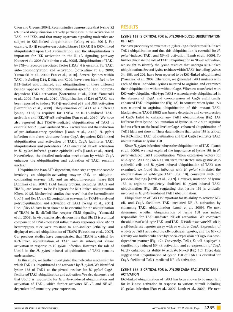
LYSINE 158 IS CRITICAL FOR H. PYLORI‐INDUCED UBIQUITINATIONOF TAK1We have previously shown that H. pylori CagA facilitates K63‐linkedTAK1 ubiquitination and that this ubiquitination is essential for H.pylori‐induced TAK1 and NF‐kB activation [Lamb et al., 2009]. Tofurther elucidate the role of TAK1 ubiquitination in NF‐kB activation,we sought to identify the lysine residues that undergo K63‐linkedubiquitination. Several lysine residues within TAK1, including lysines34, 158, and 209, have been reported to be K63‐linked ubiquitinated[Yamazaki et al., 2009]. Therefore, we generated TAK1 mutants witheach of these individual lysines mutated to arginine and examinedtheir ubiquitination with or without CagA. When co‐transfected withK63‐only ubiquitin, wild‐type TAK1 was moderately ubiquitinated inthe absence of CagA and co‐expression of CagA significantlyenhanced TAK1 ubiquitination (Fig. 1A). In contrast, when lysine 158was mutated to arginine, ubiquitination of this mutant TAK1(designated as TAK‐K158R) was barely detectable and co‐expressionof CagA failed to enhance any TAK1 ubiquitination (Fig. 1A).Different from lysine 158, mutation of lysine 34 or 209 to argininehad no effect on the basal level or CagA‐enhanced ubiquitination ofTAK1 (data not shown). These data indicate that lysine 158 is criticalfor K63‐linked TAK1 ubiquitination and that CagA facilitates TAK1ubiquitination at lysine 158.
SinceH. pylori infection induces the ubiquitination of TAK1 [Lambet al., 2009], we next explored the importance of lysine 158 in H.pylori‐induced TAK1 ubiquitination. When expression vectors forwild‐type TAK1 or TAK1‐K158R were transfected into gastric AGSepithelial cells and H. pylori‐induced ubiquitination of TAK1 wasexamined, we found that infection with H. pylori stimulated theubiquitination of wild‐type TAK1 (Fig. 1B), consistent with ourprevious findings [Lamb et al., 2009]. However, mutation of lysine158 to arginine completely abolished H. pylori‐induced TAK1ubiquitination (Fig. 1B), suggesting that lysine 158 is criticallyinvolved in H. pylori‐induced TAK1 ubiquitination.
Ubiquitination of TAK1 is important for its ability to activate NF‐kB, and CagA facilitates TAK1‐mediated NF‐kB activation byenhancing TAK1 ubiquitination [Lamb et al., 2009]. We nextdetermined whether ubiquitination of lysine 158 was indeedresponsible for TAK1‐mediated NF‐kB activation. We comparedthe abilities of wild‐type TAK1 and TAK1‐K158R to activate NF‐kB ina kB‐luciferase reporter assay with or without CagA. Expression ofwild‐type TAK1 activated the kB‐luciferase reporter, and the NF‐kBactivity was further enhanced by the co‐expression of CagA in a dose‐dependent manner (Fig. 1C). Conversely, TAK1‐K158R displayed asignificantly reduced NF‐kB activation, and co‐expression of CagAbarely enhanced its ability to activate NF‐kB (Fig. 1C). These datasuggest that ubiquitination of lysine 158 of TAK1 is essential forCagA‐facilitated TAK1‐mediated NF‐kB activation.
LYSINE 158 IS CRITICAL FOR H. PYLORI CAGA‐FACILITATED TAK1ACTIVATIONK63‐linked ubiquitination of TAK1 has been shown to be importantfor its kinase activation in response to various stimuli includingH. pylori infection [Fan et al., 2009; Lamb et al., 2009]. We next
JOURNAL OF CELLULAR BIOCHEMISTRY ACTIVATION OF TAK1 BY H. PYLORI CagA 2285
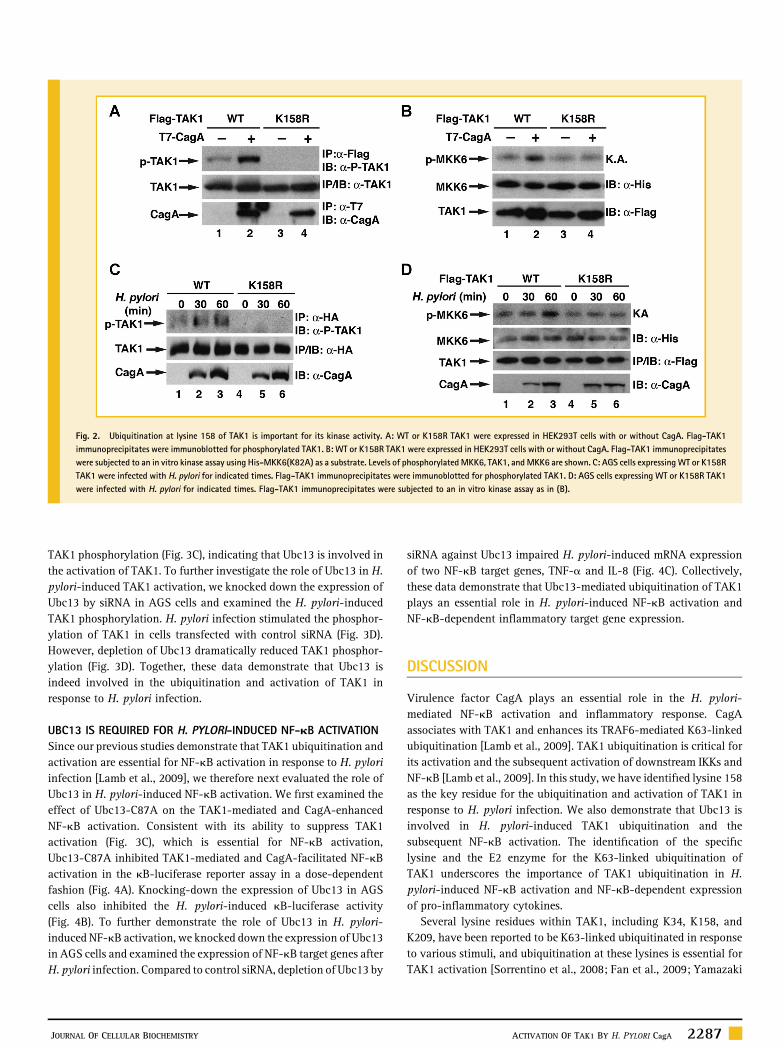
assessed whether ubiquitination of lysine 158 was involved in H.pylori‐induced TAK1 activation. Over‐expressed TAK1 displayed abasal level of kinase activity which is demonstrated by its auto‐phosphorylation of threonine 187 (Fig. 2A). Co‐expression of CagAfurther enhanced the kinase activity of wild‐type TAK1. However, co‐expression of CagA did not enhance the kinase activity of TAK1‐K158R, which displayed almost undetectable basal kinase activity(Fig. 2A). The importance of ubiquitination of K158 in TAK1 kinaseactivation was further demonstrated by an in vitro kinase assay usingMKK6 as the substrate (Fig. 2B). Wild‐type TAK1 phosphorylatedMKK6, and this phosphorylation was further enhanced by co‐expression of CagA. However, mutation of lysine 158 to argininedemolished TAK10s ability to phosphorylate MKK6, and co‐expres-sion of CagA failed to enhance the levels of phosphorylated MKK6(Fig. 2B). More importantly, when we examined the activation ofTAK1 in response to H. pylori infection, we found that H. pyloriinfection stimulated the auto‐phosphorylation of wild‐type TAK1 butnot the TAK1‐K158R mutant (Fig. 2C), indicating that K158 is criticalfor H. pylori infection‐induced TAK1 activation. Consistently, whenTAK1 activity was examined by the in vitro kinase assay, we observedthat H. pylori infection activated wild‐type TAK1 but not the TAK‐K158R mutant (Fig. 2D). All together, these data suggest thatubiquitination of lysine 158 is essential for the activation of TAK1 invivo in response to H. pylori infection.
UBC13 MEDIATES THE UBIQUITINATION AND ACTIVATION OF TAK1We have previously shown that E3 ligase TRAF6 is involved in H.pylori‐induced TAK1 ubiquitination [Lamb et al., 2009]. Recentstudies indicate that TRAF6 couples with Ubc13/Uev1a E2 enzymes tocatalyze K63‐linked ubiquitination [Wang et al., 2001], so we nextexamined whether Ubc13/Uev1a was also involved in the CagA‐facilitated ubiquitination of TAK1. First, we employed small‐interference RNA (siRNA) to knock down the expression of Ubc13,Uev1a or UbcH7 and examined the ubiquitination of TAK1. Comparedto control siRNA, depletion of Ubc13 or Uev1a impaired the basal aswell as the CagA‐enhanced ubiquitination of TAK1 (Fig. 3A).Depletion of UbcH7, an E2 enzyme which is not involved in TRAF6‐mediated ubiquitination [Wang et al., 2001], had no effect on theubiquitination of TAK1 (Fig. 3A). These data suggest a specificinvolvement of Ubc13/Uev1a in the CagA‐facilitated ubiquitinationof TAK1. To further dissect the role of Ubc13 in the H. pylori‐inducedTAK1 ubiquitination, we examined the ubiquitination of TAK1 inAGS cells transfectedwith control or Ubc13 siRNA.H. pylori infectioninduced ubiquitination of TAK1 in cells transfected with controlsiRNA; however, ubiquitination was attenuated when expression ofUbc13 was inhibited by siRNA (Fig. 3B). These data reveal that Ubc13plays an important role in H. pylori‐induced ubiquitination of TAK1.
Since ubiquitination of TAK1 is critical for its activation andUbc13is involved in H. pylori‐induced TAK1 ubiquitination, we nextdetermined whether Ubc13 is important for H. pylori‐mediated TAK1activation. We first examined the effect of a catalytically inactiveform of Ubc13 (Ubc13‐C87A) on the phosphorylation of TAK1.Ubc13‐C87A with cysteine 87 mutated to alanine lacks E2 activityand functions as a dominant negative form to suppress activity ofUbc13 [Deng et al., 2000]. Ubc13‐C87A not only blocked the basallevels of TAK1 phosphorylation but also inhibited CagA‐enhanced
Fig. 1. Lysine 158 is required for H. pylori‐induced ubiquitination andactivation of TAK1. A: Wild‐type (WT) or K158R mutant TAK1 were expressedwith K63‐only ubiquitin and CagA in HEK293T cells. Flag immunoprecipitatesfrom the lysates were immunoblotted for HA‐ubiquitin. B: WT or K158R mutantTAK1 were ectopically expressed in AGS cells, which were then infected with H.pylori as indicated. Flag immunoprecipitates from the lysates wereimmunoblotted for ubiquitin. C: HEK293T cells were transfected with IL‐8‐luciferase reporter and either WT or K158R TAK1 and increasing amounts ofCagA plasmids as indicated. Luciferase activity was measured 30 h post‐transfection.
2286 ACTIVATION OF TAK1 BY H. PYLORI CagA JOURNAL OF CELLULAR BIOCHEMISTRY
TAK1 phosphorylation (Fig. 3C), indicating that Ubc13 is involved inthe activation of TAK1. To further investigate the role of Ubc13 in H.pylori‐induced TAK1 activation, we knocked down the expression ofUbc13 by siRNA in AGS cells and examined the H. pylori‐inducedTAK1 phosphorylation. H. pylori infection stimulated the phosphor-ylation of TAK1 in cells transfected with control siRNA (Fig. 3D).However, depletion of Ubc13 dramatically reduced TAK1 phosphor-ylation (Fig. 3D). Together, these data demonstrate that Ubc13 isindeed involved in the ubiquitination and activation of TAK1 inresponse to H. pylori infection.
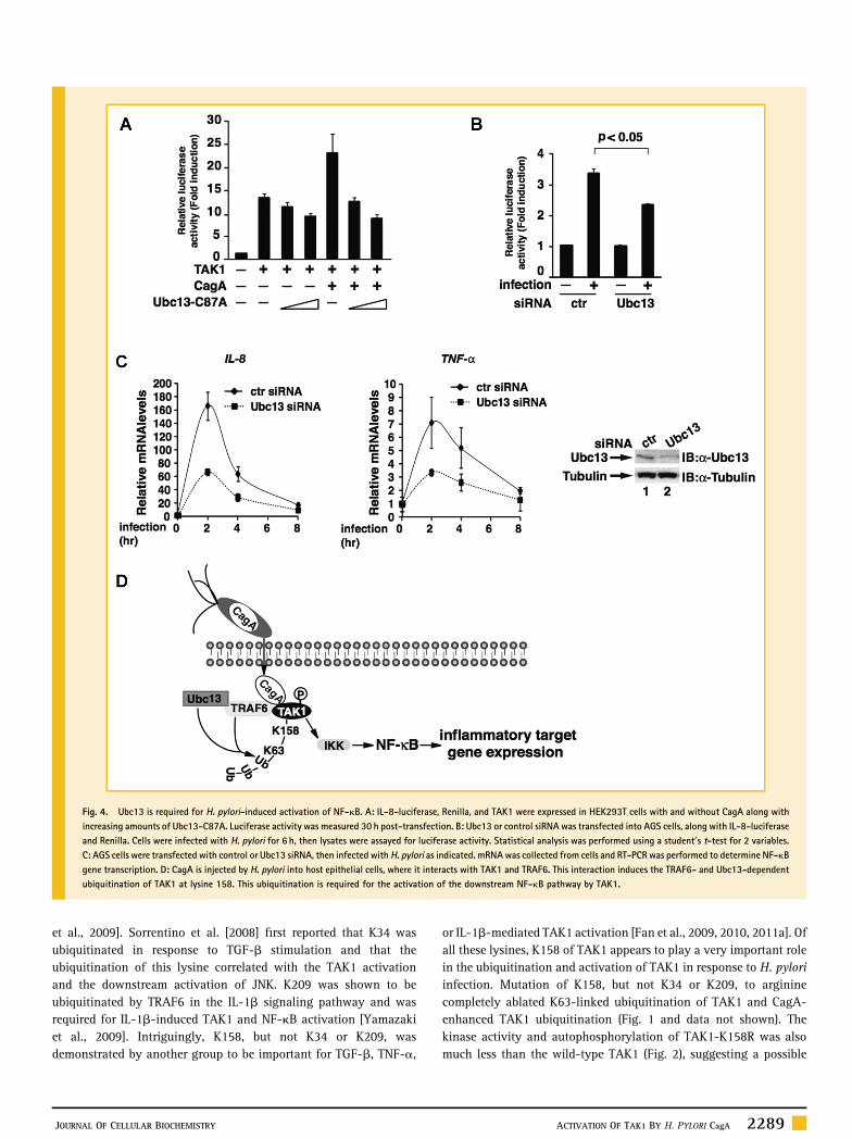
UBC13 IS REQUIRED FOR H. PYLORI‐INDUCED NF‐kB ACTIVATIONSince our previous studies demonstrate that TAK1 ubiquitination andactivation are essential for NF‐kB activation in response to H. pyloriinfection [Lamb et al., 2009], we therefore next evaluated the role ofUbc13 in H. pylori‐induced NF‐kB activation. We first examined theeffect of Ubc13‐C87A on the TAK1‐mediated and CagA‐enhancedNF‐kB activation. Consistent with its ability to suppress TAK1activation (Fig. 3C), which is essential for NF‐kB activation,Ubc13‐C87A inhibited TAK1‐mediated and CagA‐facilitated NF‐kBactivation in the kB‐luciferase reporter assay in a dose‐dependentfashion (Fig. 4A). Knocking‐down the expression of Ubc13 in AGScells also inhibited the H. pylori‐induced kB‐luciferase activity(Fig. 4B). To further demonstrate the role of Ubc13 in H. pylori‐induced NF‐kB activation, we knocked down the expression of Ubc13in AGS cells and examined the expression of NF‐kB target genes afterH. pylori infection. Compared to control siRNA, depletion of Ubc13 by
siRNA against Ubc13 impaired H. pylori‐induced mRNA expressionof two NF‐kB target genes, TNF‐a and IL‐8 (Fig. 4C). Collectively,these data demonstrate that Ubc13‐mediated ubiquitination of TAK1plays an essential role in H. pylori‐induced NF‐kB activation andNF‐kB‐dependent inflammatory target gene expression.
DISCUSSION
Virulence factor CagA plays an essential role in the H. pylori‐mediated NF‐kB activation and inflammatory response. CagAassociates with TAK1 and enhances its TRAF6‐mediated K63‐linkedubiquitination [Lamb et al., 2009]. TAK1 ubiquitination is critical forits activation and the subsequent activation of downstream IKKs andNF‐kB [Lamb et al., 2009]. In this study, we have identified lysine 158as the key residue for the ubiquitination and activation of TAK1 inresponse to H. pylori infection. We also demonstrate that Ubc13 isinvolved in H. pylori‐induced TAK1 ubiquitination and thesubsequent NF‐kB activation. The identification of the specificlysine and the E2 enzyme for the K63‐linked ubiquitination ofTAK1 underscores the importance of TAK1 ubiquitination in H.pylori‐induced NF‐kB activation and NF‐kB‐dependent expressionof pro‐inflammatory cytokines.
Several lysine residues within TAK1, including K34, K158, andK209, have been reported to be K63‐linked ubiquitinated in responseto various stimuli, and ubiquitination at these lysines is essential forTAK1 activation [Sorrentino et al., 2008; Fan et al., 2009; Yamazaki
Fig. 2. Ubiquitination at lysine 158 of TAK1 is important for its kinase activity. A: WT or K158R TAK1 were expressed in HEK293T cells with or without CagA. Flag‐TAK1immunoprecipitates were immunoblotted for phosphorylated TAK1. B: WT or K158R TAK1 were expressed in HEK293T cells with or without CagA. Flag‐TAK1 immunoprecipitateswere subjected to an in vitro kinase assay using His‐MKK6(K82A) as a substrate. Levels of phosphorylated MKK6, TAK1, and MKK6 are shown. C: AGS cells expressing WT or K158RTAK1 were infected with H. pylori for indicated times. Flag‐TAK1 immunoprecipitates were immunoblotted for phosphorylated TAK1. D: AGS cells expressing WT or K158R TAK1were infected with H. pylori for indicated times. Flag‐TAK1 immunoprecipitates were subjected to an in vitro kinase assay as in (B).
JOURNAL OF CELLULAR BIOCHEMISTRY ACTIVATION OF TAK1 BY H. PYLORI CagA 2287
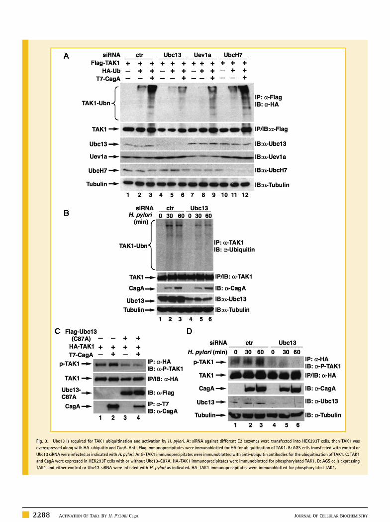
Fig. 3. Ubc13 is required for TAK1 ubiquitination and activation by H. pylori. A: siRNA against different E2 enzymes were transfected into HEK293T cells, then TAK1 wasoverexpressed along with HA‐ubiquitin and CagA. Anti‐Flag immunoprecipitates were immunoblotted for HA for ubiquitination of TAK1. B: AGS cells transfected with control orUbc13 siRNA were infected as indicated with H. pylori. Anti‐TAK1 immunoprecipitates were immunoblotted with anti‐ubiquitin antibodies for the ubiquitination of TAK1. C: TAK1and CagA were expressed in HEK293T cells with or without Ubc13‐C87A. HA‐TAK1 immunoprecipitates were immunoblotted for phosphorylated TAK1. D: AGS cells expressingTAK1 and either control or Ubc13 siRNA were infected with H. pylori as indicated. HA‐TAK1 immunoprecipitates were immunoblotted for phosphorylated TAK1.
2288 ACTIVATION OF TAK1 BY H. PYLORI CagA JOURNAL OF CELLULAR BIOCHEMISTRY
et al., 2009]. Sorrentino et al. [2008] first reported that K34 wasubiquitinated in response to TGF‐b stimulation and that theubiquitination of this lysine correlated with the TAK1 activationand the downstream activation of JNK. K209 was shown to beubiquitinated by TRAF6 in the IL‐1b signaling pathway and wasrequired for IL‐1b‐induced TAK1 and NF‐kB activation [Yamazakiet al., 2009]. Intriguingly, K158, but not K34 or K209, wasdemonstrated by another group to be important for TGF‐b, TNF‐a,
or IL‐1b‐mediated TAK1 activation [Fan et al., 2009, 2010, 2011a]. Ofall these lysines, K158 of TAK1 appears to play a very important rolein the ubiquitination and activation of TAK1 in response to H. pyloriinfection. Mutation of K158, but not K34 or K209, to argininecompletely ablated K63‐linked ubiquitination of TAK1 and CagA‐enhanced TAK1 ubiquitination (Fig. 1 and data not shown). Thekinase activity and autophosphorylation of TAK1‐K158R was alsomuch less than the wild‐type TAK1 (Fig. 2), suggesting a possible
Fig. 4. Ubc13 is required for H. pylori‐induced activation of NF‐kB. A: IL‐8‐luciferase, Renilla, and TAK1 were expressed in HEK293T cells with and without CagA along withincreasing amounts of Ubc13‐C87A. Luciferase activity was measured 30 h post‐transfection. B: Ubc13 or control siRNA was transfected into AGS cells, along with IL‐8‐luciferaseand Renilla. Cells were infected with H. pylori for 6 h, then lysates were assayed for luciferase activity. Statistical analysis was performed using a student0s t‐test for 2 variables.C: AGS cells were transfected with control or Ubc13 siRNA, then infected with H. pylori as indicated. mRNAwas collected from cells and RT‐PCR was performed to determine NF‐kBgene transcription. D: CagA is injected by H. pylori into host epithelial cells, where it interacts with TAK1 and TRAF6. This interaction induces the TRAF6‐ and Ubc13‐dependentubiquitination of TAK1 at lysine 158. This ubiquitination is required for the activation of the downstream NF‐kB pathway by TAK1.
JOURNAL OF CELLULAR BIOCHEMISTRY ACTIVATION OF TAK1 BY H. PYLORI CagA 2289
conformation change of TAK1‐K158R to completely inactivate itskinase activity. However, crystal structure of the TAK1 kinasedomain, where K158 resides, reveals that lysine 158 is on the surfaceof the protein, arguing against this possibility [Brown et al., 2005].
While it is clear that ubiquitination of K158 of TAK1 is critical forTAK1 activation, it is still not clear how the ubiquitination of TAK1activates the kinase. It is expected that, like other K63‐linkedubiquitinated signaling molecules, the polyubiquitin chain functionsas a scaffold to recruit other signaling molecules which are essentialfor the activation of TAK1 [Chen, 2012]. For example, MEKK3 hasbeen shown to be recruited to the ubiquitinated TAK1, and thisrecruitment is required for the activation of TAK1 [Yamazakiet al., 2009]. TAK1 associated proteins, such as TAB1 and TAB2/3,activate TAK1 and specifically bind to K63‐linked ubiquitin chainsthrough their zinc fingers [Kanayama et al., 2004]. Ubiquitination ofTAK1 may also facilitate the formation of TAK1‐TAB2/3 complexes,and the interaction of multiple complexes allows for cross‐activationof TAK1, although a recent study shows that free K63‐linkedubiquitin chains, not attached to any other proteins, can bind theubiquitin‐binding domain of TAB2 and activate the TAK1 complex invitro [Xia et al., 2009]. On the other hand, TAK1 ubiquitination mayaid in its activity by recruiting substrates, such as the IKK complex.The ubiquitin‐binding domain of the regulatory subunit NEMO hasbeen shown to be important in the activation of IKK [Adhikariet al., 2007; Windheim et al., 2008], and it may be that it binds to theubiquitin chains on TAK1. Supporting this possibility, Fan et al.showed that ubiquitinated TAK1 recruited NEMO and the IKKcomplex, leading to the activation of IKKs [Fan et al., 2009]. Lysine158 resides in the kinase domain of TAK1, raising another possibilitythat TAK1 ubiquitination might simply change the conformation ofthe kinase, allowing for the exposure of its catalytic domain. But theexact molecular mechanism for how ubiquitinated TAK1 activates itkinase activity needs to be further determined.
We have previously shown that E3 ligase TRAF6 is involved in H.pylori‐induced TAK1 ubiquitination and activation [Lamb et al., 2009].TRAF6 functions together with the E2 enzymes Ubc13/Uev1a to catalyzethe synthesis of K63‐linked ubiquitination [Deng et al., 2000]. Forexample, Ubc13 mediates the K63‐linked ubiquitination of TAK1 in IL‐1b signaling [Yamazaki et al., 2009]. Ubc13 is also involved in the K63‐linked ubiquitination of ELKS in response to genotoxic stress [Wuet al., 2010]. Consistent with its function as an E2 enzyme, we found thatUbc13 was involved in H. pylori‐induced TAK1 ubiquitination, sincedepletion of Ubc13 impaired H. pylori‐induced and CagA‐facilitatedTAK1 ubiquitination (Fig. 3). Furthermore, Ubc13 was required for theactivation of TAK1, activation of NF‐kB, and NF‐kB‐dependent targetgene expression (Figs. 3 and 4). The identification of Ubc13 as the E2enzyme for H. pylori‐induced TAK1 ubiquitination and activationfurther supports the notion that K63‐linked ubiquitination of TAK1 isessential for the H. pylori‐mediated inflammatory response.
Although CagA enhances Ubc13/TRAF6‐mediated K63‐linkedubiquitination of TAK1 (Fig. 4D), how this occurs is still not clear.CagA fails to enhance the interaction between TRAF6 and TAK1 (datanot shown), excluding the possibility that CagA might function as abridging factor to facilitate the TAK1‐TRAF6 interaction. It is possiblethat CagA could target TRAF6 and enhance its E3 ligase activity.Supporting this argument, we also found that CagA interacts with
TRAF6 in vitro and enhances its auto‐ubiquitination [Lambet al., 2009]. CagA is associated with the membrane when it entersthe host cell, and has been shown to oligomerize [Higashi et al., 2005;Ren et al., 2006]. It is possible that the membrane‐bound andoligomerized CagA recruits TRAF6, enhancing its E3 ligase activityand thus the ubiquitination of TAK1. Alternatively, CagA mightfacilitate the formation of the Ubc13/TRAF6 complex to determinelinkage‐specific ubiquitination of TAK1. Studies suggest that linkagespecificity may be determined either by an E2 alone or by E2‐E3combinations in vivo [Ye and Rape, 2009]. Another possibility is thatCagA may inhibit deubiquitination by de‐ubiquitinases (DUBs) suchas A20 or CYLD [Lamb and Chen, 2013]. These DUBs are capable ofremoving ubiquitin chains from TRAF6 and TAK1 [Kovalenkoet al., 2003; Boone et al., 2004; Reiley et al., 2007; Fan et al., 2011b],thereby enhancing the overall ubiquitination of TAK1.
TAK1 is activated by a wide range of stimuli, including TGF‐b,TNF‐a, IL‐1b, T‐cell receptor (TCR), and TLR signaling, to modulateimmune and inflammatory responses, and K63‐linked ubiquitinationplays a pivotal role in its activation [Chen, 2012]. It has to be notedthat TAK1 and its ubiquitination can be targeted by a variety ofpathogens to either enhance or suppress the cellular immuneresponse. In this study, we demonstrate that ubiquitination ofTAK1 at K158 is targeted by CagA for H. pylori‐induced TAK1activation and NF‐kB‐dependent inflammatory target gene expres-sion. TAK1 ubiquitination and activation has also been shown to benecessary for NF‐kB activation by the Kaposi0s sarcoma‐associatedherpesvirus G protein‐coupled receptor [Bottero et al., 2011]. There-fore, understanding the molecular mechanism for H. pylori‐inducedTAK1 ubiquitination and activation not only provides new insightsinto activation of the NF‐kB signaling cascade, but also provides newinformation for how pathogensmight hijack host signalingmoleculesto activate or suppress NF‐kB activation. Inhibiting the ubiquitina-tion of TAK1 by small molecules could be a potential therapeuticapproach to ameliorate H. pylori‐induced inflammation and theresulting gastric cancer.
MATERIALS AND METHODS
CELL LINES, RECOMBINANT PROTEINS, PLASMIDS, ANDANTIBODIESHuman AGS gastric adenocarcinoma cells and human HEK293T cellswere purchased from ATCC and cultured in DMEM supplementedwith 10% fetal bovine serum (FBS). Recombinant proteins of His‐MKK6 (K82A) and expression vectors of CagA, TAK1, and ubiquitinhave been previously described [Lamb et al., 2009]. TAK1 mutantswere generated using QuikChange site‐directed mutagenesis kit fromStratagene. Flag‐Ubc13‐C87A was received as a gift from Dr. ZhijianChen (UT Southwestern). Antibodies against CagA, TAK1, Flag, HA,ubiquitin, and His were from Santa Cruz Biotechnology Inc. Anti‐T7antibody was from Covance and anti‐phospho‐T187 TAK1 was fromCell Signaling.
H. PYLORI CULTURE AND INFECTIONH. pylori G27 strain was cultured in bisulfite‐free Brucella broth onagar media containing Ham0s F‐12 medium supplemented with 10%
2290 ACTIVATION OF TAK1 BY H. PYLORI CagA JOURNAL OF CELLULAR BIOCHEMISTRY
FBS and 5mg/ml vancomycin at 37°C in the presence of 10% CO2. H.pylori was added to AGS cells for infection at an multiplicity ofinfection (MOI) of 50–100.
TRANSIENT TRANSFECTION AND LUCIFERASE REPORTER ASSAYTransient transfection and luciferase reporter assays were performedas previously described [Lamb et al., 2009]. In each luciferaseexperiment, cells were also co‐transfected with EF1‐Renilla luciferasereporter plasmid which was used as an internal control. Firefly andrenilla luciferase activities were measured with the dual luciferaseassay system from Promega.
IMMUNOPRECIPITATION AND IMMUNOBLOTTING ANALYSISImmunoprecipitation and immunoblotting analysis were performedas previously described [Chen et al., 2002].
siRNA KNOCKDOWNUbc13, Uev1a, and UbcH7 siRNA from Ambion (Ubc13 sense:CCAGAUGAUCCAUUAGCAAtt; Uev1a sense: ACUUACAAGAUG-GACAGGGtt; UbcH7 sense: AUGUGGGAUGAAAAACUUCtt) wastransfected into AGS cells using Lipofectamine 2000 according to themanufacturer0s protocol. Twenty‐four hours post‐transfection, cellswere transfected with expression vectors for Flag‐TAK1, HA‐TAK1,HA‐Ub, and/or T7‐CagA as described, and 24 h later cells wereinfected or harvested for further experiments.
QUANTITATIVE REAL‐TIME PCR ANALYSISAGS cells were transfected with control or Ubc13 siRNA usingLipofectamine 2000 (Invitrogen) 48 h before being infected with H.pylori for various time‐points. Total RNA was extracted usingRNeasy Mini kit (Qiagen). Complementary DNAwas synthesized withOmniscript RT kit (Qiagen). Quantitative real‐time PCR wasperformed using Qiagen SYBR green PCR kit by 7300 Real‐timePCR system (ABI). PCR primers for human b‐actin, IL‐8, and TNF‐awere purchased from Qiagen.
IN VITRO KINASE ASSAYTAK1 immunoprecipitated from transfected HEK293T cells or H.pylori infected AGS cells, was washed three times with IP buffer [Chenet al., 2002] and once with 1� kinase assay buffer (10mM HEPESpH7.4, 1mM MnCl2, 5mM MgCl2, 12.5mM glycerol‐2‐phosphate,50mM Na3VO4, 2mM NaF, 0.5mM DTT), and incubated with MKK6(K82A) (1mg) in kinase assay buffer with 70mCi [g‐32P] ATP(3,000 Ci/mmol) for 10min at 30°C. Samples were separated by 10%SDS–PAGE and visualized by autoradiography.
ACKNOWLEDGMENTSWe would like to thank Z.J. Chen (HHMI) for the gift of reagents, andmembers of the Chen lab for discussion. This work is supported in partby NIH grant DK‐085158 (to L.F.C.).
REFERENCESAdhikari A, Xu M, Chen ZJ. 2007. Ubiquitin‐mediated activation of TAK1 andIKK. Oncogene 26:3214–3226.
Backert S, Naumann M. 2010. What a disorder: Proinflammatory signalingpathways induced by Helicobacter pylori. Trends Microbiol 18:479–486.
Boone DL, Turer EE, Lee EG, Ahmad RC, Wheeler MT, Tsui C, Hurley P, ChienM, Chai S, Hitotsumatsu O, McNally E, Pickart C, Ma A. 2004. The ubiquitin‐modifying enzyme A20 is required for termination of Toll‐like receptorresponses. Nat Immunol 5:1052–1060.
Bottero V, Kerur N, Sadagopan S, Patel K, Sharma‐Walia N, Chandran B. 2011.Phosphorylation and polyubiquitination of transforming growth factor beta‐activated kinase 1 are necessary for activation of NF‐kappaB by the Kaposi0ssarcoma‐associated herpesvirus G protein‐coupled receptor. J Virol 85:1980–1993.
Brown K, Vial SC, Dedi N, Long JM, Dunster NJ, Cheetham GM. 2005.Structural basis for the interaction of TAK1 kinase with its activating proteinTAB1. J Mol Biol 354:1013–1020.
Chen LF, GreeneWC. 2004. Shaping the nuclear action of NF‐kB. Nat Rev MolCell Biol 5:392–401.
Chen LF, Mu Y, Greene WC. 2002. Acetylation of RelA at discrete sitesregulates distinct nuclear functions of NF‐kB. EMBO J 21:6539–6548.
Chen ZJ. 2012. Ubiquitination in signaling to and activation of IKK. ImmunolRev 246:95–106.
Conze DB, Wu CJ, Thomas JA, Landstrom A, Ashwell JD. 2008. Lys63‐linked polyubiquitination of IRAK‐1 is required for interleukin‐1 receptor‐and toll‐like receptor‐mediated NF‐kappaB activation. Mol Cell Biol28:3538–3547.
Deng L, Wang C, Spencer E, Yang L, Braun A, You J, Slaughter C, Pickart C,Chen ZJ. 2000. Activation of the IkappaB kinase complex by TRAF6 requires adimeric ubiquitin‐conjugating enzyme complex and a unique polyubiquitinchain. Cell 103:351–361.
Fan Y, Yu Y,Mao R, Zhang H, Yang J. 2011a. TAK1 Lys‐158 but not Lys‐209 isrequired for IL‐1beta‐induced Lys63‐linked TAK1 polyubiquitination andIKK/NF‐kappaB activation. Cell Signal 23:660–665.
Fan Y, Yu Y, Shi Y, SunW, XieM, Ge N, Mao R, Chang A, Xu G, SchneiderMD,Zhang H, Fu S, Qin J, Yang J. 2010. Lysine 63‐linked polyubiquitination ofTAK1 at lysine 158 is required for tumor necrosis factor alpha‐ andinterleukin‐1beta‐induced IKK/NF‐kappaB and JNK/AP‐1 activation. J BiolChem 285:5347–5360.
Fan Y, Yu Y, Shi Y, Sun W, Xie M, Ge N, Mao R, Zhang A, Xu G, SchneiderMD, Zhang H, Fu S, Qin J, Yang J. 2009. Lysine 63‐linked polyubiquiti-nation of transforming growth factor‐[beta]‐activated kinase 1 atlysine 158 is required for tumor necrosis factor [alpha] and interleukin‐1[beta]‐induced I[kappa]B kinase/nuclear factor‐[kappa]B and c‐JUNN‐terminal kinase/activator protein 1 activation. J Biol Chem 285:5347–5360.
Fan YH, Yu Y, Mao RF, Tan XJ, Xu GF, Zhang H, Lu XB, Fu SB, Yang J. 2011b.USP4 targets TAK1 to downregulate TNFalpha‐inducedNF‐kappaB activation.Cell Death Differ 18:1547–1560.
Fukushima T, Matsuzawa S, Kress CL, Bruey JM, Krajewska M, Lefebvre S,Zapata JM, Ronai Z, Reed JC. 2007. Ubiquitin‐conjugating enzyme Ubc13 is acritical component of TNF receptor‐associated factor (TRAF)‐mediatedinflammatory responses. Proc Natl Acad Sci USA 104:6371–6376.
Ghosh S, Karin M. 2002. Missing pieces in the NF‐kappaB puzzle. Cell 109(Suppl):S81–S96.
Hatakeyama M. 2008. SagA of CagA in Helicobacter pylori pathogenesis. CurrOpin Microbiol 11:30–37.
Higashi H, Yokoyama K, Fujii Y, Ren S, Yuasa H, Saadat I, Murata‐Kamiya N,Azuma T, Hatakeyama M. 2005. EPIYA motif is a membrane‐targeting signalof Helicobacter pylori virulence factor CagA in mammalian cells. J Biol Chem280:23130–23137.
KanayamaA, Seth RB, Sun L, Ea CK, HongM, Shaito A, Chiu YH, Deng L, ChenZJ. 2004. TAB2 and TAB3 activate the NF‐kappaB pathway through binding topolyubiquitin chains. Mol Cell 15:535–548.
JOURNAL OF CELLULAR BIOCHEMISTRY ACTIVATION OF TAK1 BY H. PYLORI CagA 2291
Kovalenko A, Chable‐Bessia C, Cantarella G, Israel A, Wallach D, Courtois G.2003. The tumour suppressor CYLD negatively regulates NF‐kappaB signallingby deubiquitination. Nature 424:801–805.
Lamb A, Chen LF. 2010. The many roads traveled by Helicobacter pylori toNFkappaB activation. Gut Microbes 1:109–113.
Lamb A, Chen LF. 2013. Role of theHelicobacter pylori‐induced inflammatoryresponse in the development of gastric cancer. J Cell Biochem 114:491–497.
Lamb A, Yang XD, Tsang YH, Li JD, Higashi H, HatakeyamaM, Peek RM, BlankeSR, Chen LF. 2009. Helicobacter pylori CagA activates NF‐kappaB by targetingTAK1 for TRAF6‐mediated Lys 63 ubiquitination. EMBO Rep 10:1242–1249.
McGee DJ, Mobley HL. 2000. Pathogenesis of Helicobacter pylori infection.Curr Opin Gastroenterol 16:24–31.
Peek RM, Jr. 2005. Orchestration of aberrant epithelial signaling byHelicobacter pylori CagA. Sci STKE 2005:pe14.
Peek RM, Jr., Blaser MJ. 2002. Helicobacter pylori and gastrointestinal tractadenocarcinomas. Nat Rev Cancer 2:28–37.
Reiley WW, Jin W, Lee AJ, Wright A, Wu X, Tewalt EF, Leonard TO, NorburyCC, Fitzpatrick L, Zhang M, Sun SC. 2007. Deubiquitinating enzyme CYLDnegatively regulates the ubiquitin‐dependent kinase Tak1 and preventsabnormal T cell responses. J Exp Med 204:1475–1485.
Ren S, Higashi H, Lu H, Azuma T, Hatakeyama M. 2006. Structural basis andfunctional consequence ofHelicobacter pylori CagAmultimerization in cells. JBiol Chem 281:32344–32352.
Shibata W, Hirata Y, Maeda S, Ogura K, Ohmae T, Yanai A, Mitsuno Y, YamajiY, Okamoto M, Yoshida H, Kawabe T, Omata M. 2006. CagA protein secreted
by the intact type IV secretion system leads to gastric epithelial inflammationin the Mongolian gerbil model. J Pathol 210:306–314.
Sorrentino A, Thakur N, Grimsby S, Marcusson A, von Bulow V, Schuster N,Zhang S, Heldin CH, LandstromM. 2008. The type I TGF‐beta receptor engagesTRAF6 to activate TAK1 in a receptor kinase‐independent manner. Nat CellBiol 10:1199–1207.
Wang C, Deng L, Hong M, Akkaraju GR, Inoue J, Chen ZJ. 2001. TAK1 is aubiquitin‐dependent kinase of MKK and IKK. Nature 412:346–351.
Windheim M, Stafford M, Peggie M, Cohen P. 2008. Interleukin‐1 (IL‐1)induces the Lys63‐linked polyubiquitination of IL‐1 receptor‐associatedkinase 1 to facilitate NEMObinding and the activation of IkappaBalpha kinase.Mol Cell Biol 28:1783–1791.
Wu ZH, Wong ET, Shi Y, Niu J, Chen Z, Miyamoto S, Tergaonkar V. 2010.ATM‐ andNEMO‐dependent ELKS ubiquitination coordinates TAK1‐mediatedIKK activation in response to genotoxic stress. Mol Cell 40:75–86.
Xia ZP, Sun L, Chen X, Pineda G, Jiang X, Adhikari A, ZengW, Chen ZJ. 2009.Direct activation of protein kinases by unanchored polyubiquitin chains.Nature 461:114–119.
Yamaoka Y. 2010. Mechanisms of disease: Helicobacter pylori virulencefactors. Nat Rev Gastroenterol Hepatol 7:629–641.
Yamazaki K, Gohda J, Kanayama A, Miyamoto Y, Sakurai H, Yamamoto M,Akira S, Hayashi H, Su B, Inoue J. 2009. Two mechanistically and temporallydistinct NF‐kappaB activation pathways in IL‐1 signaling. Sci Signal 2:ra66.
Ye Y, Rape M. 2009. Building ubiquitin chains: E2 enzymes at work. Nat RevMol Cell Biol 10:755–764.
2292 ACTIVATION OF TAK1 BY H. PYLORI CagA JOURNAL OF CELLULAR BIOCHEMISTRY

![4-Phenylbutyrate modulates ubiquitination of ... · bile salts into bile [2], the MRP2/Mrp2-dependent secretion of these solutes provides the osmotic driving force for the formation](https://img.pdfslide.tips/doc/110x75/5eac2e0da3ab5b4fad4f2f47/4-phenylbutyrate-modulates-ubiquitination-of-bile-salts-into-bile-2-the-mrp2mrp2-dependent.jpg)