Embed Size (px)
Citation preview

Cellular Signalling 26 (2014) 2306–2316
Contents lists available at ScienceDirect
Cellular Signalling
j ourna l homepage: www.e lsev ie r .com/ locate /ce l l s ig
Hepatocyte growth factor triggers distinctmechanisms of Asef and Tiam1activation to induce endothelial barrier enhancement☆
Katherine Higginbotham, Yufeng Tian, Grzegorz Gawlak, Nurgul Moldobaeva, Alok Shah, Anna A. Birukova ⁎Section of Pulmonary and Critical Care Medicine, Department of Medicine, University of Chicago, Chicago, IL 60637, USA
Abbreviations: APC, Adenomatous Polyposis Coli ProteEC, endothelial cell; GEF, guanine nucleotide exchange facartery endothelial cell; MT, microtubules; nsRNA, non-speelectrical resistance; XPerT, express permeability testing a☆ Supported by NIH NHLBI grants HL089257 and HL107⁎ Corresponding author at: Lung Injury Center, Sect
Medicine, Department of Medicine, University of Chicag6026, Chicago, IL 60637, USA. Tel.: +1 773 834 2634; fax
E-mail address: [email protected]
http://dx.doi.org/10.1016/j.cellsig.2014.07.0320898-6568/© 2014 Published by Elsevier Inc.
a b s t r a c t
a r t i c l e i n f oArticle history:Received 23 April 2014Received in revised form 11 July 2014Accepted 28 July 2014Available online 4 August 2014
Keywords:HGFRac GTPaseGuanine nucleotide exchange factorEndotheliumPermeabilityCytoskeleton
Previous reports described an important role of hepatocyte growth factor (HGF) in mitigation of pulmonaryendothelial barrier dysfunction and cell injury induced by pathologic agonists and mechanical forces. HGFprotective effects have been associated with Rac-GTPase signaling pathway activated by Rac-specific guaninenucleotide exchange factor Tiam1 and leading to enhancement of intercellular adherens junctions. This studytested involvement of a novel Rac-specific activator, Asef, in endothelial barrier enhancement by HGF andinvestigated a mechanism of HGF-induced Asef activation. Si-RNA-based knockdown of Tiam1 and Asef had anadditive effect on attenuation of HGF-induced Rac activation and endothelial cell (EC) barrier enhancement.Tiam1 and Asef activation was abolished by pharmacologic inhibitors of HGF receptor and PI3-kinase. In contrastto Tiam1, Asef interactedwith APC and associatedwithmicrotubule fraction upon HGF stimulation. EC treatmentby low dose nocodazole to inhibit peripheral microtubule dynamics partially attenuated HGF-induced Asefperipheral translocation, but had negligible effect on Tiam1 translocation. These effects were associated withattenuation of HGF-induced barrier enhancement in EC pretreated with low ND dose and activation of Racand its cytoskeletal effectors PAK1 and cortactin. These data demonstrate, that in addition to microtubule-independent Tiam1 activation, HGF engages additional microtubule- and APC-dependent pathway of Asefactivation. These mechanisms may complement each other to provide the fine tuning of Rac signaling andendothelial barrier enhancement in response to various agonists.
© 2014 Published by Elsevier Inc.
1. Introduction
The lung endothelium forms a semi-selective barrier betweencirculating blood and interstitial fluid, which is dynamically regulatedby a counterbalance of barrier protective and barrier disruptive bio-active molecules present in the circulation. Mechanisms which governincreased vascular permeability have been actively investigated [1–6],while cellularmechanisms of endothelial barrier enhancement by circu-lating vasoactive agonists and growth factors are far less understood.
Hepatocyte growth factor (HGF) is a multifunctional mesenchyme-derived factor secreted by several cell types including vascular endo-thelium. Along with other bioactive substances HGF appears in lungcirculation under pathological conditions, such as acute lung injury,sepsis, lung inflammation, and ventilator induced lung injury, and has
in; EB1, End-Binding protein-1;tor; HPAEC, human pulmonarycific RNA; TER, transendothelialssay.920.ion of Pulmonary and Criticalo, 5841 S. Maryland Ave, MC-: +1 773 834 2683.(A.A. Birukova).
been implicated in lung repair, cell survival, and restoration of lungbarrier function [7–9]. Barrier protective effects of HGF have beenobserved in human pulmonary endothelial cells (ECs) [10] and cerebralendothelium [11]. HGF stimulates multiple signaling pathways in-cluding activation of Src and c-Abl tyrosine kinases [12,13], mitogenactivated protein (MAP) kinases Erk1/2 and p38, protein kinase C,phosphatidylinositol-3-kinase (PI3-kinase) and its downstreameffectorGSK-3β [10] and small GTPase Rac [8,9].
One mechanism of HGF-induced endothelial barrier enhancementinvolves activation of PI3-kinase causing stimulation of guanine nucleo-tide exchange factor (GEF) Tiam1, which facilitates exchange of GDP forGTP in the nucleotide-binding center of small GTPase Rac leading to Racactivation [14]. As result, activated Rac induces remodeling of the actincytoskeleton and increases interaction between adherens junctionproteins α,β,γ-catenin and VE-cadherin [9,10].
Tiam1 belongs to theDbl family of GEFs, and its nucleotide exchangeactivity is regulated by diverse mechanisms, including PI3-kinase-dependent, receptor tyrosine kinase-dependent, protein kinase A-dependent, and Epac-Rap1-dependent pathways [14–18]. Tiam1 isdirectly involved in Rac-mediated endothelial barrier protective effectsby a number of agonists including sphingosine-1 phosphate, HGF, highmolecular weight hyaluronan and protective oxidized phospholipids[19–21]. However, Tiam1-dependent mechanism does not fully explain
2307K. Higginbotham et al. / Cellular Signalling 26 (2014) 2306–2316
the potent HGF-induced EC barrier enhancement and stimulation of Racsignaling, as inhibition of Tiam1 did not cause complete inhibition ofHGF effects in the lung endothelium.
Another member of the Dbl family of Rac-specific GEFs, Asef hasbeen recently implicated in the regulation of the actin cytoskeleton re-modeling in epithelial and neuronal cells by activating Rac and Cdc42GTPases [22]. Constitutive Asef activation by truncated APC or Asefoverexpression decreased cell–cell adhesion andmigration of colorectaltumor cells [23], but decreased E-cadherin-mediated cell–cell adhesionandpromotedmigration of kidney epithelial cells [24]. Asef containsDblhomology (DH) domain exhibiting GEF activity, plekstrin homology(PH) domain which determines the subcellular localization and activityby interacting with phosphatidylinositol phosphate, Src homology 3(SH3) autoinhibitory domain and a region that binds tumor suppressorAdenomatous Polyposis Coli Protein (APC), which also interacts withmicrotubules [24]. Constitutive Asef activation by truncated APC orAsef overexpression decreased cell–cell adhesion andmigration of colo-rectal tumor cells [23], but decreased E-cadherin-mediated cell–cell ad-hesion and promoted migration of kidney epithelial cells [24].Involvement of Asef in regulation of vascular endothelial barrier re-mains unknown.
This study tested an involvement of Asef in pulmonary EC barrierenhancement, relations between Asef and Tiam1 in stimulating HGF-induced Rac signaling and EC permeability response, and investigateda mechanism of HGF-induced Asef intracellular translocation andactivation.
2. Materials and methods
2.1. Cell culture and reagents
Human HGF was obtained from R&D Systems (Minneapolis, MN).Cell-permeable c-Met kinase inhibitor, N-(3-Fluoro-4-(7-methoxy-4-quinolinyl)phenyl)-1-(2-hydroxy-2-methylpropyl)-5-methyl-3-oxo-phenyl-2,3-dihydro-1H-pyrazole carboxamide, and PI3-kinase kinaseinhibitor LY294002 were from EMDMillipore (Billerica, MA). Reagentsfor immunofluorescence were purchased form Molecular Probes(Eugene, OR). End-Binding protein-1 (EB1) and Rac1 antibodies werepurchased from BD Transduction Laboratories (San Diego, CA);phospho-cortactin and phospho-PAK antibodies were from Cell Signal-ing (Beverly, MA); Asef, APC, Tiam1 antibodies were from Santa CruzBiotechnology (Santa Cruz, CA);. Unless otherwise specified, allbiochemical reagents, including nocodazole, β-actin and β-tubulin anti-bodies, were obtained from Sigma (St. Louis, MO). Plasmid for bacterialexpression of Rac (G15A) mutant used in GEF activation assays was agenerous gift from Katalin Szaszi (St. Michael’s Hospital, Toronto,Canada). Human pulmonary artery endothelial cells (HPAECs) wereobtained from Lonza (East Rutherford, NJ) and used for experimentsat passages 5–7.
2.2. Si-RNA and DNA transfections
HPAECs were treated with pre-designed Asef-, Taim1-, or APC-specific siRNA. Set of three Stealth™ Select siRNA duplexes waspurchased from Invitrogen (Carlsbad, CA) in ready to use, desalted,deprotected, annealed double-strand form. Transfection of EC withsiRNA was performed using siPORT transfection reagent (Invitrogen,Carlsbad, CA) as previously described [25]. Non-specific, non-targeting
Fig. 1. Asef and Tiam1 knockdown attenuates HGF-induced EC barrier enhancement. (A) ECs gspecific RNA were stimulated with HGF (50 ng/ml, 10 min). After removal of unbound FITC-aMethods; *P b 0.05 vs. control; **P b 0.05 vs. HGF-stimulated ECs treatedwith nonspecific RNA;(0.25 mg/ml)were transfectedwith specific siRNAor non-specific RNA. After cell stimulationwavidin was removed, and FITC fluorescence signal was visualized by fluorescence microscopy.
RNA (Dharmacon, Lafayette, CO) was used as a control treatment.Seventy-two hours after transfection, cells were harvested and usedfor experiments.
2.3. Analysis of EC permeability
Endothelial permeability to macromolecules was monitored by ex-press permeability testing assay (XPerT) [26,27] available fromMillipore (Vascular Permeability Imaging Assay, cat. #17-10398). Thisassay is based on high affinity binding of cell impermeable avidin-conjugated FITC-labeled tracer to the biotinylated extracellular matrixproteins immobilized on the surface covered with EC monolayers. Inpermeability visualization experiments, 15 min after EC stimulationwith HGF, FITC–avidin solution was added directly to the culture medi-um for 3 min before termination of the experiment. Unbound FITC–avidin was washed out with PBS (pH 7.4, 37 °C), cells were fixed with3.7% formaldehyde in PBS (10 min, room temperature) and visualiza-tion of FITC–avidin on the bottoms of coverslips was performed usingNikon imaging system Eclipse TE 300 (Nikon, Tokyo, Japan) equippedwith a digital camera (DKC 5000, Sony, Tokyo, Japan). Images wereprocessed with Adobe Photoshop 7.0 software (Adobe Systems, SanJose, CA). For the permeability assay in the 96-well plates, cells wereseeded on biotinylated gelatin-coated plates (3 × 10⁴ cells/well).FITC–avidin solution was added directly to the culture medium at thefinal concentration 25 μg/ml for 3 min before termination of the exper-iment unless otherwise specified. Unbound FITC–avidin was washedout with 200 μl PBS, pH 7.4, 37 °C (two cycles, 10 sec each). Finally,100 μl PBS was added in each well, and the fluorescence of matrix-bound FITC–avidin was measured on Victor X5 Multilabel Plate Reader(Perkin Elmer, Waltham, MA) using an excitation wavelength of485 nm and emission wavelength of 535 nm, 0.1 s. Measurements oftransendothelial electrical resistance (TER) across confluent HPAECmonolayers were performed using the electrical cell-substrate im-pedance sensing system (Applied Biophysics, Troy, NY) as previouslydescribed [28,29].
2.4. Immunofluorescence staining and imaging analysis
Endothelial cells plated on glass cover slipswere treatedwith the ag-onist of interest,fixed in 3.7% formaldehyde solution in PBS for 10min at4 ° C, washed three times with PBS, permeabilized with 0.1% triton X-100 in PBS-Tween (PBST) for 30 min at room temperature, and blockedwith 2% BSA in PBST for 30 min. Incubations with primary antibodieswere performed in blocking solution (2% BSA in PBST) for 1 h at roomtemperature followed by stainingwithAlexa 488-conjugated secondaryantibodies. After immunostaining, slides were analyzed using a Nikonvideo imaging system (Nikon Instech Co., Japan) as described elsewhere[28,30]. For microtubule analysis, cells were fixed with −20 °Cmethanol and immunostaining was carried out with β-tubulin orEB1 antibodies as described previously [25,31]. In brief, the cellboundaries were outlined, and the concentric outline shapes re-duced to 70% were applied to the images to mark peripheral (outer30% of diameter) and central (inner 70%) regions. The integratedfluorescence density in the peripheral area was measured usingMetaMorph software and was calculated as a percentage of the inte-grated fluorescence density in the total cell area. The results werenormalized in each experiment.
rown in 96-well plates were transfected with Asef-specific, Tiam1-specific siRNA or non-vidin, the FITC fluorescence at the bottom of culture dish was measured as described inn= 6. (B) Pulmonary ECs grownon glass coverslipswith immobilized biotinylated gelatinith vehicle orHGF (50 ng/ml), FITC–avidin (25 μg/ml)was added for 3 min. Unbound FITC–

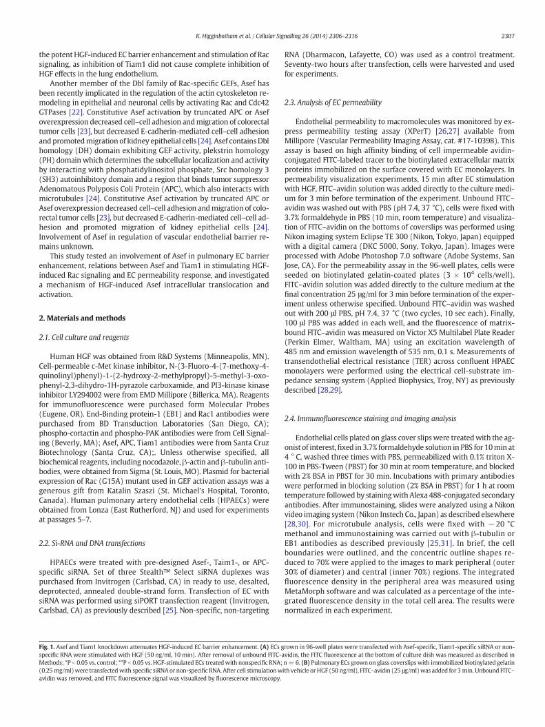
Fig. 2. Role of Asef and Tiam1 in HGF-induced activation of Rac. (A) Pulmonary ECs were transfected with Asef-specific, Tiam1-specific siRNA, or their combination, or with non-specificRNAand stimulatedwithHGF (50 ng/ml, 5 min). Rac activationwas determinedbyRac-GTP pulldown assay. Content of activated Racwas normalized to the total Rac content in EC lysates.siRNA-induced target protein depletion was verified by western blot analysis. Bar graphs represent quantitative densitometry of western blot experiments. *P b 0.05 vs. nsRNA; n = 3.(B) Cells were preincubated with vehicle, c-Met inhibitor (carboxamide 50 nM, 30 min) or PI3-kinase inhibitor (LY294002 20 μM, 30 min) followed by stimulation with HGF (5 min).Asef and Tiam1 activation was determined by pulldown assay with immobilized RacG15A and evaluated by increased GEF association with RacG15A. Content of activated Asef orTiam1 was normalized to the total GEF protein content in EC lysates. Bar graphs represent quantitative densitometry of western blot experiments. *P b 0.05 vs. HGF treatment withoutinhibitors; n = 3.
2309K. Higginbotham et al. / Cellular Signalling 26 (2014) 2306–2316
2.5. Co-immunoprecipitation, subcellularfractionation and immunoblotting
After agonist stimulation, cells were washed in cold phosphate buff-ered saline (PBS) and lysed on ice with cold TBS-NP40 lysis buffer(20 mM Tris pH 7.4, 150 mM NaCl, 1% NP40) supplemented with pro-tease and phosphatase inhibitor cocktails (Roche, Indianapolis, IN).Clarified lysates were then incubated with antibodies to APC or Asefovernight at 4 °C, washed 3–4 times with TBS-NP40 lysis buffer, andthe complexes were analyzed by Western blotting using appropriateantibodies. In fractionation studies, cytosolic (soluble) and membrane/cytoskeletal (particulate) fractions were isolated as described previous-ly [32,33]. For analysis of protein phosphorylation profile, cells were
stimulated, then lysed, and protein extracts were separated by SDS-PAGE, transferred to polyvinylidene fluoride membrane, and probedwith specific antibodies. Equal protein loading was verified by re-probing membranes with antibody to β-actin or specific protein ofinterest.
2.6. Rac and GEF activation assays
Rac activation was evaluated in pulldown assays using agarosebeads with immobilized PAK1-PBD [28]. In brief, after stimulation, celllysates were collected, and GTP-bound Rac was captured using pull-down assays with immobilized PAK1-PBD agarose. The levels of activat-ed Rac as well as total Rac content were evaluated by western blot

Fig. 3. HGF effects on APC, Asef and Tiam1 association with MT fraction and APC-Asef interactions. (A) ECs were stimulated with HGF (50 ng/ml) followed by isolation of MT-enrichedfractionation. APC, Asef and Tiam1 were detected by western blot of MT fractions and normalized to tubulin content. Bar graph represents quantitative densitometry of western blotexperiments. *P b 0.05; n = 3. (B) ECs were treated with nonspecific and APC-specific siRNA, and HGF-induced accumulation of Asef in MT fraction was assessed. Lower panels showWestern blot detection of total Asef and APC protein levels in total cell lysates. Bar graphs represent quantitative densitometry of western blot data. *P b 0.05 vs. nsRNA; n = 3. (C andD) ECs pretreated with vehicle, c-Met inhibitor or PI3-kinase inhibitor were stimulated with HGF (50 ng/ml), and APC (C) and Asef (D) proteins were immunoprecipitated under non-denaturing conditions using appropriate antibody. Presence of APC, Asef and Tiam1 in immune complexes was tested by western blot. Results are representative of three to sixindependent experiments.
2310 K. Higginbotham et al. / Cellular Signalling 26 (2014) 2306–2316
analysis. Active Asef or Tiam1 was affinity precipitated from cell lysatesaccording to previously described protocol [34] using the Rac (G15A)mutant kindly provided by K. Szaszi (St. Michael’s Hospital, Toronto,
Canada). This mutant cannot bind nucleotide and therefore has high af-finity for activated GEFs [35]. Activated Asef and Tiam1 in Rac (G15A)pulldowns were detected by Western blotting and normalized to total

Fig. 4. Dose-dependent effects of nocodazole on EC permeability and microtubule arrangement. (A) ECs were treated with the indicated concentrations of nocodazole, and changes inEC permeability were monitored by TER measurements. Shown are representative data from three independent measurements. (B) Immunofluorescence staining of microtubulecytoskeleton in EC treated with various nocodazole concentrations was performed using β-tubulin antibody.
2311K. Higginbotham et al. / Cellular Signalling 26 (2014) 2306–2316
Asef or Tiam1 in cell lysates for each sample. Precipitationwith glutathi-one–Sepharose beads containing no fusion proteins resulted in no gua-nine nucleotide exchange proteins precipitation.
2.7. Statistical analysis
Results are expressed as means ± SD of three to six independentexperiments. Stimulated samples were compared to controls byunpaired Student’s t-tests. For multiple-group comparisons, a one-wayvariance analysis (ANOVA), followed by the post hoc Fisher’s test, wasused. P b 0.05 was considered statistically significant.
3. Results
3.1. HGF-induced activation of Asef and Tiam1 mediates barrier protectiveeffects on pulmonary EC
Involvement of Asef and Tiam1 in HGF-induced EC barrier enhance-mentwas first tested in experiments withmeasurements of permeabil-ity for macromolecules. Knockdown of Asef, Tiam1 or both proteins inhuman lung EC was performed using siRNA approach. Fresh EC mono-layers with nearly established confluence were used to better illustratebarrier enhancing effects of HGF treatment and effects of protein knock-down. Bar graph (Fig. 1A) represents quantitative analysis of EC per-meability changes by measurements of fluorescence of accumulatedFITC–avidin in 96-well plates using microplate reader, as described inMethods. Single knockdown of Tiam1 or Asef partially attenuatedHGF-induced EC barrier enhancement indicated by increased binding
Fig. 5 Effect of low dose nocodazole on HGF-induced EC barrier enhancement and microtubul(0.05 nM, marked by first arrow). At the time point indicated by second arrow, cells were stimdata of four independent experiments. (B) ECs grown stimulated with HGF with or without prEB1 antibody. Insets show high magnification images of cell peripheral areas with EB1-positivfixed HPAEC monolayers; *P b 0.05, n = 6.
of FITC–avidin to biotinylated substrate under EC monolayers. Thedouble knockdown of Asef and Tiam1 exhibited additive effect on sup-pression of HGF-induced EC barrier enhancement.
Visualization of paracellular permeabilitywas performed by fluores-cence microscopy of control and stimulated EC grown on glass cover-slips after quick incubation with FITC–avidin permeability tracer(Fig. 1B). In control conditions, low levels of basal accumulation ofFITC-labeled tracer were observed at sites underlying the cell–cell junc-tion area and reflecting basal levels of paracellular permeability. HGFsignificantly decreased the basal EC monolayer permeability for FITC-labelled avidin. Barrier enhancing effect of HGFwas partially attenuatedby single knockdown of Asef or Tiam1 andwas completely abrogated inEC monolayers with double Asef/IQGAP1 knockdown.
3.2. HGF-induced activation of Rac is mediated by Asef and Tiam1.
Single Asef or Tiam1 knockdown partially attenuated HGF-inducedRac activation evaluated by Rac-GTP pulldown assays. Rac activationwas completely inhibited by double knockdown of Asef and Tiam1(Fig. 2A, left panel). siRNA-induced taget protein knockdown was con-firmed bywestern blot analysis of EC lysates (Fig. 2A, right panel). HGFactivated Asef and Tiam1 guanine nucleotide exchange activities to-wards Rac evaluated by pulldown of activated GEFs on Rac(G15A)beads. These effects were abrogated by the cell pretreatmentwith phar-macologic inhibitors of HGF receptor c-Met and PI3-kinase (Fig. 2B).These results demonstrate dual regulation of HGF-induced Rac activityby Asef and Tiam1.
e peripheral growth. (A) Human pulmonary ECs were treated with low dose nocodazoleulated with HGF (50 ng/ml), and TER was monitored over 2 h. Shown are representativeetreatment with low dose nocodazole (0.05 nM) followed by immunostaining with anti-e microtubule tips. Bar graph depicts quantitative analysis of peripheral EB1 in methanol-

Fig. 6. Effect of low dose nocodazole pretreatment on HGF-induced Asef, Tiam1 and APC peripheral translocation. Human pulmonary ECs pretreated with low dose nocodazole (0.05 nM,15 min) were stimulated with HGF (50 ng/ml). (A) Asef, Tiam1 and APC accumulation in membrane/cytoskeletal fraction was monitored by western blot. The content of examinedproteins in corresponding total cell lysates was used as a normalization control. Bar graph represents quantitative densitometry of western blot experiments. *P b 0.05 vs. HGF withoutnocodazole treatment; n = 4. (B) Intracellular redistribution of endogenous Asef an Tiam1 in HGF-stimulated endothelial cells was examined by immunofluorescence staining withappropriate antibody. Shown are representative results of three to five independent experiments.
2313K. Higginbotham et al. / Cellular Signalling 26 (2014) 2306–2316

Fig. 7. Pretreatment with low dose nocodazole suppresses HGF-induced activation of Rac pathway and EC barrier enhancement. Human pulmonary ECs pretreated with low dosenocodazole (0.05 nM, 15min)were stimulatedwith HGF (50 ng/ml). (A) Rac activationwas assessed by pulldown of Rac-GTP using PAK-PBD beads. (B) Rac-dependent PAK and cortactinphosphorylation was assessed by western blot analysis of total cell lysates. (C and D) EC permeability was evaluated by XPerT permeability assay described in Methods. After unboundFITC–avidin was removed, the FITC fluorescence signal was visualized by fluorescence microscopy. The bar graph shows quantitative analysis of EC permeability of EC monolayersgrown in 96-well plates; *P b 0.05 vs. HGFwithout nocodazole treatment; n= 6 (C). Permeability changes weremonitored in pulmonary ECs grown on glass coverslipswith immobilizedbiotinylated gelatin (0.25 mg/ml) (D).
2314 K. Higginbotham et al. / Cellular Signalling 26 (2014) 2306–2316
3.3. HGF induces selective recruitment of Asef to microtubules andassociation with APC
Asef interaction with microtubule-associated protein adenomatouspolyposis coli (APC) is required for its activation and guanine nucleotide
exchange activity towards Rac [22]. Analysis of potential Asef and Tiam1recruitment to microtubules was examined in experiments with isola-tion of microtubule enriched fraction. HGF stimulation increased thelevels of Asef and APC in EC microtubule fraction (Fig. 3A). By contrast,the basal levels of Tiam1 detected in MT fraction under non-stimulated

2315K. Higginbotham et al. / Cellular Signalling 26 (2014) 2306–2316
conditions were not affected by HGF treatment. siRNA-induced knock-down of APC abolishedHGF-induced recruitment of Asef tomicrotubulefraction suggesting APC-facilitated mechanism of Asef association withmicrotubules (Fig. 3B). Analysis of total cell lysates showed that APCknockdown did not affect total Asef levels in pulmonary EC. APC–Asefassociation was further tested in reciprocal co-immunoprecipitation as-says using APC or Asef antibody. HGF stimulated interaction betweenAsef and APC, which was abolished by cell pretreatment with c-Metand PI3-kinase inhibitors (Fig. 3CD). These data suggest that in additionto activation of nucleotide exchange activity, c-Met–PI3-kinase signal-ing is also important for Asef interaction with APC.
3.4. Inhibition of peripheral microtubule dynamics attenuates HGF-inducedbarrier enhancement.
Nocodazole (ND) is an MT depolymerizing agent capable of com-plete dissolution of microtubule network when used at high concentra-tion. However, at low concentrations nocodazole has been shown tohave no effect on gross MT cytoskeleton structure, but slow down pe-ripheral MT dynamics [36]. In the test experiments we determined theND concentration which had no considerable effect on basal EC perme-ability evaluated by measurements of transendothelial electrical resis-tance (TER) (Fig. 4A) and microtubule arrangement under basalconditions (Fig. 4B). At this low concentration (0.05 nM), NDattenuatedHGF-induced barrier enhancementmonitored by TERmeasurements inEC monolayers (Fig. 5A). We evaluated effects of HGF and low dose NDpretreatment on peripheral MT density. ECs after HGF stimulation werefixed with methanol and subjected to immunofluorescence stainingwith antibody to End-Bindingprotein-1 (EB1)which tracks the growingplus end ofmicrotubules. Analysis of EB1 staining showed higher densi-ty of EB1-positive MT tips at the distal area of HGF-stimulated cells, asshown in Fig. 5B and higher magnification insets. Pretreatment withlow doseND did not significantly affect the EB1-positiveMT tips densityin control cells, but abolished the HGF effects on peripheral MT expan-sion. Bar graph presents the quantitative analysis of immunofluores-cence data.
3.5. Differential role of peripheral microtubule dynamics in HGF-inducedtranslocation of APC/Asef and Tiam1
Effects of MT peripheral dynamics on APC, Asef and Tiam1 recruit-ment to cell cortical compartment were examined using treatmentwith low ND dose, as described above. Subcellular fractionation experi-ments showed that low dose ND treatment abolished accumulation ofAPC and Asef in the membrane/cytoskeletal fraction. Of note, low doseND treatment did not affect the HGF-induced membrane recruitmentof Tiam1 (Fig. 6A). Bar graphs represent quantitative analysis of Asef,APC and Tiam1 translocation in HGF-stimulated EC with and withoutND pretreatment. Immunofluorescence analysis of intracellular Asefand Tiam1 localization shows increased accumulation of these proteinsat the submembrane area at the cell periphery of HGF-stimulated EC.Pretreatment with low dose ND abolished the HGF-induced peripheralaccumulation of Asef but did not affect accumulation of Tiam1 (Fig. 6B).
3.6. Inhibition of peripheral microtubule dynamics attenuates HGF-inducedactivation of Rac signaling and enhancement of EC barrier
EC pretreatment with low dose ND attenuated HGF-induced activa-tion of Rac (Fig. 7A) and phosphorylation of Rac effector PAK1 and reg-ulator of actin remodeling, cortactin (Fig. 7B). This parameter reflectsactivation of Rac signaling pathway, as phosphorylation of cortactin atY421 and Y466 in response to Rac1 activation resulted in localization ofphosphorylated cortactin with F-actin in lamellipodia and podosomes[37] and regulated actin dynamics. In turn, decreased cortactin tyrosinephosphorylation was observed in cells with inhibited Rac1 and wasassociated with inhibition of cortactin peripheral localization [38].
Effects of low ND dose on the HGF-induced EC barrier enhancementwere further tested using visualization of local areas in the EC mono-layers with increased EC permeability for macromolecules. Bar graphdepicts results of quantitative analysis of permeability data (Fig. 7C).HGF significantly decreased the basal EC monolayer permeability forFITC-labeled avidin. Barrier enhancing effect of HGF was attenuated inEC monolayers pretreated with low ND dose. Visualization of the sitesof increased permeability showed that basal levels of FITC-labeled traceraccumulation at sites underlying the cell–cell junction area observed incontrol nonstimulated EC monolayers were reduced in HGF-treated EC,and HGF effect was attenuated by low dose ND pretreatment (Fig. 7D).
4. Discussion
The main finding of this study is a demonstration of a novel role ofAsef in the HGF-induced enhancement of endothelial barrier. Our dataalso show different routes of HGF-induced Tiam1 andAsef translocationto cell periphery. While HGF-induced PI3-kinase activation was essen-tial for activity of both GEFs, the HGF-induced Tiam1 activation and pe-ripheral translocation were independent on microtubule dynamics. Incontrast, HGF-induced microtubule peripheral growth was requiredfor APC-assisted translocation of Asef to cell periphery, full activationof Rac signaling and EC barrier enhancement response. HGF increasedassociation of Asef and APC with MT fraction and stimulated formationof Asef–APC complex detected by reciprocal co-immunoprecipitationassays. APC forms a complexwith EB1 atmicrotubule plus-ends and co-localizes at cell junctionswith adherens junction protein β-catenin [39].On the other hand, APC binding to Asef via its armadillo repeat domainenhances its GEF activity [40]. These features suggest a potential role forAPC in regulation of basal endothelial barrier and re-establishment ofmonolayer integrity via agonist-induced interactions with Asef andstimulation of Asef nucleotide exchange activity.
In polarized fibroblasts, APC accumulation was also observed atthe actin-rich lamellopodia of migrating cells [41]. Importantly, themost frequent APC localization occurs at the tips of microtubule-dependent cellular protrusions which appear to be the areas with ac-tivated actin dynamics and migration activity in this direction [42].These published data support the proposed role for Asef–APC com-plex in the microtubule-guided local regulation of Rac activity ob-served in our study.
The accumulation of APC at EC periphery observed in this studymaybe alsomediated through direct interactions of APC with the Rac/Cdc42effector protein, IQGAP1 [41]. Our unpublished data show presence ofIQGAP1 in the Asef co-immunoprecipitates from HGF-stimulated EC.These observations support our hypothesis that HGF-induced EC barrierenhancement ismediated by local Rac signaling viaMT-APC assistedde-livery and IQGAP1 peripheral capturing of Asef leading to its activationand local stimulation of Rac siganling.
This study used EC treatment with low dose nocodazole to furtherdissect a role of peripheral MT polymerization/depolymerization dy-namics in HGF-induced signaling by Asef and Tiam1. We determinedthe nocodazole concentration which, similarly to effects previously de-scribed in immortalized BSC-1 cell line [36], did not cause global chang-es in the pool of polymerized tubulin or MT network structure, butattenuated peripheral MT polymerization/depolymerization dynamicsin pulmonary EC. At this concentration, nocodazole inhibited HGF-induced peripheral MT expansion and significantly attenuated HGF-induced peripheral accumulation of Asef without affecting peripheraltranslocation of Tiam1. “Freezing” of peripheral microtubule dynamicspartially attenuated HGF-induced activation of Rac signaling and de-creased HGF-induced barrier enhancement in EC monolayers whichwas detected by XPerT permeability assay. These results further supporta novel role for Asef in local regulation of Rac signaling at cell peripheryvia cooperation with microtubule-dependent mechanisms.
In summary, the results of this study provide a new insight into bar-rier enhancing mechanisms stimulated by HGF and demonstrate an

2316 K. Higginbotham et al. / Cellular Signalling 26 (2014) 2306–2316
alternative mechanism of Rac stimulation via APC- and MT-mediatedperipheral delivery and activation of Asef which acts in concert withPI3K-Tiam1-dependent axis of Rac signaling.
Aknowledgements
This work was supported by the grants from National Heart, Lung,and Blood Institutes (HL89257 and HL107920).
References
[1] N. Mochizuki, Circ. J. 73 (12) (2009) 2183–2191.[2] E. Dejana, E. Tournier-Lasserve, B.M. Weinstein, Dev. Cell 16 (2) (2009) 209–221.[3] C.M. Beckers, V.W. van Hinsbergh, G.P. van Nieuw Amerongen, Thromb. Haemost.
103 (1) (2010) 40–55.[4] C. Tiruppathi, R.D. Minshall, B.C. Paria, S.M. Vogel, A.B. Malik, Vasc. Pharmacol. 39
(4–5) (2002) 173–185.[5] T. Hirase, K. Node, Am. J. Physiol. Heart Circ. Physiol. 302 (3) (2012) H499–H505.[6] K.G. Birukov, N. Zebda, A.A. Birukova, Compr. Physiol. 3 (1) (2013) 429–484.[7] K. Matsumoto, T. Nakamura, J. Biochem. (Tokyo) 119 (4) (1996) 591–600.[8] P.A. Singleton, R. Salgia, L. Moreno-Vinasco, J. Moitra, S. Sammani, T. Mirzapoiazova,
J.G. Garcia, J. Biol. Chem. 282 (42) (2007) 30643–30657.[9] A.A. Birukova, E. Alekseeva, A. Mikaelyan, K.G. Birukov, FASEB J. 21 (11) (2007)
2776–2786.[10] F. Liu, K.L. Schaphorst, A.D. Verin, K. Jacobs, A. Birukova, R.M. Day, N. Bogatcheva, D.P.
Bottaro, J.G. Garcia, FASEB J. 16 (9) (2002) 950–962.[11] I. Date, N. Takagi, K. Takagi, T. Kago, K. Matsumoto, T. Nakamura, S. Takeo, Biochem.
Biophys. Res. Commun. 319 (4) (2004) 1152–1158.[12] H.T. Chen, H.K. Tsou, C.H. Chang, PLoS ONE 7 (6) (2012) e38378.[13] A. Cipres, Y.A. Abassi, K. Vuori, Cell. Signal. 19 (8) (2007) 1662–1670.[14] Y. Zheng, Trends Biochem. Sci. 26 (12) (2001) 724–732.[15] H.C. Welch, W.J. Coadwell, L.R. Stephens, P.T. Hawkins, FEBS Lett. 546 (1) (2003)
93–97.[16] W.T. Arthur, L.A. Quilliam, J.A. Cooper, J. Cell Biol. 167 (1) (2004) 111–122.[17] K.L. O'Connor, A.M. Mercurio, J. Biol. Chem. 276 (51) (2001) 47895–47900.[18] J.M. Servitja, M.J. Marinissen, A. Sodhi, X.R. Bustelo, J.S. Gutkind, J. Biol. Chem. 278
(36) (2003) 34339–34346.[19] P.A. Singleton, S.M. Dudek, E.T. Chiang, J.G. Garcia, Faseb J. 19 (12) (2005)
1646–1656.
[20] E. Gonzalez, R. Kou, T. Michel, J. Biol. Chem. 281 (6) (2006) 3210–3216.[21] P.A. Singleton, S. Chatchavalvanich, P. Fu, J. Xing, A.A. Birukova, J.A. Fortune, A.M.
Klibanov, J.G. Garcia, K.G. Birukov, Circ. Res. 104 (8) (2009) 978–986.[22] Y. Kawasaki, T. Senda, T. Ishidate, R. Koyama, T. Morishita, Y. Iwayama, O. Higuchi, T.
Akiyama, Science 289 (5482) (2000) 1194–1197.[23] Y. Kawasaki, S. Furukawa, R. Sato, T. Akiyama, Cancer Sci. 104 (8) (2013)
1135–1138.[24] Y. Kawasaki, R. Sato, T. Akiyama, Nat. Cell Biol. 5 (3) (2003) 211–215.[25] X. Tian, Y. Tian, N. Sarich, T. Wu, A.A. Birukova, Faseb J. 26 (9) (2012) 3862–3874.[26] O. Dubrovskyi, A.A. Birukova, K.G. Birukov, Lab. Invest. 93 (2013) 254–263.[27] X. Tian, Y. Tian, G. Gawlak, N. Sarich, T. Wu, A.A. Birukova, J. Biol. Chem. 289 (8)
(2014) 5168–5183.[28] K.G. Birukov, V.N. Bochkov, A.A. Birukova, K. Kawkitinarong, A. Rios, A. Leitner, A.D.
Verin, G.M. Bokoch, N. Leitinger, J.G. Garcia, Circ. Res. 95 (9) (2004) 892–901.[29] A.A. Birukova, K.G. Birukov, K. Smurova, D.M. Adyshev, K. Kaibuchi, I. Alieva, J.G.
Garcia, A.D. Verin, FASEB J. 18 (15) (2004) 1879–1890.[30] A.A. Birukova, K. Smurova, K.G. Birukov, K. Kaibuchi, J.G.N. Garcia, A.D. Verin,
Microvasc. Res. 67 (1) (2004) 64–77.[31] Y. Komarova, C.O. De Groot, I. Grigoriev, S.M. Gouveia, E.L. Munteanu, J.M. Schober, S.
Honnappa, R.M. Buey, C.C. Hoogenraad, M. Dogterom, G.G. Borisy, M.O. Steinmetz, A.Akhmanova, J. Cell Biol. 184 (5) (2009) 691–706.
[32] A.A. Birukova, T. Zagranichnaya, E. Alekseeva, P. Fu, W. Chen, J.R. Jacobson, K.G.Birukov, Exp. Cell Res. 313 (11) (2007) 2504–2520.
[33] A.A. Birukova, I. Malyukova, V. Poroyko, K.G. Birukov, Am. J. Physiol. Lung Cell. Mol.Physiol. 293 (1) (2007) L199–L211.
[34] F. Waheed, Q. Dan, Y. Amoozadeh, Y. Zhang, S. Tanimura, P. Speight, A. Kapus, K.Szaszi, Mol. Biol. Cell 24 (7) (2013) 1068–1082.
[35] R. Garcia-Mata, K.Wennerberg,W.T. Arthur, N.K. Noren, S.M. Ellerbroek, K. Burridge,Methods Enzymol. 406 (2006) 425–437.
[36] R.J. Vasquez, B. Howell, A.M. Yvon, P. Wadsworth, L. Cassimeris, Mol. Biol. Cell 8 (6)(1997) 973–985.
[37] J.A. Head, D. Jiang, M. Li, L.J. Zorn, E.M. Schaefer, J.T. Parsons, S.A.Weed, Mol. Biol. Cell14 (8) (2003) 3216–3229.
[38] A.A. Birukova, Y. Tian, O. Dubrovskyi, N. Zebda, N. Sarich, X. Tian, Y. Wang, K.G.Birukov, J. Cell. Physiol. 227 (10) (2012) 3405–3416.
[39] M. Sharma, L. Leung, M. Brocardo, J. Henderson, C. Flegg, B.R. Henderson, J. Biol.Chem. 281 (25) (2006) 17140–17149.
[40] T. Akiyama, Y. Kawasaki, Oncogene 25 (57) (2006) 7538–7544.[41] T. Watanabe, S. Wang, J. Noritake, K. Sato, M. Fukata, M. Takefuji, M. Nakagawa, N.
Izumi, T. Akiyama, K. Kaibuchi, Dev. Cell 7 (6) (2004) 871–883.[42] I.S. Nathke, C.L. Adams, P. Polakis, J.H. Sellin, W.J. Nelson, J. Cell Biol. 134 (1) (1996)
165–179.