Embed Size (px)
Citation preview

— 1 —
名古屋大学博物館報告
Bull. Nagoya Univ. MuseumNo. 19, 1–8, 2003
Immunocytochemical and Ultrastructural Analysis of Opsin- andVasoactive Intestinal Peptide (VIP)- like Immunoreactive
Neurons in the Lateral Septum of the Pigeon
HIRUNAGI Kanjun
The Nagoya University Museum, Chikusa-ku Nagoya, 464-8601, Japan
Summary
CSF-contacting neurons of the lateral septum are considered as a putative deep brainphotoreceptor in the avian brain. By means of immunocytochemistry using antibodies to a visualpigment (RET-P1, a monoclonal antibody against opsin, Barnstable, 1980) and vasoactive intestinalpeptide (VIP), I demonstrated clusters of immunoreactive small neurons in the lateral septum ofthe pigeon. Opsin-like immunoreactive neurons and VIP-like immunoreactive neurons have similarmorphological features. Their perikarya accumulate in the subependymal regions of the ventromedialwalls of both lateral ventricles. The labelled neurons are multipolar or bipolar with pyriform orspindle-shaped cell bodies. Immunoreactive CSF-contacting neurons contact the CSF via a processthat penetrates the ependyma and terminates in a single knob-like swelling. Immunoreactive fibersoriginating in the group of cell bodies seem to give rise to dense terminal-like structures in theseptal area. Immuno-electron-microscopic investigations of these neurons revealed an accumulationof VIP- and opsin immunoreactive dense-core vesicles (100-150 nm in diameter) in ventricularterminals, perikarya and neuronal terminal-like structure with VIP- and RET-P1-immunolabellingrespectively. Based on these evidence it seems clear that VIP-and opsin-like immunoreactive neuronsof this study are the same as the neurons that express both opsin- and VIP-like immunoreactivityin the ateral septum of the ring dove (Silver et al., 1988). In this study double immunolabellingusing VIP and RET-P1 antibodies shows the coexistence of VIP and opsin in the same dense vesicles.
Introduction
Immunohistochemical studies reveal small neurons with an accumulation of VIP-like immu-noreactive (ir) in the lateral septum in avian brains (Hirunagi et al., 1995; Hof et al., 1991; Korfand Fahrenkrug, 1984; Kuenzel and Blähser, 1994; Silver et al., 1988; Yamada et al., 1982), some ofwhich are cerebrospinal fluid (CSF)-contacting. Recently, this type of small neuron has been con-sidered as a candidate for the deep encephalic photoreceptor of avian and reptilian brains. First,Silver et al. demonstrated opsin-like immunoreactivity in this type of neurons of the lateral sep-tum of birds using an opsin antibody, RET-P1, raised against a membrane fraction of rat retina(Barnstable, 1980; Silver et al., 1988). In that study, they established that some VIP-like ir neuronsof the lateral septum co-express RET-P1 immunoreactivity with an immuno-fluorescent doublelabelling method. More recently, opsin-like immunoreactivity has been reported in similar CSF-contacting neurons of the identical region of the lateral septum of the lizard Anolis carolinensis(Foster et al., 1993). Furthermore, VIP-like ir CSF-contacting neurons have been demonstrated inthe same region of the lateral septum of several reptilian species (Hirunagi et al., 1993). Withregard to the subcellular localization of VIP-like antigen, Hirunagi et al. showed that VIP-like irCSF-contacting neurons have VIP-like ir dense vesicles (approximately 100-150 nm diameter) in

— 2 —
the perikarya with dendritic processes and axon terminals of the duck (Hirunagi et al., 1995). Thisresult suggests that VIP-like ir neurons contain VIP-like neuropeptide in these electron-densevesicles. However, until now, the ultrastructural localization of opsin-like antigen has remainedelusive. In the present study we demonstrate the ultrastructural localization of opsin- and VIP-like antigens in the neurons of the lateral septum of the pigeon. I used single labelling immunocy-tochemistry for RET-P1 and VIP and a dual-labelled method using both ABC labelling andimmunogold-silver labelling at the electron microscopic level.
Material and Methods
Male and female adult pigeons were perfused transcardially with 0.75% NaCl followed by amixture of 4% paraformaldehyde and 0.1% glutaraldehyde in 0.1M phosphate buffer at pH 7.4.Brains were rapidly removed and a coronal block containing the lateral septum area post-fixed forovernight. Vibratome sections (50 µm thick) were cut and treated for the demonstration of neu-rons with RET-P1 and VIP immunoreactivities as described below. (1) Single label immunostainingfor VIP; Sections were incubated in polyclonal porcine VIP antibody (1:1000, Cambridge ResearchBiochemicals, UK) for 72 hours at 4°C. For visualizing of immunoreaction, a PAP method wasused. The details of immunostaining for VIP and the specifity of VIP antibody was describedpreviously (Hirunagi et al., 1993; Hirunagi et al., 1995). (2) Single label immunostaining for RET-P1; Sections were incubated in monoclonal opsin antibody (RET-P1) (1: 20000) diluted in 0.1MPBScontaining 1% BSA and 0.3% Triton X-100 for 72 hours at 4°C. The sites of the antibody-antigenbinding were visualized with an avidin-biotin-peroxidase (ABC) procedure (Elite ABC kit, VectorLabs, Burlingame, CA) according to the protocol. Other sections were incubated in monoclonalopsin antibody (RET-P1) (1: 20000) for 72 hours at 4°C. Sections were then incubated in goat anti-mouse IgG bound to 1nm colloidal gold (AuroProbe One, Amersham, UK) diluted 1:50 in 0.1%gelatin PBS, BSA. Sections were fixed in 2% glutaraldehyde for 10 minutes. Silver intensificationof gold was processed by the light-insensitive IntenSE-M Kit (Amersham, UK) following themanufacture’s protocol. (3) Dual label immunostaining for VIP and RET-P1; Following incubationwith RET-P1 as the first primary antisera, immunoreactivity was visualized with an ABC proce-dure as shown in (2). Sections were incubated in polyclonal porcine VIP antibody (1:1000, Cam-bridge Research Biochemicals, UK) as second primary antisera. Sections were then incubated ingoat anti-rabbit IgG bound to 1 nm colloidal gold (AuroProbe One, Amersham, UK) diluted 1:50 in0.1% gelatin PBS, BSA. Sections were fixed in 2% glutaraldehyde for 10 minutes. Silver intensifi-cation of gold was processed by the light-insensitive IntenSE-M kit (Amersham, UK), as above.For electron microscopy immunostained sections were osmicated and flat-embedded in Araldite.The details of these procedures have been described previously. Following contrast staining withuranyl acetate and lead citrate, ultra-thin sections were examined with a JEOL JEM 1210 elec-tron microscope.
Results
Using polyclonal antibody against VIP and monoclonal antibody, RET-P1, against rat opsin, anaccumulation of immunorective neurons is found in a highly circumscribed region of the lateralseptum /nucleus accumbens with each label. Light- and electron-microscopic morphological fea-tures of VIP-like and opsin-like ir neurons are identical in the lateral septum of the pigeon. Weshow results of opsin-like and VIP-like ir neurons and double labelling with RET-P1 and VIP inFig. 1, 2 respectively. Opsin-like ir perikarya accumulates in the subependymal regions of theventromedial walls of both lateral ventricles at the level of the pars medialis of the lateral septal

— 3 —
Fig 1. a. Accumulation of opsin-ir neurons in the lateral septum Arrows: Immunoreactive neuronsArrowheads: Basal processes V: Lateral ventricle Bar: 100 µm b. Opsin ir CSF-contacting neuronsin the lateral septum Arrows: ventricular processes of opsin ir CSF-contacting neurons B: Basalprocess P: Ventricular process S: cell body V: Lateral ventricle Bar: 20 µm c. Opsin ir densevesicles in immunoreactive neuron Arrows: immunoreactive vesicles E: Endoplasmic reticulumN: Nucleus Bar: 1 µm d. Opsin ir vesicles identified by 1 nm immunogold-silver enhancedmethod Arrowheads: Immunoreactive dense vesicles M: Mitochondria Bar: 1 µm e. Opsin irventricular process of the CSF-contacting neuron in the lateral septum C: Cilium in lateralventricle Bar: 1 µm f. Opsin ir neuronal terminal near small neuron in the lateral septum M:Mitochondria N: nucleus of immunonegative neuron T: Opsin ir neuronal terminal Bar: 1 µm

— 4 —
organ (Kuenzel and Blähser, 1994) (Fig. 1a). The labelled neurons are multipolar or bipolar withpyriform or spindle-shaped cell bodies. Some of these are CSF-contacting neurons, with a processthat penetrates the ependyma and terminates in a single knob-like swelling (Fig. 1b). In additionto the cluster of cell bodies, ir fibers originating in the group of cell bodies course into deeper layersof the septal area. Some of them appear to be beaded axons (Fig. 1a). Using pre-embedding meth-ods of ABC and 1 nm immunogold, electron- microscopic studies reveal that opsin-like neuronshave an accumulations of dense-core vesicles with diameter of 100-150 nm in their perikarya andventricular terminal. An ABC labelling demonstrates RET-P1-labelled dense vesicles near the en-doplasmic reticulum in the cytoplasm (Fig. 1c) and in the ventricular terminal (Fig. 1e). With a1nm immunogold silver labelling, silver-enhanced gold particles, which indicate the site of RET-P1antigen, are frequently associated with dense vesicles (Fig. 1d). In the lateral septum, terminalformations with VIP or RET-P1 immunoreactivity are distributed. VIP-and RET-P1-labelled termi-nals lie in the same regions. Most prominent immunoreactive terminal formations are observed inthe area of the nucleus septi lateralis. In this area opsin-like ir terminals form dense networks.They appeared as intensely immunoreactive spots at light-microscopic observations as VIP immu-noreactive terminals that were reported our previous paper (Hirunagi et al., 1994). Electron-micro-scopic observations show that opsin-like ir terminals are observed near immunonegative smallneurons in the lateral septum (Fig. 1f). VIP immunocytochemistry shows an accumulation of VIP irneurons in the subependymal region of the lateral septum. This type of neurons project VIP-like ir
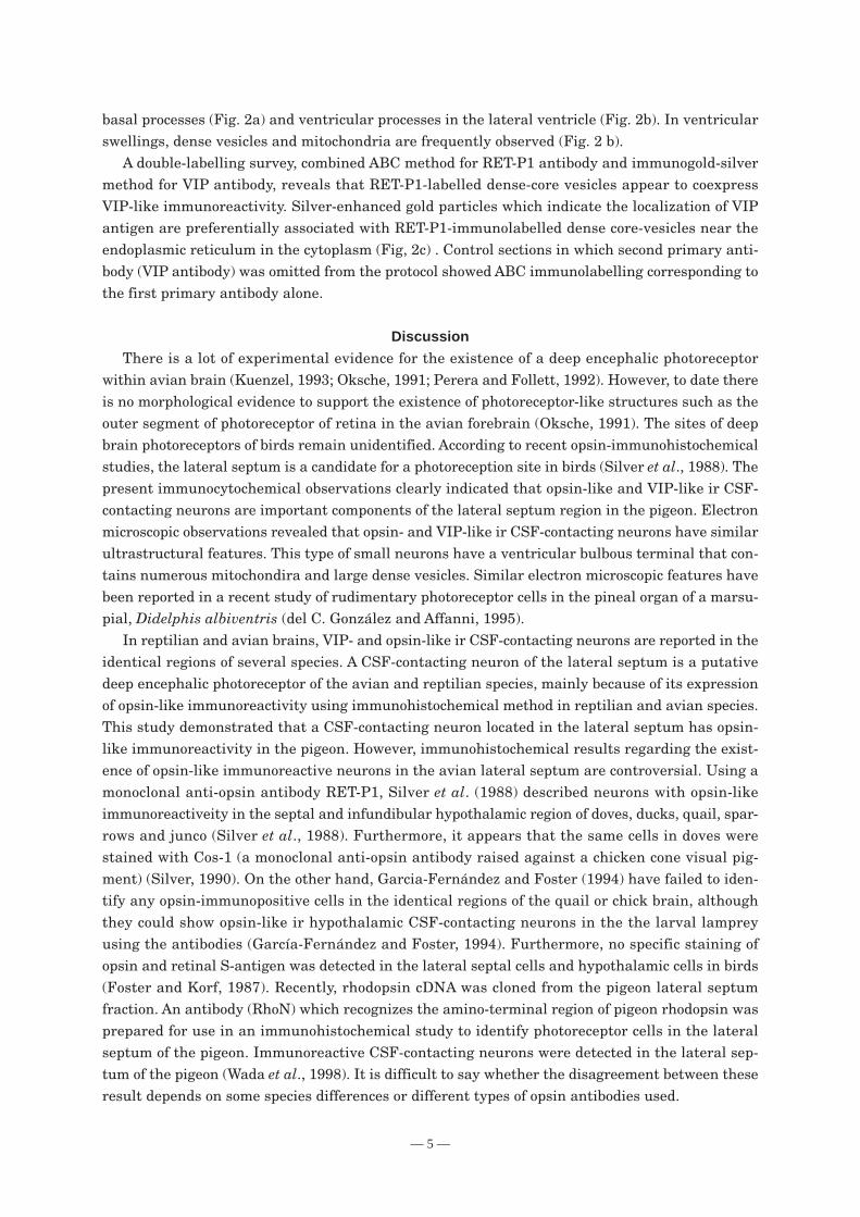
Fig. 2. a. VIP ir neuron in the lateral septum B:Basal process EP: Ependymal cell N:Nucleus of VIP ir neuron Bar: 2 µm b. VIPir ventricular process of the CSF-con-tacting neuron of the lateral septum EP:Ependymal cell M: Mitochondria in pro-cess V: Lateral ventricle Bar: 1 µm c.Double labelling of opsin and VIP anti-bodies Arrow indicates opsin ir densevesicle. Arrowheads indicate both opsin(dense vesicles) and VIP (silver-enhancedgold particles) immunoreactivity ongranules. ER: Endoplasmic reticulum Bar:1 µm
— 5 —
basal processes (Fig. 2a) and ventricular processes in the lateral ventricle (Fig. 2b). In ventricularswellings, dense vesicles and mitochondria are frequently observed (Fig. 2 b).
A double-labelling survey, combined ABC method for RET-P1 antibody and immunogold-silvermethod for VIP antibody, reveals that RET-P1-labelled dense-core vesicles appear to coexpressVIP-like immunoreactivity. Silver-enhanced gold particles which indicate the localization of VIPantigen are preferentially associated with RET-P1-immunolabelled dense core-vesicles near theendoplasmic reticulum in the cytoplasm (Fig, 2c) . Control sections in which second primary anti-body (VIP antibody) was omitted from the protocol showed ABC immunolabelling corresponding tothe first primary antibody alone.
Discussion
There is a lot of experimental evidence for the existence of a deep encephalic photoreceptorwithin avian brain (Kuenzel, 1993; Oksche, 1991; Perera and Follett, 1992). However, to date thereis no morphological evidence to support the existence of photoreceptor-like structures such as theouter segment of photoreceptor of retina in the avian forebrain (Oksche, 1991). The sites of deepbrain photoreceptors of birds remain unidentified. According to recent opsin-immunohistochemicalstudies, the lateral septum is a candidate for a photoreception site in birds (Silver et al., 1988). Thepresent immunocytochemical observations clearly indicated that opsin-like and VIP-like ir CSF-contacting neurons are important components of the lateral septum region in the pigeon. Electronmicroscopic observations revealed that opsin- and VIP-like ir CSF-contacting neurons have similarultrastructural features. This type of small neurons have a ventricular bulbous terminal that con-tains numerous mitochondira and large dense vesicles. Similar electron microscopic features havebeen reported in a recent study of rudimentary photoreceptor cells in the pineal organ of a marsu-pial, Didelphis albiventris (del C. González and Affanni, 1995).
In reptilian and avian brains, VIP- and opsin-like ir CSF-contacting neurons are reported in theidentical regions of several species. A CSF-contacting neuron of the lateral septum is a putativedeep encephalic photoreceptor of the avian and reptilian species, mainly because of its expressionof opsin-like immunoreactivity using immunohistochemical method in reptilian and avian species.This study demonstrated that a CSF-contacting neuron located in the lateral septum has opsin-like immunoreactivity in the pigeon. However, immunohistochemical results regarding the exist-ence of opsin-like immunoreactive neurons in the avian lateral septum are controversial. Using amonoclonal anti-opsin antibody RET-P1, Silver et al. (1988) described neurons with opsin-likeimmunoreactiveity in the septal and infundibular hypothalamic region of doves, ducks, quail, spar-rows and junco (Silver et al., 1988). Furthermore, it appears that the same cells in doves werestained with Cos-1 (a monoclonal anti-opsin antibody raised against a chicken cone visual pig-ment) (Silver, 1990). On the other hand, Garcia-Fernández and Foster (1994) have failed to iden-tify any opsin-immunopositive cells in the identical regions of the quail or chick brain, althoughthey could show opsin-like ir hypothalamic CSF-contacting neurons in the the larval lampreyusing the antibodies (García-Fernández and Foster, 1994). Furthermore, no specific staining ofopsin and retinal S-antigen was detected in the lateral septal cells and hypothalamic cells in birds(Foster and Korf, 1987). Recently, rhodopsin cDNA was cloned from the pigeon lateral septumfraction. An antibody (RhoN) which recognizes the amino-terminal region of pigeon rhodopsin wasprepared for use in an immunohistochemical study to identify photoreceptor cells in the lateralseptum of the pigeon. Immunoreactive CSF-contacting neurons were detected in the lateral sep-tum of the pigeon (Wada et al., 1998). It is difficult to say whether the disagreement between theseresult depends on some species differences or different types of opsin antibodies used.

— 6 —
RET-P1 ir cells of the lateral septum coexpress VIP-like immunoreactivity in the brains of ringdove, Japanese quail and duck (Silver et al., 1988). In the present study, we confirmed thecolocalization of RET-P1 and VIP in neurons of the lateral septum in the pigeon at the electronmicroscopic level using a dual labeling method. Our electron microscopic results suggested thatRET-P1 and VIP antigens colocalize in a dense core vesicle which is found in the perikarya andterminals. With regard to the subcelluar localization of VIP-like specific antigen, electron micro-scopic analysis has shown that VIP-like ir neurons of the lateral septum have an accumulation ofimmunostained large dense core vesicles (approximately 150 nm in diameter) in their cytoplasm(Hirunagi et al., 1995). Similar VIP immunoreactive core vesicles were reported in the terminalformations of the lateral septum of duck (Hirunagi et al., 1995) and pigeon (Hirunagi et al., 1994).About the subcellular localization of RET-P1 antigen, electron microscopy revealed the distribu-tion of RET-P1 antigen in the rat retina (Fekete and Barnstable, 1983). Immunoreactivity of RET-P1 antibody can be seen in the plasma membranes of both outer segments and inner segments ofrod photoreceptors. And a more diffuse reaction product was reported within the peripheral cyto-plasm of the inner segments. In this study, RET-P1 immunoreactivity has been observed in theentire neuronal elements including axon and dendritic processes of CSF-contacting neurons. Ven-tricular bulbus terminals of CSF-contacting neurons have RET-P1 immunoreactivity. However, nolamellar membrane structures, similar to those found in the outer segments of retinal photorecep-tors, have been identified electron-microscopically in the CSF-contacting neurons of lateral sep-tum of birds. Confirmation of these CSF-contacting neurons as photoreceptors awaits physiologi-cal evidence.
Acknowledgements
I would like to thank Drs. S. Ebihara and A. Adachi (Nagoya Univ.) and Dr. C. Blakemore for thegift of the antibody. I would also like to thank Dr. R. Silver (Columbia Univ.) for her kind commentsin this paper.
ReferencesBarnstable, C.J. (1980) Monoclonal antibodies which recognize different cell types in the rat retina, Nature,
286, 231-235.del C. González, M.M. and Affanni, J.M. (1995) Cells of photoreceptor line in the pineal organ of the adult
marsupial, Didelphis albiventris, Cell Tissue Res., 282, 363-366.Fekete, D.M. and Barnstable, C.J. (1983) The subcellular localization of rat photoreceptor-specific antigens,
J. Neurocytol., 12, 785-803.Foster, R.G., García-Fernández, J.M., Provencio, I. and DeGrip, W.J. (1993) Opsin localization and chromophore
retinoids identified within the basal brain of the lizard Anolis carolinensis, J. Comp. Physiol. A, 172, 33-45.
Foster, R.G. and Korf, H.-W. (1987) Immunocytochemical markers revealing retinal and pineal but nothypothalamic photoreceptor system in the Japanese quail, Cell Tissue Res., 248, 161-167.
García-Fernández, J.M. and Foster, R.G. (1994) Immunocytochemical identification of photoreceptor proteinsin hypothalamic cerebrospinal fluid-contacting neurons of the larval lamprey (Petromyzon marinaus),Cell Tissue Res., 275, 319-326.
Hirunagi, K., Kiyoshi, K., Adachi, A., Hasegawa, M., Ebihara, S. and Korf, H.-W. (1994) Electron-microscopicinvestigations of vasoactive intestinal peptide (VIP)-like immunoreactive terminal formations in the lateralseptum of the pigeon, Cell Tissue Res., 278, 415-418.
Hirunagi, K., Rommel, E. and Korf, H.-W. (1995) Ultrastructure of cerebrospinal fluid-contacting neuronsimmunoreactive to vasoactive intestinal peptide and properties of the blood-brain barrier in the lateralseptal organ of the duck, Cell Tissue Res., 279, 123-133.

— 7 —
Hirunagi, K., Rommel, E., Oksche, A. and Korf, H.-W. (1993) Vasoactive intestinal peptide-immunoreactivecerebrospinal fluid-contacting neurons in the reptilian lateral septum/nucleus accumbens, Cell TissueRes., 274, 79-90.
Hof, P.R., Dietl, M.M., Charnay, Y., Martin, J.-L., Bouras, C., Palacio, J.M. and Magistretti, P.J. (1991)Vasoactive intestinal peptide binding sites and fibers in the brain of the pigeon Columba livia: anautoradiographic and immunohistochemical study, J. Comp. Neurol., 305, 393-411.
Korf, H.-W. and Fahrenkrug, J. (1984) Ependymal and neuronal specializations in the lateral ventricle of thePekin duck, Anas platyrhynchos, Cell Tissue Res., 236, 217-227.
Kuenzel, W.J. (1993) The search for deep encephalic photoreceptors within the avian brain, using gonadaldevelopment as a primary indicator, Poult. Sci., 72, 959-967.
Kuenzel, W.J. and Blähser, S. (1994) Vasoactive intestinal polypeptide (VIP)-containing neurons: distributionthroughout the brain of the chick (Gallus domesticus) with focus upon the lateral septal organ, Cell TissueRes., 275, 91-107.
Oksche, A. (1991) The development of the concept of photoneuroendocrine system: historical perspective. InD.C. Klein, R.Y. Moore and S.M. Reppert (Eds.), Suprachiasmatic Nucleus, The Mind’s Clock, OxfordUniversity Press, New York, pp. 5-11.
Perera, A.D. and Follett, B.K. (1992) Photoperiodic induction in vitro: the dynamics of gonadotropin-releasinghormone release from hypothalamic explants of Japanese quail, Endocrinology, 131, 2898-2908.
Silver, R. (1990) Avian behavioral endocrinology: status and prospects. In W.M., S. Ishii and C.G. Scanes(Eds.), Endocrinology of Birds: Molecular to Behavioral, Japan Sci. Soc. (Springer -Verlag), Tokyo, Berlin,pp. 261-272.
Silver, R., Witkovsky, P., Horvath, P., Alones, V., Barnstable, C.J. and Lehman, M.N. (1988) Coexpression ofopsin- and VIP-like immunoreactivity in CSF-contacting neurons of the avian brain, Cell Tissue Res., 253,189-198.
Wada, Y., Okano, T., Adachi, A., Ebihara, S. and Fukada, Y. (1998) Identification of rhodopsin in the pigeondeep brain. FEBS lett., 424, 53-56.
Yamada, S., Mikami, S. and Yanaihara, N. (1982) Immunohistochemical localization of vasoactive intestinalpolypeptide (VIP)-containing neurons in the hypothalamus of the Japanese quail, Coturnix coturnix, CellTissue Res., 226, 13-26.

— 8 —
ハト外側中隔でのオプシン、VIP免疫陽性ニューロンの免疫細胞化学および電子顕微鏡による研究
蛭薙 観順(名古屋大学博物館)
外側中隔の髄液接触ニューロンは鳥類の脳深部に存在すると考えられている光受容細胞の候
補の一つである。視物質(RET-P1)と血管作用性小腸ペプチド(vasoactive intestinal peptide)
(VIP)の抗体を用いた免疫細胞化学法により、ハトの外側中隔で、それぞれ免疫陽性の小型
ニューロンの集団を検出した。この種のニューロン細胞体は、両側の側脳室の腹内側で上衣下に
集合する。両者の免疫陽性ニューロンは類似した形態であり、細胞のタイプは双極もしくは多極
性である。一方の突起を脳室方向にのばし、その先端が脳室内に終わる髄液接触ニューロンであ
る。もう一方の突起は細胞体から出て脳実質にのび、中隔領域に密度の高い神経終末の集合野を
形成するようである。RET-P1とVIP抗体を使った免疫電子顕微鏡で観察すると、免疫陽性の顆
粒(直径が100~ 150 nm)が、脳室内の終末構造、核の周囲部と神経終末内に認められる。こ
れらの所見から、VIP陽性ニューロンとRET-P1免疫陽性ニューロンは同一の細胞であると考え
られる。本研究では、さらに、電子顕微鏡レベルの二重免疫染色法により、VIPとオプシンが同
一の顆粒に共存する可能性を示唆した。