Embed Size (px)
Citation preview
In Silico protein structure and function prediction
Torgeir R. HvidstenSlides: http://www.trhvidsten.com/Teaching.html
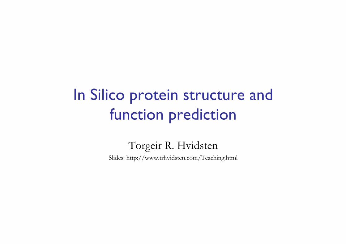
Proteins play key roles in a living system
Three examples of protein functions
− Catalysis:Almost all chemical reactions in a living cell are catalyzed by protein enzymes
− Transport:Some proteins transports various substances, such as oxygen, ions, and so on
− Information transfer:For example, hormones
Alcohol dehydrogenase oxidizes alcohols to aldehydes or ketones
Haemoglobin carries oxygen
Insulin controls the amount of sugar in the blood
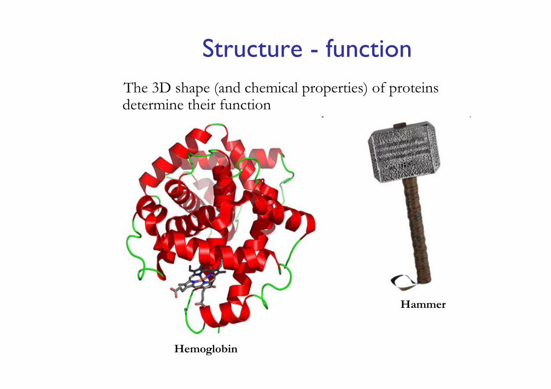
The 3D shape (and chemical properties) of proteins determine their function
Structure - function
Hemoglobin
Hammer
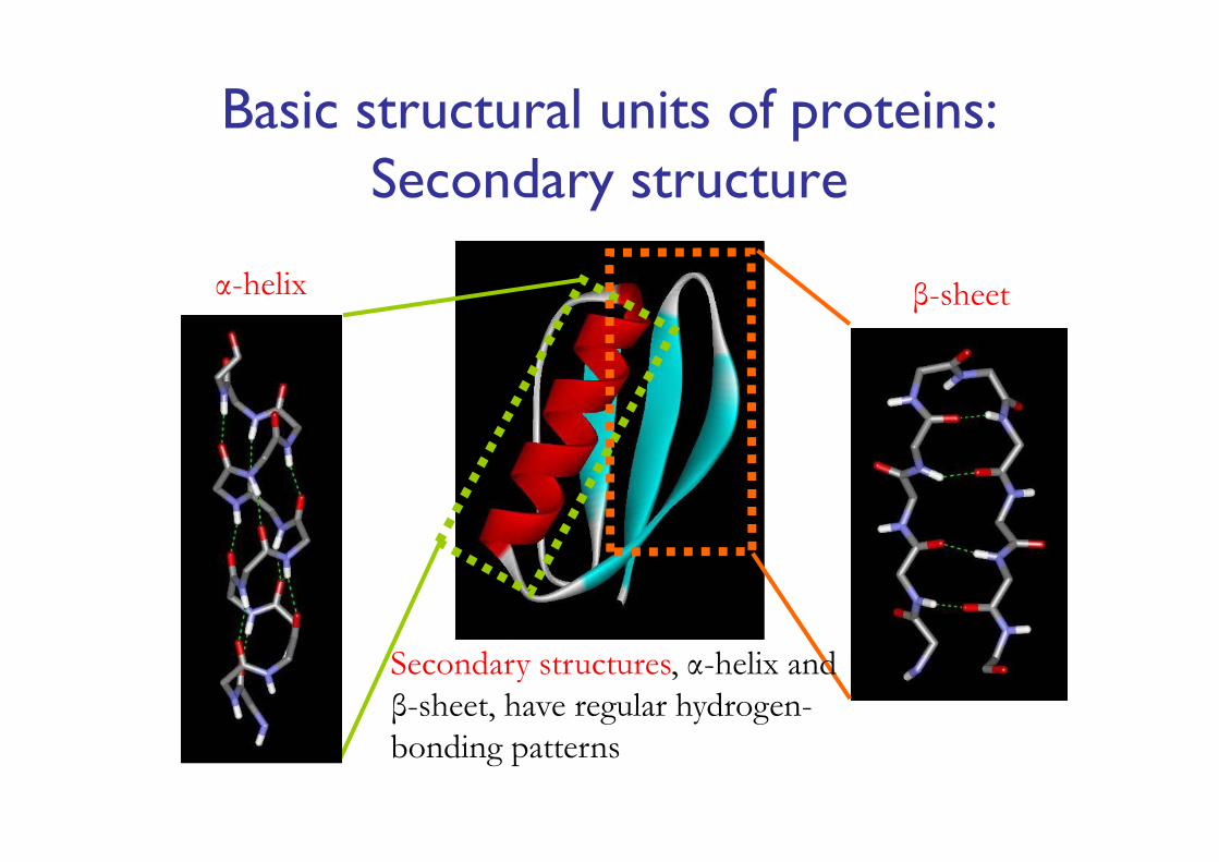
Basic structural units of proteins: Secondary structure
α-helix β-sheet
Secondary structures, α-helix and β-sheet, have regular hydrogen-bonding patterns
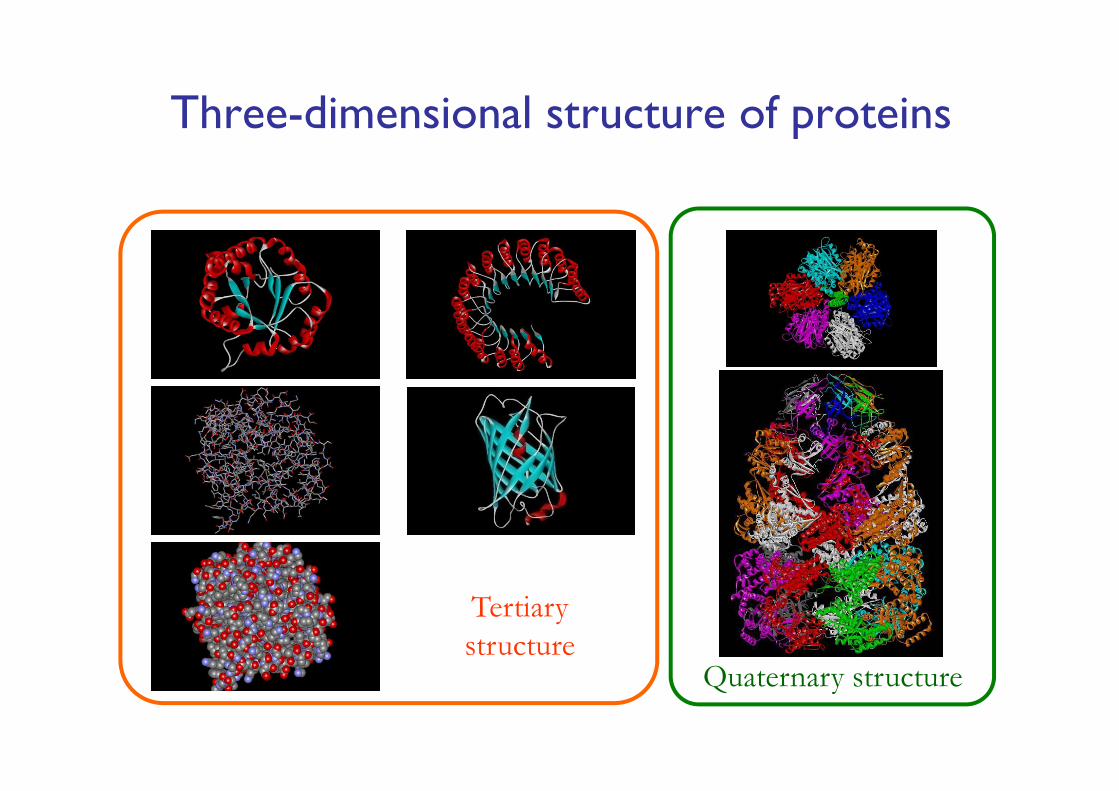
Three-dimensional structure of proteins
Tertiary structure
Quaternary structure
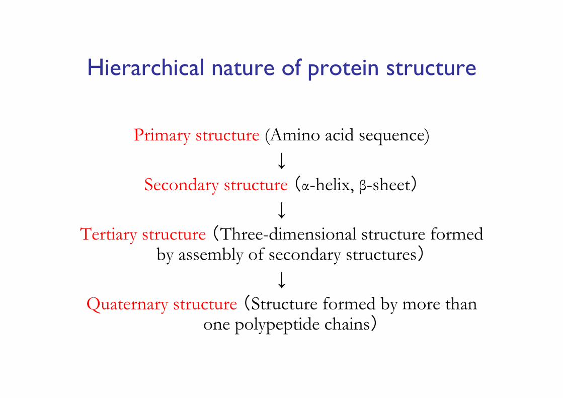
Hierarchical nature of protein structure
Primary structure (Amino acid sequence)↓
Secondary structure (α-helix, β-sheet)↓
Tertiary structure (Three-dimensional structure formed by assembly of secondary structures)
↓Quaternary structure (Structure formed by more than
one polypeptide chains)

Domains: recurrent units of proteins
The same or similar domains are found in different proteins
Each domain has a well determined compact structure and performs a specific function
Proteins evolve through the duplication and domain shuffling

Protein domains can be defined based on:
Geometry: group of residues with a high contact density, number of contacts within domains is higher than the number of contacts between domains
Kinetics: domain as an independently folding unit
Physics: domain as a rigid body linked to other domains by flexible linkers
Genetics: minimal fragment of gene that is capable of performing a specific function
Protein folds
One domain → one fold
Fold definition: two folds are similar if they have a similar topology: arrangement/orientation of secondary structure elements (architecture) and connectivity− topology = architecture + connectivity
Fold classification: structural similarity between folds is found using structure-structure comparison algorithms
Domain/fold classification Class α: a bundle of α helices connected by loops on the
surface of protein Class β: antiparallel β sheets Class α/β: mainly parallel β sheets with intervening α
helices Class α+β: mainly segregated α helices and antiparallel β
sheetsMultidomain proteins: comprise domains representing
more than one of the above four classesMembrane and cell-surface proteins: α helices
(hydrophobic) with a particular length range, traversing a membrane
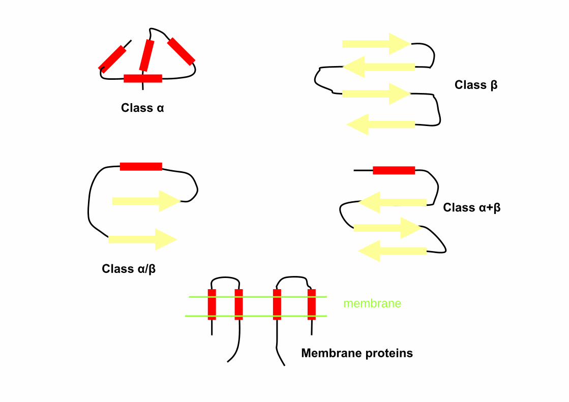
Class α
Class β
Class α/β
Class α+β
Membrane proteins
membrane
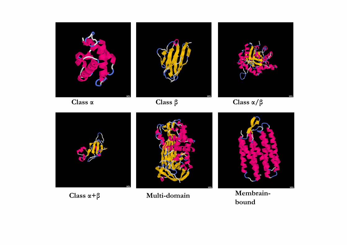
Class α Class β Class α/β
Class α+β Multi-domain Membrain-bound
Structural classification of proteins (SCOP)
The SCOP database aims to provide a detailed and comprehensive description of the structural and evolutionary relationships between all proteins whose structure is known.
Created by manual inspection and aided by automated methods
Consists of four hierarchical categories:− Class, Fold, Superfamily and Family.

SCOP
The eight most frequent SCOP folds
Why study structure?
• A full understanding of a molecular system comes from careful examination of the sequence-structure-function triad
• Below 30% protein sequence identity detection of a homologous relationship is not guaranteed by sequence alone
• Structure is much more conserved than sequence
However:• A non-redundant set of sequences is different than a non-
redundant set of structures is different than a non-redundant set of functions
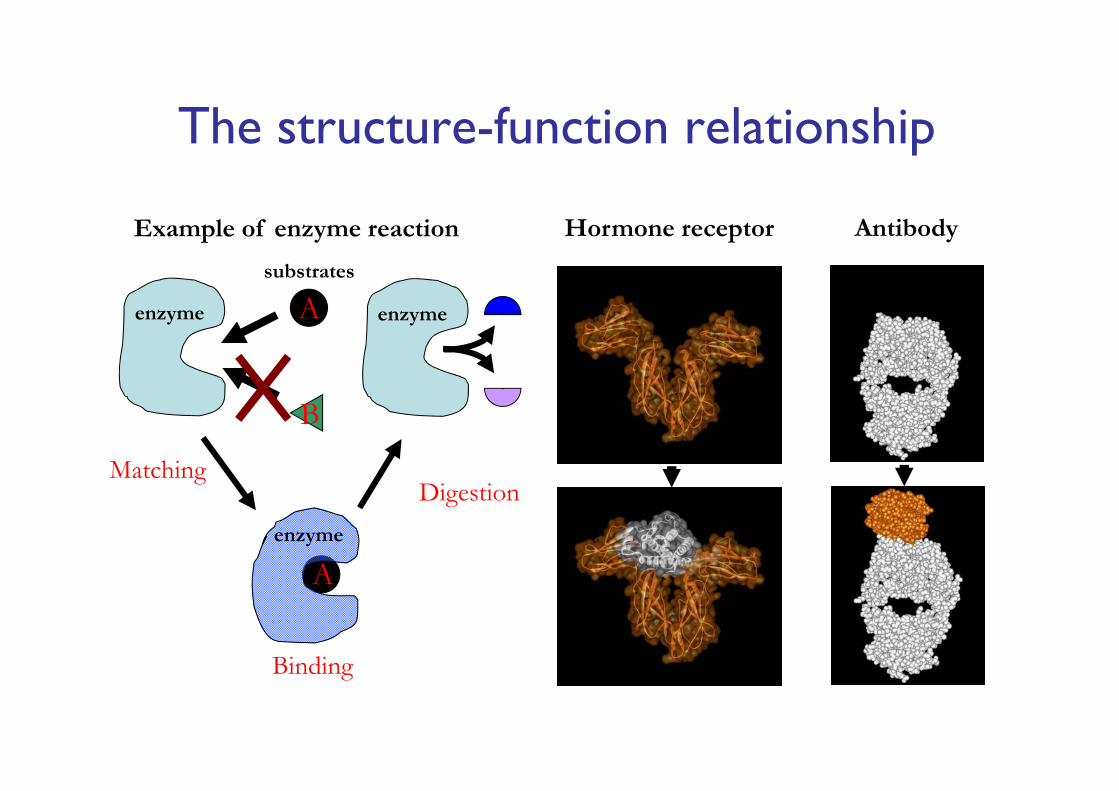
The structure-function relationship
enzyme A
B
A
Binding
Digestion
enzyme
Matching
Hormone receptor AntibodyExample of enzyme reaction
enzyme
substrates
Structure-function relationships
• The golden rule is there are no golden rules – George Bernard Shaw− Complication comes from one structure - multiple functions− Some folds are promiscuous and adopt many different functions -
superfolds
• Above 40% sequence identity, sequences tend to have the same structure and function – but there are exceptions
• Structure and function tend to diverge at ~ 25% sequence identity
• The structure-function relationship is even more complex than the relationship between sequence and structure (and not as well understood)
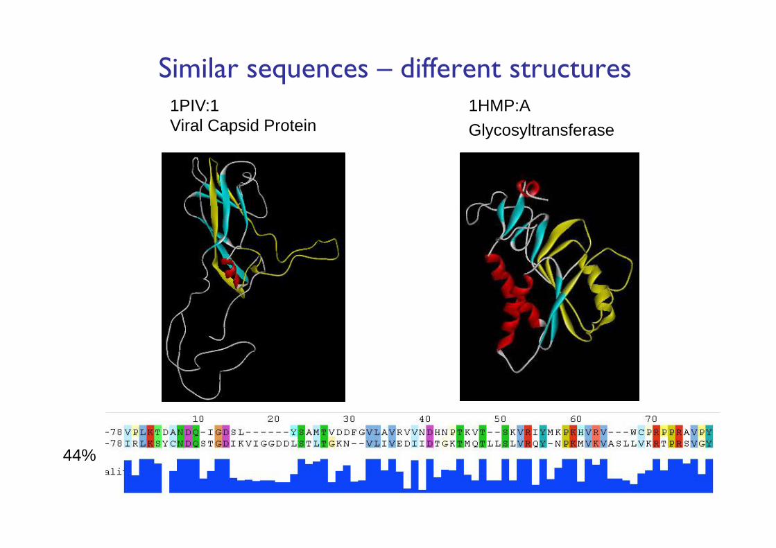
1HMP:AGlycosyltransferase
1PIV:1Viral Capsid Protein
Similar sequences – different structures
44%
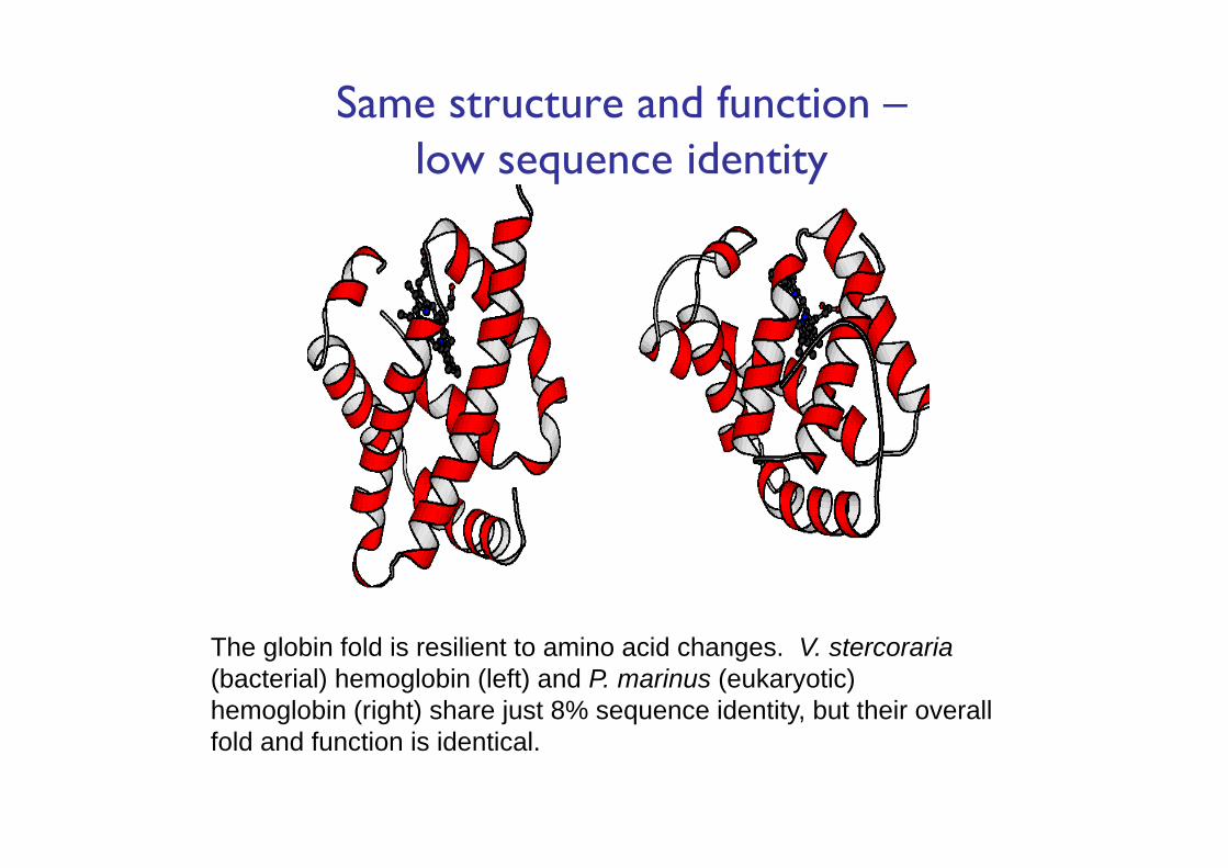
Same structure and function –low sequence identity
The globin fold is resilient to amino acid changes. V. stercoraria(bacterial) hemoglobin (left) and P. marinus (eukaryotic) hemoglobin (right) share just 8% sequence identity, but their overall fold and function is identical.
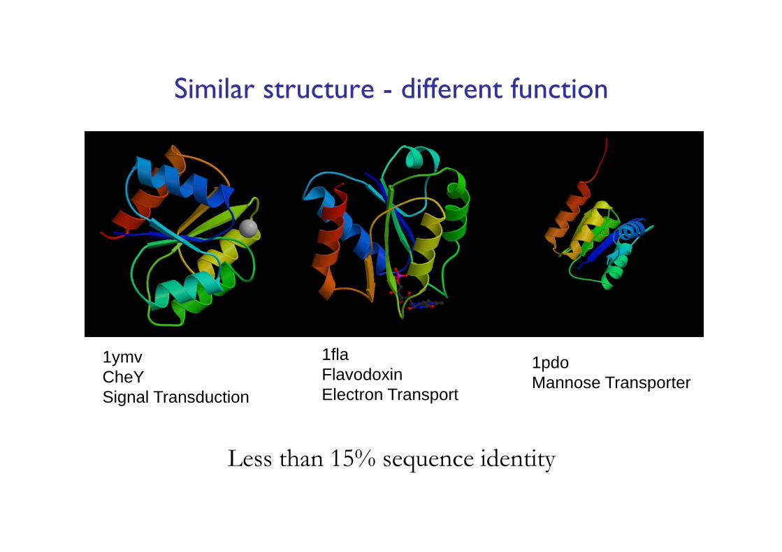
1flaFlavodoxinElectron Transport
1pdoMannose Transporter
1ymvCheYSignal Transduction
Less than 15% sequence identity
Similar structure - different function
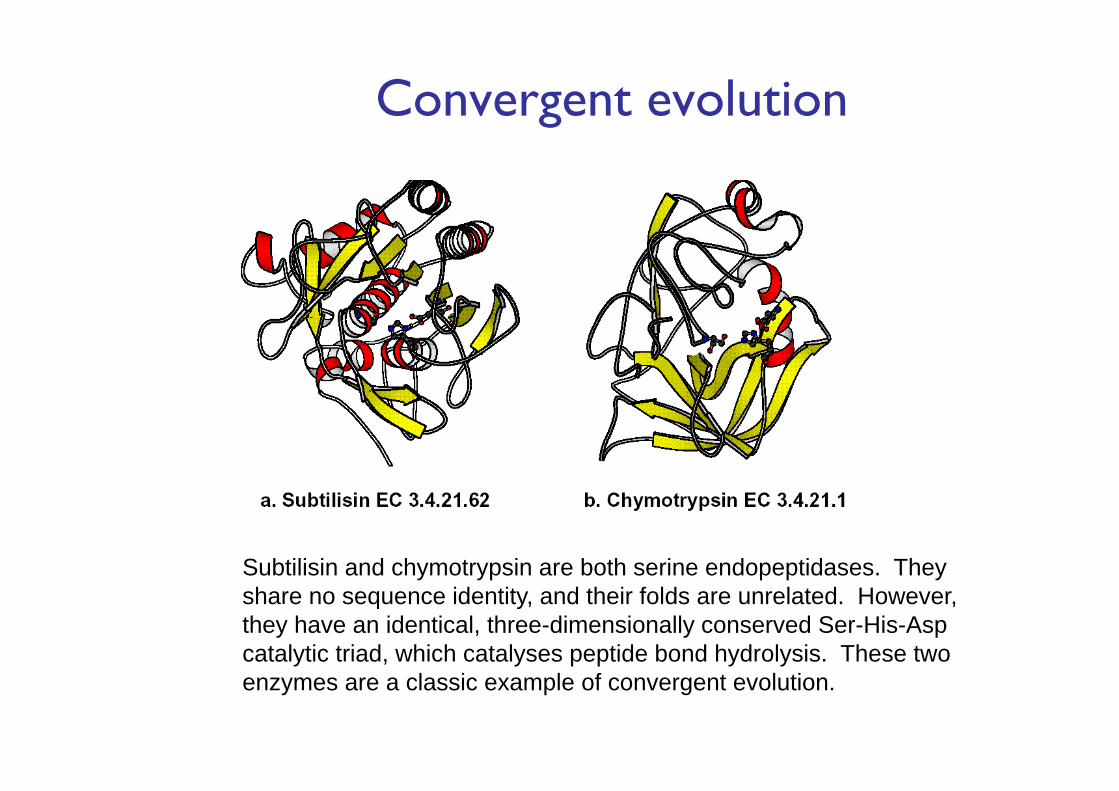
Convergent evolution
Subtilisin and chymotrypsin are both serine endopeptidases. They share no sequence identity, and their folds are unrelated. However, they have an identical, three-dimensionally conserved Ser-His-Asp catalytic triad, which catalyses peptide bond hydrolysis. These two enzymes are a classic example of convergent evolution.
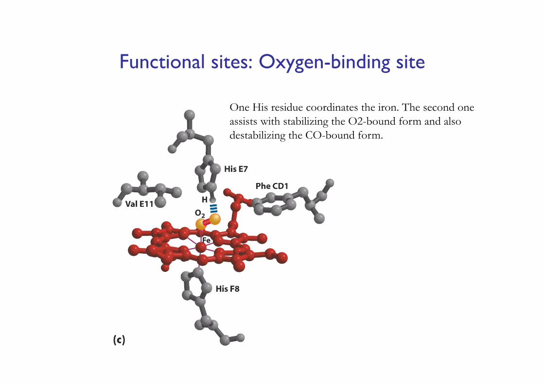
Functional sites: Oxygen-binding site
One His residue coordinates the iron. The second one assists with stabilizing the O2-bound form and also destabilizing the CO-bound form.

Computational function prediction methodsMajor challenges• The multifunctional nature of proteins
→ proteins have multiple domains hosting different function→ some domain host several functions
• The functional sites in proteins may be− better conserved than global sequence
→ low sequence similarity between functionally similar proteins− better conserved than global fold
→ the same function may be hosted by different folds• … but in some cases functional sites may be
– less conserved than global sequence→ highly similar sequences do not have the same function
– less conserved than global fold→ the same fold may host different functions
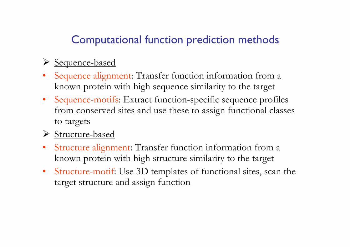
Computational function prediction methods
Sequence-based• Sequence alignment: Transfer function information from a
known protein with high sequence similarity to the target• Sequence-motifs: Extract function-specific sequence profiles
from conserved sites and use these to assign functional classes to targets
Structure-based• Structure alignment: Transfer function information from a
known protein with high structure similarity to the target• Structure-motif: Use 3D templates of functional sites, scan the
target structure and assign function
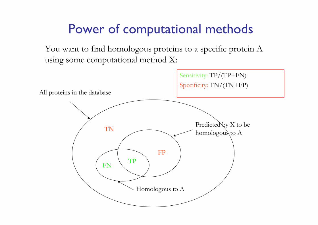
Power of computational methodsYou want to find homologous proteins to a specific protein A using some computational method X:
All proteins in the database
Homologous to A
Predicted by X to be homologous to A
TP
TN
FP
FN
Sensitivity: TP/(TP+FN)Specificity: TN/(TN+FP)

Example method: Global structure similarity
1PLS - PH domain(Human pleckstrin)
2DYN - PH domain(Human dynamin)
1PLS/2DYN:
23% sequence identity
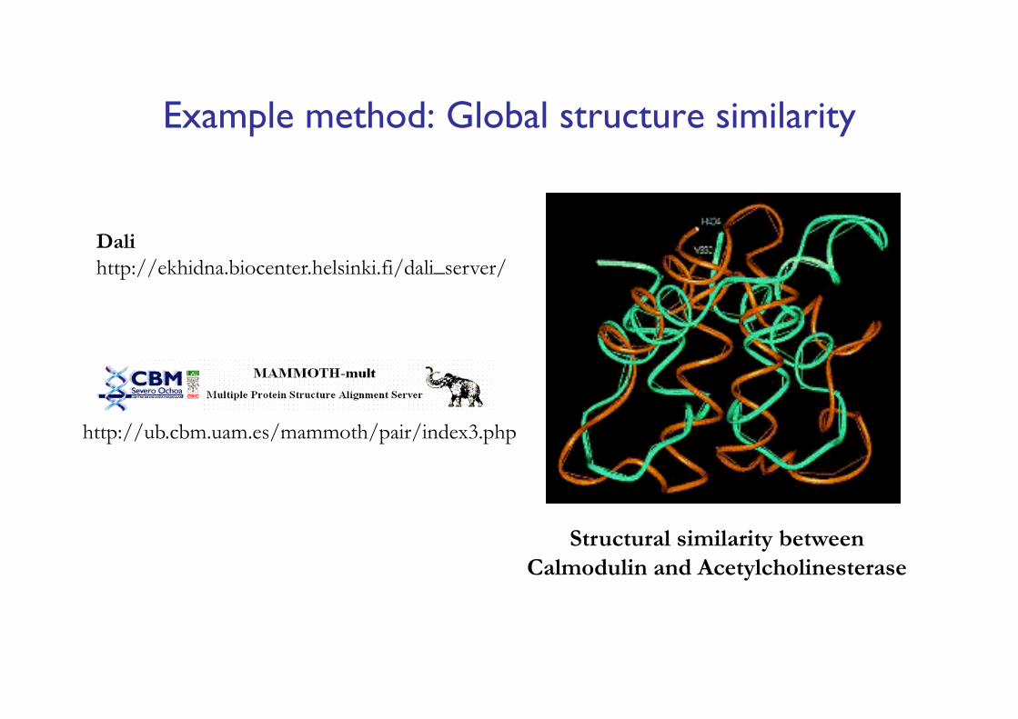
Example method: Global structure similarity
Structural similarity between Calmodulin and Acetylcholinesterase
Dalihttp://ekhidna.biocenter.helsinki.fi/dali_server/
http://ub.cbm.uam.es/mammoth/pair/index3.php
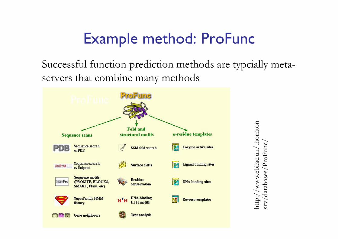
Example method: ProFuncSuccessful function prediction methods are typcially meta-servers that combine many methods
http
://w
ww.
ebi.a
c.uk/
thor
nton
-sr
v/da
taba
ses/
ProF
unc/
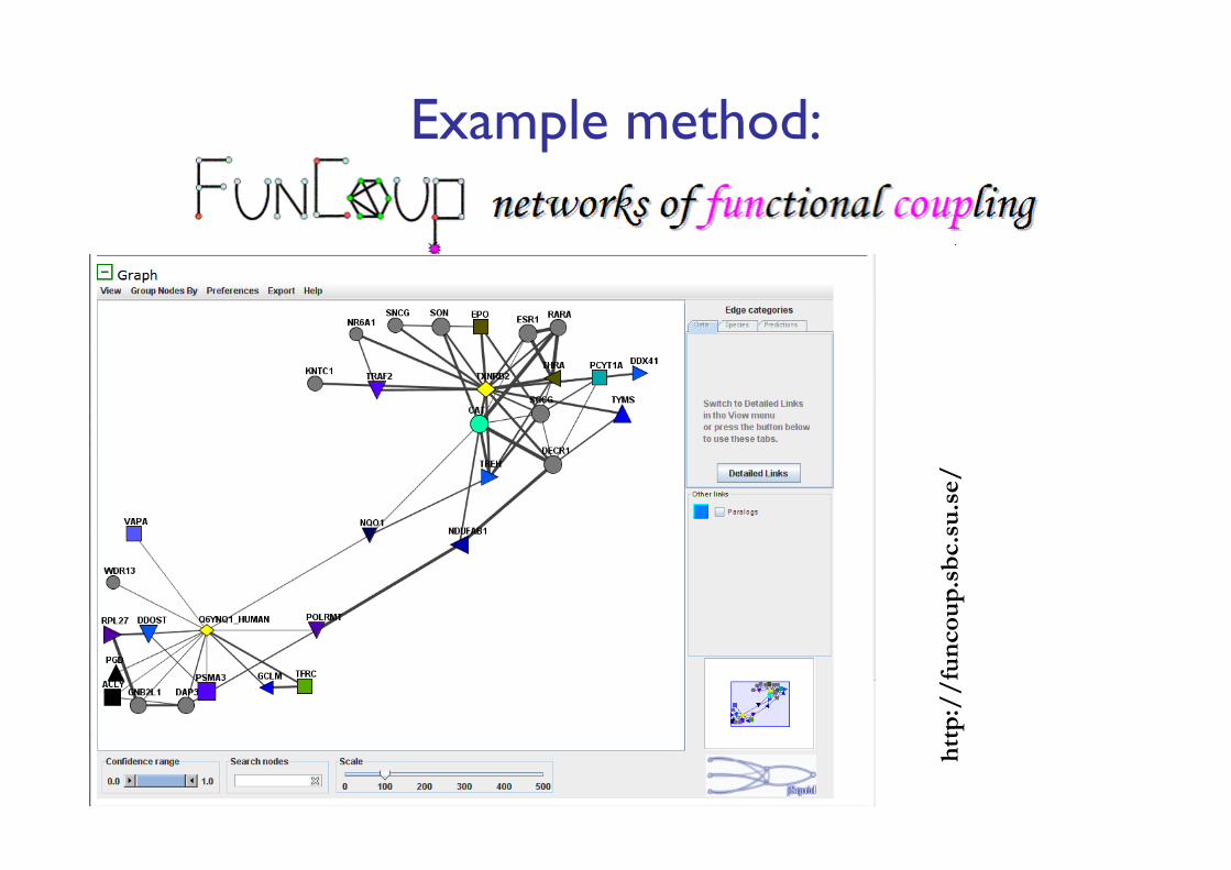
Example method:
htt
p:/
/fu
nco
up.
sbc.
su.s
e/
Structural Genomics
The biggest limitation for predicting function from structure is the low availability of structure information
Solution: Structural genomics− Solve experimentally the structure for a representative set of all protein
sequences, e.g., one or a few proteins from each fold − Predict the structure for the remaining sequences using homology
modeling, i.e., transfer structure from a structurally solved homology− Predict function from structure
Structure prediction methods are better at predicting the core of proteins than the loops
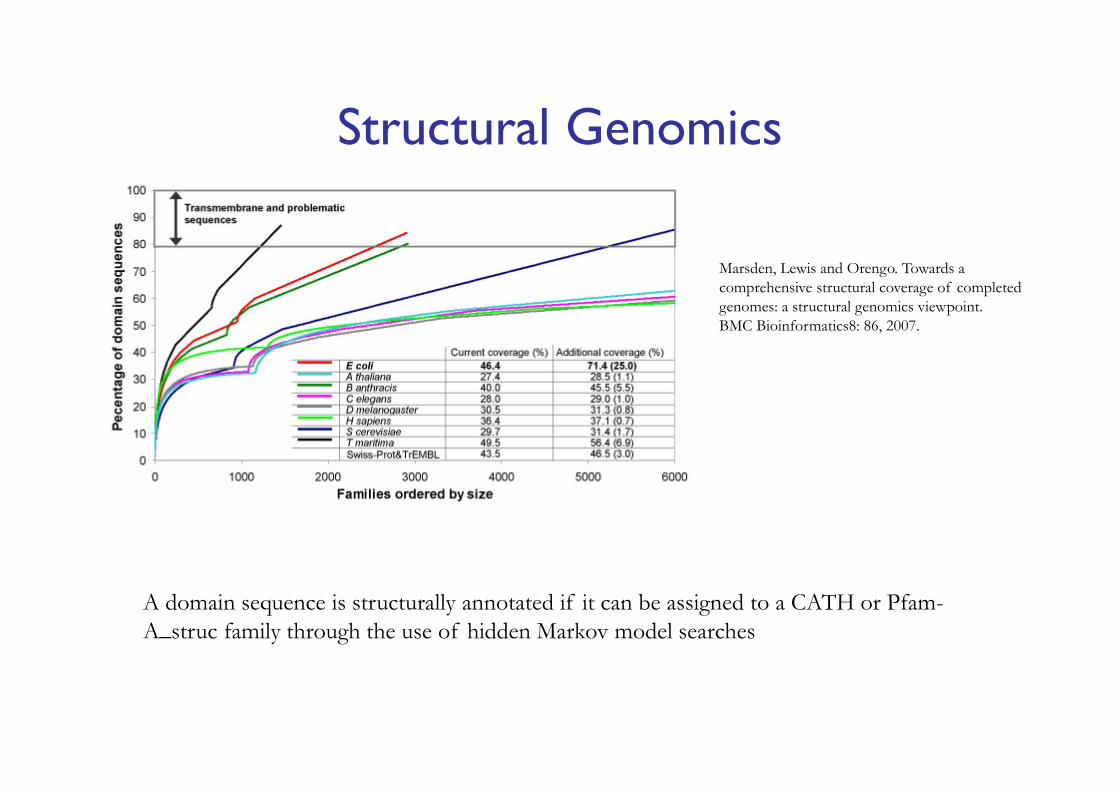
Structural Genomics
A domain sequence is structurally annotated if it can be assigned to a CATH or Pfam-A_struc family through the use of hidden Markov model searches
Marsden, Lewis and Orengo. Towards a comprehensive structural coverage of completed genomes: a structural genomics viewpoint.BMC Bioinformatics8: 86, 2007.
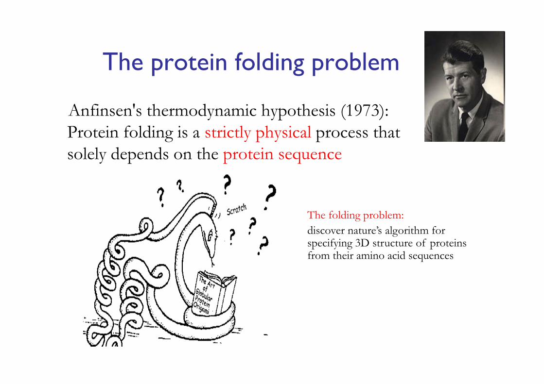
The protein folding problem
Anfinsen's thermodynamic hypothesis (1973): Protein folding is a strictly physical process that solely depends on the protein sequence
The folding problem:discover nature’s algorithm for specifying 3D structure of proteins from their amino acid sequences
Hydrophobic interactions (I)
Atomic charges dictate how folds occurGroups of C-H atoms have little charge
− Called hydrophobic or non-polar
Hydrophobic groups pack together− To avoid contact with solvent (aqueous solution)− To minimise energy
Hydrophobic and hydrophilic regions are the main driving force behind the folding process

Hydrophobic interactions (II)
Hydrophobicity vs. hydrophilicity
Van der Waals interaction
Electrostatic interactionHydrogen bondsDisulfide bonds
Folding is directed mainly by internal residues
Mutations that change surface residues are accepted more frequently and are less likely to affect protein conformations than are changes of internal residues
This is consistent with the idea of hydrophobic force-driven folding
Molten globule
Phase 1: Much of the secondary structure that is present in a native proteins forms within a few milliseconds
Phase 2: Hydrophobic collapse into the Molten globule− Slightly larger (5-15% in radius) than the native conformation− Significant amount of secondary structure formed− Side chains are still not ordered/packed− Structure fluctuation is much larger - not very
thermodynamically stable
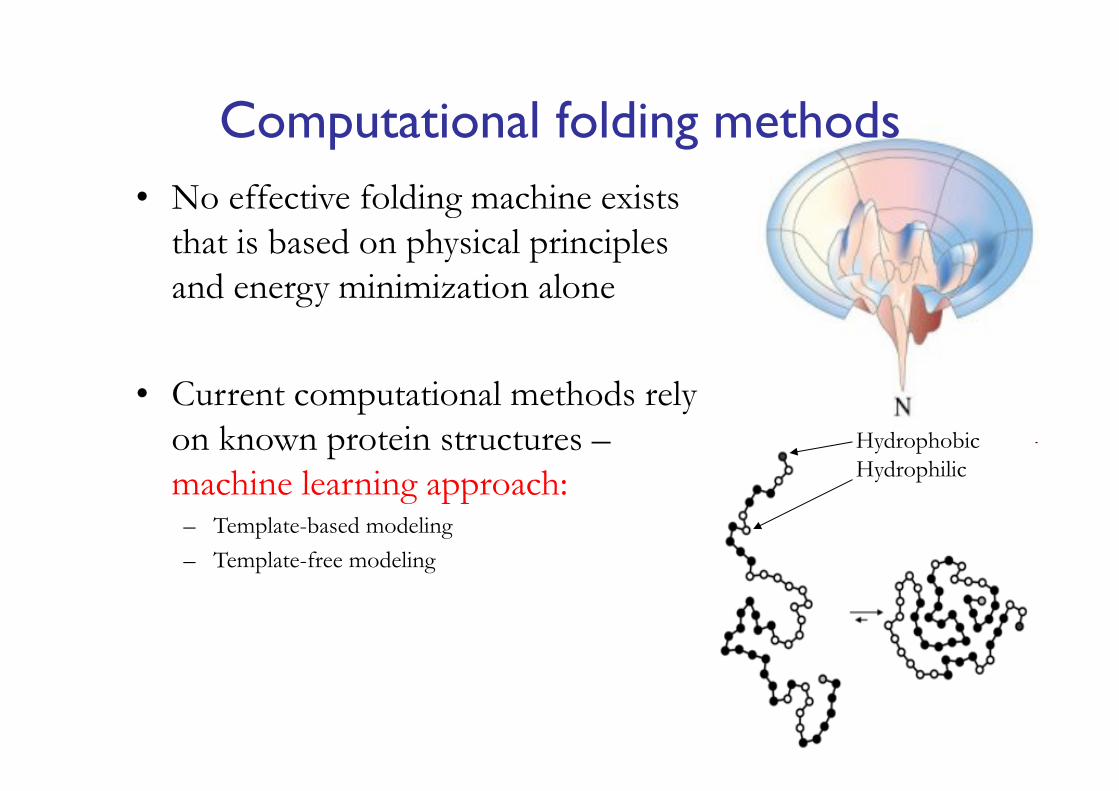
Computational folding methods• No effective folding machine exists
that is based on physical principles and energy minimization alone
• Current computational methods rely on known protein structures –machine learning approach:– Template-based modeling– Template-free modeling
HydrophobicHydrophilic

Structure represented by angels
N-terminal C-terminal

Protein folding Levinthal’s paradox
− If for each residue there are only two degrees of freedom (,)
− Assume each can have only 3 stable values− This leads to 32n possible conformations− If a protein can explore 1013 conformation per second (10
per picosecond)− Still requires an astronomical amount of time to fold a
protein Conclusion: proteins must fold in a way that does not
randomly explore each possible conformations!
Structure prediction
Protein structure prediction is the “holy grail” of bioinformatics
Since structure is so important for function, solving the structure prediction problem should allow protein design, design of inhibitors, etc
Huge amounts of genome data - what are the functions of all of these proteins?
Assumptions
Assumption 1: All the information about the structure of a protein is contained in its sequence of amino acids
Assumption 2: The structure that a (globular) protein folds into is the structure with the lowest free energy
Finding native-like conformations require:- A scoring function (potential)- A search strategy.
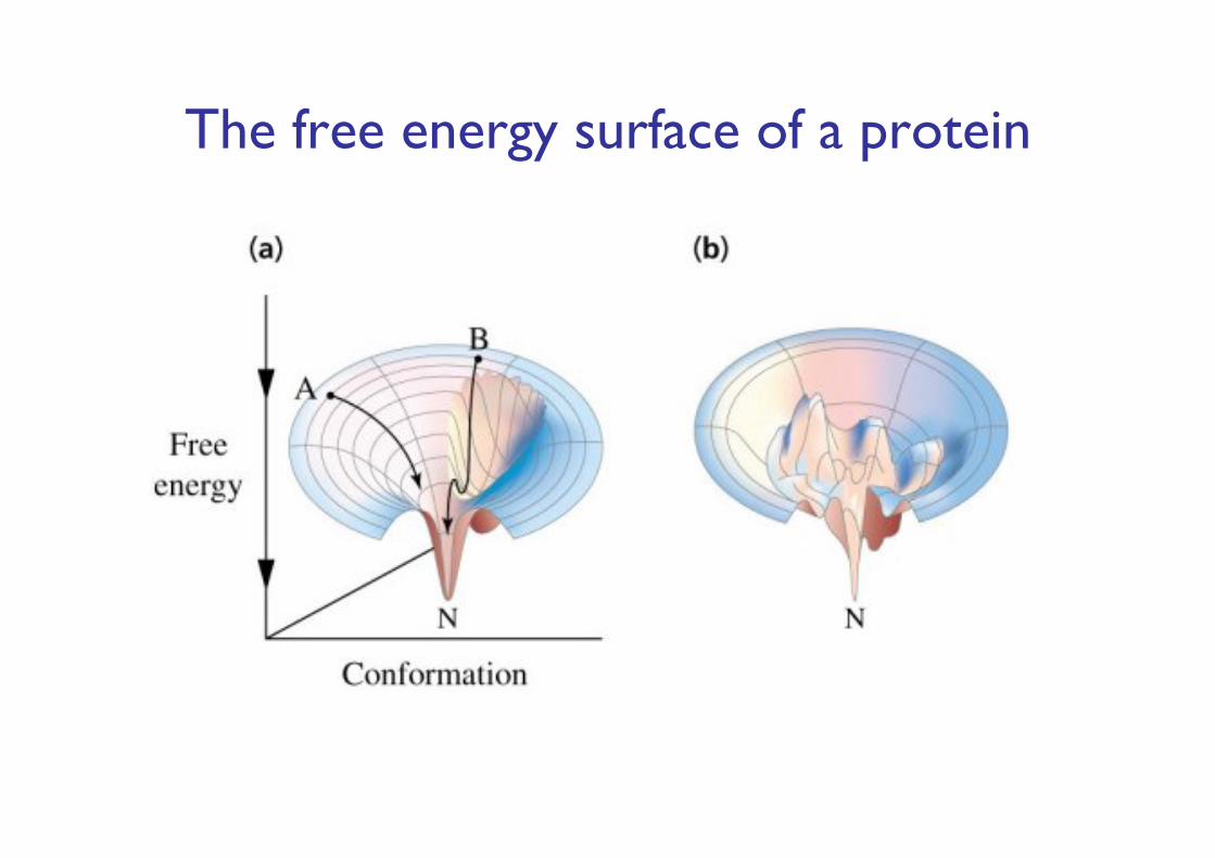
The free energy surface of a protein
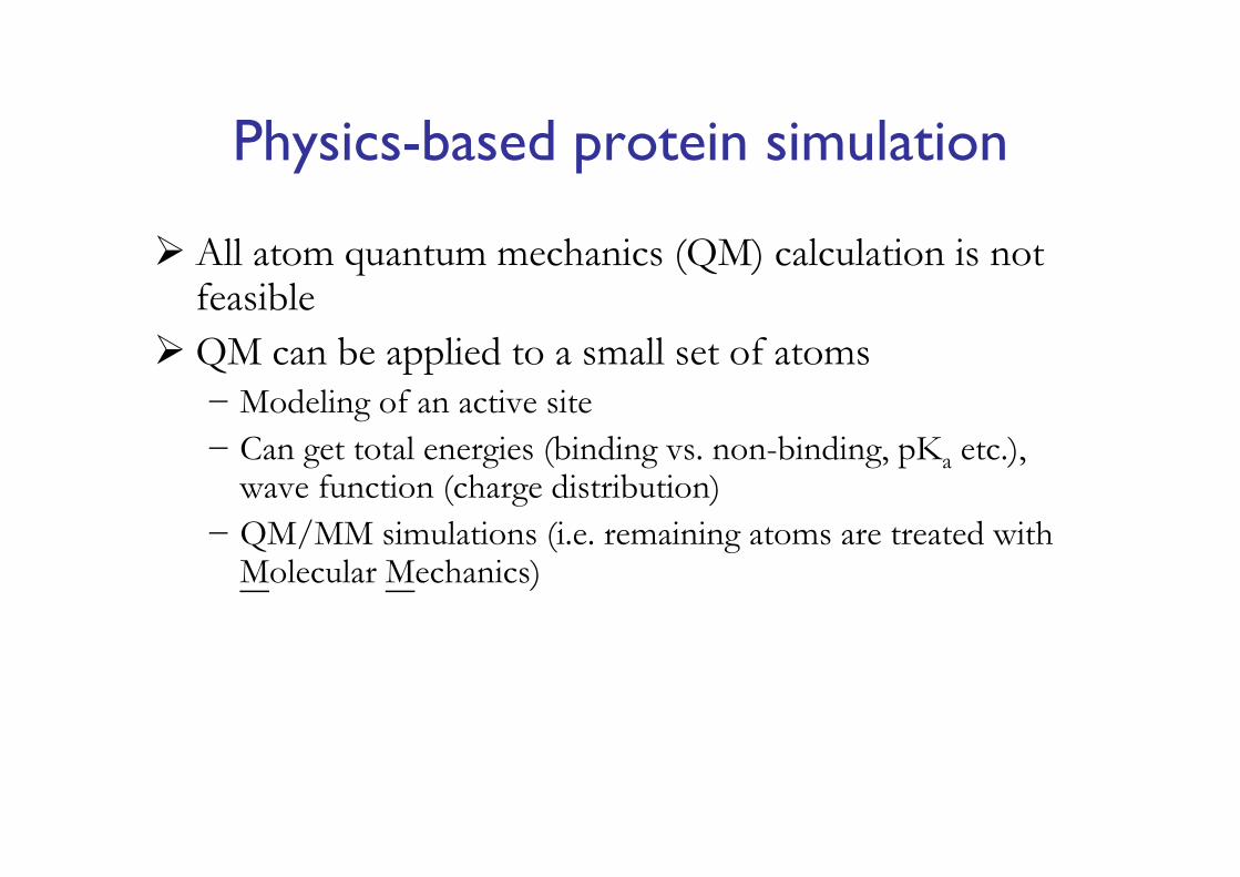
Physics-based protein simulation
All atom quantum mechanics (QM) calculation is not feasible
QM can be applied to a small set of atoms− Modeling of an active site − Can get total energies (binding vs. non-binding, pKa etc.),
wave function (charge distribution)− QM/MM simulations (i.e. remaining atoms are treated with
Molecular Mechanics)
Problems
Is the energy function correct? − Precise enough to discriminate non-native structure.− Yet simple enough for computers to carry out efficiently.
Is the conformational search good enough to cover the global minimum?
Protein folding without any prior knowledge about protein structure is a difficult task.
Protein structure prediction is often quoted as an “NP complete problem”, i.e. the complexity of the problem grows exponentially as the number of residues increases

Flavors of “knowledge-based” structure prediction
Experimental data− X-ray crystallography− NMR spectroscopy
Computational methods− Homology/comparative modeling− Fold recognition (threading)− Ab initio (de novo, new folds) methods (Ab initio: “from the
beginning”.

Comparative modelingAVGIFRAAVCTRGVAKAVDFVP
AVGIFRAAVCTRGVAKAVDFVP| || | | || ||||| ||AIGIWRSATCTKGVAKA--FVA
+

AVGIFRAAVCTRGVAKAVDFVP
AVGIFRAAVCTRGVAKAVDFVP| || | | || ||||| ||AIGIWRSATCTKGVAKA--FVA
AVGIFRAAVCTRGVAKAVDFVP| || | | || |||| ||AIGIWRSATCTKGVAK--AFVA
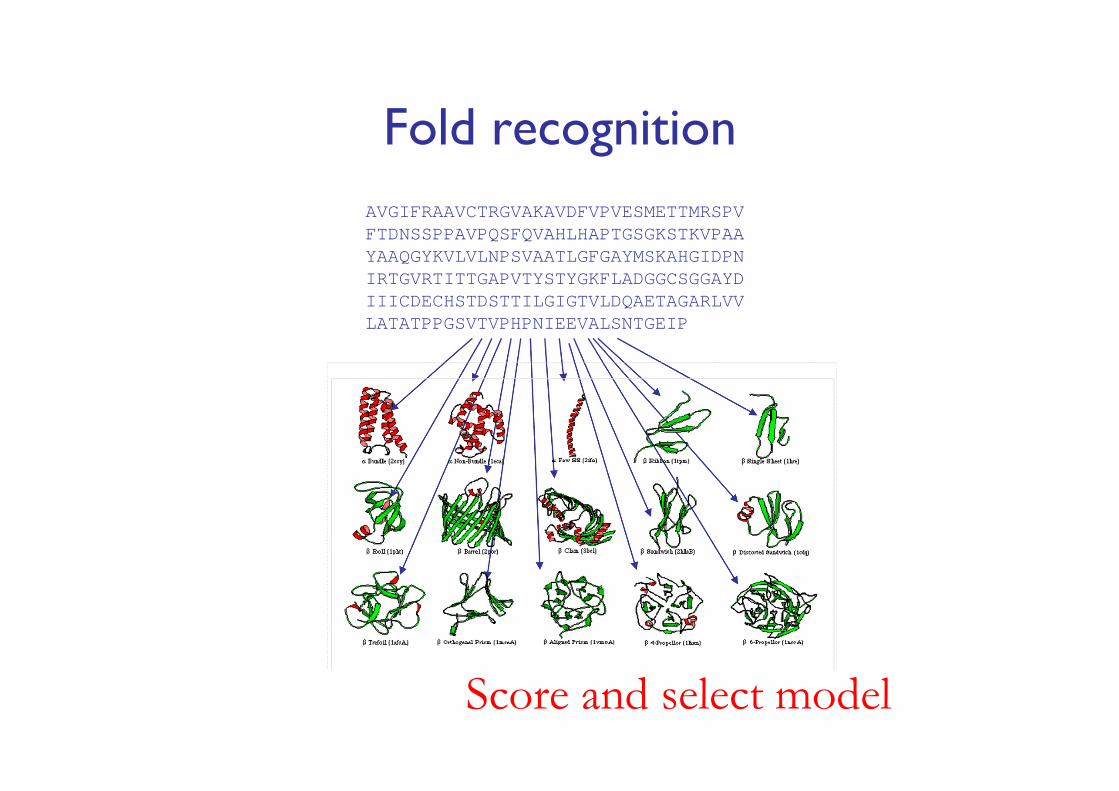
Fold recognitionAVGIFRAAVCTRGVAKAVDFVPVESMETTMRSPVFTDNSSPPAVPQSFQVAHLHAPTGSGKSTKVPAAYAAQGYKVLVLNPSVAATLGFGAYMSKAHGIDPNIRTGVRTITTGAPVTYSTYGKFLADGGCSGGAYDIIICDECHSTDSTTILGIGTVLDQAETAGARLVVLATATPPGSVTVPHPNIEEVALSNTGEIP
Score and select model
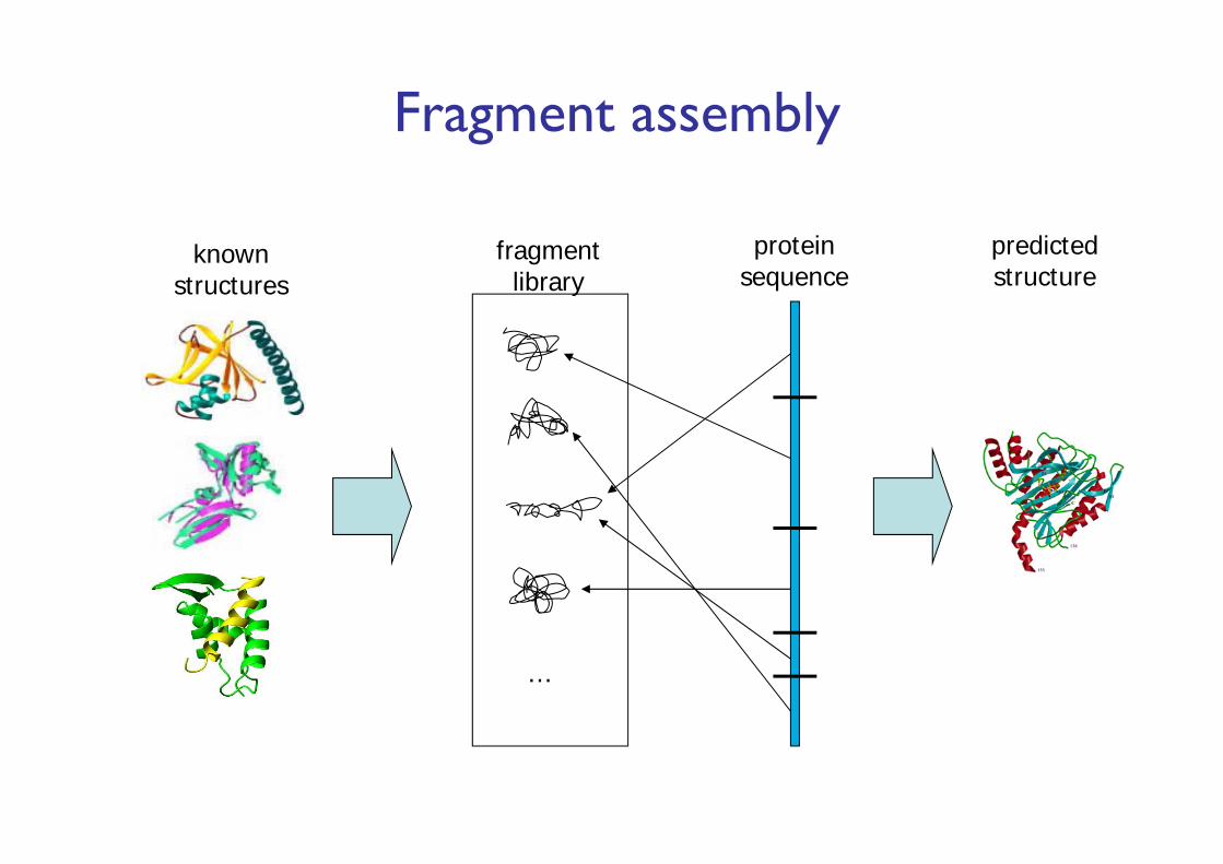
Fragment assembly
knownstructures
…
fragmentlibrary
proteinsequence
predictedstructure
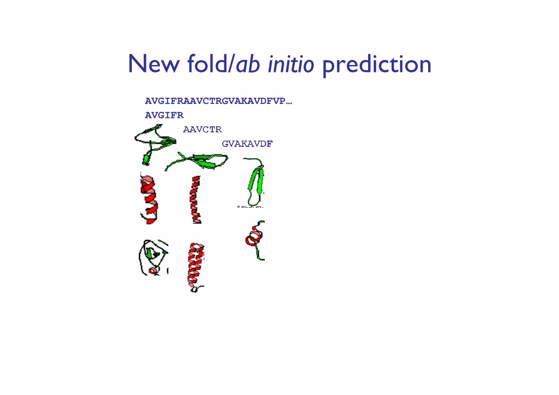
New fold/ab initio predictionAVGIFRAAVCTRGVAKAVDFVP…AVGIFR
AAVCTRGVAKAVDF
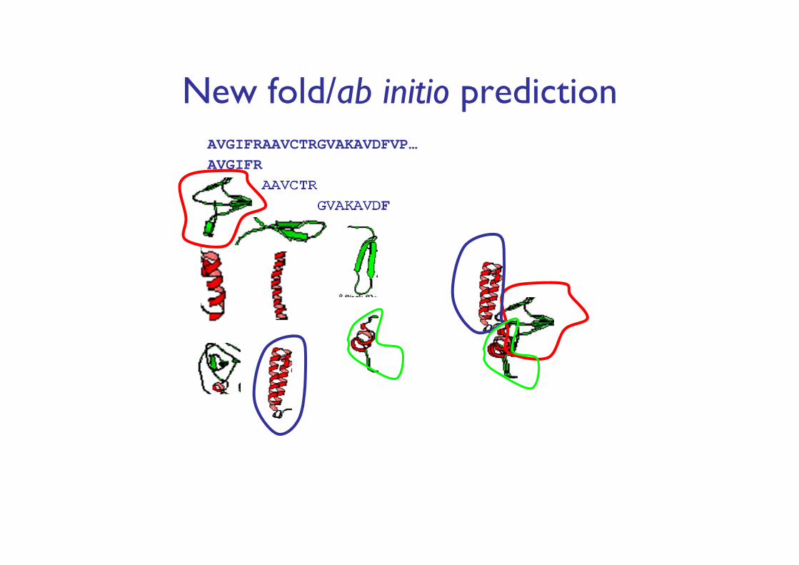
New fold/ab initio predictionAVGIFRAAVCTRGVAKAVDFVP…AVGIFR
AAVCTRGVAKAVDF
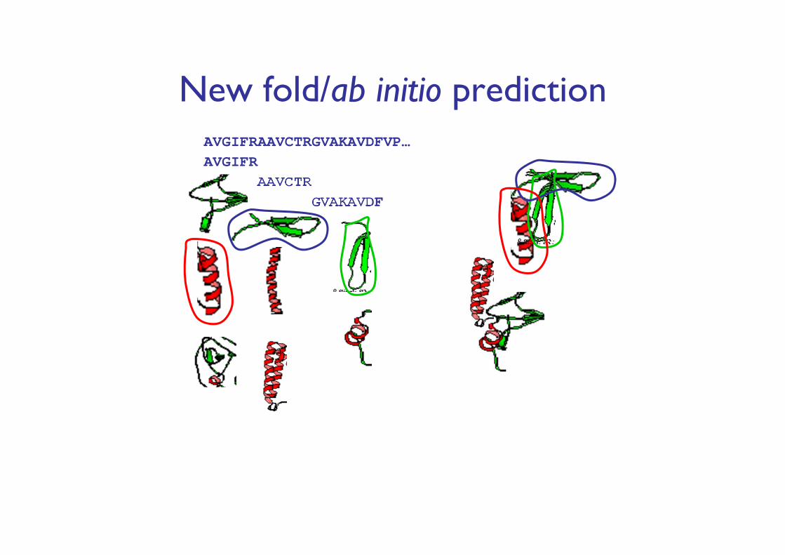
New fold/ab initio predictionAVGIFRAAVCTRGVAKAVDFVP…AVGIFR
AAVCTRGVAKAVDF
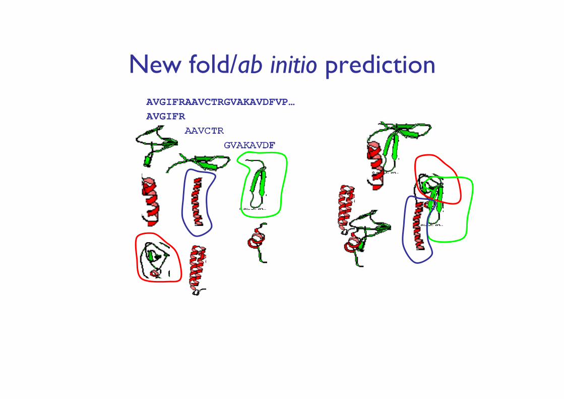
New fold/ab initio predictionAVGIFRAAVCTRGVAKAVDFVP…AVGIFR
AAVCTRGVAKAVDF

New fold/ab initio predictionAVGIFRAAVCTRGVAKAVDFVP…AVGIFR
AAVCTRGVAKAVDF
Score and select model
CASP: Community Wide Experiment on theCritical Assessment of Techniques for Protein Structure Prediction
http://www.predictioncenter.org/
Aim: obtain an in-depth and objective assessment of our current abilities and inabilities in the area of protein structure prediction
Participants will predict the structure of a set of sequences soon to be known structures
These will be true predictions, not ‘post-dictions’ made on already known structures.
Meta-methods
Meta-methods combine predictions from individual methods− E.g. 3D-Jury: http://bioinfo.pl/Meta/
Range from methods that select the best prediction to methods that improve and combine other predictions
Often include methods for all flavors of protein structure prediction
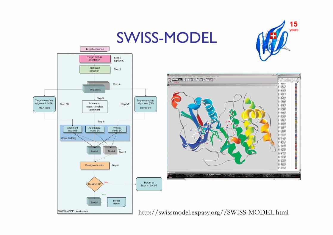
SWISS-MODEL
http://swissmodel.expasy.org//SWISS-MODEL.html
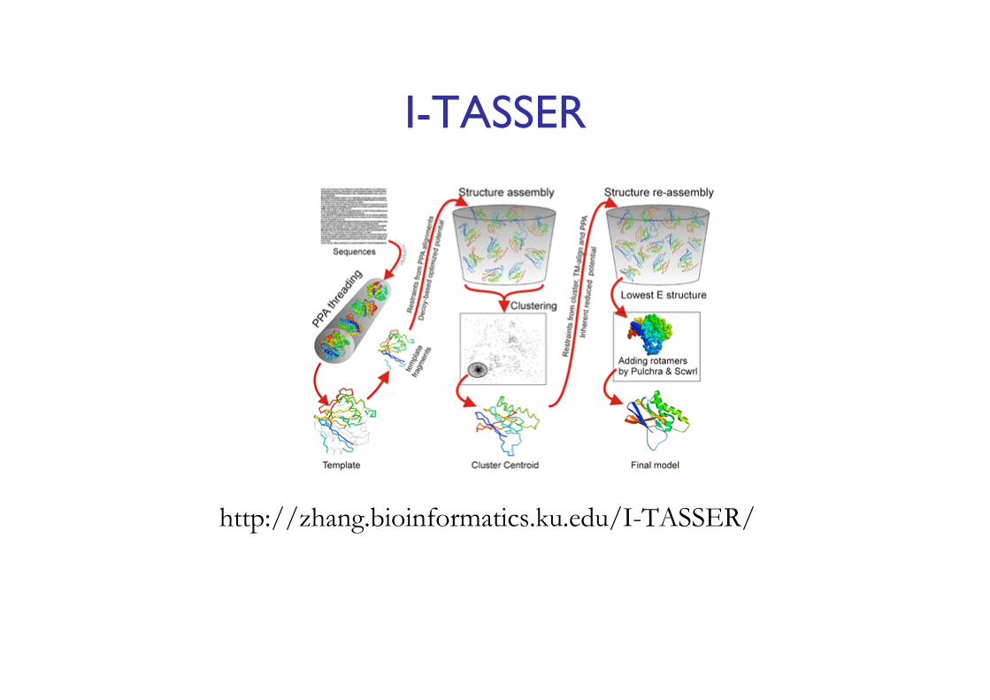
I-TASSER
http://zhang.bioinformatics.ku.edu/I-TASSER/
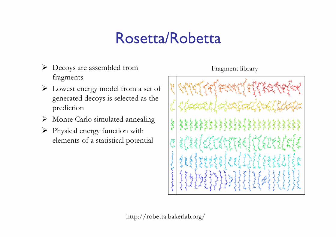
Rosetta/Robetta
http://robetta.bakerlab.org/
Decoys are assembled from fragments
Lowest energy model from a set of generated decoys is selected as the prediction
Monte Carlo simulated annealing Physical energy function with
elements of a statistical potential
Fragment library
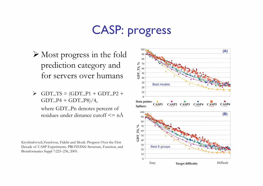
CASP: progress
Most progress in the fold prediction category and for servers over humans
GDT_TS = (GDT_P1 + GDT_P2 + GDT_P4 + GDT_P8)/4, where GDT_Pn denotes percent of residues under distance cutoff <= nÅ
Kryshtafovych,Venclovas, Fidelis and Moult. Progress Over the First Decade of CASP Experiments. PROTEINS: Structure, Function, and Bioinformatics Suppl 7:225–236, 2005.