Embed Size (px)
Citation preview

Structure-function analysis ofplant fructosyltransferases
Inauguraldissertation
zur Erlangung der Würde eines Doktors der Philosophie
vorgelegt derPhilosophisch-Naturwissenschaftlichen Fakultät
der Universität Basel
von
Denise AltenbachAus Rodersdorf, Schweiz
Basel, 2005

Genehmigt von der Philosophisch-Naturwissenschaftlichen Fakultätauf Antrag von Professor Dr. Andres Wiemken und Professor Dr. Thomas Boller.
Basel, den 7. Juni 2005
Professor Dr. Hans-Jakob WirzDekan

Structure-function analysis of plant fructosyltransferases
Inauguraldissertation
Zur Erlangung der Würde eines Doktors der Philosophie vorgelegt der Philisophisch-Naturwissenschftlichen Fakultät der Universität Basel
VonDenise AltenbachAus Rodersdorf, Schweiz
Basel, Mai 2005

Acknowledgments
I thank Prof. Dr. Andres Wiemken and Prof. Dr. Thomas Boller for giving me the
opportunity to carry out this PHD study at the Botanical Institute of the University ofBasel. Their sustained support and interest in all aspects of my work were invaluable.
Thanks to Dr. Wiemken, Dr. Boller, Dr. Marcel Lüscher, Dr. Tita Ritsema, EvelineNüesch and Dr. Alain Meyer for inspiring me with novel ideas, troubleshooting, critical
refereeing and help for successful completion of my work. I benefited a lot from their
work experience, willingness to help and motivation.The collaboration with Eveline Nüesch and Dr. Tita Ritsema inspired me and helped me
transforming many of my ideas into action. It was a pleasure to work in the lab with Dr.Tita Ritsema, Eveline Nüesch, Liesbet van Riet, Vinay Nagaraj, Alain Meyer, Marcel
Lüscher and Urs Hochstrasser. I would like to thank Jürg Oetiker, David Brodmann,Vaclav Mandak, Florian Fisch and Nadja Feddermann for support and motivation.
Thanks to all other colleagues at the Botanical Institute who contributed to my work bygenerously lending their expertise and providing words of encouragement.
My time of the PHD has been memorable due to the special friendship with EvelineNüesch, Margret Engelhard, Tita Ritsema, Liesbet van Riet, Marcel Lüscher, Urs
Hochstrasser, Claudia Heer, Lukas Lanz, Patrick Wille, Nadja Feddermann, Nika Grass,
Karin and Thommy Kubin, Tobias Wolf, Heiner Zweifel, Helene Corbiere, Alain Meyer,Ines Schnegg and Malin Elfstrand.
My warmest thanks for all their sacrifices and continuous support must go to my familyand Mathias Hefti.

Table of contents - 1
Table of contents 1
Abbreviation List 5
Summary 6
Chapter 1: General introduction 8
1.1. Fructans: occurrence, structure and physiological function 8
1.2. Enzymes involved in fructan metabolism 13
1.3. The evolution of fructosyltransferases 17
1.4. Molecular and biochemical properties of plant fructosyltransferasesand acid invertases 19
1.5. Structure-function relationships of fructosyltransferases 22
1.6. Three-dimensional structures 26
1.7. Expression systems for fructosyltransferases 29
1.8. Aim of the thesis 34
Chapter 2: The large subunit determines catalytic specificityof barley sucrose:fructan 6-fructosyltransferase (6-SFT) andfescue sucrose:sucrose 1-fructosyltransferase (1-SST) 35
2.1. Abstract 35
2.2. Introduction 36
2.3. Materials and Methods 37
2.3.1. Microbial strains and vectors used for cloning and heterologousexpression 37

Table of contents - 2
2.3.2. Cloning and mutagenesis 37
2.3.3. Construction of recombinant-tagged enzymes with exchangedlarge and small subunits 39
2.3.4. Cloning procedure to obtain the LSuB expressed alone 40
2.3.5. Expression of fructosyltransferases in Pichia pastoris 40
2.3.6. Characterization of recombinant fructosyltransferases 41
2.4. Results and Discussion 42
2.4.1. Expression of recombinant plant fructosyltransferasesin P. pastoris 42
2.4.2. Study of the possible role of a conserved motif at the startof the small subunit 42
2.4.3. Chimeric enzymes with exchanged large and small subunits 44
2.4.4. References 48
Chapter 3: Mutational analysis of the active center of plantfructosyltransferases: Festuca 1-SST and barley 6-SFT 50
3.1. Abstract 50
3.2. Introduction 51
3.3. Materials and Methods 53
3.3.1. Construction of recombinant chimeric enzymes, mutagenesisand expression 54
3.3.2. Characterization of recombinant fructosyltransferases 55
3.4. Results 57
3.4.1. Heterologous expression in the yeast Pichia pastoris 57
3.4.2. The (N/S)DPNG motif does not determine enzymatic specificity 58
3.4.3. Amino acid substitutions in the RDP- and the EC-motifreveal their necessity for fructosyl-transfer 60

Table of contents - 3
3.5. Discussion 62
3.6. References 65
Chapter 4: Are fructosyltransferases modified invertases? 67
4.1. Abstract 67
4.2. Introduction 68
4.3. Materials and Methods 71
4.3.1. Cloning and Mutagenesis of festuca 1-SST (faSST) 71
4.3.2. Cloning and Mutagenesis in onion invertase 71
4.3.3. Expression in Pichia pastoris 73
4.3.4. Characterization of recombinant enzymes 73
4.4. Results 74
4.4.1. Molecular relationships of vacuolar invertases andsucrose:sucrose 1 fructosyltransferases (1-SSTs) 74
4.4.2. Selection of amino acids correlating with invertase or1-SST activity respectively 76
4.4.3. Functional characterization of wild-type and mutagenizedenzymes expressed in Pichia pastoris 77
4.4.4. Characterization of SST-mutants 79
4.4.5. Characterization of invertase-mutants 81
4.5. Discussion 84
Chapter 5: General Discussion 90
5.1. Initial questions and experimental approach 90
5.2. “Acid invertase-like” enzymes 91

Table of contents - 4
5.3. Enzyme specificity of “acid invertase-like” enzymes 95
5.4. Outlook 97
References 99
Appendix 112
Alignment 112
Curriculum vitae 120
Publication list 121

Abbreviation List - 5
Abbreviation List
1-FFT fructan:fructan 1-fructosyltransferase1-k 1-kestose1-SST sucrose:sucrose 1-fructosyltransferase6G-FFT fructan:fructan 6G-fructosyltransferase6-k 6-kestose6-SFT sucrose:fructan 6-fructosyltransferase6-SST sucrose:sucrose 6-fructosyltransferaseAOX1 alcohol oxidase 1acINV Allium cepa vacuolar invertasebif bifurcosecDNA complementary DNAConA concanavalin ADNA deoxyribonucleic acidDP degree of polymerisationfaSST Festuca arundinacea 1-SST;
re-classified as Schedonorus arundinaceusFEH fructan exohydrolasefru fructoseFT fructosyltransferaseHPAEC High Performance Anion Exchange ChromatographyLSLB Low Salt Luria Bertani mediumLsuB large subunit of faSSTMES 2-morpholinoethanesulfonic acidmRNA messenger RNAnys nystosePCR polymerase chain reactionPEG polyethylene glycolpPICZaC Pichia pastoris shuttle vectorSDS-PAGE sodium dodecyl sulphate-polyacrylamide gel electrophoresissuc sucroseYPDS yeast peptone dextrose sorbitol

Summary - 6
Summary
Fructans are an important class of plant carbohydrates that consist of linear or
branched chains of fructosyl moieties. Their synthesis requires fructosyltransferases
(FTs) that catalyze the transfer of fructosyl units from a donor substrate (sucrose or
fructan) to an acceptor substrate (sucrose or fructan). The fructosyltransferases involved
in fructan metabolism are related to acid invertases, enzymes that cleave sucrose into
glucose and fructose. An invertase can be considered a fructosyltransferase which
transfers the fructose moiety to water. The aim of the present work was to elucidate what
determines the different catalytic activities of this enzyme group, by use of molecular
methods. In order to study such structure-function relationships we artificially introduced
mutational changes and constructed chimeric FTs (enzymes with exchanged regions).
The goal was to detect the determining regions or single amino acids. For this purpose we
optimized the expression of FTs in the methylotrophic yeast Pichia pastoris and
developed the methodology to create the chimeric constructs. Conventional cloning using
conveniently located restriction sites and the method of overlapping PCR was used.
In a first part domain exchanges between two closely related FTs from cereals
were analyzed by expressing the corresponding constructs in Pichia (Chapter 2). The two
subunits of FTs (N-terminal large subunit and C-terminal small subunit) were exchanged
between Festuca arundinacea (re-classified as Schedonorus arundinaceus)
sucrose:sucrose 1-fructosyltransferases (1-SST) and Hordeum vulgare sucrose:sucrose 6-
fructosyltransferase (6-SFT). The study revealed that it is the large subunit that carries the
structural features responsible for enzyme specificity.
In a second part we focused on the conserved motifs (S/N)DPNG, RDP and EC,
located on the large subunit, that are presumably essential in the active site of plant FTs.
For this purpose two other SST-SFT-chimeras with exchanged N-termini encompassing
these motifs, as well as Festuca 1-SST carrying single amino acid substitutions in the
RDP- and EC-motif were analyzed (Chapter 3). This study revealed the importance of the

Summary - 7
three hypothesized active site motifs for the transfructosylation reaction. All three of
them were shown to be important for enzyme activity and/or for specificity.
In a third part, we addressed the question what structural components determine
the relative transferase and hydrolase activities of FTs and vacuolar invertases via a
targeted mutational analysis based on sequence comparisons between vacuolar invertases
and 1-SSTs, the latter an example of a sucrose-using FT (Chapter 4). We chose Allium
cepa invertase and Festuca arundinacea 1-SST for our analysis. Nine amino acids
dispersed along the sequence could be identified correlating with either invertase or
1-SST activities. The selected amino acids of onion invertase were mutated to the
corresponding amino acids in Festuca 1-SST and vice versa. For both enzymes, the
mutations were analyzed independently. Functional expression in Pichia revealed shifts
in the catalytic specificity and activity, demonstrating the importance of these amino
acids outside the three highly conserved motifs (S/N)DPNG, RDP, and EC for the
enzymatic reaction (Chapter 4).
This work helped to narrow down the region potentially responsible for enzyme
specificity in plant FTs. We could pinpoint the importance of the regions with the highly
conserved motifs, and of some additional characteristic single amino acids dispersed
along the sequence, for enzyme activity and specificity.

Chapter 1- General introduction - 8
Chapter 1
General introduction
1.1. Fructans: occurrence, structure and physiological function
The disaccharide sucrose consists of glucose and fructose and is the main
transport sugar in all plants. It can furthermore serve as reserve carbohydrate. Sucrose –
dissolved in large quantities in the vacuole – and starch – stored in insoluble from in the
amyloplasts or temporary in the chloroplasts – are by far the most common reserve
carbohydrates in higher plants. Apart from these, about 15 % of flowering plants use
fructans as reserve carbohydrate (Hendry, 1993; Wiemken et al., 1995; Vijn and
Smeekens., 1999; Ritsema and Smeekens, 2003a; Ritsema and Smeekens, 2003c).
Fructans are “extensions of sucrose”: They consist of linear or branched fructose chains
attached to sucrose. As highly water soluble molecules fructans are predominantly stored
in the vacuole (Wiemken et al., 1995). Depending on the plant species, fructans mostly
reach a DP of 10 to 200 and are very diverse in structure (Vijn and Smeekens., 1999).
In plants fructans occur in many prominent orders like the Asterales, the Liliales,
and the Poales, among which are representatives of economic importance (e.g. wheat,
barley) (Hendry, 1987; Hendry, 1993). Fructans are classified according to their
differences in glycosidic linkage type (Fig. 1). Linear fructans called inulins are
composed of b(2-1) linked fructosyl units. They typically occur in the order of Asterales
(e.g. chicory). Linear fructans containing primarily or exclusively b(2-6) linkages
occurring in many forage grasses (Poaceae), are called phleins (Waterhouse and
Chatterton, 1993). Grasses often contain mixed fructan-types where b(2-1) and b(2-6)
fructosyl linkages are combined within one molecule. These fructans which occur for
example in wheat and barley, are called graminans (branched fructans). Graminans
sometimes are of even more complex structures where the fructose chains, linked b(2-1)
and b(2-6), are elongated on two sites of the starter sucrose, at the C1 of the fructose,

Chapter 1- General introduction - 9
and/or at the C6 of the glucose residue (e.g. Lolium perenne; Pavis et al., 2001; Fig. 1).
These fructans, called neo-series fructan, most often occur as inulin neo-series and are
widespread in the order of Liliales and Asparageles (e.g. onion and garlic and asparagus;
Shiomi, 1989).
In plants, fructan mainly serves as a reserve carbohydrate. Storing fructan instead
of sucrose as soluble reserve carbohydrate has several advantages: as soluble
polysaccharide fructans are osmotically less active than sucrose, and can therefore be
stored in much higher concentrations. Since fructans are highly water soluble and
accumulate in the vacuole, the largest cell compartment, storage of very large quantities
is possible. In sink organs like roots, tubers, bulbs or stems, as well as in source organs
like mature leaves, high fructan concentrations (up to 70% of dry weight) can be stored
(Wiemken et al., 1995). Generally fructans are stored if photosynthetic carbon production
exceeds demands, and are mobilized if carbon and energy is needed. An example is the
rapid breakdown of fructan stored in the leaf base upon defoliation of grasses, providing
energy and building stones for the re-growth of leaves (Wagner et al., 1986; Schnyder
and Nelson, 1987; Morvan-Bertrand et al., 2001).
If carbon fixation in a leaf exceeds export and demands, accumulation of sucrose can lead
to a feedback inhibition of photosynthesis. In this situation the ability to synthesize
fructan is a physiological advantage, since vacuolar fructan synthesis lowers the
concentration of sucrose in the cell and thus, prevents sugar-induced feedback inhibition
of photosynthesis (Housley & Pollock, 1993). For most plants the main reserve
carbohydrate is starch. It can accumulate in “source”-leaves as transitory starch in the
chloroplasts, or in reserve organs in amyloplasts. Like the accumulation of fructan,
storage of transitory starch can lower the sucrose concentration in the leaf, but storage of
equivalent amounts of starch as observed with fructans, would inevitably obstruct the
chloroplasts and consequently interfere with photosynthesis (Wiemken et al., 1995).
Starch biosynthesis decreases dramatically when the temperature drops below 10 °C,
whereas photosynthesis and fructan production are much less sensitive to low
temperature (Pollock, 1986). Thus, plants having the possibility to accumulate not only
starch, but also sucrose and/or fructan can optimally react to environmental conditions
and therefore have physiological advantages.

Chapter 1- General introduction - 10
In addition to their function as short and long term storage compounds, fructans
presumably protect against drought and freezing stress. This assumption is supported by
the fact, that fructan-accumulating plants are especially abundant in temperate and arid
climate zones with seasonal frost or drought periods, and are almost absent in tropical
regions (Hendry, 1993). Because the cell membranes are primary targets for both freezing
and desiccation injuries (Vereyken et al., 2001), fructans are supposed to be involved in
the stabilization of membranes. Indeed, it was shown using in vitro systems, that fructo-
oligosaccharides enhance membrane stability during freezing and cellular dehydration
through their affinity to phospholipids (Hincha et al., 2000; Vereyken et al., 2001;
Vereyken et al., 2003; Hincha et al., 2002). In planta it was shown, that transgenic sugar
beet (Beta vulgaris) and tobacco (Nicotiana tabaccum) transformed with a bacterial
levansucrase, had enhanced drought and freezing resistance (Pilon-Smith et al., 1999;
Konstantinova et al., 2002). Another functions of fructan metabolism is partitioning of
assimilates induced by biotic or abiotic factors (Pollock and Cairns, 1991; Suzuki and
Chatterton, 1993). A rapid sequence of accumulation and breakdown of fructans in the
growth zone of barley leaves (Roth et al., 1997) and during anthesis in Campanula
rapunculoides (Vergauwen et al., 2000) and daylily (Bieleski et al., 1993) flowers are
examples that lead to the assumption that fructans play a role in cell expansion.
Depolymerization of fructans probably contributes to the osmotic driving force involved
in cell expansion.
Interestingly also a few algae (green algae or Chlorophyta) and some
microorganisms are capable of synthesizing fructans (Hendry, 1993). Bacterial strains
such as Bacillus, Actinomyces and Streptococcus produce fructan extracellularly (Cote &
Algren, 1993; Hendry, 1993). Bacterial fructans are called levans and are generally
composed of b(2-6) linked fructosyl residues linked to a terminal sucrose and can reach a
DP of up to 100’000. Remarkably, also b(2-1) linked fructans and branched fructans are
found. There are a few reports on the synthesis of extracellular fructans by fungi, e.g. by
Aspergillus, Penicillium and Fusarium (Hendry and Wallace, 1993).

Chapter 1- General introduction - 11
In microorganisms, fructans which are extracellularly produced might act as
adhesive material around plant roots or leaves (Leigh and Coplin, 1992). Also for oral
fructan synthesizing streptococci such as Streptococcus mutans, fructans are believed to
serve as a glue and readily mobilized carbohydrate, enhancing the formation of dental
plaque (Suzuki and Chatterton, 1993).
Interest in fructans increased during the last decade due to health-promoting
effects of fructans for humans. Inulin, mainly isolated from chicory roots, is added to a
variety of products like yoghourt and “müesli” as a food additive. Long chain fructans act
as emulsifiers and give a better mouth feeling to products like fat-free yoghurt. Short
chain fructans and oligofructose can serve as sweeteners. Fructans act as “soluble food
fibers”, because the human digestive tract contains no enzymes to degrade b(2-1) and
b(2-6) glycosidic linkages. Therefore, fructans pass from the small intestine into the large
intestine without being absorbed. Only in the bowel fructans are utilized preferably by the
beneficial bifidobacteria. This effect of advantageously altering the balance in the
bacterial flora of the intestine is thought to increase gut health (Kleessen et al., 2001).
Further beneficial effects of fructan to human health are reported such as an increased
calcium resorption, or a lowering of the concentrations of insulin and cholesterol
(Jackson et al., 1999; Delzenne and Kok, 2001).

Chapter 1- General introduction - 12
Fig. 1 Representation of sucrose and the derived first representatives of different fructan types. The enzymes forsynthesis are indicated using their abbreviations. Arrows indicate the possible sites of fructose chain elongation forthe different groups. Abbreviations: 1-SST (sucrose:sucrose 1-fructosyltransferase); 6-SST (sucrose:sucrose 6-fructosyltransferase); 6-SFT (sucrose:fructan 6-fructosyltransferase); 1-FFT (fructan:fructan 6-fructosyltransferase);6-FFT (fructan:fructan 6-fructosyltransferase); 6G-FFT (fructan:fructan 6G-fructosyltransferase).
6
11
OO
O
2
O1
OO
2
1
OO 2 6
6
bifurcose
6-SFT, 1-FFT ?, 6-FFT ?
Inulin
OO
O
2
O1
sucrose
1
OO
O
2
O1
OO
2
11-kestose
OO
O
2
O1
OO 2 6
66-kestose
OO
O
2
O1
6
OO 2 6
6
1
1neo-kestose
1-SST
6-SFT
6-SST ?
6G-FFT
1-FFT
6-FFT ? 6-SFT ?
Neo-Series
Graminan
Phlein
6G-FFT, 1-FFT ?, 6-FFT or 6-SFT?

Chapter 1- General introduction - 13
1.2. Enzymes involved in fructan metabolism
Sucrose is the starting point of fructan metabolism (Fig. 1). Sucrose is synthesized in the
cytoplasm from the sequential actions of sucrose-phosphate synthase and sucrose-
phosphate phosphatase, and it can be reversibly cleaved by sucrose synthase, or
irreversibly hydrolyzed by invertases (Winter and Huber; 2000). Invertases in plants exist
in several isoforms with different biochemical properties and subcellular locations
(Sturm, 1999). Acid invertases (vacuolar invertases and cell-wall invertases) that are also
named b-fructosidases and neutral/alkaline invertases (cytosolic invertases) can be
distinguished in plants (Sturm, 1999). Besides cleaving sucrose, also hydrolysis of low
DP fructans as well as of raffinose and stachiose has been detected with acid invertases
(Marx, 1995). In contrast, alkaline invertases located in the cytosol, are sucrose specific
(Sturm, 1999). For the understanding of fructan metabolizing enzymes the acid invertases
are of special importance.
Plant fructans are derived from sucrose (Fig. 1). Their synthesis requires
fructosyltransferases (FTs) that catalyze the transfer of fructosyl units from a donor
substrate (sucrose or fructan) to an acceptor substrate (sucrose or fructan). Synthesis is
always initiated by 1-SST (sucrose:sucrose 1-fructosyltransferase), producing the shortest
fructan with a b(2-1) linkage called 1-kestose besides glucose, from two molecules of
sucrose. In this case sucrose serves as both a fructosyl donor and acceptor (Fig. 1; Fig. 2).
Chain elongation to higher DP fructan occurs via the action of 1-FFTs (fructan:fructan 1-
fructosyltransferases), 6-SFTs (sucrose:fructan 6-fructosyl transferases) and/or 6G-FFTs
(fructan:fructan 6G-fructosyltransferases), depending on fructan types synthesized by
different plant species. 1-FFT, 6-SFT and 6G-FFT are named according to the glycosidic
bond they form.
Inulin type fructans are synthesized by the elongation of 1-kestose via successive
attachment of fructosyl units by the action of 1-FFT (Edelman & Jefford, 1968; Fig.2). 1-
FFT uses one fructan as a fructosyl donor and attaches it to another fructan or sucrose,
thereby shortening one fructan and elongating another one. Sucrose can be used as
fructosyl acceptor but not as donor substrate.

Chapter 1- General introduction - 14
The two enzymes, 1-SST and 1-FFT, can only form b(2-1) linked fructans, the inulin, but
cereals such as wheat and barley form other types of fructan, the graminans, that have
primarily b(2-6) linkages between the fructosyl units (Simmen et al., 1993). The only
enzyme so far known to form b(2-6) linkages in cereals, the 6-SFT, has been purified and
cloned first from barley (Duchateau et al., 1995; Sprenger et al., 1995). The preferred
substrates of 6-SFT are sucrose and 1-kestose leading to the formation of the
tetrasaccharide bifurcose which is the smallest branched fructan, and glucose (Fig 1; Fig.
2). In the presence of sucrose as the only substrate, the activity of 6-SFT is mainly
hydrolytic, leading to the production of glucose and fructose, and only 20% of total
activity is directed into the production of 6-kestose. Evidence for the existence of a 1-
FFT in barley was obtained by Lüscher and coworkers while purifying barley 1-SST
(Lüscher et al., 2000). Studies of oat fructan showed, that also fructans of the neo-series
occur in grasses (Livingston et al., 1993). Nevertheless no 6G-FFT (Fructan:fructan 6G-
fructosyltransferase), the enzyme known to form these fructans in liliaceae has been
cloned to date in grasses. However, such an enzyme has been cloned from onion (Vijn et
al., 1997). It can use both, sucrose and low DP inulin as fructosyl acceptors, whilst
fructosyl donors can be 1-kestose and low DP inulin, but not sucrose. Although the
enzyme shows a high sequence similarity to onion invertase, it cannot hydrolyze sucrose
(Vijn et al., 1997). The two FTs known in onion are 1-SST and 6G-FFT. It was shown,
that transgenic tobacco BY2 cells expressing onion 6G-FFT and incubated with 1-kestose
produced the same fructan-pattern as it is found in onion bulbs (Ritsema et al., 2003).
Thus, no 1-FFT seems to be needed in onion for the formation of higher DP fructans of
the neo-series. In contrast, in the neo-series producing plant asparagus, 1-SST, 6G-FFT
and 1-FFT have been shown to be involved in fructan synthesis (Shiomi, N., Fructan
2004).
It is not much known what determines the typical chain lengths patterns found in
different plant species. In one case it has been shown that biochemical differences of two
1-FFTs, namely the FFT from chicory and the one form globe thistle, are determinants of
the specific chain lengths synthesized in the two plant species (Vergauwen et al., 2003;
Hellwege et al., 1998).

Chapter 1- General introduction - 15
Fig. 2 Main activities of fructosyltransferases in plants. The degree of polymerization (DP) is
indicated for fructans with m≥3 and n≥2.
Breakdown of fructan is thought to proceed via fructan exo-hydrolases (FEH;
(Fig. 2)), since increased FEH activity correlates with fructan breakdown (Marx et al.,
1997). FEHs degrade fructan polymers by splitting off terminal fructosyl residues. Up to
now, no evidence for fructan endohydrolases has been found in plants. FEHs
preferentially hydrolyzing b(2-1)-bonds (1-FEH) or b(2-6)-bonds (6-FEH) have been
distinguished (Van Damme et al., 1983). Generally sucrose has a strong inhibitory effect
on FEHs and seems not to be hydrolyzed (Bonnet and Simpson, 1993). Surprisingly, FEH
genes and activities have been recently detected in non-fructan plants, where they might
play a role in defense, acting on microbial (exogenous) fructans (Van den Ende et al.,
2004).
- 1-SST: sucrose + sucrose Æ 1-kestose + glucose
- 1-FFT: fructan DP=m + fructan DP=n Æ fructan DP=m-1 + fructan DP=n+1
- 6-SFT: sucrose + sucrose Æ 6-kestose + glucose
sucrose + 1-kestose Æ bifurcose + glucose
- 6G-FFT: fructan DP=m + 1-kestose Æ neokestose + fructan DP=m-1
- FEH: fructan DP=m Æ fructan DP=m-1 + fructose
- INV: sucrose Æ glucose + fructose

Chapter 1- General introduction - 16
In plants sugars are not only central metabolites but play a role as signal molecules
functioning as regulators of gene expression (Koch, 1996). Thus, by altering sucrose
availability, invertases, FEHs and FTs may be indirectly involved in the control of cell
differentiation and plant development.
Interestingly, bacterial fructan is generally synthesized by one single enzyme
called levansucrase (sucrose:fructan 6-fructosyltransferase). The enzyme cleaves sucrose,
releases the glucose and attaches the remaining fructosyl molecule initially to the
fructosyl residue of another molecule of sucrose and subsequently, to the growing fructan
chain (Cote and Imam, 1989). A side activity of levansucrase is the hydrolysis of sucrose
into glucose and fructose.
Glycoside hydrolases have been classified into 87 families based on the similarity
of their overall amino acid sequences, assuming that this reflects both structural and
mechanistic relationships (Henrissat, 1991). The database of “carbohydrate-active
enzymes” (CAZY, http://afmb.cnrs-mrs.fr/-cazy/CAZY/index.html) groups bacterial
invertases and levansucrases into glycoside hydrolase (GH) family 68, whilst fungal and
plant invertases and FTs fall into GH family 32. Both these families are members of the
glycoside hydrolase clan GH-J (Henrissat, 1991; Pons et al., 2000). The overall sequence
homology between the two families is less than 15% although they catalyze very similar
reactions.

Chapter 1- General introduction - 17
1.3. The evolution of fructosyltransferases
In the plant kingdom invertases with different biochemical properties and
subcellular locations are omnipresent (Sturm, 1999; see introduction 1.2.). In contrast,
FTs are of rather limited distribution, restricted to a few although partially very large
plant families (Hendry, 1993). Comparison of the amino acid sequences of plant FTs and
acid invertases revealed a very high degree of identity (see alignment in appendix and
Fig. 5 (chapter 1.5.); Sprenger et al., 1995; Vijn & Smeekens, 1999). The occurrence in
unrelated and young plant families as well as the close homology of the enzymes indicate
that the capacity for fructan synthesis is a relatively novel trait that most probably
developed independently in several genera.
If vacuolar invertases are presented in a phylogenetic tree together with FTs,
FEHs and cell-wall invertases, they cluster with FTs whilst FEHs cluster with cell-wall
invertases (Fig. 3). The following evolutionary process from invertases to FTs was
proposed (Wei and Chatterton, 2001): An ancestral invertase gene duplicated before the
divergence of monocots and dicots. One duplicate evolved into cell-wall invertase
isoforms and FEHs, and the other evolved into the vacuolar invertases and various FTs.
Because of the high degree of similarity of the amino acid sequences, it was speculated
that vacuolar invertases were recruited for generating FTs by means of small mutational
changes (Hendry, 1993; Sprenger et al., 1995; Pons et al., 1998; Vijn and Smeekens,
1999; Van Laere and Van den Ende, 2002).

Chapter 1- General introduction - 18
hvSFT mtaSFT macSFT mlpSFT mpsSFT mfaSST mlpSST mtaSST macGFT macINV maoINV macSST masSST mzmINV mciINV datINV dciSST dtoSST dhtSST dcsSST dciFFT dcsFFT dhtFFT dleINV dosINV mzmCIN mosCIN mtaCIN mleCIN dtaFEH mciFEH2 dciCIN dciFEH1 d
vacu
olar
inve
rtas
e s +
fru
cto s
y ltr
ansf
era
ses
cellw
all i
nver
tase
s +
fr
ucta
n ex
ohyd
rola
ses
0
101.5
20406080100
Fig. 3 (picture taken from Ritsema and Smeekens; 2003a): Phylogenetic tree offructosyltransferases, fructan exohydrolases and invertases in monocots (m) and dicots (d).acGFT, Alium cepa 6G-FFT (Y07838); acINV, Allium cepa invertase (AJ006067); acSFT,Agropyron cristatum 6-SFT (AF211253); acSST, Allium cepa 1-SST (AJ006066); aoINV,Asparagus officinalis invertase (AF002656); asSST, Allium sativum 1-SST (AY098442); atINV,Arabidopsis thaliana invertase (AY142666); ciCIN, Cichorium intybus cell wall invertase(Y11124); ciFEH1, Cichorium intybus 1-FEH I (AJ242538); ciFEH2, Cichorium intybus 1-FEH II(AJ295033); ciFFT, Cichorium intybus 1-FFT (U84398); ciINV, Cichorium intybus invertase(AJ419971); ciSST, Cichorium intybus 1-SST (U81520); csFFT, Cynara scolymus 1-FFT(AJ000481); csSST, Cynara scolymus 1-SST (Y09662); faSST, Festuca arundinaceae 1-SST(AJ297369); htFFT, Helianthus tuberosus 1-FFT (AJ009756); htSST, Helianthus tuberosus 1-SST(AJ009757); hvSFT, Hordeum vulgare 6-SFT (X83233); leCIN, Lycopersicon esculentum cell wallinvertase (AF506006); leINV, Lycopersicon esculentum invertase (D11350); lpSFT 6-SFT Loliumperenne (AF494041); lpSST, Lolium perenne 1-SST (AF492836); osCIN, Oryza sativa cell wallinvertase (AB073749); osINV, Oryza sativa invertase (AF019113); psSFT, Poa secunda 6-SFT(AF192394); taCIN, Triticum aestivum cell wall invertase (AF030420); taFEH, Triticum aestivumFEH (AJ508387); taSFT, Triticum aestivum 6-SFT (accession number AB029887); taSST,Triticum aestivum 1-SST (AB029888); toSST, Taraxacum officinale 1-SST (AJ250634); zmCIN,Zea mays cell wall invertase (U17695); zmINV, Zea mays invertase (U16123).
Chapter 1- General introduction - 19
1.4. Molecular and biochemical properties of plant fructosyltransferases and acid
invertases
Both, vacuolar invertases and FTs, are formed as vacuolar targeted
preproenzymes with an N-terminal signal sequence and a propeptide that are cleaved off
after protein folding and final targeting (Sturm and Chrispeels, 1990; Sprenger et al.,
1995). Typically they are encoded by one single mRNA (Fig. 4). The common feature of
plant acid invertases and FTs is to consist of an approximately 80 kD protein, which is
cleaved during maturation yielding a N-terminal large subunit and a smaller C-terminal
subunit (Fig. 4; Sturm, 1999; Sprenger et al., 1995; Lüscher et al., 2000a; Koops and
Jonker, 1996; Van den Ende et al., 1996a and 1996b; Van der Meer et al., 1998)).
Whether or not this cleavage has a physiological function is not clear, and also the
functions of individual subunits are unresolved. In contrast to these acid invertases and
plant FTs which are heterodimers, FEHs are not cleaved in two subunits. Also bacterial
fructosyltransferases consist of one subunit only.
Generally plant FTs and acid invertases are glycoproteins. The number of
potential glycosylation sites (Asn-X-Ser/Thr) varies between the different proteins.
Evidence for N-glycosylation was experimentally proven via binding to ConA/Sepharose
columns (Sprenger et al., 1995, Koops and Jonker, 1996). The hypothesized impact of N-
glycosylation on protein folding and stability still remains to be proven (Ritsema and
Smeekens, 2003a).
At the biochemical level, further similarities between plant FTs and vacuolar
invertases are evident. These enzymes differ in their preferential fructosyl donor and
acceptor substrates, but generally catalyse also fructosyl- transfers from and to alternative
substrates, albeit at a lower efficiency, depending on the type and substrate
concentrations provided, the temperature and the ionic strength (Simmen et al., 1993;
Cairns 1995; Vijn & Smeekens, 1999). For example barley (Hordeum vulgare) 6-SFT
and tall fescue (Festuca arundinacea; re-classified as Schedonorus arundinaceus) 1-SST

Chapter 1- General introduction - 20
exhibit invertase activity in addition to their main activity, depending on the environment
in which they are active. When sufficient amounts of sucrose and 1-kestose are present,
6-SFT guides 80% of its total activity into fructan synthesis, forming bifurcose, and only
20% into the hydrolysis of sucrose. If only sucrose is available as substrate, 6-SFT acts
almost purely as a hydrolase (Sprenger et al., 1995). Thus the enzyme is not only highly
homologous to vacuolar invertases at the level of amino acid sequence but also retains
considerable invertase activity. On the other hand invertases are well known to exhibit
some FT activity under certain conditions, forming 1-kestose from sucrose (Obenland et
al., 1993; Vijn et al., 1998). In biochemical terms, FTs harbor the intrinsic capacity to act
as hydrolases and vice versa.
A
B
Fig. 4 (A) Scheme of barley 6-SFT cDNA. (B) Amino acid sequence of a typical plant FT (6-SFT).Highly conserved motifs are indicated.
C-term
SDPNG RDP EC
N-term
5’ 3’1 47 248 1910
N-TermATG Stop
Amino acid sequence of a mature plant FT
5’ untranslated leader sequence
vacuolar sorting signal
SMALL SUBUNITLARGE SUBUNIT
Chapter 1- General introduction - 21
The potential close relatedness of enzymes with hydrolyzing and polymerizing
activity of glycosidic bonds was experimentally demonstrated in the case of a Bacillus
subtilis levansucrase (Chambert and Petit Glatron, 1991). The activity of the enzyme
could be readily modulated by site directed mutagenesis: if the arginine 331 was mutated
to histidine, the ratio between polymerase and hydrolase activities decreased
significantly. Moreover, the substitution of arginine 331 with lysine, leucine or serine led
to an enzyme that formed trisaccharides but no levans besides hydrolytic activity. In
attempts of sequence alignments with acid invertases and plant FTs, the arginine 331 of
levansucrase could not be assigned to a certain amino acid, but this is not surprising since
the bacterial levansucrases belong to GH family 68 and the plant enzymes to GH family
32.

Chapter 1- General introduction - 22
1.5. Structure-function relationships of fructosyltransferases
Comparison of the amino acid sequence of plant FTs and acid invertases reveals a
high degree of identity (Sprenger et al., 1995; Vijn and Smeekens, 1999). An alignment
of peptide sequences (Fig. 5) and structural analysis reveals several conserved motifs
(Vijn and Smeekens, 1999) for members of protein family GH32 (containing besides
invertases and FTs also inulinases and levanases).
The general enzymatic mechanism proposed for glycoside hydrolases of GH family 32
and 68 is a ping-pong mechanism via an enzyme-fructosyl intermediate (Chambert and
Gonzy-Treboul, 1976; Song and Jacques, 1999, Vergauwen et al., 2003). It involves the
protonation of the glycosidic oxygen followed by a nucleophile attack on the anomeric
carbon of the sugar substrate by a carboxylate group. The reaction requires three acidic
amino acids: (i) The catalytic nucleophile for the covalent binding of the fructose residue,
(ii) an acid/base catalyst that functions as a proton donor and, (iii) an amino acid that is
not directly involved in catalysis but acts as a transition state stabilizer (Ozimek et al.,
2004).
Alignments and experimental studies with yeast extracellular invertase and
bacterial levansucrase, such as affinity labelling, site-directed mutagenesis and random
mutagenesis propose three conserved regions involved in the reaction containing the
following motifs: The b-fructosidase motif (Fig. 5; region A), the EC-motif (Fig. 5;
region G) and the RDP motif (Fig. 5; region F). The b-fructosidase motif (first described
and defined by Reddy and Maley in 1990) consists of the amino acids NDPNG for all
known acid invertases, whereas it is quite variable in FTs (NDPNG, SDPNG, ADPNA,
GDPNA etc.). Since the FTs analyzed in this work either carry the motif NDPNG
(Festuca arundinacea 1-SST and Allium cepa invertase) or SDPNG (Hordeum vulgare 6-
SFT), we subsequently name the b-fructosidase motif (N/S)DPNG motif.

Chapter 1- General introduction - 23
Fig. 5 (Picture taken from Vijn and Smeekens, 1999): Comparison of amino acid sequences ofplant fructosyltransferases with those of acid invertases and cell wall invertases. Alignment ofwell-conserved regions of invertases and fructosyltransferases. Letters in bold type show almostperfectly conserved amino acids. Numbers above the comparisons represent the amino acidsequence of onion (Allium cepa) 1-SST (Ac1-SST). Region A contains the so-called Suc-bindingbox NDPNG with the well-conserved Asp. Region G contains the well-conserved Glu, which,together with the Asp, is involved in Suc hydrolysis in invertases. The following sequences wereincluded: 1-SST from onion (accession no. AJ0060660), artichoke (accession no. Y09662),Jerusalem artichoke (accession no. AJ009757), and chicory (accession no. U81520); 1-FFT fromJerusalem artichoke (accession no. AJ009756) and artichoke (accession no. AJ000481); 6G-FFTfrom onion (accession no. Y07838); 6-SFT from barley (accession no. X83233); acid/vacuolarinvertases of onion (accession no. AJ006067), asparagus (accession no. AF002656), carrot(accession no. A67163 [ DcINV] and accession no. X75351 [ DcINV1-1]), tulip (accession no.X95651), tomato (accession no. D22350), bean (accession no. U92438), mung bean (accessionno. D10265), potato (accession no. X70368); and cell wall invertases from Arabidopsis(accession no. X78424), tobacco (accession no. X81834), carrot (accession no. X78424), tomato(accession no. AB004558), wheat (accession no. AJ224681), and fava bean (accession no.Z35162). Abbreviations for the source plants are as follows: Nt, Nicotiana tabacum; Le,Lycopersicon esculentum; Dc, Daucus carota; Vf, Vicia faba; Ta, Triticum aestivum; At,Arabidopsis thaliana; Pv, Phaseolus vulgaris; Vr, Vigna radiata; Ci, Cichorium intybus; Ht,Helianthus tuberosus; Cs, Cynara scolymus; St, Solanum tuberosum; Ac, Allium cepa; Ao,Asparagus officinales; Hv, Hordeum vulgare; Tg, Tulipa gesneriana.
The b-fructosidase motif was shown to be essential for activity in yeast invertase
(GH32) where Asp23 was identified as the catalytic nucleophile (Reddy and Maley,
1990; Reddy and Maley, 1996). With respect to the catalytic nucleophile in GH68,

Chapter 1- General introduction - 24
mutational studies have been reported for FTs of Lactobacillus reuteri 121 and Bacillus
subtilis levansucrase (Ozimek et al., 2004; Meng & Fütterer, 2003). Changing the
putative catalytic nucleophile resulted in a heavily reduced total sucrose hydrolyzing
activity.
The Glu204 of the EC-motif in yeast invertase (GH32) is proposed as proton
donor during sucrose hydrolysis (Reddy and Maley, 1990; Reddy and Maley, 1996). This
conclusion is strengthened further by site-directed mutagenesis and structural analysis of
Bacillus subtilis levansucrase (GH68), where the mutation of Glu342 to Ala completely
abolished enzyme activity. The mechanism proposed by Reddy and Maley for the
cleavage of sucrose, basically involves the above mentioned nucleophile, attacking the
fructose of sucrose, and a proton donor which might be the glutamate of the EC-motif. In
the first step glucose is released and fructose is bound to the nucleophile in an ester
linkage. Then this ester linkage can be hydrolyzed using water and thereby releasing the
fructose. This mechanism for sucrose hydrolysis is also proposed for the polymerizing
activity of members of GH family 32 and GH family 68.
All studies available concerning the highly conserved RDP motif were done with
enzymes from family GH68. Studies in bacterial levansucrase propose that the Asp in this
motif forms a transient covalent fructosyl intermediate or acts as a transition state
stabilizer (Batista et al., 1999; Song and Jacques, 1999; Yanase et al., 2002). Some amino
acids in the vicinity of the RDP motif, namely Arg331 in Bacillus subtilis levansucrase
and the His-296 in Zymomonas mobilis levansucrase, were shown to be important for
maintaining the enzyme’s polymerizing activity. The hydrolyzing activity was not
affected by changing these amino acids (Chambert and Petit-Glatron, 1991; Yanase et al.,
2002). In addition, conserved aromatic amino acid residues should be considered as
potential candidates influencing the hydrolytic and the transfructosylation activities of
glycoside hydrolases and/or polymerases (T. Ritsema; personal communication).
The above described studies were performed using extracellular yeast invertase or
bacterial FTs, enzymes showing relatively low overall homologies to plant acid
invertases and FTs. But since the b-fructosidase motif, the EC-motif and the RDP motif

Chapter 1- General introduction - 25
are also very conserved in plant FTs, a similar mechanism for sucrose hydrolysis and/or
the fructosyl-transfer to sucrose and/or fructan can be expected. Similarly as proposed in
the model of yeast invertase, the corresponding glutamate (EC-motif) of plant FTs could
serve as a proton donor after a nucleophile attack on sucrose and/or fructan by the
aspartate of the b-fructosidase motif. So far only the role of the b-fructosidase motif has
been experimentally studied by mutational analysis in plant FTs (Ritsema et al., 2004).
The EC- and RDP- motifs of plant FTs have not been investigated so far. It remains to be
shown which amino acids are involved in the transfructosylation process and in
determining enzyme specificity.

Chapter 1- General introduction - 26
1.6. Three-dimensional structures
Common sequence motifs of GH family 32 and 68 are found also in GH family
43, a family comprising b-xylosidases and a–L-arabinofurosidases. By means of
modelling studies a common six-bladed b-propeller structure for the GH families 32 and
68 has been predicted (Pons et al., 2000), which is homologous to the structure of
neuraminidase. The first elucidation of the structure of a GH43 enzyme, namely of
Cellvibrio japonicus a–L-arabinase A43 (Arb43A), revealed a novel five-bladed b-
propeller fold (Nurizzo et al., 2002).
For GH68 the crystal structure from Bacillus subtilis levansucrase and for GH32
the crystal structure of the invertase from the bacterium Thermotoga maritima were
recently elucidated (Meng and Fütterer, 2003; Alberto et al., 2004). Within GH family 32
the structures of the exo-inulinase from Aspergillus awamori and of the fructan-
exohydrolase (FEH) from chicory followed (Nagem et al., 2004; Verhaest et al., 2005).
Bacillus subtilis levansucrase was crystallized in both, the ligand-free and -bound
(sucrose) state. The exo-inulinase from Aspergillus awamori was crystallized in the
fructose-bound state, and the invertase from Thermotoga maritima and the FEH from
chicory in the ligand-free states only.
All structures show a five-bladed b-propeller with a deep negatively charged
central pocket (Fig. 6). One propeller consists of five b-sheets that adopt the classical
“W” topology of four antiparallel b-strands that are named A to D. The N-terminal A
strand lines the central cavity, and the C-terminal D-strand the periphery. The b-sheets
are packed face-to-face and show a characteristic propeller blade-like twist. In contrast to
the crystallized levansucrase, the structures from enzymes of the GH family 32 are
composed of two modules, namely the five-bladed b-propeller and the C-terminal b-
sandwich (Fig. 6). The b-sandwich consists of two sheets of six b-strands. The two
modules are linked via a ten residue linker. The structures provide a template for all
members of GH family 32 including acid invertases and also plant FTs (Alberto et al.,
2004). The C-terminal b-sandwich module corresponds to the C-terminal small subunit

Chapter 1- General introduction - 27
found in plant vacuolar invertases and FTs, where it usually consist of the C-terminal
third of the protein.
The active site in the available structures is positioned at one end of the cavity at
the center of the b-propeller, with a funnel like opening towards the surface of the
molecule. This central pocket is heavily negatively charged. The pocket in the crystal
structure of B. subtilis accommodates a single sucrose molecule in the substrate-bound
structure of an inactive levansucrase mutant (Meng and Fütterer, 2003). The fructosyl
unit of sucrose is located at the bottom of the pocket and the glucose moiety on top. The
pocket is composed almost exclusively of amino acids that belong to highly conserved
sequence motifs. Included are three highly invariant acidic residues, D86, D247 and
Fig 6 A. Thermotoga maritima invertase.Picture taken from Alberto et al., J. Biol. Chem.2004; 279:18903-18910.
The central pocket is the active site. Involved inenzymatic activity are D in the b-fructosidasemotif (N/S)DPNG, D in the RDP-motif and E inthe EC-motif.
*
**
Central pocket: NDPNG; RDP; EC (*)
Fig 6 B. Cichorium intybus FEH IIa. Picturetaken from Verhaest et al., The Plant J. 2005;41:400-411.

Chapter 1- General introduction - 28
E342. D86 corresponds to the catalytic nucleophile of the b-fructosidase motif and E342
to the general acid/base catalyst of the EC-motif that were described in more detail in
chapter 1.5. The corresponding acidic amino acids can be determined via sequence
comparison for members of GH32 and correspond to D17 and E190 in Thermotoga
maritima invertase, D41 and E241 in Aspergillus awamori exo-inulinase, and D22 and
E201 in chicory FEH where they also flank the active site. The third acidic amino acid,
D247 of B. subtilis levansucrase is also flanking the active site in the other structures and
corresponds to the aspartate of the highly conserved RDP motif. It is proposed to be a
transition state stabilizer. In all structures the tree highly conserved acidic amino acids
flanking the central pocket are members of the conserved motifs (N/S)DPNG, RDP and
EC within the GH32.
Thermotoga maritima invertase (Fig. 6A) and chicory FEH (Fig. 6B) were crystallized
only in the ligand-free state, but since the catalytic modules of invertase, FEH and
levansucrase are structurally related, the comparison allowed the modeling of the position
of the substrate in the active site. The crystal structures of invertase and FEH revealed a
glycerol molecule that was present in the active site. It mimics the O4’ and O6’ hydroxyl-
groups of the fructose-moiety of sucrose or inulin respectively. This feature helped
furthermore in defining the precise position of the modeled substrate molecule in the
active site.
The crystallographic data show that the aspartate of the b-fructosidase motif and the
glutamate of the EC-motif are in the ideal position to be in close contact with their
substrate sucrose or fructan respectively.
No crystal structure of a plant fructan synthesizing enzyme or a plant acid
invertase is yet available. These plant enzymes are only up to 15% identical to members
of GH68 such as B. subtilis levansucrase and should rather be compared to members of
their own family, namely GH32. From the available structures, chicory FEH is the closest
relative to plant FTs as well as to plant acid invertases with an amino acid sequence
identity of 30 to 40%.
Mutational studies (Chapter 1.5.) combined with the available knowledge concerning
crystal structures provide further insight into the mode of action of members of GH
family 32 and 68. The question what determines enzyme specificity remains unknown.

Chapter 1- General introduction - 29
1.7. Expression systems for fructosyltransferases
The fact that vacuolar invertases show FT activity under certain condition led to a
considerable debate in the literature about the existence of specific fructan-synthesizing
enzymes (Cairns, 1995). Clear evidence for the existence of specific FTs has appeared
only with the purification of the corresponding enzymes (Simmen et al., 1993; Van den
Ende et al., 1996, Sprenger et al., 1995). The activities of the purified FTs are clearly
different from the activities of invertases. However, until today the allocation of a
sequence to either vacuolar invertases or FT is rather speculative. A reliable classification
requires functional analysis which can be achieved by the use of a gene expression
system.
The first successfully used expression system for FTs was transient expression in
tobacco (Nicotiana plumbaginifolia) protoplasts (Sprenger et al., 1995), tobacco being a
non fructan producing plant. Expression of FTs in plant cells has advantages over
bacterial or fungal expression systems. As vacuolar enzymes, FTs are N-glycosylated and
potentially the glycosylation has an effect on the activity and specificity of the enzyme.
Bacteria do not N-glycosylate proteins and in fungi, like yeast, the pattern of
glycosylation differs from that in plants with respect to the kind and amount of sugar
residues added (Elbein, 1991). Furthermore, enzyme processing during targeting differs
in plant cells from that in bacterial- or yeast-cells. Barley 6-SFT was successfully
expressed as first FT in tobacco protoplast (Sprenger et al., 1995). After a short lag phase,
the protoplasts steadily accumulated an activity that formed 1-kestose from sucrose, and
bifurcose from sucrose and 1-kestose. The experiment unequivocally proved that the
cloned cDNA encoded a functional 6-SFT. However, the protoplasts did not accumulate
higher DP fructans, and a possible hydrolytic activity of the expressed enzyme could not
be determined since the hydrolytic background activity in this expression system was too
high.

Chapter 1- General introduction - 30
In order to further characterize barley 6-SFT, it was stably expressed in the non-
fructan plant tobacco and in the inulin type fructan accumulating plant chicory (Sprenger
et al., 1997). Comparing these two plants was of special interest, since the in vivo effect
of having sucrose as the sole substrate (tobacco) could be compared to having both,
sucrose and 1-kestose as substrates (chicory). Transformants of tobacco synthesized the
trisaccharide 6-kestose and a series of higher DP fructans of the phlein type.
Transformants of chicory, a plant naturally only producing inulin, synthesized in addition
branched fructans of the graminan type. The main branched fructan found was the
tetrasaccharide bifurcose, which also represents the dominant fructan found in barley
leaves. Similarly as barley 6-SFT, onion 6G-FFT was expressed in chicory and tobacco
plants as well as in tobacco protoplasts (Vijn et al., 1997). A low activity of this enzyme
was found in protoplasts and tobacco plants, and in chicory the results were ambiguous
because of the interfering FT activities. Recently, another plant system, tobacco BY2-
cells, was successfully used to express onion 6G-FFT (Ritsema et al., 2003). The
limitation of this system was again the high background invertase activity.
The disadvantage of using plant expression systems for FTs is that all plants
contain interfering invertases. This feature is especially inconvenient since the relative
hydrolase and transferase activities of FTs are of special interest. The question what
structural components are determining the balance between the polymerase or hydrolase
activities of these enzymes is a key question. For FTs it has furthermore to be shown
what determines enzyme specificity. A convenient tool for such structure-function studies
is a suitable gene expression system.
In 1998, Hochstrasser and Co-workers introduced the Pichia pastoris yeast
expression system for FTs. The big advantage of the methylotrophic yeast Pichia pastoris
compared to plant expression systems is that it does not secrete sucrose metabolising
enzymes such as invertase. Due to the special design of the Pichia expression system
plasmids, the recombinant enzymes are secreted to the medium where no invertases
interfere with the activity of interest. The responsible element for targeting to the
extracellular space is the secretion signal of the Saccharomyces cerevisiae a-factor.
Chapter 1- General introduction - 31
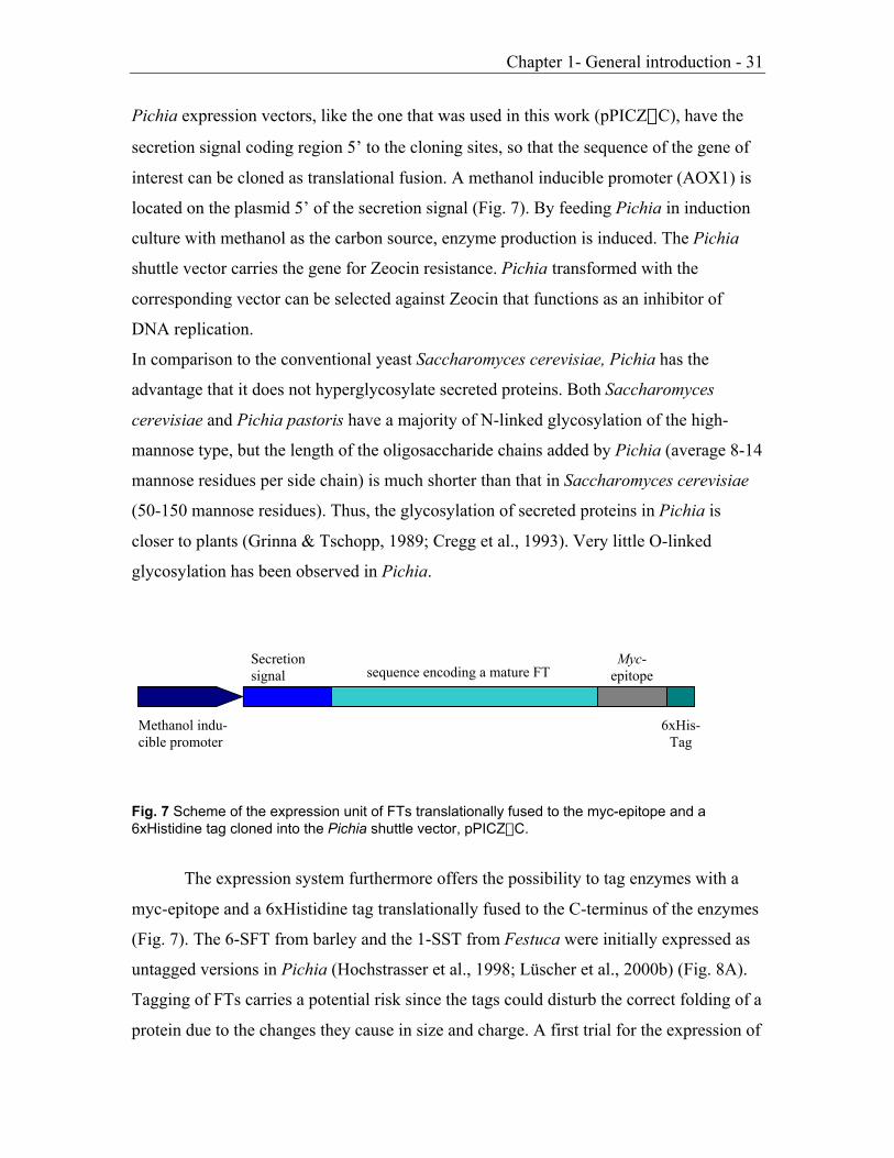
Pichia expression vectors, like the one that was used in this work (pPICZaC), have the
secretion signal coding region 5’ to the cloning sites, so that the sequence of the gene of
interest can be cloned as translational fusion. A methanol inducible promoter (AOX1) is
located on the plasmid 5’ of the secretion signal (Fig. 7). By feeding Pichia in induction
culture with methanol as the carbon source, enzyme production is induced. The Pichia
shuttle vector carries the gene for Zeocin resistance. Pichia transformed with the
corresponding vector can be selected against Zeocin that functions as an inhibitor of
DNA replication.
In comparison to the conventional yeast Saccharomyces cerevisiae, Pichia has the
advantage that it does not hyperglycosylate secreted proteins. Both Saccharomyces
cerevisiae and Pichia pastoris have a majority of N-linked glycosylation of the high-
mannose type, but the length of the oligosaccharide chains added by Pichia (average 8-14
mannose residues per side chain) is much shorter than that in Saccharomyces cerevisiae
(50-150 mannose residues). Thus, the glycosylation of secreted proteins in Pichia is
closer to plants (Grinna & Tschopp, 1989; Cregg et al., 1993). Very little O-linked
glycosylation has been observed in Pichia.
Fig. 7 Scheme of the expression unit of FTs translationally fused to the myc-epitope and a6xHistidine tag cloned into the Pichia shuttle vector, pPICZaC.
The expression system furthermore offers the possibility to tag enzymes with a
myc-epitope and a 6xHistidine tag translationally fused to the C-terminus of the enzymes
(Fig. 7). The 6-SFT from barley and the 1-SST from Festuca were initially expressed as
untagged versions in Pichia (Hochstrasser et al., 1998; Lüscher et al., 2000b) (Fig. 8A).
Tagging of FTs carries a potential risk since the tags could disturb the correct folding of a
protein due to the changes they cause in size and charge. A first trial for the expression of
Methanol indu-cible promoter
sequence encoding a mature FTSecretionsignal
Myc-epitope
6xHis-Tag

Chapter 1- General introduction - 32
a tagged FT was done with barley 6-SFT (Fig. 8; Altenbach, Diplomathesis 2000). The
tags had no influence on enzyme activity. Tagged 6-SFT retained all characteristics of its
untagged recombinant counterpart. In this work we expressed all constructs with a myc-
epitope and a 6xHistidine tag (Fig. 8B).
Previous expertise showed that the expression of FTs in Pichia is especially
successful, when only the sequence corresponding to the mature proteins were cloned
between the secretion signal and the tagging epitopes (Hochstrasser et al., 1998). The
expression level of recombinant 6-SFT carrying the entire N-terminus including the plant
vacuolar targeting signal, was found to be heavily reduced in comparison to the
expression of the sequence encoding the mature protein only. Possibly, the combination
of the a-factor secretion signal with the sequence of the vacuolar targeting signal led to
mistargeting of the proteins in the secretion process. Therefore all subsequent expression
studies were performed with the sequences encoding mature proteins.
Fig. 8 Constructs introduced into plasmid pPICZaC for expression in P. pastoris. (A) Originalconstructs with the natural stop codon. (B) Constructs where the stop codon was changed to anXbaI site, yielding recombinant proteins with C-terminal tags.
EcoRI
stop
XbaI
AMature 6-SFT
stop
myc HisAOX1 promoter
a-factor signal sequence
stop
XbaI
XbaI
stop
EcoRI XbaI
BMature 6-SFT
myc His

Chapter 1- General introduction - 33
This expression system was the ideal tool to study structural features of plant FTs
determining the balance between their specific polymerase or hydrolase activities. An
optimized expression procedure of recombinant tagged FTs was achieved in this work,
and enabled the analysis of the effect of mutational changes (Chapter 2, Chapter 3,
Chapter 4) as well as domain exchanges (Chapter 2, Chapter 3; Nüesch, Diplomathesis
2003) between different FTs.
Chapter 1- General introduction - 34
1.8. Aim of the thesis
Many plant FTs have been sequenced during the last few years, and the data clearly
indicated that a high homology exists between the different FTs and the vacuolar
invertases. At the biochemical level further similarities between plant FTs and vacuolar
invertases are evident. Depending on conditions, FTs harbor the intrinsic capacity to act
as hydrolases and vice versa. Because of the high degree of similarity at the molecular
and biochemical level it was speculated, that vacuolar invertases were recruited for
generating FTs by means of small mutational changes. It is also speculated that the
distinct fructosyl donor and acceptor specificities of different FTs are determined by
slight modifications of the amino acid sequence.
If in nature a few amino acid changes were the basis for the evolution of FTs from
invertases, it should be possible to artificially influence FT activity and specificity by
introducing mutational changes and/or by exchanging regions between different FTs. In
order to pinpoint structural requirements for enzyme activity and/or specificity, we
functionally characterized chimeric FTs (Chapter 2; Chapter 3), and FTs carrying
mutational changes (Chapter 3; Chapter 4), via expression in the yeast Pichia pastoris.
Chapter 2 - 35
Chapter 2
The large subunit determines catalytic specificity of barley
sucrose:fructan 6-fructosyltransferase (6-SFT) and fescue
sucrose:sucrose 1-fructosyltransferase (1-SST)
Denise Altenbach, Eveline Nüesch, Alain D. Meyer, Thomas Boller*,
Andres Wiemken
Zurich Basel Plant Science Center, Botanisches Institut der Universität Basel,
Hebelstrasse 1, CH-4056 Basel, Switzerland
FEBS Letters 567 (2004) 214-218
2.1. Abstract Plant fructosyltransferases are highly homologous in primary
sequence and typically consist of two subunits but catalyse widely different reactions.
Using functional expression in the yeast Pichia pastoris, we show that the substrate
specificity of festuca sucrose:sucrose 1-fructosyltransferase (1-SST) and barley
sucrose:fructan 6-fructosyltransferase (6-SFT) is entirely determined by the large
subunit. Chimeric enzymes with the large subunit of festuca 1-SST (LSuB) and the
small subunit of barley 6-SFT have the same catalytic specificity as the native festuca
1-SST, and vice versa. If the LSuB is expressed alone, it does not yield a functionally
active enzyme, indicating that the small subunit is nevertheless essential.
Key words: Cereals; Enzyme specificity; Fructosyltransferase; Pichia pastoris;
Sucrose:sucrose 1- b-D-fructosyltransferase (1-SST); Sucrose:fructan 6-b-D-
fructosyltransferase (6-SFT)
* Corresponding author

Chapter 2 - 36
2.2. Introduction
Fructans are an important class of carbohydrates in plants [1-3]. The enzymes
characteristic of plant fructan metabolism, fructosyltransferases and fructan
hydrolases, have been found to be highly homologous to the plants' soluble acid b-
fructosidases (invertases) belonging to glycoside hydrolase family 32 [2,4]. Indeed,
the first plant fructosyltransferase to be cloned, the 6-SFT from barley (Hordeum
vulgare), displayed both 6-SST/6-SFT and b-fructosidase activity [5]. In contrast, the
first 1-SST of grasses to be cloned, the one of tall fescue (Festuca arundinacea),
produced almost exclusively 1-kestose and glucose when supplied with sucrose and
had very little b-fructosidase activity [6]. In view of their striking homologies,
fructosyltransferases may have evolved from b-fructosidases by relatively few
mutational changes [4,5,7]. However, it is unknown which changes are essential for
changes of catalytic specificity.
Typically, plant acid b-fructosidases and fructosyltransferases are synthesized
as a primary translation product of ~85 kDa but then are cleaved into a large N-
terminal subunit of ~60 kDa and a small C-terminal subunit of ~25 kDa [5,8-11]. The
large subunit contains putative catalytic motifs for sucrose binding and hydrolysis,
namely the b-fructosidase motif, the RDP motif and the EC-domain [3,10]. The
importance of these motifs was experimentally proven for the b-fructosidase motif
and the EC-motif in the case of yeast invertase [12] and for the RDP motif in the case
of a bacterial fructosyltransferase [13].
We have previously established a convenient heterologous expression system
in Pichia pastoris to study fructosyltransferases of plants [6,14]. In the present work,
we describe how this system can be optimised, and we use it to investigate the
catalytic activity of chimeric enzymes generated by exchanging the large and small
subunit of 6-SFT and 1-SST, respectively. Our results show that it is the large subunit
of the enzyme which determines its catalytic properties.

Chapter 2 - 37
2.3. Materials and Methods
2.3.1. Microbial strains and vectors used for cloning and heterologous expression
Escherichia coli strain DH5a was used for amplification of the recombinant
plasmids pK18, pBluescript KS+ (Stratagene, Amsterdam, The Netherlands),
pPICZaC. Pichia pastoris strain X-33 (wild type), and the pPICZaC shuttle vector
were obtained from Invitrogen BV (Leek, The Netherlands).
2.3.2. Cloning and mutagenesis
Constructs P1 and F1 in pPICZaC, representing the native coding sequences
of barley 6-SFT (EMBL X83233) and festuca 1-SST (EMBL AJ297369), respectively
(Fig. 1A,B), were described earlier [6,14]. To obtain myc- and 6xhis-tagged versions
of the two enzymes, their coding regions were excised with EcoRI and XbaI and
subcloned into pK18. The stop codons were then altered to XbaI restriction sites by
PCR, using primers P3f and P3r for 6-SFT and SST001 and SST002 for 1-SST
(Table 1). The resulting PCR products were purified and digested with AgeI and XbaI
for 6-SFT and with NruI and XbaI for 1-SST, respectively, ligated into the
correspondingly digested parent plasmid, and excised from the plasmid by EcoRI and
XbaI. These fragments were cloned in frame with the myc-epitope and his-tag into the
Pichia shuttle vector, leading to pPICZaC-P3 and pPICZaC-F2 (Fig. 1C,D). Note
that we used the tags here simply to verify that the recombinant proteins are
expressed, but that we would like to use them, in the future, to purify high levels of
recombinant proteins to produce antibodies against them; there are still no specific
antibodies against invertases or plant fructosyltransferases available.

Chapter 2 - 38
Fig. 1 Constructs introduced into plasmid pPICZaC for expression in P. pastoris. A, B:Original constructs with the natural stop codon. C,D: Constructs where the stop codon waschanged to an XbaI site, yielding recombinant proteins with C-terminal tags. E: Swap1: largesubunit of festuca 1-SST with the small subunit of barley 6-SFT fused to it. F: Swap2 largesubunit of barley 6-SFT with the small subunit of festuca 1-SST fused to it. G: large subunit offestuca 1-SST expressed alone.
stop
XbaI
XbaI
stop
EcoRI XbaI
1-SST6-SFT
Subunit cleavage site
Swap2F myc His
stop
EcoRI XbaI
1-SST 6-SFT
Subunit cleavage site
Swap1E myc His
EcoRI
1-SST stop
XbaI
LSuBG myc His
F1EcoRI
stop
XbaI
BMature 1-SST
stop
myc His
P1EcoRI
stop
XbaI
AMature 6-SFT
stop
myc His
AOX1 promoter
a-factor signal sequence
P3
stop
EcoRI XbaI
CMature 6-SFT
myc His
F2
stop
D
EcoRI XbaI
Mature 1-SST
myc His

Chapter 2 - 39
Table 1Oligonucleotides used for cloning
Primer Sequence
P3f 5’-GTTCAACAACGCCACCGGTGCCAGC-3’
P3r 5’-CTTATTAATGACGAGTCTAGAGAACTTGATTGAAGATAC-3’
SST001 5’-CATTGTGCAGAGCTTCGCGATGGGTGGGAGGATTAC-3’
SST002 5’-CTCGACTTGGTTTCATCTCTAGAGCGTCGTTCGTGAAGATATG-3’
ADDA008 5’-GTGGCTGCCCTCAACGACACCAACGTTGGCTACAACTGCAG-3’
ADDA009 5’-CTGCAGTTGTAGCCAACGTTGGTGTCGTTGAGGGCAGCCAC-3’
SST004 5’-CCGACGTGGATGCTCTAGACGAGGCCGATGTCAGC-3’
SST005 5’-GCTGACATCGGCCTCGTCTAGAGCATCCACGTCGG-3’
SFT001 5’-TCCATTCAGTCAGTTCCTAGGACGGTGGCTCTG–3’
P4001 5’-GCTGACATCGGCCTCGTTGAGGGCAGCCACGGCGGAAGCATC-3’
P4002 5’-CGCCGTGGCTGCCCTCAACGAGGCCGATGTCAGCTACAAC–3’
SST006 5’-GAGTTTTTGTTCTAGAGCGTCG-3’
Mutant P3[DTN], in which the first three amino acids (EAD) of the small subunit of
barley 6-SFT are changed into the motif DTN typical of FFTs, was generated by site
directed mutagenesis using standard procedures (QuikChangeTM Site-Directed
Mutagenesis Kit, Strategene), based on the pK18-clone containing the tagged version
of barley 6-SFT. Primers ADDA008 and ADDA009 (Table 1) were designed to
introduce the desired mutation and an additional restriction site (AclI).
2.3.3. Construction of recombinant tagged enzymes with exchanged large and small
subunits
To obtain Swap1 (Fig. 1E), a construct with the large subunit of 1-SST at the
N-terminus followed by the small subunit of 6-SFT, we made use of a PstI restriction
site present close to the beginning of the small subunit in both festuca 1-SST and
barley 6-SFT. In 1-SST there is another PstI site; therefore the 1-SST containing
plasmid pPICZaC-F1 was digested using KpnI, XbaI, and the resulting SST-fragment

Chapter 2 - 40
was ligated into pBluescript leading to pBluescript1. This plasmid was digested with
PstI and XbaI to cut off the small subunit of the festuca 1-SST. Digesting of pK18-P3
with PstI and XbaI yielded the DNA-sequence corresponding to the small subunit of
barley 6-SFT. This was ligated into pBluescript1 containing the KpnI-PstI-fragment
of festuca 1-SST, leading to the plasmid pBlueSwap1. To include the entire mature
sequence of the large subunit of festuca 1-SST in the swap-construct, the plasmids
pBlueSwap1 and pPICZaC-F1 were digested KpnI and XbaI, and the fragments
ligated. This resulted in plasmid pPICZaC-Swap1.
For Swap2 (Fig. 1F), the large subunit of barley 6-SFT was coupled to the small
subunit of festuca 1-SST by overlapping PCR [15], using the primer pairs
SFT001/P4001 and SST006/P4002, respectively, to amplify the coding regions of the
C-terminus of the large subunit of barley 6-SFT (from a conveniently located XmaJI
site to the subunit cleavage site) and the small subunit of festuca 1-SST. The PCR-
amplified fragments were mixed in equimolar amounts, denatured and re-annealed for
a second PCR using SFT001 and SST006 as primers. The full-length product created
in this way was digested with XmaJI and XbaI and ligated into the XmaJI/XbaI
digested pPICZaC-P3, resulting in pPICZaC-Swap2.
2.3.4. Cloning procedure to obtain the LsuB expressed alone
A XbaI site was introduced in the plasmid pK18-F2 5bp upstream of the
beginning of the small subunit by site directed mutagenesis (QuikChangeTM Site-
Directed Mutagenesis Kit, Stratagene), using primers SST004 and SST005 (table 1).
This allowed construction of a tagged version of the large subunit (Fig. 1G).
All inserts in pPICZaC were sequenced after cloning and found to correspond exactly
to the desired constructs.
2.3.5. Expression of fructosyltransferases in Pichia pastoris
The sequences were all cloned in frame behind the a-factor signal sequence of
the expression vector pPICZaC, to allow entry into the secretory pathway. Competent

Chapter 2 - 41
Pichia pastoris cells were transformed according to the EasyComp transformation
protocol (EasySelectTM Pichia Expression Kit, Invitrogen BV).
Expression in Pichia pastoris was performed as described [14], with minor
modifications. The P. pastoris strain X-33 was transformed with 4 mg of PmeI-
linearized constructs and plated on selective YPDS/Zeocin plates. To screen for
activity, some of the newly grown colonies were inoculated in liquid culture. The
best-growing colony was selected and used for further experiments. This strain was
then grown in liquid culture, and the transgene was induced with 1% methanol, added
at 15, 24, 36 and 42h of induction. Generally, the cultures were used for experiments
after 48h of induction. The culture medium was harvested by centrifugation,
concentrated 50-fold by dialysis against solid PEG 35000, and then desalted using
desalting columns equilibrated with 50 mM MES (NaOH) buffer (pH 5.75).
2.3.6. Characterization of recombinant fructosyltransferases
Enzyme assays were performed for 1-5h at 27°C, with 100 mM sucrose (suc)
or with 50 mM of all other substrates and the products formed were analysed by anion
exchange chromatography as described [16]. Catalytic specificity was tested with the
following substrates: sucrose, 1-kestose, nystose and a combination of sucrose with 1-
kestose. A maximum of 7% of the substrates was used up after 5 h of incubation. In
order to keep the substrate concentration constant during the experiments, the amount
of protein used per enzyme assays was ca 0.5 mg for festuca 1-SST and 5 to 10 mg for
all other constructs .

Chapter 2 - 42
2.4. Results and Discussion
2.4.1. Expression of recombinant plant fructosyltransferases in Pichia pastoris
Previously, we introduced Pichia pastoris as a heterologous expression system
for plant fructosyltransferases such as barley 6-SFT [14] and festuca 1-SST [6]. The
fact that Pichia does not produce any invertases or fructosyltransferases makes it
especially suitable for this purpose since there are no background activities that might
interfere with our activities of interest. In this early work, we used pPICZaC without
making use of the potential of this plasmid to express a myc-epitope and a 6xhistidine
tag at the C-terminus of the recombinant protein. We now constructed barley 6-SFT
and festuca 1-SST containing these tags (Fig. 1C, D) and compared their expression
levels and enzymatic specificities with the parent untagged versions (Fig. 1A, B). In
both cases, the tagged versions were expressed to a similar degree as the
corresponding untagged parent enzymes, and the tagged versions had the same
catalytic specificity as their native parent enzymes (data not shown). Western blots
with antibodies against the tags revealed a major band of approximately 85 kDa (data
not shown), indicating that the recombinant enzymes are well expressed but not
cleaved into two subunits, as demonstrated previously for recombinant untagged 1-
SST in Pichia pastoris[6].
Since the yield of recombinant proteins in our original protocol [6,14] was
relatively low, we attempted to improve it. Previously, we had used 0.5% (v/v)
methanol, applied every 24h, to induce the transgenes (which are under control of the
AOX1 promoter, see Fig. 1). Both with 1-SST and 6-SFT, we achieved an
approximately 50 times higher yield of enzyme activity when we added methanol at
1% (v/v) final concentration at 0, 15, 24, 36, and 42h of induction (data not shown).
2.4.2. Study of the possible role of a conserved motif at the start of the small subunit
The putative catalytic domains (b-fructosidase motif, RDP motif, EC-motif) of
fructosyltransferases are located on the large subunit. In order to search for domains
possibly important for enzyme specificity on the small subunit, we compared

Chapter 2 - 43
invertases and fructosyltransferases, and found the first three amino acids at the
(putative) N-terminus of the small subunit to be conserved. In fact, for barley 6-SFT,
the N-terminus of the small subunit was experimentally determined [5] and shown to
start with the motif EAD. Remarkably, most of the related plant b-fructosidases, 1-
SSTs, 6-SFTs, and also 6G-FFTs have the triplet EAD, while none of the currently
known fructan:fructan fructosyltransferases (FFTs) has this motif. The FFTs of
Cynara scolymus (AJ000481) and Cichorium intybus (U84398) show the motif DTN
at the corresponding position in sequence alignments. To investigate the importance
of these differences on enzymatic specificity, we replaced the motif EAD in our
construct P3 by DTN, using site-directed mutagenesis.
Fig. 2 Catalytic activities of recombinant tagged barley 6-SFT (P3) with the native EAD motif(A, C) and comparison to the mutated version (P3[DTN]) (B, D). Time course of productformation over a period of 0 to 5 h with 100 mM sucrose (A, B) or 100 mM sucrose and 50mM 1-kestose (C, D) as substrates.

Chapter 2 - 44
The enzymatic activities of the tagged recombinant barley 6-SFT (P3, with the
native EAD sequence) and its derivative, the mutated 6-SFT (P3[DTN]) were
compared, using either sucrose alone or a mixture of sucrose and 1-kestose as
substrate. When sucrose was offered as the sole substrate, fructose, 1-kestose and 6-
kestose-levels linearly increased with incubation time in exactly the same way in both
enzyme preparations (Fig. 2A,B). Similarly, with sucrose and 1-kestose as substrates,
the products fructose, 6-kestose and bifurcose were formed by both enzymes in an
almost identical manner (Fig. 2C,D).Thus the mutational change of the motif EAD to
DTN does not affect enzymatic activity of recombinant 6-SFT.
2.4.3. Chimeric enzymes with exchanged large and small subunits
To learn more about the function of the two subunits of plant
fructosyltransferases, we studied the enzymatic properties of chimeric enzymes which
contained the large subunit of festuca 1-SST coupled to the small subunit of barley 6-
SFT (Swap1) and vice versa (Swap2). Furthermore we also expressed the large
subunit of festuca 1-SST alone.
The activity of the hybrid enzymes was markedly reduced compared to the
“wild-type”-enzymes. The small subunit might play a role in the level of activity and
this in a cooprative way with it’s “own” large subunit. However the recombinant
proteins with exchanged (swapped) subunits were functional fructosyltransferases and
had catalytic activities that were qualitatively almost identical to the parent enzyme
contributing the large subunit.
Thus, when incubated with 100 mM sucrose, Swap1 produced mainly 1-kestose, a
small amount of fructose and traces of nystose, just like the parent enzyme
contributing the large subunit, 1-SST (Fig. 3A). There was no measurable production
of 6-kestose with Swap1, as might have been expected if the small subunit derived
from 6-SFT contributed to catalytic specificity. Similarly, when Swap2 was incubated
with 100 mM sucrose, it exhibited the same enzymatic activity as the parent enzyme
contributing the large subunit, 6-SFT. Swap2 primarily acted as a b-fructosidase,
yielding fructose, and produced 1-kestose and 6-kestose in the same proportions as the
parent enzyme, 6-SFT (Fig. 3B).
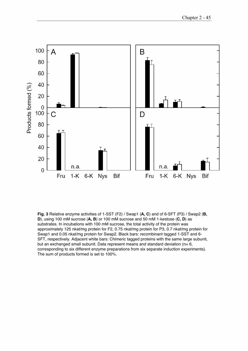
Chapter 2 - 45
Fig. 3 Relative enzyme activities of 1-SST (F2) / Swap1 (A, C) and of 6-SFT (P3) / Swap2 (B,D), using 100 mM sucrose (A, B) or 100 mM sucrose and 50 mM 1-kestose (C, D) assubstrates. In incubations with 100 mM sucrose, the total activity of the protein wasapproximately 125 nkat/mg protein for F2, 0.75 nkat/mg protein for P3, 0.7 nkat/mg protein forSwap1 and 0.05 nkat/mg protein for Swap2. Black bars: recombinant tagged 1-SST and 6-SFT, respectively. Adjacent white bars: Chimeric tagged proteins with the same large subunit,but an exchanged small subunit. Data represent means and standard deviation (n= 6,corresponding to six different enzyme preparations from six separate induction experiments).The sum of products formed is set to 100%.

Chapter 2 - 46
The same picture emerged when enzymatic activities were compared in the
presence of sucrose and 1-kestose as substrates. Swap1 yielded nystose and fructose
in the same proportions as the parent enzyme delivering the large subunit, 1-SST, but
no bifurcose as would have been expected if the enzyme contributing the small
subunit, 6-SFT, had a role in enzymatic specificity (Fig. 3C). Vice versa, Swap2
yielded bifurcose in the same proportion as the parent enzyme delivering the large
subunit, 6-SFT, but no nystose as would have been expected if the enzyme
contributing the small subunit, 1-SST, had a role in enzymatic specificity (Fig. 3D).
1-SST acts as a fructan exohydrolase when incubated with 1-kestose or
nystose alone [6]. This is also true for Swap1, comprising the large subunit of 1-SST:
Both the parent enzyme 1-SST and Swap1 yielded considerable amounts of fructose
when incubated with 1-kestose or nystose (Fig. 4A,B). Interestingly, both the parent
enzyme and Swap1 additionally act as a FFT when incubated with 1-kestose, as
indicated by the fact that about 15% of the products represent nystose (Fig. 4A).
Fig. 4 Relative enzyme activities of 1-SST(F2, black columns) and Swap1 (whitecolumns), using 50 mM 1-kestose (A) or50 mM nystose (B) as substrates. Datarepresent means and standard deviation(n= 6, corresponding to six differentenzyme preparations from six separateinduction experiments). The sum ofproducts formed is set to 100%.

Chapter 2 - 47
In contrast, both 6-SFT and recombinant Swap2, comprising the large subunit
of 6-SFT, displayed no measurable product formation in incubations with either 50
mM 1-kestose alone or nystose alone (data not shown).
To obtain further insight into the importance of the small subunit, we
attempted to express a tagged version of the large subunit of festuca 1-SST alone
(LSuB). However, we could not observe any enzyme activity in the corresponding
preparations from the P. pastoris culture fluid (data not shown). In addition, immune
blots with antibodies directed against the tags did not reveal any antigenic proteins in
the preparations from the culture fluid; however, an immune-responsive protein of the
correct size (approximately 50 kD) was present in preparations obtained by boiling
induced cells in the presence of 0.5% SDS and 500 mM NaCl, followed by de-salting
(data not shown). These data indicate that the large subunit alone is translated
correctly in Pichia, but not correctly folded and thus not secreted. The retained protein
showed no activity in enzymatic assays using lysed cells (data not shown).
In conclusion, our data show that the small subunit of fructosyltransferases is
essential for functional expression of the protein, but has no influence on the precise
catalytic specificity of the enzyme. It is the large subunit alone which determines
catalytic specificity and the nature of the products formed.
We are currently performing more detailed domain-exchange experiments within the
large subunit, in conjunction with site-directed mutagenesis of some of its highly
conserved domains, in order to narrow down the regions important for catalytic
specificity.
Acknowledgments: We thank V. Mandak for excellent help with HPLC-
analysis and P. Wille, M. Elfstrand and T. Ritsema for critical reading of the
manuscript. This project was funded, in part, by the Swiss National Science
Foundation.

Chapter 2 - 48
2.4.4. References
[1] Wiemken, A., Sprenger, N. and Boller, T. (1995) in: InternationalSymposium on Sucrose Metabolism, pp. 179-188 (Pontis, H.G.,Salerno, G.L. and Echeverria, E.J., Eds.) American Society of PlantPhysiology Press, Rockville, MA.
[2] Vijn, I. and Smeekens, S. (1999) Plant Physiol. 120, 351-359.[3] Ritsema, T. and Smeekens, S. (2003) Curr. Opin. Plant Biol. 6, 223-
230.[4] Pons, T., Olmea, O., Chinea, G., Beldarrain, A., Marquez, G., Acosta,
N., Rodriguez, L. and Valencia, A. (1998) Proteins - Structure, Functionand Genetics 33, 383-395.
[5] Sprenger, N., Bortlik, K., Brandt, A., Boller, T. and Wiemken, A. (1995)Proc. Natl. Acad. Sci. USA 92, 11652-11656.
[6] Lüscher, M., Hochstrasser, U., Vogel, G., Aeschbacher, R., Galati, V.,Nelson, C.J., Boller, T. and Wiemken, A. (2000) Plant Physiol. 124,1217-1227.
[7] Van Laere, A. and Van den Ende, W. (2002) Plant Cell Environ. 25,803-813.
[8] Koops, A.J. and Jonker, H.H. (1996) Plant Physiol. 110, 1167-1175.[9] Van den Ende, W., Van Wonterghem, D., Verhaert, P., Dewil, E., Van
Laere, A. (1996) Planta 199, 493-502.[10] Sturm, A. (1999) Plant Physiol. 121, 1-7.[11] Van den Ende, W., Michiels, A., Van Wonterghem, D., Vergauwen, R.
and Van Laere, A. (2000) Plant Physiol. 123, 71-79.[12] Reddy, A. and Maley, F. (1996) J. Biol. Chem. 271, 13953-13958.[13] Batista, F.R., Hernandez, L., Fernandez, J.R., Arrieta, J., Menendez,
C., Gomez, R., Tambara, Y. and Pons, T. (1999) Biochem. J. 337, 503-506.
[14] Hochstrasser, U., Lüscher, M., De Virgilio, C., Boller, T. and Wiemken,A. (1998) FEBS Lett. 440, 356-360.

Chapter 2 - 49
[15] Horton, R.M., Hunt, H.D., Ho, S.N., Pullen, J.K. and Pease, L.R. (1989)Gene 77, 61-68.
[16] Lüscher, M., Hochstrasser, U., Boller, T. and Wiemken, A. (2000) NewPhytol. 145, 225-232.

Chapter 3 - 50
Chapter 3
Mutational analysis of the active center of plant
fructosyltransferases: Festuca 1-SST and barley 6-SFT
Denise Altenbach*, Eveline Nüesch*, Tita Ritsema, Thomas Boller,
Andres Wiemken
* These authors contributed equally to the work
Botanisches Institut der Universität Basel, Zurich Basel Plant Science Center,
Hebelstrasse 1, CH-4056 Basel, Switzerland
Submitted to FEBS Letters
3.1. Abstract
The active center of the glycoside hydrolase family 32 contains the three
characteristic motifs (N/S)DPNG, RDP, and EC. We replaced the N-terminal region
including the (N/S)DPNG motif of barley 6-SFT (sucrose:fructan 6-
fructosyltransferase) by the corresponding region of Festuca 1-SST (sucrose:sucrose
1-fructosyltransferase). The chimeric enzyme, expressed in Pichia, retained the
specificity of 6-SFT. Attempts to replace a larger piece at the N-terminus including
also the RDP motif failed. A point mutation introduced in the RDP motif of 1-SST
abolished enzymatic activity. Interestingly, point mutations of the EC-motif resulted
in an enzyme which had lost the capability to form 1-kestose and glucose from
sucrose but still accepted 1-kestose, producing fructose and sucrose as well as
nystose.

Chapter 3 - 51
3.2. Introduction
Fructans are polymers of fructose used as storage carbohydrates by many
plants. They are synthesized from sucrose by specific fructosyltransferases (FTs). The
linkages between the fructosyl-residues are either exclusively of the b(2-1) or the
b(2-6) type, or both linkage types occur in branched fructans [1]. The degree of
polymerization and distribution of linkage types, depending on FT specificities, are
characteristic of different plant species. Fructan synthesis in plants is initiated
generally by a sucrose:sucrose 1-fructosyltransferase (1-SST) using sucrose both as
fructosyl donor and acceptor to produce 1-kestose, the shortest b(2-1) linked fructan,
and glucose. In the case of grasses such as wheat and barley, the next step of fructan
synthesis is mediated by a sucrose:fructan 6-fructosyltransferase (6-SFT), which again
uses sucrose as fructosyl donor but fructans, preferably 1-kestose, as acceptor,
forming b(2-6) linkages. Thus, by fructosyl transfer to 1-kestose the smallest
branched fructan, the tetrasaccharide bifurcose, is formed. It remains to be shown
whether or not additional FTs are involved in fructan synthesis of grasses.
Many plant FTs have been sequenced during the last few years, and the data
clearly indicate a high homology to the vacuolar, acid invertases (b-fructosidases).
These enzymes are all members of the glycoside hydrolase family 32 (GH32) and
share three highly conserved regions characterized by the motifs (N/S)DPNG (also
called b-fructosidase motif), RDP, and EC [2]. A function in sucrose hydrolysis has
been experimentally shown for the (N/S)DPNG- and EC-motifs in the case of yeast
invertase, and for the RDP motif in the case of bacterial FTs belonging to the family
GH68 [3-5]. An invertase of the bacterium Thermotoga maritima was the first GH32
member of which the crystal structure has been determined [6], followed by structural
analyses of an Aspergillus awamori exo-inulinase and of a chicory fructan-
exohydrolase [7,8]. These structures show all a five bladed b-propellers with a deep
negatively charged central pocket and a b-sandwich. The three highly conserved
motifs mentioned above flank this central pocket. The aspartate of the (N/S)DPNG
motif is proposed to act as a nucleophile, the glutamate of the EC-motif as a proton
donor, and the aspartate of the RDP motif as transition state stabilizer in the
transfructosylation reaction [2].

Chapter 3 - 52
From the GH32 members structurally elucidated, chicory FEH is the closest
relative of plant FTs and vacuolar invertases, with an amino acid sequence identity of
30 to 40%. For plant FTs, no crystal structures are available yet, but the three
conserved domains of chicory FEH flanking the central pocket, defined by the motifs
(N/S)DPNG, RDP and EC, are highly conserved also in these FTs. Moreover, the
(N/S)DPNG-motif has been shown by mutational studies to be essential for the
activity of an onion FT [9]. For the RDP- and EC-motif of plant FT’s, such studies
have not been made so far. Another common feature of plant FTs and vacuolar
invertases is that they usually are composed of a large and a small subunit due to a
posttranslational processing [10-12], as indicated in Fig. 1A. . In a previous study
using barley 6-SFT [10] and Festuca 1-SST [13], we found that the large subunit,
which harbours all three conserved domains mentioned above, determines the
catalytic specificity [14].
Our present work continues the comparative study of barley 6-SFT and
Festuca 1-SST and provides the first experimental evidence that the motifs of all the
three conserved domains are of functional importance in the case of the two plant
FT’s studied. We narrowed down the region of the proteins responsible for enzyme
specificity by exchanging domains including the (N/S)DPNG-motif between Festuca
1-SST and barley 6-SFT, and by introducing point mutations in the EC- and RDP-
motif of Festuca 1-SST.

Chapter 3 - 53
3.3. Materials and Methods
Fig. 1. Constructs for expression in P. pastoris. A: Festuca 1-SST. Conserved motifs areindicated. B: Barley 6-SFT. C: Chimera1: N-terminus of mature Festuca 1-SST comprisingthe (N/S)DPNG motif, with the C-terminus of barley 6-SFT comprising the RDP and EC motifsfused to it. D: Chimera2: N-terminus of mature Festuca 1-SST comprising the (N/S)DPNGand RDP motif, with the C-terminus of barley 6-SFT comprising the EC motif fused to it. E, F:Festuca 1-SST with selected sites for amino acid substitutions. Numbers indicate positions inthe corresponding amino acid sequence, counted from the N-terminus of the open readingframes.

Chapter 3 - 54
3.3.1. Construction of recombinant chimeric enzymes, mutagenesis and expression
The Pichia shuttle plasmids pPICZaC-F2 and pPICZaC-P3 were described
before [14]. They carry a Zeocin resistance gene and contain the following coding
sequences, cloned in frame behind the a-factor signal sequence: pPICZaC-F2, mature
Festuca 1-SST (from Festuca arundinacea, re-classified as Schedonorus
arundinaceus; EMBL AJ297369 [13]; Fig. 1A); pPICZaC-P3, mature barley 6-SFT
(from Hordeum vulgare; EMBL X83233 [10]; Fig. 1B). They were used for the
construction of Chimera1 and Chimera2 (Fig. 1C and D).
Table 1
Oligonucleotides used for cloning
Primer Sequence
SST023.2 5’-CAAGGACTTCCGCGCCCCCCTCACCGCCTG-3’
SST024.2 5’-CAGGCGGTGAGGGGGGCGCGGAAGTCCTTG-3’
SST015 5’-CATGTACGACTGCATCGACCTCTACCCCGTCGG-3’
SST016 5’-CCGACGGGGTAGAGGTCGATGCAGTCGTACATG-3’
SST017 5’-CATGTACGCGTGCATCGACCTCTACCCCGTCGG-3’
SST018 5’-CCGACGGGGTAGAGGTCGATGCACGCGTACATG-3’
SST-002 5’-GAAGCATCGATGAATTCCGCC-3’
RDP-001 5’-CATCCTGCTCTACACCGGCGCCACCAACGCCTCCGCC-3’
SFT-002 5’-GAGCCACCGTCCTAGGAAC3’
RDP-002 5’-GGCGGAGGCGTTGGTGGCGCCGGTGTAGAGCAGGATG-3’
EC-001 5’-GGACGACGACGGCCACGCAGGCATCGCCATGATGTACAAG-3’
EC-002 5’-CTTGTACATCATGGCGATGCCTGCGTGGCCGTCGTCGTCC-3’

Chapter 3 - 55
For Chimera1 (Fig. 1C), the N-terminal region of mature Festuca 1-SST was
coupled to the C-terminus of barley 6-SFT by overlapping PCR [15], using primer
pair SST-002/RDP-002 to amplify from the EcoRI cloning site to bp 639 in 1-SST
and SFT-002/RDP-001 to amplify 6-SFT from bp 523 to bp 1162. The PCR-amplified
fragments were used as templates in a second PCR with primers SST-002 and SFT-
002. The full-length product created in this way was cloned into pPICZaC-P3 using
EcoRI and XmaJI resulting in pPICZaC-Chimera1.
Chimera2 (Fig. 1D) was created correspondingly using primers SST-002/EC-
002 and SFT-002/EC-001 and primers SST-002 and SFT-002 in the second PCR,
followed by EcoRI/XmaJI cloning resulting in pPICZaC-Chimera2.
Mutant SST(D260A), in which the aspartate in the RDP motif of Festuca 1-
SST was changed into alanine (Fig. 1E) , as well as the mutant SST(E318A) and
SST(E318D), in which the glutamate of tje EC motif was changed into alanine or
aspartate (Fig. 1F), respectively, were generated using the QuikChangeTM Site-
Directed Mutagenesis Kit (Strategene), based on the pK18-clone containing the
tagged version of Festuca 1-SST as described [14]. Primers SST023.2 and SST024.2
(Table 1) were designed to introduce the desired mutation for SST(D260A), SST017
and SST018 for SST(E318A) and SST015 and SST016 for SST(E318D).
SST(D260A), SST(E318A) and SST(E318D) were cloned EcoRI/XbaI into the Pichia
shuttle vector pPICZaC. All inserts in pPICZaC were sequenced after cloning and
found to carry the desired mutation or the desired chimera. All constructs carry a C-
terminal 6_histidine tag and are cloned behind the a-factor secretion signal.
Transformation to Pichia, expression and further processing were described before
[14].
3.3.2. Characterization of recombinant fructosyltransferases
Enzyme assays were performed at 27°C with 100 mM sucrose (suc) or with 50
mM of all other substrates, and the products formed were analysed by anion exchange
chromatography as described [16]. Catalytic specificity was tested with the following
substrates: sucrose, 1-kestose, nystose and a combination of sucrose and 1-kestose. A
maximum of 7% of the substrates was used up after the incubations. In order to keep

Chapter 3 - 56
the substrate concentration constant during the experiments, the amount of protein
used per enzyme assay was ca 0.5 mg for Festuca 1-SST and 5 to 10 mg for all other
constructs.
For western blots, methanol-chloroform precipitated enzyme preparations (5 to
10 mg each) were separated on a 10% gel by SDS-PAGE, electro blotted to a
nitrocellulose membrane (Schleicher & Schuell GmbH) and probed with monoclonal
anti-His and alkaline-phosphatase secondary antibodies.

Chapter 3 - 57
3. 4. Results
3.4.1. Heterologous expression in the yeast Pichia pastoris
To study structure-function relationships in plant FTs, the Pichia pastoris
yeast expression system is a powerful tool [14]. The chimeric constructs were
constructed with overlapping PCR using Festuca arundinacea 1-SST (Fig. 1A) and
Hordeum vulgare 6-SFT (Fig. 1B). In Chimera1 (Fig. 1C), the N-terminal part
encompassing the (N/S)DPNG motif is from 1-SST, whereas the C-terminal part
encompassing the RDP motif and the EC-motif is from 6-SFT. In Chimera2 the N-
terminal part encompassing the (N/S)DPNG- and RDP-motif is from 1-SST, whereas
the C-terminal part encompassing the EC-motif is from 6-SFT (Fig. 1D). Point
mutations were investigated using Festuca 1-SST (Fig. 1E, F).
Fig. 2. Western Blot analysis of recombinant methanol-chloroform precipitated enzymepreparations (5-10 mg each), probed with anti-His and alkaline-phosphatase secondaryantibodies. The size of the bands is indicated in kD. P0: Pichia transformed with empty vector;1-SST: recombinant 1-SST; 6-SFT: recombinant 6-SFT; Chimera1, Chimera2; SST(E318A),SST(E318D), SST(D260A): see Fig. 1.
P0 1-SST 6-SFT Chimera2 Chimera1 SST(E318D)
SST(D260A)
85 kD
SST(E318A)
35 kD

Chapter 3 - 58
To verify proper expression of the constructs, western blots were performed
using antibodies against the C-terminal His tag. Extracts of Pichia transformed with
the empty vector pPICZaC did not show any bands on the blots (Fig. 2, lane P0). In
contrast, clear bands were revealed on blots with extracts of Pichia transformed with
the various constructs. Normally, Festuca 1-SST and barley 6-SFT isolated form
plants consist of two subunits and show two bands on SDS-PAGE [10,13]. However,
upon expression in Pichia, most constructs yielded primarily a band of approximately
85 kD. This indicates that the recombinant proteins are well expressed but not cleaved
into two subunits, as was already previously demonstrated for recombinant untagged
SST [13]. A faint band at 35 kD was apparent for some of the constructs, indicating
release of a fragment possibly corresponding to the small subunit in Pichia (Fig. 2);
since the antibodies are directed against the C-terminal tag, fragments corresponding
to the large subunit cannot be seen on this western blot. For Chimera2, the 85 kD is
even fainter than the 35 kD band, indicating that this product is weakly expressed or
unstable in Pichia.
3.4.2. The (N/S)DPNG motif does not determine enzymatic specificity
Chimera1 (Fig. 1C), with the NDPNG motif of 1-SST, was tested with
different substrates and its activity was compared to the activities of recombinant 1-
SST and 6-SFT, with the motif in the form of SDPNG (Fig. 3). In incubations with
100 mM sucrose it was mainly hydrolytically active, as can be seen by the
predominant production of fructose (Fig. 3A). The other products formed by
Chimera1 in incubations with sucrose were 1-kestose, 6-kestose and marginal
amounts of bifurcose. The products formed and their relative amounts were the same
as for recombinant 6-SFT. In corresponding incubations, recombinant 1-SST produces
mainly 1-kestose (80%). Also in incubations with a combination of sucrose and 1-
kestose, the products formed by Chimera1 were fructose, 6-kestose and bifurcose, and
their relative amounts were the same as for recombinant 6-SFT (Fig. 3B). In contrast,
the corresponding incubation with 1-SST lead to the production of fructose and
nystose. These analyses of Chimera1 showed that it retained all the characteristics of
recombinant 6-SFT. Its 1-SST derived N-terminal part, including the conserved b-
fructosidase motif NDPNG, does not influence enzymatic properties.

Chapter 3 - 59
Preparations of Chimera2 (Fig. 1D) did not show any enzymatic activity. In
addition, as shown above (Fig. 2), this chimera yielded only low amounts of protein,
indicating that this highly conserved region between the EC-motif and the RDP motif
is important for protein stability.
Fig. 3. Relative enzyme activities of recombinant 1-SST, 6-SFT and Chimera1 using 100 mMsucrose (A) or 100 mM sucrose and 50 mM 1-kestose (B) as substrates. In incubations with100 mM sucrose, the total activity of the protein was approximately 125 nkat/mg protein for 1-SST, 0.75 nkat/mg protein for 6-SFT and 0.7 nkat/mg protein for Chimera1. Data representmeans and standard deviation (n= 5, corresponding to five different enzyme preparationsfrom five separate induction experiments). The sum of products formed is set to 100%. F:fructose, 1K: 1-kestose, 6K: 6-kestose, B: bifurcose, N: nystose.
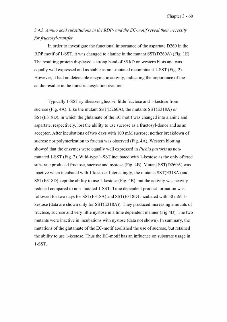
Chapter 3 - 60
3.4.3. Amino acid substitutions in the RDP- and the EC-motif reveal their necessity
for fructosyl-transfer
In order to investigate the functional importance of the aspartate D260 in the
RDP motif of 1-SST, it was changed to alanine in the mutant SST(D260A) (Fig. 1E).
The resulting protein displayed a strong band of 85 kD on western blots and was
equally well expressed and as stable as non-mutated recombinant 1-SST (Fig. 2).
However, it had no detectable enzymatic activity, indicating the importance of the
acidic residue in the transfructosylation reaction.
Typically 1-SST synthesizes glucose, little fructose and 1-kestose from
sucrose (Fig. 4A). Like the mutant SST(D260A), the mutants SST(E318A) or
SST(E318D), in which the glutamate of the EC motif was changed into alanine and
aspartate, respectively, lost the ability to use sucrose as a fructosyl-donor and as an
acceptor. After incubations of two days with 100 mM sucrose, neither breakdown of
sucrose nor polymerization to fructan was observed (Fig. 4A). Western blotting
showed that the enzymes were equally well expressed in Pichia pastoris as non-
mutated 1-SST (Fig. 2). Wild-type 1-SST incubated with 1-kestose as the only offered
substrate produced fructose, sucrose and nystose (Fig. 4B). Mutant SST(D260A) was
inactive when incubated with 1-kestose. Interestingly, the mutants SST(E318A) and
SST(E318D) kept the ability to use 1-kestose (Fig. 4B), but the activity was heavily
reduced compared to non-mutated 1-SST. Time dependent product formation was
followed for two days for SST(E318A) and SST(E318D) incubated with 50 mM 1-
kestose (data are shown only for SST(E318A)). They produced increasing amounts of
fructose, sucrose and very little nystose in a time dependent manner (Fig 4B). The two
mutants were inactive in incubations with nystose (data not shown). In summary, the
mutations of the glutamate of the EC-motif abolished the use of sucrose, but retained
the ability to use 1-kestose. Thus the EC-motif has an influence on substrate usage in
1-SST.

Chapter 3 - 61
Fig. 4. HPAEC-PAD profile of products formed in enzyme assays of 1-SST, SST(D260A) andSST(E318A). Recombinant enzymes were incubated with 100 mM sucrose (A) or 50 mM 1-kestose (B) as substrate. Products formed by 1-SST were analyzed after 1h of incubation.Products formed in incubations of SST(D260A) and SST(E318A) were analyzed from 0h to 2days. G: glucose, F: fructose, S: sucrose, K: 1-kestose, Nys: nystose.

Chapter 3 - 62
3.5. Discussion
Our biochemical data on the enzymatic activities of mutated plant
fructosyltransferases are of interest in view of the recent elucidation of the structure of
some related enzymes. Crystal structures became available recently for members of
the glycoside hydrolase families GH32 and GH68, i.e. enzymes that catalyze similar
reactions although they share only 15% overall sequence identity. They all form five
bladed b-propellers with a deep negatively charged central pocket representing the
active site [6-8,17]. Members of GH32 additionally have a b-sandwich attached to the
propeller that possibly corresponds to the small subunit found in plant acid invertases
and FTs. In the structure of Bacillus subtilis levansucrase (GH68), the central pocket
contains three acidic amino acid residues essential for the catalytic mechanism: the
nucleophile (D86), the general acid/base catalyst (E342) and the transition state
stabilizer (D247) [17]. These catalytic residues are also present in the active site of all
enzymes of family GH32 for which structures are available, and they correspond to
the conserved motifs (N/S)DPNG, EC and RDP within GH32.
In plant enzymes belonging to this family, i.e. the FTs and acid invertases,
only the (N/S)DPNG (b-fructosidase) motif was biochemically characterized via
domain-exchanges and point mutations. It was shown to be important for product
specification but not for the specification of the type of fructosyl donor used; thus its
influence on enzyme specificity seems to act via acceptor specification [9]. The
mutation of the nucleophile itself (D85) resulted in an enzymatically inactive protein
(Ritsema et al., submitted).
In our present work, the results with Chimera1 gave further insight into the
function of the b-fructosidase motif, which has the usual sequence NDPNG in 1-SST
but the less frequent sequence SDPNG in 6-SFT. The chimeric enzyme retained all
the characteristics of recombinant 6-SFT although its N-terminal part, including the
canonical NDPNG motif, was derived from 1-SST. 1-SST and 6-SFT can both use
sucrose as an acceptor and donor substrate, but they differ in their preferential
fructosyl acceptor substrate. Chimera1 prefers the same fructosyl acceptor substrates
like barley 6-SFT. Since 1-SST and 6-SFT have the same preferred fructosyl-donor

Chapter 3 - 63
(sucrose), exchanging the N-terminus containing the b-fructosidase motif might give
no change in donor specificity. The C-terminal part of the chimera is influencing
acceptor binding which is different for 1-SST and 6-SFT (sucrose or 1-kestose
respectively).
The point mutation SST(D260A) of the RDP motif yielded an inactive
product. The aspartate residue of this motif has been proposed to act as transition state
stabilizer [4,5,17,18]. It forms strong hydrogen bonds with the substrate sucrose, but
is too far away from either the C2’ hydroxyl or the glycosidic oxygen of sucrose to be
directly involved in the catalytic mechanism, i.e. in hydrolyzing the bond between
glucose and fructose of sucrose [6,17]. The mutation (D260A) might increase the
distance to the substrate and therefore be a hindrance for the formation of hydrogen
bonds, but a conformational change of SST(D260A) cannot be excluded.
The acidic amino acids in the other two motifs have been found to be directly
involved in catalysis. This was first described for yeast invertase, a member of the
GH32 family: The aspartate in the (N/S)DPNG (b-fructosidase) motif (Asp23) was
shown to be the catalytic nucleophile while the glutamate of the EC-motif (Glu204)
was identified as the general acid/base catalyst [3]. Our results with the point mutation
of the corresponding glutamate in 1-SST (SST(E318A); SST(E318D)) are particularly
intriguing, since they indicate a resulting change in the enzyme's catalytic activity.
Both mutated forms lost the ability to use sucrose as a substrate, showing neither
cleavage into glucose and fructose nor polymerization to 1-kestose. This result is in
accordance with the corresponding mutation of E204A in yeast invertase, where also
the sucrose cleaving capacity was heavily reduced. Yeast invertase has only one
substrate (sucrose) whereas Festuca 1-SST can also use 1-kestose. Wild type 1-SST
produces fructose, sucrose and nystose when incubated with 1-kestose. The mutants
SST(E318A) and SST(E318D)) formed these products from 1-kestose as well,
although with markedly reduced efficiency. The 3D-structure of the active site of
Bacillus subtilis levansucrase in the presence of sucrose shows that the glutamate of
the EC-domain points towards the bond between the glucose and the fructose moiety
of sucrose [17]. The fructosyl unit of sucrose is located at the bottom of the pocket
and the glucose moiety on top. Since 1-kestose has two fructose moieties attached to

Chapter 3 - 64
glucose, its terminal fructosyl residue may contact an alternative acid/base catalyst in
the active site. Due to the low overall homologies of Festuca 1-SST to the crystal
structures available, it cannot be predicted which glutamate might replace E318 in
catalysis.
The general glycosyl hydrolase reaction mechanism in members of the GH32
and GH68 families is based on three acidic amino acid residues in the active center.
Neither the available crystal structures nor the data obtained with mutational analysis
can unequivocally answer the question what structural components determine enzyme
specificity. Functional differences such as N-glycosylation or amino acid changes
outside active site residues may play a role in the selection of different substrates[8]
and will be investigated in the future.
Acknowledgements
We thank V. Mandak for excellent help with HPLC-analysis and D. Brodmann
for help with figures. This project was funded, in part, by the Swiss National Science
Foundation.

Chapter 3 - 65
3.6. References[1] Pollock, C.J. and Cairns, A.J. (1991) Ann. Rev. Plant Physiol. Plant
Mol. Biol. 42, 77-101.[2] Pons, T., Naumoff, D.G., Martinez-Fleites, C. and Hernandez, L. (2004)
Proteins 54, 424-432.[3] Reddy, A. and Maley, F. (1996) J. Biol. Chem. 271, 13953-13958.[4] Batista, F.R., Hernandez, L., Fernandez, J.R., Arrieta, J., Menendez,
C., Gomez, R., Tambara, Y. and Pons, T. (1999) Biochem. J. 337, 503-506.
[5] Yanase, H., Maeda, M., Hagiwara, E., Yagi, H., Taniguchi, K. andOkamoto, K. (2002) J. Biochem. 132, 565-572.
[6] Alberto, F., Bignon, C., Sulzenbacher, G., Henrissat, B. and Czjzek, M.(2004) J. Biol. Chem. 279, 18903-18910.
[7] Nagem, R.A.P., Rojas, A.L., Golubev, A.M., Korneeva, O.S.,Eneyskaya, E.V., Kulminskaya, A.A., Neustroev, K.N. and Polikarpov, I.(2004) J. Mol. Biol. 344, 471-480.
[8] Verhaest, M., Van den Ende, W., Le Roy, K., De Ranter, C.J., VanLaere, A. and Rabijns, A. (2005) Plant J. 41, 400-411.
[9] Ritsema, T., Verhaar, A., Vijn, I. and Smeekens, S. (2004) Plant Mol.Biol. 54, 853-863.
[10] Sprenger, N., Bortlik, K., Brandt, A., Boller, T. and Wiemken, A. (1995)Proc. Nat. Acad. Sci. USA 92, 11652-11656.
[11] Sturm, A. (1999) Plant Physiol. 121, 1-7.[12] Van den Ende, W., Michiels, A., Van Wonterghem, D., Vergauwen, R.
and Van Laere, A. (2000) Plant Physiol. 123, 71-79.[13] Lüscher, M., Hochstrasser, U., Vogel, G., Aeschbacher, R., Galati, V.,
Nelson, C.J., Boller, T. and Wiemken, A. (2000) Plant Physiol. 124,1217-1227.
[14] Altenbach, D., Nüesch, E., Meyer, A.D., Boller, T. and Wiemken, A.(2004) FEBS Lett. 567, 214-218.
[15] Horton, R.M., Hunt, H.D., Ho, S.N., Pullen, J.K. and Pease, L.R. (1989)Gene 77, 61-68.

Chapter 3 - 66
[16] Lüscher, M., Hochstrasser, U., Boller, T. and Wiemken, A. (2000) NewPhytol. 145, 225-232.
[17] Meng, G.Y. and Fütterer, K. (2003) Nature Struct. Biol. 10, 935-941.[18] Ozimek, L.K., van Hijum, S.A.F.T., van Koningsveld, G.A., van der
Maarel, M.J.E.C., van Geel-Schutten, G.H. and Dijkhuizen, L. (2004)FEBS Lett. 560, 131-133.

Chapter 4 - 67
Chapter 4
Are fructosyltransferases modified invertases?
Collaborative work with Dr. Tita Ritsema
Zurich Basel Plant Science Center, Botanisches Institut der Universität Basel,
Hebelstrasse 1, CH-4056 Basel, Switzerland
4.1. Abstract
Enzymes polymerizing sucrose to fructans (fructosyltransferases, FTs) are highly
homologous to enzymes hydrolyzing sucrose (acid invertases) and belong to glycoside
hydrolase family 32. Recently, the first crystal structures were determined within this
family. They consists of a five bladed b-propeller with a deep negatively charged central
pocket representing the active site, and a b-sandwich. There are three highly conserved
motifs flanking the central pocket that are functionally essential: the (N/S)DPNG-motif,
the RDP-motif and the EC-motif. However, it remains unclear what structural
components determine polymerization or breakdown of sucrose. Therefore we decided to
perform a mutational analysis based on sequence comparisons between acid invertases
and sucrose:sucrose 1-fructosyltransferases (1-SSTs), the latter an example of sucrose-
using FTs, and chose Allium cepa invertase and Festuca arundinacea (re-classified as
Schedonorus arundinaceus) 1-SST for our analysis. Nine amino acids dispersed along the
sequence could be identified correlating with either invertase or 1-SST activity,
respectively. The selected amino acids of onion invertase were mutated to the
corresponding amino acids in Festuca 1-SST and vice versa. For both enzymes, the
mutations were analyzed independently. Functional expression in the yeast Pichia
pastoris revealed shifts in the catalytic specificity and activity, demonstrating the
importance of amino acids outside the three highly conserved motifs ((N/S)DPNG, RDP,
EC) for the enzymatic reaction.

Chapter 4 - 68
4.2. Introduction
Fructans, are a class of highly water-soluble polysaccharides consisting of linear
or branched fructose chains attached to sucrose. In plants, they occur in many prominent
orders like the Asterales, the Liliales, and the Poales, among which are representatives of
economic importance (e.g. wheat, barley) (Hendry, 1993). As highly water soluble
molecules fructans are predominantly stored in the vacuole (Wiemken et al., 1995).
Besides their function as a reserve carbohydrate, fructans are suggested to be involved in
cell expansion (Bieleski, 1993) and enhanced tolerance to cold and drought via
stabilization of cellular membranes (Hincha et al., 2000; Konstantinova et al., 2002;
Vereyken et al., 2003).
Fructan synthesis in plants starts with the conversion of sucrose to the shortest
b(2,1) linked fructan 1-kestose. The enzyme that performs this reaction, sucrose:sucrose
1-fructosyltransferase (1-SST), is found in all fructan-producing plants. It uses two
molecules of sucrose, one as fructosyl donor and the other one as fructosyl acceptor for
the synthesis of the trisaccharide 1-kestose. In a second step, chain elongation to higher
DP fructans occurs via the action of fructan:fructan 1-fructosyltransferase (1-FFT),
sucrose:fructan 6-fructosyltransferase (6-SFT) and/or fructan:fructan 6G-
fructosyltransferase (6G-FFT), depending on the species-specific type of fructan
synthesized by the plant (Vijn and Smeekens; 1999). These fructosyltransferases (FTs)
display different donor and acceptor specificities using either sucrose and/or fructan as
fructosyl donor or acceptor, respectively, and are named according to the substrates they
use and according to the linkage type they form (b(2,1) or b(2,6)). Fructan cleavage is
catalyzed by fructan exohydrolases (FEHs).
Functionally related to FTs are invertases, transferring the fructosyl residue from
sucrose not to a saccharide-chain but to water, producing glucose and fructose. Vacuolar
invertases resemble SSTs at the biochemical level: SSTs generally show a low but
distinct hydrolase activity and vice versa invertases produce traces of 1-kestose,
particularly at high sucrose concentrations (≥ 100 mM; Sprenger et al., 1995).

Chapter 4 - 69
Invertases are ubiquitous in plants whilst fructosyltransferases are restricted to several
unrelated families (Hendry, 1987). Peptide sequences of plant FTs generally have a very
high similarity to amino acid sequences of different invertases (Sprenger et al., 1995; Van
den Ende et al., 1996; Vijn et al., 1997). If vacuolar invertases are presented in
phylogenetic trees with FTs, fructan exohydrolases (FEHs) and cell-wall invertases, the
vacuolar invertases always cluster with FTs whilst the FEHs cluster with cell-wall
invertases (Fig. 1; Wei and Chatterton, 2001). Close relationships at the biochemical and
molecular level between fructan biosynthesizing enzymes and invertases support the idea,
that the fructan synthesizing enzymes evolved from vacuolar invertases by relatively few
mutational changes (Sprenger et al., 1995; Vijn and Smeekens, 1999). As the initiator of
fructan synthesis, 1-SSTs are thought to be the closest relatives to vacuolar invertases.
Both enzymes use sucrose as substrate: Invertases for hydrolyzation and SSTs for
polymerization.
Plant fructan metabolizing enzymes and acid invertases are grouped in glycoside
hydrolase family 32 (GH32), based on overall sequence homologies (Henrissat, 1991).
Three highly conserved motifs are a characteristic for the GH32 family. They consist of
the following conserved sequences: (N/S)DPNG (also called b-fructosidas motif), RDP
(RDP motif) and EC (EC-motif). The acidic amino acids (marked in bold) of these motifs
have been proposed to function as active site residues (Reddy and Maley, 1996; Pons et
al., 2004). These three motifs can explain the cleavage of glycosidic bonds in general, but
not what structural components determine enzyme specificity. It still has to be shown
what features determine fructosyltransfer to a saccharide (FT activity) or to water
(invertase activity).
To address the questions of structural components determining different enzyme
specificities, we performed a targeted mutational approach, based on sequence
comparisons between acid invertases and 1-SSTs. Sequence comparisons of acid
invertases and 1-SSTs revealed nine amino acids dispersed along the sequence to
correlate with either invertase or 1-SST activity respectively (Fig. 2). For our analysis we
chose Allium cepa invertase (acINV) and Festuca arundinacea 1-SST (faSST; re-
classified as Schedonorus arundinaceus), two enzymes that have been very efficiently

Chapter 4 - 70
expressed in Pichia pastoris (Lüscher et al., 2000; acINV by T. Ritsema). The selected
nine amino acids of acINV were mutated to the corresponding amino acid in faSST and
vice versa (Fig. 3). For both enzymes, the mutations were analyzed independently. The
functional expression of the mutated enzymes in the yeast system Pichia pastoris
revealed shifts in the catalytic specificity and activity of invertase and 1-SST
respectively.

Chapter 4 - 71
4.3. Materials and Methods
4.3.1. Cloning and mutagenesis of Festuca 1-SST (faSST)
FaSST mutants carrying single mutations at specific sites of the sequence (A, B,
C, D, E, F, G, H and X) were generated using the QuikChangeTM Site-Directed
Mutagenesis Kit (Strategene), based on the pK18-clone containing the tagged version of
Festuca 1-SST (Altenbach et al., 2004).
Primers SSTAf and SSTAr (Table 1) were designed to introduce the desired mutation at
site A in faSST, SSTBf and SSTBr for the mutation at site B etc.
Wild type sequences and mutated sequences were cloned EcoRI/XbaI into the Pichia
shuttle vector pPICZaC. Competent E. coli cells were transformed with pPICZaC-
faSST-A, pPICZaC-faSST-B etc. respectively, and selected for Zeocin‘ resistance. All
inserts in pPICZaC were sequenced after cloning and found to carry the desired
mutation.
4.3.2. Cloning and mutagenesis in onion invertase
The onion invertase present in pBlue (Vijn et al, 1998) was digested with SacI
and XhoI and cloned into the mutagenesis vector pALTER-1 (Promega) digested with
SacI and SalI (SalI and XhoI create the same overhang). Mutagenesis was performed
using Altered Sites II (Promega) as described in the manual and mutations were checked
by sequencing.
For cloning into the Pichia vector pPICZaC (Invitrogen) wild type invertase and mutants
were PCRed with primers containing ClaI and XbaI restriction sites. After digestion of
the product and the pPICZaC vector with ClaI and XbaI, they were ligated and the
presence of inserts was confirmed by PCR.
Chapter 4 - 72
Table 1
Oligonucleotides used for cloning, mutations are indicated in bold
Primer Sequence
SSTAf 5’-CAGCCAGAGAAGCACTGGATGAACGATCCCAACG-3’
SSTAr 5’-CGTTGGGATCGTTCATCCAGTGCTTCTCTGGCTG-3’SSTBf 5’-GCCGTGTCCAAGGACCTAGTGAACTGGCGCCAC-3’SSTBr 5’-GTGGCGCCAGTTCACTAGGTCCTTGGACACGGC-3’
SSTCf 5’-CTAGCCCAGGTCCAGAACCTCGCCACGCCCGCC-3’
SSTCr 5’-GGCGGGCGTGGCGAGGTTCTGGACCTGGGCTAG-3’
SSTDf 5’-GCCAACCCCATCCTCGTCCCGCCCCCCGGTATC-3’
SSTDr 5’-GATACCGGGGGGCGGGACGAGGATGGGGTTGGC-3’
SSTEf 5’-GACGGCACCGGCATGTGGGAGTGCATCGACCTC-3’
SSTEr 5’-GAGGTCGATGCACTCCCACATGCCGGTGCCGTC-3’
SSTFf 5’-TACGAGTGCATCGACTTCTACCCCGTCGGTGGC-3’
SSTFr 5’-GCCACCGACGGGGTAGAAGTCGATGCACTCGTA-3’
SSTGf 5’-CGCATCGTCTGGGCCTGGATCGGCGAGACCGAC-3’
SSTGr 5’-GTCGGTCTCGCCGATCCAGGCCCAGACGATGCG-3’
SSTHf 5’-ACCAAGGGCTGGGCATCTCTCATGACGATTCCAAG-3’
SSTHr 5’-CTTGGAATCGTCATGAGAGATGCCCAGCCCTTGGT-3’SSTXf 5’-TACGCGCTCGGGAGGTATGACGCCGCCGCCAAC-3’
SSTXr 5’-GTTGGCGGCGGCGTCATACCTCCCGAGCGCGTA-3’
Primer Sequence
INVA 5’-GTGAAGAACTATATGAACGATC-3’
INVB 5’-CTCCAGAGACATGGTCCACTGGA-3’
INVC 5’-CCAAGTCCAGTGCTTAGCGGTC-3’
INVD 5’-CCCCATCCTGTACCCTCCCCCA-3’
INVE 5’-GTTGGTATGTACGAATGCGTGG-3’
INVF 5’-ATGCGTGGATCTCTACCCGGT-3’
INVG 5’-CTTTGGAGTTACATTGGAGAAA-3’
INVH 5’-GGGCTGGGCTAATCTTCAGGGT-3’
INVX 5’-ATCGGGACGTTCGATCCTGCAC-3’

Chapter 4 - 73
4.3.3. Expression in Pichia pastoris
The wild-type enzymes and all mutants were present in frame behind the yeast a-
factor signal sequence of the expression vector pPICZaC, to allow entry into the
secretory pathway of Pichia pastoris. Competent Pichia pastoris cells were either
transformed according to the EasyComp transformation protocol (EasySelectTM Pichia
Expression Kit, Invitrogen BV) or by electroporation as described in the manual and
selected for Zeocine‘ resistance.
Induction of protein expression in Pichia pastoris was performed as described (Altenbach
et al., 2004) with minor modifications: The growth medium was supplemented by 1%
(v/v) methanol at 16, 24 and 40h of induction. After 45h of induction the cultures were
centrifuged (10 min, 3000 rpm) and the supernatants containing the recombinant enzymes
were used for all subsequent steps.
4.3.4. Characterization of recombinant enzymes
To characterize the enzymes, the supernatants were diluted 1:10 to have a final
concentration of 50 mM MES-buffer pH 5.6 and the desired sucrose concentration per
assay. Enzyme assays contained sucrose concentrations as indicated (1 mM to 1 M). They
were incubated for 20 minutes at 27°C and thereafter stopped by heating for 2 min at
95°C. The products formed were analysed by high performance anion exchange
chromatography (HPAEC) as described (Lüscher et al., 2000).

Chapter 4 - 74
4.4. Results
4.4.1. Molecular relationships of vacuolar invertases and sucrose:sucrose 1-
fructosyltransferases (1-SSTs)
Sequence similarity between 1-SSTs and vacuolar invertases reaches 80%. In a
phylogenetic tree acid invertases and 1-SSTs exhibit separate but closely related clusters
(Fig. 1). There are some exceptions: for example the vacuolar invertase from barley
seems to be more closely related to SSTs and SFTs than to vacuolar invertases from Zea
mais and Arabidopsis thaliana that are catalyzing the same reaction. SSTs from grass
species (Festuca arundinacea, Lolium perenne, Hordeum vulgare and Triticum aestivum)
build a cluster. They are more closely related to vacuolar invertases from
monocotyledonous species (Hordeum vulgare, Allium cepa or Zea mais) than to the SSTs
from dicotyledonous species included in the phylogenetic tree (Helianthus tuberosus and
Chicorium intybus; Fig. 1).
Interestingly, enzymes from one species or family that confer different types of
reactions are sometimes more closely related than enzymes that catalyze the same
reaction but belong to different species or families. For example the different enzymes
from Allium cepa (1- SST, 6G-FFT and the vacuolar invertase) are clustering with the
SST form Allium sativum in the phylogenetic tree (Fig. 1). The tree furthermore
illustrates that vacuolar invertases are more homologous to FTs than to cell-wall
invertases and FEHs.
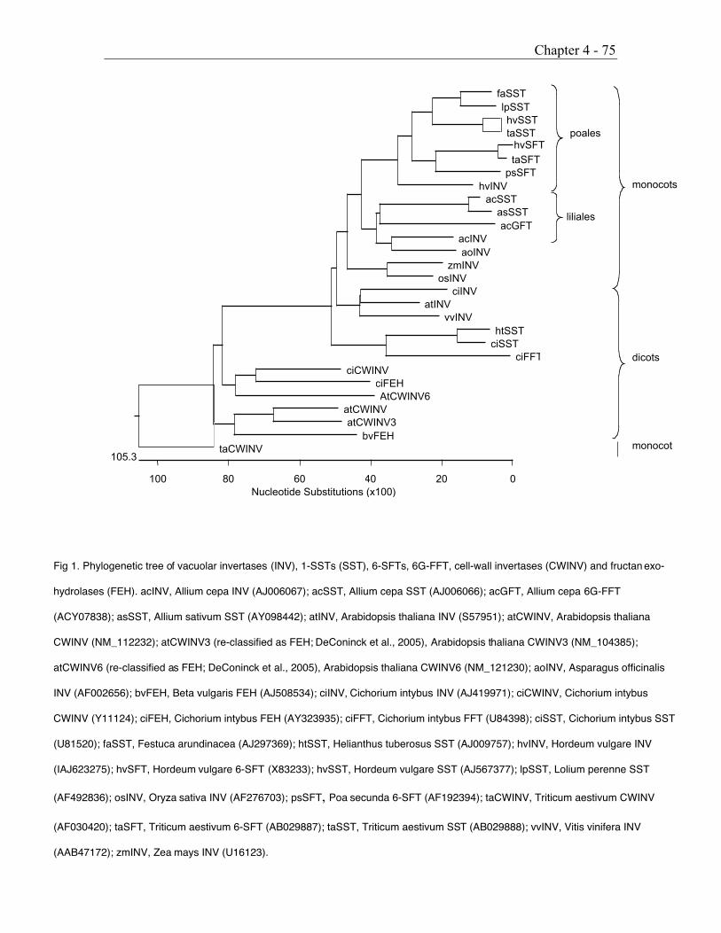
Chapter 4 - 75
Fig 1. Phylogenetic tree of vacuolar invertases (INV), 1-SSTs (SST), 6-SFTs, 6G-FFT, cell-wall invertases (CWINV) and fructan exo-
hydrolases (FEH). acINV, Allium cepa INV (AJ006067); acSST, Allium cepa SST (AJ006066); acGFT, Allium cepa 6G-FFT
(ACY07838); asSST, Allium sativum SST (AY098442); atINV, Arabidopsis thaliana INV (S57951); atCWINV, Arabidopsis thaliana
CWINV (NM_112232); atCWINV3 (re-classified as FEH; DeConinck et al., 2005), Arabidopsis thaliana CWINV3 (NM_104385);
atCWINV6 (re-classified as FEH; DeConinck et al., 2005), Arabidopsis thaliana CWINV6 (NM_121230); aoINV, Asparagus officinalis
INV (AF002656); bvFEH, Beta vulgaris FEH (AJ508534); ciINV, Cichorium intybus INV (AJ419971); ciCWINV, Cichorium intybus
CWINV (Y11124); ciFEH, Cichorium intybus FEH (AY323935); ciFFT, Cichorium intybus FFT (U84398); ciSST, Cichorium intybus SST
(U81520); faSST, Festuca arundinacea (AJ297369); htSST, Helianthus tuberosus SST (AJ009757); hvINV, Hordeum vulgare INV
(IAJ623275); hvSFT, Hordeum vulgare 6-SFT (X83233); hvSST, Hordeum vulgare SST (AJ567377); lpSST, Lolium perenne SST
(AF492836); osINV, Oryza sativa INV (AF276703); psSFT, Poa secunda 6-SFT (AF192394); taCWINV, Triticum aestivum CWINV
(AF030420); taSFT, Triticum aestivum 6-SFT (AB029887); taSST, Triticum aestivum SST (AB029888); vvINV, Vitis vinifera INV
(AAB47172); zmINV, Zea mays INV (U16123).
liliales
monocots
poales
dicots
monocot
Nucleotide Substitutions (x100)0
105.3
20406080100
faSSTlpSSThvSSTtaSST
HvSFTtaSFT
psSFThvINV
acSSTasSSTacGFT
acINVaoINV
zmINVosINV
ciINVatINV
vvINVhtSST
ciSSTciFFT
ciCWINVciFEHAtCWINV6
atCWINVatCWINV3
bvFEHtaCWINV
hvSFT

Chapter 4 - 76
4.4.2. Selection of amino acids correlating with invertase or 1-SST activity respectively
We chose a directed mutational approach based on sequence comparisons
between vacuolar invertases and SSTs for the elucidation of amino acids that possibly
influence enzyme specificity. To this intend, sequences of vacuolar invertases and SSTs
of monocotyledonous and dicotyledonous species were aligned and also vacuolar
invertases from non-fructan producing species such as Arabidopsis thaliana (Fig. 2). We
screened for amino acids in the sequence that specifically differed between invertases and
SSTs. In particular, differences in amino acid polarity, hydrophobicity or hydrophilie and
in the type of aromatic amino acid were considered. Nine amino acids dispersed along the
sequence could be identified which correlated either with invertase or with 1-SST activity
(Fig. 2 Sites A to H and X). These nine sites are located outside the highly conserved
motifs (Fig. 3). Only site A flanks the highly conserved motif (N/S)DPNG, supposed to
be the catalytic nucleophile in the transfructosylation reaction, and site E and F flank the
EC-motif which is probably the proton donor needed for catalysis (Fig. 2, Fig. 3).
Remarkably, the majority of the amino acids found to be either invertase or SST specific
turned out to be aromatic.
Fig 2. Alignment of the nine selected sites of 1-SSTs and vacuolar invertases. Abbreviations as infigure 1.
A B C D E + F X G HFa SST KHYMNDPNG139 KDMVN175 QVQCLAT206 ILYPPPG252 GMYECIDLYP324 GRFDA364 WAYIG410 WANLMT428
Hv SST KYYQNDPNG109 KDLIH146 QVTCLAE197 VVFPPPG223 GEYECIDLYA295 GRFDA336 WACVG382 WANLQS400
Ac SST NHFMADPNA100 KDMIH137 QVQNLAE188 ILMPPPG214 GMLECVDLYP285 GTFDL330 WGYVG376 WASVQN394
As SST YHFMGDPNA100 KDMIH137 QVQNLAE188 ILMPPPG214 GMLECVDLYP285 GTFDL330 WGYVG376 WANVLN394
Lp SST KHYMNDPNG129 KDLVN165 QVQCLAV216 ILFPPPG242 GMYECLDIYP314 GWFDA354 WAFVG400 WASLMS418
Ht SST KNFISDPDG111 KDMIN147 QVQCLAY198 VLLPPPG224 GMWECVDLYP293 GSYDI334 WGYVG385 WANILN398
Ci SST KNFISDPDG123 RDMIN159 QLQCLAY210 ILFPPPG236 GMWECVDLYP305 GTFDP346 WGYVG392 WANILN410
Ac Inv KNWMNDPNG167 RDLVH203 QVQNLAV254 ILVPPPG280 GMWECVDFYP350 GTYDP396 WSWIG442 WASLQG460
Hv Inv KNWMNDPNG140 RDLLR177 QVQCLAF228 VMYPPPG255 GMWECIDLYP324 GRYDA371 WGWVG417 WASLQS335
Ao Inv KNWMNDPNG135 KDLLS171 QVQNLAV222 ILVPPPG249 GMWECIDFYP319 GTYDP364 WGWIG410 WASLQG428
Zm Inv KNWMNDPNG142 RDLLH178 QVQNLAE230 VLVPPPG256 GMWECVDFYP328 GTYDP376 WGWVG422 WASVQS440
Os Inv NNWMNDPNG131 RDLIH167 QVQNLAE218 VLVPPPG244 GMWECVDLYP314 GTYDG358 WGWIG404 WASLQS422
Ci Inv KNWMNDPNG141 KDLIN177 QVQNLAY228 VLVPPPG254 GMWECVDFYP322 GTYDA363 WSWIK409 WASLMG427
At Inv KNWMNDPNG37 KDLIH73 QVQNLAY124 VLVPPPG150 GMWECVDFYP218 GTYDD359 WGWIG307 WASVQG325
Vv Inv KNWMNDPNG139 KDLIE186 QVQNLAY237 VLVPPPG263 GMWECVDLYP331 GTYSL372 WGWIG418 WASVQS436

Chapter 4 - 77
Fig 3. Model of Festuca 1-SST showing the positions of conserved active site motifs (above) andof the amino acids chosen for mutagenesis (below). Sites are designated A, B to X.
Table 2. Overview of the selected sites
Site FaSST AcINVA Y133W W161YB M173L L201MC C203N N251CD Y248V V276YE Y317W W343YF L322F F348LG Y408W W440YH N425S S457NX F362Y Y394F
4.4.3. Functional characterization of wild-type and mutagenized enzymes expressed in
Pichia pastoris
Studying structure function relationships of FTs and invertases demands an
appropriate expression system. The expression of these enzymes in the yeast Pichia
pastoris has been shown to be ideal, since this yeast is devoid of sucrose metabolizing
enzymes such as invertases (Lüscher et al., 2000). For the mutational analysis we chose
Allium cepa invertase (acINV) and Festuca arundinacea 1-SST (faSST), two enzymes
that were known to be very efficiently expressed in Pichia (Lüscher et al., 2000; acINV
by T. Ritsema). The amino acids at the nine selected sites of acINV were mutated to the
N-term
Large Subunit
Mature Protein
RDP ECC-term
NDPNG
A: Y¤ W
B: M¤ LE: Y¤ W
C: C¤ NF: L¤ F
D: Y¤ V X: F¤ Y
G: Y¤ W
H: N¤ S
Small Subunit

Chapter 4 - 78
corresponding amino acids in faSST and vice versa (Fig. 3, Table 2). For both enzymes
these different mutations were analyzed separately.
Initially recombinant wild-type faSST and acINV were functionally characterized
in order to subsequently detect possible activity shifts upon introducing the mutations.
Invertases are saturable with sucrose and have Km-values from 2-20 mM. In contrast,
fructosyltransferases do not show Michaelis-Menten kinetics and are essentially non
saturable, the Km has to be estimated (Ritsema et al., 2003; Van den Ende et al., 1996). It
seems that SSTs need a higher sucrose concentration than invertases for proper activity.
With increasing concentrations of sucrose, the recombinant faSST produced increasing
amounts of glucose and 1-kestose besides small amount of fructose (Fig. 4A). The
hydrolase activity was relatively low at all offered sucrose concentrations as compared to
the polymerase activity. Saturation appeared only to be reached at sucrose concentrations
≥ 800 mM. In contrast, recombinant acINV reached its saturation already at about 20 mM
sucrose (Fig. 4B). At sucrose concentrations of ≥ 100 mM, a distinct but very small
amount of 1-kestose was found. In summary, faSST produces mainly 1-kestose and
glucose, whereas acINV acts almost purely as a hydrolase. The differences in the
saturation characteristics for sucrose of the two enzymes, and their relatively low side
activities, namely hydrolase activity in the case of faSST, and polymerase activity in the
case of acINV, are the ideal basis for analyzing possible shifts in enzymatic specificity
upon mutational changes.
Fig 4. Characterization of recombinant faSST (A) and acINV (B). Enzyme preparations wereincubated with increasing concentrations of sucrose (27°C, 20’). Product formation (nmol glucose,fructose, 1-kestose) was quantified on HPAEC-PAD.
SST
0
2
4
6
8
10
12
14
0 200 400 600 800 1000
sucrose concentration
nmol
pro
duct
Glucose Fructose Kestose
0
0.5
1
1.52
2.5
33.5
4
4.5
0 50 100 150 200
sucrose concentration
nmol
pro
duct
glucose fructose kestose
InvertaseA B

Chapter 4 - 79
4.4.4. Characterization of SST-mutants
Like the wild-type enzymes, genes encoding mutated enzymes were expressed in
the yeast Pichia pastoris. Enzyme activity was assayed with sucrose. The faSST mutants
at sites D and G were not active. At site D, tyrosine248 was changed to valine (Table 2),
representing a change from an aromatic amino acid with an OH-group to a hydrophobic
amino acid. The site is located next to a highly conserved proline-triplet, that probably
forms a turn in the tertiary structure (Fig. 2). In the inactive faSST mutant at site G,
tyrosine408 was changed to tryptophan, which is also an aromatic amino acid but a much
bigger one, that might disturb the protein structure.
Fig 5. Relative hydrolytic activity of wild-type and mutated recombinant SSTs. The active faSSTmutants at sites A, B, C, E, F, H and X (Table 2 gives an overview) were incubated with differentconcentrations of sucrose (sucrose concentrations are indicated in mmol on the x-axis). Relativeactivities of polymerization and hydrolysis of sucrose were determined, taking 1-kestoseproduction as a measure for polymerization and fructose production as a measure for hydrolysis.The sum of polymerizing and hydrolyzing activities was set to be 100%, and % hydrolase activityis shown. Standard deviations are indicated and are calculated form two independent inductionexperiments.
0
20
40
60
80
100
100
200
400
600
800
1000 10
020
040
060
080
010
00 100
200
400
600
800
1000 10
020
040
060
080
010
00 100
200
400
600
800
1000 10
020
040
060
080
010
00 100
200
400
600
800
1000 10
020
040
060
080
010
00
faSST A B C E F H X
% fr
ucto
se

Chapter 4 - 80
All the other single amino acid mutations resulted in active recombinant proteins.
These active faSST mutants were tested by incubation with different concentrations of
sucrose (Fig. 5). At site A, the amino acid tyrosine133 of faSST was changed to
tryptophan, as occurring in all known vacuolar invertases. The aligned SSTs carry a
tyrosine or a phenylalanine at this site. Site A is in the vicinity of the highly conserved
motif (N/S)DPNG. The mutant revealed no change in relative polymerase (80-90%) or
hydrolas (10-20%) activities as compared to the recombinant wild-type faSST upon
incubation with different concentrations of sucrose (Fig. 5). At site B, methionine173 of
faSST was replaced by leucine which is hydrophobic as well. The mutation at site E is
like the one at site A, a change of a tyrosine317 to tryptophan, and for the mutation at site
F, leucine322 which is aliphatic and apolar, was changed to the also apolar but aromatic
phenylalanine. The mutations at sites A, B, E and F had no effect on faSST specificity as
compared to the wild-type form.
The mutants at sites C and X in faSST had almost doubled relative hydrolytic
activity in comparison to wild type faSST. I.e. fructose production doubled with respect
to all sucrose concentrations offered (Fig. 5). At site C, the cysteine203 of faSST was
changed to asparagine, as occurring in all invertases of the alignment except for barley
invertase (Fig. 2). This alteration can cause a loss of a disulfide bond. The mutant at site
X also showed a doubling of the relative hydrolytic activity in comparison to wild-type
faSST. The phenylalanine362 in faSST was mutated to the omnipresent tyrosine of
invertases in the corresponding site. The mutation represents an alteration from
hydrophobic aromatic to polar aromatic.
In comparison to wild type faSST, the mutant at site H, where asparagine425 was
changed to serine, had almost exchanged ratios of hydrolysis versus polymerization. The
enzymes relative hydrolytic activity was approximately 80%, whereas its relative
polymerizing activity was reduced to approximately 20% (Fig. 5). Although the faSST
carrying the mutation at site H was mainly hydrolytically active, it did not become a
typical invertase. Its capacity to synthesize 1-kestose was still too high, and it was not
saturated at low sucrose concentrations such as wild type acINV (Fig. 6, compare to Fig.
4).

Chapter 4 - 81
Fig 6. Characterization of the faSST mutant at site H. Enzyme preparations were incubated withincreasing concentrations of sucrose (27°C, 20’). Product formation (nmol glucose, fructose, 1-kestose) was quantified on HPAEC-PAD.
4.4.5. Characterization of invertase-mutants
Genes encoding mutated onion invertase (acINV) were expressed in the yeast
Pichia pastoris and enzymatic activity was assayed with sucrose similar to the wild-type
enzyme. The acINV mutants at site D and X were not active. At site D, valine276 was
changed to tyrosine. Like in faSST carrying mutation D, the site for mutagenesis is
located next to a highly conserved proline-triplet, probably representing a turn in the 3D-
structure. At site X, tyrosine394 was replaced in acINV with phenylalanine. This looks a
relatively small change in polarity only, keeping the aromatic ring. Nevertheless, acINV
carrying the mutation at siteX was inactive, and the faSST mutant at site X showed
doubled relative hydrolytic activity.
All other single mutants of acINV were active. As before, the enzyme
preparations of the mutants were analyzed with different concentrations of sucrose (Fig.
7) and the relative activities of hydrolysis versus polymerization were determined.
Furthermore the Km for sucrose was evaluated (Fig. 8). AcINV mutated at sites B, C, E,
F or H showed the same biochemical properties as the wild-type invertase, with respect to
kestose production and Km-values. At site B, leucine201 was replaced with methionine,
both molecules are hydrophobic. At site C, asparagine251 was changed to cysteine. The
change at site E is like at site A, a change from tryptophan343 to tyrosine, but such as the
mutation at site E in faSST, showing no change in enzymatic specificity. In acINV
0
0.5
1
1.5
2
2.5
0 100 200 300 400 500 600
sucrose concentration (mM)
nm
ol p
rod
uct
Glucose Fructose Kestose

Chapter 4 - 82
carrying the mutation at site F, the apolar character of phenylalanine348 was unchanged,
but the aromatic character was lost due to the mutation to leucine. For the acINV mutant
at site H, that also showed unchanged enzymatic characteristics, serine457 was mutated to
the chemically more reactive asparagine.
Fig 7. Relative polymerizing activity of wild-type and mutated recombinant invertase. The activeacINV mutants at sites A, B, C, E, F, G and H (Table 2 gives an overview) were incubated withdifferent concentrations of sucrose (sucrose concentrations are indicated in mmol on the x-axis).Relative activities of polymerization and hydrolysis of sucrose were determined, taking 1-kestoseproduction as a measure for polymerization and fructose production as a measure for hydrolysis.The sum of polymerizing and hydrolyzing activities was set to be 100%, and % polymeraseactivity is shown. Standard deviations are indicated and are calculated form two independentinduction experiments with two assays per sucrose concentration.
0%
1%
2%
3%
4%
5%
6%
7%
20 4010
020
0 20 4010
020
0 20 4010
020
0 20 4010
020
0 20 4010
020
0 20 4010
020
0 20 4010
020
0 20 4010
020
0
acINV A B C E F G H
% k
esto
se
0.02.04.06.08.0
10.012.014.016.018.020.0
acINV A B C E F G H
Km
(m
M)
Fig 8. Km-values for sucrose of invertasemutants. Onion invertase and the mutants wereassayed with increasing concentrations ofsucrose. The Michaelis-menten konstant (Km)was calculated using Lineweaver-Burk-plots.
Chapter 4 - 83
The invertase mutated at sites A or G, showed an increased kestose production as
compared to recombinant wild-type acINV (Fig. 7). Also the Km-value for sucrose was
significantly increased (Fig. 8). Site A is close to the highly conserved motif (N/S)DPNG.
The conserved tryprophan in invertases at this site was mutated to tryrosine. Amazingly,
the corresponding mutation in faSST had no, or only little effect. Like the mutations at
site A and E, the mutation at site G represents a replacement of tryptophan440 with
tyrosine. This mutation in invertase led to an activity shift towards more polymerizing
activity whilst in the case of faSST it led to inactivity.
In summary, SST mutants at sites C, H and X showed an increased relative hydrolytic
activity and invertase mutants at sites A and G showed increased Km-values and
increased fructosyltransferase activity, demonstrating the importance of amino acids
outside highly conserved motifs for the enzymatic reaction.

Chapter 4 - 84
4.5. Discussion
For GH family 32, where plant FTs and vacuolar invertases belong to, the acidic
amino acids (indicated in bold) of three highly conserved motifs (N/S)DPNG, RDP and
EC are proposed as active site residues (Pons et al., 2000; Ozimek et al,, 2004; Verhaest
et al., 2005). Recently the 3D-structures of three enzymes of this family were resolved:
the invertase from the bacterium Thermotoga maritima (Alberto et al., 2004), the exo-
inulinase from Aspergillus awamori (Nagem et al., 2004) and the fructan-exohydrolase
(FEH) from chicory (Verhaest et al., 2005). Each structure shows a five bladed b-
propellers with a deep negatively charged central pocket, representing the active site, and
a b-sandwich. The three highly conserved motifs flank the central pocket, and can be
brought in connection with the general reaction mechanism for the cleavage of glycosidic
bonds. Nevertheless, how enzyme specificity is regulated is unknown. Despite the close
molecular and biochemical relationships of vacuolar invertases and SSTs, and the
availability of crystal structures, the question what structural features determine the
hydrolyzing or polymerizing character of a distinct enzymes, remains unanswered.
In order to detect the amino acids in FTs and vacuolar invertases which are
determining enzyme specificity, we performed a targeted mutational analysis. It was
based on sequence comparisons between vacuolar invertases and SSTs. Upon a search in
sequence alignments for amino acids correlating with either invertase or SST activities,
nine amino acids dispersed along the sequence which fulfilled these selection criteria
were identified. Three of these nine sites turned out to contain tryptophan in all aligned
invertases, whilst at the corresponding site SST’s contain either tyrosine or phenylalanine
at site A, tyrosine, leucine or tryptophan at site E and tyrosine, cysteine or phenylalanine
at site G. Site A and site E flank the highly conserved motifs (N/S)DPNG and EC,
respectively. Thus, the selection pressure to preserve tryptophan seems to be much higher
in vacuolar invertases than in FTs, where more amino acid variation is found.
Nevertheless also SSTs generally seem to keep the aromatic nature of the amino acids at

Chapter 4 - 85
the above described sites. Maybe the replacement of the highly conserved tryptophans of
invertases was a step in the evolution from vacuolar invertases towards FTs.
Interestingly, mutants at sites A and G in acINV, that showed increased kestose
production and Km-values, were mutations of the highly conserved tryptophan to
tyrosine. In faSST the mutation at site A, which changed the tyrosine to the larger
aromatic molecule tryptophan, had no effect on enzyme specificity. The amino acid
corresponding to site A was furthermore analyzed in onion 6G-FFT (Ritsema et al.,
2005). Like in faSST, there is a tyrosine present at the corresponding site of 6G-FFT
which was mutated to tryprophan. Corresponding to the result obtained for the faSST
mutant at site A, also mutated onion 6G-FFT showed no changes in enzyme specificity.
Whereas the mutant at site G of acINV showed an increased kestose production and an
increased Km-value, the faSST mutated at site G was inactive. The two mutations leading
to increased Km-values in invertase, at site A and at site G, also showed increased
production of 1-kestose. Possibly the capacity to synthesize 1-kestose is linked to an
increase in Km. If SSTs evolved from vacuolar invertases one evolutionary step could be
the increase in Km for sucrose. A possible function of this biochemical feature could be
to prevent fructan synthesis if sucrose concentrations are low and sucrose is needed for
other physiological processes.
The mutations affecting SST activity towards increased relative hydrolytic
activity were mutations at sites C, H and X. Here the mutation at site X involved aromatic
amino acids. Phenylalanine was changed to tyrosine, the amino acid that is present in all
invertases analyzed by the alignment. In general, the conservation of distinct amino acids
in invertases seems to be much higher than in SSTs. AcINV mutated at site X was
inactive. In summary, the majority of the sites we found to affect hydrolyzing activities
versus polymerizing activities, turned out to be of aromatic character. In the literature
mainly aromatic amino acids are discussed to have affinity to sugar molecules e.g. by
constituting a channel for transport of long chain sugars across membranes in the
maltoporines; the maltoporin appears to be passive while the sugar glides screwlike along
an aromatic lane (Dutzler et al., 2002).
Chapter 4 - 86
Whereas in the invertase only amino acid substitutions altering aromatic residues
changed enzyme specificity, in SST also mutations at other sites, namely C and H, led to
activity shifts. At site C the amino acid cysteine occurs predominantly in SSTs. An
exception are the SSTs from Allium cepa and Allium sativum, carrying an asparagine at
this site like the invertases compared, except the Hordeum vulgare invertase. A mutation
at site C in faSST led to a doubling of the hydrolytic activity relative to transferase
activity in comparison to wild-type faSST. In contrast, the acINV carrying the mutation at
site C had unchanged enzymatic properties. Cystein has the potential to form disulfide
bonds that can severely influence the tertiary structure of proteins. It is unclear if the
tertiary structure was affected and led to a change in enzyme specificity in the faSST
mutated at site C, or if the mutation of the cysteine to the chemically more reactive
asparagine was the cause for the doubling in relative hydrolase activity. None of the 3D-
structures within the GH32 family indicated disulfide bonds.
The alignment shows that SSTs predominantly carry asparagines at site H with two
exceptions, Allium cepa SST and Lolium perenne SST, where a serine is present. The
alignment revealed always a serine in the corresponding site of vacuolar invertases. Both,
serine and asparagine are polar amino acids, but whereas serine can only interact with
other amino acids or water molecules via its hydroxyl-group, asparagine is capable of
undergoing reactions with either their carbonyl-group or the amino-group. The mutant at
site H of faSST showed increased relative hydrolyzing activity of sucrose. Whereas wild-
type faSST guided 80% of its total activity into the polymerization of sucrose to 1-
kestose, the mutant was mainly hydrolytically active. The relative hydrolyzing activity of
the mutant was 80%. Nevertheless, the faSST mutant at site H did not become a typical
invertase, since it synthesized more 1-kestose than wild type invertase did, and it was
essentially not saturable in incubations with sucrose. No alteration of enzyme specificity
was detected for the acINV mutant at site H.
Mutations at site D led to inactivity of both, acINV and faSST. Most probably the activity
was lost due to a disturbance of the tertiary structure. The site is located next to a highly
conserved proline-triplet. The grouping of prolines in the amino acid sequence often leads
to turns or loops. This is also seen in the 3D-structures (Alberto et al., 2004). Already
Chapter 4 - 87
slight disturbance of the vicinity of this highly conserved area might be a reason for
inactivity of our constructs.
For plant FTs and vacuolar invertases no crystal structure is yet available. The
data obtained in this work were theoretically analyzed using the 3D-structure of the
invertase from the bacterium Thermotoga maritima. This bacterial invertase was chosen
for the interpretation of the data, since like onion invertase and Festuca 1-SST it is a
sucrose metabolizing enzyme. The exo-inulinase from Aspergillus awamori and the FEH
form chicory use inulin.
Based on an alignment of Thermotoga maritima invertase, onion invertase and Festuca 1-
SST, the probable positions of the sites A, B, C, D, E, F, G, H and X could be localized in
the sequence of Thermotoga maritima invertase. The sites are all located on the b-
propeller domain in the apparent crystal structure, which carries the active site (data not
shown). Usually vacuolar invertases and FTs consist of two subunits, a large and a small
one. The b-propeller domain probably represents the large subunit that has been shown to
determine enzyme specificity in barley 6-SFT and Festuca 1-SST (Altenbach et al.,
2004). The fact, that the positions of the amino acids correlating either with invertase or
with SST activity can be assigned to amino acids on the b-propeller domain, strengthens
the argument that these amino acids could influence enzyme specificity.
It is probable that enzyme specificity of invertases and FTs is also depending on
the N-glycosylation. This possibility is currently discussed and experimentally
investigated (Verhaest et al., 2005; Le Roi et al., Fructan 2004). The 3D-structure of
chicory FEH shows a b-propeller with a b-sandwich attached to it. Verhaest and co-
workers could show that the cavity between the two domains forms a cleft, emerging
from the active site, which is believed to be the inulin-binding site. This idea is supported
by the presence of four glycerol molecules in this cleft. The cleft is also present in the
structure of the invertase form Thermotoga maritima, but the groove is occluded near the
active site. The role of the cleft present in chicory FEH was investigated via the
introduction of an N-glycosylation site within the cleft (Le Roi et al., Fructan 2004). This
glycosylation site is normally absent in FEHs, but present in invertases. The recombinant

Chapter 4 - 88
glycosylated protein showed a strongly reduced activity with inulin. Thus, differences in
glycosylation might influence substrate binding and therefore have an effect on enzyme
specificity. None of our mutations (A to X) affected a glycosylation site.
Fig. 9 Thermotoga maritima invertase (Alberto et al., 2004). (A) View from the top into the activesite. Active site residues (the aspartates of the motifs (N/S)DPNG and RDP, and the glutamate ofthe EC- motif) are indicated in green. Amino acids (at sites A, C, G, H and X) that influencedhydrolysis versus polymerization in onion invertase and Festuca 1-SST are indicated in red. (B)Side-view illustrating amino acid substitutions that led to a change in enzymatic specificity. Theyare located on the rim of the central pocket.
The assigned localization of the nine sites in the 3D-structure of Thermotoga
maritima invertase revealed, that those amino acids that influenced hydrolysis versus
polymerization, are located at the rim of the central pocket. They correspond to sites A
and G in acINV and to sites C and H in faSST. Being located opposite the entrance of the
central pocket, site X is an exception in this respect. Possibly the sites A, C, G, and H that
are located at the rim of the active site, influence substrate recognition and/or guidance of
the sugar substrate to the active site. Molecules located on this rim could hinder or
facilitate the entry of substrates and thereby have an influence on substrate specificity.
H
G
A
X
C
H
G
A
X
C
X C
A
G
H
A B
Chapter 4 - 89
Pons and coworkers used a sequence-structure compatibility search for the theoretical
elucidation of sites that influence enzyme activity and specificity in different glycoside
hydrolase families (Pons et al., 2004). They speculated that positions far away from the
active site are involved in fructosyltransfer reactions as well as in the hydrolysis of long-
chain glycans while positions close to the active site are important for the orientation of
substrates and the hydrolysis of short glycans. These theoretical data are in accordance
with our mutagenesis data with invertase and SST since both enzymes cleave a short
glycan (sucrose) and transfer the fructosyl units either to water or another sucrose,
respectively. Thus, the mutations of amino acids that are located close to the active site
(A, C, G, and H) influence relative hydrolase versus transferase activities of a short
glycan, namely sucrose.
The three conserved motifs (N/S)DPNG, RDP and EC could be brought in
connection with the general mechanism for cleavage of glycosidic bonds within GH32.
The present work reveals amino acids located outside these highly conserved motifs that
influence enzyme specificity. The balance between polymerization and breakdown
reactions by invertases and SSTs could separately be modulated via directed amino acid
substitutions. We therefore speculate that differences in the amino acid sequence in
combination with differences in glycosylation influence substrate recognition and/or
binding and therefore have an effect on enzyme specificity.
It is unlikely that one single mutation is sufficient to change an invertase to an
SST and vice versa. In order to get further insight into what components are important in
polymerization and/or hydrolyzation using sucrose as a substrate, SST and invertase
mutants carrying multiple mutations have to be investigated.

Chapter 5- General discussion - 90
Chapter 5
5. General Discussion
5.1. Initial questions and experimental approach
The main goal of my thesis was to find structural components (motifs) important
for the catalytic function and specificity of plant FTs and vacuolar invertases.
Comparisons of the deduced primary amino acid sequences of plant FTs and vacuolar
invertases, as well as related enzymes from fungi and bacteria, were the basis for the
selection of potentially interesting sites or motifs. We used domain swaps and site
directed mutagenesis, followed by expression of the resulting constructs in the yeast
Pichia pastoris, to elucidate the relative importance of these sites or motifs for enzymatic
function.

Chapter 5- General discussion - 91
5. 2. “Acid invertase-like” enzymes
Utilization of sucrose as a source of carbon and energy depends on its cleavage
into hexoses. Sucrose can be hydrolyzed to its components glucose and fructose by the
enzyme invertase. In plants, invertases are ubiquitous. They occur as acid invertases
(vacuolar invertases and cell-wall invertases) and alkaline (or neutral) invertases.
If the protein sequence of any acid plant invertase is “blasted” against sequences of
proteins from the kingdom of bacteria, the highest similarities are detected with bacterial
invertases, levanases, inulinases, sucrose 6-phosphate hydrolases and levansucrases (Fig.
1). Similarly, “blasting” of an invertase sequence against proteins from the fungal
kingdom, retrieves the sequences of invertases, inulinases, and of fructosyltransferases
(Heyer and Wendenburg; 2001). If the protein sequence of a plant invertase is “blasted”
against sequences of plant proteins, the first sequences retrieved correspond to invertases,
FEHs and FTs. Interestingly, there are no “hits” when any invertase is “blasted” against
all available sequences of animal proteins. Animals hydrolyze sucrose with the enzyme
sucrase, a structurally totally unrelated enzyme. There are five “hits” when an invertase is
blasted against sequences of proteins from the kingdom of Protista (e.g. Leishmania), and
only one “hit” against sequences of Archaea proteins (Haloarcula marismortui).
Monera
Eubacteria
- Invertase
- Levanase
- Inulinase
- Sucrose 6-phosphate hydrolase
Levansucrase
Archaea?
-only one example of
Sucrose 6-phosphate hydrolase
Plantae
- Invertase
- FEH
- FT
Fungi- Invertase
- Inulinase
- FT
Fig. 1 Kingdoms harboring “acid invertase-like” enzymes.
Animalia
Protista
- Probable Invertase
- Probable Sucrose
6-phosphate
hydrolase

Chapter 5- General discussion - 92
Plants contain an additional type of invertase, namely neutral/alkaline invertase. This
enzyme shares no homology in its primary amino acid sequence with the acid invertase.
Interestingly only neutral invertases and no acid invertases are reported for blue algae
(cyanobacteria) such as for example Anabaena variabilis (Schilling and Ehrnsperger;
1985; Porchia et al., 1999). There are no homologues of neutral invertases in any other
kingdom.
In summary, the class of “acid invertase-like” enzymes is predominantly found in
the kingdoms of bacteria, fungi and plants. Their abundance in these only distantly
related organisms implies an extremely large protein family. Thus the phylogenetic tree
of plant acid invertases, FTs and FEHs, which is shown in chapter 4 (Fig. 1), represents
only a very small branch of a huge tree of “acid invertase-like” enzymes. An example of
an even smaller branch is provided by an unrooted tree of plant cell-wall invertases and
FEHs (Fig. 2; Van den Ende et al., 2004). Although plant FEHs show no invertase
activity, their amino acid sequences are highly homologous especially to cell-wall type
invertases (Van den Ende et al., 2002). For example, the sugar beet 6-FEH does not
cluster with the 1-FEHs from chicory plants in the tree, but appears in a different
subgroup that also contains invertases from Arabidopsis thaliana (AtcwINV1, AtcwINV3
and AtcwINV5). AtcwINV6 groups together with FEHs from fructan plants in the
phylogenetic tree. It was recently demonstrated that AtcwINV3 and AtcwINV6 are not
invertases but are real FEHs with different substrate specificities. By contrast, AtcwINV1
was found to be a typical invertase (De Coninck et al., 2005). The tree illustrates the close
relatedness of plant cell-wall invertases and FEHs.
Two additional “acid invertase-like” enzymes are included in this unrooted tree which are
named microbial FEHs (Van den Ende et al., 2004): from bacterial origin, the levanase
from Bacillus subtilis (6-FEH), and from fungal origin, the exo-inulinase from
Aspergillus awamori (1-FEH). These enzymes from bacteria or fungi catalyze almost the
same reaction as the plant enzymes but are located far away in the phylogenetic tree.
The illustration of the inulinase from Aspergillus awamori and the levanase from Bacillus
subtilis in the tree nicely shows that functionally closely related enzymes are widely
dispersed on a huge tree of “acid invertase-like” enzymes (Fig. 2).

Chapter 5- General discussion - 93
Fig 2. (taken from Van den Ende et al., 2004): Phylogenetic tree of plant cell wall invertases andFEHs. For comparison, two microbial FEHs are included as outliers (green branches). FEHs areindicated in green. Enzymes that have a low iso-electric point are underlined. The six Arabidopsissequences (AtCWINV1–AtCWINV6) are boxed. For a detailed list of the Accession numbers, seehttp://www.kuleuven.ac.be/bio/dev/nederlands/cwtree.htm. The cell wall-type glycosyl hydrolasesare tentatively divided into three major groups A–C. Group A is further separated into twosubgroups I (monocots) and II (dicots). Group C is separated into three subgroups I (dicots), II(monocots) and III (monocots). The phylogenetic relationships were analyzed using ClustalW(http://www.ebi.ac.uk/clustalw/) and TREEVIEW [43]. The scale bar indicates a distance value of0.1. Abbreviations: FEHs, fructan exohydrolases; INV, cell wall invertase; INV?, putative cell wallinvertase (functionality not determined).

Chapter 5- General discussion - 94
For each of the three distantly related kingdoms that harbor “acid invertase-like”
proteins (bacteria, fungi and plants) crystal structures became available recently: (i) the
invertase from the bacterium Thermotoga maritima (Alberto et al., 2004), (ii) the exo-
inulinase from the fungus Aspergillus awamori (Nagem et al., 2004) and (iii), the fructan-
exohydrolase (FEH) from chicory (Verhaest et al., 2005). All structures show a five-
bladed b-propeller with a deep negatively charged central pocket and a b-sandwich
attached to it (for details see chapter 1.6.). Thus there is a large family of “acid invertase-
like hydrolases” dispersed along different kingdoms of organisms, with almost identical
three-dimensional structures. Through a superposition of the structures, it was observed
that the active site is located in the b-propeller domain. It seems to be a characteristic that
the length of the proteins is conserved. The b-sandwich appears to be necessary for a
functional enzyme, although it is distant from the active site. The N-terminal b-propeller
corresponds to the N-terminal large subunit, and the C-terminal b-sandwich module to
the C-terminal small subunit found in plant vacuolar invertases and FTs, where it usually
consists of the C-terminal third of the protein.
Our results provide further information with respect to the function of the two subunits:
The analysis of chimeras between Festuca 1-SST and barley 6-SFT (Chapter 2) showed
that the product forming activity was entirely determined by the large N-terminal subunit.
This agrees well with the fact that the active site is located on the b-propeller in all crystal
structures elucidated so far. If only the large subunit of Festuca 1-SST was expressed in
Pichia, no FT activity could be detected. The amount of the recombinant protein found in
the Pichia culture filtrate was very low, possibly because of incorrect targeting in the
yeast cells or because of degradation by Pichia proteases. The small subunit might thus
have an effect on protein folding, proper post-translational processing, cellular targeting
or on the control of enzyme activity, but it is not involved in determining enzyme
specificity.

Chapter 5- General discussion - 95
5.3. Enzyme specificity of “acid invertase-like” enzymes
Enzyme activities are manifold among “acid invertase-like” enzymes. They can
basically be subdivided into fructosylhydrolases and fructosyltransferases. According to
our view, the catalytic specificity of these enzymes basically involves an active site for
the fructosyl donor and an active site for the fructosyl acceptor.
The simplest reaction is catalyzed by invertases. They initially bind the fructosyl moiety
of sucrose (donor) and, in a second step, release it and transfer it to water (acceptor). 1-
SSTs also initially cleave the bond between the glucose and fructose moiety of sucrose
and bind the fructosyl residue, but then transfer it to another sucrose (acceptor). In
contrast to invertases, 1-SSTs must therefore harbor a binding-site for the fructosyl
acceptor sucrose. This can be considered as a first step in the evolution from vacuolar
invertases towards FTs. A third enzyme which is also capable of using sucrose as
fructosyl donor is 6-SFT. Like invertases, 6-SFTs cleave the bond between glucose and
fructose of sucrose, bind the fructosyl moiety, but then transfer it to either sucrose or
fructan (forming kestose or bifurcose). Like 1-SSTs, 6-SFTs must therefore have a
binding-site for a fructosyl acceptor, but with a different specificity than 1-SSTs. Thus a
further step in the evolution of specific SFTs was likely the emergence of a preference for
a fructan as fructosyl acceptor. FFTs lost the capacity to use sucrose as fructosyl donor,
but instead use a fructan of DP≥3 as donor. The fructosyl acceptor can be either sucrose
or a fructan. The basic difference of FFTs compared to the previously described enzymes
is that the donor-binding site can only accept fructan.
What can be learned from the crystal structures published up to now? Based on
the extensive overlap of the active site of the available 3D-structures of “acid invertase-
like” enzymes, the fructosyl donor binding site can be relatively well derived, because the
exo-inulinase from Aspergillus awamori was crystallized in the fructose-bound state. In
contrast, the invertase from Thermotoga maritima and the FEH from chicory were
crystallized in the ligand-free states only. The donor binding site includes the three motifs
that were also emerging from biochemical studies, namely the (N/S)DPNG, the RDP and
the EC-motif. The reaction scheme in the case of 1-FEH has been interpreted as follows

Chapter 5- General discussion - 96
(Verhaest et al., 2005): a fructan of the inulin type initially binds to the active site, where
the glycosidic oxygen of the inulin is protonated by the glutamate of the EC-motif. This
step is followed by a nucleophile attack of the aspartate in the (N/S)DPNG motif, forming
a covalent fructosyl-intermediate. The third step of the reaction is the hydrolysis of the
fructosyl-intermediate releasing fructose and the enzyme in its ligand-free state. The
same scheme can be applied to invertases and plant FTs, where instead of inulin, sucrose,
1-kestose or another fructan acts as fructosyl donor and binds to the aspartate of the
(N/S)DPNG motif. Thus the fructosyl donor binding step is basically solved. However, it
is currently unknown what structures account for donor preferences. Furthermore, it is
unknown where the fructosyl acceptor binds and what determines acceptor preferences.
Our results show that small changes in amino acid sequence can be the basis for
differences in donor and acceptor preferences.
1. The proteins with mutations of the glutamate of the EC-motif in Festuca 1-SST, were
unable to use sucrose, but retained the ability to use 1-kestose (Chapter 3). Our results are
particularly intriguing, since they indicate a change in the enzyme's catalytic activity
caused by one single amino acid substitution.
2. Both, invertases and 1-SSTs use sucrose as fructosyl donor, but differ in their fructosyl
acceptor which is water or another sucrose, respectively. In our mutational analysis of
nine single amino acid residues in onion invertase and Festuca 1-SST, the mutated
enzymes revealed changed catalytic specificity, gradually demonstrating the importance
of single amino acids on donor and acceptor preferences (Chapter 4).

Chapter 5- General discussion - 97
5.4. Outlook
We could show that the large N-terminal subunit entirely determines enzyme
specificity in barley 6-SFT and Festuca 1-SST. The C-terminal small subunit was shown
to have no impact on catalytic specificity, but might play a role in protein folding and
stability or the control of enzyme activity. It would be interesting to further study the
function of the small subunit by exchanging the subunits of enzymes which are less
related than 1-SST and 6-SFT. e.g. by using enzymes with completely different substrate
preferences like invertases and FFTs or SSTs and FEHs etc.
Furthermore, the functions of the post-translational cleavage process, if any,
remains elusive. FTs which are cleaved in the two subunits in planta, are basically
uncleaved but nevertheless active if expressed in Pichia. Specific plant factors, such as
proteolytic enzymes in the plant vacuole might direct the cleavage process of plant FTs;
such factors might be absent in Pichia pastoris. The expression of plant FTs in Pichia
provides a “natural” tool for differences in post-translational modifications. Cellular
targeting and glycosylation patterns differ in plants and the yeast system. These post-
translational differences might have an impact on enzyme specificity and hence explain
the differences in the activities between plant purified and recombinant FTs, as seen for
barley 6-SFT (Hochstrasser et al., 1998). Studying translational modifications of FTs in
Pichia might provide important knowledge of structure-function relationships. A
promising approach for structure-function elucidations would be a mutational analysis of
the potential glycosylation sites present in plant FTs and vacuolar invertases.
Our studies of conserved motifs revealed the importance of the (N/S)DPNG-, the
RDP- and the EC-motif in the transfructosylation reaction of plant FTs, and helped to
narrow down the region responsible for enzyme specificity. We could show via the
analysis of chimeric enzyme, that the region responsible for enzyme specificity is located
between the (N/S)DPNG motif and the small subunit in barley 6-SFT and Festuca 1-SST.
More detailed domain exchanges within the large subunit, in conjunction with other FTs
such as FFTs and FEHs and in conjunction with site-directed mutagenesis studies of
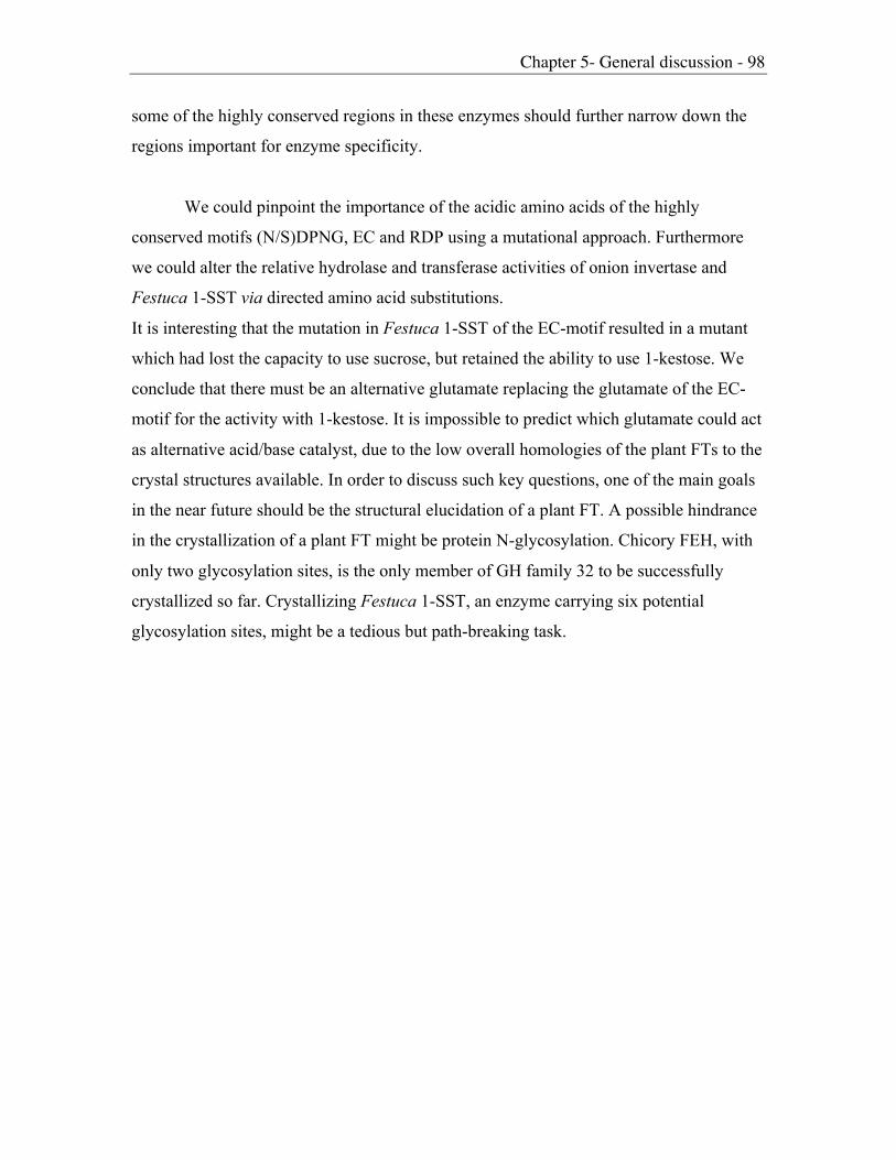
Chapter 5- General discussion - 98
some of the highly conserved regions in these enzymes should further narrow down the
regions important for enzyme specificity.
We could pinpoint the importance of the acidic amino acids of the highly
conserved motifs (N/S)DPNG, EC and RDP using a mutational approach. Furthermore
we could alter the relative hydrolase and transferase activities of onion invertase and
Festuca 1-SST via directed amino acid substitutions.
It is interesting that the mutation in Festuca 1-SST of the EC-motif resulted in a mutant
which had lost the capacity to use sucrose, but retained the ability to use 1-kestose. We
conclude that there must be an alternative glutamate replacing the glutamate of the EC-
motif for the activity with 1-kestose. It is impossible to predict which glutamate could act
as alternative acid/base catalyst, due to the low overall homologies of the plant FTs to the
crystal structures available. In order to discuss such key questions, one of the main goals
in the near future should be the structural elucidation of a plant FT. A possible hindrance
in the crystallization of a plant FT might be protein N-glycosylation. Chicory FEH, with
only two glycosylation sites, is the only member of GH family 32 to be successfully
crystallized so far. Crystallizing Festuca 1-SST, an enzyme carrying six potential
glycosylation sites, might be a tedious but path-breaking task.

References - 99
References
Alberto, F., Bignon, C., Sultzenbacher, G., Henrissat, B. and Czjzek, M. (2004): The
three-dimensional structure of invertase (b-fructosidase) from Theromotoga maritima
reveals a bimodular arrangement and an evolutionary relationship between retaining and inverting glycosidases. J. Biol. Chem. 279,18903-18910.
Altenbach, D.; Diploma Thesis (2000): Heterologous Expression of an Epitope Tagged
Version of Barley 6-SFT (Sucrose:Fructan 6-Fructosyltransferase) for StructureFunction Analysis. Diploma Thesis, Universität Basel.
Altenbach D., Nüesch, E., Meyer, A.D., Boller, T. and Wiemken, A. (2004): The largesubunit determines catalytic specificity of barley sucrose:fructan 6-
fructosyltransferase and fescue sucrose:sucrose 1-fructosyltransferase FEBS Letters567, 214-218.
Batista, F.R., Hernandez, L., Fernandez, J.R., Arrieta, J., Menendez, C., Gomez, R.,
Tambara, Y. and Pons, T. (1999): Substitution of Asp-309 by Asn in the Arg-Asp-Pro (RDP) motif of Acetobacter diazotrophicus levansucrase affects sucrose
hydrolysis,but not enzyme specificity. Biochem. J. 337, 503-506.
Bieleski R.L. (1993): Fructan hydrolysis driven petal expansion in the ephemeral daylily
flower. Plant Physiol. 103, 213-219.
Bonnet, G.D. and Simpson, R.J. (1993): Fructan-hydrolyzing activities from Loliumrigidum gaudin. New Phytol. 123, 443-451.
Cairns, A.J. (1995): Effects of enzyme concentration on oligofructan synthesis from
sucrose. Phytochemistry. 40, 705-708.
Chambert, R. and Gonzy-Treboul, G. (1976): Levansucrase of Bacillus subtilis: kineticand thermodynamic aspects of transfructosylation processes. Eur. J. Biochem. 62,55-64.

References - 100
Chambert, R. and Petit-Glatron, M.F. (1991): Polymerase and hydrolase activities ofBacillus subtilis levansucrase can be separately modulated by site-directed
mutagenesis. Biochem. J. 279, 35-41.
Cote, G. L. & Imam, S. H. (1989). Purification and properties of an extracellular
levansucrase from Erwinia herbi-cola. Carbohyd Res 190, 299-307.
Cote G.L. and Algreen, J.A. (1993): Metabolism in microorganisms. Part1: Levan andlevansucrase. In Suzuki, M and Chatterton, N.J. Science and Technology of fructan,
CRC-press, Boca Raton, FL 141-168.
Cregg, J.M., Verdvick, T.S. and Taschke, W.C. (1993): Recent advantages in theexpression of foreign genes in Pichia pastoris. Biotechnology (NY) 11, 905-910.
De Coninck, B., Le Roi, K., Francis, I., Clerens, S., Vergauwen, R., Halliday, A.M., Smith,S.M., Van Laere, A. and Van Den Ende, W. (2005): Arabidopsis AtcwINV3 and 6 are
not invertases but are fructan exohydrolases (FEHs) with different substratespecificities. Plan, Cell and Environment 28, 432-443.
Delzenne, N.M. and Kok, N. (2001): Effects of fructan-type prebiotics on lipid metabolism.
Amer J Clin Nutr. 73:456S-458S
Duchateau, N., Bortlik, K., Simmen, U., Wiemken, A. and Bancal, P. (1995):
Sucrose:Fructan 6-Fructosyltransferase, a Key Enzyme for Diverting Carbon fromSucrose to Fructan in Barley Leaves. Plant Physiol. 107, 1249-1255.
Dutzler, R., Schirmer, T., Karplus, M. and Fischer, S. (2002): Translocation mechanism of
long sugar chains across the Maltoporin membrane channel. Structure. 10, 1273-1284
Edelman, J. and Jefford, T.G. (1968): The mechanism of fructosan metabolism in higher
plants as exemplified in Helianthus tuberosus L. New Phytol. 67, 517-531.

References - 101
Elebein A.D. (1991): The role of N-linked oligosaccharides in glycoprotein function.
TIBTECH 9, 346-352.
Fang, G., Weiser, B., Visosky, A., Moran, T. and Burger, H. (1999): PCR-mediated
recombination: A general method applied to construct chimeric infectious molecularclones of plasma-derived HIV-1 RNA. Nature Medicine 5 (2), 239-242.
Grinna, L.S. and Tschopp, J.F. (1989): Size distribution and general structural features of
N-linked oligosaccharides from the methylotrophic yeast, Pichia pastoris. Yeast, 5(2),107-115
Hellwege, E.M., Raap, M., Gritscher, D., Willmitzer, L. and Heyer, A.G. (1998):
Differences in chain length distribution of inulin from Cynara scolymus and Helianthustuberosus are reflected in a transient plant expression system using the respective 1-
FFT cDNAs. FEBS Letters 427(1), 25-28.
Hendry, G. (1987): The ecological significance of fructan in contemporary flora. NewPhytol. 106, 201-216.
Hendry, G. (1993): Evolutionary Origins and Natural Functions of Fructans – AClimatological, Biogeographic and Mechanistic Appraisal. New Phytol. 123(1), 3-14.
Hendry, G. and Wallace, R.K. (1993): The origin, distribution and evolutionary
significance of fructans. In Suzuki, M and Chatterton, N.J. Science and Technology of
fructan, CRC-press, Boca Raton.
Henrissat, B. (1991): A classification of glycosyl hydrolases based on amino acidsequence similarities. Biochem J. 280, 309-316.
Heyer, A. and Wendenburg, R. (2001): Gene cloning and functional characterization by
heterologous expression of the fructosyltransferase of Aspergillus sydowi IAM 2544.Applied and Env. Microbiol. 67, 363-370.

References - 102
Hincha, D., Hellwege, E., Heyer, A. and Crowe, J. (2000): Plant fructans stabilize
phophatidylcholine liposomes during freeze-drying. Eur. J. Biochem. 267, 535-540.
Hincha, D., Zuther, E., Hellwege, E. and Heyer, A. (2002): Specific effects of fructo- and
gluco-oligosaccharides in the preservation of liposomes during drying. Glycobiology12 (2), 103-110.
Hochstrasser, U., Lüscher, M., DeVirgilio, C., Boller, T. and Wiemken, A. (1998):
Expression of a functional barley sucrose-fructan 6-fructosyltransferase in themethylotrophic yeast Pichia pastoris. FEBS Letters 440 (3), 356-360.
Horton, R.M., Hunt, H.D., Ho, S.N., Pullen, J.K. and Pease, L.R. (1989): Engineering
hybrid genes without the use of restriction enzymes: gene splicing by overlapextension. Gene 77 (1), 61-68.
Housley, T.L. and Pollock C.J. (1993). The metabolism of fructans in higher plants. InSuzuki, M. and Chatterton, N.J. (eds.), Science and Technol. Of fructans., CRCPress, Boca Raton, 191-225.
Jackson, K.G., Taylor, G.R., Clohessy, A.M. and Williams, C.M. (1999): The effect of thedaily intake of inulin on fasting lipid, insulin and glucose concentration in middle-aged
men and women. Br. J. Nutr. 82, 23-30
Kawakami, A. and Yoshida, M. (2002): Molecular characterization of sucrose:sucrose 1-
fructosyltransferase and sucrose:fructan 6-fructosyltransferase associated withfructan acculmulation in winter wheat during cold hardening. Biosci. Biotechnol.Biochem. 66(11), 2297-2305.
Kleessen, B., Hartmann, T. and Blaut, M. (2001): Oligofructose and long-chain inulin:influence on the gut microbial ecology of rats associated with a human faecal flora.
British J. of Nutr. 86, 291-300.
Koch, K.E. (1996): Carbohydrate-modulated gene expression in plants. Annu Rev. PlantPhysiol 47, 509-540.

References - 103
Konstantinova, T., Parvanova, D., Atanassov, A. and Djilianov, D. (2002): Freezingtolerant tobacco, transformed to accumulate osmoprotectants. Plans Sci. 163, 157-
164
Koops and Jonker (1996): Purification and characterization of the enzymes of fructan
biosynthesis in tubers of Helianthus tuberosus colombia. Plant Physiol. 110: 1167-1175.
Leigh, J.A. and Coplin, D.L. (1992): Exopolysaccharides in plant-bacterial interactions.
Annu. Rev. of Microbiol. 46, 307-346.
Le Roi, K., Van Laere, A. and Van den Ende, W. (2004): Glycosylation affects substratespecificity in 1-FEH. Fifth international Fructan Symposium, Havana, Cuba : Abstract
Nr 49
Livingston III, D.P., Chatterton, N.J. and Harrison, P.A. (1993): Structure and quantity of
fructan oligomers in oat (Avena spp.). New Phytol. 123, 725-734.
Lüscher, M., Erdin, C., Sprenger, N., Hochstrasser, U., Boller, T. and Wiemken, A.(1996):Inulin synthesis by a combination of purified fructosyltransferases from tubers of
Helianthus tuberosus. FEBS Letters 385(1-2), 39-42.
Lüscher, M., Hochstrasser, U., Vogel, G., Aeschbacher, R., Galati, V., Nelson, C.J.,
Boller, T. and Wiemken, A. (2000a): Cloning and Functional Analysis ofSucrose:Sucrose 1-Fructosyltransferase from Tall Fescue. Plant Physiol. 124, 1217-
1227.
Lüscher, M., Hochstrasser, U., Boller, T. and Wiemken, A. (2000b): Isolation ofsucrose:sucrose 1-fructosyltransferase (1-SST) from barley (Hordeum vulgare). NewPhytol. 145, 225-232.

References - 104
Martinez, N.G., Tognetti, J.A. and Pontis, H.G. (2001): Protein kinase and phosphatase
activities are involved in fructan synthesis initiation mediated by sugars. Planta 213,640-646.
Marx, S.P. (1995): Fructan hydrolysis in plants: identification, purification, andcharacterization of linkage-specific fructan-b-fructosidases. Dissertation ETH Zürich
No. 11237.
Marx, S.P., Nösberger, J. and Frehner, M. (1997): Seasonal variation of fructan-b-
fructosidase (FEH) activity and characterization of a b-(2-1)-linkage-specific FEH from
tubers of Jerusalem artichoke (Helianthus tuberosus). New Phytol. 135, 267-277.
Meng, G. and Fütterer, K. (2003): Structural framework of fructosyl transfer in Bacillus
subtilis levansucrase. Nature Structural Biol. 10, 935-941
Morvan-Bertrand, A. Boucaud, J., LeSaos, J. and Prudhomme, M.P. (2001): Roles of the
fructans from leaf sheaths and form the elongating leaf bases in the regrowthfollowing defoliation in Lolium perenne L. Planta 213, 109-120
Müller, J., Aeschbacher, R., Sprenger, N., Boller, T. and Wiemken, A. (2000):
Disaccharide-Mediated Regulation of Sucrose:Fructan-6-Fructosyltransferase, a KeyEnzyme of Fructan Synthesis in Barley Leaves. Plant Physiol. 123, 265-273.
Nagaraj, V.J., Riedl, R., Boller, T., Wiemken, A. and Meyer, A.D. (2001): Light and sugar
regulation of the barley sucrose:fructan 6-fructosyltransferase promoter. J. PlantPhysiol. 158, 1601-1607.
Nagaraj, V.J., Altenbach, D., Galati, V., Lüscher, M., MeyerA.D., Boller, T. andWiemken, A. (2004): Distinct regulation of sucrose-sucrose-1-fructosyltransferase (1-
SST) and sucrose-fructan-6-fructosyltransferase (6-SFT), the key enzymes of fructansynthesis in barley leaves: 1-SST as the pacemaker. New Phytol. 161, 735-748)
Nagem, R.A.P., Rojas, A.L., Golubev, A.M., Korneeva, O.S., Eneyskaya, E.V.,
Kulminskajy, A.A., Neustroev, K.N. and Polikarpov, I. (2004): Crystal Structure

References - 105
of Exo-inulinase from Aspergillus awamori: The Enzyme Fold and Structural
Determinantsof Substrate Recognition. J. Mol. Biol. 344, 471-480.
Nüesch E.; Diploma Thesis (2003): Functional analysis of protein domains in plant
fructosyltransferases using the Pichia pastoris expression system. Diploma Thesis,Universität Basel.
Nurizzo, D. et al., (2002) Cellvibrio japonicus a-L-arabinanase 43A has a novel five-
bladed b-propeller fold. Nat. Sruct. Biol. 9, 665-668.
Obenland, D., Simmen, U., Boller, T. and Wiemken, A. (1991): Regulation of Sucrose-
Sucrose-Fructosyltransferase in Barley Leaves. Plant Physiol. 97, 811-813.
Obenland, D., Simmen, U., Boller, T. and Wiemken, A. (1993): Purification andcharacterization of three soluble invertases from barley (Hordeum vulgare L.) leaves.
Plant Physiol. 89, 549-556.
Ozimek, L.K., Van Hijum, S.A.F.T., Van Koningsveld, G.A., Van der Maarel, M.J.E.C.,
Van Geel-Schutten, G.H. and Dijkhuizen, L. (2004): Site directed mutagenesis study ofthe three catalytic residues of the fructosyltransferase of Lactobacillus reuteri 121.
FEBS Letters 560, 131-133.
Pavis, N., Chatterton N.J., Harrison, P.A., Baumgartner, S., Praznik, W., Boucaud, J. andPrud’homme, M.P. (2001): Structure of fructans in roots and leaf tissues of Loliumperenne. New Phytol. 150, 83-95.
Pilon-Smits, E., Terry, N., Sears, T. and Van Dun, K. (1999): Enhanced droughtresistance in fructan-producing sugar beet. Plant Physiol. 37, 313-317.
Pollock, C.J. and Cairns, A.J. (1991): Fructan Metabolism in Grasses and Cereals. Annu.Rev. Plant Physiol. Plant Mol. Biol. 42, 77-101.
Pollock, C.J. (1986): Fructans and the metabolism of sucrose in vascular plants. NewPhytol. 104, 1-24.

References - 106
Pons, T., Olmea, O., Chinea, G., Beldarrain, A., Marquez, G., Acosta, N., Rodriguez, L.and Valencia, A. (1998): Structural Model for Family 32 of Glycosyl-Hydrolase
Enzymes. Proteins 33, 383-395.
Pons, T., Hernandez, L., Batista, F.R. and Chinea, G. (2000): Prediction of a common b-
popeller catalytic domain for fructosyltransferases of different origin and substratespecificity. Protein Science 9, 2285-2291.
Pons, T., Naumoff, D.G., Martinez-Fleites, C. and Hernandez, L. (2004): Three acidic
residues are at the active site of a b-propeller architecture in glycoside hydrolase
families 32, 43, 62, and 68. Proteins 54, 424-432.
Porchia, A.C., Currati, L. and Salerno, G.L. (1999): Sucrose metabolism in cyanobacteria:sucrose synthase from Anabena sp. strain PCC 7119 is remarkably different from the
plant enzymes with respect to substrate affinity and amino-terminal sequence. Planta210, 34-40
Reddy, A. and Maley, F. (1990): Identification of an Active-site Residue in Yeast
Invertase by Affinity Labeling and Site-directed Mutagenesis. J. Biol. Chem. 265,10817-10820.
Reddy, A. and Maley, F. (1996): Studies on Identifying the Catalytic Role of Glu-204 inthe Active Site of Yeast Invertase. J. Biol. Chem. 271, 13953-13958.
Ritsema, T. and Smeekens, S. (2003a): Fructans: beneficial for plants and humans.
Current Opinion in Plant Biology 6, 223-230.
Ritsema, T., Joling, J. and Smeekens, S. (2003b):Patterns of fructan synthesized byonion fructan:fructan 6G-fructosyltransferase expressed in tobacco BY2 cells- is
fructan:fructan 1-fructosyltransferase needed in onion? Ney Phytol. 160, 61-67.
Ritsema, T. and Smeekens, S. (2003c): Engineering fructan metabolism in plants. J.Plant Physiol. 160, 811-820

References - 107
Ritsema T., Verhaar, A., Vijn, I. and Smeekens, S. (2004): Fructosyltransferase mutants specify a function for the b-fructosidase motif of the sucrose-binding box in specifying
the fructan type synthesized. Plant Mol. Biol. 54, 853-863.
Ritsema, T., Verhaar, A., Vijn, I. and Smeekens, S.(2005): Using natural variation
to investigate the function of individual amino acids in the sucrose-binding box of fructose:fructose 6G-fructosyltransferase inproduct formation. Plant Mol. Biol , in
press.
Roth, A., Lüscher, M., Sprenger, N., Boller, T. and Wiemken, A. (1997): Fructan andfructan-metabolizing enzymes in the growth zone of barley leaves. New Phytol. 136,73-79.
Schilling, N. and Ehrnsperger, K. (1985): Cellular differentiation of sucrose metabolism in
Anabaena variabilis. Z. Naturforsch. 40, 776-779.
Schnyder, H. and Nelson, C.J. (1987): Growth rates and carbohydrate fluxes within theelongation zone of tall fescue leaf blades. Plant Physiol. 85, 548-553
Shiomi, N. (1989): Properties of fructosyltransferases involved in the synthesis of fructan
in Liliaceous plants. J Plant Physiol. 134, 151-155.
Shiomi, N. (2004): Cloning and expression of genes encoding fructosyltransferases from
asparagus plants in food technology. Fifth international Fructan Symposium, Havana,Cuba : Abstract Nr 67
Simmen, U., Obenland, D., Boller, T. and Wiemken, A. (1993): Fructan Synthesis in
Excised Barley Leaves. Plant Physiol. 101, 459-468.
Song, D. and Jacques, N. (1999): Mutation of aspartic acid residues in thefructosyltransferase of Streptococcus salivarius ATCC 25975. Biochem. J. 344, 259-
264.

References - 108
Sprenger, N., Bortlik, K., Brandt, A., Boller, T. and Wiemken, A. (1995): Purification,
cloning, and funtional expression of sucrose:fructan 6-fuctosyltransferase, a keyenzyme of fructan synthesis in barley. Proc. Natl. Acad. Sci. USA 92, 11652-11656.
Sprenger, N., Schellenbaum, L., vanDun, K., Boller, T. and Wiemken, A. (1997): Fructansynthesis in transgenic tobacco and chicory plants expressing barley sucrose:fructan
6-fructosyltransferase. FEBS Letters 400, 355-358.
Sturm, A. (1999): Invertases. Primary structures, functions and roles in plant developmentand sucrose partitioning. Plant Physiol. 121, 1-7.
Sturm, A. and Crispeels, N.J. (1990): cDNA cloning of carrot extracellular b-fructosidase
and its expression in response to wounding and bacterial infection. Plant Cell 2, 1107-1119.
Suzuki, M. and Chatterton, N.J. (1993): Science and technology of fructans. CRC Press.Florida
Van den Ende, W., Van Wonterghem, D., Dewil, E., Verhaert, P., De Loof, A. and Van
Laere, A. (1996a): Purification and characterization of 1-SST, the key enzymeinitiating fructan biosynthesis in young chicory roots (Cichorium intybus). Physiol.Plant. 98, 455-466.
Van den Ende, W., Van Wonterghem, D., Verhaert, P., Dewil, E. and Van Laere, A.
(1996b): Purification and characterization of fructan:fructan 1-fructosyl transferasefrom chicory (Cichorium intybus L) roots. Planta 199, 493-502.
Van den Ende, W., Michiels, A., Van Wonterghem, D., Vergauwen, R. and Van Laere, A.
(2000): Cloning, developmental, and tissue specific expression of sucrose:sucrose 1-fructosyltransferase from Taraxacum officinale. Fructan localization in roots. Plant Physiol. 123, 71-79.
Van den Ende, W., Michiels, A., De Roover, J., Verhaert, P. and Van Laere, A. (2000):Cloning and functional analysis of chicory root fructan 1-exohydrolase I (1-FEH I): a

References - 109
vacuolar enzyme derived from a cell-wall invertase ancestor? Mass fingerprinting of
the 1-FEH enzyme. Plant Journal 24, 447-456.
Van den Ende, W., Michiels, A., De Roover, J. and Van Laere, A. (2002): Fructanbiosynthetic and breakdown enzymes in dicots evolved from different invertases.
Expression of fructan genes throughout chicory development. Scientific world journal2, 1273-1287.
Van den Ende, W., De Conincke, B. and Van Laere, A. (2004): Plant fructan
exohydrolases: a role in signalling and defense? Trends in Plant Sci. 9, 523-527.
Van der Meer, I.M., Koops, A.J., Hakkert, J.C. and Van Tunen, A.J. (1998): Cloning of thefructan biosynthesis pathway of Jerusalem artichoke. Plant Journal 15(4), 489-500.
Van Laere, A. and Van den Ende, W. (2002): Inulin metabolism in dicots: chicory as amodel system. Plant Cell and Environment 25(6), 803-813.
Vereyken, I.J., Chupin, V., Demel R.A., Smeekens, S. and de Kruijff, B. (2001): Fructans
insert between the headgroups of phospholipids. BBA-Biomembranes 1510(1-2), 307-320.
Vereyken, I.J., van Kuik, J.A., Evers, T.H., Rijken, P.J. and de Kruijff. B (2003): Structural
requirements of the fructan-lipid interaction. Biohysical J. 84, 3147-3154
Vergauwen, R., Van den Ende, W. and Van Laere, A. (2000): The role of fructan in
flowering of Campanula rapunculoides. J. Exp. Bot. 51(348),1261-1266.
Vergauwen, R., Van Laere, A. and Van den Ende, W. (2003): Properties offructan:fructan 1-fructosyltransferases from Chicory and Globe Thistle, two
asteracean plants storing greatly different types of inulin. Plant Physiol. 133, 391-401.

References - 110
Verhaest, M., Van den Ende, W., Le Roi, K., De Ranter, C.J., Van Laere, A. and Rabijns,
A. (2005): X-ray diffraction structure of a plant glycosyl hydrolase family 32 protein:fructan 1-exohydrolase Iia of Cichorium intybus. The Plant J. 41, 400-411
Vijn, I., van Dijken, A., Sprenger, N., van Dun, K., Weisbeek, P., Wiemken, A. andSmeekens, S. (1997): Fructan of the inulin neoseries is synthesized in transgenic
chicory plants (Cichorium intybus L.) harbouring onion (Allium cepa L.) fructan:fructan6G-fructosyltransferase. Plant Journal 11(3), 387-398.
Vijn, I., van Dijken, A., Lüscher, M., Bos, A., Smeets, E., Weisbeck, P., Wiemken, A. and
Smeekens, S. (1998): Cloning of Sucrose:Sucrose 1-Fructosyltransferase from Onionand Synthesis of Structurally Defined Fructan Molecules from Sucrose. Plant Physiol.117, 1507-1513.
Vijn, I. and Smeekens, S. (1999): Fructan: More than a Reserve Carbohydrate? PlantPhysiol. 120, 351-359.
Wagner, W. and Wiemken, A (1986): Properties and subellular localization of fructanhydrolase in the leaves of barley (Hordeum vulgare L. cv. Gerbel). J. Plant Physiol.123, 359-372
Wagner, W., Wiemken, A. and Matile, P. (1986): Regulation of Fructan Metabolism inLeaves of Barley (Hordeum vulgare L. cv Gerbel). Plant Physiol. 81, 444-447.
Wagner, W. and Wiemken, A. (1987): Enzymology of Fructan Synthesis in Grasses. PlantPhysiol. 85, 706-710.
Wang, N. and Nobel, P.S. (1998): Phloem transport of fructans in the crassulacean acid
metabolism species Agava deserti. Plant Physiol 116, 709-714.
Waterhouse, A.L. & Chatterton, N.J. (1993). Glossary of fructan terms. In M Suzuki, NJChatterton, eds, Science and Technology of Fructans. CRC Press, Boca Raton, FL,
1-7.

References - 111
Wei, J.Z. and Chatterton, N.J. (2001): Fructan biosynthesis and fructosyltansferase
evolution: Expression of the 6-SFT (sucrose: fructan 6-fructosyltransferase) gene incrested wheatgrass (Agropyron cristatum). J. Plant Physiol. 158, 1203-1213.
Wiemken, A., Sprenger, N. and Boller, T. (1995): Fructan - an extension of sucrose bysucrose. In: H.G. Pontis, G.L. Salerno, E.J. Echeverria (eds): Sucrose Metabolism,
Biochemistry, Physiology and Molecular Biology. American Society of PlantPhysiologists, Rockville, MD, pp 178-189.
Winter, H. and Huber, S.C. (2000): Regulation of sucrose metabolism in higher plants:
localization and regulation of activity of key enzymes. Crit. Rev. Plant Sci. 19, 31-67.
Yanase, H., Maeda, M., Hagiwara, E., Yagi, H., Taniguchi, K. and Okamoto, K. (2002):Identification of Functionally Important Amino Acid Residues in Zymomonas mobilisLevansucrase. J. Biochem. 132, 565-572.

Appendix - 112
1 -MESSAVVP-GTTAPLLPYAYAPLPSSADDAREN---QSSGG----VRWR-VCAAVLAAS faSST 1 -MESSAVVVQGTTAPLLPYAYAPLPSSADDAREN---QSSGGG---VRWR-ACA---ASA lpSST 1 -----------------------MARRSSGPAWP---EVAR-----VRRR-ACRLGRAGA hvSST 1 -MDSSRVIL-IPGTPPLPYAYEQLPSSSADAKGI---EEERAGGGGLRWR-ACAAVLAAS taSST 1 -MESRDIES-------SPALNAPLLQA-S-----------PP----IKSSKLKVALLATS acSST 1 -MMASSTTT----TPLILHDDPENLPELT--------GSP------TTRRLSIAKVLSGI htSST 1 -MASSTTAT----TPLILRDETQICPQLA--------GSP------VGRRLSMANVLSGI ciSST 1 -MDSRGITP-------G--AYAPLPSS-GDD------QRGGG----VRYCLACVATVLAA psSFT 1 -MGSHGKPP-------LPYAYKPLPSDAADG------KRTGC----MRWS-ACATVLTAS hvSFT 1 -MGSHGKPP-------LPYAYKPLPSD-ADG------ERAGC----TRWR-VCAVALTAS taSFT 1 -MDAQDIES-------R----HPLIG----A------R--------PRRRALRSLSILLA acGFT 1 -MQTPEPFT----DLEHEP-HTPLLDHHH--------NPPPQ----TTTKPLFTRVVSGV htFFT 1 -MKTAEPLS----DLEDASNRTPLLDHP---------APPPA----AVKKQSFVRVLSSI ciFFT 1 -MPTMDTTDR--------GSYAQLPDDAEAGSAH---RRRTGPLCAAILLTSAALLLAVA hvINV 1 -MEARDGGSAP-----LPCSYAPLPEDAEAATTVGRARRTAGPLCAALMLVTAAVLLMVA taINV 1 -MSSDDLES-------PPSSYLPIPPS-DEF------HDQPP----PLRSWLRLLSIPLA acINV 1 MASSRDVES-------PPTSYAPLPSD-DEQ------RPGSA----PPRSRLRLIAIAMP aoINV 1 MMDGAAPLLP--ETSPESRQQRDPERG----------KRRTPVLPAVVASAVVLLGLAAL osINV 1 MMASLSDVENSITTSTLPYSYAPLPNG----------EQPAGNDRPVHSSRKTALFLVSV ciINV 1 ------------------------------------------------------------ atINV 1 -MASAAAPL-----IVSLLCCTALAVH-------------------------AAHVVYPE taCWINV 1 ------------------------------------------------------MSNSSN ciCWINV 1 -MTKEVCSN-----IGLWLLLTLLIG--N-----------------YVVNLEASHHVYKR atCWINV 1 --------------------------------------------------------MADV atCWINV6 1 -MAPNNGSW-----LVLSISMMLLSHGMI-----------------IIAKDQAIHHHDDD bvFEH 1 -MKKSLSSF-----IVLCFLVIILETG-------------------------RVKATSRN ciFEH* 1 ------------------------------------------------------------ tmINV* 1 -----------------------------------------------------MAPLSKA aaINU*
51 ALAVLIVVGLLAGGRVDRGP---------AGGDVASAAVPAVP----MEIPR-------- faSST 50 LVVLLVVVGFFAGGRVDLGQ---------DGEVSATSSVPGS------------------ lpSST 29 RRRRRGLRAGVAG-R--------------DAVAGSVPSIPAT------EFPR-------- hvSST 55 AVVALVVAAAVFG-ASGAGW---------DAVAASVPATPAT------EFPR-------- taSST 37 TSVLLLIAAFFAV-KYSV------------FDSGSGLLKDDPP----------------- acSST 42 -LVSVLVIGALVALINNQT------------YESPSATTFVTQL---------------- htSST 42 -LVFVLVICVLVAVIHDQSQQIMATNNHQGGDKPTSAATFTAPL---------------- ciSST 40 SAVVVVVAFVSVG--------------------G-VRVLLAG------------------ psSFT 42 AMAVVVVGATLLA--------------------G-LRMEQA------------------- hvSFT 41 AMVVVVVGATLLA--------------------G-FRVDQA------------------- taSFT 31 AALLLGLVLFYA------------------NGTGSGTAVDP-------VR---------- acGFT 43 TFVLFFFGFAIVFIVLNQQ------------N-SS--VRIVTNS---------------- htFFT 43 TLVSLFFVLAFVLIVLNQQ------------D-STNATANLALP---------------- ciFFT 49 ALAGVRVAGQLPVAGVI--------------MSGQPTTVDVVP----TTSTS-------- hvINV 55 ALAGVRLAGQLPADGIVG-------------VSGDQTTVDAA-----MMSTS-------- taINV 42 LMFLLFLATFLSN--LES------------PPSDSGLVSDPVTFDVNPAVVRRGKDAGVS acINV 43 PILLLALAAALF---LSG------------SGAVTDLVAGPSP-DTMPEIVR-------- aoINV 49 FLVYG-FHDGGDGRAAVLAP---------GTVEVAASSSRGVVEGVSEKS---------- osINV 51 FLAVASLVALFAGNGPVLPK---------NLNTSAVPSTAGMPEKVAPLS---------- ciINV 1 -----------------------------------------------IR----------- atINV 30 LQSLEATEVD------------------------------K------------------- taCWINV 7 ASESLFPATS-----------------------------EQ------------------- ciCWINV 36 LTQSTNTKSPSVN---------------------------Q------------------- atCWINV 5 MEQNLLQTAVLN------------------------------------------------ atCWINV6 38 HDDMLINDHQMIN-------------------------DDD------------------- bvFEH 30 LNDVIMLANQQI---------------------------EQ------------------- ciFEH* 1 ------------------------------------------------------------ tmINV* 8 LSVFMLMGITYAFN---------------------------------------------- aaINU*

Appendix - 113
90 ------------------------SRGKDFGVSEKASGA------YSADGG---FPWSNA faSST 83 ------------------------SRGKDSGVSEKES---------PADGG---FPWSNA lpSST 60 ------------------------SRGKEHGVSEKTSGA------YSANGG---FPWSNA hvSST 91 ------------------------SRGKEHGVSEKTSGA------YSAN-A---FPWSNA taSST 67 -------------------------------SDSE------------------DYPWTNE acSST 73 -----------------------------PNIDLKRVPG---------------KLDSSA htSST 85 -----------------------------LQVDLKRVPG---------------KLESNA ciSST 61 ------------------------------GTGDSKD----------AD-A---FPWSNS psSFT 62 -------------------------------VDEEAA----------AG-G---FPWSNE hvSFT 61 -------------------------------VDEEAA----------GG-----FPWSNE taSFT 56 -------------------------------VDN-------------------EFPWTND acGFT 72 -----------------------------EKSFIRYSQ--------------------TD htFFT 74 -----------------------------EKSSAQHYQ--------------------SD ciFFT 83 ------------------------SRGPEYGVSEKTSGAGAHGGMLGADAG-NAFPWSNA hvINV 89 ------------------------SRGPESGVSEKTSGAAAHGGMLGADASGNAFPWSNA taINV 88 DKTSGVDSGFVLDPVAVDANSVVVHRGKDAGVSDKTSGVDS-GLLKDSPLG--PYPWTNQ acINV 79 -----------------------TDRGVEEGVSSKSSGAGP-GLLTSVSHG--QYPWTNK aoINV 89 -------------------------TTPALRLG--G----------GAVR---DYAWTNS osINV 92 -------------------------RGVEKGVSEKSFHP-----LLGADN---SFPWSNN ciINV 3 ----------------------------HEKVE--------------------AFPWNNT atINV 41 -------------------------------------------------------EL--- taCWINV 19 -------------------------------------------------------PY--- ciCWINV 50 -------------------------------------------------------PY--- atCWINV 17 ------------------------------------------------------------ atCWINV6 54 -------------------------------------------------------PY--- bvFEH 44 -------------------------------------------------------PY--- ciFEH* 1 ------------------------------------------------------------ tmINV* 22 ------------------------------------------------------------ aaINU*
117 MLQWQRTGFHFQPEKHYMNDPNGPVYYGGWYHLFYQYNPKGDSWG--NIAWAHAVSKDMV faSST 107 MLQWQHTGFHFQPLKHYMNDPNGPVYYGGWYHLFYQHNPYGDSWG--NVSWGHAVSKDLV lpSST 87 MLQWQRTGFHFQPDKYYQNDPNGPVYYGGWYHFFYQYNPSGSVWEP-QIVWGHAVSKDLI hvSST 117 MLQWQRTGYHFQPDKYYQNDPNGPVYYGGWYHFFYQYNPSGSVWEP-QIVWGHAVSKDLI taSST 78 MLKWQRTGYHFQPPNHFMADPNAAMYYKGWYHFFYQYNPNGSAWDY-SISWGHAVSKDMI acSST 89 EVEWQRSTYHFQPDKNFISDPDGPMYHMGWYHLFYQYNPQSAIWG--NITWGHSVSKDMI htSST 101 DVEWQRSAYHFQPDKNFISDPDGPMYHMGWYHLFYQYNPESAIWG--NITWGHSVSRDMI ciSST 77 MLQWQRAGFHFQTEKNFMSDPNGPVYYRGYYHLFYQYNTKGVVWDD-GIVWGHVVSRDLV psSFT 77 MLQWQRSGYHFQTAKNYMSDPNGLMYYRGWYHMFYQYNPVGTDWDD-GMEWGHAVSRNLV hvSFT 75 MLQWQRSGYHFQTAKNYMSDPNGLMYYRGWYHMFFQYNPVGTDWDD-GMEWGHAVSRNLV taSFT 66 MLAWQRCGFHFRTVRNYMNDPSGPMYYKGWYHLFYQHNKDFAYWG--NITWGHAVSRDLI acGFT 83 RLSWERTAFHFQPAKNFIYDPDGQLFHMGWYHMFYQYNPYAPVWG--NMSWGHSVSKDMI htFFT 85 RLTWERTAYHFQPAKNFIYDPNGPLFHMGWYHLFYQYNPYAPIWG--NMSWGHAVSKDMI ciFFT 118 MLQWQRTGFHFQPEKNWMNDPNGPVYYKGWYHLFYQYNPDGAIWGN-KIAWGHAASRDLL hvINV 125 MLQWQRTGFHFQPEKNWMNDPNGPVYYKGWYHLFYQYNPDGAIWGN-KIAWGHAASRDLL taINV 145 MLSWQRTGFHFQPVKNWMNDPNGPLYYKGWYHFFYQYNPEGAVWG--NIAWGHAVSRDLV acINV 113 MLSWQRTGFHFQPEKNWMNDPNGPLYYKGWYHFFYQYNPNAAVWG--DIAWGHAVSKDLL aoINV 109 MLSWQRTAFHFQPPNNWMNDPNGPLYYKGWYHLFYQWNPDSAVWG--NITWGHAVSRDLI osINV 119 MLDWQRTAFHFQPKKNWMNDPNGPVFYNGWYHLFYQYHPDAPVWG--KIVWGHAVSKDLI ciINV 15 ILSWQRTAFHFQPEKNWMNDPNGPLFYKGWYHFFYQYNPNAAVWG--DIVWGHAVSKDLI atINV 43 -----RTGYHFQPPKHWINDPNGPMYYKGLYHLFYQYNPKGAVWG--NIIWAHSVSTDLV taCWINV 21 -----RTAFHFQPPQNWMNDPNGPMCYNGVYHLFYQYNPFGPLWNL-RMYWAHSVSHDLI ciCWINV 52 -----RTGFHFQPPKNWMNDPNGPMIYKGIYHLFYQWNPKGAVWG--NIVWAHSTSTDLI atCWINV 17 -----RTSFHFQPQRNWLNDPNAPMYYKGFYHLFYQNNPLAPEFSRTRIIWGHSVSQDMV atCWINV6 56 -----RTAYHFQSPKNWMNDPNGPMIYKGIYHLFYQYYPYDPVWHT-EIVWGHSTSTDLI bvFEH 46 -----RTGYHFQPPSNWMNDPNGPMLYQGVYHFFYQYNPYAATFGD-VIIWGHAVSYDLV ciFEH* 1 ---MFKPNYHFFPITGWMNDPNGLIFWKGKYHMFYQYNPRKPEWG--NICWGHAVSDDLV tmINV* 22 YDQPYRGQYHFSPQKNWMNDPNGLLYHNGTYHLFFQYNPGGIEWG--NISWGHAISEDLT aaINU*
(N/S)DPNG-motif

Appendix - 114
175 NWRHLPLAMVPDQ--WYDSNGVLTGSITVLP--------DGQ--VILLYTGNTD------ faSST 165 NWRHLPVALVPDQ--WYDINGVLTGSITVLP--------DGR--VILLYTGNTD------ lpSST 146 HWRHLPPALVPDQ--WYDIKGVLTGSITVLP--------DGK--VIPLYTGNTE------ hvSST 176 HWRHLPPALVPDQ--WYDIKGVLTGSITVLP--------DGK--VILLYTGNTE------ taSST 137 HWLHLPVAMVPDH--WYDSKGVWSGYATTLP--------DGR--IIVLYTGGTD------ acSST 147 NWFHLPFAMVPDH--WYDIEGVMTGSATVLP--------NGQ--IIMLYSGNAY------ htSST 159 NWFHLPFAMVPDH--WYDIEGVMTGSATVLP--------NGQ--IIMLYTGNAY------ ciSST 136 HWRHLPIAMVPDH--WYDNMGVLSGSITVLN--------SGR--LVMIYTGVFSNTTDRS psSFT 136 QWRTLPIAMVADQ--WYDILGVLSGSMTVLP--------NGT--VIMIYTGATN-----A hvSFT 134 QWRTLPIAMVADQ--WYDILGVLSGSMTVLP--------NGT--VIMIYTGATN-----A taSFT 124 NWQHLPVAVGPDH--WYDISGVWTGSIIVVS--------EDR--VVMLFTGGTK------ acGFT 141 NWYELPVAMVPTE--WYDIEGVLSGSTTVLP--------NGQ--IFALYTGNAN------ htFFT 143 NWFELPVALTPTE--WYDIEGVLSGSTTALP--------NGQ--IFALYTGNAN------ ciFFT 177 RWRHLPVAMSPDQ--WYDINGVWSGSATVLP--------DGR--IVMLYTGSTN------ hvINV 184 RWRHLPVAMSPDQ--WYDINGVWSGSATVLP--------DGR--IVMLYTGSTN------ taINV 203 HWTHLPLAMVPDQ--WYDINGVWTGSATILP--------DGQ--IVMLYTGATN------ acINV 171 SWRHLPLAMVPDR--WYDINGVWTGSATILP--------DGR--IIMLYTGATN------ aoINV 167 HWLHLPLAMVPDH--WYDINGVWTGSATQLP--------DGR--IVMLYTGATE------ osINV 177 NWRHLPIAMETDE--WYDEQGVWTGSATILP--------NGE--LVVLYTGSTN------ ciINV 73 HWLYLPIAMVPDQ--WYDANGVWTGSATFLD--------DGS--IVMLYTGSTD------ atINV 96 DWVALEPGIYPSK--PFDINGCWSGSATILP--------NGV--PVIMYTGIEP-----K taCWINV 75 NWIHLDLAFAPTE--PFDINGCLSGSATVLP--------GNK--PIMLYTGIDT-----E ciCWINV 105 NWDPHPPAIFPSA--PFDINGCWSGSATILP--------NGK--PVILYTGIDP-----K atCWINV 72 NWIQLEPALVPSE--SFDINSCWSGSATILP--------DGR--PVILYTGLDV-----N atCWINV6 110 NWTQQPIALSPSE--PYDINGCWSGSITILP--------QNK--PVILYTGINN-----K bvFEH 100 NWIHLDPAIYPTQ--EADSKSCWSGSATILP--------GNI--PAMLYTGSDS-----K ciFEH* 56 HWRHLPVALYP----DDETHGVFSGSAVE---------KDGK--MFLVYTYYR--DPTHN tmINV* 80 HWEEKPVALLARGFGSDVTEMYFSGSAVADVNNTSGFGKDGKTPLVAMYTSYYPVAQTLP aaINU*
217 TL------AQVQCLATPADPSDPLLREWIKHP-ANPILY----PPP---GIGLKDFRDPL faSST 207 TF------SQVQCLAVPADPSDPLLRSWIKHP-ANPILF----PPP---GIGLKDFRDPL lpSST 188 TF------AQVTCLAEPADPSDPLLREWVKHP-ANPVVF----PPP---GIGMKDFRDPT hvSST 218 TF------AQVTCLAEPADPSDPLLREWVKHP-ANPVVF----PPP---GIGMKDFRDPT taSST 179 QL------VQVQNLAEPADPSDPLLIEWKKSN-GNPILM----PPP---GVGPHDFRDPF acSST 189 DL------SQVQCLAYAVNSSDPLLIEWKKYE-GNPVLL----PPP---GVGYKDFRDPS htSST 201 DL------SQLQCLAYAVNSSDPLLLEWKKYE-GNPILF----PPP---GVGYKDFRDPS ciSST 184 GM------MEVQCLAVPAEPNDPLLRRWTKHP-ANPVLV----HPP---GIKDMDFRDPT psSFT 179 SA------VEVQCIATPADPNDPLLRRWTKHP-ANPVIW----SPP---GVGTKDFRDPM hvSFT 177 SA------VEVQCIATPADPTDPLLRRWTKHP-ANPVIW----SPP---GVGTKDFRDPM taSFT 166 SF------DQSINLAEAADPSDPLLLKWIKYD-NNPILW----PPP---GIVRDDFRDPN acGFT 183 DF------SQLQCKAVPVNLSDPLLIEWVKYE-DNPILY----TPP---GIGLKDYRDPS htFFT 185 DF------SQLQCKAVPLNTSDPLLLEWVKYE-NNPILF----TPP---GIGLKDYRDPS ciFFT 219 AS------VQVQCLAFPTDPSDPLLINWTKYE-NNPVMY----PPP---GVGEKDFRDPT hvINV 226 AS------VQVQCLAFPTDPSDPLLINWTKYE-NNPVMY----PPP---GVGEKDFRDPT taINV 245 ES------VQVQNLAVPADQSDTLLLRWKKSE-ANPILV----PPP---GIGDKDFRDPT acINV 213 ES------VQVQNLAVPADLSDPLLLEWTKVDDANPILV----PPP---GVGATDFRDPT aoINV 209 ES------VQVQNLAEPADPNDPLLREWSKAE-ANPVLV----PPP---GIGLTDFRDPT osINV 219 ES------VQVQNLAYPADPSDPLLIKWVKYP-GNPVLV----PPP---GIDNKDFRDPT ciINV 115 GF------VQVQNLAYPEDPSDPLLLKWVKFS-GNPVLV----PPP---GIGAKDFRDPT atINV 139 E-------TPSAERRVPGQPLRPFLRKWVKPD-YNPIIN----PDH---GINASAFRDPT taCWINV 118 N-------RQVQNLAVPKDLSDPYLREWVKHT-GNPIIS----LPE---EIQPDDFRDPT ciCWINV 148 N-------QQVQNIAEPKNLSDPYLREWKKSP-LNPLMA--PDAVN---GINASSFRDPT atCWINV 115 NK------QQVTVVAEPKDVSDPLLREWVKPK-YNPVMV----PPS---NVPFNCFRDPT atCWINV6 153 N-------YQVQNLALPKNLSDPYLKEWIKLP-QNPLMAGTPTNNN---NINASSFRDPS bvFEH 143 S-------RQVQDLAWPKNLSDPFLREWVKHP-KNPLIT----PPE---GVKDDCFRDPS ciFEH* 99 KG-----EKETQCVAMS-ENG---L-DFVKYDG-NPVIS----KPP---EEGTHAFRDPK tmINV* 140 SGQTVQEDQQSQSIAYSLDDG---L-TWTTYDAANPVIP----NPPSPYEAEYQNFRDPF aaINU*
RDP-motif
Appendix - 115
263 TAWFDHSDNTWRTVIGSKDDDG--HAGIILSYKTKDFVNYELMPGNMHRGPDGTGMYECI faSST 253 TAWFEHSDNTWRTIIGSKDDDG--HAGIVLSYKTTDFVNYELMPGNMHRGPDGTGMYECL lpSST 234 TAWFDASDGTWRTIIGSKNDSD--HSGIVFSYKTKDFVSYELMPGYMYRGPKGTGEYECI hvSST 264 TAWFDESDGTWRTIIGSKNDSD--HSGIVFSYKTKDFLSYELMPGYMYRGPKGTGEYECI taSST 225 PVWYNESDSTWHMLIGSKDD---NHYGTVLIYTTKDFETYTLLPDILHKTKDSVGMLECV acSST 235 TLWSGPDG-EYRMVMGSKHNET---IGCALIYHTTNFTHFELKEEVLHAVPH-TGMWECV htSST 247 TLWMGPDG-EWRMVMGSKHNET---IGCALVYRTTNFTHFELNEEVLHAVPH-TGMWECV ciSST 230 TAWFDESDSTYRTVIGSKDDNQGDHAGFAMVYKTKDFLSFQRIPGILHR-VEHTGMWECM psSFT 225 TAWYDESDETWRTLLGSKDDHDGHHDGIAMMYKTKDFLNYELIPGILHR-VVRTGEWECI hvSFT 223 TAWYDESDDTWRTLLGSKDDNNGHHDGIAMMYKTKDFLNYELIPGILHR-VERTGEWECI taSFT 212 PIWYNASESTYHIVVGSKNDSL-QHTGIALVYLTKDFKKFDLLPTVLHSVDK-VGMWECV acGFT 229 TVWTGPDG-KHRMIMGTKRGN----TGMVLVYYTTDYTNYELLDEPLHSVPN-TDMWECV htFFT 231 TVWTGPDG-KHRMIMGTKINR----TGLVLVYHTTDFTNYVMLEEPLHSVPD-TDMWECV ciFFT 265 TAWFDGPDDMWRLVIGPKDDR---HAGMVMTYKTKDFMDYELVPGLLHR-VPGTGMWECI hvINV 272 TAWFDGSDDTWRLVIGSKDDH---HAGMVMTYKTKDFIDYELVPGLLHR-VPGTGMWECI taINV 291 TAWYEPSDDTWRIVIGSKDS---SHSGIAIVYSTKDFINYKLIPGILHAVER-VGMWECV acINV 260 TAWFEPSDSTWRIAIGTKDA---DHSGVALVYSTKDFLNYTLLPGTLHTVKH-VGMWECI aoINV 255 TAWRNPADSAWRITIGSKDRD---HAGLALVYKTEDFLHYDLLPTLLHVVKG-TGMWECV osINV 265 TAWKTPEG-KWRITIGSK-IN---KTGISLVYDTEDFKTFELLDGLLHAVPG-TGMWECV ciINV 161 TAWKTSSG-KWRITIGSK-IN---RTGISLIYDTTDFKTYEKHDTLLHQVPN-TGMWECV atINV 184 TAWYGPDG-HWRLVVGSKEN----MRGIAVLYRSRDFRRWIKAHHSLHAG-L-TGMWECP taCWINV 163 TTWLEEDG-TWRLLVGSQKD----KTGIAFLYHSGDFVNWTKSDSPLHKVSG-TGMWECV ciCWINV 195 TAWLGQDK-KWRVIIGSKIH----RRGLAITYTSKDFLKWEKSPEPLHYDDG-SGMWECP atCWINV 161 EAWKGQDG-KWRVLIGAKEKDT--EKGMAILYRSDDFVQWTKYPVPLLESEG-TGMWECP atCWINV6 202 TAWQLSDG-KWRVIVGTQQG----KRGLAVLFTSDDFVKWNNTGNPLHSTEG-NGIWECP bvFEH 188 TAWLGPDG-VWRIVVGGDRD----NNGMAFLYQSTDFVNWKRYDQPLSSADA-TGTWECP ciFEH* 141 VNRSNGE---WRMVLGSGKDEK---IGRVLLYTSDDLFHWKYEGVIFEDETT--KEIECP tmINV* 192 VFWHDES---QKWVVVTSIAE----LHKLAIYTSDNLKDWKLVSEFGPYNAQ-GGVWECP aaINU*
321 DLYPVGGNS--------SE---MLGGDDSPD----VLFVLKESSDD-------ERHDYYA faSST 311 DIYPVGGNS--------SE---MLGGDSSPE----VLFVLKESAND-------EWHDYYA lpSST 292 DLYAVGGGS------RASD---MYN-STAED----VLYVLKESSDD-------DRHDWYS hvSST 322 DLYAVGGGR------KASD---MYN-STAED----VLYVLKESSDD-------DRHDWYS taSST 282 DLYPVATTG--NQIGNGLE----MKGGSGKG----IKHVLKASMDD-------ERHDYYA acSST 290 DLYPVSTVH-----TNGLD-----MVDNGPN----VKYVLKQSGDE-------DRHDWYA htSST 302 DLYPVSTTH-----TNGLD-----MKDNGPN----VKYILKQSGDE-------DRHDWYA ciSST 289 DFYPVGG----------G-------DNSSSE----VLYVIKASMND-------ERHDYYA psSFT 284 DFYPVGRR---------S-------SDNSSE----MLHVLKASMDD-------ERHDYYS hvSFT 282 DFYPVGRR---------T-------SDNSSE----MLHVLKASMDD-------ERHDYYS taSFT 270 EVYPVATTG--PLLHKAIDNFDVDRVLDRST----VKHVLKASMND-------EWHDYYA acGFT 283 DFYPVSLTN-----DSALD-----MAAYGSG----IKHVIKESWEG-------HGMDWYS htFFT 285 DLYPVSTIN-----DSALD-----IAAYGPD----MKHVIKESWEG-------HGMDWYS ciFFT 321 DLYPVGGVRG-IDMTDAVT---AASNNGGDD----VLHVMKESSDD-------DRHDYYA hvINV 328 DLYPVGGLRG-IDMTEAVA---AASNNGGGD----VLHVMKESSDD-------DRHDYYA taINV 347 DFYPVATADS-SHANHGLD----PSARPSPA----VKHVLKASMDD-------DRHDYYA acINV 316 DFYPIATSG--AGANRGLD----PSVRPSKL----VKHVLKESSDD-------DRQDWYA aoINV 311 DLYPVSTSP---AVEDGLE----TSTPPGPG----VKHVLKASLDD-------DRNDYYA osINV 319 DFYPISKQ-----GENGLD----TSVD-GPG----VKHVVKASMDD-------DRNDYYA ciINV 215 DFYPVSKT-----QVNGLD----TSVN-GPD----VKHVIKASMDD-------TRIDHYA atINV 237 DFYPVAVAGGRRHHRSGVD----TRELHDSTVAAEVKYVLKVSLDV-------TRYEYYT taCWINV 217 DFFPVWVDS-----TNGVD----TSIINPSNR---VKHVLKLGIQD-------HGKDCYL ciCWINV 249 DFFPVTRFG-----SNGVE----TSSFGEPNEI--LKHVLKISLDD-------TKHDYYT atCWINV 217 DFFPVSITG-----KEGVD----TSVNNAS-----VRHVLKASFGG-------N--DCYV atCWINV6 256 DFFPVYVGK-----SLGAD----TSIIGDD-----VKHVLKLSLFD-------TQYEYYT bvFEH 242 DFYPVPLNS-----TNGLD----TSVYGGS-----VRHVMKAGFEG-------H--DWYT ciFEH* 193 DLVRIGEK---------------------DI----LIYSITS-----------TNSVLFS tmINV* 244 GLVKLPLDS-------------------GNS----TKWVITSGLNPGGPPGTVGSGTQYF aaINU*
EC-motif

Appendix - 116
359 LGRFDAAANIWTPIDQELDLGI--GLRYDWG-KYYASKSFYDQKKNRRIVWAYIG--ETD faSST 349 LGWFDAAANTWTPQDPEADLGI--GLRYDWG-KYYASKSFYDPIKNRRVVWAFVG--ETD lpSST 331 LGRFDAAANKWTPIDDELELGV--GLRYDRG-KYYASKSFYDPVKKRRVVWACVG--ETD hvSST 361 LGRFDAAANKWTPIDEELELGV--GLRYDWG-KYYASKSFYDPVKKRRVVWAYVG--ETD taSST 325 IGTFDLESFSWVPDDDTIDVGV--GLRYDYG-KFYASKTFYDQEKKRRILWGYVG--EVD acSST 329 IGSYDIVNDKWYPDDPENDVGI--GLRYDFG-KFYASKTFYDQHKKRRVLWGYVG--ETD htSST 341 VGTFDPEKDKWYPDDPENDVGI--GLRYDYG-KFYASKTFYDQHQKRRVLWGYVG--ETD ciSST 321 LGRYDAAGNTWTPLDPELDLGM--GLRYDWG-KFYASTSFYDPIKRRRVVLGYVG--ETD psSFT 317 LGTYDSAANTWTPIDPELDLGI--GLRYDWG-KFYASTSFYDPAKNRRVLMGYVG--EVD hvSFT 315 LGTYDSAANRWTPIDPELDLGI--GLRYDWG-KFYASTSFYDPAKKRRVLMGYVG--EVD taSFT 317 IGTFDPIGNKWTPDDETVDVGI--GLRYDWG-KFYASRTFFDPLKQRRIIWGYIG--EVD acGFT 322 IGTYDAINDKWTPDNPELDVGI--GLRCDYG-RFFASKSLYDPLKKRRITWGYVG--ESD htFFT 324 IGTYDVINDKWTPDNPELDVGI--GLRVDYG-RFFASKSLYDPLKKRRVTWGYVA--ESD ciFFT 366 LGRYDATKNTWTPLDVDADLGI--GLRYDWG-KFYASKTFYDPAKKRRVLWGWVG--ETD hvINV 373 LGRYDAAKNTWTPLDSDADVGI--GLRYDWG-KFYASKTFYDPSKKRRVLWGWVG--ETD taINV 391 IGTYDPAQNTWVPDDASVDVGI--GLRYDWG-KFYASKTFYDHAKKRRILWSWIG--ETD acINV 359 IGTYDPDTNKWTPDDESLDVGI--GLRYDLG-KFYASKTFYDQEKKRRVLWGWIG--ESD aoINV 353 IGTYDGETDTWTPDNADIDVGI--GLRYDYG-KFYASKTFYDPVGRRRVLWGWIG--ETD osINV 358 IGTYDAYKGKWTPDNPTLDVGI--GLRYDYG-IYYASKTFYDQNKQRRVLWSWIK--ETD ciINV 254 IGTYDDSNATWVPDNPSIDVGISTGLRYDYG-KYYASKTFYDQNKGRRILWGWIG--ESD atINV 286 IGWYDHAKDRYTPDLDFPDND--YGLRYDYG-DFYASKSFYDPVKKRRVLWGWAN--ESD taCWINV 258 IGKYSADKENYVPEDELTLS----TLRLDYG-MYYASKSFFDPVKNRRIMTAWVN--ESD ciCWINV 291 IGTYDRVKDKFVPDNGFKMDG--TAPRYDYG-KYYASKTFFDSAKNRRILWGWTN--ESS atCWINV 254 IGKYSSETEDFSADYEFTNTS--ADLRYDHG-TFYASKAFFDSVKNRRINWGWVI--ETD atCWINV6 295 IGRYDIEKDIYVPDEGSIESD--LGLRYDYG-KFYASKSFFDDETNRRILWGWVN--ESS bvFEH 279 IGTYSPDRENFLPQNGLSLTGSTLDLRYDYG-QFYASKSFFDDAKNRRVLWAWVP--ETD ciFEH* 217 MGELK--EGKLNVEKR---------GLLDHGTDFYAAQTFFGT--DRVVVIGWLQSWLRT tmINV* 281 VGEFD--GTTFTPDADTVYPGNSTANWMDWGPDFYAAAGYNGLSLNDHVHIGWMNNWQYG aaINU*
414 SEQADITKGWANLMTIPRTVELDKK-TRTNLIQWPVEELDTLRRN-STDLSGITVDAGSV faSST 404 SEQADKAKGWASLMSIPRTVELDKK-TRTNLIQWPVEEIETLRRN-VTDLGGITVEAGSV lpSST 386 SERADITKGWANLQSIPRTVELDEK-TRTNLIQWPVEELDTLRIN-TTDLSGITVGAGSV hvSST 416 SERADITKGWANLQSIPRTVELDEK-TRTNLVQWPVEELDALRIN-TTDLSGITVGAGSV taSST 380 SKADDILKGWASVQNIARTILFDAK-TRSNLLVWPVEELDALRTS-GKEFNGVVVEPGST acSST 384 PQKYDLSKGWANILNIPRTVVLDLE-TKTNLIQWPIEETENLRSKKYDEFKDVELRPGAL htSST 396 PPKSDLLKGWANILNIPRSVVLDTQ-TGTNLIQWPIEEVEKLRSTKYDEFKDVELRPGSL ciSST 376 SRRSDEAKGWASIQSIPRTVALDKK-TWTNLLLWPVQEIETLRAN-ATAFNGITIDTGSV psSFT 372 SKRADVVKGWASIQSVPRTVALDEK-TRTNLLLWPVEEIETLRLN-ATELTDVTINTGSV hvSFT 370 SKRADVVKGWASIQSVPRTIALDEK-TRTNLLLWPVEEIETLRLN-ATELSDVTLNTGSV taSFT 372 SQKADIAKGWASLQGIPRSVLYDVK-TGTNVLTWPIEEMEGLRMA-RKDFSGIKIKKGST acGFT 377 SADQDLSRGWATVYNVGRTIVLDRK-TGTHLLHWPVEEVESLRYN-GQEFKEIKLEPGSI htFFT 379 SADQDLNRGWATIYNVARTIVLDRK-TGTHLLHWPAEEIESLRYD-GREFKEIELAPGSI ciFFT 421 SESADVAKGWASLQSTPRAVVLDTK-TGSNLLQWPVEEVETLRTN-STDIGGVTIDRGSV hvINV 428 SEHADVAKGWASLQSIPRTVVLDTK-TGSNLLQWPVEEVETLRTN-STNLGGVTVEHGSV taINV 446 SETADIAKGWASLQGVPRTVLLDVK-TGSNLITWPVVEIESLRTR-PRDFSGITVDAGST acINV 414 SESADILKGWASLQGIPRTVLYDLR-TGSNLITWPIEEVESLRSN-LHDFSGITIDKGST aoINV 408 SERADILKGWASLQSIPRTVMLDTK-TGSNLLQWPVVEVENLRMR-GKSFDGLDVSPGSV osINV 413 TEASDIKKGWASLMGVPRTIVLDKK-TQSNIIQWPVEEINLLRTN-LTVFKTVVVESGSL ciINV 311 SEAADVQKGWSSVQGIPRTVVLDTR-THKNLVQWPVEEIKSLRLS-SKKFDMTIG-PGTV atINV 341 TVPDDRNKGWAGIQAIPRKIFLS-R-SGRQLIQWPVEEIKSLRAKHVN-VSNKAVKGGEY taCWINV 311 SEADVIARGWSGVQSFPRSLWLD-K-NQKQLLQWPIEEIEMLHQNEVS-FHNKKLDGGSS ciCWINV 346 SVEDDVEKGWSGIQTIPRKIWLD-R-SGKQLIQWPVREVERLRTKQVKNLRNKVLKSGSR atCWINV 309 SKEDDFKKGWAGLMTLPREIWMD-T-SGKKLMQWPIEEINNLRTKSVSLDDCYEFKTGST atCWINV6 350 IQADDIKKGWSGVQAIPRTVVLD-K-SGKQLVQWPLAEVDMLRENDVE-LPSQVIKGGSL bvFEH 336 SQADDIEKGWAGLQSFPRALWID-R-NGKQLIQWPVEEIEELRQNQVN-LQNKNLKPGSV ciFEH* 264 GLYPTKREGWNGVMSLPRELYVENN----ELKVKPVDELLALRKR--K-----VFETAKS tmINV* 339 ANIPTYP--WRSAMAIPRHMALKTIGSKATLVQQPQEAWSSISNK--RPIYSRTFKTLSE aaINU*

Appendix - 117
472 IRL-PLHQGAQI-DIEASFQLNSSD--VDALTEA-----DVSYNCSTS-GA-AVRGALGP faSST 462 IHL-PLQQGGQL-DIEASFRLNSSD--IDALNEA-----DVGFNCSSSDGA-AVRGALGP lpSST 444 VPL-HLHQTAQL-DTEATFHIDASA--IEAVNEA-----DVSYNCTTSSGA-ATRGALGP hvSST 474 AFL-PLHQTAQL-DIEATFRIDASA--IEALNEA-----DVSYNCTTSSGA-ATRGALGP taSST 438 YHL-DVGTATQL-DIEAEFEINKEA--VDAVVEA-----DVTYNCSTSDGA-AHRGLLGP acSST 443 VPL-EIGTATQL-DIVATFEIDQKM--LESTLEA-----DVLFNCTTSEGS-VARSVLGP htSST 455 VPL-EIGTATQL-DISATFEIDQKK--LQSTLEA-----DVLFNCTTSEGS-VGRGVLGP ciSST 434 FLL-PIRQGTQL-DIEASFRLNASA--VAAISEA-----DVGYNCSSSGGA-ATRGVLGP psSFT 430 IHI-PLRQGTHARHAEASFHLDASA--VAALNEA-----DVGYNCSSSGGA-VNRGALGP hvSFT 428 IHI-PLRQGTQL-DIEATFHLDASA--VAALNEA-----DVGYNCSSSGGA-VNRGALGP taSFT 430 VELSDFGDAFQI-DIEAEFTISKEA--LEATIEA-----DVGYNCSSSGGA-AIRGTLGP acGFT 435 IPL-DIGTATQL-DIVATFEVDQAA--LNATSET-----DDIYGCTTSLGA-AQRGSLGP htFFT 437 MPL-DIGPATQL-DIVATFEVEQET--FMRTSDT-----NGEYGCTTSAGA-TERGSLGP ciFFT 479 FAL-NLHRATQL-DIEASFRLDQLD--IAASNEA-----DVGYNCSTSGGA-AGRGKLGP hvINV 486 FPL-SLHRATQL-DIEASFRLDPLD--VAAAKEA-----DVGYNCSTSGGT-TGRGTLGP taINV 504 FKL-DVGGAAQL-DIEAEFKISSEE--LEAVKEA-----DVSYNCSSSGGA-AERGVLGP acINV 472 FHL-DVHGAAQL-DIEAEFKINEES--LSAEAENG---TGVMYNCSGGGGA-AERGLLGP aoINV 466 VPL-DVGKATQL-DIEAVFEVDTSA--ADGVVT---EAGAAAYSCGTGGGA-VGRGLMGP osINV 471 VPL-NLPSASQL-DIVAEFEVDKKT--VERLNG-----ADIAYDCAKNGGA-AQRGALGP ciINV 368 VPV-DVGSATQL-DIEAEFEIKTDD--LKLFFDDDSVEADNKFSCETNGGS-TARGALGP atINV 398 FKIDGFK-SVQS-DVEAAFVIKNLE-KAEKFDTAWQ--TDAQGLCKKL-NS-HVKGGVGP taCWINV 368 LEVLGIT-ASQA-DVKISFKLANLE-EAEELDPSW---VDPQLICSEN-DA-SKKGKFGP ciCWINV 404 LEVYGVT-AAQA-DVEVLFKVRDLE-KADVIEPSW---TDPQLICSKM-NV-SVKSGLGP atCWINV 367 FEISGIT-AAQA-DVEVTFNLPFLENNPEILDADQ---VDDATLFD-R-DS-SVGCVYGP atCWINV6 407 VEISQIT-ASQA-DVEISFKIPESN-YVEELDSTC---TNPQILCSQK-GA-SIKGRFGP bvFEH 393 LEIHGIA-ASQA-DVTISFKLEGLK-EAEVLDTTL---VDPQALCNER-GA-SSRGALGP ciFEH* 313 GTF-LLDVKENSYEIVCEFSG--------------------------------------- tmINV* 395 GST-NTTTTGETFKVDLSFSAKSKA-------S----------------------T---- aaINU*
521 FGLLVLAN-----GRTEQTAVYFYVSKG--VDGALQTHFCHDESRST--QAKDVVNRMIG faSST 512 FGLLVFAD-----GRHEQTAAYFYVSKG--LDGSLLTHYCHDESRST--RAKDVVSRVVG lpSST 494 FGLLVLAN----RALTEQTGVYFYLSKG--LDGALRTHFCHDELRSS--HASDVVKRVVG hvSST 524 FGLLVLAN----RALTEQTGVYFYVSKG--LDGGLRTHFCHDELRSS--HASDVVKRVVG taSST 488 FGLLVLAN----EKMTEKTATYFYVSRN--VDGGLQTHFCQDELRSS--KANDITKRVVG acSST 493 FGVVVLAD----AQRSEQLPVYFYIAKD--IDGTSRTYFCADETRSS--KDVSVGKWVYG htSST 505 FGIVVLAD----ANRSEQLPVYFYIAKD--TDGTSKTYFCADESRSS--TDKDVGKWVYG ciSST 484 FGLLVLA----D-TRGEQTAVYFYVSRG--LDGGLRTSFCNDESRSS--WARDVTKRVVG psSFT 481 FGLLVLAAG--D-RRGEQTAVYFYVSRG--LDGGLHTSFCQDELRSS--RAKDVTKRVIG hvSFT 478 FGLLVLAAG--D-RRGEQTAVYFYVSRG--LDGGLHTSFCQDELRSS--RAKDVTKRVIG taSFT 481 FGLLVLAN----QDLTENTATYFYVSKG--IDGSLITHFCQDETRSS--KANDIVKRVVG acGFT 485 FGLAVLAD----GTLSELTPVYFYIAKK--ADGGVSTHFCTDKLRSS--LDYDGERVVYG htFFT 487 FGIAVLAD----GTLSELTPVYFYISKK--TDGSVATHFCTDKLRSS--LDYDGERVVYG ciFFT 529 FGLLVLADARRYGGDAERTAVYFYVARG--LDGGLHTHFCHDEMRSS--HANDIVKRVVG hvINV 536 FGLLVLADARHHSGDMERTGVYFYVARG--LDGGLRTHFCHDETRSS--HANDIVKKVVG taINV 554 FGLLVLAN----QDLTEQTATYFYVSRG--MDGGLNTHFCQDEKRSS--KASDIVKRIVG acINV 524 FGLLVLAN----SDLTEQTAAYFYVSRG--VDGELQTHFCQDEMRSS--KANDIVKSVVG aoINV 518 FGLLVLAD----DQLSERTAVFFYLVKG--VDGNLTTFFCQDELRSS--KANDLVKRVYG osINV 521 FGFSVLAH----EGLAEHTPVYFYVAKG--VDGNLQTFFCADQSRSS--NANDVDKSIYG ciINV 423 FGFSVLAD----ENLSEQTPVYFYVTKG--KHSKLNTVFCTDTSRST--LANDVVKPIYG atINV 451 FGLWLLAS----DDLKERTAVFFRVFKN--NDTSYVVLMCNDPTRSS--YESLIYRPTFA taCWINV 420 FGLLALAS----SDLREQTAIFFRVFR---KNGRYVVLMCSDQSRSS--MKNGIEKRTYG ciCWINV 456 FGLMVLAS----KNLEEYTSVYFRIFKARQNSNKYVVLMCSDQSRSS--LKEDNDKTTYG atCWINV 419 FGLLALAS----SDLSEQTAIFFKVIR---RGNGYAVVMCSSEKRSS--LRDNIKKSSHG atCWINV6 459 FGLLTLAS----MGLEEYTAVFFRIFKG---PNKYVVLMCSDQTRSS--LNPTTDKLSFG bvFEH 445 FGLLAMAS----KDLKEQSAIFFRVFQN--QLGRYSVLMCSDLSRST--VRSNIDTTSYG ciFEH* 333 -EIELRMG----NESEEVVITK---SRD-------E--LIVDTTRSG-VSGGEVRKS--- tmINV* 421 FAIALRAS----ANFTEQTLVGYDFAKQ-------Q--IFLDRTHSGDVSFDETFASVYH aaINU*
large subunit small subunit (faSST)

Appendix - 118
572 SIVPVLDG--ETFSVRVLV------DHSIVQSFAMGGRITATSRAYPTEAIYAAAGVYLF faSST 563 GTVPVLDG--ETFSVRVLV------DHSIVQSFVMGGRTTVTSRAYPTEAIYAAAGVYLF lpSST 546 STVPVLDG--EDFSVRVLV------DHSSVQSFAMGGRLTTTSRAYPTEAIYAAAGVYLF hvSST 576 STVPVLDG--EDFSVRVLV------DHSIVQSFAMGGRLTATSRAYPTEAIYAAAGVYMF taSST 540 HTVPVLHG--ETFSLRILV------DHSIVESFAQKGRAVATSRVYPTEAIYDSTRVFLF acSST 545 SSVPVLPG--EKYNMRLLV------DHSIVEGFAQNGRTVVTSRVYPTKAIYNAAKVFLF htSST 557 SSVPVLGG--ENYNMRLLV------DHSIVEGFAQGGRTVVTSRVYPTKAIYGAAKIFLF ciSST 535 STVPVLDG--EVLSMRVLV------DHSIVQSFAMGGRVTATSRVYPTEAIYAAAGVYVF psSFT 534 STVPVLDG--EALSMRVLV------DHSIVQGFDMGGRTTMTSRVYPMES-YQEARVYLF hvSFT 531 STVPVLDG--EAFSMRVLV------DHSIVQGFAMGGRTTMTSRVYPMEA-YQEAKVYLF taSFT 533 GTVPVLDG--ETFAVRILV------DHSVIESFAMGGRTSATSRAYPTEAINSAARVFLF acGFT 537 GTVPVLDD--EELTMRLLV------DHSIVEGFAQGGRTVITSRAYPTKAIYEQAKLFLF htFFT 539 STVPVLDG--EELTMRLLV------DHSVVEGFAMGGRTVMTSRVYPTKAIYEGAKIFLF ciFFT 585 NTVPVLDG--EELSVRVLV------DHSIVESFAMGGRLTATSRVYPTEAIYANAGVYLF hvINV 592 NIVPVLDG--EEFSVRVLV------DHSIVESFAMGGRLTATSRVYPTEAIYANAGVYLF taINV 606 HSVPVLDG--ESFALRILV------DHSIVESFAQGGRASATSRVYPTEAIYNNARVFVF acINV 576 GTVPVLKG--ETLSLRILV------DHSIVESFAQGGRASATSRVYPTEAIYSSAKVFLF aoINV 570 SLVPVLDG--ENLSIRILVSLSSNVDHSIVEGFAQGGRTCITSRVYPTKAIYESAKIFLF osINV 573 SIVPVLKG--EKLAMRILV------DHSIVESFAQEGRTCITSRVYPTKAINNNAQLFLF ciINV 475 SFVPVLKG--EKLTMRILV------DHSIVEGFAQGGRSCITSRVYPTKAIYGATKLFLF atINV 503 GFVNVDITKTKKIALRTLI------DQSVVESFGAGGKTCILTRVYPRKAVGDNAHLFVF taCWINV 471 AFVDIDPQQ-DEISLRTLI------DHSIVESFGGRGKTCITTRVYPTLAIGEQARLFAF ciCWINV 510 AFVDINPH--QPLSLRALI------DHSVVESFGGKGRACITSRVYPKLAIGKSSHLFAF atCWINV 470 AFLDIDPRH-EKISLRCLI------DHSIIESYGVGGKTVITSRVYPKLAIGEAAKLYVF atCWINV6 510 IFVDVDPIN-EDLSLRILI------DHSIVESFSAKGKSCITARVYPTMAINDKAKLYVF bvFEH 497 AFVDIDPRS-EEISLRNLI------DHSIIESFGAGGKTCITSRIYPKFVNNEEAHLFVF ciFEH* 372 ---TVEDE--ATNRIRAFL------DSCSVEFFFN-DSIAFSFRIHPEN-------VYNI tmINV* 468 GPLTPDST--GVVKLSIFV------DRSSVEVFGGQGETTLTAQIFPSSD--A---VHAR aaINU*
624 NNATGATVTAERLVVYEMASADNHIFTNDDL faSST 615 NNATSATITAEGLVVYEMASAESRAFLADDM lpSST 598 NNATGTSVTAEKLIVHDMDSSYNHIYTDDDLVVVG hvSST 628 NNATGTSVTAEKLVVHDMDSSYNHIYTDDDLVVVD taSST 592 NNATSATVTAKSVKIWHMNSTHNHPFPGFPAP acSST 597 NNATGISVKA-SIKIWKMGEAELNPFPLPGWTFEL htSST 609 NNATGISVKV-SLKIWKMAEAQLDPFPLSGWSS ciSST 587 NNATGASVTAERLVMHEMDSVDNNQIFLVDDL psSFT 585 NNATGASVTAERLVVHEMDSAHNQLSNEDDGMYLHQVLESRHVTIGSKVGKTGI hvSFT 582 NNATGASVTAERLVVHEMDSAHNQLSNMDDHSYVQ taSFT 585 NNATGVDVIAESVKIWQMNSTYNDFYHFAHVLKES acGFT 589 NNATGTSVKA-SLKIWQMASAPIHQYPFFVLKA htFFT 591 NNATHTSVKA-SLKIWQIASVRIQPYPF ciFFT 637 NNATGIQVTTTRLVVHEMDSS hvINV 644 NNATSARVNVTRLVVHEMDSSYNQAYMTSL taINV 658 NNATGAKVTAQSLKVWHMSTAINEIYDPATSVM acINV 628 NNATGASITAQSLKIWHMNSTLSRPFDFNDGAQAQ aoINV 628 NNATNVRVTAKSLKIWELNSAYIRPYVD osINV 625 NNATATKVTA-SVNVWQMNSAHV ciINV 527 NNAIDATVTT-SFTVWQMNNAFIHPYSSDDLGVPSST atINV 557 NNGE-SDIKLTNLRAWEMKTPTMNKRLGQ taCWINV 524 NHGT-ESVEISELSAWSMKKAQMKVEEP ciCWINV 562 NYGY-QSVDVLNLNAWSMNSAQIS atCWINV 523 NDGE-NGVIMTSLEAWSMRNAQINSNPTY atCWINV6 563 NNGT-EDVKITKLSAWSMKKAQINLSTDNTSNMSYNSNKVEKEEF bvFEH 550 NNGT-QNVKISEMSAWSMKNAKFVVDQSVKSAA ciFEH* 413 LSVK---SNQVKLEVFELENIWL tmINV* 515 LASTGGTTEDVRADIYKIASTWN aaINU*
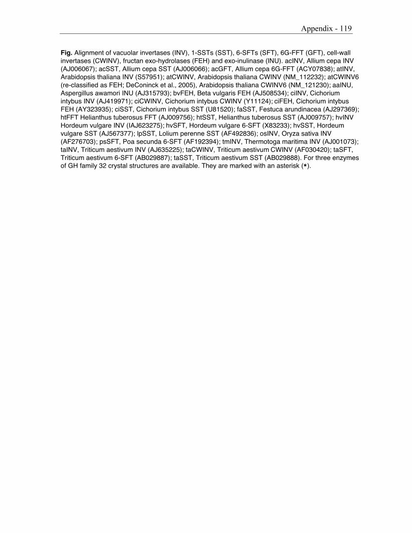
Appendix - 119
Fig. Alignment of vacuolar invertases (INV), 1-SSTs (SST), 6-SFTs (SFT), 6G-FFT (GFT), cell-wallinvertases (CWINV), fructan exo-hydrolases (FEH) and exo-inulinase (INU). acINV, Allium cepa INV(AJ006067); acSST, Allium cepa SST (AJ006066); acGFT, Allium cepa 6G-FFT (ACY07838); atINV,Arabidopsis thaliana INV (S57951); atCWINV, Arabidopsis thaliana CWINV (NM_112232); atCWINV6(re-classified as FEH; DeConinck et al., 2005), Arabidopsis thaliana CWINV6 (NM_121230); aaINU,Aspergillus awamori INU (AJ315793); bvFEH, Beta vulgaris FEH (AJ508534); ciINV, Cichoriumintybus INV (AJ419971); ciCWINV, Cichorium intybus CWINV (Y11124); ciFEH, Cichorium intybusFEH (AY323935); ciSST, Cichorium intybus SST (U81520); faSST, Festuca arundinacea (AJ297369);htFFT Helianthus tuberosus FFT (AJ009756); htSST, Helianthus tuberosus SST (AJ009757); hvINVHordeum vulgare INV (IAJ623275); hvSFT, Hordeum vulgare 6-SFT (X83233); hvSST, Hordeumvulgare SST (AJ567377); lpSST, Lolium perenne SST (AF492836); osINV, Oryza sativa INV(AF276703); psSFT, Poa secunda 6-SFT (AF192394); tmINV, Thermotoga maritima INV (AJ001073);taINV, Triticum aestivum INV (AJ635225); taCWINV, Triticum aestivum CWINV (AF030420); taSFT,Triticum aestivum 6-SFT (AB029887); taSST, Triticum aestivum SST (AB029888). For three enzymesof GH family 32 crystal structures are available. They are marked with an asterisk (*).

121
Publications 03 Distinct regulation of sucrose-sucrose-1-fructosyltransferase (1-SST) and sucrose-fructan-6-fructosyltransferase (6-SFT), the key enzymes of fructan synthesis in barley leaves: 1-SST as the pacemaker. New Phytologist 04 (161: 735-748)Nagaraj, V.J., Altenbach, D., Galati, V., Lüscher, M., MeyerA.D., Boller, T., Wiemken, A.
04 The large subunit determines catalytic specificity of barley sucrose:fructan 6-fructosyltransferase (6-SFT) and fescue sucrose:sucrose1-fructosyltransferase (1-SST) FEBS Letters 04 (567: 214-218)D. Altenbach, E. Nüesch, A.D. Meyer, T. Boller, A. Wiemken
05 Mutational analysis of the active center of fructosyltransferases: Festuca 1-SST and barley 6-SFT. SubmittedD. Altenbach, E. Nüesch, T. Ritsema, T. Boller, A. Wiemken
Conferences 00 Fructan 2000, Arolla (CH)Poster presentation of the Diploma Thesis
04 Fructan and Human Health, Lugano (CH)Oral presentation: Modifying plant fructosyltransferase activities using domain-shuffling
04 Fructan 2004, Havana (Cuba)Poster presentation: the collaborative work with the University Utrecht:Are fructosyltransferases adapted Invertases?
![[PPT]Cell Structure & Function - TypePadsimpson.typepad.com/files/cell-structurewoyce2010.ppt · Web viewCell Structure & Function](https://img.pdfslide.tips/doc/110x75/5aa4d86e7f8b9a517d8c79e9/pptcell-structure-function-viewcell-structure-function-.jpg)
![BCH-350 Plant Cell - جامعة القاسم الخضراءuoqasim.edu.iq/e_Learning/lec_file/Structure of plant cell.pdf · [STRUCTURE & FUNCTION OF PLANT CELL] ... Plastid (chloroplast](https://img.pdfslide.tips/doc/110x75/5b24c26a7f8b9a483a8b475a/bch-350-plant-cell-of-plant-cellpdf.jpg)