Embed Size (px)
Citation preview
MOLECULAR AND CELLULAR BIOLOGY, Apr. 2008, p. 2125–2137 Vol. 28, No. 70270-7306/08/$08.00�0 doi:10.1128/MCB.00740-07Copyright © 2008, American Society for Microbiology. All Rights Reserved.
Inducible Expression of Chimeric EWS/ETS Proteins Confers Ewing’sFamily Tumor-Like Phenotypes to Human Mesenchymal
Progenitor Cells�†Yoshitaka Miyagawa,1 Hajime Okita,1* Hideki Nakaijima,1 Yasuomi Horiuchi,1 Ban Sato,1
Tomoko Taguchi,1 Masashi Toyoda,3 Yohko U. Katagiri,1 Junichiro Fujimoto,2Jun-ichi Hata,1 Akihiro Umezawa,3 and Nobutaka Kiyokawa1
Department of Developmental Biology, National Research Institute for Child Health and Development, 2-10-1, Okura, Setagaya-ku,Tokyo 157-8535, Japan1; National Research Institute for Child Health and Development, 2-10-1, Okura,
Setagaya-ku, Tokyo 157-8535, Japan2; and Department of Reproductive Biology,National Research Institute for Child Health and Development, 2-10-1,
Okura, Setagaya-ku, Tokyo 157-8535, Japan3
Received 27 April 2007/Returned for modification 13 July 2007/Accepted 7 January 2008
Ewing’s family tumor (EFT) is a rare pediatric tumor of unclear origin that occurs in bone and soft tissue.Specific chromosomal translocations found in EFT cause EWS to fuse to a subset of ets transcription factorgenes (ETS), generating chimeric EWS/ETS proteins. These proteins are believed to play a crucial role in theonset and progression of EFT. However, the mechanisms responsible for the EWS/ETS-mediated onset remainunclear. Here we report the establishment of a tetracycline-controlled EWS/ETS-inducible system in humanbone marrow-derived mesenchymal progenitor cells (MPCs). Ectopic expression of both EWS/FLI1 andEWS/ERG proteins resulted in a dramatic change of morphology, i.e., from a mesenchymal spindle shape toa small round-to-polygonal cell, one of the characteristics of EFT. EWS/ETS also induced immunophenotypicchanges in MPCs, including the disappearance of the mesenchyme-positive markers CD10 and CD13 and theup-regulation of the EFT-positive markers CD54, CD99, CD117, and CD271. Furthermore, a prominent shiftfrom the gene expression profile of MPCs to that of EFT was observed in the presence of EWS/ETS. Togetherwith the observation that EWS/ETS enhances the ability of cells to invade Matrigel, these results suggest thatEWS/ETS proteins contribute to alterations of cellular features and confer an EFT-like phenotype to humanMPCs.
Ewing’s family tumor (EFT) is a rare childhood cancer aris-ing mainly in bone and soft tissue. Since EFT has a poorprognosis, it is important to elucidate the underlying patho-genic mechanisms for establishing a more effective therapeuticstrategy. EFT is characterized by the presence of chimericgenes composed of EWS and ets transcription factor genes(ETS) formed by specific chromosomal translocations, i.e.,EWS/FLI1, t(11;22)(q24;q12); EWS/ERG, t(21;22)(q12;q12);EWS/ETV1, t(7;22)(p22;q12); EWS/E1AF, t(17;22)(q12;q12);and EWS/FEV, t(2;22)(q33;q12) (26). The products of thesechimeric genes behave as aberrant transcriptional regulatorsand are believed to play a crucial role in the onset and pro-gression of EFT (3, 36). Indeed, recent studies have revealedthat the induction of EWS/FLI1 proteins can trigger transfor-mation in certain cell types, including NIH 3T3 cells (36),C2C12 myoblasts (12), and murine primary bone marrow-de-rived mesenchymal progenitor cells (MPCs) (6, 45, 52). How-ever, studies have also indicated that overexpression of EWS/FLI1 provokes apoptosis and growth arrest in mouse normal
embryonic fibroblasts and primary human fibroblasts (10, 31),hence hampering understanding of the precise role of EWS/ETS proteins in the development of EFT. The function ofEWS/ETS proteins would be greatly influenced by cell type,and thus the cells that can originate EFTs might be moresusceptible to the tumorigenic effects of EWS/ETS.
Although the cell origin of EFT is still unknown, the expres-sion of neuronal markers in spite of the occurrence in bone andsoft tissues has kept open the debate as to a potential mesen-chymal or neuroectodermal origin. As described above, ectopicexpression of EWS/FLI1 results in dramatic changes in mor-phology and the formation of EFT-like tumors in murine pri-mary bone marrow-derived MPCs but not in murine embry-onic stem cells (6, 45, 52), supporting the notion that MPCs area plausible cell origin of EFT (45). However, others argue thatMPCs cannot be considered progenitors of EFT without fur-ther evidence of similarity between human EFT and MPC-EWS/FLI1-induced tumors in mice (29, 46).
The development of experimental systems using murinespecies is useful for elucidating the mechanisms behind thepathogenesis of EFT. However, several differences betweenhuman and murine systems cannot be ignored; these differ-ences include the expression patterns of surface antigens inMPCs, for instance (7, 44, 51, 53). Moreover, human cells aredifficult to transform in vitro, and the transformed cells of miceseem to produce a more aggressive tumor than those of hu-
* Corresponding author. Mailing address: Department of Develop-mental Biology, National Research Institute for Child Health andDevelopment, 2-10-1, Okura, Setagaya-ku, Tokyo 157-8535, Japan.Phone: 81-3-3416-0181. Fax: 81-3-3417-2496. E-mail: [email protected].
† Supplemental material for this article may be found at http://mcb.asm.org/.
� Published ahead of print on 22 January 2008.
2125
on April 2, 2018 by guest
http://mcb.asm
.org/D
ownloaded from
on A
pril 2, 2018 by guesthttp://m
cb.asm.org/
Dow
nloaded from
on April 2, 2018 by guest
http://mcb.asm
.org/D
ownloaded from
mans (1). The findings suggest the existence of undefined cell-autonomous mechanisms that render human cells resistant tomalignant transformation. Therefore, the use of human cellmodels is ideal for clarifying how EFT develops. Models of theonset of EFT have been generated using primary fibroblasts(31) and rhabdomyosarcoma cells (23). However, these celltypes are not appropriate for studying the origins of EFT, anda model that precisely recapitulates EWS/ETS-mediated EFTformation is required.
UET-13 cells are obtained by prolonging the life span ofhuman bone marrow stromal cells by use of the retroviraltransgenes hTERT and E7 (38, 50), retain the ability to dif-ferentiate into not only mesodermal derivatives but also neu-ronal progenitor-like cells, and are considered a good modelfor studying the cellular events in human MPCs. Therefore, wehave examined the biological effect of EWS/ETS in humanMPCs by use of UET-13 cells by exploiting tetracycline-induc-ible systems for expressing EWS/ETS (EWS/FLI1 and EWS/ERG). Here we report that overexpression of EWS/ETSmediates an EFT-like phenotype, including morphology, im-munophenotype, and gene expression profile, with enhance-ment of the Matrigel invasion ability of UET-13 cells.
MATERIALS AND METHODS
Cell cultures and establishment of UET-13TR-EWS/ETS cell lines. UET-13cells were cultured in Dulbecco’s modified Eagle’s medium (DMEM) with 10%Tet system approved fetal bovine serum (T-FBS) (Takara) at 37°C under ahumidified 5% CO2 atmosphere. EFT cell lines (EES-1 [20], SCCH196 [21],RD-ES and SK-ES1 [5], NCR-EW2 and NCR-EW3 [19], and W-ES [13]) andneuroblastoma (NB) cell lines (NB69 and NB9 [15] and GOTO [47]) werecultured in RPMI 1640 with 10% FBS. A rhabdomyosarcoma cell line, NRS-1(40), was cultured in Eagle’s minimal essential medium with 10% FBS. The celllines used in this study are listed in Table 1.
UET-13 cells were seeded at a density of 5 � 104 cells per well in 24-well tissueculture plates 1 day prior to transfection. For introducing the tetracycline-induc-ible system, UET-13 cells were transfected with pcDNA6-TR (Invitrogen) by useof Lipofectamine 2000 (Invitrogen). After 72 h, the medium was replaced withfresh medium containing 200 �g/ml of blasticidin S (Invitrogen). Individualresistant clones were selected for a month and designated UET-13TR cells.UET-13TR cells were further transfected with pcDNA4-EWS/ETSs constructedas described below, and individual resistant clones were selected in DMEMcontaining 10% T-FBS and 200 to 300 �g/ml of Zeocin (Invitrogen). The Zeocin-resistant clones were expanded and tested for the induction of EWS/ETS ex-pression upon the addition of tetracycline by use of reverse transcription-PCR(RT-PCR) as described below.
Plasmid construction. A gateway cassette (bases 1 to 1705) was amplified frompBLOCK-iT3-DEST (Invitrogen) by PCR, and the PCR product was insertedinto the EcoRV site of pcDNA4-TO (Invitrogen) (termed pcDNA4-DEST).Since the type II EWS/FLI1 is a stronger transactivator than the type I product
(32), we used the type II variant in the present study. EWS/ERG was isolatedfrom W-ES, an EFT cell line, joining EWS exon 7 and ERG exon 9. Full-lengthEWS/FLI1 type II and EWS/ERG cDNAs were amplified from cDNAs preparedfrom NCR-EW2 and W-ES cells, respectively, by PCR as described below andcloned into the XmnI-EcoRV sites of pENTR11 (Invitrogen). The resultingpENTR11-EWS/ETSs were recombined with pcDNA4-DEST by use of LRrecombination reaction as instructed by the manufacturer (Invitrogen) to con-struct the tetracycline-inducible EWS/ETS expression vector pcDNA4-EWS/ETSs.
Western blot analysis. UET-13 transfectants were cultivated with or without 3�g/ml of tetracycline for 72 h. Western blot analysis was performed as previouslydescribed (37). Briefly, the cell lysates were prepared and separated on a 10%sodium dodecyl sulfate-polyacrylamide gel electrophoresis gel and transferredonto a polyvinylidene difluoride membrane. The membranes were blocked with5% skimmed milk in phosphate-buffered saline (PBS) containing 0.01% Tween20 (Sigma) and incubated with primary antibodies. As the primary antibodies,anti-Fli-1, anti-Erg-1/2/3 (Santa Cruz Biotechnology), and anti-actin (Sigma)were used. Horseradish peroxidase-conjugated anti-rabbit or anti-mouse immu-noglobulin G (IgG) antibodies (DakoCytomation) were used as secondary anti-bodies. Blots were detected by chemiluminescence using an ECL Plus Westernblotting detection system (GE Healthcare Bio-Science Corp.) and exposed toX-ray film (Kodak) for 5 to 30 min.
MTT assay and detection of apoptosis. Growth curves of UET-13 transfectantswere determined using the 3-(4,5-dimethylthiazol-2-yl)-2,5-diphenyltetrazoliumbromide (MTT) assay as described previously (18). The apoptosis was detectedusing an annexin V-fluorescein isothiocyanate (FITC) apoptosis detection kit(Biovision) according to the manufacturer’s instructions and analyzed by flowcytometry (Cytomics FC500; Beckman Coulter).
Immunofluorescence analysis. After 1 week of culture in the absence orpresence of tetracycline, UET-13 cells and the transfectants were harvested with0.25% trypsin plus EDTA (IBL). The cells (2 � 105) were incubated with mousemonoclonal antibodies for 20 min. In the case of fluorescence-labeled antibodies,the cells were washed with PBS and then analyzed. In the case of primaryunconjugated mouse antibodies, the cells were washed and then incubated withFITC-conjugated goat anti-mouse IgG antibody (Jackson ImmunoResearchLaboratories) for 20 min. Cell fluorescence was detected using a Cytomics FC500instrument as described previously (27).
Antibodies against the following human antigens were used: CD10, CD13,CD14, CD29, CD34, CD40, CD44, CD45, CD49e, CD54, CD56, CD61, CD90,CD105, CD117, and CD166 from Beckman Coulter; CD73 from BD Bio-sciences-Pharmingen; CD55 from Abcam; CD59 from Cedarlane Laboratories;and CD133 and CD271 from Miltenyi Biotec GmbH.
Immunocytochemistry. Cells were grown on collagen type I-coated coverglasses (Iwaki). After 72 h with or without tetracycline, cells were fixed for 30 minin 4% paraformaldehyde and permeabilized in PBS containing 0.2% TritonX-100 (Sigma) for 30 min. Subsequently, they were washed with PBS and blockedin PBS containing 0.1% Triton X-100 and 1% bovine serum albumin (Sigma) for30 min before being incubated with a monoclonal anti-CD99 antibody, i.e., 12E7(1:100) (DakoCytomation) or O13 (1:200) (Thermo), and polyclonal anti-Fli-1antibody (1:100) (Santa Cruz) for 1 h. Bound antibodies were visualized withappropriate secondary antibodies, i.e., Alexa Fluor 488 goat anti-mouse IgG(heavy plus light chains) highly cross-adsorbed and Alexa Fluor 546 goat anti-rabbit IgG (heavy plus light chains) highly cross-adsorbed (Invitrogen) for 1 h at1:300. Nuclei were counterstained with 4�,6�-diamidino-2-phenylindole (DAPI)or propidium iodide (PI) (Sigma). For the visualization of whole cells, cells weretreated with Celltracker Blue (Invitrogen) for 30 min and then fixed. Fluores-cence was observed and analyzed using a confocal laser scanning microscope andimage software (either FV500 from Olympus or LSM510 from Carl Zeiss).Precise measurements of cell size, nuclear size, and the nucleus-to-cytoplasm(N/C) ratio were performed using Image J (16).
RT-PCR analysis. Total RNA was extracted from cells by use of an RNeasy kit(Qiagen) and reverse transcribed using a first-strand cDNA synthesis kit (GEHealthcare Bio-Science Corp). RT-PCR was performed with a HotstarTaq mas-ter mix kit (Qiagen). As an internal control, human GAPDH cDNA was alsoamplified. The sequences of gene-specific primers for RT-PCR were as follows:for EWS/FLI1 (forward), 5�-ATGGCGTCCACGGATTACAGTACCT-3�; forEWS/FLI1 (reverse), 5�-GGGTCTTCTTTGACACTCAATCG-3�; for EWS/ERG (forward), 5�-ATGGCGTCCACGGATTACAGTACCT-3�; for EWS/ERG (reverse), 5�-TTAGTAGTAAGTGCCCAGATGAGAA-3�; for GAPDH(forward), 5�-CCACCCATGGCAAATTCCATGGCA-3�; and for GAPDH (re-verse), 5�-TCTAGACGGCAGGTCAGGT/CCACC-3�. PCR products wereelectrophoresed with a 1% agarose gel and stained with ethidium bromide.
TABLE 1. Cell lines used in this study and fusion transcript types
Cell line Diagnosis Fusion transcripttype Reference
EES-1 EFT EWS/FLI1 type I 20SCCH196 EFT EWS/FLI1 type I 21RD-ES EFT EWS/FLI1 type II 5SK-ES1 EFT EWS/FLI1 type II 5NCR-EW2 EFT EWS/FLI1 type II 19NCR-EW3 EFT EWS/E1AF 19W-ES EFT EWS/ERG 13NB69 NB 15NB9 NB 15GOTO NB 47NRS-1 RMS PAX3/FKHR 40
2126 MIYAGAWA ET AL. MOL. CELL. BIOL.
on April 2, 2018 by guest
http://mcb.asm
.org/D
ownloaded from
Real-time RT-PCR. Real-time RT-PCR was performed using TaqMan univer-sal PCR master mix and TaqMan gene expression assays and an inventoriedassay on an ABI Prism 7900HT sequence detection system (Applied Biosystems)according to the manufacturer’s instructions. The human GAPDH gene was usedas an internal control for normalization.
DNA microarray analysis. Total RNA isolated from cells was reverse tran-scribed and labeled using one-cycle target labeling and control reagents asinstructed by the manufacturer (Affymetrix). The labeled probes were hybridizedto the human genome U133 Plus 2.0 array (Affymetrix). The arrays were per-formed in a single experiment and analyzed using GeneChip operating software,version 1.2 (Affymetrix). Background subtraction, normalization, and principalcomponent analysis (PCA) were performed by GeneSpring GX 7.3 software(Agilent Technologies). Signal intensities were prenormalized based on the me-dian of all measurements on that chip. To account for the difference in detectionefficiencies between the spots, prenormalized signal intensities on each genewere normalized to the median of prenormalized measurements for that gene.The data were filtered using the following steps. (i) Genes that were scored asabsent in all samples were eliminated. (ii) Genes for which the signal intensitieswere lower than 100 were eliminated. (iii) Performing cluster analysis using
filtering genes, we selected the genes that exhibited increased expression ordecreased expression in tetracycline-treated cells. Accession numbers for themicroarray data are given below.
Invasion assay. The invasion assay was performed using Matrigel (BD Bio-science) according to the previous description (34) with some modification.Polycarbonate filter inserts containing 8-�m pores (BD Falcon) were coated with50 �l of a 6:1 mixture of culture medium and Matrigel and placed into 24-wellculture plates containing DMEM supplemented with 10% T-FBS as chemoat-tractants. Cells (2.5 � 104) treated with or without tetracycline for 72 h weresuspended in DMEM containing 0.01% T-FBS and plated on top of each filterinsert. After 20 h in culture in the presence or absence of tetracycline, nonin-vading cells were removed from upper surface of the filter with a cotton swab.The invading cells on the lower surface of the filter were fixed with formalin,stained with hematoxylin-eosin, and counted in five fields per membrane withlight microscopy. As a control, cells were also cultured on uncoated filter inserts.The invasion efficiency was presented as the ratio of the number of invading cellson Matrigel-coated inserts to that on uncoated inserts. Experiments were per-formed in triplicate, and the means with standard deviations of the values areshown in the graphs in Fig. 8.
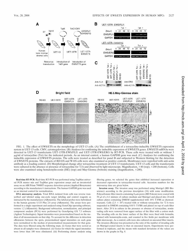
FIG. 1. The effect of EWS/ETS on the morphology of UET-13 cells. (A) The establishment of a tetracycline-inducible EWS/ETS expressionsystem in UET-13 cells. CMV, cytomegalovirus. (B) Analyses for confirming the inducible expression of EWS/ETS genes. EWS/ETS mRNAs weredetected in UET-13 transfectants UET-13TR-EWS/FLI1 and UET-13TR-EWS/ERG by RT-PCR. These cells were treated with or without 3�g/ml of tetracycline (Tet) for the indicated periods. As an internal control, a human GAPDH gene was used. (C) Analyses for confirming theinducible expression of EWS/ETS proteins. The cells were treated as described for panel B and subjected to Western blotting for the detectionof EWS/ETS proteins. The extracts of RD-ES and W-ES cells were also examined as positive controls. Membranes were reprobed with anti-actinantibody as a loading control. (D) Morphological change after tetracycline treatment of UET-13 transfectants. UET-13 cells and the transfectantswere cultured in the absence or presence of tetracycline for 72 h and observed by light microscopy. Magnification, �40 (top); �200 (bottom). Cellswere also examined using hematoxylin-eosin (HE) (top) and May-Giemsa (bottom) staining (magnification, �200).
VOL. 28, 2008 EFFECTS OF EWS/ETS EXPRESSION IN HUMAN MPCs 2127
on April 2, 2018 by guest
http://mcb.asm
.org/D
ownloaded from
Microarray data accession numbers. Microarray data have been deposited inthe Gene Expression Omnibus database GEO (www.ncbi.nlm.nih.gov/geo) (ac-cession numbers GSE8665 and GSE8596).
RESULTS
EWS/ETS expression results in morphological changes inUET-13 cells. To investigate how the expression of EWS/ETSaffects human MPCs, we used UET-13 cells as a model ofhuman MPCs and expressed EWS/FLI1 (UET-13TR-EWS/FLI1) and EWS/ERG (UET-13TR-EWS/ERG) in a tetracy-cline-inducible manner (Fig. 1A). As shown in Fig. 1B and C,we confirmed that the tetracycline treatment could induceEWS/ETS expression by RT-PCR analysis and Western blot-ting. The inducibility upon the addition of doxycycline wascomparable to that upon the addition of tetracycline.
Using these cell systems, first we examined the effect ofEWS/ETS expression on morphology in UET-13 transfectants.When tetracycline was added to the culture, the morphologiesof both UET-13TR-EWS/FLI1 and UET-13TR-EWS/ERGcells were dramatically changed (Fig. 1D). Tetracycline-treatedUET-13TR-EWS/ETS cells consisted of a mixture of smallround-to-polygonal cells and short spindle cells. The cell mor-phology resembled that of EFT cell lines. To assess the repro-
ducibility of this phenotypic change, other UET-13TR-EWS/ETS clones were examined, and similar morphological changeswere observed. Since tetracycline treatment did not affect themorphology of UET-13TR cells (Fig. 1D), it was suggestedthat the morphological alteration in UET-13 cells from a mes-enchymal cell shape to small round cells, one of the charac-teristics of EFT, can be attributed to EWS/ETS expression.
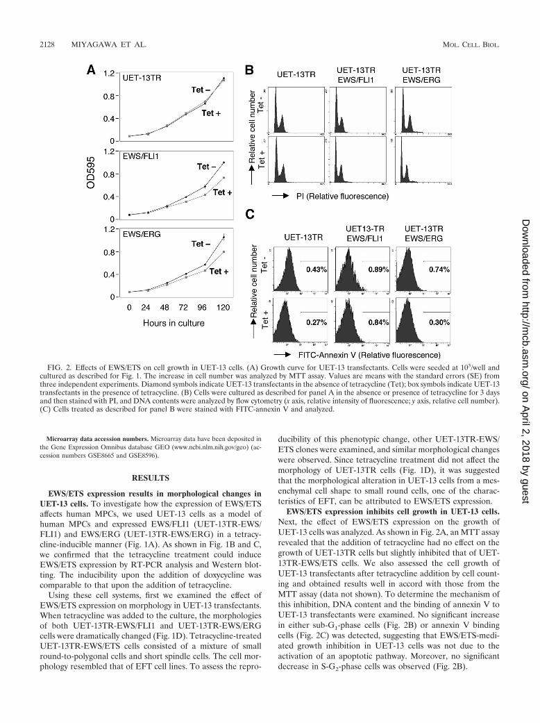
EWS/ETS expression inhibits cell growth in UET-13 cells.Next, the effect of EWS/ETS expression on the growth ofUET-13 cells was analyzed. As shown in Fig. 2A, an MTT assayrevealed that the addition of tetracycline had no effect on thegrowth of UET-13TR cells but slightly inhibited that of UET-13TR-EWS/ETS cells. We also assessed the cell growth ofUET-13 transfectants after tetracycline addition by cell count-ing and obtained results well in accord with those from theMTT assay (data not shown). To determine the mechanism ofthis inhibition, DNA content and the binding of annexin V toUET-13 transfectants were examined. No significant increasein either sub-G1-phase cells (Fig. 2B) or annexin V bindingcells (Fig. 2C) was detected, suggesting that EWS/ETS-medi-ated growth inhibition in UET-13 cells was not due to theactivation of an apoptotic pathway. Moreover, no significantdecrease in S-G2-phase cells was observed (Fig. 2B).
FIG. 2. Effects of EWS/ETS on cell growth in UET-13 cells. (A) Growth curve for UET-13 transfectants. Cells were seeded at 103/well andcultured as described for Fig. 1. The increase in cell number was analyzed by MTT assay. Values are means with the standard errors (SE) fromthree independent experiments. Diamond symbols indicate UET-13 transfectants in the absence of tetracycline (Tet); box symbols indicate UET-13transfectants in the presence of tetracycline. (B) Cells were cultured as described for panel A in the absence or presence of tetracycline for 3 daysand then stained with PI, and DNA contents were analyzed by flow cytometry (x axis, relative intensity of fluorescence; y axis, relative cell number).(C) Cells treated as described for panel B were stained with FITC-annexin V and analyzed.
2128 MIYAGAWA ET AL. MOL. CELL. BIOL.
on April 2, 2018 by guest
http://mcb.asm
.org/D
ownloaded from
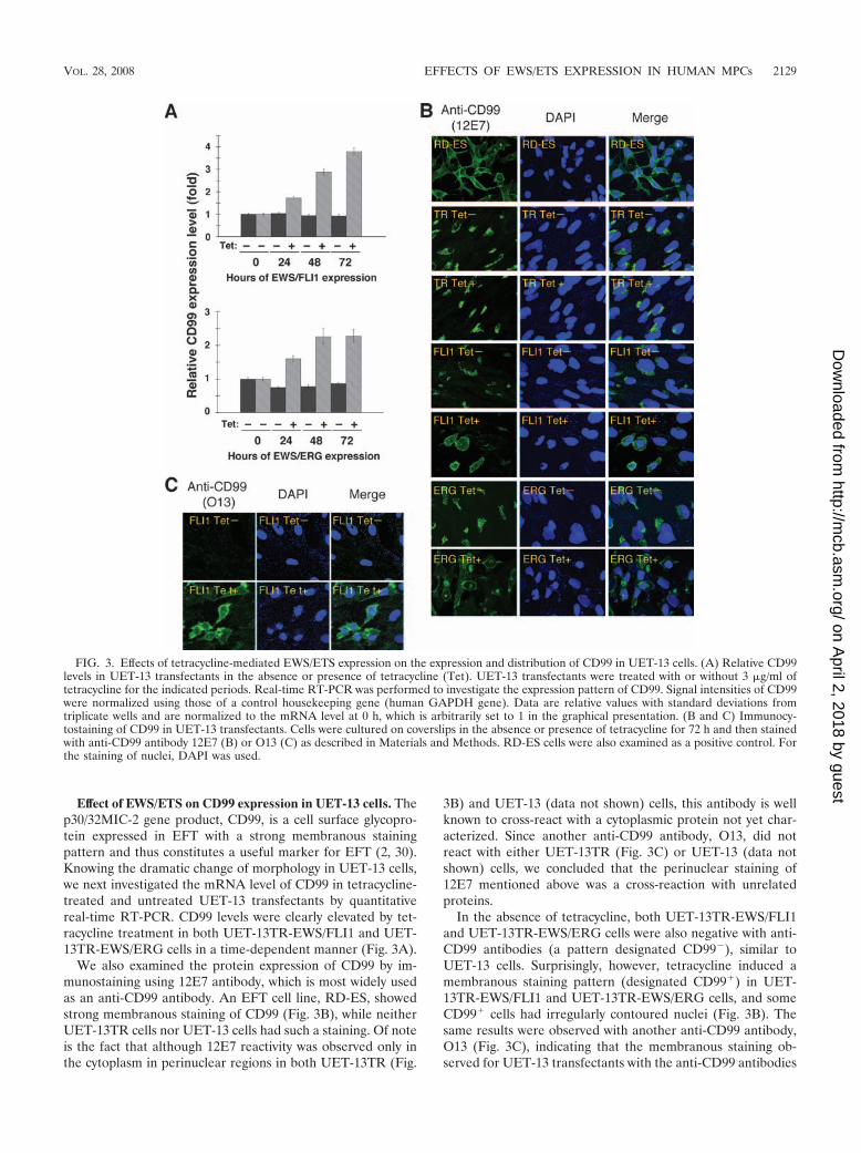
Effect of EWS/ETS on CD99 expression in UET-13 cells. Thep30/32MIC-2 gene product, CD99, is a cell surface glycopro-tein expressed in EFT with a strong membranous stainingpattern and thus constitutes a useful marker for EFT (2, 30).Knowing the dramatic change of morphology in UET-13 cells,we next investigated the mRNA level of CD99 in tetracycline-treated and untreated UET-13 transfectants by quantitativereal-time RT-PCR. CD99 levels were clearly elevated by tet-racycline treatment in both UET-13TR-EWS/FLI1 and UET-13TR-EWS/ERG cells in a time-dependent manner (Fig. 3A).
We also examined the protein expression of CD99 by im-munostaining using 12E7 antibody, which is most widely usedas an anti-CD99 antibody. An EFT cell line, RD-ES, showedstrong membranous staining of CD99 (Fig. 3B), while neitherUET-13TR cells nor UET-13 cells had such a staining. Of noteis the fact that although 12E7 reactivity was observed only inthe cytoplasm in perinuclear regions in both UET-13TR (Fig.
3B) and UET-13 (data not shown) cells, this antibody is wellknown to cross-react with a cytoplasmic protein not yet char-acterized. Since another anti-CD99 antibody, O13, did notreact with either UET-13TR (Fig. 3C) or UET-13 (data notshown) cells, we concluded that the perinuclear staining of12E7 mentioned above was a cross-reaction with unrelatedproteins.
In the absence of tetracycline, both UET-13TR-EWS/FLI1and UET-13TR-EWS/ERG cells were also negative with anti-CD99 antibodies (a pattern designated CD99�), similar toUET-13 cells. Surprisingly, however, tetracycline induced amembranous staining pattern (designated CD99�) in UET-13TR-EWS/FLI1 and UET-13TR-EWS/ERG cells, and someCD99� cells had irregularly contoured nuclei (Fig. 3B). Thesame results were observed with another anti-CD99 antibody,O13 (Fig. 3C), indicating that the membranous staining ob-served for UET-13 transfectants with the anti-CD99 antibodies
FIG. 3. Effects of tetracycline-mediated EWS/ETS expression on the expression and distribution of CD99 in UET-13 cells. (A) Relative CD99levels in UET-13 transfectants in the absence or presence of tetracycline (Tet). UET-13 transfectants were treated with or without 3 �g/ml oftetracycline for the indicated periods. Real-time RT-PCR was performed to investigate the expression pattern of CD99. Signal intensities of CD99were normalized using those of a control housekeeping gene (human GAPDH gene). Data are relative values with standard deviations fromtriplicate wells and are normalized to the mRNA level at 0 h, which is arbitrarily set to 1 in the graphical presentation. (B and C) Immunocy-tostaining of CD99 in UET-13 transfectants. Cells were cultured on coverslips in the absence or presence of tetracycline for 72 h and then stainedwith anti-CD99 antibody 12E7 (B) or O13 (C) as described in Materials and Methods. RD-ES cells were also examined as a positive control. Forthe staining of nuclei, DAPI was used.
VOL. 28, 2008 EFFECTS OF EWS/ETS EXPRESSION IN HUMAN MPCs 2129
on April 2, 2018 by guest
http://mcb.asm
.org/D
ownloaded from
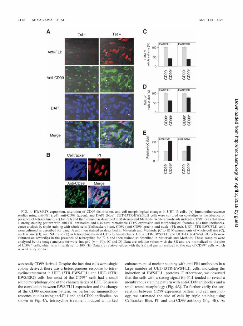
was really CD99 derived. Despite the fact that cells were singlecolony derived, there was a heterogeneous response to tetra-cycline treatment in UET-13TR-EWS/FLI1 and UET-13TR-EWS/ERG cells, but most of the CD99� cells had a smallround morphology, one of the characteristics of EFT. To assessthe correlation between EWS/FLI1 expression and the changeof the CD99 expression pattern, we performed immunofluo-rescence studies using anti-Fli1 and anti-CD99 antibodies. Asshown in Fig. 4A, tetracycline treatment induced a marked
enhancement of nuclear staining with anti-Fli1 antibodies in alarge number of UET-13TR-EWS/FLI1 cells, indicating theinduction of EWS/FLI1 proteins. Furthermore, we observedthat the cells with a strong signal for Fli1 tended to reveal amembranous staining pattern with anti-CD99 antibodies and asmall round morphology (Fig. 4A). To further verify the cor-relation between CD99 expression pattern and cell morphol-ogy, we estimated the size of cells by triple staining usingCelltracker Blue, PI, and anti-CD99 antibody (Fig. 4B). As
FIG. 4. EWS/ETS expression, alteration of CD99 distribution, and cell morphological changes in UET-13 cells. (A) Immunofluorescencestudies using anti-Fli1 (red), anti-CD99 (green), and DAPI (blue). UET-13TR-EWS/FLI1 cells were cultured on coverslips in the absence orpresence of tetracycline (Tet) for 72 h and then stained as described in Materials and Methods. White arrowheads indicate CD99� cells that havea strong staining pattern with anti-Fli1 antibodies and also have remarkable CD99 expression and morphological features. (B) Immunofluores-cence analysis by triple staining with whole cells (Celltracker; blue), CD99 (anti-CD99; green), and nuclei (PI; red). UET-13TR-EWS/FLI1 cellswere cultured as described for panel A and then stained as described in Materials and Methods. (C to E) Measurements of whole-cell size (C),nuclear size (D), and N/C ratio (E) in tetracycline-treated UET-13 transfectants. UET-13TR-EWS/FLI1 and UET-13TR-EWS/ERG cells werecultured on coverslips in the presence of tetracycline for 72 h and then stained as described in Materials and Methods. These samples wereanalyzed by the image analysis software Image J (n � 50). (C and D) Data are relative values with the SE and are normalized to the sizeof CD99� cells, which is arbitrarily set to 100. (E) Data are relative values with the SE and are normalized to the size of CD99� cells, whichis arbitrarily set to 1.
2130 MIYAGAWA ET AL. MOL. CELL. BIOL.
on April 2, 2018 by guest
http://mcb.asm
.org/D
ownloaded from
presented in Fig. 4C and D, the results clearly showed that themajority of CD99� cells were significantly smaller in bothwhole-cell size and nuclear size than the CD99� cells. More-over, CD99� cells also had a substantially increased N/C ratio(Fig. 4E). These results indicated that EWS/ETS expressionpromoted CD99 expression in UET-13 cells, and CD99 expres-sion status is correlated with the degree of morphologicalchange.
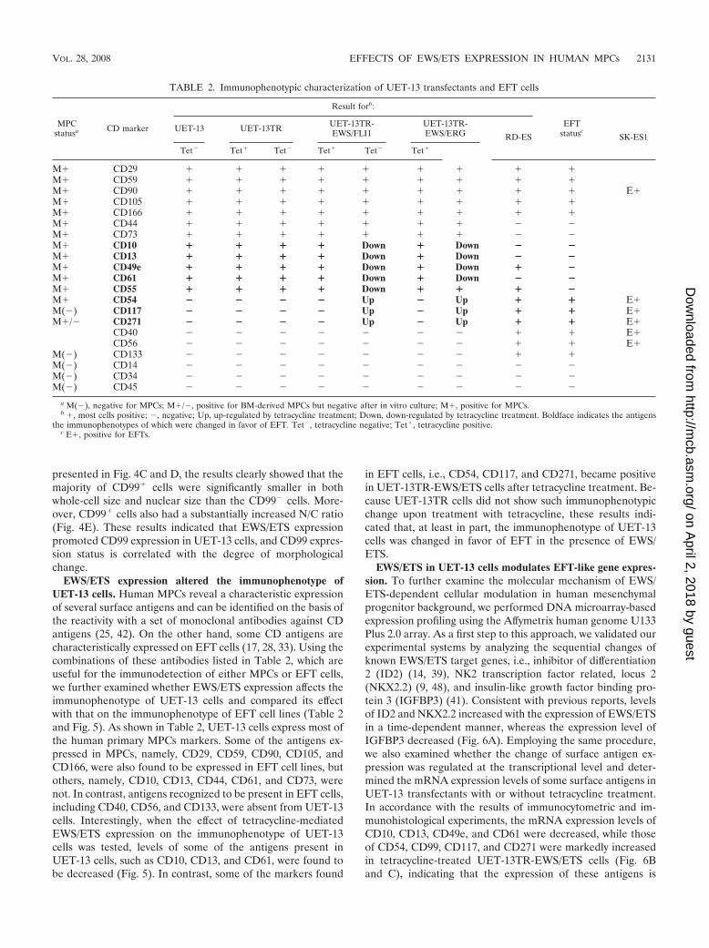
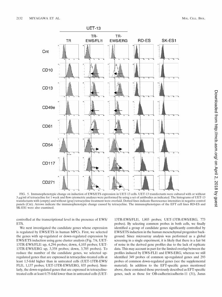
EWS/ETS expression altered the immunophenotype ofUET-13 cells. Human MPCs reveal a characteristic expressionof several surface antigens and can be identified on the basis ofthe reactivity with a set of monoclonal antibodies against CDantigens (25, 42). On the other hand, some CD antigens arecharacteristically expressed on EFT cells (17, 28, 33). Using thecombinations of these antibodies listed in Table 2, which areuseful for the immunodetection of either MPCs or EFT cells,we further examined whether EWS/ETS expression affects theimmunophenotype of UET-13 cells and compared its effectwith that on the immunophenotype of EFT cell lines (Table 2and Fig. 5). As shown in Table 2, UET-13 cells express most ofthe human primary MPCs markers. Some of the antigens ex-pressed in MPCs, namely, CD29, CD59, CD90, CD105, andCD166, were also found to be expressed in EFT cell lines, butothers, namely, CD10, CD13, CD44, CD61, and CD73, werenot. In contrast, antigens recognized to be present in EFT cells,including CD40, CD56, and CD133, were absent from UET-13cells. Interestingly, when the effect of tetracycline-mediatedEWS/ETS expression on the immunophenotype of UET-13cells was tested, levels of some of the antigens present inUET-13 cells, such as CD10, CD13, and CD61, were found tobe decreased (Fig. 5). In contrast, some of the markers found
in EFT cells, i.e., CD54, CD117, and CD271, became positivein UET-13TR-EWS/ETS cells after tetracycline treatment. Be-cause UET-13TR cells did not show such immunophenotypicchange upon treatment with tetracycline, these results indi-cated that, at least in part, the immunophenotype of UET-13cells was changed in favor of EFT in the presence of EWS/ETS.
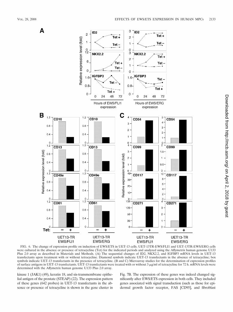
EWS/ETS in UET-13 cells modulates EFT-like gene expres-sion. To further examine the molecular mechanism of EWS/ETS-dependent cellular modulation in human mesenchymalprogenitor background, we performed DNA microarray-basedexpression profiling using the Affymetrix human genome U133Plus 2.0 array. As a first step to this approach, we validated ourexperimental systems by analyzing the sequential changes ofknown EWS/ETS target genes, i.e., inhibitor of differentiation2 (ID2) (14, 39), NK2 transcription factor related, locus 2(NKX2.2) (9, 48), and insulin-like growth factor binding pro-tein 3 (IGFBP3) (41). Consistent with previous reports, levelsof ID2 and NKX2.2 increased with the expression of EWS/ETSin a time-dependent manner, whereas the expression level ofIGFBP3 decreased (Fig. 6A). Employing the same procedure,we also examined whether the change of surface antigen ex-pression was regulated at the transcriptional level and deter-mined the mRNA expression levels of some surface antigens inUET-13 transfectants with or without tetracycline treatment.In accordance with the results of immunocytometric and im-munohistological experiments, the mRNA expression levels ofCD10, CD13, CD49e, and CD61 were decreased, while thoseof CD54, CD99, CD117, and CD271 were markedly increasedin tetracycline-treated UET-13TR-EWS/ETS cells (Fig. 6Band C), indicating that the expression of these antigens is
TABLE 2. Immunophenotypic characterization of UET-13 transfectants and EFT cells
MPCstatusa CD marker
Result forb:
EFTstatuscUET-13 UET-13TR UET-13TR-
EWS/FLI1UET-13TR-EWS/ERG RD-ES SK-ES1
Tet� Tet� Tet� Tet� Tet� Tet�
M� CD29 � � � � � � � � �M� CD59 � � � � � � � � �M� CD90 � � � � � � � � � E�M� CD105 � � � � � � � � �M� CD166 � � � � � � � � �M� CD44 � � � � � � � � �M� CD73 � � � � � � � � �M� CD10 � � � � Down � Down � �M� CD13 � � � � Down � Down � �M� CD49e � � � � Down � Down � �M� CD61 � � � � Down � Down � �M� CD55 � � � � Down � � � �M� CD54 � � � � Up � Up � � E�M(�) CD117 � � � � Up � Up � � E�M�/� CD271 � � � � Up � Up � � E�
CD40 � � � � � � � � � E�CD56 � � � � � � � � � E�
M(�) CD133 � � � � � � � � �M(�) CD14 � � � � � � � � �M(�) CD34 � � � � � � � � �M(�) CD45 � � � � � � � � �
a M(�), negative for MPCs; M�/�, positive for BM-derived MPCs but negative after in vitro culture; M�, positive for MPCs.b �, most cells positive; �, negative; Up, up-regulated by tetracycline treatment; Down, down-regulated by tetracycline treatment. Boldface indicates the antigens
the immunophenotypes of which were changed in favor of EFT. Tet�, tetracycline negative; Tet�, tetracycline positive.c E�, positive for EFTs.
VOL. 28, 2008 EFFECTS OF EWS/ETS EXPRESSION IN HUMAN MPCs 2131
on April 2, 2018 by guest
http://mcb.asm
.org/D
ownloaded from
controlled at the transcriptional level in the presence of EWS/ETS.
We next investigated the candidate genes whose expressionis regulated by EWS/ETS in human MPCs. First, we selectedthe genes with up-regulated or down-regulated expression byEWS/ETS induction using gene cluster analysis (Fig. 7A; UET-13TR-EWS/FLI1 up, 4,294 probes; down, 4,103 probes; UET-13TR-EWS/ERG up, 3,358 probes; down, 3,705 probes). Toreduce the number of the candidate genes, we selected up-regulated genes that are expressed in tetracycline-treated cells atleast 1.5-fold higher than in untreated cells (UET-13TR-EWS/FLI1, 1,137 probes; UET-13TR-EWS/ERG, 835 probes). Simi-larly, the down-regulated genes that are expressed in tetracycline-treated cells at least 0.75-fold lower than in untreated cells (UET-
13TR-EWS/FLI1, 1,803 probes; UET-13TR-EWS/ERG, 773probes). By selecting common probes in both cells, we finallyidentified a group of candidate genes significantly controlled byEWS/ETS induction in the human mesenchymal progenitor back-ground. Since microarray analysis was performed as a globalscreening in a single experiment, it is likely that there is a fair bitof noise in the derived gene profiles due to the lack of replicatedata. This may account in part for the limited overlap between theprofiles induced by EWS-FLI1 and EWS-ERG, whereas we stillidentified 349 probes of common up-regulated genes and 293probes of common down-regulated genes (see the supplementalmaterial). In addition to the EFT-specific genes mentionedabove, these contained those previously described as EFT-specificgenes, such as those for OB-cadherin/cadherin-11 (31), Janus
FIG. 5. Immunophenotypic change on induction of EWS/ETS expression in UET-13 cells. UET-13 transfectants were cultured with or without3 �g/ml of tetracycline for 1 week and flow cytometric analyses were performed by using a set of antibodies as indicated. The histograms of UET-13transfectants with (empty) and without (gray) tetracycline treatment were overlaid. Dotted lines indicate fluorescence intensities in negative controlpanels (Cnt). Arrows indicate the immunophenotypic change caused by tetracycline. The immunophenotypes of the EFT cell lines RD-ES andSK-ES1 were also examined.
2132 MIYAGAWA ET AL. MOL. CELL. BIOL.
on April 2, 2018 by guest
http://mcb.asm
.org/D
ownloaded from
kinase 1 (JAK1) (49), keratin 18, and six-transmembrane epithe-lial antigen of the prostate (STEAP) (22). The expression patternof these genes (642 probes) in UET-13 transfectants in the ab-sence or presence of tetracycline is shown in the gene cluster in
Fig. 7B. The expression of these genes was indeed changed sig-nificantly after EWS/ETS expression in both cells. They includedgenes associated with signal transduction (such as those for epi-dermal growth factor receptor, FAS [CD95], and fibroblast
FIG. 6. The change of expression profile on induction of EWS/ETS in UET-13 cells. UET-13TR-EWS/FLI1 and UET-13TR-EWS/ERG cellswere cultured in the absence or presence of tetracycline (Tet) for the indicated periods and analyzed using the Affymetrix human genome U133Plus 2.0 array as described in Materials and Methods. (A) The sequential changes of ID2, NKX2.2, and IGFBP3 mRNA levels in UET-13transfectants upon treatment with or without tetracycline. Diamond symbols indicate UET-13 transfectants in the absence of tetracycline; boxsymbols indicate UET-13 transfectants in the presence of tetracycline. (B and C) Microarray studies for the determination of expression profilesof surface antigens in UET-13 transfectants. UET-13 transfectants were treated with or without 3 �g/ml of tetracycline for 72 h. mRNA levels weredetermined with the Affymetrix human genome U133 Plus 2.0 array.
VOL. 28, 2008 EFFECTS OF EWS/ETS EXPRESSION IN HUMAN MPCs 2133
on April 2, 2018 by guest
http://mcb.asm
.org/D
ownloaded from
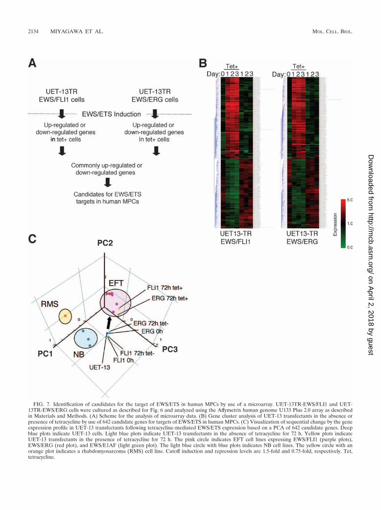
FIG. 7. Identification of candidates for the target of EWS/ETS in human MPCs by use of a microarray. UET-13TR-EWS/FLI1 and UET-13TR-EWS/ERG cells were cultured as described for Fig. 6 and analyzed using the Affymetrix human genome U133 Plus 2.0 array as describedin Materials and Methods. (A) Scheme for the analysis of microarray data. (B) Gene cluster analysis of UET-13 transfectants in the absence orpresence of tetracycline by use of 642 candidate genes for targets of EWS/ETS in human MPCs. (C) Visualization of sequential change by the geneexpression profile in UET-13 transfectants following tetracycline-mediated EWS/ETS expression based on a PCA of 642 candidate genes. Deepblue plots indicate UET-13 cells. Light blue plots indicate UET-13 transfectants in the absence of tetracycline for 72 h. Yellow plots indicateUET-13 transfectants in the presence of tetracycline for 72 h. The pink circle indicates EFT cell lines expressing EWS/FLI1 (purple plots),EWS/ERG (red plot), and EWS/E1AF (light green plot). The light blue circle with blue plots indicates NB cell lines. The yellow circle with anorange plot indicates a rhabdomyosarcoma (RMS) cell line. Cutoff induction and repression levels are 1.5-fold and 0.75-fold, respectively. Tet,tetracycline.
2134 MIYAGAWA ET AL. MOL. CELL. BIOL.
on April 2, 2018 by guest
http://mcb.asm
.org/D
ownloaded from
growth factor receptor 1) and development (such as jagged-1 andfrizzled-4, -7, and -8). Interestingly, in addition to the surfaceantigens presented in Fig. 6B and C, the expression profiling ofEWS/ETS-expressing UET-13 cells displayed the modulation ofseveral genes associated with cell adhesion, cytoskeletal structure,and membrane trafficking, such as those for collagen-11 and -21,ephrin receptor-A2, -B2, and -B3, ephrin-B1, claudin-1, integrin-�11, -�M, and -�2, CD66 (carcinoembryonic antigen-related celladhesion molecule-1), and CD102 (intercellular cell adhesionmolecule-2). They also included genes of chemokines CCL-2 and-3. These data raise the possibility that EWS/ETS can contributeto the membrane condition in human MPCs via the regulation ofthese cell surface molecules and chemokines.
Using these genes, we performed a PCA to visualize the shiftin the gene expression pattern among the 642 probes. Asshown in Fig. 7C, the plots of UET-13 transfectants treatedwith tetracycline became closer to those of EFT cells than tothose of UET-13 transfectants without tetracycline treatment.These results indicated that the expression pattern of thesegenes was altered from that of UET-13 cells to that of EFTcells in an EWS/ETS-dependent manner. Since the gene ex-pression profile of UET-13 cells is similar to those of other celltypes of mesenchymal origin (data not shown), our resultshighlighted that the phenotypic alteration from mesenchymeto EFT-like cells in UET-13 cells induced by tetracycline treat-ment was accompanied by a change in the global gene expres-sion profile.
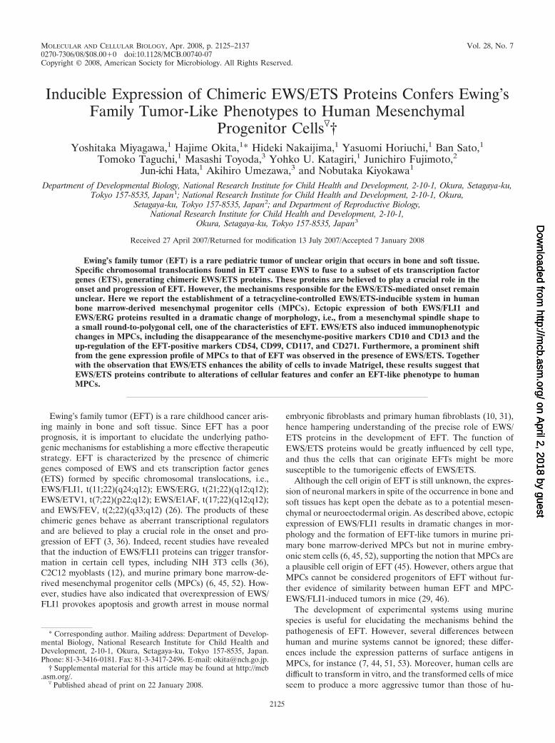
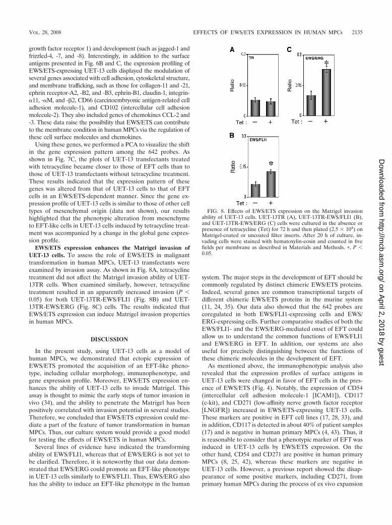
EWS/ETS expression enhances the Matrigel invasion ofUET-13 cells. To assess the role of EWS/ETS in malignanttransformation in human MPCs, UET-13 transfectants wereexamined by invasion assay. As shown in Fig. 8A, tetracyclinetreatment did not affect the Matrigel invasion ability of UET-13TR cells. When examined similarly, however, tetracyclinetreatment resulted in an apparently increased invasion (P 0.05) for both UET-13TR-EWS/FLI1 (Fig. 8B) and UET-13TR-EWS/ERG (Fig. 8C) cells. The results indicated thatEWS/ETS expression can induce Matrigel invasion propertiesin human MPCs.
DISCUSSION
In the present study, using UET-13 cells as a model ofhuman MPCs, we demonstrated that ectopic expression ofEWS/ETS promoted the acquisition of an EFT-like pheno-type, including cellular morphology, immunophenotype, andgene expression profile. Moreover, EWS/ETS expression en-hances the ability of UET-13 cells to invade Matrigel. Thisassay is thought to mimic the early steps of tumor invasion invivo (34), and the ability to penetrate the Matrigel has beenpositively correlated with invasion potential in several studies.Therefore, we concluded that EWS/ETS expression could me-diate a part of the feature of tumor transformation in humanMPCs. Thus, our culture system would provide a good modelfor testing the effects of EWS/ETS in human MPCs.
Several lines of evidence have indicated the transformingability of EWS/FLI1, whereas that of EWS/ERG is not yet tobe clarified. Therefore, it is noteworthy that our data demon-strated that EWS/ERG could promote an EFT-like phenotypein UET-13 cells similarly to EWS/FLI1. Thus, EWS/ERG alsohas the ability to induce an EFT-like phenotype in the human
system. The major steps in the development of EFT should becommonly regulated by distinct chimeric EWS/ETS proteins.Indeed, several genes are common transcriptional targets ofdifferent chimeric EWS/ETS proteins in the murine system(11, 24, 35). Our data also showed that the 642 probes arecoregulated in both EWS/FLI1-expressing cells and EWS/ERG-expressing cells. Further comparative studies of both theEWS/FLI1- and the EWS/ERG-mediated onset of EFT couldallow us to understand the common functions of EWS/FLI1and EWS/ERG in EFT. In addition, our systems are alsouseful for precisely distinguishing between the functions ofthese chimeric molecules in the development of EFT.
As mentioned above, the immunophenotypic analysis alsorevealed that the expression profiles of surface antigens inUET-13 cells were changed in favor of EFT cells in the pres-ence of EWS/ETS (Fig. 4). Notably, the expression of CD54(intercellular cell adhesion molecule-1 [ICAM1]), CD117(c-kit), and CD271 (low-affinity nerve growth factor receptor[LNGFR]) increased in EWS/ETS-expressing UET-13 cells.These markers are positive in EFT cell lines (17, 28, 33), andin addition, CD117 is detected in about 40% of patient samples(17) and is negative in human primary MPCs (4, 43). Thus, itis reasonable to consider that a phenotypic marker of EFT wasinduced in UET-13 cells by EWS/ETS expression. On theother hand, CD54 and CD271 are positive in human primaryMPCs (8, 25, 42), whereas these markers are negative inUET-13 cells. However, a previous report showed the disap-pearance of some positive markers, including CD271, fromprimary human MPCs during the process of ex vivo expansion
FIG. 8. Effects of EWS/ETS expression on the Matrigel invasionability of UET-13 cells. UET-13TR (A), UET-13TR-EWS/FLI1 (B),and UET-13TR-EWS/ERG (C) cells were cultured in the absence orpresence of tetracycline (Tet) for 72 h and then plated (2.5 � 104) onMatrigel-coated or uncoated filter inserts. After 20 h of culture, in-vading cells were stained with hematoxylin-eosin and counted in fivefields per membrane as described in Materials and Methods. �, P 0.05.
VOL. 28, 2008 EFFECTS OF EWS/ETS EXPRESSION IN HUMAN MPCs 2135
on April 2, 2018 by guest
http://mcb.asm
.org/D
ownloaded from
(25), and it has been speculated that the expression of thesemolecules in MPCs is induced in vivo via interaction with thebone marrow microenvironment and that the necessary stimuliare absent from ex vivo culture conditions. Therefore, theimmunophenotype of UET-13 cells rather might be related tothat of ex vivo-expanded primary human MPCs. In addition, itmay be possible that EWS/ETS expression led to the reexpres-sion of these disappeared markers in UET-13 cells without thenecessary stimuli. In this case, the maintenance of CD271expression outside of the bone marrow microenvironmentmight be a characteristic of EFT. Thus, our results proved thatboth EWS/FLI1 and EWS/ERG can be major causes of theexpression of these markers and that human MPCs that pre-cisely recapitulate the expression are strong candidates for thecell origins of EFT cells. The findings also imply that theseantigens are suitable targets for diagnostic tools and new ther-apeutic agents. In fact, imatinib mesylate, which demonstratesanticancer activity against malignant cells expressing BCR-ABL as well as CD117 and platelet-derived growth factor re-ceptor, inhibits proliferation and increases sensitivity to vin-cristine and doxorubicin in EFT cells (17).
Notably, our results also indicate that UET-13 cells, whichhave the MPC phenotype, possess the potential to acquire anEFT-like phenotype upon the expression of EWS/ETS. Unlikewhat is seen for human primary fibroblasts (31), ectopic EWS/ETS expression induces an EFT-like morphological change inhuman MPCs, suggesting that the cell type affects susceptibilityto the events following EWS/ETS expression. In murine MPCs,retrovirally transduced EWS/FLI1 has been reported to inducethe expression of CD99, a most useful marker for EFT, thoughthe results are controversial (6, 45). However, our direct evi-dence obtained with UET-13 cells clearly demonstrated thatCD99 expression is induced by EWS/ETS proteins in humanMPCs. Moreover, we showed that the expression of CD99might correlate with EWS/ETS-mediated morphologicalchange, whereas the functional role of CD99 and the correla-tion between CD99 expression status and EWS/ETS-mediatedmorphological change in the development of EFT remain un-clarified.
Consistent with the morphological and immunophenotypicchanges, the expression pattern of a set of genes in EWS/ETS-expressing UET-13 cells shifted to that in EFT cells (Fig. 7C).Although EWS/ETS expression enhanced the ability ofUET-13 cells to invade Matrigel, it did not promote migratoryability and surface-independent growth, as assessed by migra-tion assay and soft agar colony formation assay (data notshown). We also failed to develop EFT-like tumors by injectingEWS/ETS-inducing UET-13 cells into irradiated nude micetreated with tetracycline (data not shown). These results implythat EWS/ETS expression is not sufficient to induce the fulltransformation in UET-13 cells, and other genetic abnormal-ities not regulated by EWS/ETS could still be required for thefull transformation of human MPCs into EFT cells. An iden-tification of these genes will greatly improve our understandingof the additional genetic lesions that occur after EWS/ETSexpression. The genes expressed in EFT cell lines but not inEWS/ETS-expressing UET-13 cells would be candidates forsuch genes.
In summary, we reported the development of an inducibleEWS/ETS expression system in UET-13 cells as a model for
the development of EFT in MPCs. In our system, the chimericgenes alone are sufficient to confer EFT-like phenotypes, EFT-specific gene expression pattern, and partial but not full fea-tures of malignant transformation. Further analysis using oursystem should elucidate the pathogenic mechanism by whichEFTs develop from MPCs, especially the initiating events me-diated by EWS/ETS expression. Our system should also aid inthe identification of novel targets of the EWS/ETS-mediatedpathway as potential anticancer targets.
ACKNOWLEDGMENTS
This work was supported in part by health and labor sciences re-search grants (the 3rd-Term Comprehensive 10-Year Strategy for Can-cer Control [H19-010], Research on Children and Families [H18-005and H19-003], Research on Human Genome Tailor Made, and Re-search on Publicly Essential Drugs and Medical Devices [H18-005[)and a grant for child health and development from the Ministry ofHealth, Labor and Welfare of Japan, JSPS (Kakenhi 18790263). Thiswork was also supported by a CREST, JST grant from the JapanHealth Sciences Foundation for Research on Publicly Essential Drugsand Medical Devices and the Budget for Nuclear Research of theMinistry of Education, Culture, Sports, Science and Technology, basedon screening and counseling by the Atomic Energy Commission. Y.Miyagawa is an awardee of a research resident fellowship from theFoundation for Promotion of Cancer Research (Japan) for the 3rd-Term Comprehensive 10-Year Strategy for Cancer Control.
We are grateful to T. Motoyama for the NRS-1 cell line. We re-spectfully thank S. Yamauchi for her secretarial work and M. Itagakifor many helpful discussions and support.
REFERENCES
1. Akagi, T. 2004. Oncogenic transformation of human cells: shortcomings ofrodent model systems. Trends Mol. Med. 10:542–548.
2. Ambros, I. M., P. F. Ambros, S. Strehl, H. Kovar, H. Gadner, and M.Salzer-Kuntschik. 1991. MIC2 is a specific marker for Ewing’s sarcoma andperipheral primitive neuroectodermal tumors. Evidence for a common his-togenesis of Ewing’s sarcoma and peripheral primitive neuroectodermaltumors from MIC2 expression and specific chromosome aberration. Cancer67:1886–1893.
3. Arvand, A., and C. T. Denny. 2001. Biology of EWS/ETS fusions in Ewing’sfamily tumors. Oncogene 20:5747–5754.
4. Bertani, N., P. Malatesta, G. Volpi, P. Sonego, and R. Perris. 2005. Neuro-genic potential of human mesenchymal stem cells revisited: analysis by im-munostaining, time-lapse video and microarray. J. Cell Sci. 118:3925–3936.
5. Bloom, E. T. 1972. Further definition by cytotoxicity tests of cell surfaceantigens of human sarcomas in culture. Cancer Res. 32:960–967.
6. Castillero-Trejo, Y., S. Eliazer, L. Xiang, J. A. Richardson, and R. L. Ilaria,Jr. 2005. Expression of the EWS/FLI-1 oncogene in murine primary bone-derived cells results in EWS/FLI-1-dependent, Ewing sarcoma-like tumors.Cancer Res. 65:8698–8705.
7. Colter, D. C., I. Sekiya, and D. J. Prockop. 2001. Identification of a sub-population of rapidly self-renewing and multipotential adult stem cells incolonies of human marrow stromal cells. Proc. Natl. Acad. Sci. USA 98:7841–7845.
8. Conget, P. A., and J. J. Minguell. 1999. Phenotypical and functional prop-erties of human bone marrow mesenchymal progenitor cells. J. Cell. Physiol.181:67–73.
9. Davis, S., and P. S. Meltzer. 2006. Ewing’s sarcoma: general insights from arare model. Cancer Cell 9:331–332.
10. Deneen, B., and C. T. Denny. 2001. Loss of p16 pathways stabilizes EWS/FLI1 expression and complements EWS/FLI1 mediated transformation. On-cogene 20:6731–6741.
11. Deneen, B., S. M. Welford, T. Ho, F. Hernandez, I. Kurland, and C. T.Denny. 2003. PIM3 proto-oncogene kinase is a common transcriptionaltarget of divergent EWS/ETS oncoproteins. Mol. Cell. Biol. 23:3897–3908.
12. Eliazer, S., J. Spencer, D. Ye, E. Olson, and R. L. Ilaria, Jr. 2003. Alterationof mesodermal cell differentiation by EWS/FLI-1, the oncogene implicatedin Ewing’s sarcoma. Mol. Cell. Biol. 23:482–492.
13. Fujii, Y., Y. Nakagawa, T. Hongo, Y. Igarashi, Y. Naito, and M. Maeda. 1989.Cell line of small round cell tumor originating in the chest wall: W-ES. Hum.Cell 2:190–191. (In Japanese.)
14. Fukuma, M., H. Okita, J. Hata, and A. Umezawa. 2003. Upregulation of Id2,an oncogenic helix-loop-helix protein, is mediated by the chimeric EWS/etsprotein in Ewing sarcoma. Oncogene 22:1–9.
15. Gilbert, F., G. Balaban, P. Moorhead, D. Bianchi, and H. Schlesinger. 1982.
2136 MIYAGAWA ET AL. MOL. CELL. BIOL.
on April 2, 2018 by guest
http://mcb.asm
.org/D
ownloaded from
Abnormalities of chromosome 1p in human neuroblastoma tumors and celllines. Cancer Genet. Cytogenet. 7:33–42.
16. Girish, V., and A. Vijayalakshmi. 2004. Affordable image analysis using NIHImage/ImageJ. Indian J. Cancer 41:47.
17. Gonzalez, I., E. J. Andreu, A. Panizo, S. Inoges, A. Fontalba, J. L. Fernan-dez-Luna, M. Gaboli, L. Sierrasesumaga, S. Martin-Algarra, J. Pardo, F.Prosper, and E. de Alava. 2004. Imatinib inhibits proliferation of Ewingtumor cells mediated by the stem cell factor/KIT receptor pathway, andsensitizes cells to vincristine and doxorubicin-induced apoptosis. Clin. Can-cer Res. 10:751–761.
18. Hansen, M. B., S. E. Nielsen, and K. Berg. 1989. Re-examination and furtherdevelopment of a precise and rapid dye method for measuring cell growth/cell kill. J. Immunol. Methods 119:203–210.
19. Hara, S., E. Ishii, S. Tanaka, J. Yokoyama, K. Katsumata, J. Fujimoto, andJ. Hata. 1989. A monoclonal antibody specifically reactive with Ewing’ssarcoma. Br J. Cancer 60:875–879.
20. Hatori, M., H. Doi, M. Watanabe, H. Sasano, M. Hosaka, S. Kotajima, F.Urano, J. Hata, and S. Kokubun. 2006. Establishment and characterizationof a clonal human extraskeletal Ewing’s sarcoma cell line, EES1. Tohoku J.Exp. Med. 210:221–230.
21. Homma, C., Y. Kaneko, K. Sekine, S. Hara, J. Hata, and M. Sakurai. 1989.Establishment and characterization of a small round cell sarcoma cell line,SCCH-196, with t(11;22)(q24;q12). Jpn. J. Cancer Res. 80:861–865.
22. Hubert, R. S., I. Vivanco, E. Chen, S. Rastegar, K. Leong, S. C. Mitchell, R.Madraswala, Y. Zhou, J. Kuo, A. B. Raitano, A. Jakobovits, D. C. Saffran,and D. E. Afar. 1999. STEAP: a prostate-specific cell-surface antigen highlyexpressed in human prostate tumors. Proc. Natl. Acad. Sci. USA 96:14523–14528.
23. Hu-Lieskovan, S., J. Zhang, L. Wu, H. Shimada, D. E. Schofield, and T. J.Triche. 2005. EWS-FLI1 fusion protein up-regulates critical genes in neuralcrest development and is responsible for the observed phenotype of Ewing’sfamily of tumors. Cancer Res. 65:4633–4644.
24. Im, Y. H., H. T. Kim, C. Lee, D. Poulin, S. Welford, P. H. Sorensen, C. T.Denny, and S. J. Kim. 2000. EWS-FLI1, EWS-ERG, and EWS-ETV1 on-coproteins of Ewing tumor family all suppress transcription of transforminggrowth factor beta type II receptor gene. Cancer Res. 60:1536–1540.
25. Jones, E. A., S. E. Kinsey, A. English, R. A. Jones, L. Straszynski, D. M.Meredith, A. F. Markham, A. Jack, P. Emery, and D. McGonagle. 2002.Isolation and characterization of bone marrow multipotential mesenchymalprogenitor cells. Arthritis Rheum. 46:3349–3360.
26. Khoury, J. D. 2005. Ewing sarcoma family of tumors. Adv. Anat. Pathol.12:212–220.
27. Kiyokawa, N., Y. Kokai, K. Ishimoto, H. Fujita, J. Fujimoto, and J. I. Hata.1990. Characterization of the common acute lymphoblastic leukaemia anti-gen (CD10) as an activation molecule on mature human B cells. Clin. Exp.Immunol. 79:322–327.
28. Konemann, S., T. Bolling, A. Schuck, J. Malath, A. Kolkmeyer, K. Horn, D.Riesenbeck, S. Hesselmann, R. Diallo, J. Vormoor, and N. A. Willich. 2003.Effect of radiation on Ewing tumour subpopulations characterized on asingle-cell level: intracellular cytokine, immunophenotypic, DNA and apop-totic profile. Int. J. Radiat. Biol. 79:181–192.
29. Kovar, H., and A. Bernard. 2006. CD99-positive “Ewing’s sarcoma” frommouse bone marrow-derived mesenchymal progenitor cells? Cancer Res.66:9786.
30. Kovar, H., M. Dworzak, S. Strehl, E. Schnell, I. M. Ambros, P. F. Ambros,and H. Gadner. 1990. Overexpression of the pseudoautosomal gene MIC2 inEwing’s sarcoma and peripheral primitive neuroectodermal tumor. Onco-gene 5:1067–1070.
31. Lessnick, S. L., C. S. Dacwag, and T. R. Golub. 2002. The Ewing’s sarcomaoncoprotein EWS/FLI induces a p53-dependent growth arrest in primaryhuman fibroblasts. Cancer Cell 1:393–401.
32. Lin, P. P., R. I. Brody, A. C. Hamelin, J. E. Bradner, J. H. Healey, and M.Ladanyi. 1999. Differential transactivation by alternative EWS-FLI1 fusionproteins correlates with clinical heterogeneity in Ewing’s sarcoma. CancerRes. 59:1428–1432.
33. Lipinski, M., K. Braham, I. Philip, J. Wiels, T. Philip, C. Goridis, G. M.Lenoir, and T. Tursz. 1987. Neuroectoderm-associated antigens on Ewing’ssarcoma cell lines. Cancer Res. 47:183–187.
34. Lochter, A., A. Srebrow, C. J. Sympson, N. Terracio, Z. Werb, and M. J.Bissell. 1997. Misregulation of stromelysin-1 expression in mouse mammarytumor cells accompanies acquisition of stromelysin-1-dependent invasiveproperties. J. Biol. Chem. 272:5007–5015.
35. May, W. A., A. Arvand, A. D. Thompson, B. S. Braun, M. Wright, and C. T.Denny. 1997. EWS/FLI1-induced manic fringe renders NIH 3T3 cells tumor-igenic. Nat. Genet. 17:495–497.
36. May, W. A., S. L. Lessnick, B. S. Braun, M. Klemsz, B. C. Lewis, L. B.Lunsford, R. Hromas, and C. T. Denny. 1993. The Ewing’s sarcoma EWS/FLI-1 fusion gene encodes a more potent transcriptional activator and is amore powerful transforming gene than FLI-1. Mol. Cell. Biol. 13:7393–7398.
37. Miyagawa, Y., J. M. Lee, T. Maeda, K. Koga, Y. Kawaguchi, and T. Kusak-abe. 2005. Differential expression of a Bombyx mori AHA1 homologueduring spermatogenesis. Insect Mol. Biol. 14:245–253.
38. Mori, T., T. Kiyono, H. Imabayashi, Y. Takeda, K. Tsuchiya, S. Miyoshi, H.Makino, K. Matsumoto, H. Saito, S. Ogawa, M. Sakamoto, J. Hata, and A.Umezawa. 2005. Combination of hTERT and bmi-1, E6, or E7 inducesprolongation of the life span of bone marrow stromal cells from an elderlydonor without affecting their neurogenic potential. Mol. Cell. Biol. 25:5183–5195.
39. Nishimori, H., Y. Sasaki, K. Yoshida, H. Irifune, H. Zembutsu, T. Tanaka,T. Aoyama, T. Hosaka, S. Kawaguchi, T. Wada, J. Hata, J. Toguchida, Y.Nakamura, and T. Tokino. 2002. The Id2 gene is a novel target of transcrip-tional activation by EWS-ETS fusion proteins in Ewing family tumors. On-cogene 21:8302–8309.
40. Ogose, A., T. Motoyama, T. Hotta, and H. Watanabe. 1995. In vitro differ-entiation and proliferation in a newly established human rhabdomyosarcomacell line. Virchows Arch. 426:385–391.
41. Prieur, A., F. Tirode, P. Cohen, and O. Delattre. 2004. EWS/FLI-1 silencingand gene profiling of Ewing cells reveal downstream oncogenic pathways anda crucial role for repression of insulin-like growth factor binding protein 3.Mol. Cell. Biol. 24:7275–7283.
42. Quirici, N., D. Soligo, P. Bossolasco, F. Servida, C. Lumini, and G. L.Deliliers. 2002. Isolation of bone marrow mesenchymal stem cells by anti-nerve growth factor receptor antibodies. Exp. Hematol. 30:783–791.
43. Reyes, M., T. Lund, T. Lenvik, D. Aguiar, L. Koodie, and C. M. Verfaillie.2001. Purification and ex vivo expansion of postnatal human marrow meso-dermal progenitor cells. Blood 98:2615–2625.
44. Reyes, M., and C. M. Verfaillie. 2001. Characterization of multipotent adultprogenitor cells, a subpopulation of mesenchymal stem cells. Ann. N. Y.Acad. Sci. 938:231–235.
45. Riggi, N., L. Cironi, P. Provero, M. L. Suva, K. Kaloulis, C. Garcia-Echev-erria, F. Hoffmann, A. Trumpp, and I. Stamenkovic. 2005. Development ofEwing’s sarcoma from primary bone marrow-derived mesenchymal progen-itor cells. Cancer Res. 65:11459–11468.
46. Riggi, N., M. L. Suva, and I. Stamenkovic. 2006. Ewing’s sarcoma-like tu-mors originate from EWS-FLI-1-expressing mesenchymal progenitor cells.Cancer Res. 66:9786.
47. Sekiguchi, M., T. Oota, K. Sakakibara, N. Inui, and G. Fujii. 1979. Estab-lishment and characterization of a human neuroblastoma cell line in tissueculture. Jpn. J. Exp. Med. 49:67–83.
48. Smith, R., L. A. Owen, D. J. Trem, J. S. Wong, J. S. Whangbo, T. R. Golub,and S. L. Lessnick. 2006. Expression profiling of EWS/FLI identifiesNKX2.2 as a critical target gene in Ewing’s sarcoma. Cancer Cell 9:405–416.
49. Staege, M. S., C. Hutter, I. Neumann, S. Foja, U. E. Hattenhorst, G. Hansen,D. Afar, and S. E. Burdach. 2004. DNA microarrays reveal relationship ofEwing family tumors to both endothelial and fetal neural crest-derived cellsand define novel targets. Cancer Res. 64:8213–8221.
50. Takeda, Y., T. Mori, H. Imabayashi, T. Kiyono, S. Gojo, S. Miyoshi, N. Hida,M. Ita, K. Segawa, S. Ogawa, M. Sakamoto, S. Nakamura, and A. Umezawa.2004. Can the life span of human marrow stromal cells be prolonged bybmi-1, E6, E7, and/or telomerase without affecting cardiomyogenic differ-entiation? J. Gene Med. 6:833–845.
51. Tondreau, T., N. Meuleman, A. Delforge, M. Dejeneffe, R. Leroy, M. Massy,C. Mortier, D. Bron, and L. Lagneaux. 2005. Mesenchymal stem cells de-rived from CD133-positive cells in mobilized peripheral blood and cordblood: proliferation, Oct4 expression, and plasticity. Stem Cells 23:1105–1112.
52. Torchia, E. C., S. Jaishankar, and S. J. Baker. 2003. Ewing tumor fusionproteins block the differentiation of pluripotent marrow stromal cells. Can-cer Res. 63:3464–3468.
53. Woodbury, D., E. J. Schwarz, D. J. Prockop, and I. B. Black. 2000. Adult ratand human bone marrow stromal cells differentiate into neurons. J. Neuro-sci. Res. 61:364–370.
VOL. 28, 2008 EFFECTS OF EWS/ETS EXPRESSION IN HUMAN MPCs 2137
on April 2, 2018 by guest
http://mcb.asm
.org/D
ownloaded from
MOLECULAR AND CELLULAR BIOLOGY, June 2008, p. 3882 Vol. 28, No. 110270-7306/08/$08.00�0 doi:10.1128/MCB.00557-08
ERRATUM
Inducible Expression of Chimeric EWS/ETS Proteins Confers Ewing’s FamilyTumor-Like Phenotypes to Human Mesenchymal Progenitor Cells
Yoshitaka Miyagawa, Hajime Okita, Hideki Nakaijima, Yasuomi Horiuchi, Ban Sato, Tomoko Taguchi,Masashi Toyoda, Yohko U. Katagiri, Junichiro Fujimoto, Jun-ichi Hata,
Akihiro Umezawa, and Nobutaka KiyokawaDepartment of Developmental Biology, National Research Institute for Child Health and Development, 2-10-1, Okura, Setagaya-ku,
Tokyo 157-8535, Japan; National Research Institute for Child Health and Development, 2-10-1, Okura, Setagaya-ku,Tokyo 157-8535, Japan; and Department of Reproductive Biology, National Research Institute for
Child Health and Development, 2-10-1, Okura, Setagaya-ku, Tokyo 157-8535, Japan
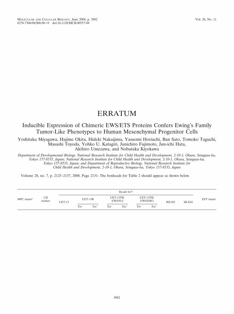
Volume 28, no. 7, p. 2125–2137, 2008. Page 2131: The boxheads for Table 2 should appear as shown below.
MPC statusa CDmarker
Result forb:
EFT statusc
UET-13UET-13R UET-13TR-
EWS/FLIUET-13TR-EWS/ERG RD-ES SK-ES1
Tet� Tet� Tet� Tet� Tet� Tet�
3882