Embed Size (px)
Citation preview

Article
Chimeric Vaccines Designed byImmunoinformatics-Activated Polyfunctional andMemory T cells that Trigger Protection againstExperimental Visceral Leishmaniasis
Rory Cristiane Fortes De Brito 1,† , Jeronimo Conceição Ruiz 2,3,† ,Jamille Mirelle de Oliveira Cardoso 1, Thais Lopes Valentim Di Paschoale Ostolin 1 ,Levi Eduardo Soares Reis 1, Fernando Augusto Siqueira Mathias 1,Rodrigo Dian de Oliveira Aguiar-Soares 1, Bruno Mendes Roatt 1,4 , Rodrigo Corrêa-Oliveira 5,Daniela de Melo Resende 2,3 and Alexandre Barbosa Reis 1,4,*
1 Laboratório de Imunopatologia, Núcleo de Pesquisas em Ciências Biológicas/NUPEB, Universidade Federalde Ouro Preto, Ouro Preto 35400-000, Brazil; [email protected] (R.C.F.D.B.);[email protected] (J.M.d.O.C.); [email protected] (T.L.V.D.P.O.);[email protected] (L.E.S.R.); [email protected] (F.A.S.M.);[email protected] (R.D.d.O.A.-S.); [email protected] (B.M.R.)
2 Grupo Informática de Biossistemas e Genômica, Instituto René Rachou, Fiocruz Minas,Belo Horizonte 30190-002, Brazil; [email protected] (J.C.R.);[email protected] (D.d.M.R.)
3 Programa de Pós-graduação em Biologia Computacional e Sistemas, Instituto Oswaldo Cruz, Fiocruz,Rio de Janeiro 21040-360, Brazil
4 Instituto Nacional de Ciência e Tecnologia em Doenças Tropicais (INCT-DT), Salvador 40110-160, Brazil5 Laboratório de Imunologia Celular e Molecular, Instituto René Rachou, Fiocruz Minas,
Belo Horizonte 30190-002, Brazil; [email protected]* Correspondence: [email protected]; Tel.: +55-31-3559-1694† These authors contributed equally to this study.
Received: 7 March 2020; Accepted: 27 March 2020; Published: 27 May 2020�����������������
Abstract: Many vaccine candidates against visceral leishmaniasis (VL) have been proposed; however,to date, none of them have been efficacious for the human or canine disease. On this basis, the designof leishmaniasis vaccines has been constantly changing, and the use of approaches to select specificepitopes seems to be crucial in this scenario. The ability to predict T cell-specific epitopes makesimmunoinformatics an even more necessary approach, as in VL an efficient immune response againstthe parasite is triggered by T lymphocytes in response to Leishmania spp. immunogenic antigens.Moreover, the success of vaccines depends on the capacity to generate long-lasting memory andpolyfunctional cells that are able to eliminate the parasite. In this sense, our study used a combinationof different approaches to develop potential chimera candidate vaccines against VL. The first pointwas to identify the most immunogenic epitopes of Leishmania infantum proteins and construct chimerascomposed of Major histocompatibility complex (MHC) class I and II epitopes. For this, we usedimmunoinformatics features. Following this, we validated these chimeras in a murine model in athorough memory study and multifunctionality of T cells that contribute to a better elucidation of theimmunological protective mechanisms of polyepitope vaccines (chimera A and B) using multicolorflow cytometry. Our results showed that in silico-designed chimeras can elicit polyfunctional T cellsproducing T helper (Th)1 cytokines, a strong immune response against Leishmania antigen, and thegeneration of central and effector memory T cells in the spleen cells of vaccinated animals that wasable to reduce the parasite burden in this organ. These findings contribute two potential candidatevaccines against VL that can be used in further studies, and help in this complex field of vaccinedevelopment against this challenging parasite.
Vaccines 2020, 8, 252; doi:10.3390/vaccines8020252 www.mdpi.com/journal/vaccines

Vaccines 2020, 8, 252 2 of 20
Keywords: reverse vaccinology; immunoinformatics; chimera vaccine; Leishmania infantum;polyfunctional T cells; memory T cells; rational design of vaccines
1. Introduction
During the last decades, the way in which the immune system works and the protectivemechanisms related to vaccination for complex parasites has been extensively interpreted, and thecentral dogma of vaccination has been mounted [1]. Some studies support the idea that an antigen iscapable of inducing long-lasting immunological memory and protective immunity against re-challengewith the same pathogen. Although this concept has been successful in generating potent vaccinesfor many diseases, leishmaniasis caused by the protozoan parasite Leishmania remains a threateningexception. Thus, the design of leishmaniasis vaccines has been constantly changing, and the useof polyepitope vaccines seems to gain prominent space in this scenario. In one of the pioneeringstudies of visceral leishmaniasis (VL) polyepitope vaccines, a DNA vaccine containing Leishmaniadonovani GP63 protein T cell epitopes was proposed. The authors evaluated the immunogenicity ofthe vaccine in immunized and challenged BALB/c mice showing increased production of Interferongama (IFN-γ) and Interleukin (IL)-2 in splenocytes of vaccinated animals. In addition, this vaccinereduced parasite load in the spleen and liver of challenged mice [2]. In light of this, seeking to expandthe antigenic repertoire of vaccines, the authors of [3] constructed a multiepitope DNA vaccine thatencoded four protein-fused peptides, lipophosphoglycan (LPG)-3, Leishmania major stress inducibleprotein (LmSTI)-1, cysteine peptidase B (CPB), and cysteine peptidase C (CPC). They evaluatedthe cytotoxic activity of lymphocytes and IFN-γ production in transgenic mouse (holding humanMHC alleles, Human leukocyte antigen (HLA)-DRB1 * 0101/HLA-A * 0201), and the results revealedincreased cytotoxic pleaseactivity and IFN-γ production after immunization [3]. Along the same lineof studies, the authors of [4] constructed DNA vaccines based on peptides selected from L. donovaniantigens (CPA, CPB, Kinetoplastid membrane protein (KMP)11, Thiol specific-antioxidant protein(TSA), and Elongation factor 1 (P74) that showed a significant reduction of parasite burden in thespleen after immunization using a unique preparation of these antigens as DNA vaccine when themice were challenge with L. donovani promastigotes [4].
The basic assumption that introduction of an antigen into a host will generate protective immunityagainst the pathogen appears to be invalid. Thus, there are possible reasons for the failures andthe possible approaches that may bring success to generation of Leishmania vaccine. First, the trafficof T cells between lymph nodes and the microenvironment on the site of infection is essential foractivation and maturation of the right cells [1,5]. Besides this, the major challenge faced by theimmunologists is how to identify antigens capable of generating long-lasting immunological memory.Therefore, several approaches to the evaluation of immunological memory have been developed usingmulticolor flow cytometry, which aims to identify and evaluate effector and memory T lymphocytesubpopulations to validate different vaccine candidates [6]. The ability to predict T cell-specific epitopesmakes immunoinformatics an even more necessary approach, as in VL an efficient immune responseagainst specific epitopes of the parasite is triggered by T lymphocytes in response to some Leishmaniaspp. [7–9]. Thus, some research groups have been proposing vaccine candidates on the basis of specificclass I and II MHC-binding epitopes mapped to known proteins [3,4,10].
Therefore, the development of polyepitope vaccines is a promising field that has been studiedin recent years. In this sense, our study used a combination of different approaches to developcandidate vaccines against VL. The first point was to identify the best tools to map immunogenicepitopes and construct chimeras composed of MHC class I and II epitopes. For this, we usedimmunoinformatics features described by [9]. Afterwards, we validated these chimeras in BALB/c micein a thorough memory study and multifunctionality of T cells that contributes to a better elucidation ofthe immunological protective mechanisms of chimeras (A and B) constituted of polyepitope vaccines.

Vaccines 2020, 8, 252 3 of 20
2. Materials and Methods
2.1. Ethical Statement
The study was performed according to the recommendation of the National Institute of Health,USA. The protocol number 2015/03 was approved by the Ethical Committee for the Use of ExperimentalAnimals (CEUA) of the Universidade Federal de Ouro Preto, Ouro Preto, MG, Brazil. All theexperiments were performed to minimize animal suffering.
2.2. Epitope Mapping and Chimeric Proteins Design
The chimeric proteins used in this study were designed on the basis of epitopes mapped alongLeishmania infantum proteins already described in the literature as vaccine candidates (Table 1).For epitope mapping, an integrative immunoinformatics approach described by [9,11] was used.The epitopes for T cells were selected on the basis of the highest scores provided by the predictivealgorithms. We used a combination of NetCTL and NetMHC to predict MHC class I epitopesand NetMHCII for MHC class II epitopes, thus promiscuous epitopes were selected on the basisof their affinity towards human and mouse MHC alleles that are available for those algorithms.The algorithm NetCTL version 1.2 makes predictions of peptide–MHC class I binding and proteasomalC terminal cleavage, both using artificial neural networks, and transporter associated with antigenprocessing (TAP) transport efficiency using weight matrix. The tree predictions are then integrated [12].The thresholds used were 0.15 for the weight on C terminal cleavage, 0.05 for weight on TAP transportefficiency, and 1.0 for epitope identification. NetMHC version 3.0. predicts binding of peptides todifferent HLA alleles using artificial neural networks and weight matrices, and the prediction is basedon IC50 values where strong binder epitopes have an IC50 below 50 nM, and weak binder epitopes havean IC50 value below 500 nM [13]. NetMHCII version 1.0 was used, which predicts binding of peptidesto 14 different HLA-DR alleles using position specific weight matrices (PSSM) [14]. The thresholds arebased on IC50—50 nM for strong binder epitopes, and 500 nM for weak binder threshold score.
To compose the chimeric vaccines, these epitopes were repeated in tandem to enhance theimmune response after immunization. Chimeric protein A has epitopes from histone protein (H2A),acid ribosomal protein P2 (LiP2a), acid ribosomal protein P0 (LiP0), Leishmania homologue of activatedC kinase (LACK), and cysteine peptidase C (CPC). Chimeric protein B is composed of epitopes ofcysteine peptidase proteins A and B (CPA and CPB), surface antigenic protein (PSA-50S), and specificamastigote protein A2 (A2). The linker GPGPG amino acid sequence was used to link the epitopes andincrease protein stability and processing [15] (Figure S1).
2.3. Prediction of Epitope Conservancy among Leishmania Species
The proteome of L. donovani (BPK282A1 strain), Leishmania major (MHOM/IL/81/Friedlin strain),and Leishmania braziliensis (MHOM/BR/1966/M2903 strain) were retrieved from The KinetoplastidGenomics Resource (TritrypDB). The proteome of Leishmania amazonensis (MHOM/BR/71973/M2269strain) was retrieved from [16]. To evaluate the conservancy of the selected epitopes among theseLeishmania species, the Immune epitope database (IEDB) epitope conservancy tool (http://tools.immuneepitope.org/tools/conservancy/) was used.
2.4. Prediction of Antigenicity, Allergenicity, and Physicochemical Properties of the Two Chimeras
To predict the antigenicity of the two constructs (chimeras A and B), ANTIGENpro was used.This algorithm is based on sequences, pathogen-independent prediction method, and an alignment-freeprediction method. For the allergenicity prediction, we used AlgPred, which uses a combinationof predictive algorithms as described by [17]. The server uses several algorithms (SVMc + MAST +
IgEepitope + ARPs BLAST) to check allergenicity of a query sequence, the prediction of allergens isbased on similarity of known epitope with any region of a given protein. The server results are reliableand have 85% precision at −0.4 threshold.

Vaccines 2020, 8, 252 4 of 20
To define the physical and chemical parameters associated with the constructs, ProtParam webserver (from EXPASY) was used to define various physicochemical properties. Parameters as molecularweight (kDa), theoretical isoelectric point (pI), estimated half-life, and grand average of hydropathywere assessed.
2.5. Tertiary Structure Prediction and Refinement of the Models
The 3D structures of predicted chimera constructs were assessed by utilizing RaptorX [18], a webserver for structure prediction. To improve the quality of the templates, a refinement of the tertiarystructures was performed using GalaxyRefine web server [19]. The validation of refined models wascarried out using ProSA [20], a web server. This server can predict the quality of the model which isindicated in the form of z-score. This score must be in the range of the characteristic for native proteins,otherwise it can indicate an erroneous structure. After that, Ramachandran plots were generated usingRAMPAGE server [21] to determine the overall quality of predicted and refined models.
2.6. Molecular Docking to Highlight the Binding Affinity between the Chimeras and Mouse and HumanTLR Receptors
For molecular docking, we used ClusPro2.0, which utilizes the Fourier correlation algorithm,filtering out the models with the amalgamation of desolvation and electrostatic energies. On the basisof the lowest binding energy, docked complexes were selected for further analysis [22]. The evaluationof the binding affinity between the refined constructs (chimeras A and B) and mouse and humantoll-like receptors (TLRs) 3 and 4 was performed. The tertiary structures of TLR3 and TLR4 wereretrieved from the PDB database [23,24]—for TLR3 we used id:3CIG (mouse) and id:2A0Z (human).Regarding TLR4, id:5IJB and id:4G8A were used, corresponding to mouse and human, respectively.
2.7. Chimera Proteins Synthesis
The proteins (chimeras A and B) were synthetized by GenScript company; the genes wereconstructed, cloned, and expressed in prokaryote system (Escherichia coli) according to the manufacturer.The purification was performed by nickel column followed by characterization using SDS-PAGEand Western blot (Figure S2). The purified proteins were endotoxin free (<1 EU/µg) and with purityhigher than 90% by SDS-PAGE. The endotoxins were measured according to the Food and DrugAdministration FDA-approved techniques for endotoxin detection, therefore Genscript used Limulusamoebocyte lysate (LAL) assay. The specific LAL kinetic turbidimetric form was performed, which candetect down to 0.01 EU/mL.
2.8. Parasites
Promastigotes of L. infantum, strain OP46 (MCAN/BR/2008/OP46), maintained by passage inSyrian golden hamsters were cultured at 22–24 ◦C in medium LIT (liver infusion tryptose) with100 U of penicillin G sodium and 100 µg of streptomycin sulfate per milliliter, and were sub-culturedin the same medium at an average density of 1 × 10⁸ cells/ml as described by [25]. The parasiteswere used for soluble Leishmania antigen (SLA) preparation, as described by [26], as well as for miceexperimental challenge.
2.9. Immunization Regimens and Challenge of the Mice
For vaccine efficacy studies, eight 6- to 8-week-old female BALB/c mice per group (Centro deCiência Animal (CCA) facility) were randomized by corporal weight and immunized three timesbiweekly subcutaneously in the back with 100 µl of vaccine formulations per mouse. The experimentalgroups were divided in SAL (animals that received sterile saline, 0.9% NaCl, pH 7.2–7.4), SAP (animalsinoculated with 60 µg of saponin), ChiA+SAP (animals that received 10 µg of the chimeric protein Aassociated with 60 µg of saponin), and ChiB+SAP (animals inoculated with 10 µg of chimeric proteinB associated with 60 µg of saponin). The doses were stablished on the basis of certain studies using

Vaccines 2020, 8, 252 5 of 20
chimeric proteins where 10 µg of the chimeric proteins per dose was in the average [27,28]. Mice werechallenged by injecting 1 × 10⁷ of stationary L. infantum strain OP46 promastigotes intravenously,15 days after the last immunization.
2.10. Analyses of Polyfunctional T cell Phenotypes
Fourteen days after the last immunization, polyfunctional T cell phenotypes were assessedin splenocytes of mice. The evaluation was performed before infection because we wanted toanalyze the vaccine immune responses triggered in BALB/c mice in more detail, and because theidentification of these poly-functional T cells is more evident after the last immunization but before theinfection [6,29]. Cell suspensions were incubated in RPMI supplemented with 1% L-glutamine and 10%fetal bovine serum and plated in 96-well round-bottom (Costar, USA) culture plates at a concentrationof 5 × 10⁵ cells per well. Cells were cultured for 24 h at 37 ◦C with 5% CO2 in the presence ofSLA (50 µg/ml). Brefeldin A (SIGMA) was added (10 µg/ml) at 20 h of incubation. Afterwards,cells were blocked with anti-mouse CD16/CD32 (Mouse BD FC block, 0,5 µg/well) harvested, washed,treated with Phosphate buffer saline (PBS) plus an inert protein (serum albumin 5%), and stained withanti-mouse CD3 FITC (clone 145.2C11), anti-mouse CD4 BV605 (clone RM4-5), anti-mouse CD8α BV786(clone 53-6.7), anti-mouse CD62L BV510 (clone MEL-14), and anti-mouse CD44 APC (clone IM7) (BDBiosciences Bioscience, USA) at room temperature for 30 minutes. Cells were fixed with FACS fixingsolution (10 g/L paraformaldehyde, 10.2 g/L sodium cacodylate, and 6.63 g/L sodium chloride, pH 7.2),washed, and treated with PBS buffer containing 0.5% saponin for permeabilization. Cells were stainedwith anti-mouse IFN-γ AF700 (clone XMG1.2), anti-mouse tumor necrosis factor (TNF-α )PE-Cy7(clone LG.3A10), and anti-mouse IL-2 PE (clone JES6-5H4) (BD Biosciences Bioscience, San Jose, CA,USA). Cells were acquired (300,000 events) on LSR Fortessa cytometer (BD Biosciences, USA) usingFACSDiva software. For analysis in FlowJo software, dead cells were excluded after FVS450 stain andalive cells were gated for CD4+ and CD8+ T cells and intracellular cytokine production. Polyfunctionalcells were analyzed through the Boolean gate strategy.
2.11. Proliferation Assay and Intracellular Cytokine Stain
The proliferation of antigen experienced T cells was assessed by Carboxyfluorescein diacetatesuccinimidyl ester (CFSE) assay, 30 days after mice challenge, as described by [11]. Briefly, spleen’s cellsuspensions were incubated in 5 µM CFDA-SE and the labeled cells were washed thoroughly beforeplating in 96-well round-bottom culture plates at a concentration of 5 × 10⁵ cells per well. Cells werecultured for 5 days at 37 ◦C with 5% CO2 in the presence of SLA (50 µg/ml). After incubation, cells weretreated with 10 µg/ml of brefeldin A (Sigma) for 4 h. Afterwards, cells were blocked with anti-mouseCD16/CD32 (0.5 µg/well) and harvested, washed, treated with PBS and an inert protein (serum albumin5%), and stained with anti-mouse CD3 BV650 (clone 145.2C11), anti-mouse CD4 BV605 (clone RM4-5),and anti-mouse CD8α BV786 (clone 53-6.7) (BD Biosciences Bioscience, USA) at room temperature for30 minutes. Cells were fixed with FACS fixing solution (10 g/L paraformaldehyde, 10.2 g/L sodiumcacodylate, and 6.63 g/L sodium chloride, pH 7.2), washed, and permeabilized. Cells were stained withanti-mouse IFN-γ AF700 (clone XMG1.2), anti-mouse TNF-α PE-Cy7 (clone LG.3A10), and anti-mouseIL-2 PE (clone JES6-5H4) (BD Biosciences Bioscience, USA). Cells were acquired (300,000 events) onLSR Fortessa cytometer (BD Biosciences, USA) using FACSDiva software. For analysis in FlowJosoftware, dead cells were excluded after FVS780 stain and alive cells were gated for CFSE-stainedCD4+ and CD8+ T cells and intracellular cytokine production.
2.12. Analyses of Memory T cell Phenotypes
Central and effector memory T cells were analyzed 30 days post-challenge, as described by [11].Splenocytes from animals were plated at 5× 10⁵ cells per well in duplicate in 96-well round-bottom plates.Cells were treated with the same conditions described above. After 5 days of culture, cells were thenprepared for flow cytometry analysis. Samples were blocked with anti-mouse CD16/CD32 (0.5 µg/well)

Vaccines 2020, 8, 252 6 of 20
and stained with surface markers at room temperature using the following antibodies: anti-mouseCD3 FITC (clone 17A2), anti-mouse CD4 BV605 (clone RM4-5), anti-mouse CD8α PerCP-Cy5.5 (clone53-6.7), anti-mouse CD44 APC (clone IM7), anti-mouse CD45RA BV711 (clone 14.8), anti-mouse CD62LAF700 (clone MEL-14), anti-mouse CD127 BV510 (clone SB/199), and anti-mouse CD197 BV421 (clone4B12). The events were acquired (300,000 cells) on an LSR Fortessa cytometer (BD Biosciences) usingFACSDiva software. For analysis, dead cells were excluded after FVS780 stain and live cells were usedfor further analyses.
2.13. Evaluation of the Parasite Burden in the Spleen by Quantitative PCR
Parasite burden in the spleen was analyzed 30 days after infection by real-time PCR (qPCR)quantification, as described by [30]. Briefly, at the end of the study, the mice were euthanized,and the spleens were extracted and used for genomic DNA isolation with the CTAB reagent.The L. infantum spleen burden was assessed by PCR quantification of DNA polymerase (forwardprimer: 5′-TGTCGCTTGCAGACCAGATG-3′; reverse primer: 5′-GCATCGCAGGTGTGAGCAC-3′;Taqman probe: VIC 5′-AGGAAACCTGTGGAGCC-3′ MGB NFQ). The amounts of mousechromosomal DNA in extracted spleen were analyzed using murine TNF-α primers (5-TCCCTCTCATCAGTTCTATGGCCCA-3; 5-CAGCAAGCATCTATGCACTTAGACCCC-3) that amplifya 170 bp product. The PCR reactions were conducted as follows: one incubation step at 50 ◦C for2 min and an initial denaturation step at 95 ◦C for 10 min, followed by 40 cycles of denaturation at95 ◦C for 15 s and annealing-extension at 60 ◦C for 1 min. All samples were run on MicroAmp optical96-well reaction plates (Applied Biosystems) sealed with MicroAmp optical adhesive film (AppliedBiosystems). Each 96-well reaction plate contained a standard curve in triplicate (efficiency, 96.0%;r2 = 0.99) in duplicate samples. The result was expressed as number of amastigotes DNA copies perspleen of mice.
2.14. Statistical Analysis
The data were analyzed through Graph Pad Prism 8.0 software and expressed as mean plus/minusstandard deviation (SD). First, the normality of the data was assessed using the Shapiro–Wilk testand statistical differences were analyzed by one-way ANOVA and Kruskal–Wallis test followed byDunnett’s and Dunn’s multiple comparison tests, respectively. Correlation analyses were performedby Pearson’s r test. Differences were considered significant when p-value <0.05.
3. Results
3.1. In Silico Construction of the Chimeras A and B
Herein, a total of 12 epitopes (ligands for mouse and human MHC class I and II alleles) werepredicted to construct chimera A and chimera B. These epitopes were mapped in the sequence ofnine proteins described in the literature as potential vaccine candidates for VL. The peptides arerepresented in Table 1, where it can be seen that most of them were highly conserved across L. donovaniand L. major species. To construct the chimeras, the epitopes were repeated in tandem to enhance theimmunogenicity, and GPGPG sequence was used as a linker. The final constructions of the chimeraswere predicted to be highly antigenic (predicted probability of antigenicity chimera A = 0.896793 andchimera B = 0.790044), and they were also revealed to be nonallergic in nature. Prediction by SVMmethod based on amino acid composition (chimera A score = −0.96747231 and threshold = −0.4;chimera B score = −0.55381796, threshold = −0.4). Prediction was based on the SVM method whichwas based on dipeptide composition (chimera A score = 0.27467095, threshold = −0.2; chimera Bscore = 0.14230073 and threshold = −0.2). For predictions based on BLAST analyses, no hits werefound for both chimera sequences and they did not contain experimentally proven Immunoglobulin E(IgE) epitope.

Vaccines 2020, 8, 252 7 of 20
Table 1. MHC class I and II epitopes of known Leishmania infantum proteins that have been shown as candidate vaccines for leishmaniasis. The amino acid sequence,alleles, and conservancy of these promiscuous epitopes are represented in the table. The epitopes were selected through an immunoinformatics pipeline describe by[9,11] and they showed a high score for the algorithms NetMHC, NetCTL, and NetMHCII.
Chimera Protein Epitope Sequence MHC Class I and II Alleles
Conservancy of the Selected Epitope across LeishmaniaSpecies (%)
L. donovani L. major L. amazonensis L. braziliensis
Chimera A
Histone protein (H2A) KKRCRLNPR H2-Dk 100 89 100 66
DDISSLLKNVTLSHS HLA-DRB1*1302 HLA-DRB1*0101 HLA-DRB1*0401 HLA-DRB1*0404 HLA-DRB1*1501 100 87 100 66
Acid ribosomal protein P2 (LiP2a) AAKMSAMPAASSGAA HLA-DRB1*0901 HLA-DRB1*0101 100 100 100 93
MSTKYLAAY HLA-A*01 HLA-A*26 HLA-B*62 HLA-A*03 HLA-B*58 100 100 100 100
Acid ribosomal protein P0 (LiP0) VDAFKNLLAVSVATSYEF HLA-DRB1*0101 HLA-DRB1*0701 HLA-DRB1*1501 HLA-DRB1*1302 100 100 100 94
AHRVKAPAR H2-Dk 100 100 100 89
Leishmania homologue ofactivated C kinase (LACK)
WSADGNTLY HLA-A*01 HLA-A*26 HLA-B*62 HLA-B*58 100 100 100 100
DRSIRMWDLRNGQCQ HLA-DRB4*0101 HLA-DRB1*1501 100 100 100 93
ATERSLSVY HLA-A*01 HLA-A*26 HLA-B*62 100 100 100 78
Cysteine peptidase C (CPC)GYLVSGKSL HLA-A*24 H2-Kd 100 100 100 78
WTASADNGY HLA-A*01 HLA-A*26 HLA-B*62 HLA-B*58 100 100 100 89
LVKYKGGTSYSVKGE HLA-DRB1*0101 HLA-DRB1*0701 HLA-DRB1*1501 100 100 100 60
Chimera B
Cysteine peptidase A (CPA)MTEDYMGMY HLA-A*01 HLA-A*26 B62 89 100 67 100
AKRRRLPTT H2-Dk 100 100 78 78
RPDFMNMTPRGVPLE HLA-DRB1*0101 HLA-DRB1*0701 HLA-DRB1*1501 HLA-DRB1*1302 HLA-DRB5*0101 100 87 93 67
Cysteine peptidase B (CPB)SKKFSHPSL HLA-B*39 100 66 89 56
AGALVMGTALLTESA HLA-DRB1*0101 HLA-DRB1*0404 HLA-DRB1*1302 100 60 80 67
RTDRQSCLY HLA-A*01 HLA-B*58 HLA-A*03 100 78 89 78
Surface antigenic protein(PSA-50S)
DSWSRLQGLTSLTLS HLA-DRB1*0101 HLA-DRB1*1501 53 87 53 53
LPPEWAAMP H2-Dd 78 89 78 78
LTDERTCLV HLA-A*01 HLA-A*02 67 100 78 67
Amastigote protein A2 (A2)GKGLRAPPL H2-Dk 100 100 78 56
SQAGDVFAL HLA-B*39 HLA-B*62 HLA-B*44 HLA-A*02 HLA-B*27 100 89 78 56
GPHLRGGAVTSSVVT HLA-DRB1*0101 HLA-DRB1*0701 100 87 67 60

Vaccines 2020, 8, 252 8 of 20
Regarding physicochemical properties, chimera A was found to have 40 kDa of molecular weightand Chimera B has 38 kDa. The theoretical isoeletric points (pI) for the chimeras A and B were found tobe 9.8 and 9.57, respectively, showing a basic nature. For chimeras A and B, an in silico estimated half-lifeof 30 h in mammalian reticulocytes and 20 and 10 h in yeast and E. coli, respectively, was observed.Both the chimeras are hydrophilic in nature according to grand average of hydropathicity (GRAVY).
3.2. Tertiary Structure Prediction and Binding Affinity between the Chimeras and Mouse and HumanTLR Receptors
RaptorX webserver was used to predict the tertiary structure and three-dimensional (3D) modelof the chimera proteins. Further, the refinement of the chimeras’ models was assessed using GalaxyRefine tool. To validate the 3D models, RAMPAGE and ProSA were used, and the initial modelof the chimeras A and B showed 85.4% and 88.5% of residues, respectively, in the favored regions,whereas after refinement, the models reached to 90.1% and 92.7%, respectively. The z-score returnedfor chimera A was -2.02 and for chimera B it was -4.01, and this showed that the quality of the predictedmodel is acceptable (Figure 1a,b).
After the molecular docking of chimera proteins and human (Figure 2a) and mice (Figure 2b)TLR3/TLR4 receptors, several models were generated for the eight complexes. Among them, we selectedonly models that presented the lowest energy and where the receptors were properly occupied by theconstructs; thus, Figure 2 shows the best-docked complexes. Toll-like receptors (TLRs) are a group ofreceptors that can recognize pathogen-associated molecular patterns (PAMPs), and they play a crucialrole in modulating immune response. TLR4 and TLR3 are expressed on cell membrane surface and onendosomes, respectively. Their activation results into functioning of intracellular signalling pathway ofnuclear factor kappa-B (NF-κB) and cytokine production, leading to innate immune system activationand ultimately long-lasting adaptive immunity that is important against various pathogens [31]. Thus,we wanted to measure in silico the capacity of the chimeras to bind those TLRs, suggesting that vaccinesshould have the capacity to enter into immune cells and be able to trigger an immune response.

Vaccines 2020, 8, 252 9 of 20
1
Chimera A Chimera B
(a) (b)
Figure 1. Illustrative representation of 3D structures for chimeras A and B and the validation parametersprovided by Rampage and ProSA web servers. Chimera A (a) is represented by orange color and chimeraB (b) is represented by green color. ProSA analysis and Ramachandran diagrams are represented in thefigure for both chimeras (above 3D structures). The quality of the models is given by the z-score (−3.05)predicted by the ProSA, whereas Ramachandran diagrams show the distribution of the amino acid infavored, allowed, and disallowed regions.

Vaccines 2020, 8, 252 10 of 20
(TLRs) are a group of receptors that can recognize pathogen-associated molecular patterns (PAMPs), and they play a crucial role in modulating immune response. TLR4 and TLR3 are expressed on cell membrane surface and on endosomes, respectively. Their activation results into functioning of intracellular signalling pathway of nuclear factor kappa-B (NF-κB) and cytokine production, leading to innate immune system activation and ultimately long-lasting adaptive immunity that is important against various pathogens [31]. Thus, we wanted to measure in silico the capacity of the chimeras to bind those TLRs, suggesting that vaccines should have the capacity to enter into immune cells and be able to trigger an immune response.
Figure 2. Interaction of refined models for chimeras A and B with toll-like receptors using molecular docking (ClusPro2.0 web server). Representation of the best docked-complex between chimeras and human (a) and mouse (b) toll-like receptor (TLR)3 (blue) and TLR4 (grey). Chimera A (a) is represented by orange, chimera B is represented by green.
3.3. Chimeras A and B Designed by Immunoinformatics Tools Elicited a Different Pattern of Polyfunctional T Cells in the Spleen of Immunized mice
In this step, BALB/c mice were immunized (2 weeks of interval) with the chimeras, and the animals were sacrificed 14 days after the last dose (Figure 3a). To determine the capacity of the chimeras to elicit and develop polyfunctional central and effector memory cells, single, double, and triple intracellular cytokine productions were evaluated in T cells using multicolor flow cytometry. The gating strategy is shown in Figure 3b. The analysis of single-cell cytokine production indicated that IFN-γ and TNF-α produced by central memory (CM) CD4+ T cells (Figure 3c) were significantly higher in the ChiA+SAP and ChiB+SAP groups when compared to the control groups (SAL, SAP). There was no difference for IL-2 single production between the groups. On the other hand, the double cytokine production (IL-2 + IFN-γ IL-2 + TNF-α and IFN-γ + TNF-α) was higher in the vaccinated groups comparing to SAL and SAP, although there were no statistical differences in the triple cytokine production in the immunized groups. For effector memory (EM) CD4+ T cells (Figure 3d), ChiB+SAP was able to induce higher single (IFN-γ and TNF-α), double (IL-2 + TNF-α and IFN-γ + TNF-α), and triple (IFN-γ + IL-2 + TNF-α) cytokine production when compared to SAL and SAP. Regarding the subpopulation of CM CD8+ T cells (Figure 3e), it a higher production of triple cytokine in the cells of the immunized mice (ChiA+SAP and ChiB+SAP) was observed when compared to SAL and ChiB+SAP-immunized mice in comparison to SAP group. In addition, ChiB+SAP promoted an enhancement in the percentage of CM CD8+ producing double cytokines (IL-2 + TNF-α and IFN-γ + TNF-α) when compared to the control groups. Changes in the patterns of cytokine production were found in EM CD8+ T cells (Figure 3f), where ChiB+SAP induced higher frequency of cells producing single (except IL-2), double, and triple cytokines when compared to SAL and SAP. Taken together,
Figure 2. Interaction of refined models for chimeras A and B with toll-like receptors using moleculardocking (ClusPro2.0 web server). Representation of the best docked-complex between chimeras andhuman (a) and mouse (b) toll-like receptor (TLR)3 (blue) and TLR4 (grey). Chimera A (a) is representedby orange, chimera B is represented by green.
3.3. Chimeras A and B Designed by Immunoinformatics Tools Elicited a Different Pattern of Polyfunctional TCells in the Spleen of Immunized Mice
In this step, BALB/c mice were immunized (2 weeks of interval) with the chimeras, and the animalswere sacrificed 14 days after the last dose (Figure 3a). To determine the capacity of the chimeras to elicitand develop polyfunctional central and effector memory cells, single, double, and triple intracellularcytokine productions were evaluated in T cells using multicolor flow cytometry. The gating strategy isshown in Figure 3b. The analysis of single-cell cytokine production indicated that IFN-γ and TNF-αproduced by central memory (CM) CD4+ T cells (Figure 3c) were significantly higher in the ChiA+SAPand ChiB+SAP groups when compared to the control groups (SAL, SAP). There was no differencefor IL-2 single production between the groups. On the other hand, the double cytokine production(IL-2 + IFN-γ IL-2 + TNF-α and IFN-γ + TNF-α) was higher in the vaccinated groups comparing toSAL and SAP, although there were no statistical differences in the triple cytokine production in theimmunized groups. For effector memory (EM) CD4+ T cells (Figure 3d), ChiB+SAP was able to inducehigher single (IFN-γ and TNF-α), double (IL-2 + TNF-α and IFN-γ + TNF-α), and triple (IFN-γ + IL-2+ TNF-α) cytokine production when compared to SAL and SAP. Regarding the subpopulation of CMCD8+ T cells (Figure 3e), it a higher production of triple cytokine in the cells of the immunized mice(ChiA+SAP and ChiB+SAP) was observed when compared to SAL and ChiB+SAP-immunized micein comparison to SAP group. In addition, ChiB+SAP promoted an enhancement in the percentageof CM CD8+ producing double cytokines (IL-2 + TNF-α and IFN-γ + TNF-α) when compared tothe control groups. Changes in the patterns of cytokine production were found in EM CD8+ T cells(Figure 3f), where ChiB+SAP induced higher frequency of cells producing single (except IL-2), double,and triple cytokines when compared to SAL and SAP. Taken together, these data show that the chimericvaccines constructed through immunoinformatics induced an immunogenicity and increased thepolyfunctionality central and effector T cell subpopulations.

Vaccines 2020, 8, 252 11 of 20
Figure 3. Percentage of polyfunctional T cells in spleens of immunized mice with ChiA+SAP (animals that received 10 μg of the chimeric protein A associated with 60 μg of saponin) and ChiB+SAP (animals inoculated with 10 μg of chimeric protein B associated with 60 μg of saponin). Polyfunctional T cells were evaluated by percentage of intracellular IL-2, TNF-α, and IFN-γ producing T cells (central and effector memory) at the same time. (a) BALB/c mice were immunized three times and polyfunctional T cells were assessed in spleen 14 days after the last immunization using multicolor flow cytometry. (b) Representative plot of the gating strategy to characterize the multifunctional T cell producers of intracellular IFN-γ, TNF-α, and IL-2 using the Boolean gate strategy. (c) The percentage of CD3+CD4+CD44highCD62Lhigh and (d) CD3+CD4+CD44highCD62Llow cells producing single, double, and triple cytokines after in vitro stimulation with SLA (soluble Leishmania antigen). (e) The percentage of CD3+CD8+CD44highCD62Lhigh and (f) CD3+CD8+CD44highCD62Llow producing single, double, and triple cytokines in vitro after stimulation with SLA. The groups SAL (animals that received sterile saline, 0.9% NaCl, pH 7.2–7.4), SAP (animals inoculated with 60 μg of saponin), ChiA+SAP, and ChiB+SAP are represented by the colors light grey, dark grey, orange, and green, respectively, and pizza graphs represent the pattern of cytokine production. Data are expressed as means plus/minus standard deviation of two independent experiments (n = 8). Significant differences between the groups are represented by p-values: * p-value < 0.05, ** p-value < 0.005, *** p-value < 0.0005.
3.4. Chimeras a and B Not Only Induced Proliferation of T Cells but Also Enhanced the Production of Intracellular Cytokines by These Cells in the Spleen of Immunized Mice and after Being Challenged with L. Infantum
Figure 3. Percentage of polyfunctional T cells in spleens of immunized mice with ChiA+SAP (animalsthat received 10 µg of the chimeric protein A associated with 60 µg of saponin) and ChiB+SAP(animals inoculated with 10 µg of chimeric protein B associated with 60 µg of saponin). PolyfunctionalT cells were evaluated by percentage of intracellular IL-2, TNF-α, and IFN-γ producing T cells(central and effector memory) at the same time. (a) BALB/c mice were immunized three times andpolyfunctional T cells were assessed in spleen 14 days after the last immunization using multicolorflow cytometry. (b) Representative plot of the gating strategy to characterize the multifunctional T cellproducers of intracellular IFN-γ, TNF-α, and IL-2 using the Boolean gate strategy. (c) The percentage ofCD3+CD4+CD44highCD62Lhigh and (d) CD3+CD4+CD44highCD62Llow cells producing single, double,and triple cytokines after in vitro stimulation with SLA (soluble Leishmania antigen). (e) The percentageof CD3+CD8+CD44highCD62Lhigh and (f) CD3+CD8+CD44highCD62Llow producing single, double,and triple cytokines in vitro after stimulation with SLA. The groups SAL (animals that receivedsterile saline, 0.9% NaCl, pH 7.2–7.4), SAP (animals inoculated with 60 µg of saponin), ChiA+SAP,and ChiB+SAP are represented by the colors light grey, dark grey, orange, and green, respectively,and pizza graphs represent the pattern of cytokine production. Data are expressed as means plus/minusstandard deviation of two independent experiments (n = 8). Significant differences between the groupsare represented by p-values: * p-value < 0.05, ** p-value < 0.005, *** p-value < 0.0005.
3.4. Chimeras a and B Not Only Induced Proliferation of T Cells but Also Enhanced the Production ofIntracellular Cytokines by These Cells in the Spleen of Immunized Mice and after Being Challenged withL. Infantum
To evaluate the efficiency of the chimeras A and B to promote specific immune responses in vivo,BALB/c mice were immunized with three doses of chimeras with saponin (2-week intervals) andthen challenged with promastigotas of L. infantum. Thirty days after challenge, they were euthanized(Figure 4a). The results of the proliferation of splenocyte and intracytoplasmic cytokine productionwere expressed as culture index stimulated by the control culture (SC/CC). The representativeplot of the gating strategy for CFSE-labelled cells is shown in Figure S3. According to the data(Figure 4b), ChiA+SAP induced a higher CD4+ and CD8+ T cell proliferation when compared to SALand SAP groups, whereas ChiB+SAP induced a significant CD4+ T cell proliferation compared toSAL. Regarding intracellular IFN-γ production by CD4+ and CD8+ T lymphocytes, we observeda significant increase in ChiA+SAP and ChiB+SAP compared to SAL and SAP groups (Figure 4c).

Vaccines 2020, 8, 252 12 of 20
Further, ChiA+SAP and ChiB+SAP were capable of increasing the production of TNF-α by CD4+ Tcells and only ChiA+SAP increased the production of this cytokine by CD8+ T cells when compared tocontrol groups (Figure 4d).
To evaluate the efficiency of the chimeras A and B to promote specific immune responses in vivo, BALB/c mice were immunized with three doses of chimeras with saponin (2-week intervals) and then challenged with promastigotas of L. infantum. Thirty days after challenge, they were euthanized (Figure 4a). The results of the proliferation of splenocyte and intracytoplasmic cytokine production were expressed as culture index stimulated by the control culture (SC/CC). The representative plot of the gating strategy for CFSE-labelled cells is shown in Figure S3. According to the data (Figure 4b), ChiA+SAP induced a higher CD4+ and CD8+ T cell proliferation when compared to SAL and SAP groups, whereas ChiB+SAP induced a significant CD4+ T cell proliferation compared to SAL. Regarding intracellular IFN-γ production by CD4+ and CD8+ T lymphocytes, we observed a significant increase in ChiA+SAP and ChiB+SAP compared to SAL and SAP groups (Figure 4c). Further, ChiA+SAP and ChiB+SAP were capable of increasing the production of TNF-α by CD4+ T cells and only ChiA+SAP increased the production of this cytokine by CD8+ T cells when compared to control groups (Figure 4d).
Figure 4. Splenic immune response after mice immunization with chimeric vaccines and challenge with L. infantum. (a) BALB/c mice were immunized three times and challenged with L. infantum promastigotes. Four weeks later, splenocytes were obtained and proliferation and intracellular cytokine production (IFN-γ and TNF-α) were assessed through flow cytometry. (b) Graphs represent
Figure 4. Splenic immune response after mice immunization with chimeric vaccines and challengewith L. infantum. (a) BALB/c mice were immunized three times and challenged with L. infantumpromastigotes. Four weeks later, splenocytes were obtained and proliferation and intracellular cytokineproduction (IFN-γ and TNF-α) were assessed through flow cytometry. (b) Graphs represent the index ofT cell proliferation (culture index stimulated by the control culture (SC/CC) ratio) of T-CD4+ and T-CD8+
cells after in vitro stimulation with SLA (soluble Leishmania antigen). (c) Representation of IFN-γ and(d) TNF-α production indexes (SC/CC ratio) of T-CD4+ and T-CD8+ cells after in vitro stimulationwith SLA. Indexes were calculated on the basis of SLA-stimulated (SC) cultures divided by the controlculture (CC). Data are expressed as means ± SD of two independent experiments (n = 8). The p-valuesrepresent the difference between the groups: * p-value < 0.05, ** p-value < 0.005, *** p-value < 0.0005,and **** p-value < 0.0001.
Vaccines 2020, 8, 252 13 of 20
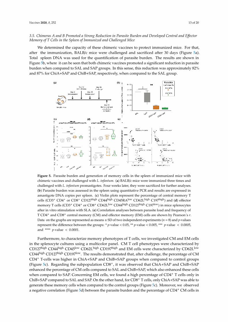
3.5. Chimeras A and B Promoted a Strong Reduction in Parasite Burden and Developed Central and EffectorMemory of T Cells in the Spleen of Immunized and Challenged Mice
We determined the capacity of these chimeric vaccines to protect immunized mice. For that,after the immunization, BALB/c mice were challenged and sacrificed after 30 days (Figure 5a).Total spleen DNA was used for the quantification of parasite burden. The results are shown inFigure 5b, where it can be seen that both chimeric vaccines promoted a significant reduction in parasiteburden when compared to SAL and SAP groups. In this sense, this reduction was approximately 82%and 87% for ChiA+SAP and ChiB+SAP, respectively, when compared to the SAL group.
Figure 5. Parasite burden and generation of memory cells in the spleen of immunized mice with chimeric vaccines and challenged with L. infantum. (a) BALB/c mice were immunized three times and challenged with L. infantum promastigotes. Four weeks later, they were sacrificed for further analyses. (b) Parasite burden was assessed in the spleen using quantitative PCR and results are expressed in amastigote DNA copies per spleen. (c) Violin plots represent the percentage of central memory T cells (CD3+ CD4+ or CD8+ CD127high CD44high CD45RAlow CD62Lhigh C197high) and (d) effector memory T cells (CD3+ CD4+ or CD8+ CD62Llow CD44high CD127high C197low) in mice splenocytes after in vitro stimulation with SLA. (e) Correlation analyses between parasite load and frequency of T CD4+ and CD8+ central memory (CM) and effector memory (EM) cells are shown by Pearson´s r. Data on the graphs are represented as means ± SD of two independent experiments (n = 8) and p-values represent the difference between the groups: * p-value < 0.05, ** p-value < 0.005, *** p-value < 0.0005, and **** p-value < 0.0001.
The final construct for chimeras A and B has epitopes with high score for the algorithms used, conservancy across L. donovani and L. major, and highly antigenic and non-allergic for dogs and/or human use. Their predicted physicochemical features demonstrated that they are stable, hydrophilic (negative values for GRAVY), and basic nature. The 3D structures were validated using Ramachandran plot and ProSA webserver, indicating that the quality of the models is satisfactory [41–45]. Various studies have been shown that chimera constructs that have an affinity to toll-like receptors (mainly TLR-3, TLR-4, and TLR-9) may have the ability to enhance the immune response against intracellular pathogens [42–46]. In this sense, we performed molecular docking showing the interaction between chimeras A and B with mouse and human TLR-3 and TLR-4. For the conformational stability of docked complexes, energy minimization was achieved to minimize the potential energy of the complete systems [44]. In summary, both evaluated chimeras interacted with mice and human TLR-3 and TLR-4 demonstrating, the in silico capacity to be immunogenic. The cloning and expression of the chimeras was well succeeded using E. coli system, as shown in Figure S2.
A critical feature of successful vaccines against Leishmania spp. is their ability to induce multifunctionality regarding cytokine production and immunological memory [5,6,47]. Therefore,
Commented [m1]: There is no figure 5 e in the Figure 5,
please confirm and provide it.
Commented [RB2R1]: The figure was corrected
Figure 5. Parasite burden and generation of memory cells in the spleen of immunized mice withchimeric vaccines and challenged with L. infantum. (a) BALB/c mice were immunized three times andchallenged with L. infantum promastigotes. Four weeks later, they were sacrificed for further analyses.(b) Parasite burden was assessed in the spleen using quantitative PCR and results are expressed inamastigote DNA copies per spleen. (c) Violin plots represent the percentage of central memory Tcells (CD3+ CD4+ or CD8+ CD127high CD44high CD45RAlow CD62Lhigh C197high) and (d) effectormemory T cells (CD3+ CD4+ or CD8+ CD62Llow CD44high CD127high C197low) in mice splenocytesafter in vitro stimulation with SLA. (e) Correlation analyses between parasite load and frequency ofT CD4+ and CD8+ central memory (CM) and effector memory (EM) cells are shown by Pearson´s r.Data on the graphs are represented as means ± SD of two independent experiments (n = 8) and p-valuesrepresent the difference between the groups: * p-value < 0.05, ** p-value < 0.005, *** p-value < 0.0005,and **** p-value < 0.0001.
Furthermore, to characterize memory phenotypes of T cells, we investigated CM and EM cellsin the splenocyte cultures using a multicolor panel. CM T cell phenotypes were characterized byCD127high CD44high CD45low CD62Lhigh CD197high and EM cells were characterized by CD62Llow
CD44high CD127high CD197low. The results demonstrated that, after challenge, the percentage of CMCD4+ T-cells was higher in ChiA+SAP and ChiB+SAP groups when compared to control groups(Figure 5c). Regarding the subpopulation CD8+, it was observed that ChiA+SAP and ChiB+SAPenhanced the percentage of CM cells compared to SAL and ChiB+SAP, which also enhanced these cellswhen compared to SAP. Concerning EM cells, we found a high percentage of CD4+ T cells only inChiB+SAP compared to SAL and SAP. On the other hand, for CD8+ T cells, only ChiA+SAP was able togenerate these memory cells when compared to the control groups (Figure 5c). Moreover, we observeda negative correlation (Figure 5d) between the parasite burden and the percentage of CD4+ CM cells in

Vaccines 2020, 8, 252 14 of 20
the spleen (Pearson’s r = −0.4919; p-value <0.05). In addition, we found the same negative correlationfor CD8+ CM cells (Pearson’s r = −0.4926, p-value <0.05), CD4+ EM cells (Pearson’s r = −0.5601,p-value <0.05), and CD8+ EM cells (Pearson’s r = −0.5333, p-value <0.05) (Figure 5e).
4. Discussion
The use of immunoinformatics capable of predicting immunodominant epitopes has been usedin numerous studies. This methodology has already been described as being highly accurate formapping immunogenic epitopes to Leishmania proteins [2,9,11,32]. Therefore, immunoinformatics canresult in a huge gain for the design of multiepitope vaccines against neglected diseases, in this case,visceral leishmaniasis.
In this context, the present study focused on the design of multi-epitope chimeric vaccines thatcan afford a robust level of protective immunity against L. infantum. For that, we mapped potential Tcell epitopes on nine known proteins that have immunogenic capabilities that have been described inthe literature. Therefore, according to the computational methodology proposed by [9], we mappedthe highest score epitopes provided by immunoinformatics concerning the histone H2A, Lip2a, Lip0,LACK, and CPC (chimera A) proteins, as well as CPA, CPB, PSA-50S, and A2 proteins (chimeraB) (Figure S1). Thus, we could observe that the components chosen to construct both chimera Aand chimera B are antigens that stimulate lymphocyte proliferation and IFN-γ production. Further,the profile of cells stimulated with these antigens may also have a balanced Th1/Th2 polarization.This is observed by induction of humoral response with the production of immunoglobulins, but atthe same time the antigens are able to trigger IFN-γ production and in vitro cell proliferation of mousesplenocytes [33–40].
The final construct for chimeras A and B has epitopes with high score for the algorithms used,conservancy across L. donovani and L. major, and highly antigenic and non-allergic for dogs and/or humanuse. Their predicted physicochemical features demonstrated that they are stable, hydrophilic (negativevalues for GRAVY), and basic nature. The 3D structures were validated using Ramachandran plot andProSA webserver, indicating that the quality of the models is satisfactory [41–45]. Various studieshave been shown that chimera constructs that have an affinity to toll-like receptors (mainly TLR-3,TLR-4, and TLR-9) may have the ability to enhance the immune response against intracellularpathogens [42–46]. In this sense, we performed molecular docking showing the interaction betweenchimeras A and B with mouse and human TLR-3 and TLR-4. For the conformational stability of dockedcomplexes, energy minimization was achieved to minimize the potential energy of the completesystems [44]. In summary, both evaluated chimeras interacted with mice and human TLR-3 and TLR-4demonstrating, the in silico capacity to be immunogenic. The cloning and expression of the chimeraswas well succeeded using E. coli system, as shown in Figure S2.
A critical feature of successful vaccines against Leishmania spp. is their ability to inducemultifunctionality regarding cytokine production and immunological memory [5,6,47]. Therefore,we chose IFN-γ, TNF-α, and IL-2, focusing on the objective of performing the multifunctional study toqualify both central and effector memory T cells. Some studies have already shown that multifunctionalT cells that produce these cytokines are more effective when compared to individual productionfor parasite elimination and infection control [5,6,10,48]. Thus, our data reinforce that chimeras Aand B associated with the adjuvant have this feature to change the patterns of cytokine production.These vaccines can elicit multifunctional CD4+ and CD8+ of CM and EM T cells, which are crucial forthe elimination of the parasite [5,6,10,48]. Additionally, the Chimeric vaccines trigged T lymphocyteactivation through the proliferation of CD4+ and CD8+ subpopulations. The results of T lymphocyteproliferation in the vaccine groups after L. infantum stimulation further support the findings thatIFN-γ is released after this activation, and we can observe the generation of long-term immunologicalmemory against Leishmania spp. [10,49–52]. Gamma interferon (IFN-γ, a cytokine considered centralfor protection against Leishmania spp.) is evaluated and investigated in practically all studies involvingvaccines against leishmaniasis. This cytokine is critical for monitoring, screening, and designing

Vaccines 2020, 8, 252 15 of 20
effective vaccines against these intracellular protozoa [4,10,32,53,54]. Another effector cytokine isTNF-α, which plays a crucial role in the control of intracellular protozoan [55,56]. Our data support therole of these cytokines to orientate a type 1 response that is paramount to protection against Leishmaniaspp. [6,57]. Moreover, our results suggest that the protection conferred by the chimeric multiepitopeprotein could be associated with IFN-γ production by both T lymphocyte subpopulations.
To date, several vaccine candidates against the different types of leishmaniasis have been proposedand with promising results in murine models, but none of them have achieved the market for humanimmunization and VL prevention. This may be due to issues related to the different levels of immunememory generation that are currently agreed upon the guidelines for vaccine development in differentresearch groups [6,52,58,59]. In our study, we characterized memory T cell phenotype in the spleen ofthe immunized animals. The results showed that both chimeric vaccines were able to trigger centralmemory in both CD4+ T lymphocytes and CD8+ T lymphocytes. Some studies have shown that thesecells play a fundamental role in the immune response, being responsible for the renewal of immunesystem memory cells [60]. There is a compartmentalization of these memory cells, as demonstratedin studies in which central memory cells differentiate and expand in the spleen and may recirculateinto secondary lymphoid organs [5]. Regarding the findings of effector memory cells, only ChiB+SAPwas able to generate CD4+ EM T cells, and ChiA+SAP promoted an increase in CD8+ EM T cells.Effector memory cells have the important capacity to migrate to the sites of infection [61]. Thus,the chimeric vaccines tested in this study were able to generate effector memory lymphocytes in thesplenic compartment accompanied by a decrease in parasitic load on the organ.
It is difficult to point out which profile, central or effector memory, would be responsible forprotection in experimental models. This is due to the heterogeneity of responses generated inleishmaniasis immunoprophylaxis studies in different species or animal models, in which there isno consensus on which memory T cell profile would characterize the vaccine candidate as idealor not [6]. Thus, it is believed that both profiles add a lot to this process of parasite control andelimination [29,50,59]. Our results corroborate with these studies, as we found negative correlationbetween the percentage of CM and EM T cells and parasite burden in the spleen. This shows thatthe augment of memory cells probably leads to parasite control and their elimination in the organs.In the splenic compartment there is the establishment of a chronic infection that occurs later and isnot self-resolving as observed in the liver, which makes this organ one of the main targets in studiesevaluating the efficacy of vaccines in BALB/c model [62]. Given the above information, our findingsdemonstrated an important parasite reduction in the spleen of vaccine groups of 82% and 87%,respectively, for ChiA+SAP and ChiB+SAP.
Few studies evaluating polyepitope chimeric vaccines have been reported in the literature.Within the context of VL, there is a restricted number of studies on chimeric vaccines.Regarding challenges with L. infantum, parasite burden results in the spleen obtained by us weresuperior when compared to the 42% load reduction after 10 weeks of infection in a study conductedby [63]. Regarding the L. donovani challenge, the authors of [4] observed a surprising 91% reductionin parasite burden on the same organ after 21 days of infection. It is interesting to note that in thisstudy we used 10 µg per dose of each of the chimeras, much smaller amounts compared to otherstudies that used 20, 25, 100, or even 200 µg of multiepitope chimeric protein [10,63], indicating thehigher capacity of our chimeras in generating immunogenicity, which must be directly associated withparasite control. We highlight the possibility of missing or omitting potential epitopes due to in silicoprediction as a possible disadvantage noted in studies employing immunoinformatics. On the otherhand, this methodology allows large-scale screening, leading to the mapping of important epitopesover a short period of time, which can potentially be very advantageous when used for leishmaniasisvaccines [3].

Vaccines 2020, 8, 252 16 of 20
5. Conclusions
Taking all these findings together, this study is promising in the field of development ofmultiepitope chimeric vaccines for VL, being rationally designed using immunoinformatics andemploying different computational approaches. Thus, the vaccines elicited multifunctional T cells andinduced immunogenicity with CD4+ and CD8+ T lymphocyte proliferation and IFN-γ and TNF-αproduction. Besides this, vaccines instigated the development of central memory and effector of Tlymphocytes in mouse splenocytes, which led to a decrease in parasite load in splenic tissue. To achievesuccess in vaccine development for leishmaniasis, many rational approaches and new techniques forevaluation of polyfunctional and memory cells should be taken. In this sense, this study will contributea large amount to the field of development of vaccines against VL, a critical neglected disease affectingthousands of people around the world.
6. Patent
The patent of the chimeric vaccines used in this study was deposited under the register numberBR10201800819 in the Instituto Nacional da Propriedade Industrial (INPI), Brazil.
Supplementary Materials: The following are available online at http://www.mdpi.com/2076-393X/8/2/252/s1:Figure S1: Illustration of the Chimera’s constructions. The multi-epitope vaccine sequence consisting of 24MHC class I and II ligands. GPGPG sequence was used as linker to join the epitopes. H2A = Histone protein2, LiP2a = Acid ribosomal protein P2, LiP0 = Acid ribosomal protein P0, LACK = Leishmania homologue ofactivated C kinase, CPC = Cysteine peptidase C, CPA = Cysteine peptidase A, CPB = Cysteine peptidase B,PSA-50S = Surface antigenic protein, A2 = Amastigote protein A2, Figure S2: Evaluation of the expression ofchimeric proteins A and B in the E. coli system. The expression was confirmed by SDS-PAGE as described in (a)and (b) for Chimeras A and B respectively. M1 = protein marker 1 (120 to 10 kDa); 1 = Bovine Serum Albumin(2 µg); 2 = 2 µg of chimeric protein A (~40 kDa) or B (~38 kDa). Also, the expression of the Chimera A (c) andChimera B (d) was confirmed by Western Blot using anti-His antibody. M2 = protein marker 2 (120 to 22 kDa);3 = 2 µg of chimeric protein A (c) and B (d), Figure S3: Representative plot of the gating strategy for CFSE-labelledcells to evaluate the proliferation of splenocytes. The first step was to select the single cells, then the live cellsusing (FVS780). To characterize the T-lymphocytes anti-CD3, anti-CD4 and anti-CD8 were used followed by theproliferation analyses selecting the CFSElow-labelled cells.
Author Contributions: All the authors participated with suggestions and the development of this manuscript;R.C.F.D.B., J.M.d.O.C., T.L.V.D.P.O., L.E.S.R., F.A.S.M., and R.D.d.O.A.-S. performed all the experiments. R.C.F.D.B.,J.C.R., and B.M.R. performed the analyses of data. R.C.F.D.B., J.C.R., J.M.O.C., T.L.V.D.P.O., L.E.S.R., F.A.S.M.,B.M.R., D.d.M.R., J.C.R., A.B.R., and R.D.d.O.A.-S. participated in drafting the article and/or revising it critically forimportant intellectual content and created the figures and tables. R.C.-O., D.d.M.R., J.C.R., and A.B.R. participatedin the study conception, critical revision of the article, and supervision. All authors have read and agreed to thepublished version of the manuscript.
Funding: This research received no external funding.
Acknowledgments: The authors acknowledge the Brazilian agencies CNPq(MCTI/CNPq/CT-BIOTEC-GENOPROT no. 560943/2010-5; MCTI/CNPq/CT-BIOTEC no. 27/2013, 301526/2015-0,486618/2013-7, 310104/2018-1, and 435224/2018-2), FAPEMIG (APQ 03505-13 - PROGRAMA DE PESQUISAPARA O SUS – PPSUS MS/CNPq/FAPEMIG/SES, PRONEX APQ-01373-14, APQ-01661-13, PPM-00710-15,APQ-02638-17, APQ-02556-18, and APQ-00931-18), CAPES (this study was financed in part by the Coordenaçãode Aperfeiçoamento de Pessoal de Nível Superior - Brasil (CAPES) - Finance Code 001), UFOP, and FIOCRUZ forfinancial support. R.C.F.B., L.E.S.R., and D.M.R. are grateful for CAPES fellowships, and A.B.R., J.C.R., and B.M.R.are also grateful to CNPq for fellowships.
Conflicts of Interest: The authors declare that there is no conflict of interest.
References
1. Zutshi, S.; Kumar, S.; Chauhan, P.; Bansode, Y.; Nair, A.; Roy, S.; Sarkar, A.; Saha, B. Anti-LeishmanialVaccines: Assumptions, Approaches, and Annulments. Vaccines (Basel) 2019, 7, 156. [CrossRef]
2. Sachdeva, R.; Banerjea, A.C.; Malla, N.; Dubey, M.L. Immunogenicity and efficacy of single antigen Gp63,polytope and polytopeHSP70 DNA vaccines against visceral leishmaniasis in experimental mouse model.PLoS ONE 2009, 4, e7880. [CrossRef] [PubMed]

Vaccines 2020, 8, 252 17 of 20
3. Seyed, N.; Taheri, T.; Vauchy, C.; Dosset, M.; Godet, Y.; Eslamifar, A.; Sharifi, I.; Adotevi, O.; Borg, C.;Rohrlich, P.S.; et al. Immunogenicity evaluation of a rationally designed polytope construct encodingHLA-A*0201 restricted epitopes derived from Leishmania major related proteins in HLA-A2/DR1 transgenicmice: Steps toward polytope vaccine. PLoS ONE 2014, 9, e108848. [CrossRef] [PubMed]
4. Das, S.; Freier, A.; Boussoffara, T.; Das, S.; Oswald, D.; Losch, F.O.; Selka, M.; Sacerdoti-Sierra, N.;Schonian, G.; Wiesmuller, K.H.; et al. Modular multiantigen T cell epitope-enriched DNA vaccine againsthuman leishmaniasis. Sci. Transl. Med. 2014, 6, 234ra56. [CrossRef] [PubMed]
5. Seder, R.A.; Darrah, P.A.; Roederer, M. T-cell quality in memory and protection: Implications for vaccinedesign. Nat. Rev. Immunol. 2008, 8, 247–258. [CrossRef]
6. Darrah, P.A.; Patel, D.T.; De Luca, P.M.; Lindsay, R.W.; Davey, D.F.; Flynn, B.J.; Hoff, S.T.; Andersen, P.;Reed, S.G.; Morris, S.L.; et al. Multifunctional TH1 cells define a correlate of vaccine-mediated protectionagainst Leishmania major. Nat. Med. 2007, 13, 843–850. [CrossRef]
7. De Groot, A.S. Immunomics: Discovering new targets for vaccines and therapeutics. Drug Discov. Today2006, 11, 203–209. [CrossRef]
8. De Groot, A.S.; Berzofsky, J.A. From genome to vaccine—New immunoinformatics tools for vaccine design.Methods 2004, 34, 425–428. [CrossRef]
9. Brito, R.C.; Guimaraes, F.G.; Velloso, J.P.; Correa-Oliveira, R.; Ruiz, J.C.; Reis, A.B.; Resende, D.M.Immunoinformatics Features Linked to Leishmania Vaccine Development: Data Integration of Experimentaland In Silico Studies. Int. J. Mol. Sci. 2017, 18, 371. [CrossRef]
10. Alves-Silva, M.V.; Nico, D.; Morrot, A.; Palatnik, M.; Palatnik-de-Sousa, C.B. A chimera containing CD4+ andCD8+ T-cell epitopes of the Leishmania donovani nucleoside hydrolase (NH36) optimizes cross-protectionagainst Leishmania amazonesis infection. Front. Immunol. 2017, 8, 100. [CrossRef]
11. De Brito, R.C.F.; Cardoso, J.M.O.; Reis, L.E.S.; Mathias, F.A.S.; Aguiar-Soares, R.D.O.; Teixeira-Carvalho, A.;Roatt, B.M.; Correa-Oliveira, R.; Ruiz, J.C.; Resende, D.M.; et al. Synthetic Peptides Elicit Strong CellularImmunity in Visceral Leishmaniasis Natural Reservoir and Contribute to Long-Lasting PolyfunctionalT-Cells in BALB/c Mice. Vaccines (Basel) 2019, 7, 162. [CrossRef] [PubMed]
12. Larsen, M.V.; Lundegaard, C.; Lamberth, K.; Buus, S.; Lund, O.; Nielsen, M. Large-scale validation of methodsfor cytotoxic T-lymphocyte epitope prediction. BMC Bioinform. 2007, 8, 424. [CrossRef] [PubMed]
13. Buus, S.; Lauemoller, S.L.; Worning, P.; Kesmir, C.; Frimurer, T.; Corbet, S.; Fomsgaard, A.; Hilden, J.;Holm, A.; Brunak, S. Sensitive quantitative predictions of peptide-MHC binding by a ‘Query by Committee’artificial neural network approach. Tissue Antigens 2003, 62, 378–384. [CrossRef] [PubMed]
14. Nielsen, M.; Lundegaard, C.; Lund, O. Prediction of MHC class II binding affinity using SMM-align, a novelstabilization matrix alignment method. BMC Bioinform. 2007, 8, 238. [CrossRef] [PubMed]
15. Xue, X.; Ding, F.; Zhang, Q.; Pan, X.; Qu, L.; Pan, W. Stability and potency of the Plasmodium falciparumMSP1-19/AMA-1(III) chimeric vaccine candidate with Montanide ISA720 adjuvant. Vaccine 2010, 28,3152–3168. [CrossRef]
16. Real, F.; Vidal, R.O.; Carazzolle, M.F.; Mondego, J.M.; Costa, G.G.; Herai, R.H.; Wurtele, M.; de Carvalho, L.M.;Carmona e Ferreira, R.; Mortara, R.A.; et al. The genome sequence of Leishmania (Leishmania) amazonensis:Functional annotation and extended analysis of gene models. DNA Res. 2013, 20, 567–581. [CrossRef]
17. Khan, M.; Khan, S.; Ali, A.; Akbar, H.; Sayaf, A.M.; Khan, A.; Wei, D.Q. Immunoinformatics approachesto explore Helicobacter Pylori proteome (Virulence Factors) to design B and T cell multi-epitope subunitvaccine. Sci. Rep. 2019, 9, 13321. [CrossRef]
18. Kallberg, M.; Wang, H.; Wang, S.; Peng, J.; Wang, Z.; Lu, H.; Xu, J. Template-based protein structure modelingusing the RaptorX web server. Nat. Protoc. 2012, 7, 1511–1522. [CrossRef]
19. Heo, L.; Park, H.; Seok, C. GalaxyRefine: Protein structure refinement driven by side-chain repacking.Nucleic Acids Res. 2013, 41, W384–W388. [CrossRef]
20. Wiederstein, M.; Sippl, M.J. ProSA-web: Interactive web service for the recognition of errors inthree-dimensional structures of proteins. Nucleic Acids Res. 2007, 35, W407–W410. [CrossRef]
21. Lovell, S.C.; Davis, I.W.; Arendall, W.B., 3rd; de Bakker, P.I.; Word, J.M.; Prisant, M.G.; Richardson, J.S.;Richardson, D.C. Structure validation by Calpha geometry: Phi, psi and Cbeta deviation. Proteins 2003, 50,437–450. [CrossRef] [PubMed]
22. Vajda, S.; Yueh, C.; Beglov, D.; Bohnuud, T.; Mottarella, S.E.; Xia, B.; Hall, D.R.; Kozakov, D. New additions tothe ClusPro server motivated by CAPRI. Proteins 2017, 85, 435–444. [CrossRef] [PubMed]

Vaccines 2020, 8, 252 18 of 20
23. Berman, H.M.; Westbrook, J.; Feng, Z.; Gilliland, G.; Bhat, T.N.; Weissig, H.; Shindyalov, I.N.; Bourne, P.E.The Protein Data Bank. Nucleic Acids Res. 2000, 28, 235–242. [CrossRef] [PubMed]
24. PDB. Available online: http://www.rcsb.org/ (accessed on 11 November 2019).25. Moreira, N.; Vitoriano-Souza, J.; Roatt, B.M.; Vieira, P.M.; Ker, H.G.; de Oliveira Cardoso, J.M.; Giunchetti, R.C.;
Carneiro, C.M.; de Lana, M.; Reis, A.B. Parasite burden in hamsters infected with two different strainsof leishmania (Leishmania) infantum: “Leishman Donovan units” versus real-time PCR. PLoS ONE 2012,7, e47907. [CrossRef]
26. Reis, A.B.; Teixeira-Carvalho, A.; Vale, A.M.; Marques, M.J.; Giunchetti, R.C.; Mayrink, W.; Guerra, L.L.;Andrade, R.A.; Correa-Oliveira, R.; Martins-Filho, O.A. Isotype patterns of immunoglobulins: Hallmarks forclinical status and tissue parasite density in Brazilian dogs naturally infected by Leishmania (Leishmania)chagasi. Vet. Immunol. Immunopathol. 2006, 112, 102–116. [CrossRef]
27. Coler, R.N.; Goto, Y.; Bogatzki, L.; Raman, V.; Reed, S.G. Leish-111f, a recombinant polyprotein vaccine thatprotects against visceral Leishmaniasis by elicitation of CD4+ T cells. Infect. Immun. 2007, 75, 4648–4654.[CrossRef]
28. Dias, D.S.; Ribeiro, P.A.F.; Martins, V.T.; Lage, D.P.; Costa, L.E.; Chavez-Fumagalli, M.A.; Ramos, F.F.;Santos, T.T.O.; Ludolf, F.; Oliveira, J.S.; et al. Vaccination with a CD4(+) and CD8(+) T-cell epitopes-basedrecombinant chimeric protein derived from Leishmania infantum proteins confers protective immunityagainst visceral leishmaniasis. Transl. Res. 2018, 200, 18–34. [CrossRef]
29. Sanchez-Sampedro, L.; Gomez, C.E.; Mejias-Perez, E.; Sorzano, C.O.; Esteban, M. High quality long-term CD4+
and CD8+ effector memory populations stimulated by DNA-LACK/MVA-LACK regimen in Leishmaniamajor BALB/c model of infection. PLoS ONE 2012, 7, e38859. [CrossRef]
30. Reis, L.E.S.; Brito, R.C.F.; Cardoso, J.M.O.; Mathias, F.A.S.; Aguiar Soares, R.D.O.; Carneiro, C.M.;de Abreu Vieira, P.M.; Ramos, G.S.; Frezard, F.J.G.; Roatt, B.M.; et al. Mixed Formulation of Conventionaland Pegylated Meglumine Antimoniate-Containing Liposomes Reduces Inflammatory Process and ParasiteBurden in Leishmania infantum-Infected BALB/c Mice. Antimicrob. Agents Chemother. 2017, 61, e00962-17.[CrossRef]
31. Vaure, C.; Liu, Y. A comparative review of toll-like receptor 4 expression and functionality in different animalspecies. Front. Immunol. 2014, 5, 316. [CrossRef]
32. Zandieh, M.; Kashi, T.; Taheri, T.; Zahedifard, F.; Taslimi, Y.; Doustdary, M.; Habibzadeh, S.; Eslamifar, A.;Shokri, F.; Rafati, S.; et al. Assessment of protection induced by DNA and live vaccine encoding LeishmaniaMHC class I restricted epitopes against L. major challenge in Balb/c mice model. Microb. Biochem. Technol.2015, 7, 427–438. [CrossRef]
33. Soto, M.; Alonso, C.; Requena, J.M. The Leishmania infantum acidic ribosomal protein LiP2a inducesa prominent humoral response in vivo and stimulates cell proliferation in vitro and interferon-gamma(IFN-gamma) production by murine splenocytes. Clin. Exp. Immunol. 2000, 122, 212–218. [CrossRef][PubMed]
34. Rafati, S.; Nakhaee, A.; Taheri, T.; Taslimi, Y.; Darabi, H.; Eravani, D.; Sanos, S.; Kaye, P.; Taghikhani, M.;Jamshidi, S.; et al. Protective vaccination against experimental canine visceral leishmaniasis using acombination of DNA and protein immunization with cysteine proteinases type I and II of L. infantum.Vaccine 2005, 23, 3716–3725. [CrossRef] [PubMed]
35. Fernandes, A.P.; Costa, M.M.S.; Coelho, E.A.F.; Michalick, M.S.M.; de Freitas, E.; Melo, M.N.; Tafuri, W.L.;Resende, D.M.; Hermont, V.; Abrantes, C.D.; et al. Protective immunity against challenge with Leishmania(Leishmania) chagasi in beagle dogs vaccinated with recombinant A2 protein. Vaccine 2008, 26, 5888–5895.[CrossRef]
36. Khoshgoo, N.; Zahedifard, F.; Azizi, H.; Taslimi, Y.; Alonso, M.J.; Rafati, S. Cysteine proteinase typeIII is protective against Leishmania infantum infection in BALB/c mice and highly antigenic in visceralleishmaniasis individuals. Vaccine 2008, 26, 5822–5829. [CrossRef]
37. Ramos, I.; Alonso, A.; Marcen, J.M.; Peris, A.; Castillo, J.A.; Colmenares, M.; Larraga, V. Heterologousprime-boost vaccination with a non-replicative vaccinia recombinant vector expressing LACK confersprotection against canine visceral leishmaniasis with a predominant Th1-specific immune response. Vaccine2008, 26, 333–344. [CrossRef]

Vaccines 2020, 8, 252 19 of 20
38. Baharia, R.K.; Tandon, R.; Sahasrabuddhe, A.A.; Sundar, S.; Dube, A. Nucleosomal histone proteins of L.donovani: A combination of recombinant H2A, H2B, H3 and H4 proteins were highly immunogenic andoffered optimum prophylactic efficacy against Leishmania challenge in hamsters. PLoS ONE 2014, 9, e97911.[CrossRef]
39. Oliva, G.; Nieto, J.; Foglia Manzillo, V.; Cappiello, S.; Fiorentino, E.; Di Muccio, T.; Scalone, A.; Moreno, J.;Chicharro, C.; Carrillo, E.; et al. A randomised, double-blind, controlled efficacy trial of the LiESP/QA-21vaccine in naive dogs exposed to two leishmania infantum transmission seasons. PLoS Negl. Trop. Dis. 2014,8, e3213. [CrossRef]
40. Pereira, L.; Abbehusen, M.; Teixeira, C.; Cunha, J.; Nascimento, I.P.; Fukutani, K.; dos-Santos, W.; Barral, A.;de Oliveira, C.I.; Barral-Netto, M.; et al. Vaccination with Leishmania infantum acidic ribosomal P0 butnot with nucleosomal histones proteins controls Leishmania infantum infection in hamsters. PLoS Negl.Trop. Dis. 2015, 9, e0003490. [CrossRef]
41. Nezafat, N.; Eslami, M.; Negahdaripour, M.; Rahbar, M.R.; Ghasemi, Y. Designing an efficient multi-epitopeoral vaccine against Helicobacter pylori using immunoinformatics and structural vaccinology approaches.Mol. Biosyst. 2017, 13, 699–713. [CrossRef]
42. Khatoon, N.; Pandey, R.K.; Prajapati, V.K. Exploring Leishmania secretory proteins to design B and T cellmulti-epitope subunit vaccine using immunoinformatics approach. Sci. Rep. 2017, 7, 8285. [CrossRef][PubMed]
43. Kumar Pandey, R.; Ojha, R.; Mishra, A.; Kumar Prajapati, V. Designing B- and T-cell multi-epitope basedsubunit vaccine using immunoinformatics approach to control Zika virus infection. J. Cell. Biochem. 2018,119, 7631–7642. [CrossRef] [PubMed]
44. Pandey, R.K.; Bhatt, T.K.; Prajapati, V.K. Novel Immunoinformatics Approaches to Design Multi-epitopeSubunit Vaccine for Malaria by Investigating Anopheles Salivary Protein. Sci. Rep. 2018, 8, 1125. [CrossRef][PubMed]
45. Chauhan, V.; Rungta, T.; Goyal, K.; Singh, M.P. Designing a multi-epitope based vaccine to combat KaposiSarcoma utilizing immunoinformatics approach. Sci. Rep. 2019, 9, 2517. [CrossRef] [PubMed]
46. Nezafat, N.; Karimi, Z.; Eslami, M.; Mohkam, M.; Zandian, S.; Ghasemi, Y. Designing an efficient multi-epitopepeptide vaccine against Vibrio cholerae via combined immunoinformatics and protein interaction basedapproaches. Comput. Biol. Chem. 2016, 62, 82–95. [CrossRef]
47. Apostolico, J.S.; Lunardelli, V.A.S.; Yamamoto, M.M.; Cunha-Neto, E.; Boscardin, S.B.; Rosa, D.S. Poly(I:C)Potentiates T Cell Immunity to a Dendritic Cell Targeted HIV-Multiepitope Vaccine. Front. Immunol. 2019,10, 843. [CrossRef]
48. Selvapandiyan, A.; Dey, R.; Nylen, S.; Duncan, R.; Sacks, D.; Nakhasi, H.L. Intracellular replication-deficientLeishmania donovani induces long lasting protective immunity against visceral leishmaniasis. J. Immunol.2009, 183, 1813–1820. [CrossRef]
49. Okwor, I.B.; Jia, P.; Mou, Z.; Onyilagha, C.; Uzonna, J.E. CD8+ T cells are preferentially activatedduring primary low dose leishmania major infection but are completely dispensable during secondaryanti-Leishmania immunity. PLoS Negl. Trop. Dis. 2014, 8, e3300. [CrossRef]
50. Reed, S.G.; Coler, R.N.; Mondal, D.; Kamhawi, S.; Valenzuela, J.G. Leishmania vaccine development:Exploiting the host-vector-parasite interface. Expert Rev. Vaccines 2016, 15, 81–90. [CrossRef]
51. Passero, L.F.; Carvalho, A.K.; Bordon, M.L.; Bonfim-Melo, A.; Carvalho, K.; Kallas, E.G.; Santos, B.B.;Toyama, M.H.; Paes-Leme, A.; Corbett, C.E.; et al. Proteins of Leishmania (Viannia) shawi confer protectionassociated with Th1 immune response and memory generation. Parasites Vectors 2012, 5, 64. [CrossRef]
52. Dey, R.; Dagur, P.K.; Selvapandiyan, A.; McCoy, J.P.; Salotra, P.; Duncan, R.; Nakhasi, H.L. Live attenuatedLeishmania donovani p27 gene knockout parasites are nonpathogenic and elicit long-term protectiveimmunity in BALB/c mice. J. Immunol. 2013, 190, 2138–2149. [CrossRef] [PubMed]
53. Uzonna, J.E.; Spath, G.F.; Beverley, S.M.; Scott, P. Vaccination with phosphoglycan-deficient Leishmaniamajor protects highly susceptible mice from virulent challenge without inducing a strong Th1 response.J. Immunol. 2004, 172, 3793–3797. [CrossRef] [PubMed]
54. Martins, V.T.; Lage, D.P.; Duarte, M.C.; Carvalho, A.M.; Costa, L.E.; Mendes, T.A.; Vale, D.L.;Menezes-Souza, D.; Roatt, B.M.; Tavares, C.A.; et al. A recombinant fusion protein displaying murineand human MHC class I- and II-specific epitopes protects against Leishmania amazonensis infection.Cell. Immunol. 2017, 313, 32–42. [CrossRef]

Vaccines 2020, 8, 252 20 of 20
55. Bogdan, C.; Schroppel, K.; Lohoff, M.; Rollinghoff, M.; Solbach, W. Immunization of susceptible hosts with asoluble antigen fraction from Leishmania major leads to aggravation of murine leishmaniasis mediated byCD4+ T cells. Eur. J. Immunol. 1990, 20, 2533–2540. [CrossRef] [PubMed]
56. Manna, L.; Reale, S.; Vitale, F.; Gravino, A.E. Evidence for a relationship between Leishmania load andclinical manifestations. Res. Vet. Sci. 2009, 87, 76–78. [CrossRef] [PubMed]
57. Blackwell, J.M. Genetic susceptibility to leishmanial infections: Studies in mice and man. Parasitology 1996,112 (Suppl. S1), S67–S74. [CrossRef] [PubMed]
58. Banerjee, A.; Bhattacharya, P.; Dagur, P.K.; Karmakar, S.; Ismail, N.; Joshi, A.B.; Akue, A.D.; KuKuruga, M.;McCoy, J.P., Jr.; Dey, R.; et al. Live Attenuated Leishmania donovani Centrin Gene-Deleted Parasites InduceIL-23-Dependent IL-17-Protective Immune Response against Visceral Leishmaniasis in a Murine Model.J. Immunol. 2018, 200, 163–176. [CrossRef]
59. Rodrigues, A.; Claro, M.; Alexandre-Pires, G.; Santos-Mateus, D.; Martins, C.; Valerio-Bolas, A.;Rafael-Fernandes, M.; Pereira, M.A.; Pereira da Fonseca, I.; Tomas, A.M.; et al. Leishmania infantumantigens modulate memory cell subsets of liver resident T lymphocyte. Immunobiology 2017, 222, 409–422.[CrossRef]
60. Farber, D.L.; Yudanin, N.A.; Restifo, N.P. Human memory T cells: Generation, compartmentalization andhomeostasis. Nat. Rev. Immunol. 2014, 14, 24–35. [CrossRef]
61. Sprent, J. T memory cells: Quality not quantity. Curr. Biol. 2002, 12, R174–R176. [CrossRef]62. Loeuillet, C.; Banuls, A.L.; Hide, M. Study of Leishmania pathogenesis in mice: Experimental considerations.
Parasites Vectors 2016, 9, 144. [CrossRef] [PubMed]63. Martins, V.T.; Duarte, M.C.; Lage, D.P.; Costa, L.E.; Carvalho, A.M.; Mendes, T.A.; Roatt, B.M.;
Menezes-Souza, D.; Soto, M.; Coelho, E.A. A recombinant chimeric protein composed of human andmice-specific CD4(+) and CD8(+) T-cell epitopes protects against visceral leishmaniasis. Parasite Immunol.2017, 39, e12359. [CrossRef] [PubMed]
© 2020 by the authors. Licensee MDPI, Basel, Switzerland. This article is an open accessarticle distributed under the terms and conditions of the Creative Commons Attribution(CC BY) license (http://creativecommons.org/licenses/by/4.0/).