Embed Size (px)
Citation preview
Interventional Radiology in Trauma
Vikash Prasad, MD, FRCPCVascular and Interventional Radiology
The Moncton Hospital

Disclosures
• None relevant to this presentation• Shareholder Johnson and Johnson
Goal
• To describe the role and appropriate utilization of Interventional Radiology in trauma care using a case based approach
Objectives
• Describe Interventional Radiology modalities available for various trauma care situations
• Discuss the interventional modalities that may improve outcome or reduce adverse events in trauma
Background
• Embolization for pelvic trauma first described in 1972
• Angiography used to be used for diagnostic purposes, but has been supplanted by CT
• CT:– grade solid organ injuries– detect hemorrhage– detect vascular abnormalities (pseudoaneurysm, intimal
dissection, arteriovenous fistula, and vascular occlusion)– help predict which hemodynamically stable patients may benefit from nonoperative management
Background
• Diagnostic Angiogrphy– Indications for emergency catheter angiography in the trauma patient include clinical signs or symptoms of hemorrhage or CT evidence of ongoing hemorrhage or vascular injury
– For penetrating abdominal trauma, abdominal angiography rarely is indicated, because emergency laparotomy usually is indicated
Background
• Transcatheter embolization is usually considered preferable to surgical treatment when:– surgical access is difficult– patient is a poor operative risk– selective transcatheter embolization may limit the amount of normal tissue or parenchyma necrotized
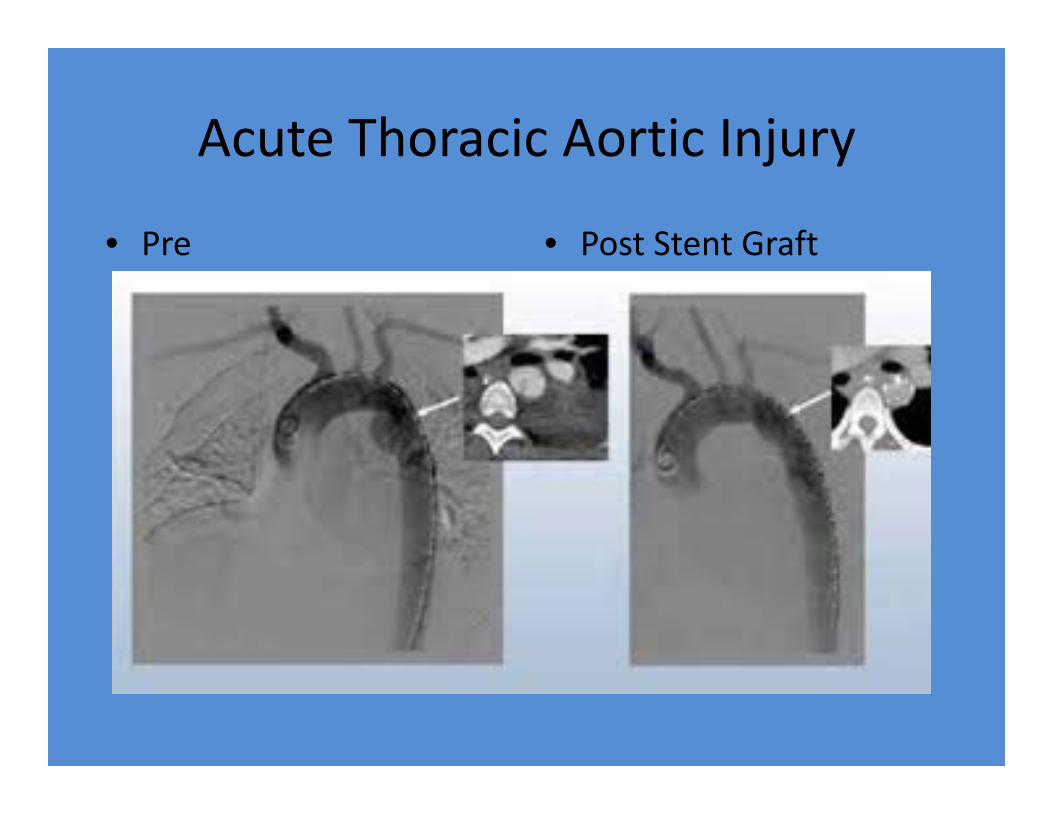
Acute Thoracic Aortic Injury
• 10‐25% survive long enough to present to the hospital
• Of those that make it to ER approximately 30% are fatal within 6 hours, and 40% are fatal within 24 hours if undiagnosed and left untreated.
• Only 2‐10% of untreated patients survive longer than 6 months
Acute Thoracic Aortic Injury
• locations of tears:– Aortic isthmus, 80‐90%– Ascending aorta, 5‐9%– Diaphragmatic aorta, 1‐3%
• Treatment:– Control blood pressure until repair– Open surgery or stent graft (TEVAR)
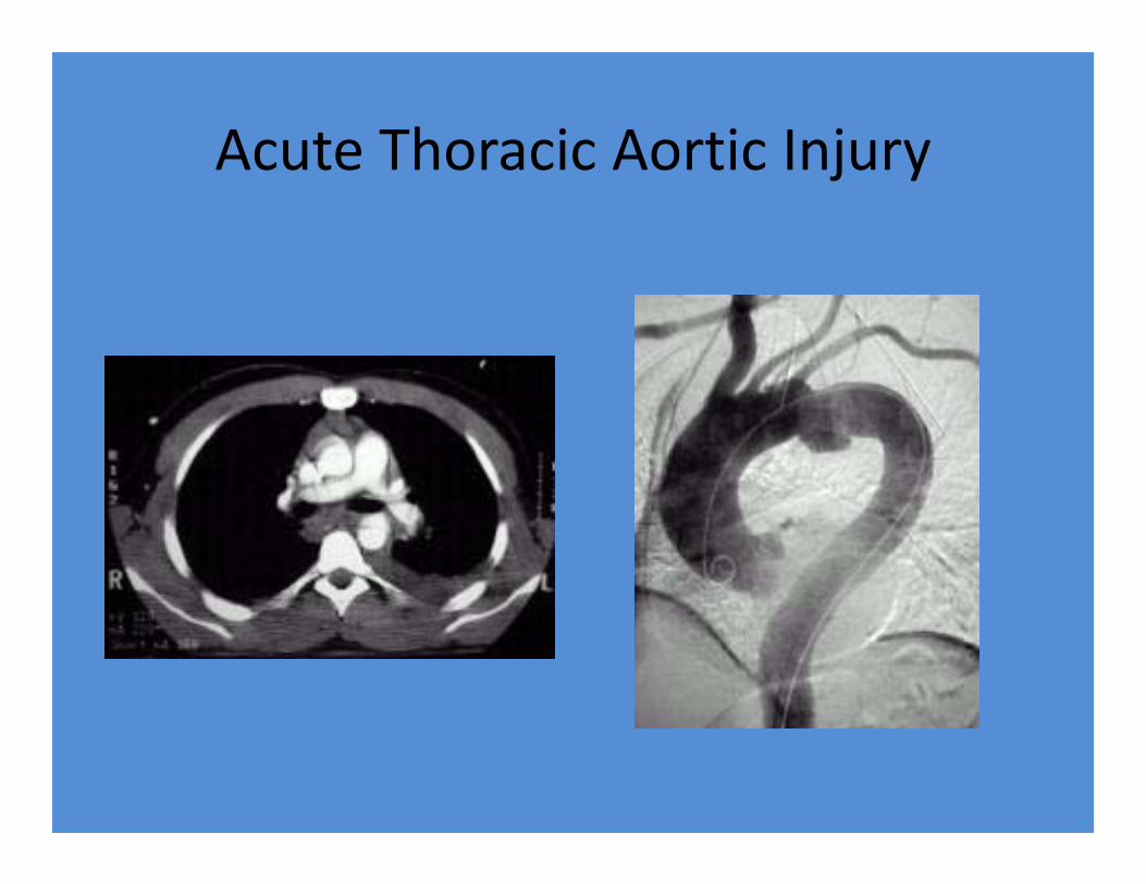
Acute Thoracic Aortic Injury
Acute Thoracic Aortic Injury
• Pre • Post Stent Graft
Abdominal Trauma
• Liver & spleen susceptible to blunt & penetrating trauma
• Management controversial• Embolotherapy:
– Feasible:• Liver – dual blood supply• Spleen – rich collateral network
– Succesesful >85%
Splenic Laceration
• Spleen plays important role in preventing overwhelming sepsis by encapsulated organisms such as pneumococcus
• Attempt splenic preservation in trauma• 70% of patients with blunt splenic injuries may be treated nonoperatively, with success rates of 71‐97%
• Nonoperative management of splenic injuries is effective in more than 95% of children
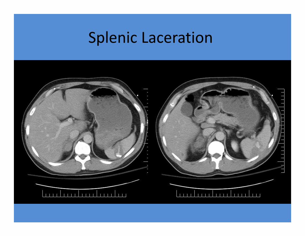
Splenic Laceration• If CT evidence of splenic injury is seen in a hemodynamically stable
patient, celiac and splenic angiography is employed. • Transcatheter embolization is used for blunt splenic trauma. The
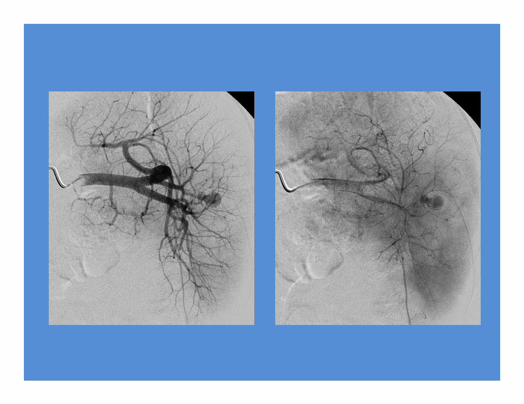
indication is extravasation or vascular injury. • Techniques include the following:
– Superselective distal embolization using a microcatheter and microcoils, polyvinyl alcohol particles, or microspheres at the bleeding site when possible
– Proximal coil embolization just distal to the dorsal pancreatic artery and proximal to the pancreatic magna artery to decrease the head of pressure and to preserve distal collateral flow
– Nonselective distal embolization using smaller particles such as Gelfoam pledgets
– Combination of proximal and distal embolization
Splenic Laceration
• For grade IV splenic injuries:– Sclafani et al reported an 84% salvage rate– Shanmuganathan et al reported a 94% salvage rate when using splenic embolization
• Complications of splenic embolization include inadvertent embolization, splenic infarction and/or abscess, and splenic artery dissection.
Splenic Laceration
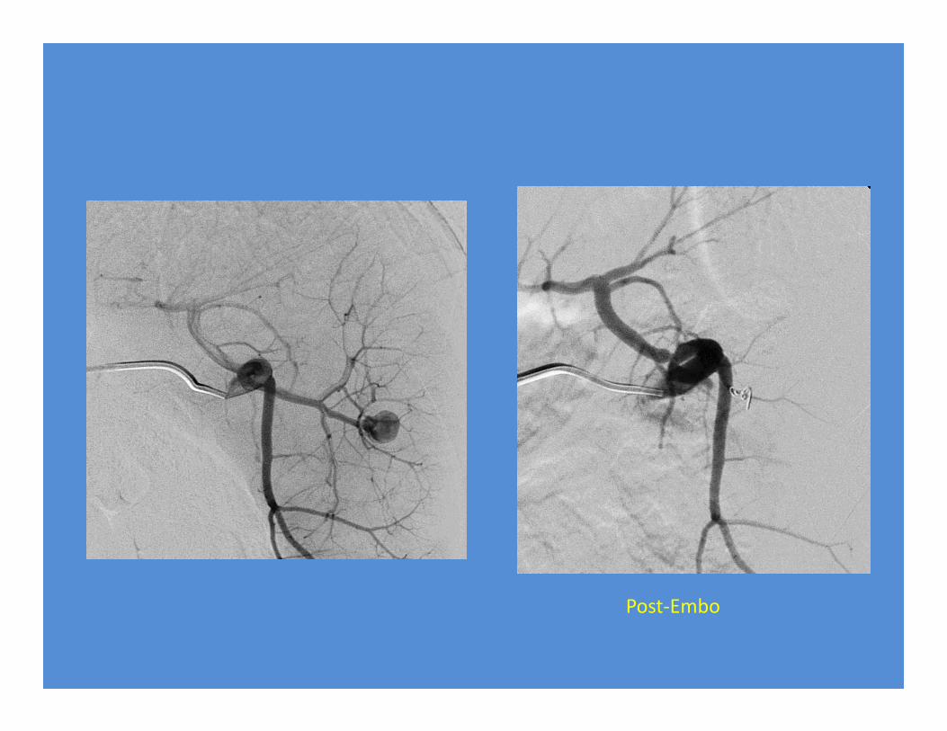
Post‐Embo
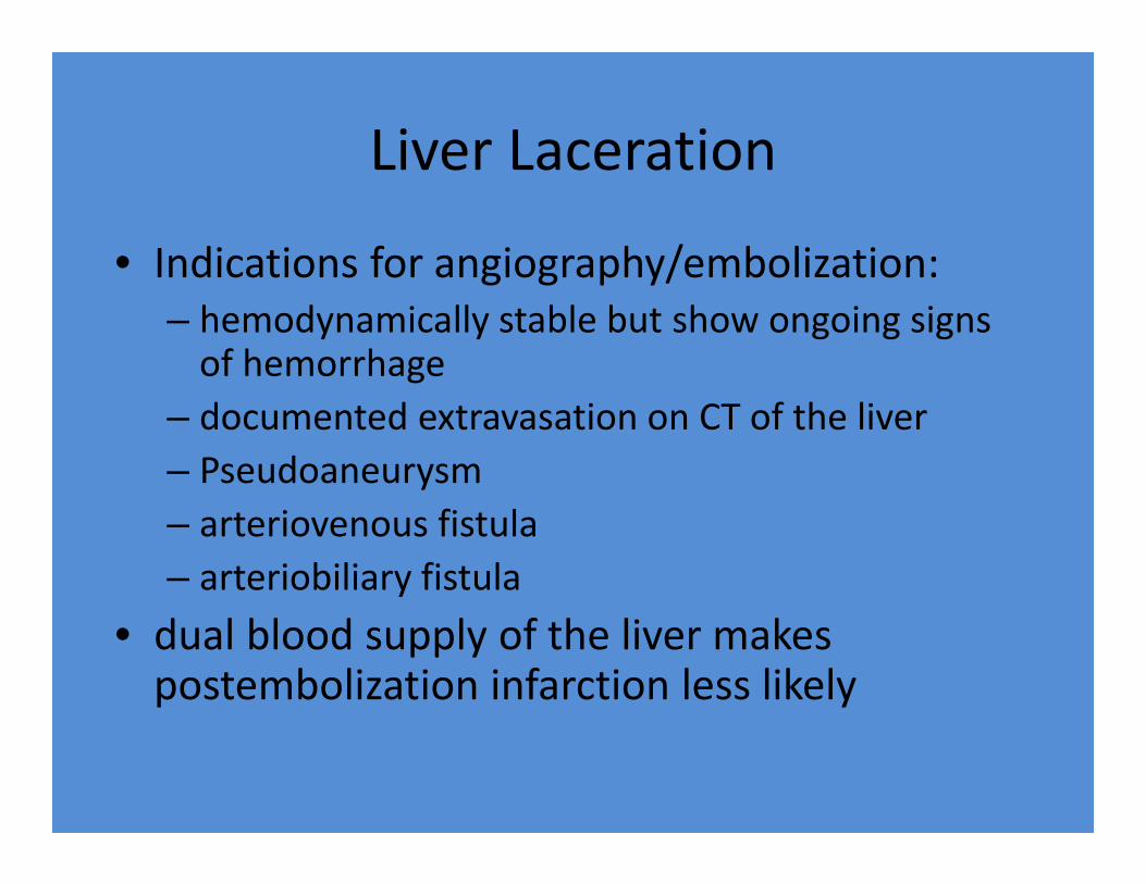
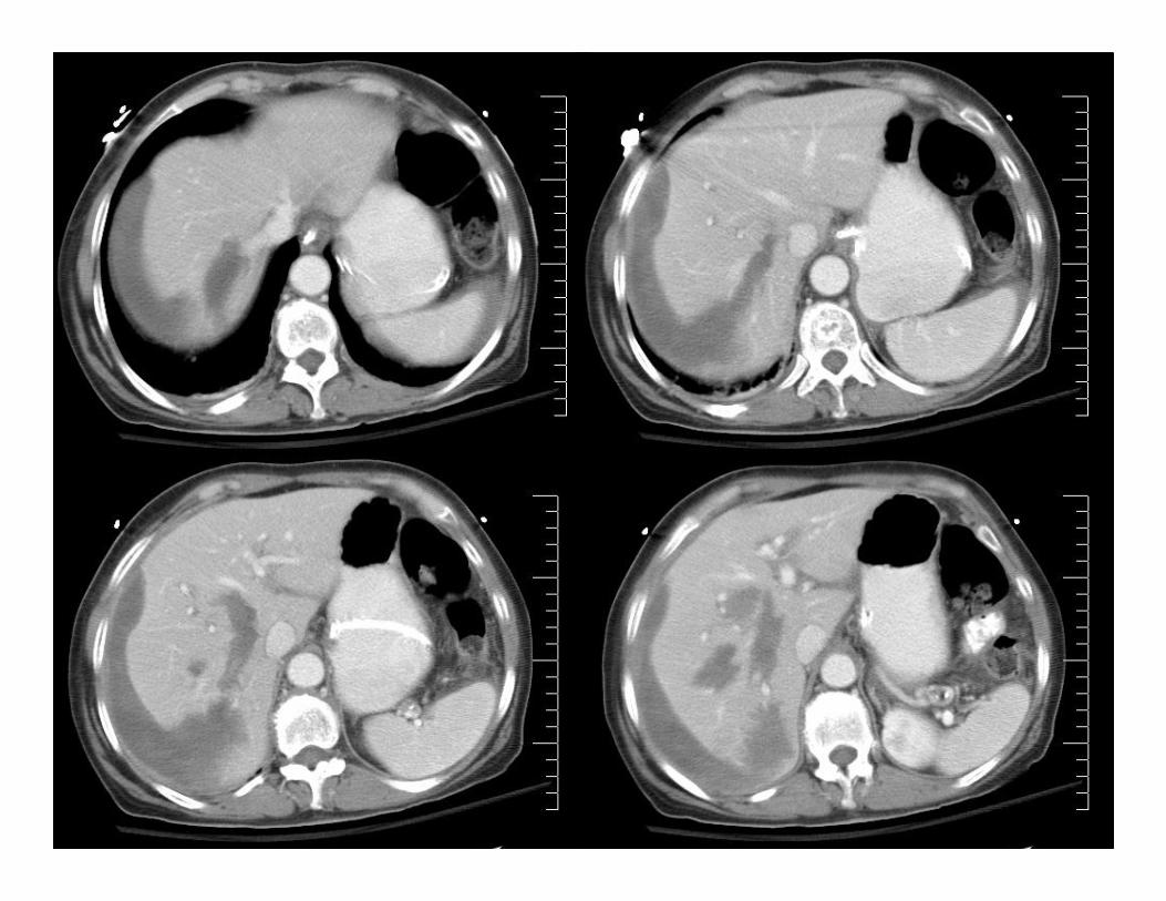
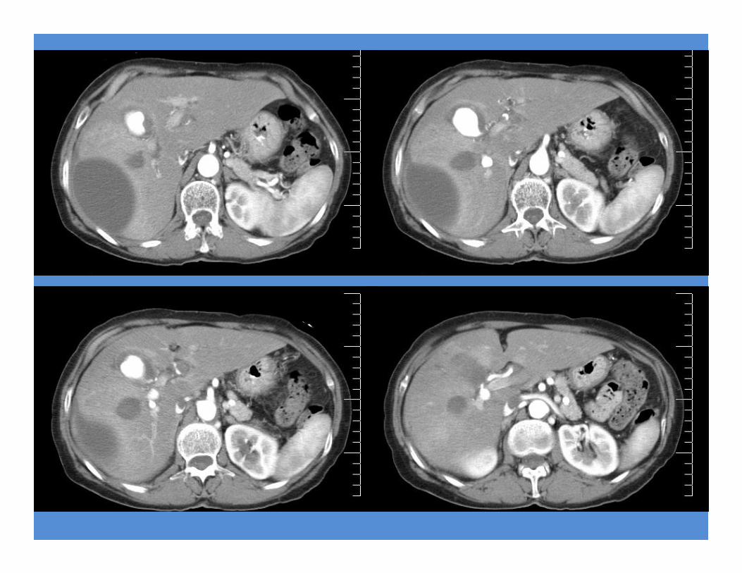
Liver Laceration
• Indications for angiography/embolization:– hemodynamically stable but show ongoing signs of hemorrhage
– documented extravasation on CT of the liver – Pseudoaneurysm– arteriovenous fistula– arteriobiliary fistula
• dual blood supply of the liver makes postembolization infarction less likely
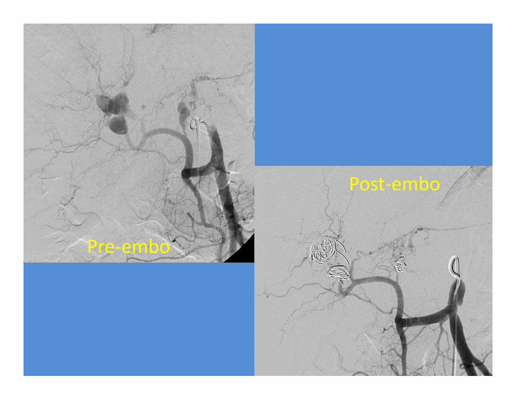
Pre‐embo
Post‐embo
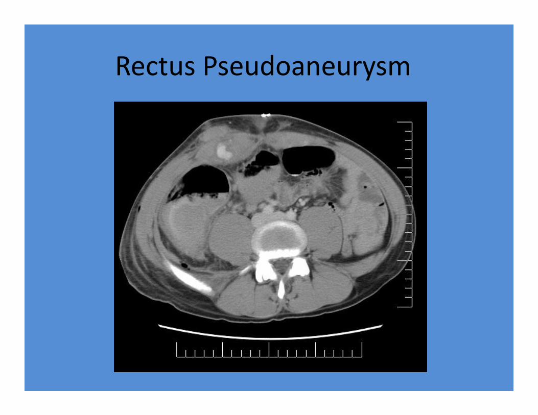
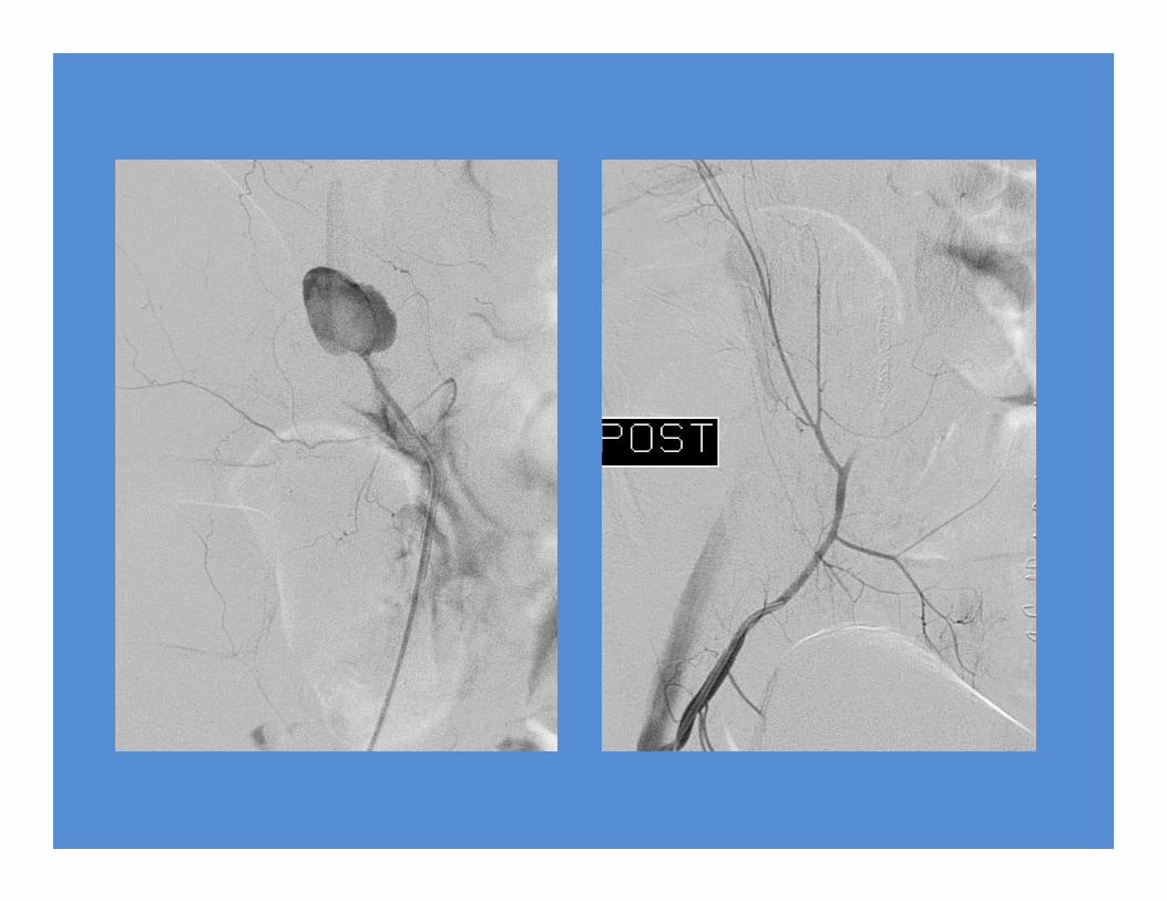
Rectus Pseudoaneurysm
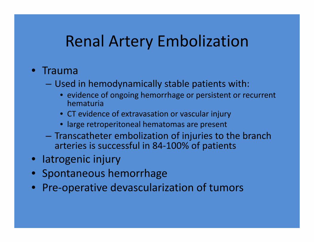
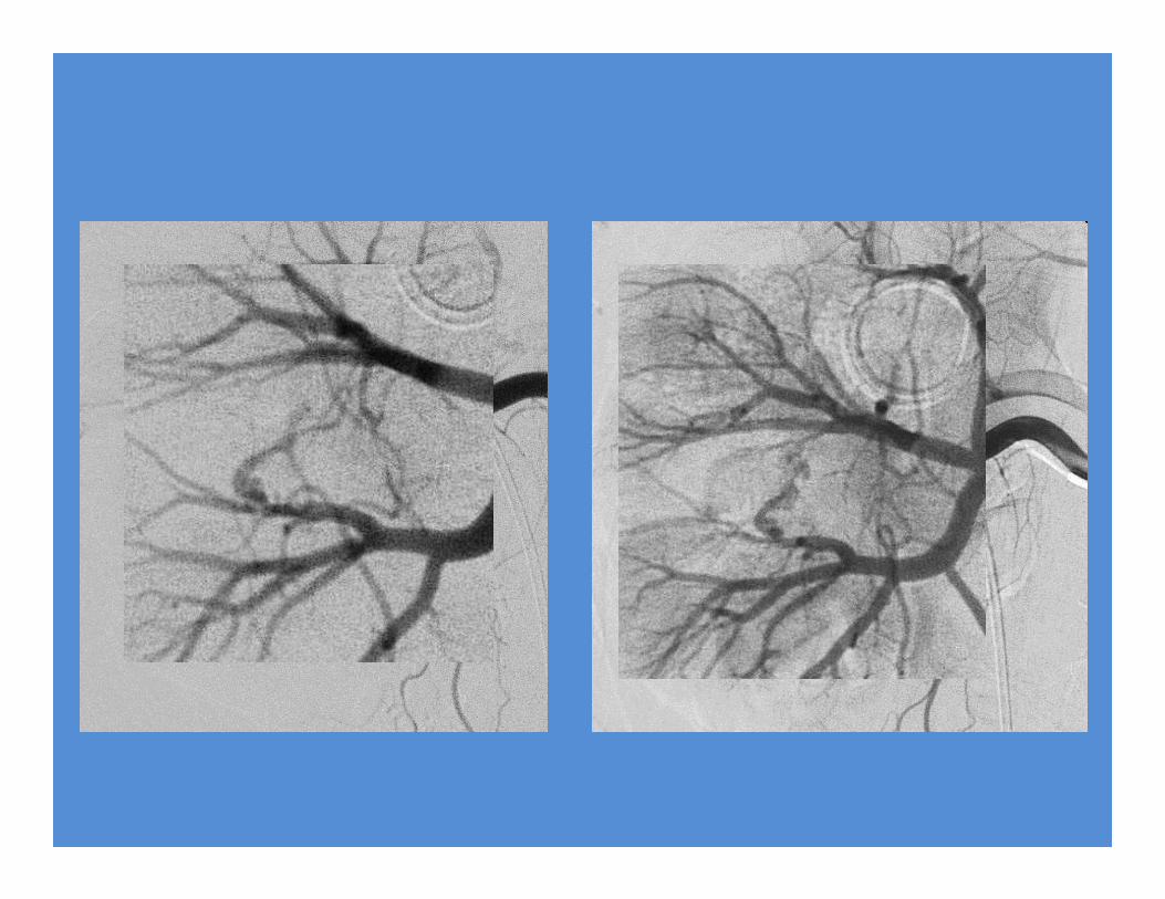
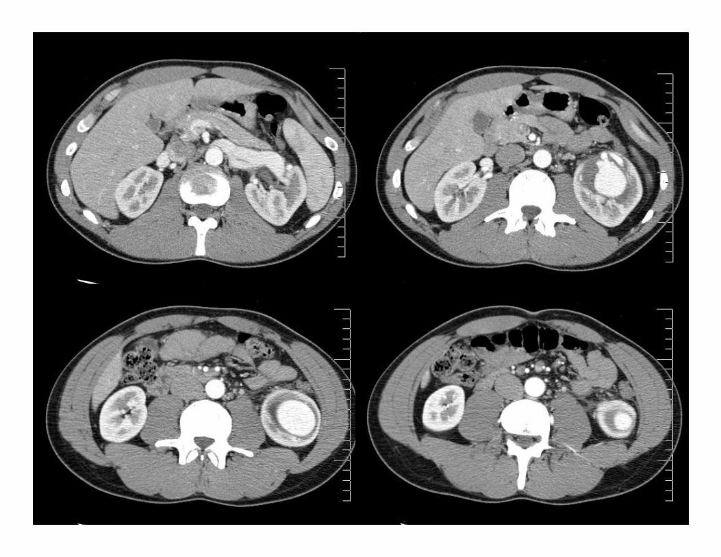
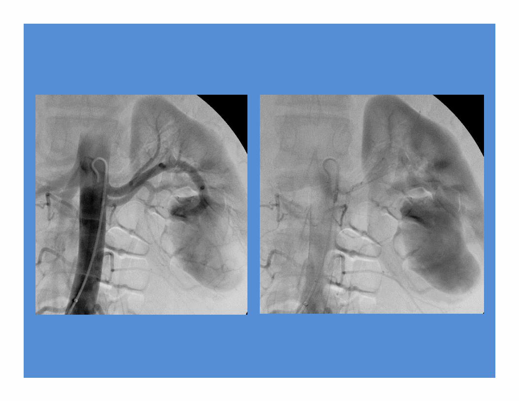
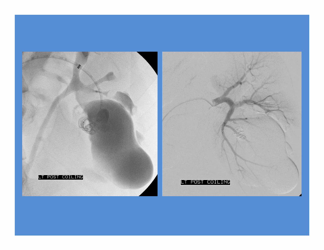
Renal Artery Embolization
• Trauma– Used in hemodynamically stable patients with:
• evidence of ongoing hemorrhage or persistent or recurrent hematuria
• CT evidence of extravasation or vascular injury• large retroperitoneal hematomas are present
– Transcatheter embolization of injuries to the branch arteries is successful in 84‐100% of patients
• Iatrogenic injury• Spontaneous hemorrhage• Pre‐operative devascularization of tumors
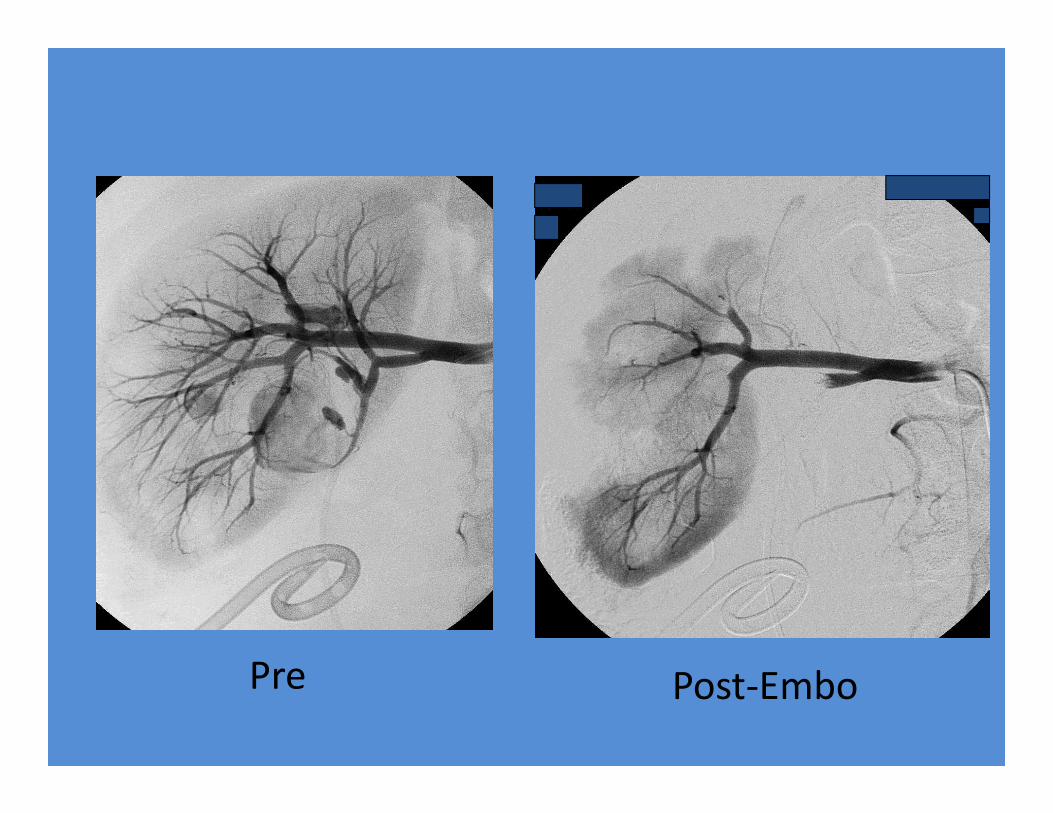
Post‐EmboPre
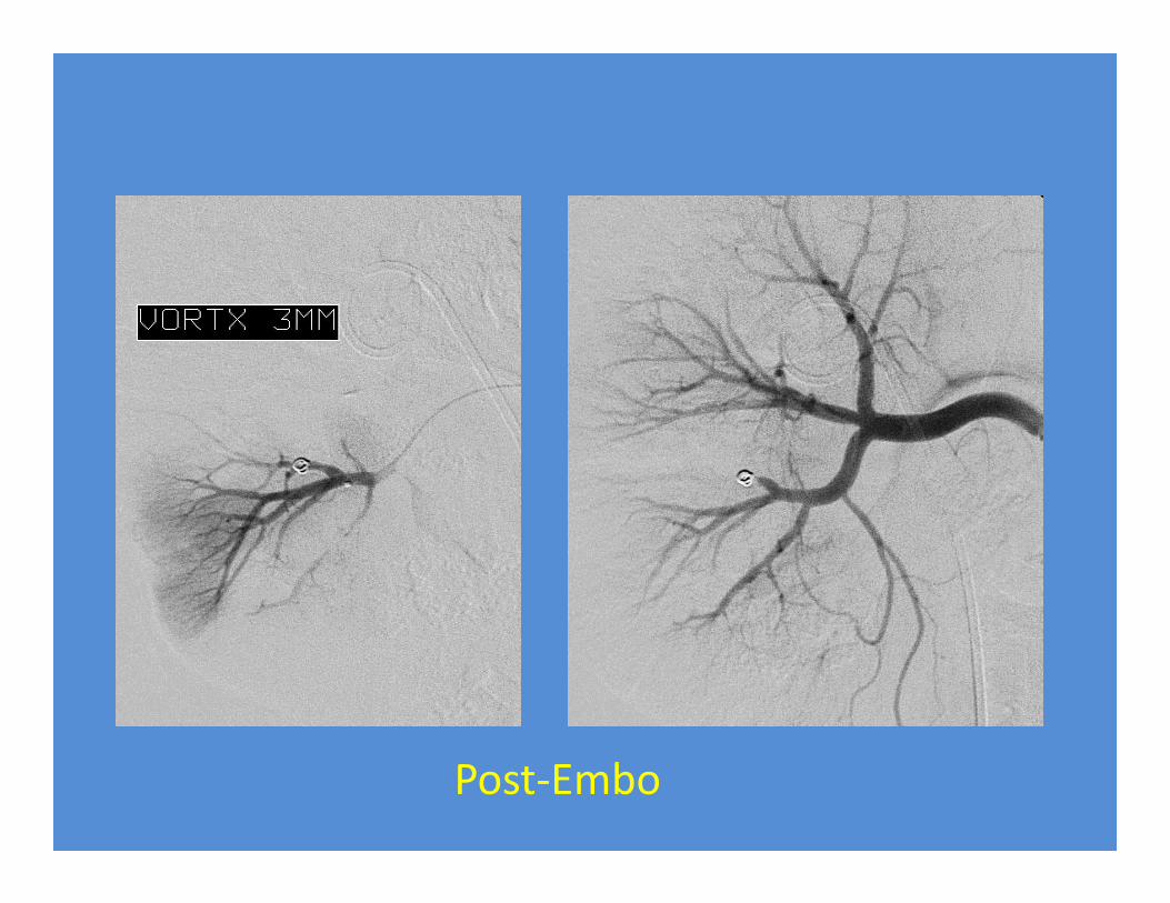
Post‐Embo
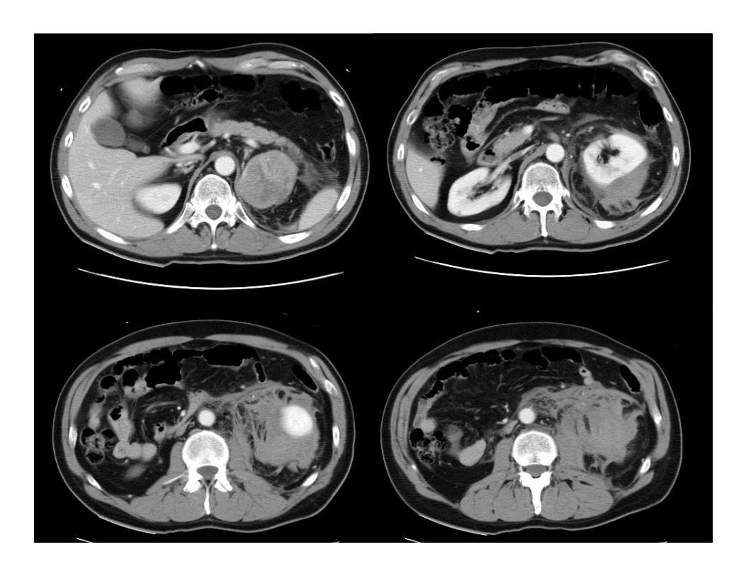
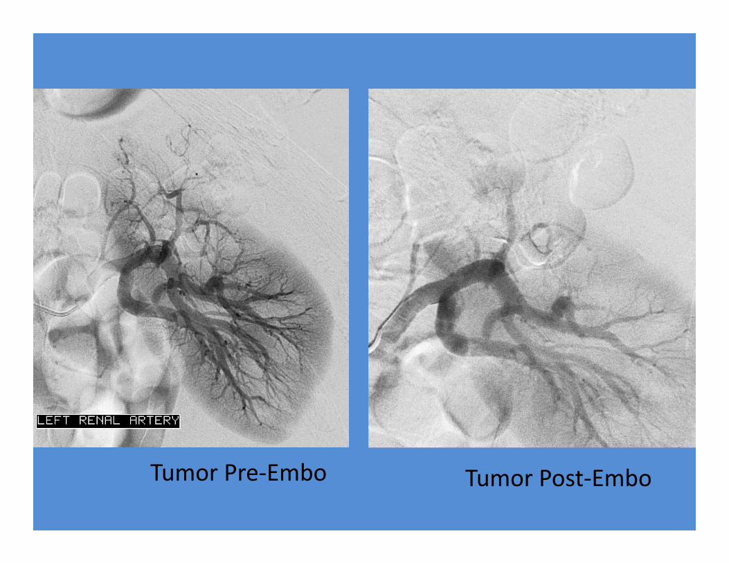
Tumor Pre‐Embo Tumor Post‐Embo
Peripheral Vascular Trauma
• Indications for angiography:– Pulse deficit– Ischemia– Expanding hematoma– Pulsatile bleeding– Bruit/thrill– Isolated neurologic deficit
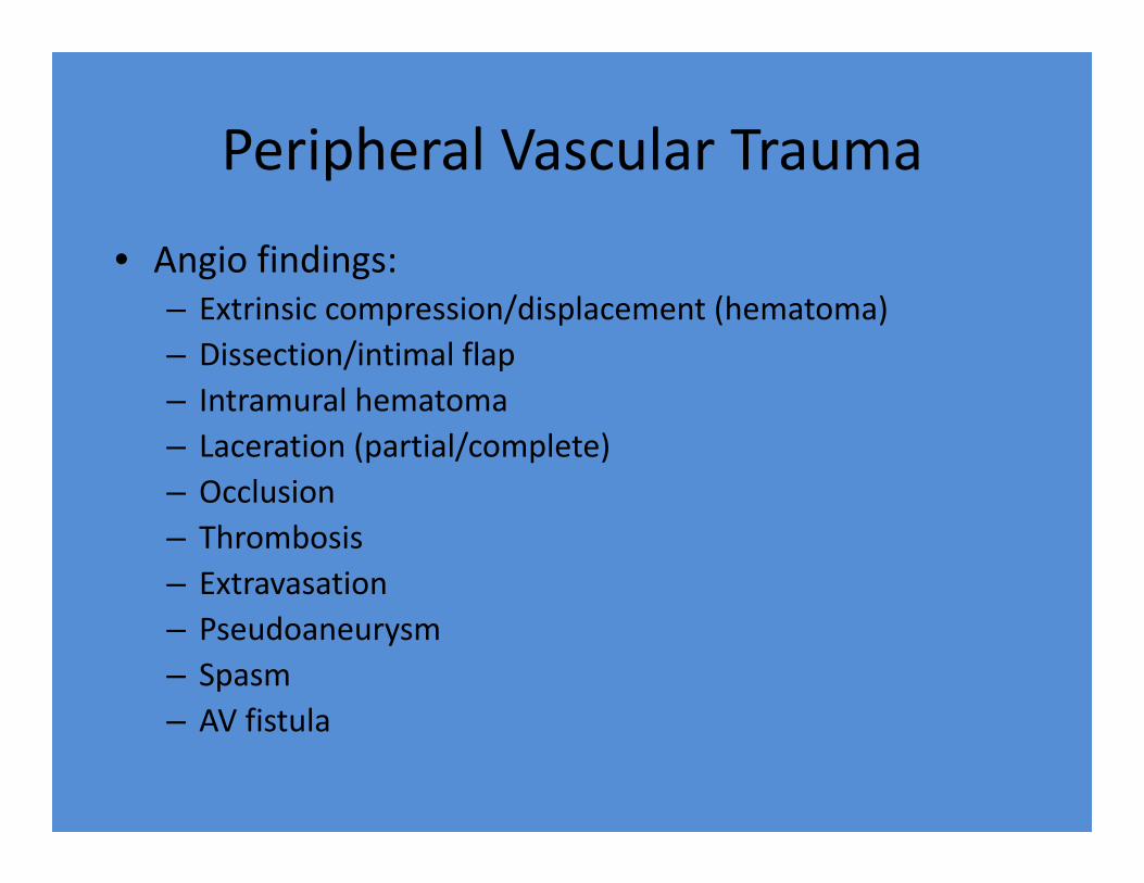
Peripheral Vascular Trauma
• Angio findings:– Extrinsic compression/displacement (hematoma)– Dissection/intimal flap– Intramural hematoma– Laceration (partial/complete)– Occlusion– Thrombosis– Extravasation– Pseudoaneurysm– Spasm– AV fistula
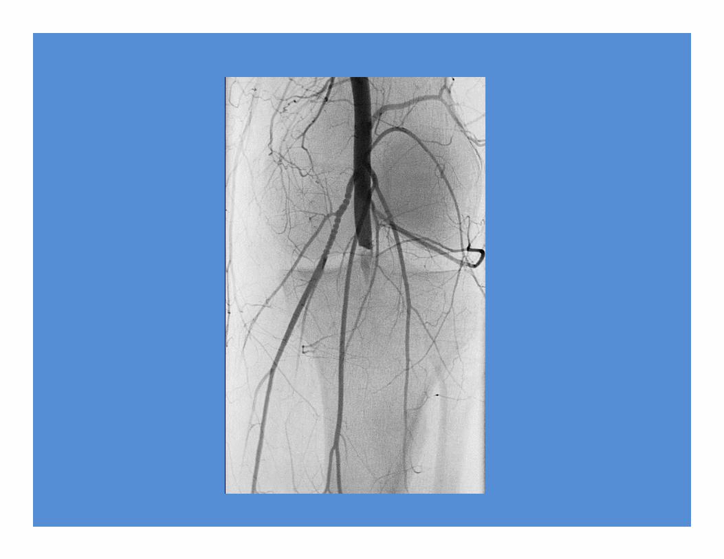
Distal femur fracture with popliteal artery transection
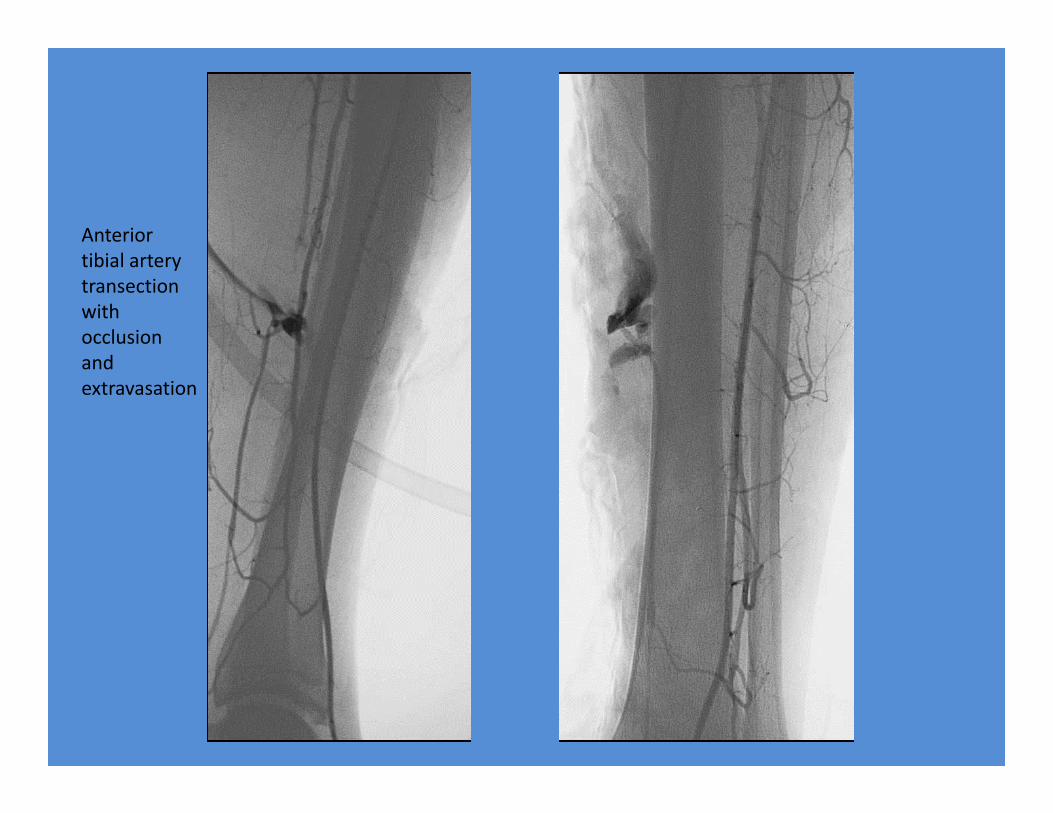
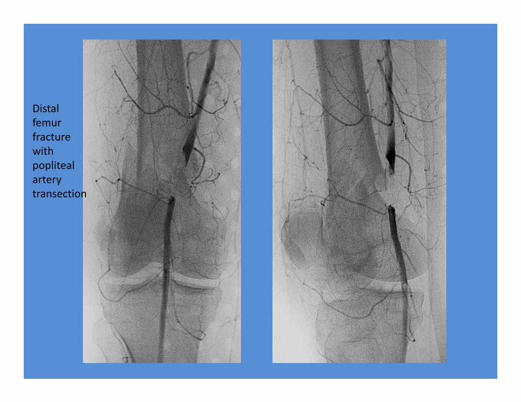
Anterior tibial artery transection with occlusion and extravasation
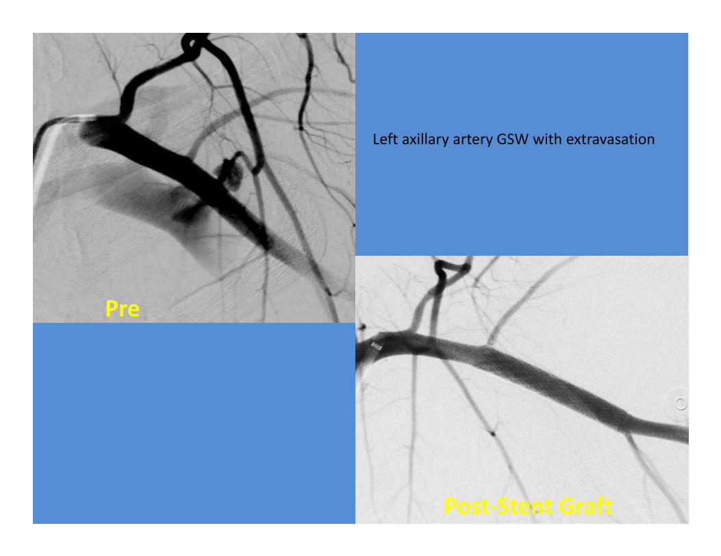
Post‐Stent Graft
Pre
Left axillary artery GSW with extravasation
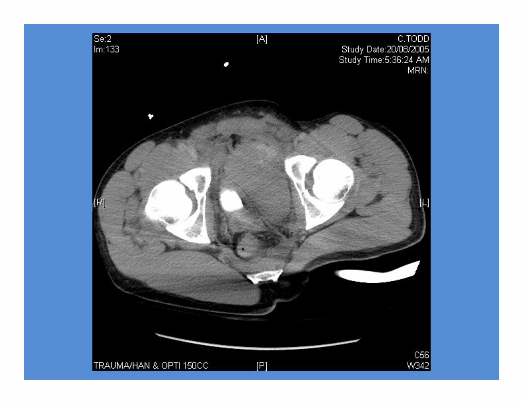
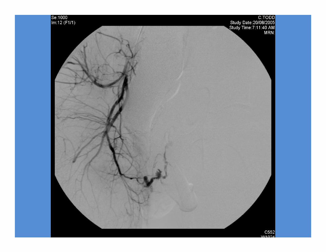
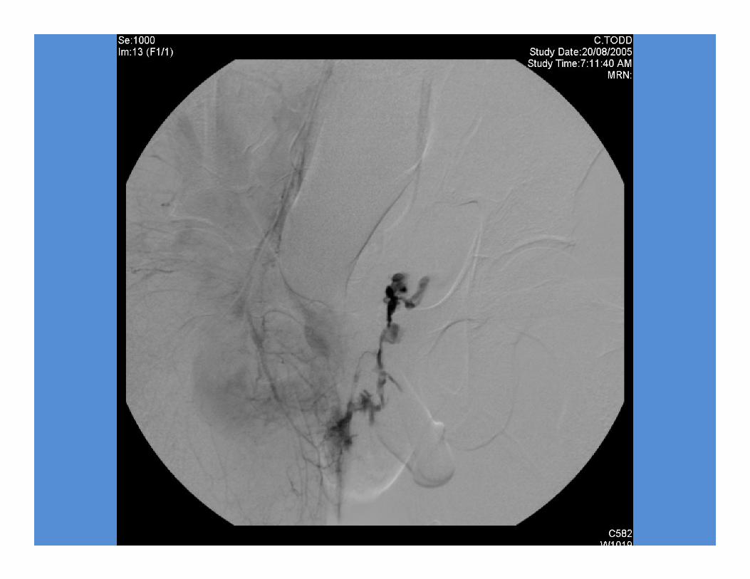
Pelvic Embolization
• Hemorrhage from pelvic trauma with associated fracture can carry up to a 50% mortality rate
• Spontaneous hemostasis can develop secondary to a tamponade effect in a stable pelvic fracture
• However, when bleeding is brisk, a dilutionalcoagulopathy may occur that increases the risk of persistent hemorrhage
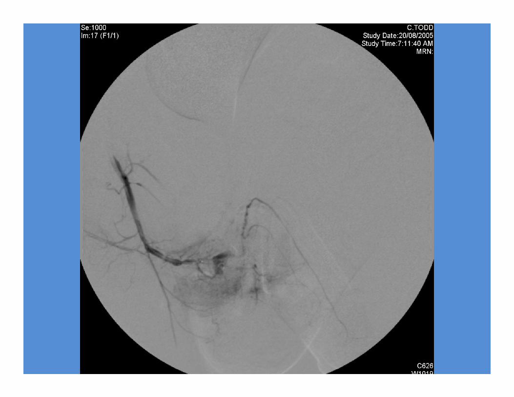
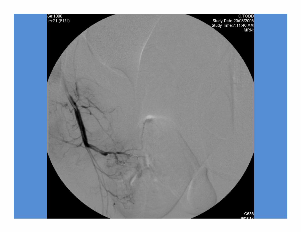
• aim of transcatheter embolization is to reduce pulse pressure and blood flow to the bleeding sites, allowing the body's hemostatic mechanisms to become effective
Pelvic Embolization
• Percutaneous transcatheter embolization has been shown to be safe and efficacious when used to treat pelvic hemorrhage
• Endovascular management of hemorrhage following pelvic fracture has been described since the 1970s
• success rate of stopping hemorrhage is 85‐100%. (despite high technical success rates, the mortality rate is approximately 50% because of concomitant injuries)
Pelvic Embolization
• Pelvic transcatheter embolization complications include the following:– Inadvertent embolization —Occurs only rarely, provided catheter position is satisfactory and the embolization procedure is terminated once occlusion is established.
– Ischemic tissue necrosis or infarction —Occurs only rarely, provided particle sizes remain larger than 500 microns, in cases involving extensive distal collateralization of the pelvic vasculature
– Impotence in men —Difficult to differentiate from impotence of neurogenic etiology related to injuries to the lumbosacral plexus
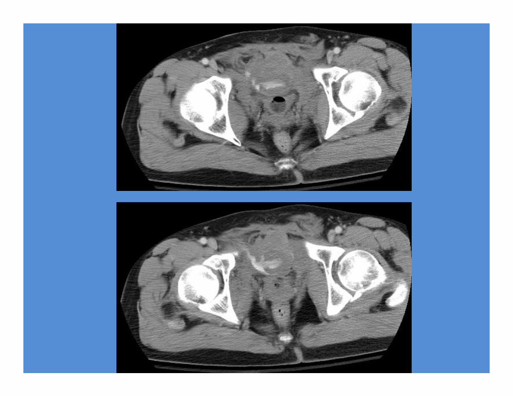
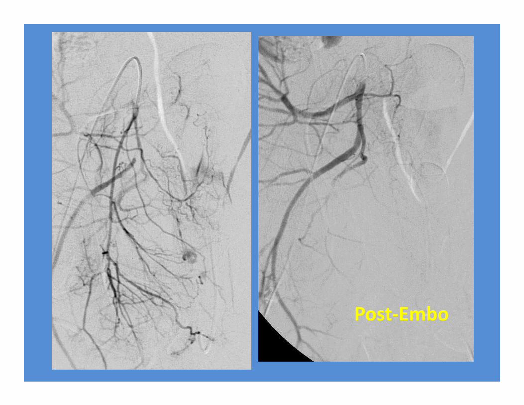
Post‐Embo
Motorcycle accident
28 y.o. male
IVC Filters
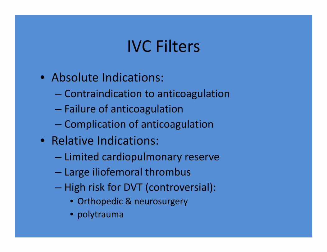
• Absolute Indications:– Contraindication to anticoagulation– Failure of anticoagulation– Complication of anticoagulation
• Relative Indications:– Limited cardiopulmonary reserve– Large iliofemoral thrombus– High risk for DVT (controversial):
• Orthopedic & neurosurgery• polytrauma
IVC Filters
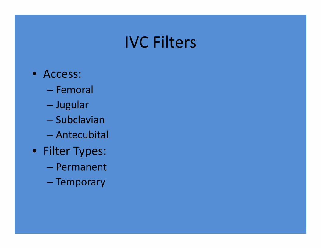
• Access:– Femoral– Jugular– Subclavian– Antecubital
• Filter Types:– Permanent– Temporary
Temporary IVC Filters
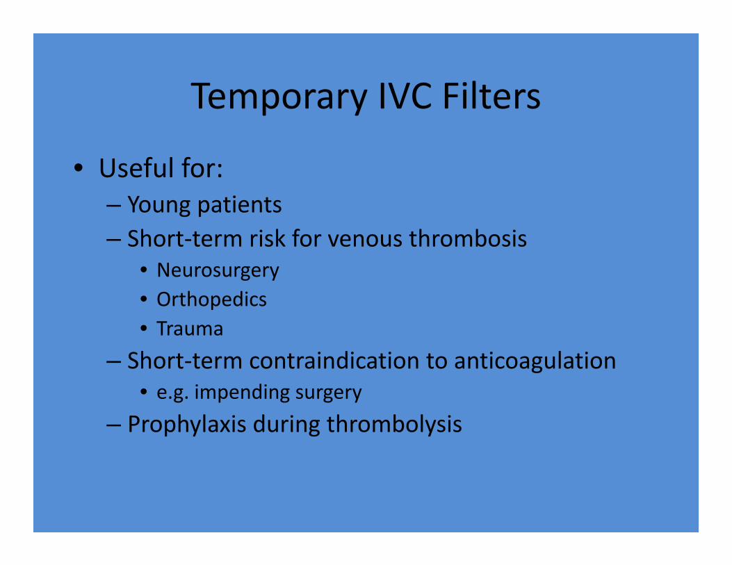
• Useful for:– Young patients– Short‐term risk for venous thrombosis
• Neurosurgery• Orthopedics• Trauma
– Short‐term contraindication to anticoagulation• e.g. impending surgery
– Prophylaxis during thrombolysis
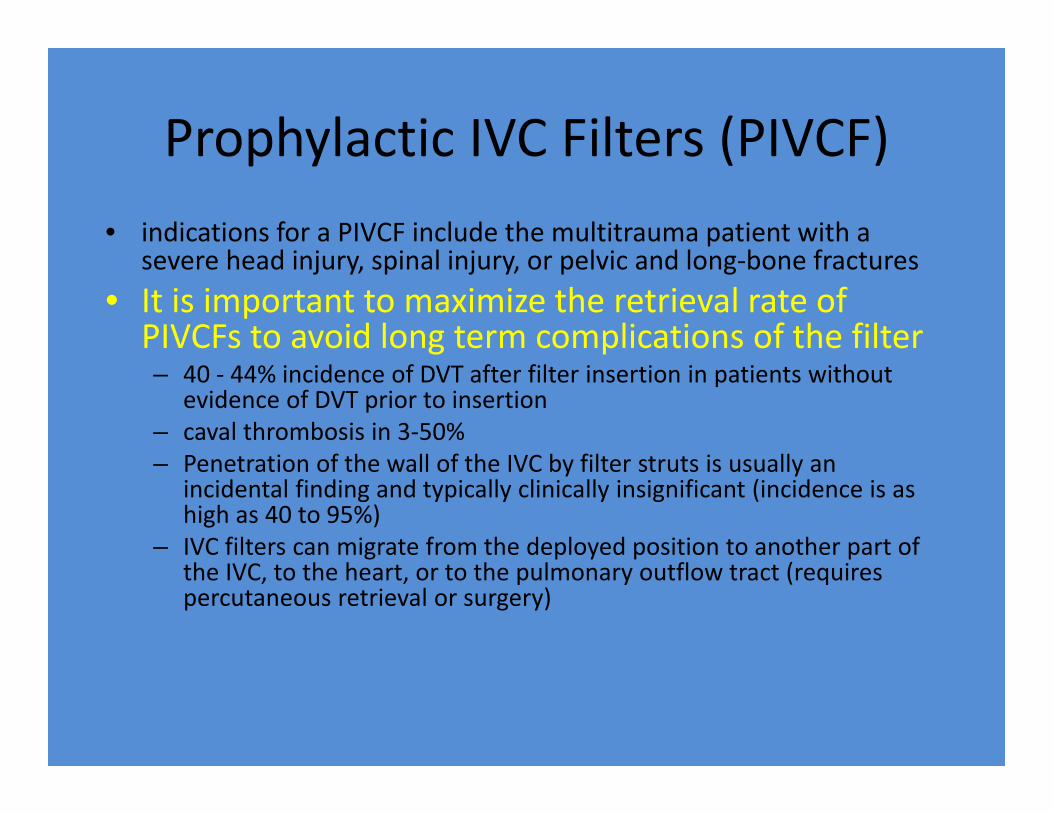
Prophylactic IVC Filters (PIVCF)• indications for a PIVCF include the multitrauma patient with a
severe head injury, spinal injury, or pelvic and long‐bone fractures• It is important to maximize the retrieval rate of PIVCFs to avoid long term complications of the filter– 40 ‐ 44% incidence of DVT after filter insertion in patients without
evidence of DVT prior to insertion– caval thrombosis in 3‐50%– Penetration of the wall of the IVC by filter struts is usually an
incidental finding and typically clinically insignificant (incidence is as high as 40 to 95%)
– IVC filters can migrate from the deployed position to another part of the IVC, to the heart, or to the pulmonary outflow tract (requires percutaneous retrieval or surgery)
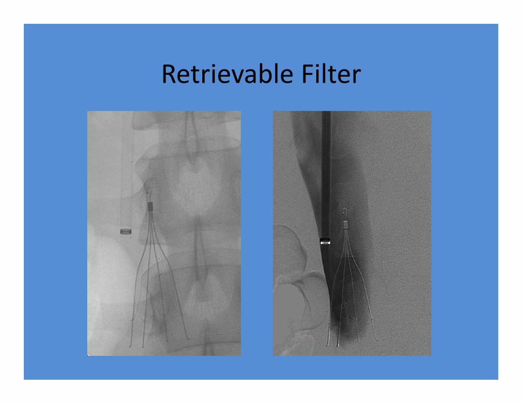
Retrievable Filter
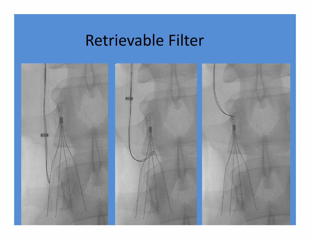
Retrievable Filter
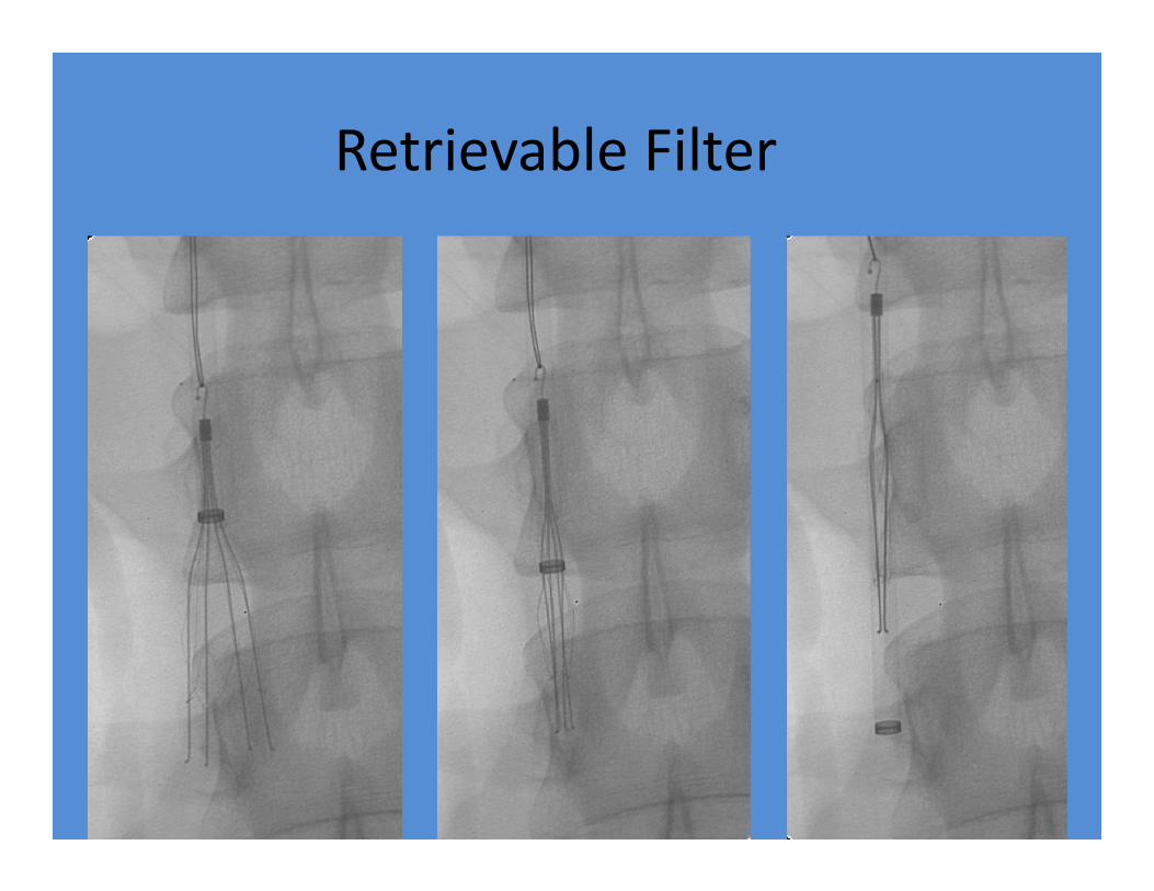
Retrievable Filter
Summary
• Many services available• Awareness of what IR can do for you• Communication & co‐operation important
Succinct online review of Interventional Radiology in Trauma:
http://emedicine.medscape.com/article/423295‐overview