Embed Size (px)
Citation preview
Intro to Mammography
Erinn Cooke, MD
Overview
• Screening recommendations• Patient positioning• Screening vs Diagnostic exams• BIRADS reporting• Case example
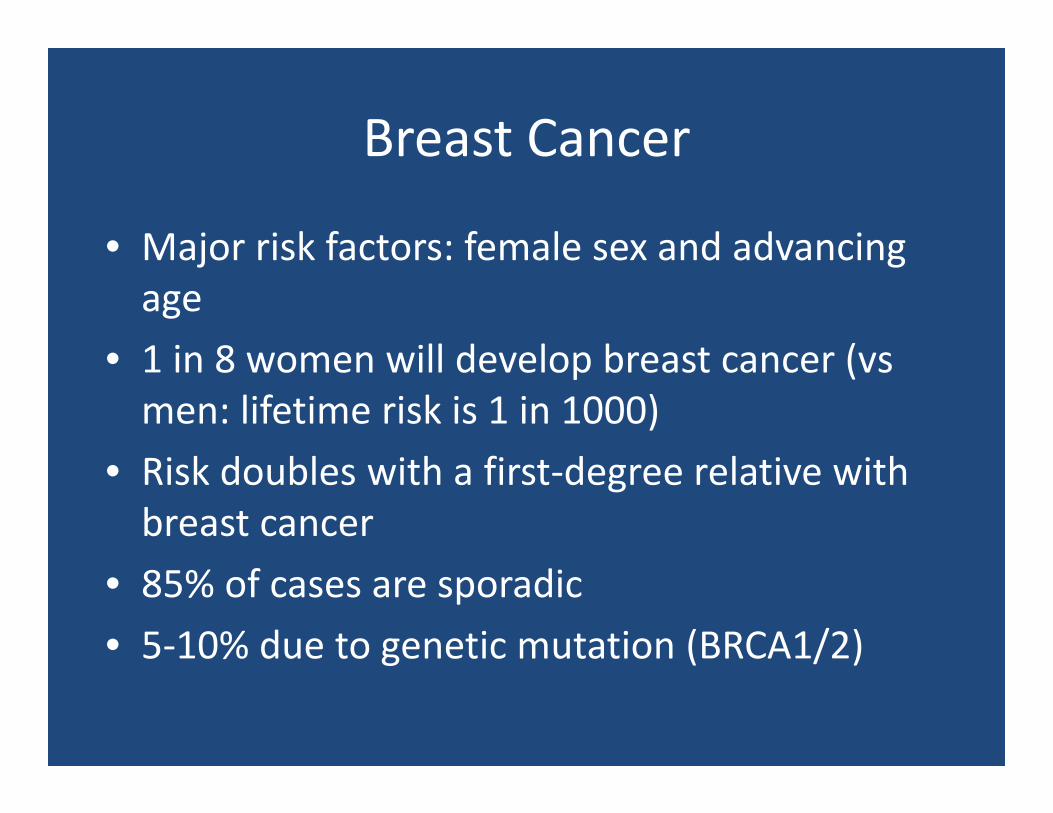
Breast Cancer
• Major risk factors: female sex and advancing age
• 1 in 8 women will develop breast cancer (vsmen: lifetime risk is 1 in 1000)
• Risk doubles with a first‐degree relative with breast cancer
• 85% of cases are sporadic• 5‐10% due to genetic mutation (BRCA1/2)
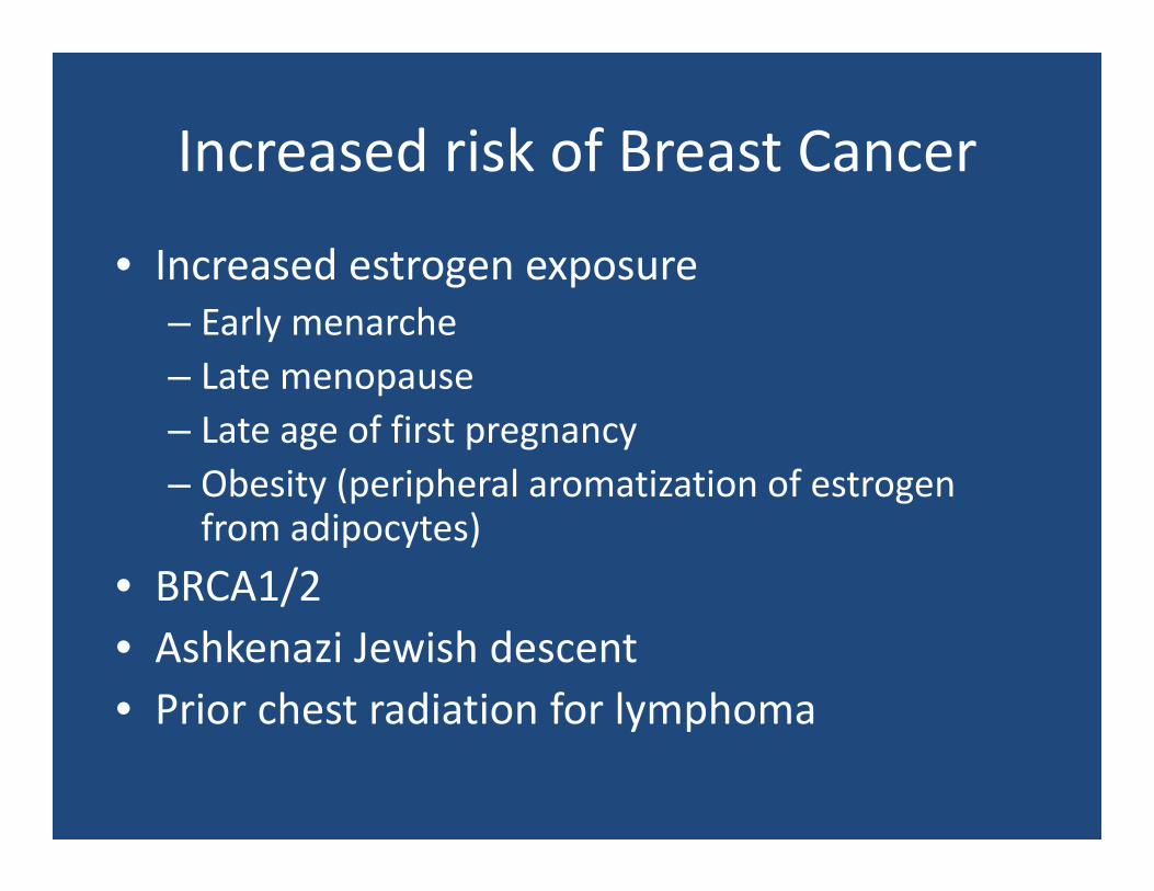
Increased risk of Breast Cancer
• Increased estrogen exposure– Early menarche– Late menopause– Late age of first pregnancy– Obesity (peripheral aromatization of estrogen from adipocytes)
• BRCA1/2• Ashkenazi Jewish descent• Prior chest radiation for lymphoma
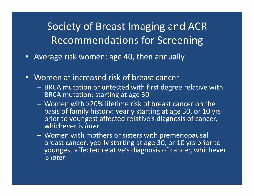
Society of Breast Imaging and ACR Recommendations for Screening
• Average risk women: age 40, then annually
• Women at increased risk of breast cancer– BRCA mutation or untested with first degree relative with BRCA mutation: starting at age 30
– Women with >20% lifetime risk of breast cancer on the basis of family history: yearly starting at age 30, or 10 yrsprior to youngest affected relative’s diagnosis of cancer, whichever is later
– Women with mothers or sisters with premenopausal breast cancer: yearly starting at age 30, or 10 yrs prior to youngest affected relative’s diagnosis of cancer, whichever is later
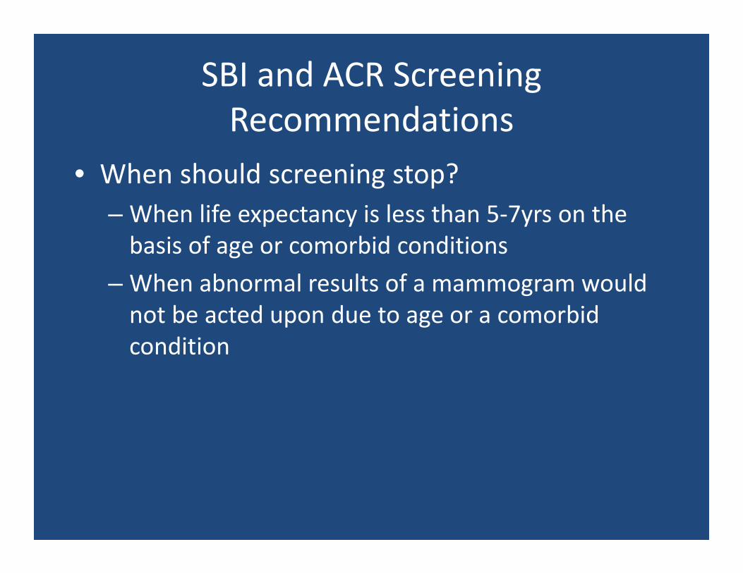
SBI and ACR Screening Recommendations
• When should screening stop?– When life expectancy is less than 5‐7yrs on the basis of age or comorbid conditions
– When abnormal results of a mammogram would not be acted upon due to age or a comorbid condition
High Risk Lesions
• LCIS: lobular carcinoma in situ– Associated with 6% of breast cancers– 90% occur in premenopausal women
• ADH: atypical ductal hyperplasia– Nonobligate precursor to DCIS– In women with positive family hx: RR of 9.7– Women age 20‐30: RR 7.0
High Risk Syndromes
• BRCA1/2– BRCA 1: up to 85% lifetime risk of breast cancer– BRCA‐2: up to 65% lifetime risk, but tends to occur at a later age
• Cowden Syndrome (PTEN gene)• Li‐Fraumeni syndrome (p53 gene)• Muir‐Torres syndrome (MSH2 and MLH1 gene)• Peutz‐Jeghers syndrome (STK11 gene)
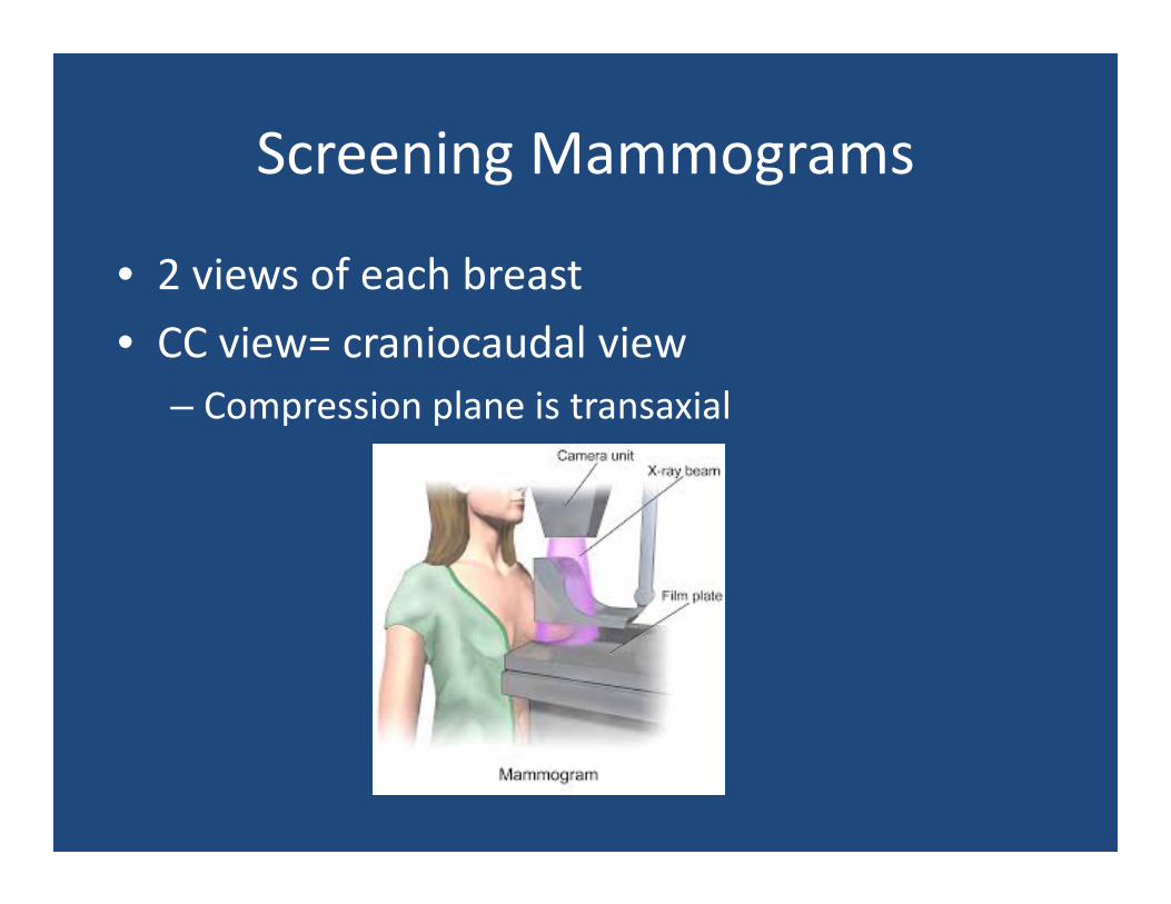
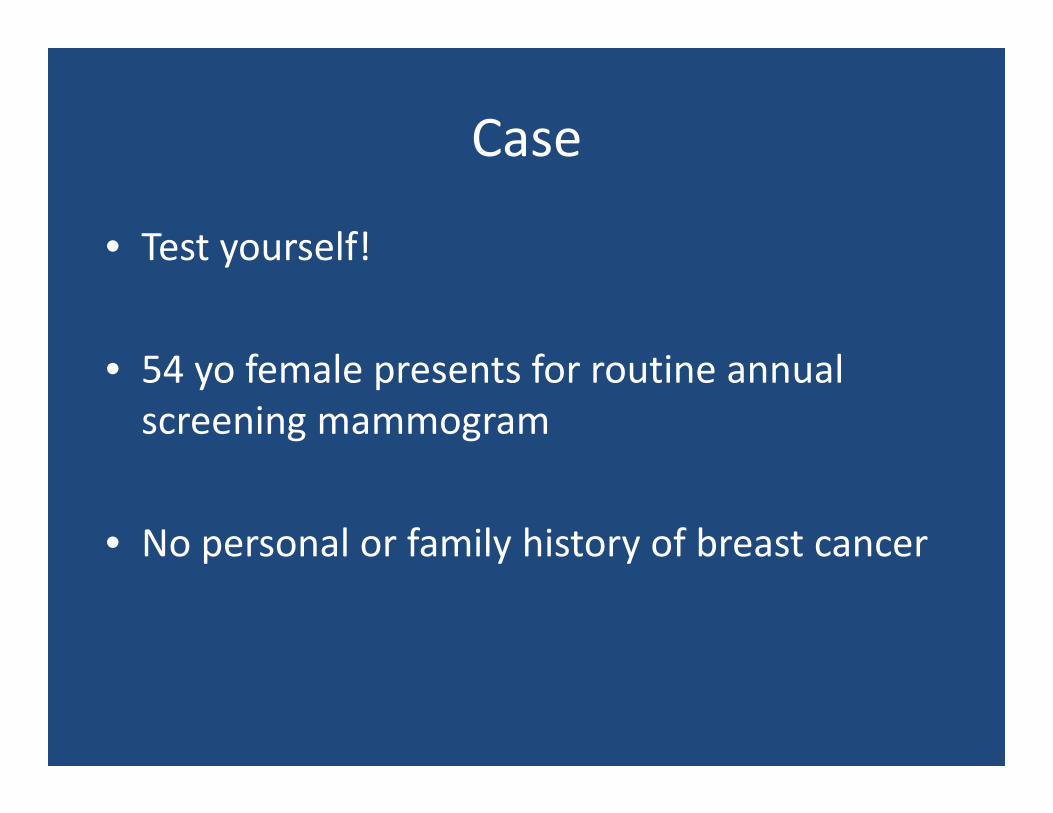
Screening Mammograms
• 2 views of each breast• CC view= craniocaudal view
– Compression plane is transaxial
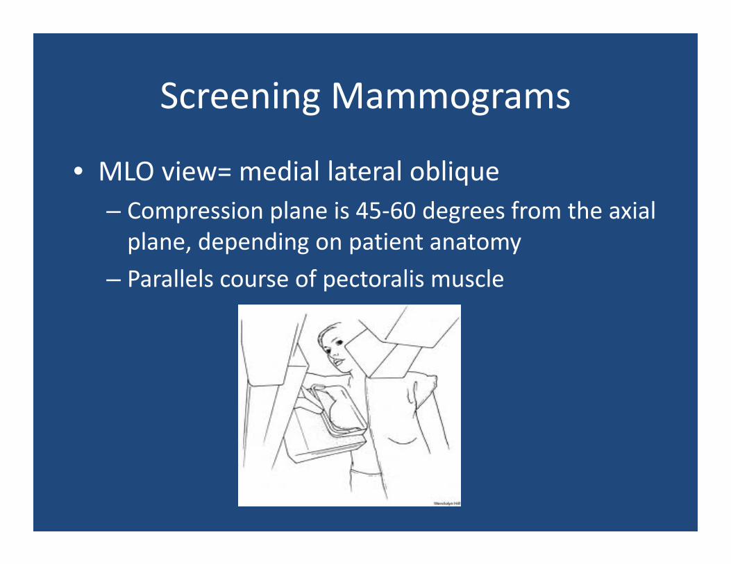
Screening Mammograms
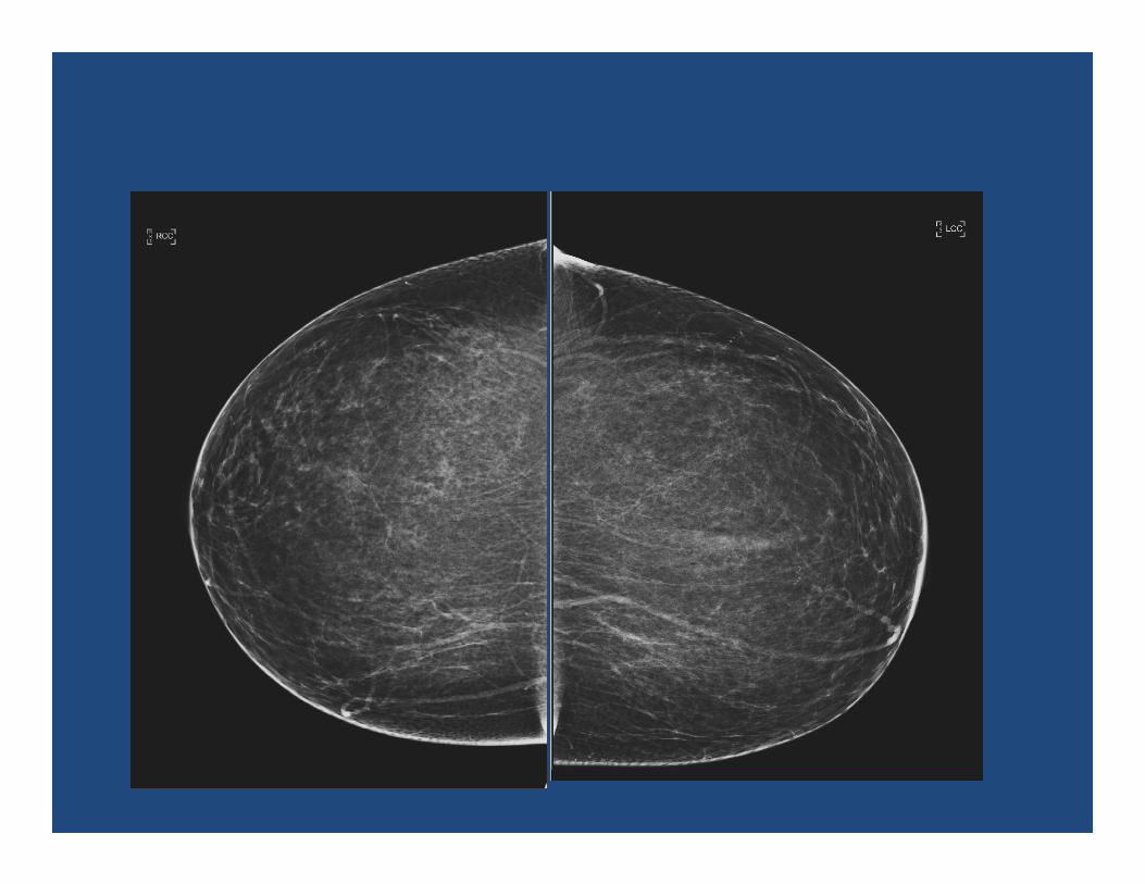
• MLO view= medial lateral oblique– Compression plane is 45‐60 degrees from the axial plane, depending on patient anatomy
– Parallels course of pectoralis muscle
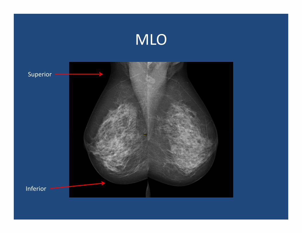
MLO
Superior
Inferior
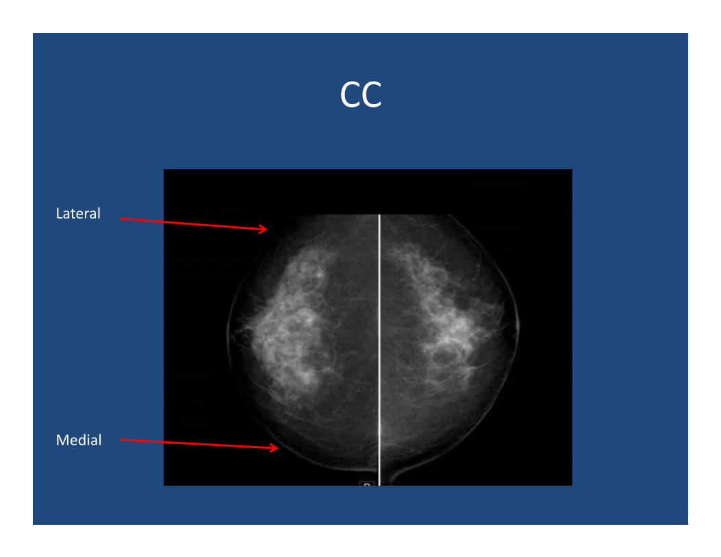
CC
Lateral
Medial
Additional Views
• Cleavage view: pulls in medial tissue of both breasts
• Exaggerated CC view: pulls in either lateral or medial tissue
• Implant displaced views
Diagnostic Mammogram
• Different from a screening mammogram!• Occurs when there is a breast “problem”
– the patient has a palpable lesion, nipple discharge, nipple inversion, etc.
– the patient is called back from a screening mammogram for additional views
– short interval follow‐up was recommended– personal history of breast cancer
Diagnostic Mammogram
• Can be one or both breasts• Can include CC and MLO view• Can include tomosynthesis• Spot compression• Magnification• Diagnostic work‐up often includes ultrasound
Spot Compression
• Used for an asymmetry or soft tissue lesion• Purpose is to “remove” overlapping tissue, and better characterize lesions
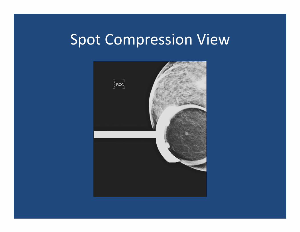
Spot Compression View
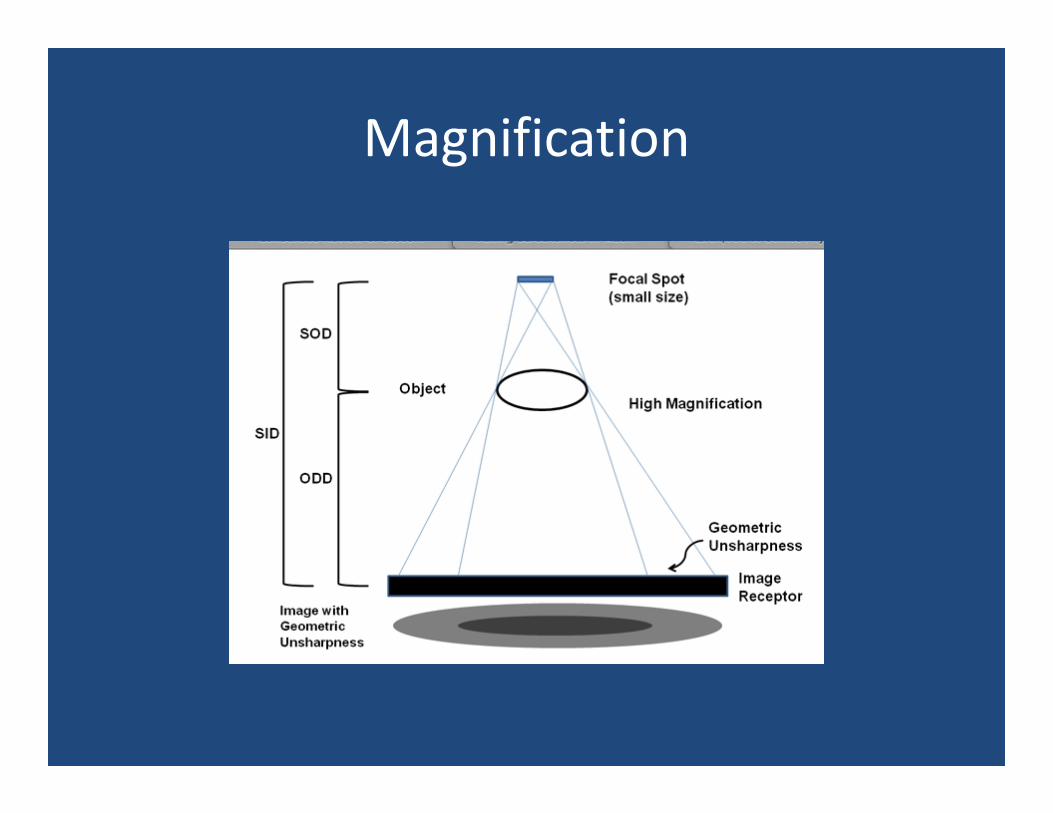
Magnification
• Used to evaluate calcifications
• Introduces an air‐gap– Reduces scatter
• Small focal spot
Magnification
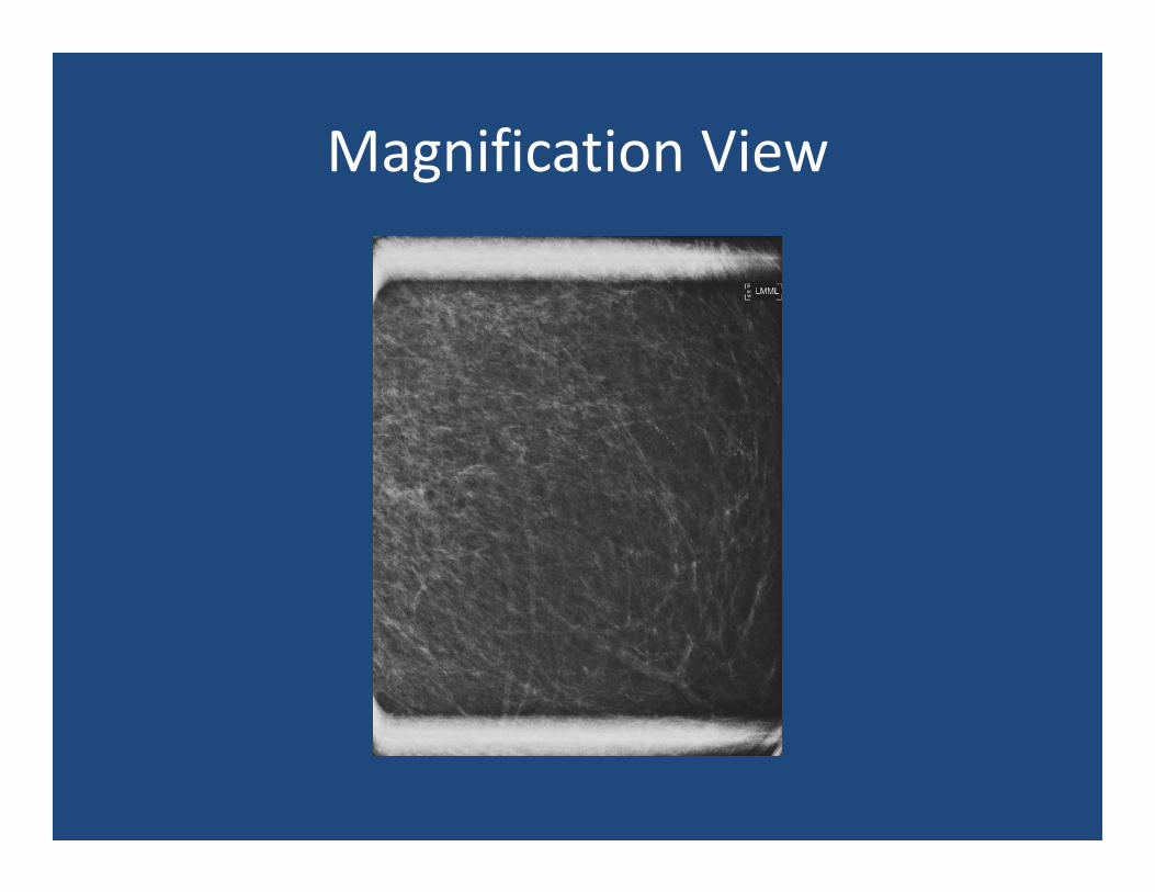
Magnification View
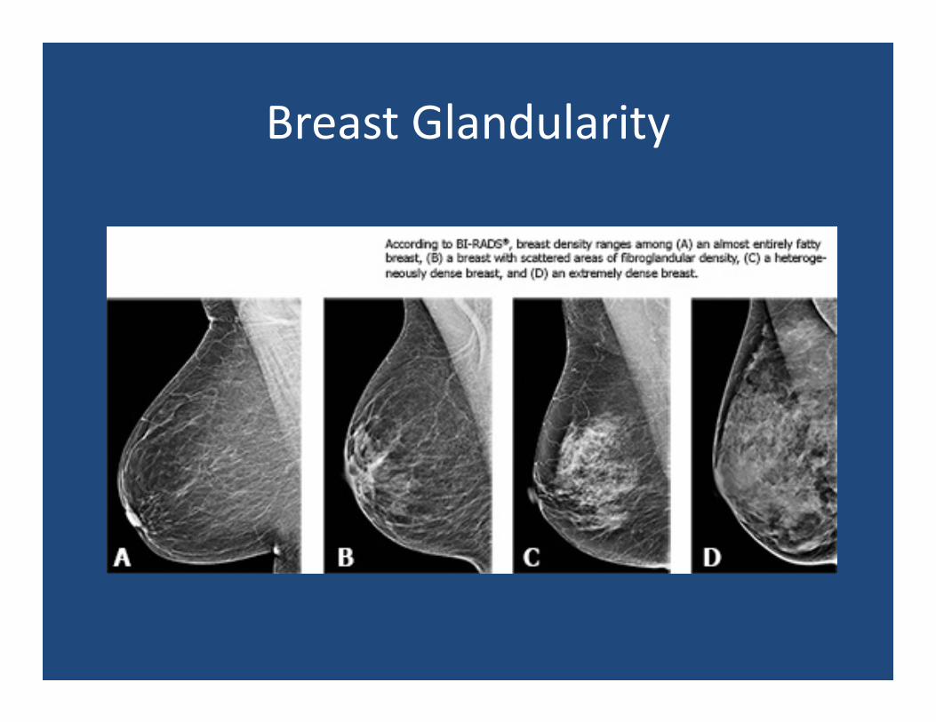
Breast Glandularity
• Screening mammogram reports should include a statement about breast tissue glandularity
• Almost entirely fat: <25% fibroglandular tissue• Scattered: 25‐50% fibroglandular tissue• Heterogeneously dense: 51‐75% fibroglandular
• Extremely dense: >75% fibroglandular
Breast Glandularity
BIRADS
• An overall assessment, assigned to a number category
• Include course of action (routine annual screening, recommend biopsy, short time interval follow‐up, etc)
BIRADS
• 0= incomplete. Additional imaging is needed– For screening mammograms ONLY
• 1= negative. Normal breasts• 2= benign
– Findings described in the report are benign. No additional workup or follow‐up is needed
• These are the only BIRADS categories for screening mammograms!
BIRADS
• 3= Probably benign. Short interval follow‐up is recommended– Lesions in this category have <2% chance of malignancy
– Never appropriate for a screening mammogram• 4= Suspicious. Biopsy is recommended
– 4a= low suspicion for malignancy– 4b= intermediate suspicion– 4c= high suspicion – Probability of malignancy ranges from >2% to <95%
BIRADS
• 5= Highly suggestive of malignancy– Probability of malignancy is >95%– Biopsy or surgical treatment is recommended
• 6= Known malignancy– Lesions have already been biopsied and proven as cancer
– Treatment plan is usually already in place
Case
• Test yourself!
• 54 yo female presents for routine annual screening mammogram
• No personal or family history of breast cancer
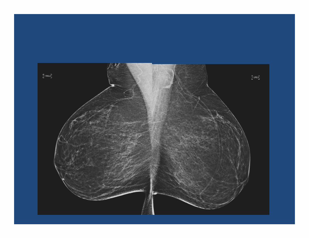
How would you characterize breast glandularity?
A. Almost entirely fatB. ScatteredC. Heterogeneously denseD. Extremely dense
How would you characterize breast glandularity?
A. Almost entirely fatB. ScatteredC. Heterogeneously denseD. Extremely dense

Case
• What would you describe in the report?
• Hint: compare to old studies!
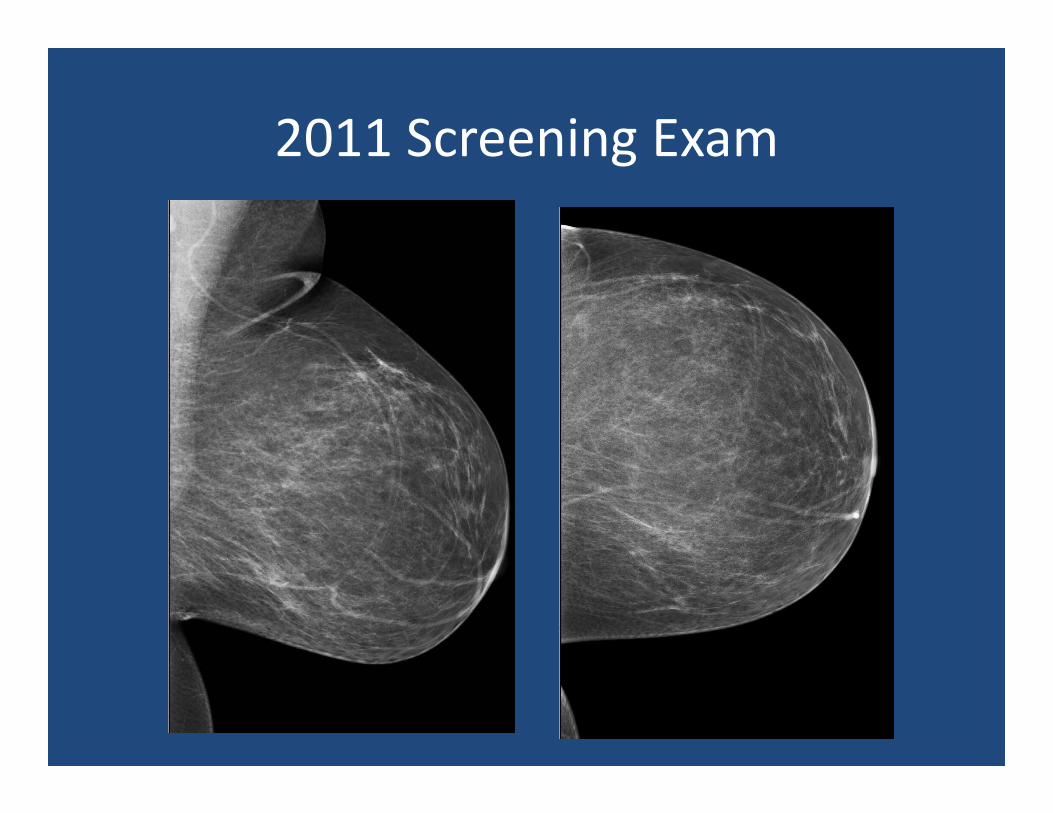
2011 Screening Exam

Current Screening Exam
Current Screening Exam
• There are new calcifications in the upper outer left breast
• Calcifications are linear, branching and in a ductal distribution
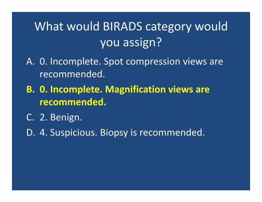
What would BIRADS category would you assign?
A. 0. Incomplete. Spot compression views are recommended.
B. 0. Incomplete. Magnification views are recommended.
C. 2. Benign.D. 4. Suspicious. Biopsy is recommended.
What would BIRADS category would you assign?
A. 0. Incomplete. Spot compression views are recommended.
B. 0. Incomplete. Magnification views are recommended.
C. 2. Benign.D. 4. Suspicious. Biopsy is recommended.
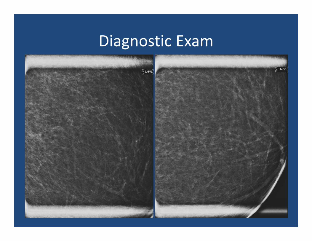
Diagnostic Exam
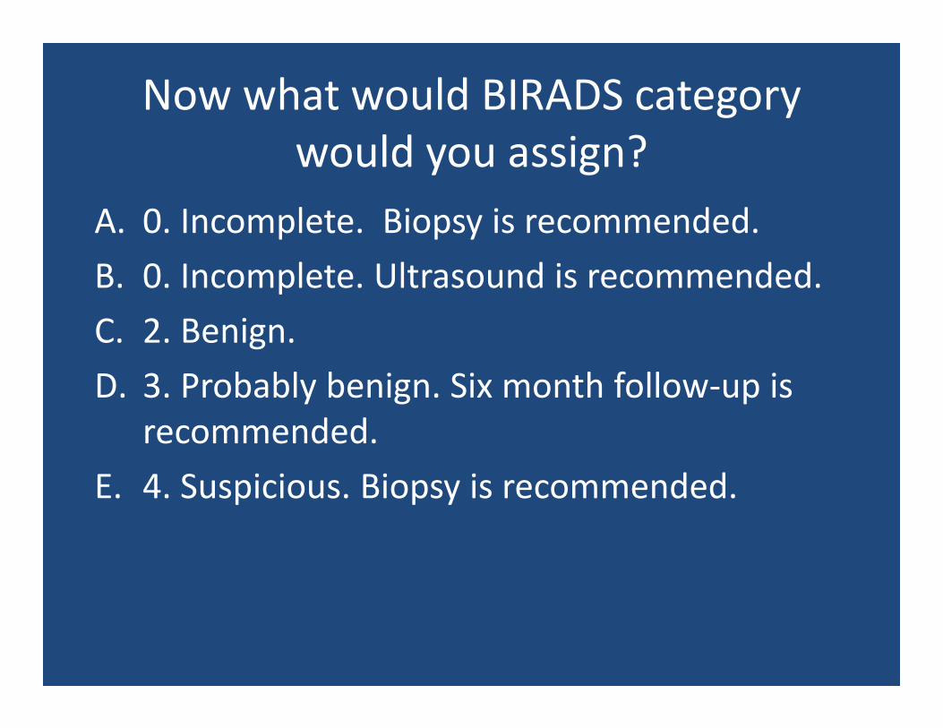
Now what would BIRADS category would you assign?
A. 0. Incomplete. Biopsy is recommended.B. 0. Incomplete. Ultrasound is recommended.C. 2. Benign.D. 3. Probably benign. Six month follow‐up is
recommended.E. 4. Suspicious. Biopsy is recommended.
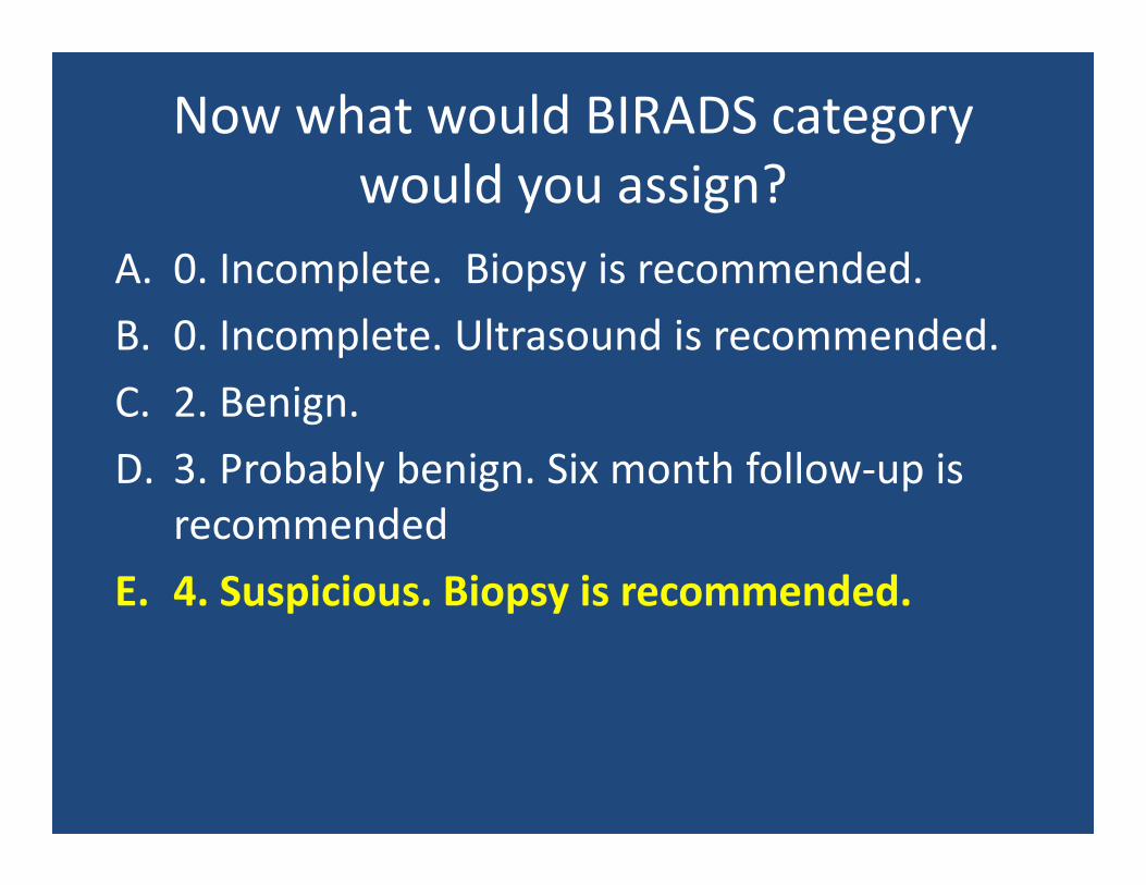
Now what would BIRADS category would you assign?
A. 0. Incomplete. Biopsy is recommended.B. 0. Incomplete. Ultrasound is recommended.C. 2. Benign.D. 3. Probably benign. Six month follow‐up is
recommendedE. 4. Suspicious. Biopsy is recommended.
Case
• Stereotactic biopsy was performed.• Path showed DCIS
References• Lee, CH et al. 2010. Breast Cancer Screening with Imaging:
Recommendations from the Society of Breast Imaging and the ACR on the use of Mammography, Breast MRI, Breast ultrasound and Other Technologies for the Detection of Clinically Occult Breast Cancer. Journal of the American College of Radiology. 7(1): 18‐27.