Embed Size (px)
Citation preview

INVESTIGATION OF CURING MECHANISM OF FLEXIBLE AQUEOUS POLYMER COATINGS AND EVALUATION OF
MONTMORILLONITES AS ANTI-TACKING AGENTS
Dissertation zur Erlangung des akademischen Grades des
Doktors der Naturwissenschaften (Dr. rer. nat.)
eingereicht im Fachbereich Biologie, Chemie, Pharmazie
der Freien Universität Berlin
vorgelegt von
MUHAMMAD IRFAN
aus Pakistan
September, 2011

1. Gutachter: Prof. Dr. Roland Bodmeier
2. Gutachter: Prof. Dr. Philippe Maincent Tag der mündlichen Prüfung: 7th September, 2011.

To my parents and family,
with love and gratitude

Acknowledgements
I very humbly thank to God, the Almighty, who provided me strength to complete this small
effort.
First of all, I am very grateful to my supervisor Prof. Dr. Roland Bodmeier for providing me
the opportunity to be part of his research team. I am so thankful to him for all his support
throughout my Ph.D. work and without his generous guidance and encouragement I would
not have been able to complete my research.
I am grateful to Prof. Dr. Philippe Maincent for co-evaluating this thesis. Thanks to Prof. Dr.
Heinz Pertz, Prof. Dr. Gerhard Wolber and Dr. Lothar Schwabe for acting as members of my
thesis advisor committee.
I cannot forget to thank Dr. Abid Riaz Ahmed and Dr. Andrei Dashevsky for their never-
ending support and fruitful discussions throughout my Ph.D. studies. Their scientific in put
helped me a lot to complete my doctoral studies.
It is also my honour to thank Higher Education Commission (HEC) of Pakistan for providing
financial support. In addition, I would like to thank German Academic Exchange Service
(DAAD) for its collaboration with HEC to maintain all records.
Thanks to all my colleagues; Dr. Mesut Ciper, Dr. Martin Körber, Dr. Burkhard Dickenhorst,
Dr. Zahra Ghalanbor, Dr. Katrin Steiner, Dr. Duangratana Shuwisitkul, Dr. Araya Raiwa,
Mirko Voigt, Julia Herrmann, Nutsawadee Apichatwatana, Muhaimin, Armin Hoessini,
Mohammad Gaith Zoubari, May Darwich, Anis Chaerunisaa, Jelena Teodosic, Barbara
Gröbner, Rebaz, Angelika Schwarz, Gabriela Karsubke, Stefan Walter, Eva Ewest and
Andreas Krause for their support and providing a friendly atmosphere during my stay at the
institute.
Lastly, I would like to express my deepest gratitude to my parents and the rest of family for
their love, their patience and their everlasting support throughout my whole life and if this
meant for them a little for me it is all.


Table of Contents 1 Introduction ................................................................................................... 1
1.1 General ....................................................................................................................... 1
1.2 Coating applications .................................................................................................. 1
1.3 Controlled release drug delivery systems ................................................................ 2
1.4 Controlled release coated dosage forms .................................................................. 4
1.5 Mechanism of drug release from coated pellets ...................................................... 7
1.6 Coating equipment and process parameters ........................................................... 9
1.6.1 Coating equipment ............................................................................................... 9
1.6.2 Process parameter ............................................................................................... 11
1.7 Polymer coating systems ......................................................................................... 12
1.8 Aqueous polymer dispersions ................................................................................. 15
1.8.1 Polymers ............................................................................................................. 16
1.8.1.1 Polyvinyl acetate ......................................................................................... 16
1.8.1.2 Acrylates ..................................................................................................... 16
1.8.1.3 Ethyl cellulose ............................................................................................. 17
1.8.2 Mechanism of film formation ............................................................................ 17
1.8.3 Additives ............................................................................................................ 20
1.8.3.1 Plasticizers .................................................................................................. 20
1.8.3.2 Pore-formers ............................................................................................... 20
1.8.3.3 Anti-tacking agents ..................................................................................... 21
1.8.4 Adhesive force of polymeric coatings ................................................................ 22
1.8.5 Curing ................................................................................................................. 24
1.8.6 Storage stability .................................................................................................. 25
1.9 Tackiness problems ................................................................................................. 27
1.10 Montmorillonites ..................................................................................................... 28
1.11 Research objectives .................................................................................................. 30
2 Materials and Methods ............................................................................... 31 2.1 Materials ................................................................................................................... 31
2.2 Methods .................................................................................................................... 33
2.2.1 Preparation of drug layered pellets ..................................................................... 33
2.2.2 Coating of pellets ............................................................................................... 33
2.2.3 Preparation of films ............................................................................................ 34
2.2.4 Preparation of model tablets ............................................................................... 34
2.2.5 Coating processability of polymer dispersions .................................................. 35

2.2.6 Characterization of the coated pellets ................................................................ 35
2.2.6.1 Curing of pellets .......................................................................................... 35
2.2.6.2 Tackiness of coated pellets ......................................................................... 35
2.2.6.3 In vitro drug release .................................................................................... 35
2.2.6.4 Macroscopic/SEM pictures and video monitoring ..................................... 36
2.2.6.5 Water uptake and weight loss ..................................................................... 36
2.2.6.6 Osmolality inside pellets ............................................................................. 36
2.2.6.7 Swelling studies .......................................................................................... 37
2.2.7 Characterization of films .................................................................................... 37
2.2.7.1 Water uptake and weight loss ..................................................................... 37
2.2.7.2 Permeability ................................................................................................ 37
2.2.7.3 Mechanical properties ................................................................................. 37
2.2.7.4 Tackiness of casted films ............................................................................ 38
2.2.7.5 Macroscopic pictures .................................................................................. 39
2.2.8 Characterization of the model tablets ................................................................. 39
2.2.8.1 Adhesive force of the coating ..................................................................... 39
2.2.9 Evaluation of curing effect ................................................................................. 40
2.2.10 Determination of contact angle .......................................................................... 40
3 Results and discussion ................................................................................ 42 3.1 Curing mechanism of Kollicoat® SR 30 D coatings .............................................. 42
3.1.1 Drug release from Kollicoat® SR 30 D coated pellets ....................................... 42
3.1.2 Water uptake and weight loss of coated pellets/free films ................................. 44
3.1.3 Mechanical properties and permeability of films ............................................... 45
3.1.4 Video monitoring of coated pellets during drug release .................................... 47
3.1.5 Swelling of coated pellets .................................................................................. 49
3.1.6 Osmolality inside coated pellets ......................................................................... 51
3.1.7 Drug release in a high osmolality medium ......................................................... 52
3.1.8 Determination of the adhesive force of coating ................................................. 54
3.1.9 Effect of drug loading ........................................................................................ 55
3.1.10 Effect of the type of drug layering (solution vs. suspension) ............................. 57
3.1.11 Curing as a function of time ............................................................................... 58
3.1.12 Conclusions ........................................................................................................ 60
3.2 Effect of starter core, drug solubility and drug loading on the curing effect of Kollicoat® SR 30 D coatings ............................................................................................... 61
3.2.1 Effect of starter core and drug solubility ............................................................ 61

3.2.2 Water uptake and weight loss of coated pellets ................................................. 63
3.2.3 Video monitoring of coated pellets during drug release .................................... 66
3.2.4 Effect of drug loading ........................................................................................ 69
3.2.5 Conclusions ........................................................................................................ 71
3.3 Approaches to overcome the curing effect of Kollicoat® SR 30 D coatings ....... 72
3.3.1 Seal coating of starter cores ............................................................................... 72
3.3.2 Improving the adhesion of coatings ................................................................... 73
3.3.3 Making the coatings brittle ................................................................................. 75
3.3.4 Use of pore-formers in coatings ......................................................................... 78
3.3.5 Long term storage stability ................................................................................. 80
3.3.6 Conclusions ........................................................................................................ 82
3.4 Curing mechanism of Eudragit® NE 30 D coatings .............................................. 83
3.4.1 Drug release and supportive studies ................................................................... 83
3.4.2 Additional investigated aspects .......................................................................... 85
3.4.3 Conclusions ........................................................................................................ 88
3.5 Evaluation of montmorillonites as anti-tacking agents ........................................ 89
3.5.1 Stability of the mixed dispersions ...................................................................... 89
3.5.2 Wettability of the MMTs and talc ...................................................................... 90
3.5.3 Tackiness of various polymers ........................................................................... 90
3.5.4 Effect of the MMTs and talc on tackiness .......................................................... 92
3.5.5 Effect of plasticizer concentration on tackiness ................................................. 95
3.5.6 Effect on Tg of polymers ................................................................................... 96
3.5.7 Coating processability of polymer dispersions .................................................. 97
3.5.8 Tackiness of the coated pellets during curing .................................................. 104
3.5.9 Mechanical properties of polymeric films ....................................................... 105
3.5.10 Drug release from coated pellets ...................................................................... 107
3.5.11 MMTs as external anti-tacking agent ............................................................... 109
3.5.12 Conclusions ...................................................................................................... 111
4 Summary .................................................................................................... 112 5 Zusammenfassung ..................................................................................... 116 6 References .................................................................................................. 121 7 Publications ................................................................................................ 141 8 Curriculum Vitae ...................................................................................... 142


Chapter 1. Introduction
1
1 Introduction
1.1 General
The coating of pharmaceutical dosage form started in the early ninth century B.C., with
Egyptian. The pills and hand shaped spherical mass containing drug, sugar and other diluents
were the primary dosage form at that time. These dosage forms were coated with a variety of
materials including talc, gelatin, sugar, gold and silver. Most of these coatings were
ineffective due to a chemical attack in the digestive tract.
The candy making industry was the first to develop and enhance the art of coating and later on
the pharmaceutical industry adapted the art of sugar coating for its own purposes. The first
sugar coated pills were developed in 1854 in Philadelphia, the USA. Enteric coatings were
started in 1880. In 1953, the first compression coated tablets were introduced and in 1954 the
foundation for the film coatings was developed (Johnson, 2007).
1.2 Coating applications
There exist numerous reasons for the coating of food and pharmaceutical products e.g.,
vegetables and fruits are being coated to protect them from temperature, moisture and attack
of microbes. Various types of oil based waxes, gums, gelatin, carbohydrates and proteins are
used to coat vegetables and fruits. For example, amylose ester of fatty acids/protein bilayer
has been used to coat the dried food to prevent stickiness (Grunnerson and Bruno, 1990).
The pharmaceutical dosage forms are also coated to protect the incorporated active
ingredients against light, oxygen, moisture, mechanical burden and the degradation in gastric
juice. Economic and safety reasons have to be taken into consideration such as product
appearance, identification, taste and odour masking of bitter drugs. A variety of release
profiles and mechanisms can be adapted: enteric coating, colon targeting, pulsatile release,
extended release or fast dissolving. In order to achieve the desired release profile, different
polymers have to be selected, which are characterized by different solubility and swelling
properties in water, gastric and intestinal fluids.

Chapter 1. Introduction
2
1.3 Controlled release drug delivery systems
Controlled release, sustained release, sustained action, prolonged action, extended action and
time release are the terms used to represent the systems that are designed to achieve a
prolonged therapeutic effect by slow and continuous release of active ingredients over
extended period of time (Coswar, 1974; Patwardhan and Das, 1983). This period may vary
from days to months (in the case of parenteral dosage forms) or hours (in the case of orally
administered dosage forms) (Urquhart, 1981).
Controlled release drug delivery systems (DDS) are defined by the FDA as those formulations
designed to release an active constituent at rates which differ significantly from those of their
corresponding immediate release forms (Gundert-Remy and Moller, 1990).The advantages of
controlled release delivery systems include:
reduced dosing frequency
reduced total amount of administered drug
increased safety margin of potent drugs
reduced incidence of local and systemic side effects and improved patient compliance
ability to maintain a constant level of active agent for a long period of time
On the other hand, the disadvantages of such systems are:
does not allow proper termination of therapy
has less flexibility in dose adjustment
significant patient variation and economical factors due to costly processes and
equipment involved during manufacturing
With regard to the mechanism applied to control the drug release, the design of oral controlled
release devices is based on one of the following categories:
diffusion-controlled systems
osmotically-driven systems
chemically-controlled systems

Chapter 1. Introduction
3
Diffusion-controlled systems
The diffusion–controlled systems can be further divided into membrane–reservoir systems
and monolithic-matrix systems. Reservoir systems are made up of a drug core (reservoir)
surrounded by a release rate controlling membrane, usually applied by a coating process. In
general, the rate controlling membrane consists of a polymeric material plus additives,
modifying the properties of the polymer (Chien, 1985). Those polymeric membranes are
insoluble in aqueous fluids, but can swell and are permeable for drug molecules allowing
diffusion of the drug through the membrane resulting in zero-order drug release, which can be
described by the following equation:
c = k • t
where k is the proportionality factor and c is the concentration of drug released at time t. This
relationship is valid if the following prerequisites are taken into consideration: undissolved
drug is present in the device, perfect sink conditions, constant surface, membrane thickness
and composition, negligible mass transfer coefficient for the portioning of the drug into and
out of the membrane.
In contrast, a matrix system consists of a drug homogenously distributed throughout a carrier.
The distributed drug may be in the form of monolithic solution or monolithic dispersion
depending on the solubility of the drug in the matrix. These systems can be prepared by a
variety of methods, e.g., compression, centrifugal pelletization, extrusion and solvent casting
or granulation. The drug release from these systems can be approximately described by a
square root of time kinetics (Higuchi, 1961; Higuchi, 1963).
Osmotically-driven systems
Osmotically driven systems use osmotic pressure as driving force for the controlled delivery
of drugs. A simple osmotic system consists of a core containing drug with or without
osmotically active agent, a semi-permeable (i.e. permeable only to water but not to solute)
membrane having an orifice for the release of drug. The drug release occurs through the
orifice by hydrostatic pressure (Theeuwes, 1985; Marucci et al., 2010; Muschert et al., 2009).
The drug release rate from a simple osmotic system can be described by the following
mathematical equation:
dM/dt = AK/h (Δπ˗Δp).C

Chapter 1. Introduction
4
where dM/dt is drug release rate, A is the membrane area, K is the membrane permeability, h
is the membrane thickness, Δπ and Δp are the osmotic and hydrostatic pressure difference
between the inside and outside of the system, respectively, and C is the drug concentration
inside the system.
Chemically-controlled systems
Chemically–controlled systems, such as drug-polymer complexes, ion exchange resins, and
prodrugs release the drug by a chemical reaction, e.g., hydrolysis, enzymatic digestion, and
ionic dissociation (Sriwongjanyna, 1996). The advantages of those systems include high drug
loading, simple drug release mechanism and good stability.
1.4 Controlled release coated dosage forms
Pellets
Pellet has narrated a variety of systematically performed, geometrically defined agglomerates
obtained from diverse starting materials utilizing different processing conditions. Pelletization
is an agglomeration process that converts fine powder or granules of bulk drugs and
Pelletization
agitation compaction layering globulation
balling compression spheronization/ powder solution/ spray spray extrusion suspension drying congealing
Figure 1: Classification of pelletization processes.
excipients into small, free flowing, spherical units, referred to as pellets. Pellets range in size
between 0.5-1.5 mm, though, other sizes could also be prepared, depending on the processing

Chapter 1. Introduction
5
technologies employed (Ghebre-Sellassie, 1989). The most widely used pelletization
processes in pharmaceutical industry are schematically illustrated in Figure 1.
Pellets exhibit less risk of local toxicity than tablets due to their uniform distribution
throughout the gastrointestinal tract (Khosla and Davis, 1990; Dressman et al., 1998). In the
case of single unit dosage forms, the destruction of the film would result in dose dumping and
loss of sustained release properties. The pellets are less susceptible to risk of dose dumping
compared to the reservoir type single unit dosage forms (Ghebre-Sellassie, 1989; Bodmeier,
1997). Due to improved safety and efficacy of drug, and having flexibility in design and
development of dosage form, the pellets are preferred over tablets (Daumensil, 1994;
Bodmeier, 1997). The advantages of multiple units over single unit dosage forms are enlisted
in Table 1.
Table 1 Comparison of single and multiple unit dosage forms
Single unit Multiple unit
Susceptible to risk of dose dumping
and local irritation
Gastric residence time is highly
influenced by food intake
Inter- and intra variability in rate and
extent of absorption
Restricted risk of dose dumping or
local irritation
Gastric residence time is less
influenced by food intake
More reproducible absorption
Since pellets are uniformly distributed throughout gastrointestinal tract, they invariably
maximize drug absorption, reduce peak plasma fluctuations, and minimize potential side
effects without appreciably lowering drug bioavailability.
Pellets provide immense flexibility during the development of oral dosage forms. Pellets
containing different drugs can be blended and formulated in a single dosage form. This helps
in delivery of chemical incompatible drugs, acting at the same or different sites within the
gastrointestinal tract. Additionally, the pellets of different release rate can be combined to
achieve desired release profile (Ghebre-Sellassie, 1989). Importantly, the drug loading up to
90% with good content uniformity is achievable (Mesiha and Valles, 1993). Other

Chapter 1. Introduction
6
technological advantages include low surface area to volume ratio, ideal shape for application
of film coatings, good flowability, low friability and narrow particle size distribution
(Reynolds, 1970).
Tablets
Tablets are defined as solid dosage forms produced by compaction of a formulation
containing the drug and certain filler or excipients selected to aid in the processing and
properties of the drug product. Compressed tablets are traditionally used for oral dosage
forms, since they are convenient, easy to apply, portable, and less expensive than other dosage
forms (Bandelin, 1989). The purpose of tablets is determined by their design.
Conventional film coatings are applied to improve product appearance, comfort of swelling,
to provide the moisture protection and unpleasant taste masking of the drug. Generally, prior
to the coating the immediate release tablet is formulated. Thus, tablet disintegration mainly
influences the effective surface area available for drug dissolution. In the case of immediate
release tablets, the quality and quantity of disintegrant play a vital role (Massimo et al., 2000).
Another important reason for the coating of tablets is to protect the drugs and excipients
sensitive to moisture (Rudnic and Kottke, 1996; Du and Hoag, 2001). Commonly,
hydroxypropyl methylcellulose (HPMC) is applied for this purpose (Plaizier-Vercammen and
De Nerve, 1993; Budavari et al, 1996) but also polyvinyl acetate (Kollicoat® SR 30 D) and
methacrylic acid copolymers (e.g., Eudragit® L 100) are being used for moisture protective
properties (Lehmann, 1997). Importantly, care has to be taken for the selection of coating
polymer, so that the resulting drug release kinetic within the stomach remains unaltered. Thus,
the moisture protective coatings must rapidly dissolve and/or rupture upon contact with the
release medium. Polymeric film coatings surrounding the drug-containing tablets can be
applied to mask the bitter and/or unpleasant taste of drugs leading to improved patient
compliance (Roy, 1994; Besse et al., 2001). HPMC is the most commonly used polymer for
this purpose (Nagai et al., 1997; Kokubo et al., 2001).To improve the taste masking
properties, ethylcellulose is also used as coating material (Bodmeier and Siepmann, 1999) and
frequently added to HPMC-based coatings (Lehmann, 1994; Siepmann et al., 2007). Also thin
acrylic films can be applied (Lehmann, 1994; McTeigue et al., 2002).

Chapter 1. Introduction
7
Soft gelatin capsules
Several advantages of soft gelatin capsules derive from the fact that the drug is in liquid form
or at least dissolved, solubilized, or suspended in a liquid vehicle. In addition, a higher degree
of homogeneity is possible in liquid systems that can not be in powder blends (Augsburger,
1995). Thus, the preparation of drugs into soft gelatin capsules can solve many troubles
involved in the tabletting (e.g., poor compaction, lack of content or weight uniformity, and
other powder flow or mixing problem). Soft gelatin capsules are appropriate for use with
compounds that are sensitive to oxidation or those that are photosensitive because the gelatin
shell can provide a barrier to oxygen and can be rendered opaque by use of opacifiers. As in
the solid state the photochemical process occurs on the product surface, a capsule and a tablet
have different light scattering characteristics and different ratios of surface area to volume
resulting in a variation in the photodegradation of the active principle (Tonnesen, 2001).
Another derived advantage from the liquid nature of the fill is an immediate release of the
contents with potential enhanced bioavailability. The proper choice of vehicle may promote
fast dispersion of capsule contents and drug dissolution (Augsburger, 1995). The most
commonly used vehicles for soft gelatin capsules fills are oils and polyethylene glycols.
Migration of water and plasticizer from the shell to shell could lead to crystallization of
poorly soluble drugs, as well as brittleness in the capsule shell. Thus, selecting appropriate
filling materials and shell composition is important in the design of soft gelatin capsule
dosage forms (Shah et al., 1992).
1.5 Mechanism of drug release from coated pellets
The major mechanism of drug release from coated pellets form is given by the solubility of
the polymer in the gastrointestinal tract. Additionally, the drug release is also influenced by
the pKa of polymer, coating thickness, and acidic and basic excipients of the pellet core. For
the water insoluble polymers, the drug release follows different phases. The medium first
hydrates/penetrates the coating, dissolve the drug and produces a saturated drug solution,
assuming sufficient drug in the pellet core. Then drug diffuses at a constant release rate out of
the core as long as a saturated solution is present.
Several possible mechanisms following this principle are given below (Dressman et al, 1995):
a) Solution/diffusion through the continuous plasticized polymer phase

Chapter 1. Introduction
8
This mechanism is followed by the systems that form a continuous phase in which plasticizer
and excipients are homogenously dispersed. This mechanism is favourable for the coating of
organic solution, which forms complete film without cracks and where the drug has relatively
high affinity for the polymer compared to water. The release rate can be described as under:
J= Pm/σ (Cs-Cb)
Pm = Dε/τβ K= D K
Where J is the flux, σ the coating thickness, Pm the permeability coefficient, Cs and Cb are the
concentration of drug at the drug-coating interface and the bulk respectively, D the molecular
diffusivity of the drug, K the distribution coefficient of the drug between polymer and fluid in
the core, ε the volume fraction of the chain openings, β the chain immobilization factor, t the
tortuosity factor and D the apparent diffusivity.
b) Solution/diffusion through plasticized channels
This mechanism seems to be unlikely, because the formation of continuous plasticizer
channels represents an extreme condition and the plasticizer will be mostly distributed more
or less uniformly throughout the polymer. Moreover, the drug solubility in the plasticizer
channels would have to be higher than in water.
c) Diffusion through aqueous pores
The drug releases through the aqueous pores when the coating is not homogenous and pores
are formed due to incomplete film formation. This usually happens in the case of aqueous
polymeric coating when unfavourable processing conditions are used. The permeability
coefficient Pp can here be expressed as:
Pp = εp Dp/τp
εp is the volume fraction of the aqueous channels and τp is the tortuosity of the aqueous
channels, Dp is the aqueous diffusivity of the drug.
d) Osmotically driven release
Drug release can be driven by an osmotic pressure difference between core and release
medium for a porous coating. This is important for highly water soluble core materials, such
as sucrose in nonpareils and highly water soluble drugs. The release rate of the drug through
pores of the coating can be described by:
J = K σ Δπ (Ci-Cm)

Chapter 1. Introduction
9
Where K is the filtration coefficient, σ the reflection coefficient of the coating, Δπ the osmotic
pressure difference, Ci and Cm the interior and media concentrations, respectively.
However, several studies stated that the overall drug release mechanism, acting with the
investigated dosage forms with water-insoluble polymers, is a combination of two or more of
the above mentioned mechanisms (Hoffman, 1986; Ozturk et al, 1990). Additional other
factors like swelling of the core or the film are capable to contribute to the release mechanism
of these systems. It was believed that swelling of the coating was accompanied by an osmotic
driven release mechanism for ethylcellulose-coantaining membrane (Hjartstam and Hjertberg,
1998).
1.6 Coating equipment and process parameters
1.6.1 Coating equipment
Several coating technologies are being used for the coating of solid cores (Mehta, 1989;
Christensen and Bertelsen, 1997). Conventional coating pans, which are traditionally used for
sugar coating, have been improved by different modifications for a better air flow, drying
capacity and coating uniformity. For example, perforated pans allow an intensive contact of
the drying air with the cores. Hi-coater (Vector), Accela Cota (Thomas) and Dria-Coater or
the Glatt are the famous coating machines.
Fluidized bed equipment is available for the coating of smaller cores, such as pellets, granules
and poweder. Different applied techniques include the top, bottom (Wurster) or tangentially
(rotary granulator) spraying mode (Jones, 1994; Deasy, 1991). A typical description of a
fluidized bed coater with top and bottom spray nozzles is given in Figure 2. The top spray
process is less efficient in film-coating than the Wurster and tangential process (Mehta et al,
1986) and also less effective in drug layering than rotary equipment (Iyer et al, 1993). The
rotary granulator technique allows pelletization and subsequent coating as a one step process
(Vecchio et al, 1998). These methods use water or organic liquids as solvents or dispersion
media which must be removed by drying during coating process.
In the hot melt coating technique the coating material is applied in a melted state onto the
substrate. The main advantage of this method is the reduced processing time, because no
solvent is required. Hydrophobic coating materials, such as waxes, fatty bases and
hydrogenated oils are used, which should have a melting point less than 80 °C and an

Chapter 1. Introduction
10
acceptable thermostability. Hot melt coating can be performed in a modified fluidized-bed
coater, where the melted material is delivered onto the substrate without solidification
(Achanta et al, 1997; Jozwiakowski, 1990).
Figure 2: A fluidized bed coater with top and Wurster bottom spary nozzles.
Dry coating is a novel coating method, which applies the micronized ploymer powder directly
onto tablets or pellets with a powder feeder, while simultaneously spraying a plasticizer. In
the first step, polymer particles are deposited onto cores and in the second step they are cured
by heating for a short time to achieve film formation. This technique was first developed for
the manufacturing of enteric coated dosage forms. It has the advantage of reduced processing
time and less need of energy due to the absence of solvent evaporation (Obara et al, 1999). A
special technique is the electrostatic tablet coating, which uses an electrical field for the
powder deposition onto core and afterwards particles are formed into a film by applying of
heat.
Another solvent free coating method is based on photocurable polymers. Wang and coworkers
used a liquid photocurable formulation based on norbone-endcapped polydimethylsiloxane,
which was applied onto nonpareil beads in a laboratory-scaled pan-coater. A relatively
impermeable coating was achieved by crosslinking the silicone based polymer upon
ultraviolet irradiation (Wang et al, 1995).

Chapter 1. Introduction
11
1.6.2 Process parameter
The manufacturing of coated dosage form is a complex process and homogenous film has to
be formed onto the core to control the drug release. In fact, several processes take place
simultaneously during the coating process. These include atomization of the spraying liquid
and droplet formation, contact and spreading over the surface of the substrate, evaporation of
the liquid and coalescence of the particles to form a film (Christensen and Bertelsen, 1997;
Jones, 1988). The air volume in fluid-bed processes should promote the particle movement,
prevent agglomeration and dry the coated substrate. Several studies have been made to
investigate and optimize critical process parameters for manufacturing of coated dosage
forms, which could have a significant influence on the release of controlled release dosage
forms.
Some critical parameters of fluidized bed coating are as under:
Fluidization air volume, which affects fluidization pattern and particle velocity
Fluidization air temperature, which is important for the evaporation of the solvent and
the softening of the latex particles, but is limited because of stickiness of the product
Fluidization air humidity, which should be kept constant to minimize batch-to-batch
variability of the coating process and should be limited to maximize the drying
efficiency of the coater
Solid application rate, which depends on the solid concentration and the liquid spray
rate, is restricted by over wetting and the tackiness of the coating
Atomization air pressure, which controls the droplet size and spraying pattern
Importantly, the properties of the substrate such as density, diameter and stickiness influence
the coating process (Christensen and Bertelsen, 1997). The flow of particles in a Wurster
column and the uniformity of particle coverage were investigated (Cheng et al., 2000; Cheng,
2000). The release profile was tremendously affected by difference in fluidization pattern and
velocities and it has been shown with different coater chamber geometries or spray mode
(Yang et al., 1992) or with beads of various sizes. The beads with high density and diameter
had thicker films and thus a slower drug release than smaller ones. The release rate was
shown to be directly proportional to the surface area of the coated cores (Rangarsson and

Chapter 1. Introduction
12
Johansson, 1988). Inappropriate process conditions during manufacturing of the coated
dosage forms can lead to stability problems.
Product temperature
The product temperature and a subsequent curing phase are of particular importance for
aqueous systems, in comparison to organic coating (Lorck et al., 1997). Inlet-air temperature
was found to be most critical variable in this regard (Parikh et al., 1993). A too low product
temperature can be one determinant, which can affect the completeness of coalescence. The
optimal product temperature for complete coalescence should be approximately 10-20 °C
above the minimum film formation. The complete coalescence to a homogenous film is only
possible, when the polymer chains are mobile enough to facilitate deformation and fusion.
Above the optimum temperature, too fast water evaporation on the pellet surface could lead to
spray loss. The time necessary for the capillary forces to achieve complete coalescence of the
particles could be insufficient, leading to a faster drug release. A low dispersion concentration
could lead to a slow drug release because the capillary forces could act longer, since more
water was available for evaporation (Laicher et al., 1993). Furthermore, the coalescence
temperature can be important for the mechanical properties of films, as shown for Surelease®
(Parikh et al., 1993; Obara and McGinity, 1995). Besides at high coating temperatures, the
problem of tackiness during coating can also occur because of interaction of drug with spray-
dispersion ingredients. This was pronounced, when ibuprofen pellets were coated with
Aquacoat® ECD dispersion already at low product temperature (Bodmeier and Paeratakul,
1994). The reason for this behavior could be the formation of a eutectic system with ibuprofen
and cetyl alcohol, the stabilizer of Aquacoat® ECD (Schmid, 2000).
1.7 Polymer coating systems
Polymer is generally dissolved in a suitable solvent, aqueous or non-aqueous for application
as a coating material. The high cost of organic solvent, high price of solvent recovery
systems, strict air quality controls, and environmental toxicity and explosiveness have
motivated pharmaceutical and food supplement to not use organic solvents during the coating
process. Presently, the water-insoluble polymers are available in a form which makes them
usable from aqueous systems. As a result, aqueous-based systems have been developed and
used instead of organic polymer solutions because of their environmental and economic
advantages. However, because water has a high heat of evaporation, aqueous-based systems
Chapter 1. Introduction
13
that might require processing times seemed initially to have a serious economical
disadvantage despite of their environmental advantages. In addition to the conventional
liquid-based systems, dry powder coating has been developed as an alternative technology. It
is generic designation for a variety of processes for applying a coating to substrate using
polymeric powder. One major advantage of this coating system is that it is environmentally
benign, producing none of the organic or aqueous waste streams, which normally are
presented in conventional liquid-based coatings. The continuous success for dry powder
coating is also related to its known economical advantages, including reduction in processing
time and in costs of environmental safety, compliance and energy use (Leong et al., 1999;
Obara et al., 1999).
Organic coating
Organic solvents are preferred over aqueous to form a continuous film especially for water
insoluble polymers. Alcohols (e.g., ethanol, isopropanol) are the mostly commonly used
solvents. As some polymers consist of structural units that form hydrogen bonds, the water is
good co-solvent. 3-5% of water is suggested to be added to a mixture of ethanol-acetone for
polymethacrylate copolymers (Lehmann et al., 1989). An important aspect is the relatively
high viscosity of polymeric solutions, which depends on the molecular weight and affinity of
the polymer to the solvents. High viscosity polymer solutions are obtained due to spreading of
polymer chain when the solvent has high affinity to the polymer chain. With the solvent
which has a lower affinity to the polymer, some polymer chain aggregation and shrinkage of
the polymer leads to lower viscosity. For that reason, the mixtures of the solvents can provide
better dissolution properties of the polymer as well as lower solution viscosity, in relation to
the concentration of the solid polymer.
For an organic polymer solution, a continuous film can be formed throughout the surface of
the substrate after the solvent is evaporated. In contrast to polymer latexes, polymer in organic
solution will form the film at room temperature, irrespective of the Tg of the polymer. The gel
formation is the most important phase, and the solvents that can not gel yield poor films,
which were indicated by poor transparency (Spitael and Kinget, 1980). Since the polymer
coils in good solvents will interpenetrate to a more compact structure than the poor one, the
compact molecules in solution will remain compact in the film state. It should be noted that
the sprayed films show comparatively a higher degree of porosity than the casted films,
Chapter 1. Introduction
14
because the droplet-like nature created during spraying, which remains apparent in the
resulting final film structure (Spitael and Kinget, 1977).
Using the mixed solvent system, it contains a good solvent of high evaporation rate under the
coating conditions. It is sometimes very useful in the mechanism of film formation upon
coating phase. The corresponding processes are as follows: firstly, a droplet of polymer
solution reaches the core surface; it has a high spreading tendency due to the low viscosity of
the diluted polymer solution, secondly, the more volatile solvent is a better choice for the
polymer due to its quick evaporation, the polymer solution in the poor solvent becomes less
sticky and gelation takes place at higher polymer concentration. However, there is also higher
tendency of the polymer to retain the solvent of higher affinity in the film (Lehmann, 1994).
Dry powder coating
Paint industries have been interested in the preparation of films from dry powder
formulations. Powder coating, which directly attaches polymer particles without using any
organic solvents, is a promising alternative approach. Compared to liquid-borne coatings, the
film formation process of dry powder coating is different since it is happening in the molten
phase. Softening, melting and cure completion are the principal stages in the film formation of
powder coating (Leong et al., 1999; Wulf et al., 2000; Pfeffer et al., 20001). A new alternative
procedure known as dry powder coating has been developed with potential applications for
film-coated dosage forms. In an effort to adjust to the new trends and enhance the versatility
of the polymer a novel coating technology with micronized hydroxypropyl methylcellulose
acetate succinate (HPMCAS) powder coating onto drug-loaded pellets and tablets was
developed by Obara and his co-workers (Obara et al., 1999). Compared to an aqueous
HPMCAS dispersion, the enterically coated pellets and tablets with HPMCAS powder require
a higher polymer amount for achieving gastric resistance. A big advantage of dry powder
coating is shorter processing time.
Aqueous dispersion coating
Polymeric material that is insoluble in water is commercially manufactured as aqueous
colloidal dispersion for extended release coatings. They are classified as true latexes or
pseudo latexes depending on the technique of production. With organic coating, the polymer
pattern is fully-entangled chains because the polymer is dissolved in the molecular state. The
film formation in organic coating process occurs due to simple loss of the solvent due to close
association of solvent to polymer. In contrast to aqueous dispersion systems where water

Chapter 1. Introduction
15
serves only as a carrier for the dispersed particles and not as solvent, the continuous phase
does not have a high level of interaction with the polymer molecules. Particle deformation
must occur in order to obtain a complete film formation (Osterwald, 1985; Iyer et al., 1990;
Sun et al., 1999).
1.8 Aqueous polymer dispersions
Aqueous polymer dispersions have been gaining a tremendous attraction for the coating of
solid dosage form for last two decades. They provide the possibility of applying water
insoluble polymer to substrates without using organic solvents, thus preventing the
environmental risks associated with them. Aqueous polymer dispersion coatings offer the
following advantages over organic polymer solution coatings:
Table 2 Comparison of organic solution and aqueous dispersion polymeric coatings
Organic coating Aqueous coating
Expensive process due to use of
organic solvents
High risks of environmental hazards
and toxicity
Not possible to coat high solid content
Economical process because water is
used as a solvent
No risk of environmental hazards at
all
High solid contents with low viscosity
can be applied
Aqueous colloidal polymer dispersions are classified as true latexes or pseudolatexes on the
basis of production technique. These latexes are prepared by emulsion of a monomer or by
emulsification of performed polymer. Different emulsification methods are available to
prepare colloidal dispersions. These methods include solution emulsification (solvent
evaporation), phase inversion and self emulsification. All these emulsion polymerization
processes require the addition of initiators that function by free radical, anionic or cationic
polymerization mechanisms. They are also stabilized with aid of anionic or nonionic
surfactants. The conversion of water insoluble polymers into colloidal aqueous polymer
dispersion was developed at emulsion polymer institute, Lehigh University under direction of

Chapter 1. Introduction
16
Dr. John W. Vanderhoff (Vanderhoff et al., 1979) and it was applied to pharmaceutical
polymers at Physical Pharmacy Department, Purdue University.
1.8.1 Polymers
1.8.1.1 Polyvinyl acetate
A new aqueous polymer dispersion, Kollicoat® SR 30 D, produced by an emulsion
polymerization process, was developed and is available with a solid content of 30%. It
consists of 27% polyvinyl acetate, 2.7% polyvinyl pyrrolidone (PVP) as a pore former, and
0.3% sodium laurylsulfate (Kollicoat® SR 30 D technical information, 2007). The dispersion
can be used for pH-independent extended release formulations or with thin film coatings for
taste masking purposes (Dashvesky et al., 1999; Kolter and Ruchatz, 1999). In blended films,
the polymer is used for the development of particular kinds of drug release systems, e.g. colon
targeting (Rock et al., 2000). If unplasticized, it has a MFT of 18 °C and results in brittle films
in dry state. Plasticizers are added to improve mechanical properties of the coating and the
final MFT depends on the type and amount of plasticizer added.
1.8.1.2 Acrylates
Acrylates are used in different industries. Poly (methyl methacrylate) is a well known
synthetic material, known as Plexiglas, which has important technical properties, such as
hardness, low specific gravity and an excellent long term-stability. For pharmaceutical
purposes, acrylates of different chemical compositions and solubility properties are available
under the trademark Eudragit® (Eudragit® technical information, 2010). (Meth) acrylate
copolymers, which are insoluble over the entire physiological pH range, are especially used
for extended release dosage forms.
Eudragit® NE 30 D
The neutral poly (ethylacrylate-methylmethacrylate) [poly(-EA-MMA)] with ratios of 2:1
(Eudragit® NE 30 D/ NE 40 D) is present in aqueous latex dispersions, produced by emulsion
polymerization. It is mainly used for transdermal formulation or buccal patches, wet
granulation and film coating. Soft films are formed from polymer dispersion without the need
of a plasticizer below room temperature. For coating, anti-tacking agents are useful to reduce
the stickiness of the polymer.

Chapter 1. Introduction
17
Eudragit® RL/RS 30 D
Cationic polymers of poly (ethylacrylate-methylmethacrylate) trimethylammonio
ethylmethacrylate chloride [poly(-EA-MMA-TAMCl)] with ratios of 1:2:0:1 (Eudragit® RS
30 D), and 1:2:0:2 (Eudragit® RL 30 D), respectively, are pseudo latexes with a solid content
of 30%. The colloidal polymer particles are stabilized by the positively charged quaternary
ammonium groups, which have chloride ions as counterions. Since Eudragit® RL 30 D
possesses the double amount of ionized groups; it is more hydrophilic and has a higher
tendency of swelling in water than Eudragit® RS 30 D. Drug release can be controlled from
coated dosage forms by the film thickness and by the mixing proportions of Eudragit® RL 30
D and RS 30 D, which determines the film permeability.
1.8.1.3 Ethyl cellulose
Ethylcellulose is a hydrophobic coating material used for controlled drug release, moisture
protection and taste masking. It is a semi-synthetic polymer manufactured from cellulose and
transferred with sodium hydroxide to alkali cellulose (Rekhi and Jambhekar, 1995).
Ethylcellulose is insoluble in gastro-intestinal tract and assures pH independent drug release
profiles due to its neutral side chains (Siepmann et al., 2007). It is widely used in oral drug
delivery as film former, since it is non-toxic, non-allergenic and non-irritant. Ethyl cellulose
water permeability is very low around one tenth of cellulose acetate (Bindschaedler et al.,
1983).
1.8.2 Mechanism of film formation
Film formation from aqueous polymer dispersions is entirely different from the conventional
organic coatings, where the polymer solution undergoes a sol gel transition upon solvent
evaporation to the final film. For latexes, it is complex and quality determinant in
pharmaceutical coating. Film-forming polymer latex is deposited from an aqueous colloidal
dispersion of discrete polymer spheres and the formation of a continuous film is then entirely
dependent on the minimum film formation temperature (MFT) of the polymer. The MFT is
defined as the minimum temperature at which latex-cast films become continuous and clear
(Lehmann, 1994; Keshikawa and Nakagami, 1994; O’ Donnnel and McGinity, 1997)
(Eckersley and Rudin, 1990; Steward et al., 2000). Typically, the coalescence of latex
particles takes place only above the MFT. In order to achieve MFT above the coating
temperature for the aqueous polymer dispersions, the addition of plasticizers is required
(Bodmeier et al., 1997). The plasticizers not only improve the flexibility and toughness of the

Chapter 1. Introduction
18
resulting films, but also soften the dispersed polymer particles and facilitate their deformation
and final coalescence (Harris and Ghebre-Sellassie, 1997).
The mechanism of film formation during drying is of theoretical and practical interest since
film properties are related to the performance of the resulting coatings (Vanderhoff, 1970;
Onion, 1986a; Eckersley and Rudin, 1990; Hogan 1995a; Wheatley and Steuernagel, 1997).
The general mechanism of latex film formation is as follows: compaction, deformation,
cohesion and polymer chain inter-diffusion. Each stage is characterized by the corresponding
phase of the latex layer on the substrate and the associated changes in the evaporation rate of
the aqueous dispersion medium.
Theoretical considerations
The film formation process of aqueous polymer dispersions can be described in three phases:
1) evaporation, particle concentration and ordering, 2) particle deformation, and 3) polymer
chain inter-diffusion across particle boundaries (Vanderhoff, 1970; Onion, 1986a; Eckersley
and Rudin, 1990; Hogan, 1995a; Wheatley and Steuernagel, 1997; Steward et al, 2000). A
description of film formation mechanism from aqueous polymer dispersion is elaborated
below in Figure 3.
Figure 3: Mechanism of film formation from aqueous colloidal polymer dispersion.

Chapter 1. Introduction
19
Phase1: Water evaporation and particle concentration
The first phase is the longest period of the three phases and last until the polymer has reached
approximately 60-70% volume fraction, or until the surface area of the latex’s liquid-air
interface starts to reduce as a consequences of solid film formation. Initially, the polymer
particles move freely with Brownian motion, colliding with one another and rebounding
elastically. Water evaporates from the latex surface, concentrating the latex surface and solid
contents. The ability of polymer particles to move about becomes more and more limited,
until some point they come into contact with one another and all particles motion ceases.
Phase II. Deformation of latex particles
This phase begins when the polymer particles come into contact, and iridescence may be seen
on the latex surface. The evaporation rate per unit of open wet latex remains constant, but the
overall rate of evaporation decreases significantly during the second phase. Since the drying
progresses further, the polymer particles are no longer mobile in the bulk latex and pack in an
ordered array, as a hexagonal close packed lattice. It has been suggested that hexagonal close
packing is theoretically possible, as well as cubic close packing. However, both shapes are not
easily distinguishable, since they share many geometrical features.
Phase III polymer chain inter-diffusion across particle boundaries
This phase starts with the initial formation of a continuous film. The remaining water leaves
the film initially via inter-particle channels and then by diffusion through the fused polymer
skin, but the rate of evaporation eventually slows down. It is during this final phase that soft
latex becomes more homogenous and gains its mechanical properties. As polymer chain inter-
diffusion takes place a process variously termed such as maturation, autohesion, or further
gradual coalescence (FGC) and particle interface tend to become less distinct. A drastic
change in film properties between phase II and III is that the initially brittle cohered particles
turn more ductile due to polymer chain entanglements.
In 1958, Voyutskii theory proposed that the surface tension forces (Dillon-Matheson-
Bradford) and capillary forces (Brwon) were inadequate to account for the physical properties
indicated by the latex films, instead, these resulted from conglomeration or “autohesion” such
as mutual inter-diffusion of free polymer chain ends across the particle-particle interface in
the coalesced film. As a result, the mutual inter-diffusion of polymer chain ends makes the
latex film homogenous and thus improves its physical-mechanical properties.

Chapter 1. Introduction
20
1.8.3 Additives
Different types of excipients are usually added to aqueous polymeric dispersion before
coating for several reasons. Most commonly used are plasticizers, anti-tacking agents and
pore formers. These excipients strongly influence film properties, coating process and release
rate of the coated dosage form. The coating formulations need to be optimized in order to
have optimized coating conditions.
1.8.3.1 Plasticizers
Plasticizers are usually high-boiling point organic solvents used to impart flexibility to brittle
and hard polymers. They generally act by reducing cohesive intermolecular forces within the
polymer chain leading to different changes in polymer properties. For instance, plasticizers
cause reduction in tensile strength, increase in elongation and flexibility and reduction in glass
transition temperature. The addition of plasticizers is required to reduce minimum film
formation temperature (MFT) below the coating temperature (Bindschaedler et al., 1983).
During the plasticization process, the plasticizer diffuses into polymer particles and promotes
the particle deformation and coalescence to form homogenous film. For good plasticization
effect, the plasticizer should be compatible with the polymeric particles.
Plasticizers, which are not compatible to polymers, can cause coagulation of the dispersion
and this phenomenon has been shown for ethyl acrylate/methacrylic acid copolymer
formulations (Kollicoat MAE 30 D) (Flöβer et al., 2000; Dangel et al., 2000). Therefore, the
selection of appropriate type and concentration should be made very carefully. The
concentration below the appropriate concentration could have anti-plasticizing effect (Guo,
1994; Guo et al., 1992). Addition of polyvinyl alcohol (PVA) to hydroxypropyl
methylcellulose (HPMC) films lead to increase in Tg, because of an increase in crystallinity
(Okhamafe and York, 1984).
1.8.3.2 Pore-formers
Pore formers are often added to the coating formulations to adjust the drug release of
extended release coatings. Commonly used pore-formers include 1) low molecular-weight
materials like sugars (e.g., sucrose, lactose, sorbitol), salts (e.g., sodium chloride, calcium
phosphate) and 2) hydrophilic polymers (e.g., polyethylene glycol, polyvinylpyrrolidone and
HPMC) or surfactants such as sodium lauryl sulfate (Muhammad et al., 1992; Li et al., 1990;
Erdmann et al., 2000). During dissolution, theses pore-formers leach out of the coating

Chapter 1. Introduction
21
membrane resulting in more permeable membrane and increase in drug release. The
concentration of the pore former can control the release kinetics, as shown for ethyl cellulose
coatings and different polysorbates as additives (Samani, 1999). The drug release from
Aquacoat coating was increased by using urea as pore-former which made the film more
porous (Appel and Zentre, 1991). Also the drug release from the Aquacoat coatings was
stabilized under stress conditions by adding pore-formers (Siepmann et al., 2007; Siepmann et
al., 2008; Muschert et al., 2009). Calcium phosphate is an example for an inorganic salt,
which is insoluble in dispersions with a neutral or alkaline pH. In the acidic artificial gastric
fluid, it is soluble and leaves a microporous membrane coating (Bodmeier and Paeratakul,
1991; Bodmeier and Paeratakul, 1990).
In a recent study, Aquacoat ECD and Kollicoat SR 30 D dispersions were found to be stable
with up to 50% addition of polyvinylpyrrolidone and polyvinyl alcohol-polyethylene glycol
graft copolymers (Dashevsky et al., 2010).
1.8.3.3 Anti-tacking agents
Anti-tacking or separating agents and pigments are commonly added to aqueous polymer
formulations to reduce agglomeration or sticking of coated particles during the coating
process. Talc, magnesium stearate, glyceryl monostearate (GMS) and titanium dioxide are
most commonly used pigments as anti-tacking agents in aqueous film coating.
Talc is used as an anti-adherent agent, which is known to have a tendency for sedimentation
and blocking of nozzles during the spraying process. Therefore, the coating dispersion should
be continuously kept under stirring during the coating process. The amount of pigments in the
aqueous dispersion must be optimized without exceeding the maximum carrying capacity of
the polymer or critical pigment volume concentration (CPVC). The pigment concentration has
a strong influence on the final film properties such as a mechanical strength and permeability
(Patton, 1979). An increase in drug release rate was found, which was explained with the
adsorption capacity, the high specific surface area and the high affinity of colloidal silica for
polar components like water (Vecchio et al., 1995). Polymer dispersions can have a high
binding capacity for pigments. To prevent sticking, up to 200% talc, based on dry polymer
mass, were incorporated into coatings of Eudragit® RL/RS 30 D plasticized with high levels
(up to 30%) of TEC (Maejima and McGinity, 2001).
GMS was shown to have a ten-time higher anti-tacking effectiveness than talc and can be
used at lower concentrations. The addition of talc and GMS lead to decrease in flexibility of

Chapter 1. Introduction
22
and the effect was increased as a function of concentration (Wesseling and Bodmeier, 1999;
Peterit et al., 1999). Magnesium stearate was effective as anti-tacking agent for
methylcellulose coated granules but it strongly increased the drug release by making film
rough. An orange peeling effect of film coating was observed, when the granules swelled in
the release medium (Wan and Lai, 1993). During coating, fine particles have a particular high
tendency of agglomeration. This could be reduced by adding NaCl to an aqueous spray
solution of hydroxyl propylcellulose (Fukumori, 1993). It was suggested that the suppression
of agglomeration was caused by a reduction of viscosity of the spray solution through salting
out of the polymer (Yuasa, 1997).
1.8.4 Adhesive force of polymeric coatings
Adhesion force between a polymer and substrate is very important pre-requisite for the coated
dosage forms (Nadkarni et al., 1975; Rowe, 1977; Felton and McGinity, 1999). The decrease
in adhesion force due to accumulation of moisture at coating-substrate interface potentially
affects the mechanical protection and the stability of the coated dosage forms (Okhamafe and
York, 1985). Therefore, to have a stable product over long period of time, the adhesion force
between coated polymer and substrate should not be changed during storage.
Generally, the adhesion of the polymer-substrate have been affected by two major forces 1)
the strength of interfacial bonds and 2) the internal stresses within the film. The factors that
affect these two parameters will also have a significant influence on the adhesion of the
coatings. A distinction must be made between “fundamental” and “practical” adhesion. The
former refers to the intermolecular interactions between the polymer and the substrate (Mittal,
1980) and the latter refers to the numerical value that results from a variety of testing
methods, including shear and tensile tests.
The earliest method for determine the adhesive force of polymeric coating to substrate
surfaces was the “Scotch tape” test where a piece of adhesive tape was applied to the coating
surface and then peeled off (Strong, 1935). It was obviously qualitative method and did not
provide accurate measurement of adhesive force. However, the first quantitative method to
determine the adhesion force was developed by Heavens in 1950. In this method, the tip of
hard stylus is drawn across the surface of film and critical load required to completely detach
the film from substrate is determined and related to polymer adhesion. Another important way
of determining quantitative adhesive force is the “butt adhesion” method. In this method,
double sided-adhesive tape is placed between the tablet surface and the upper plate and a

Chapter 1. Introduction
23
uniform displacement rate should be used to remove the film from substrate (Rowe, 1980).
Felton and McGinity used a Chatillon digital force gauge and motorized test stand to conduct
butt adhesion experiments (Figure 4). The apparatus was connected to a personal computer
and force-deflection diagrams were constructed from the data, which allowed the
visualization of the development of the force within the sample during the adhesion
experiments.
Figure 4: Schematic representation of a butt adhesion test.
The adhesive force can also be affected by the shape of the tablets. The force required to
remove the film from the surface of the biconvex tablets was lower than the same films coated
onto flat-faced tablets (Rowe, 1977). However, the flat faced tablets are preferred due to
proper sticking of adhesive tape (Fisher, 1976; Lehtola, 1995; Felton and McGinity, 1996).
Felton and McGinity used flat-faced punches with a beveled edge to achieve a more uniform
adhesion of the polymeric film.
Film thickness could also influence the adhesive force. It was shown that for films up to a
thickness of 35 µm resulted in decreased adhesion of an organic-based cellulosic polymer,
while films greater than 35 µm in thickness exhibited increased adhesion with increased film
thickness and similar results were reported for aqueous and organic-based hydroxypropyl
cellulose and aqueous-based acrylic polymeric films (Felton and McGinity, 1996; Johnson,
1986).

Chapter 1. Introduction
24
The surfactants are used to enhance the wettability of polymer dispersions on tablets (Felton
et al., 1997). The adhesion of polymeric coatings could also be affected by the drug-excipients
and drug polymer interactions (Sarisuta et al., 2006; Khan and Fell, 2001).
1.8.5 Curing
The process in which coated dosage forms are stored at elevated temperatures to promote
further gradual coalescence of the film is known as curing. It can also be defined as the input
of energy into film-coated system after the desired film coat level is applied (Hamed and
Sakr, 2003). Curing of film-coated dosage forms is an important component in the film-
formation mechanism from aqueous latexes. During coating process, the curing takes place to
a certain extent itself. However, this is fairly inadequate. To assure the completion of
coalescence, the dosage form is generally exposed to elevated temperature after the coating.
This can be done in the coating machine using a process known as post-coating fluidization
(Harris and Ghebre-Sellassie, 1986) or by placing the coated dosage forms in an oven
(Goodhart et al., 1984; Lippold, et al., 1989).
As the film formation process from the aqueous polymeric dispersions depend on capillary
forces to draw together and deform the latex particles and is highly influenced by the amount
of water in the polymeric film and environmental temperature. The increased temperature and
amount of water in the polymeric films decrease the Tg of colloidal particles, resulting in an
increased mobility of the polymer chains, which in turn enhances the further gradual
coalescence of the latex particles. As a result, better film formation takes place. On the other
hand, decreased temperature and reduced amount of water in polymeric film will not produce
enough capillary forces to bring together and deform latex particles, which in turn results in
incomplete film formation. Therefore, in order to achieve complete film formation, which
facilitates to have stable release profile, the proper curing conditions are required. This is
known as “conventional curing”. This conventional curing is most commonly recommended
for the aqueous polymers, which have high glass transition temperature (Tg) and minimum
film formation temperature (MFT). Heating of the films above Tg facilitates polymer
movement and relaxation. For example, curing at 60 °C for at least one hour was
recommended for Aquacoat® ECD coated dosage forms by the manufacturer (FMC, the USA)
and it has also been reported in literature (Bodmeier and Paeratakul, 1991; Gilligan and Wan,
1991).

Chapter 1. Introduction
25
The curing process is dependent on both the time and temperature used during the curing
process. The curing rates can be accelerated by increasing the storage temperature and relative
humidity because of fast kinetic factors responsible for coalescence (Dressman et al., 1995;
Körber et al., 2010; Amighi and Moes, 1996b; Bianchini et al., 1993).
Drug release from Kollicoat® SR 30 D coated pellets was unchanged by increasing the curing
time (Dashevsky et al., 2005). This was attributed to complete film formation during coating
process due to a low MFT of plasticized Kollicoat® SR 30 D coatings. In contrast, a strong
curing effect depending on the plasticizer type and curing conditions has also been reported
with Kollicoat® SR 30 D coated pellets (Shao et al., 2002).
A curing at 60 °C for 8 h was found to be sufficient to form complete film with Aquacoat®
ECD coated pellets (Wesseling and Bodmeier, 2001) which could be further minimized by
increasing the plasticizer concentrations (Bodmeier and Paeratakul, 1994b; Amighi and Moes,
1996a).
Additionally, the controlled humidity can accelerate the curing step significantly. This
happens because water facilitates polymer particle coalescence and it acts as plasticizer for
many polymers (Liu and Williams, 2002b; Williams III and Liu, 2000).
The extent of curing effect can also be affected by the type of plasticizer and coating level.
For example, drug release decreased with increasing harshness (time, temperature and relative
humidity) of curing conditions, when using triethyl citrate as a plasticizer whereas with
dibutyl sebacate and Myvacet this relationship was only seen at low coating levels (Yang et
al., 2010).
Furthermore, the drug migration into the coatings can also occur during the curing step which
results an increase in drug release rather than decrease. In order to overcome this problem, a
sub coating was applied between drug layer and polymer coating (Hamed and Sakr, 2003).
With Eudragit NE 30 D coated pellets, the crystallization of surfactant occurs depending on
the curing conditions (Bajdik et al., 2003). It has also shown that Eudragit NE 30 D coatings
are more sensitive to change in release profile at low curing temperatures (Kucera et al., 2009;
Lin et al., 2003).
1.8.6 Storage stability
The purpose of long term stability is to provide evidence on how the quality of a drug
substance or drug product varies with time under the influence of a variety of environmental
factors such as temperature, humidity, and light, and to establish a re-test period for the drug

Chapter 1. Introduction
26
substance or a shelf life for the drug product under recommended storage conditions
(European medicine Agency; ICH guidelines). The long term stability data is particularly
needed for the registration of new chemical entities. In general, a drug substance should be
evaluated under storage conditions (with appropriate tolerances) that test its thermal stability
and, if applicable, its sensitivity to moisture. The storage conditions and the length of studies
chosen should be sufficient to cover storage, shipment, and subsequent use. Different storage
conditions are recommended by international committee on harmonization (ICH). For
example, 25°C ± 2 °C/60% RH ± 5% RH for 12 months, 30 °C ± 2 °C/65% RH ± 5% RH for
6 months and 40 °C ± 2 °C/75% RH ± 5% RH for 6 months are less, intermediate and
accelerated long term stability conditions respectively.
Contradictory results have been reported in the literature regarding the effect of long-term
storage on drug release from coated dosage forms. The ibuprofen release from coated pellets
was increased after storage (Bodmeier and Paeratakul, 1994) and theophylline release was
decreased upon storage (Yuen et al., 1993; Goodhart et al., 1984; Bando and McGinity,
2006).
The change in release profile over long term storage can be caused by the numerous factors.
For instance, incorporation of inadequate amount of plasticizer in the formulation can result in
polymer films that are brittle and need longer curing times to exhibit stable films. It has been
shown that theophylline release from coated pellets with Eudragit RS 30D containing 10% or
20% triethylcitrate (TEC) depended on plasticizer concentration (Amighi and Moes, 1996b).
Another reason could be the physical instabilities in the coating that leads to cracks and
chipping of the coating. In addition, the researchers have attributed these problems to an
increase in water contents of the films rather than a decrease (Chowhan et al., 1982). In the
case of Aquacoat® ECD coatings, the faster drug release was associated with brittle films or
the formation of micro-ruptures in the film during storage (Wesseling and Bodmeier, 2001).
The decrease in the free volume of the film and increased compaction of polymer structure
due to further gradual coalescence as the ageing progresses was also reported one reason for
unstable release profile (Guo et al., 1993). This could also lead to change in water vapor
permeability of the films (Guo et al., 1991). Additionally, the presence of endogenous
excipients in the aqueous polymeric dispersion can also lead to serious stability issues such as
increase in drug release rates. It has been shown that crystallization of the surfactants affects
the dissolution rate of the drug from coated pellets (Amighi and Moes, 1997, Bajdik et al.,
2003).

Chapter 1. Introduction
27
The decrease in drug release was reported upon storage under elevated humidity (Siepmann et
al., 2007; Wu and McGinity, 2000) which was linked to better film formation due to further
gradual coalescence resulting in decreased permeabilities for water and drug.
The storage at 40 °C-75% RH of Kollicoat® SR 30 D coated pellets also resulted in a
decreased drug release due to continuous film formation (Shao et al., 2002). In contrary, an
extended lag time without any significant change in release profile was observed with
Kollicoat® SR 30 D coated pellets upon 1 month storage at 40 °C-75% RH (Ensslin et al.,
2008).
On the contrary, storage stability at 40 °C-75% RH from Aquacoat® ECD: HPMC coated
pellets was improved only by using thermal/humidity curing or very high temperature (80 °C)
during 24 h (Körber et al., 2010).
In some recent studies, the storage stability of aqueous polymeric coated pellets was improved
by adding pore-formers to the coating formulations. The enhanced stability was attributed to
the presence of more trapped water in these systems during film formation which facilitates
particle coalescence (Kranz and Gutsche, 2009; Muschert et al., 2009; Siepmann et al., 2008).
The incorporation of talc up to 200% in the coatings also provided a stable release profile
from Eudragit® RS 30 D coated pellets. In fact, the polymeric particles were embedded in the
skeleton of talc around the pellets, which led to an inhomogeneous film formation. Upon
curing, the smoothness of film was further decreased due to the densification of polymeric
particles and the drug released through the pores without any change (Maejima and McGinity,
2001; Ahmed et al, 2008).
1.9 Tackiness problems
Tackiness is defined as the property of a material to form a bond of measurable strength with
another material which happens to be in immediate contact. Tack can also be defined as the
ability of two materials to resist separation after bringing their surfaces into contact for a short
time under light pressure (Wetzel, 1957). The strength of tack is dependent on the extent of
inter-diffusion of molecules across interface (Voyutskii, 1971). Additionally, the extent of
deformity of polymer chain and Vander Wall forces has a huge impact on tack strength.
Autohesion or autohesive is the term used to describe the tack between two chemically similar
surfaces. Commonly, during the coating of dosage forms with aqueous polymeric dispersions,
the sticking of substrates occurs during the coating process and upon curing/storage. This

Chapter 1. Introduction
28
sticking problem is more likely observed with small substrates like mini-tablets, pellets and
granules, also with sticky and flexible polymers (Eudragit® RS & RL 30 D, Eudragit® NE &
NM 30 D and Kollicoat® SR 30 D etc) due to the tackiness of polymeric films. This leads to a
massive problem of handling the coated substrate caused by sticking to each other and with
the walls of Wurster and chamber. In some worse cases, a stoppage of process is caused by
irreversible agglomeration of several coated substrates or a complete batch (pellets and
granules) especially at higher product temperatures and plasticizer contents (Bodmeier and
Paeratakul, 1991). Moreover, tackiness increases the number of coating defects and impairs
the quality and efficiency of the coated batch. Therefore, a suitable balance has to be
established between sufficiently high product temperatures and non-agglomeration
(Wesseling et al., 1999).
In order to reduce the problems of sticking, different pigments and anti-tacking agents are
added to dispersions before coating. In this case, the most commonly used anti-tacking agents
are talc, magnesium stearate and glycerol monostearate (GMS). Talc and magnesium stearate
are used in higher amounts (50%–100%), while GMS is used in smaller quantities (10%–
20%) but it has a disadvantage of multiple steps preparation before adding into the aqueous
dispersions. Talc and magnesium stearate are usually not fully dispersed in the formulation
due to their hydrophobic nature and, as a result, blockage of nozzle is sometimes evident
during coating.
1.10 Montmorillonites
Montmorillonites (MMTs) belong to the smectite group of clays and is made up of silica
tetrahedral sheets layered between alumina octahedral sheets (Lagaly and Ziesmer, 2003;
Leszczyńskaa et al., 2007; Joshi et al., 2010). The general chemical structure of
montmorillonites is shown in Figure 5. The imperfection of the crystal lattice and the
isomorphous substitution induce a net negative charge that leads to the adsorption of alkaline
earth metal ions in the interlayer space. Such imperfection is responsible for the activity and
exchange reactions with organic compounds. MMTs also contain dangling hydroxyl end-
groups on the surfaces (Khalil et al., 2005).

Chapter 1. Introduction
29
Figure 5: Chemical structure of montmorillonites.
MMTs have a large surface area and a high aspect ratio; exhibit good adsorption ability,
cation exchange capacity and drug carrying capability. MMTs are mostly used to enhance
mechanical properties, reduce flammability and improve gas barrier properties in plastic and
paper industry (Majumdara et al., 2003). In pharmaceutical field, researchers have
investigated MMTs for different purposes such as for the formation of drug-clay and polymer-
clay intercalated nanocomposites to attain controlled release (Pospisil et al., 2001; Menga et
al., 2009; Joshi et al., 2009; Brostow et al., 2010; Pongjanyakul and Rongthong, 2010), and to
small extent as a filler, glidant and lubricant. However, no work so far has been done to
investigate the anti-tacking properties of MMTs.
Cloisite® Ca++ and Nanofill® 116 are two naturally occurring MMTs with Ca++ and Na+ on
their surfaces, respectively. Both of these MMTs have a large surface area, a high aspect ratio
and can be completely dispersed in water due to their higher hydrophilicity (Rockwood,
2010).

Chapter 1. Introduction
30
1.11 Research objectives
The present was divided into two parts. The major objectives of the first part were:
i) To investigate and understand the curing mechanism of Kollicoat® SR 30 D and
Eudragit® NE 30 D coated pellets (flexible coatings). In this regard, effect of starter core, drug
solubility, drug loading, adhesion of coating to core and flexibility of coating, on curing was
evaluated in detail.
ii) To purpose different approaches to eliminate the curing effect of Kollicoat® SR 30
D and Eudragit® NE 30 D coated pellets. In addition, storage stability under stress conditions
was also focused.
The objectives of the second part were:
i) To investigate and evaluate the anti-tacking effect of two naturally occurring
montmorillonites (MMTs) (Cloisite® Ca++ and Nanofill® 116) in comparison with talc, a
conventionally used anti-tacking agent, using different aqueous polymer dispersions.
ii) To compare the anti-tacking effect of the MMTs and talc on mechanical properties
of casted films, coating processability of aqueous polymer dispersions and drug release from
coated pellets.


Chapter 2. Materials and Methods
31
2 Materials and Methods
2.1 Materials
The experimental materials were obtained from different commercial suppliers and were used
as received.
Cores
Non-pareils (Suglets® sugar spheres NF; 710-850 µm, NP Pharma S.A., Bazainville, France),
microcrystalline cellulose cores (Celphere® CP 708; Asahi Kasai Chemical, Tokyo, Japan).
Drugs
Propranolol HCl, theophylline, carbamazepine, metoprolol tartrate (BASF AG;
Ludwigshafen, Germany), tramadol HCl (Heumann pharma GmbH, Germany).
Polymers
An aqueous dispersion of polyvinyl acetate (Kollicoat® SR 30 D; BASF AG, Ludwigshafen,
Germany), polyvinyl alcohol-polyethylene glycol graft copolymer, (Kollicoat® IR; BASF AG,
Ludwigshafen, Germany), an aqueous dispersion of ethyl acrylate and methyl methacrylate
copolymer (Eudragit® NE 30 D and Eudragit® NM 30 D; Evonik, Darmstad, Germany), an
aqueous dispersion of ethyl acrylate and methyl methacrylate copolymer with a low content of
a methacrylic acid ester (Eudragit® RL 30 D and Eudragit® RS 30 D; Evonik, Darmstad,
Germany), hydroxypropyl methylcellulose (HPMC) (Methocel® E5; Colorcon, Orpington,
UK), hydroxypropyl methylcellulose (HPMC) (Pharmacoat® 606; Shin-Etsu Chemical,
Tokyo, Japan).

Chapter 2. Materials and Methods
32
Plasticizer
Triethylcitrate (TEC) (Citroflex®-2; Morflex, Greensboro, NC, USA).
Other excipients
Talc (Luzenac pharma, Europe, Toulouse, France), Magnesium stearate (Caelo, Caeasar &
Loretz GmbH, Hilden, Germany), two naturally occurring montmorillonites (MMTs) with
calcium ions (Cloisite® Ca++) and with sodium ions (Nanofill® 116) (Rockwood additives,
Moosburg, Germany), Titanium dioxide (Evonik, Darmstad, Germany), lactose (Ludipress®;
BASF, Ludwigshafen, Germany), Silicon dioxide (Aerosil® 200; Evonik, Darmstad,
Germany), Sodium chloride (Natriumchlorid; Carl Roth GmbH, Karlsruhe, Germany), Silica
gel (Silica Gel Orange; Carl Roth GmbH, Karlsruhe, Germany).
Solvents
Isopropanol.
Buffer
0.01N HCl.

Chapter 2. Materials and Methods
33
2.2 Methods
2.2.1 Preparation of drug layered pellets
The model drugs (carbamazepine, theophylline, propranolol HCl, tramadol HCl and
metoprolol tartrate) were layered onto non-pareil and microcrystalline cellulose cores. Drug
layering solutions (15% w/w solid contents) were prepared in an isopropanol: water (88:12)
mixture using HPMC (E5) (5% w/w based on drug mass) as a binder. Drug layering was
performed in a fluidized bed coater (Aeromatic Strea-I, Binzen, Germany) to achieve 2%, 5%,
10%, 20%, 30% and 50% weight gain (based on initial weight of cores). Optionally, in the
case of propranolol HCl layering, the binder HPMC (E5) was increased to 25% w/w (based
on drug mass) or a sub coating of HPMC (E5) 10% w/w (based on initial drug layered pellets)
was applied. The process parameters were as follows: batch size = 900 g, inlet
temperature = 52 °C, product temperature = 32 ± 2 °C (carbamazepine and theophylline) or
40 ± 2 °C (propranolol HCl and tramadol HCl) or 50 ± 2 °C (metoprolol tartrate), air
flow = 80-100 m3/h, nozzle diameter = 1.2 mm, spray pressure = 1.2 bar, spray rate = 6-
8 g/min, final drying at 40 °C for 15 min.
2.2.2 Coating of pellets
Kollicoat® SR 30 D
The aqueous dispersion was plasticized with TEC (10% w/w based on dry polymer mass) and
talc (35%-100% w/w based on dry polymer mass) or titanium dioxide (20%-35% based on
dry polymer mass) were added as anti-tacking agents to avoid sticking during the coating
process. The final solid contents were adjusted to 15% w/w by adding deionized water. The
coating was performed in a fluidized bed coater (Glatt GPCG-1, Glatt GmbH, Binzen,
Germany) to obtain coating level of 20% w/w (based on dry polymer mass). The coating
parameters were as follow: batch size = 900 g, inlet temperature = 34 °C, product
temperature = 28-30 ±2 °C, air flow = 72 m3/h, nozzle diameter = 1.2 mm, spray pressure = 2
bar, spray rate = 6-8g/min, final drying at 40 °C for 10 min. After coating, 0.5% (w/w) of
colloidal silica (Aerosil® 200) was mixed with pellets to avoid sticking.
Eudragit® NE 30 D
Talc (35%-100% w/w based on dry polymer mass) or titanium dioxide (20%-35% based on
dry polymer mass) were added as anti-tacking agents into polymer dispersion and the final

Chapter 2. Materials and Methods
34
solid contents were adjusted to 15% w/w by adding deionized water. The coating was
performed in a fluidized bed coater (Mini-Glatt 4, Glatt GmbH, Binzen, Germany) to achieve
coating level of 20%w/w (based on dry polymer mass). The coating parameters were as
follow: batch size = 80 g, inlet temperature = 28 °C, product temperature = 18-20±2 °C, air
flow = 0.2 m3/h, nozzle diameter = 0.5 mm, spray pressure = 0.9 bar, spray rate = 0.8-1g/min,
final drying at 20 °C for 10 min. After coating, 0.5% (w/w) of colloidal silica (Aerosil® 200)
was mixed with pellets to avoid sticking
2.2.3 Preparation of films
Sprayed films
The coating dispersions of Kollicoat® SR 30 D and Eudragit® NE 30 D were also used to
prepare films with the help of an Airbrush having a nozzle diameter of 0.75 mm (Paasche
Chicago, IL, USA) onto teflon plates (14x14 cm2) under controlled drying conditions. The
thickness of the films (50-100 µm) was measured using a foil thickness gauge (Minitest 600;
Erischen, Hemer, Germany). The films were cured at 60 °C or 60 °C-75 % RH for 24 h and
were stored in a desiccator for 48 h until further use.
Casted films
The aqueous polymer dispersions of Eudragit® RL & RS 30 D and Kollicoat® SR 30 D were
plasticized with 10% and 20% of triethylcitrate (based on dry polymer mass) for 12 h. No
plasticizer was added to Eudragit® NE & NM 30 D dispersions. Cloisite® Ca++, Nanofill® 116
and talc were dispersed in 10%, 20% and 35% (based on dry polymer mass) concentrations in
deionized water using ultra-turrax for 5 minutes, and then added into the polymer dispersions.
The solid contents were adjusted to 15% w/w by adding deionized water and the whole
formulations were stirred for 12 h. The films were casted onto teflon mounted levelled glass
plates (area of casting = 16 Х 16 cm2) by pouring 60 g of the final formulations. The films
were dried in an oven (Heraeus T 6120, Hanau, Germany) for 48 h at 40 °C and 35% relative
humidity (RH) and then were further equilibrated at ambient condition for 2 h. After peeling
the films from teflon surface, the thickness of the films (330-360 µm) was measured with a
foil thickness gauge (Model 497, Erichsen, Hemer, Germany).
2.2.4 Preparation of model tablets
The model tablets of 10 mm diameter were prepared using lactose. 0.5% Mg stearate was
added as a lubricant. The mixture was mixed using kitchen aid for 10 minutes and the tablets

Chapter 2. Materials and Methods
35
were prepared on a single punch rotary machine (Korsch, Berlin, Germany). The hardness of
tablets was kept constant (90 N) and was measured using a hardness tester (Erweka,
Heusenstamm, Germany). The tablets were drug layered and polymers coated like the pellets,
in a drum coater (Lab-coater GC-300, Switzerland). The tablets were cured at 60 °C or 60 °C-
75 % RH for 24 h and were stored in a desiccator for 48 h until further use.
2.2.5 Coating processability of polymer dispersions
The coating processability of different aqueous polymeric dispersions (Kollicoat® RS 30 D,
Eudragit® RL and RS 30 D, Eudragit® NE 30 D and NM 30 D) was evaluated using a
fluidized bed coater (Mini-Glatt, Wurster insert, Glatt GmbH, Binzen, Germany). For each
process, 70 g of propranolol HCl layered sugar cores were used. The composition of tested
formulations was exactly similar to the one for the casted films. The coating processability
was evaluated based on the flow of pellets, agglomeration tendency, spray rate, product
temperature, nozzle blockage and yield.
2.2.6 Characterization of the coated pellets
2.2.6.1 Curing of pellets
The coated pellets were cured at 60 °C and 60 °C-75 % relative humidity (RH) for 24 h in
aluminium pans in an oven. 75% RH was attained by pouring saturated solution of sodium
chloride in a desiccator. After curing, the pellets were equilibrated in a desiccator for 24 h
before further use
2.2.6.2 Tackiness of coated pellets
3g of the coated pellets were put into aluminium pans and into glass bottles (100 ml of pH-2
media) without addition of any anti-tacking agent. The aluminium pans and bottles were
placed at 40 °C, 40 °C-75 % RH, 60 °C and 60 °C-75 % RH for 24 h. After the curing, the
pellets were passed through a sieve of 2 mm mesh size. The tackiness (agglomeration
tendency) was evaluated based on the weight of agglomerated and non-agglomerated pellets.
2.2.6.3 In vitro drug release
In vitro drug release in 0.01 N HCl (900 ml) was performed using the USP paddle apparatus
(Vankel VK 300, Vankel Industries, Edison, NJ, USA) at 37 °C (rpm=100, n=3). At pre-
determined time intervals, the 3ml samples were collected and analyzed UV-
spectrophotometrically (propranolol HCl: λ = 289, theophylline: λ = 270, carbamazepine: λ =

Chapter 2. Materials and Methods
36
289 and metoprolol tartrate: λ = 274) (Shimadzu UV-2101PC UV-Vis Scanning
spectrophotometer; Shimadzu Europe, Duisburg, Germany). Optionally, the osmolality of
release medium was adjusted to 1000-1500 mOsmol/kg with sodium chloride.
2.2.6.4 Macroscopic/SEM pictures and video monitoring
The macroscopic pictures of pellets before and after the drug release were taken using a light
macroscope (Inteq® informationtechnik, GmbH, Berlin, Germany). Additionally, the video
monitoring of pellets during drug release was performed using an image analyzing software
(IQ Easy measure®, Inteq® informationtechnik, GmbH, Berlin, Germany). In order to observe
the thickness of different drug loadings and proper distribution of anti-tacking agents in the
coating, the scanning electron micrograph (SEM) analysis of the cross-section of the coated
pellets was performed. Also the macroscopic pictures of the coated pellets were taken before
and after curing to investigate the agglomeration tendency.
2.2.6.5 Water uptake and weight loss
The water uptake was determined by using approximately 500 mg (accurately weighed)
pellets by the following method: USP paddle apparatus (Vankel VK 300, Vankel Industries,
Edison, NJ, USA) (900 ml 0.01 N HCl, 37 °C, 100 rpm, n=3). After predetermined time
points, the pellets were filtered, carefully blotted and weighed (wet weight). The wet pellets
were then dried in an oven at 60 °C for 24 h and finally in a desiccator for 48 h and weighed
(dry weight). The water uptake and weight loss was calculated as follows:
Water uptake (%) = (wet weight – dry weight)/dry weight * 100
Weight loss (%) = (initial weight – dry weight)/initial weight * 100
2.2.6.6 Osmolality inside pellets
The osmolality was determined using approximately 500 mg (accurately weighed) pellets by
the following method: USP paddle apparatus (Vankel VK 300, Vankel Industries, Edison, NJ,
USA) (900 ml 0.01 N HCl, 37 °C, 100 rpm, n=3). At predetermined time points, the pellets
were filtered and the surface water was removed using a vacuum pump. After that, the pellets
were homogenized, centrifuged and the osmolality of the filter liquid was measured using an
osmometer (Osmomat 030, Gonotec GmbH, Berlin, Germany).

Chapter 2. Materials and Methods
37
2.2.6.7 Swelling studies
The pictures of pellets at definite time intervals were selected from the video recordings and
the diameter of 50 pellets was measured under light macroscope (Inteq, Berlin, Germany). %
swelling was calculated as follows:
Swelling % = (Volumewet – Volumedry)/Volumedry*100 %
2.2.7 Characterization of films
2.2.7.1 Water uptake and weight loss
The water uptake and weight loss of the sprayed films was determined by the same method
used for the pellets.
2.2.7.2 Permeability
The permeability of uncured and cured sprayed films was determined using a diffusion cell
apparatus. The film was tightly fixed between the donor and acceptor compartment. The
donor compartment was completely filled with a saturated solution of the drug and the cell
was placed into plastic box containing 900 ml of 0.01 N HCl. The whole set up was
transferred in a horizontal shaker (GFL shaking incubator 3033; GFL mbH, Burgwedel,
Germany) (37 °C, 80 rpm, n=3). At predetermined time intervals, the 3 ml samples were
collected and analyzed UV-spectrophotometrically (Shimadzu UV-2101PC UV-Vis Scanning
spectrophotometer; Shimadzu Europe, Duisburg, Germany).
2.2.7.3 Mechanical properties
The mechanical properties of the spayed and casted films were measured by a puncture test
using Texture Analyzer (Stable Micro Systems, Surrey, U.K.). The films were fixed on a film
holder (n=6). The load cell (50 kg) was used and the puncture probe (spherical end: 5mm
diameter) was attached to the load cell. The puncture probe was driven downward with a
cross head speed of 0.1 mm/s to the centre of the film holder’s hole. Load versus
displacement curve were recorded until the rupture of the films and used to determine the
mechanical properties as follows:
Puncture strength = F/A
where F is the load required to puncture the film and A is the cross-sectional area of the edge
of the film located in the path

Chapter 2. Materials and Methods
38
% elongation at break =
Where R is the radius of the film exposed in the cylindrical hole of the holder and D is the
displacement to puncture.
2.2.7.4 Tackiness of casted films
The test specimen (n=6) were placed with one end of each polymer film in clamps (width 2.5
cm2 of Texture analyzer (Stable Micro Systems, Surrey, U.K.) as illustrated in Figure 6.
Figure 6: Set up for determination of force (F)-displacement (d) curves from polymeric films.
The upper clamp, which was attached to a driving load cell (50 Kg maximum load), was
driven upwards at a crosshead speed of 15 mm/min while the lower clamp remained in the
original position. The force needed to separate the polymer films was recorded. The average
value was obtained from the constant force portion of the force vs. displacement (Wesseling
et al., 1999).
A typical force vs. displacement diagram exemplifies the detachment force in Figure 7. The
curves were divided into three phases. In first phase a steep increase in force was recorded
resulting into maximum force. This was the point just prior to detachment of the films from
each other. The second phase reflected a slight decrease in the force followed by the last
Upper clamp
Lower clamp Test specimen

Chapter 2. Materials and Methods
39
0
3
6
9
12
15
18
0 20 40 60 80
Forc
e, N
Distance, mm
phase with a continued constant force until the films were completely separated. The last
phase represented the detachment of the films from each other. The recorded force values in
the last phase of the curve were averaged and considered as a measure of the tackiness of the
polymer films.
Figure 7: Example of force vs. displacement curves showing detachment force of polymer
films.
2.2.7.5 Macroscopic pictures
The distribution of anti-tacking agents in the casted polymeric films was observed under
Inteq-macroscopic (IQ Easy measure®, Inteq®, informationtechnik, GmbH, Berlin, Germany).
Additionally, the macroscopic pictures of the coated pellets were taken before and after curing
to investigate the agglomeration tendency.
2.2.8 Characterization of the model tablets
2.2.8.1 Adhesive force of the coating
The adhesive force of the coating of the uncured and cured tablets was determined using a
Texture Analyzer (Stable Micro Systems, Surrey, U.K.). The coating of the tablets was cut
from the centre (7mm) with the help of round sharp knife and the probe was fixed to the

Chapter 2. Materials and Methods
40
coating with double sided adhesive tape. The force was recorded at the point of detachment of
the coating from the tablet (n=6).
Figure 8: Set up for determining the adhesive force of coating using a model tablet.
2.2.9 Evaluation of curing effect
The presence of curing effect was evaluated by using f2 similarity factor f2 < 50 → curing
effect and f2 > 50 → no curing effect (Moore and Flanner et al.; 1996; Polli et al., 1997).
Similarity factor (f2) =
where f2 is similarity factor, n is the number of observations, Rt is percentage of drug
dissolved from reference formulation (uncured), and Tt is percentage of drug dissolved from
test formulation (cured).
2.2.10 Determination of contact angle
The contact angle (CA) on the surface of the MMTs and the talc was measured by pouring
deionized water and the polymer dispersions using a CA analyzer (Krüss, GmbH, Hamburg,
Germany). 1 g of the each tested powder was put on a horizontally levelled glass slide and the
surface of the powder was smoothened with a spatula. After that, a drop of deionized water
Tablet
Chapter 2. Materials and Methods
41
was placed on the surface of the tested powder with the help of a microsyringe. The CA on
both sides of the drop was measured to assume symmetry and horizontal level. The same
procedure was repeated for the polymer dispersions. The measurements were performed in
triplicate.


Chapter 3. Results and Discussion
42
3 Results and discussion
3.1 Curing mechanism of Kollicoat® SR 30 D coatings
The main objective of this part of work was to investigate the curing mechanism of Kollicoat®
SR 30 D coated pellets using sugar cores [known as nonpareil (NP) cores] and propranolol
HCl as a model drug. In this regard, effect of osmotically active cores, drug loading, adhesion
of coating to the cores and flexibility of coating on curing was investigated in detail.
Additionally, the drug release mechanism from the coated pellets, water uptake and the
mechanical properties of free films were evaluated in order to have a deep insight of curing
phenomena. The curing effect of Kollicoat® SR 30 D coated pellets has been reported (Shao
et al., 2002) but so far no clear understanding of the curing mechanism has been provided.
3.1.1 Drug release from Kollicoat® SR 30 D coated pellets
The curing effect was investigated using propranolol HCl (10% w/w based on initial weight
of cores) loaded sugar pellets (710-850 µm) coated with Kollicoat® SR 30 D (20% w/w based
on dry polymer mass). The coated pellets were cured at 60 °C (elevated temperature) and 60
°C-75%RH (elevated temperature and humidity) for 24 h and the drug release was performed
in 0.01 N HCl (pH-2) medium. The presence of curing effect was assessed by comparing the
release profiles of uncured and cured pellets using f2 similarity factor; f2 < 50 → curing effect
and f2 > 50 → no curing effect (Polli et al., 1997).
The drug release from the coated pellets was significantly decreased after curing and the
effect was more pronounced at 60 °C-75%RH (Figure 9A). The f2 similarity values of 40 and
20 were obtained after curing at 60 °C and 60 °C-75%RH, respectively, which were
suggestive of a strong curing effect. Moreover, the t50 of the pellets cured at 60 °C-75%RH
was delayed to 18 h in comparison with 7 h of the uncured pellets. However, in order to
evaluate the shape of profiles after the complete drug release, the dissolution studies were
further continued till 36 h (Figure 9B). Importantly, both the uncured and cured pellets
showed a typical sigmoidal release pattern with a lag time of 2 h followed by a continuous
release phase and lastly a plateau. The sigmoidal release profile from the oral controlled

Chapter 3. Results and Discussion
43
release pellets and tablets has been observed several times (Ensslin et al., 2008; Narisawa et
al., 1997; Ito et al., 2005; Wei et al., 2006). Interestingly, the macroscopic pictures of the
uncured and cured pellets after the drug release showed a big difference in swelling and the
A)
B)
Figure 9: Drug release from uncured and cured propranolol HCl (10% w/w) loaded sugar
pellets coated with Kollicoat® SR 30D (TEC 10% w/w, talc 35% w/w),coating level 20% w/w,
A) 18 h release and B) 36 h release.
0
25
50
75
100
0 6 12 18 24 30 36
drug
rele
ased
, %
time, h
Uncured
60 °C/24h
60 °C-75%RH/24h
F2 (60 °C) = 45F2 (60 °C-75%RH) = 23
0
25
50
75
100
0 3 6 9 12 15 18
drug
rele
ased
, %
time, h
Uncured
60 °C/24h
60 °C-75%RH/24h
F2 (60 °C) = 40F2 (60 °C-75%RH) = 20
Chapter 3. Results and Discussion
44
effect was more pronounced after curing at 60 °C-75%RH. The uncured pellets had moderate
swelling whereas the pellets cured at 60 °C-75%RH were extensively swollen like balloons.
This significant difference in the swelling led to an interesting aspect which needed to be
further investigated.
Kollicoat® SR 30 D is a flexible polymer having a minimum film formation (MFT) of 18 °C
which is further reduced to 1 °C by the addition of triethylcitrate (TEC) 10% (w/w based on
dry polymer mass) before coating. Due to a low MFT and a high recommended product
temperature of 28-32 °C, the film formation is claimed to be completed after the coating
process (Kollicoat® SR 30 D technical information, 2007) which supposed that the further
gradual coalescence, a conventional known cause for curing, of the film might not be the
reason for the observed curing effect. In order to find out the exact reason, the further
supportive studies like the water uptake of pellets/free films, the mechanical properties and
the permeability of free films, osmolality inside the pellets and the video monitoring of the
pellets were carried out.
Importantly, the curing effect at 60 °C-75%RH was more pronounced therefore it was more
focused in further studies.
3.1.2 Water uptake and weight loss of coated pellets/free films
The water uptake and weight loss studies of the free films and the coated pellets were
performed. The results showed that the water uptake of the uncured and cured pellets was
almost similar till 6 h and afterwards the cured pellets started to absorb more water which
increased continuously as a function of time (Figure 10A). For example, the pellets cured at
60 °C-75%RH showed a water uptake of approximately 800% after 18 h whereas the uncured
pellets took about 250%. In comparison, the weight loss of the uncured and cured pellets was
in accordance with the drug release results (Figure 10B). However, the decreased water
uptake of the uncured pellets resulted in increased release rate whereas the increased water
uptake of the cured pellets revealed decreased release rates. This indicates that there might be
a change in the permeability and mechanical properties of coatings upon curing which were
further investigated. The water uptake and weight loss of the free films did not show
significant change after curing (Figure 10C & D). This suggests that the higher water uptake
of the cured pellets might not be linked to only changes in the films upon curing.
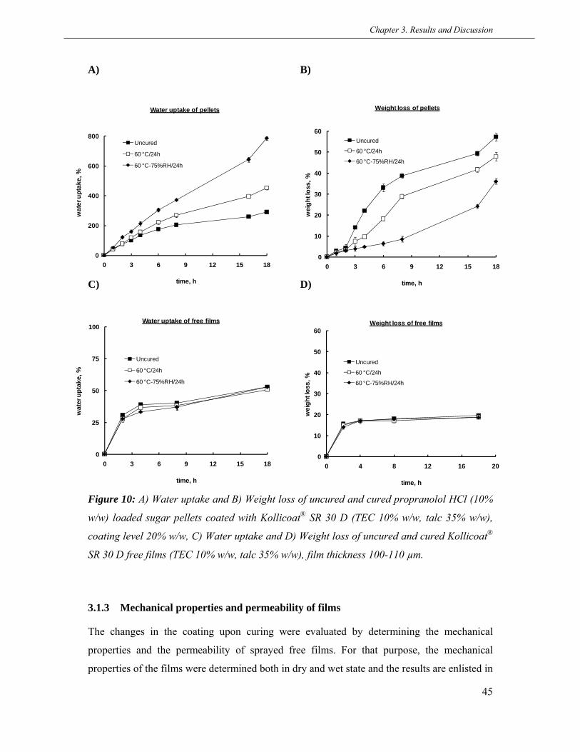
Chapter 3. Results and Discussion
45
0
25
50
75
100
0 3 6 9 12 15 18
wat
er u
ptak
e, %
time, h
Water uptake of free films
Uncured
60 °C/24h
60 °C-75%RH/24h
0
10
20
30
40
50
60
0 4 8 12 16 20
wei
ght l
oss,
%
time, h
Weight loss of free films
Uncured
60 °C/24h
60 °C-75%RH/24h
0
10
20
30
40
50
60
0 3 6 9 12 15 18
wei
ght l
oss,
%
time, h
Weight loss of pellets
Uncured
60 °C/24h
60 °C-75%RH/24h
0
200
400
600
800
0 3 6 9 12 15 18
wat
er u
ptak
e, %
time, h
Water uptake of pellets
Uncured
60 °C/24h
60 °C-75%RH/24h
A) B)
C) D)
Figure 10: A) Water uptake and B) Weight loss of uncured and cured propranolol HCl (10%
w/w) loaded sugar pellets coated with Kollicoat® SR 30 D (TEC 10% w/w, talc 35% w/w),
coating level 20% w/w, C) Water uptake and D) Weight loss of uncured and cured Kollicoat®
SR 30 D free films (TEC 10% w/w, talc 35% w/w), film thickness 100-110 µm.
3.1.3 Mechanical properties and permeability of films
The changes in the coating upon curing were evaluated by determining the mechanical
properties and the permeability of sprayed free films. For that purpose, the mechanical
properties of the films were determined both in dry and wet state and the results are enlisted in

Chapter 3. Results and Discussion
46
Dry state Uncured
60 °C/24h
60 °C-75%RH/24h
Wet state Uncured
60 °C/24h
60 °C-75%RH/24h 8.1 ± 1.2
333.6 ± 8.2
317.4 ± 6.2
340.6 ± 7.2
189.7 ± 7.5
97.1 ± 5.1
197.6 ± 5.1
7.6 ± 1.9
9.0 ± 0.8
8.0 ± 0.3
6.6 ± 0.1
7.8 ± 2.2
Testing state Curing conditionMechanical properties, n = 9
M. V. ± S. D. M. V. ± S. D.Puncture strength, N/mm2 Elongation, %
Table 3. In the dry state, no significant differences were observed in the mechanical properties
of the uncured and films cured at 60 °C-75%RH. However, the films cured at 60 °C showed a
significant decrease in the puncture strength and % elongation which could be due to the loss
of water at elevated temperature (Wu et al., 2000; Zheng et al., 2003). However, the
mechanical properties of the uncured and cured films were almost similar in the wet state
which is an indicative of no changes in the flexibility of coatings upon curing.
The permeability of the uncured and cured films was determined using a diffusion cell
apparatus. The saturated solution of propranolol HCl was filled in the donor compartment and
the samples collected from the acceptor compartment were analyzed UV
spectrophotometrically to calculate the amount of permeated drug. It is important to state that
a negligible amount of the drug could permeate through both the uncured and cured films
suggesting no changes in the permeability upon curing (data not shown). However, the
permeability of polymeric film often decreases upon curing as the film undergoes further
gradual coalescence which causes an increase in film density and tortuosity (Gutierrez-Rocca
et al., 1993; Guo et al., 1991; Zheng et al., 2005) but this phenomenon usually applies to
brittle polymers where the film formation may not complete and a post thermal curing step is
often required to promote the complete film formation.
Table 3 Effect of curing on the mechanical properties of Kollicoat® SR 30 D free film (TEC
10% w/w, talc 35% w/w) (thickness = 100 ± 5µm).
Clearly, no changes in the mechanical properties of the films in the wet state and in the
permeability of the films upon curing eliminated the speculation that “the further gradual
coalescence of the polymer particles might be the reason for the observed curing effect”. This

Chapter 3. Results and Discussion
47
also confirmed that the film formation from the Kollicoat® SR 30 D formulations was
complete after the coating process. Further, the videos monitoring of the pellets during
dissolution was performed to assess the difference in their swelling behaviour. In addition,
osmolality inside the pellets was determined to understand the drug release mechanism.
3.1.4 Video monitoring of coated pellets during drug release
The videos of the uncured and cured pellets during dissolution were recorded to observe
differences in their swelling behaviour. The video recordings were thoroughly analyzed and,
surprisingly, the swelling behaviour of the uncured and pellets cured at 60 °C-75%RH was
found to be completely different. In this regard, some pictures from the video recording at
definite time interval were selected and put together in Figure 12. The detailed swelling
behaviour of the uncured and pellets cured at 60 °C-75%RH is described below in two
scenarios:
First scenario (observed for uncured pellets): It was noticed that the coatings of the uncured
pellets started to swell locally after medium penetration. This began to happen after nearly 1 h
start of dissolution, and the coating continued to swell/extend towards that particular direction
which eventually ruptured followed by a collapse (called as localized swelling and rupturing).
In fact, majority of the uncured pellets underwent this procedure within 2-3 h and a few of
them in 3-4 h which correlates closely with the lag time. This could be explained by the fact
that the propranolol HCl 10% loading formed neither a thick nor a thin layer around the sugar
Figure 11: SEM images of cross section of sugar pellets loaded with propranolol HCl 10%
w/w.
Weak point

Chapter 3. Results and Discussion
48
cores. Hence, the medium rapidly penetrated only at “weak point” where probably the drug
layer was thin (confirmed from SEM images in Figure 11) which resulted in rapid
solubilisation of the sugar cores and arising of osmotic pressure towards that “weak point”
(Figure 12). This is why the coating only started to swell extensively towards that particular
direction (approx. 300% as estimated from the pictures) until ruptured by exceeding the
elongation limit and then finally collapsed (Figure 12). Therefore, the drug released through
visible macro ruptures by an osmotically driven mechanism. Moreover, despite of the visible
macro ruptures, the drug release followed a typical sigmoidal pattern rather than an immediate
release which also confirms the collapsing of coating due to its higher flexibility.
Uncured 60 °C-75%RH/24h
1 h
2 h
4 h
Figure 12: Pictures from video monitoring of uncured and cured (60 °C-75%RH/24 h)
propranolol HCl (10% w/w) loaded sugar pellets coated with Kollicoat® SR 30D during
dissolution (TEC 10% w/w, talc 35% w/w), coating level 20% w/w.
Local swelling
Rupture
Uniform swelling
Collapse

Chapter 3. Results and Discussion
49
Second scenario (observed for pellets cured at 60 °C-75%RH): The pellets cured at 60 °C-
75%RH revealed entirely different swelling behaviour in comparsion with the uncured. For
example, the localized swelling and rupturing of the coatings was not seen at all. In contrast,
the cured pellets started to swell uniformly after the medium penetration and continued as a
function of time without exceeding the elongation limit (approx. 150% estimated from the
pictures). This occurred because the osmotic pressure inside the pellets was distributed
equally towards all directions and no visible macro cracks were observed in the coatings. This
indicates that the drug released after a certain lag time necessary for the thinning of the
coating and the formation of non-visible micro pores. The coating extended until equilibrium
of inner/outer liquids and did not collapse. Due to this, the pellets appeared extremely swollen
(5 times) after release. A sigmoidal release pattern from the cured pellets also suggests an
osmotically driven drug release mechanism.
However, the pellets cured at 60 °C exhibited a dual type of swelling behaviour during
dissolution. Some of the pellets underwent localized swelling/rupturing whereas others
followed a low uniform swelling. As a result, the drug release profile from these pellets was
slower than the uncured and faster than the pellets cured at 60 °C-75%RH which also
suggests that elevated temperature with elevated humidity was the main reason for the
extensive uniform swelling and ultimately for the strong curing effect.
Moreover, the extensive uniform swelling of pellets upon curing at 60 °C-75%RH could be
possibly due to decrease in the adhesive force of coating which was further investigated.
3.1.5 Swelling of coated pellets
The swelling extent of pellets during dissolution was measured for the better understanding of
the drug release mechanism. The increase in diameter and % swelling as a function of time
are shown in Figure 13A & B. The result depicted that both the uncured and cured pellets had
the same extent of swelling till 2 h, afterwards the cured pellets started to swell more.
Interestingly, the swelling of the uncured pellets was decreased to a small extent after 4 h
while the pellets cured at 60 °C continued to swell slowly. In comparison, the pellets cured at
60 °C-75%RH exhibited a significant increase in swelling as a function of time. After the
dissolution, the uncured pellets had a swelling less than 100% whereas the pellets cured at 60
°C-75%RH showed a swelling of over 500% (Figure 13B). The first increase followed by
decrease in swelling of the uncured pellets agrees with the rupturing and the collapsing of the

Chapter 3. Results and Discussion
50
coating which, in turn, also closely correlates with the lag time and to the osmotically driven
release mechanism. On the other hand, the extensive uniform swelling of the pellets cured at
60 °C-75%RH was an indication of the absence of macro rupture and the slow release of drug
via micro crack in the coating. A small increase in the swelling of 60 °C cured pellets
A)
B)
Figure 13: A) Increase in diameter and B) % increase in swelling of uncured and cured
propranolol HCl (10% w/w) loaded sugar pellets coated with Kollicoat® SR 30 D during
dissolution (TEC 10% w/w, talc 35% w/w), coating level 20% w/w.
% swelling of pellets
0
100
200
300
400
500
600
0 3 6 9 12 15 18
time, h
swel
ling,
%
Uncured
60 °C/24h
60 °C-75%RH/24h
Diameter of pellets
0
0.4
0.8
1.2
1.6
2
2.4
2.8
0 3 6 9 12 15 18
time, h
diam
eter
, mm
Uncured
60 °C/24h
60 °C-75%RH/24h

Chapter 3. Results and Discussion
51
0
1000
2000
3000
0 4 8 12 16 20
time, h
mO
smol
/kg
Uncured
60 °C/24h
60 °C-75%RH/24h
represents that the low uniform swelling was dominant over the localized, which led to a
decrease in drug release in comparison with the uncured pellets.
3.1.6 Osmolality inside coated pellets
To assess the effect of osmotic pressure difference on the curing, the osmolality inside the
uncured and cured pellets was measured. Surprisingly, the osmolality inside the uncured and
cured pellets was almost similar despite of a big difference in drug release, water uptake-
weight loss and swelling behaviour (Figure 14). This could be due to that the increased drug
release of the uncured pellets was balanced by the decreased water uptake whereas the
decreased drug release of the cured pellets was equilibrated by the increased water uptake
and, as a result, the osmolality inside the pellets remained unchanged. In spite of the similar
osmotic pressure inside the uncured and cured pellets, the difference in the swelling behaviour
could be explained by the following fact: in case of the uncured pellets, the localized
swelling/rupturing of the coating occurred due to applying of osmotic pressure towards one
particular direction whereas the uniform swelling of the pellets cured at 60 °C-75%RH cured
was caused by the equal distribution of the osmotic pressure towards the whole coating.
Figure 14: Osmolality inside uncured and cured propranolol HCl (10% w/w) loaded sugar
pellets coated with Kollicoat® SR 30 D (TEC 10% w/w, talc 35% w/w), coating level 20%
w/w.

Chapter 3. Results and Discussion
52
However, an osmotically driven drug release mechanism from both the uncured and cured
pellets was confirmed. The similar osmotically driven mechanism from the pellets coated with
Kollicoat® SR 30 D and with some other polymers has been reported (Ensslin et al., 2008;
Ozturk et al., 1990; Narisawa et al., 1996).
3.1.7 Drug release in a high osmolality medium
In order to suppress the localized swelling of the uncured pellets caused by a higher inside
osmotic pressure, the drug release was carried out in a high osmolality medium. The
osmolality (30 mOsmol/kg) of pH-2 medium was raised to 1000 and 1500 mOsmol/kg by
adding sodium chloride (NaCl). As expected, the curing effect was minimized as a function of
increasing the osmolality of medium. For example, the curing effect was eliminated at 60 °C
(f2=57) and was reduced to some extent at 60 °C-75%RH by using a medium of 1000
mOsmol/kg osmolality (f2=37) (Figure 15A). In contrast, the curing effect was completely
overcome both at 60 °C (f2=55) and 60 °C-75%RH (f2=53) by further increasing osmolality to
1500 mOsmol/kg (Figure 15B). Additionally, the drug release was slowed down due to the
decreased pressure inside the pellets confirming the osmotic driven mechanism.
The release profile of the uncured pellets approached closer to the cured ones using 1500
mOsmol/kg medium, which also indicates the suppression of macro rupture in coatings of the
uncured pellets (later confirmed under video monitoring). The overall swelling of pellets was
significantly decreased due to decreased osmotic force.
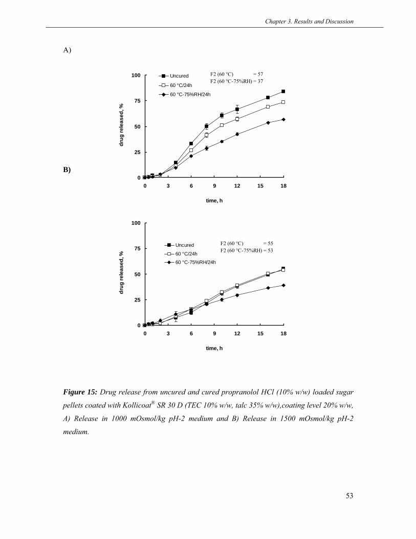
Chapter 3. Results and Discussion
53
0
25
50
75
100
0 3 6 9 12 15 18
time, h
drug
rele
ased
, %
Uncured
60 °C/24h
60 °C-75%RH/24h
F2 (60 °C) = 55F2 (60 °C-75%RH) = 53
A)
B)
Figure 15: Drug release from uncured and cured propranolol HCl (10% w/w) loaded sugar
pellets coated with Kollicoat® SR 30 D (TEC 10% w/w, talc 35% w/w),coating level 20% w/w,
A) Release in 1000 mOsmol/kg pH-2 medium and B) Release in 1500 mOsmol/kg pH-2
medium.
0
25
50
75
100
0 3 6 9 12 15 18
time, h
drug
rele
ased
, %
Uncured
60 °C/24h
60 °C-75%RH/24h
F2 (60 °C) = 57F2 (60 °C-75%RH) = 37

Chapter 3. Results and Discussion
54
0
5
10
15
20
25
Uncured 60 °C-24h 60 °C-75%RH-24h
Forc
e, N
3.1.8 Determination of the adhesive force of coating
The adhesive force of coatings to the core was measured in order to evaluate the changes
upon curing. It was difficult to determine the adhesive force of coatings using the pellets.
Therefore, the model tablets were prepared exactly similar to the pellets and the detachment
force of the coating was measured on a Texture Analyzer (Stable Micro Systems, Surrey,
U.K.).
The results revealed that the adhesive force of the coating was significantly decreased after
curing at 60 °C-75%RH/24 h (Figure 16) which could be due to the weakening of interfacial
bonding of the coating to the core caused by the penetration of water and the creation of new
stresses within the polymeric film. Another reason could be the change in swelling coefficient
of the core and coating. The similar results have been reported when a decrease in adhesion of
pigmented and non-pigmented cellulosic films occurred during storage at 37 °C and 75%RH
(Okhamafe and York, 1985). In contrast, the adhesive force was decreased to a small extent
after curing at 60 °C-24 h. This suggests that humidity plays a major role for decreasing the
adhesive force of coatings and a small decrease at 60 °C-24 h might be caused by the
increased internal stresses within coatings due to evaporation of residual water in polymeric
films (Felton and McGinity, 1997).
Figure 16: Effect of curing on the adhesive force of the Kollicoat® SR 30 D coating (TEC
10% w/w, talc 35% w/w) using model tablets (n=9), coating level 20% w/w.

Chapter 3. Results and Discussion
55
It can be stated that the decreased adhesive forces of the coating upon curing at 60 °C-
75%RH/24h led to the extensive uniform swelling of the pellets which could be the one
reason for the observed curing effect.
3.1.9 Effect of drug loading
The effect of thickness of drug layer on curing was evaluated by layering propranolol HCl
onto sugar cores in different loadings ranging from 2% to 50%. The drug layered cores were
coated, cured and the release studies were carried out. The results depicted that very low (2%-
5%) and very high (30%-50%) drug loadings did not show curing effect whereas 10%
(discussed earlier) and 20% displayed a strong curing effect (Figure 17A-F). No curing effect
with low and high drug loadings could be explained because of similar swelling behaviour of
uncured and cured pellets observed during video monitoring. The details are as under:
2% and 5% drug loadings: the uniform swelling of both uncured and cured pellets was
observed without any signs of localized swelling. This could be due to the quick dissolution
of thin drug layer (confirmed from SEM images in Figure 18A) permitting the rapid
penetration of medium from all sides. Consequently, the osmotic pressure was distributed
equally towards the whole coating and the pellets underwent uniform swelling. Moreover, the
release profiles of 2% and 5% drug loaded pellets were nearly similar like the release profile
of 10% loaded pellets cured at 60 °C-75%RH (Figure 17A-C). This could be because of their
similar swelling and release patterns.
10% and 20% drug loadings: the pellets with 10% and 20% drug loading had a strong curing
effect due to a completely different swelling of the uncured and cured pellets (which has been
explained in section 3.1.4) (Figure 17C & D).
30% and 50% drug loadings: the uncured and cured pellets with high drug loadings also
followed uniform swelling during video monitoring. This could be due that 30% and 50%
loading made a thick drug layer (confirmed from SEM images in 18B) which delayed the
penetration of medium to the cores. As a result, the gradually increasing osmotic pressure was
directed towards the whole coating and the pellets showed uniform swelling. The drug
released from the uncured and cured pellets through the same mechanism without any curing
effect (Figure 17E & F).

Chapter 3. Results and Discussion
56
Drug loading 2%
0
25
50
75
100
0 3 6 9 12 15 18
time, h
drug
rele
ased
, %
Uncured
60 °C/24h
60 °C-75%RH/24h
F2 (60 °C) = 52F2 (60 °C-75%RH) = 53
Drug loading 5%
0
25
50
75
100
0 3 6 9 12 15 18
time, h
drug
rele
ased
, %
Uncured
60 °C/24h
60 °C-75%RH/24h
F2 (60 °C) = 52F2 (60 °C-75%RH) = 50
Drug loading 10%
0
25
50
75
100
0 3 6 9 12 15 18
time, h
drug
rele
ased
, %
Uncured
60 °C/24h
60 °C-75%RH/24h
F2 (60 °C) = 40F2 (60 °C-75%RH) = 20
Drug loading 20%
0
25
50
75
100
0 3 6 9 12 15 18
time, h
drug
rele
ased
, %
Uncured
60 °C/24h
60 °C-75%RH/24h
F2 (60 °C) = 62F2 (60 °C-75%RH) = 21
Drug loading 30%
0
25
50
75
100
0 3 6 9 12 15 18
time, h
drug
rele
ased
, %
Uncured
60 °C/24h
60 °C-75%RH/24h
F2 (60 °C) = 71F2 (60 °C-75%RH) = 57
0
25
50
75
100
0 3 6 9 12 15 18
drug
rele
ased
, %
time, h
Drug loading 50%
Uncured60 °C/24h
60 °C-75%RH/24h
F2 (60 °C) = 91F2 (60 °C-75%RH) = 72
A) B)
C) D)
E) F)
Figure 17: Effect of drug loading on curing effect of propranolol HCl w/w loaded sugar
pellets coated with Kollicoat® SR 30 D (TEC 10% w/w, talc 35% w/w), coating level 20%
w/w, A) 2%, B) 5%, C) 10%, D) 20%, E) 30% and F) 50%.

Chapter 3. Results and Discussion
57
A)
B)
Figure 18: SEM images of cross section of sugar pellets with different propranolol HCl w/w
loadings, A) 2% and B) 50%.
Despite of the similar swelling behaviour of 2%, 5%, 30% and 50% drug loaded pellets, the
faster release rates with 30% and 50% drug loadings could be due to the excessive amount of
drug.
Clearly, the curing effect was observed when both the uncured and cured pellets followed a
completely different swelling behaviour which, in turn, was strongly depended on drug layer
thickness/loading.
3.1.10 Effect of the type of drug layering (solution vs. suspension)
To explain the effect of type of drug layering (solution vs. suspension) on the curing, the drug
layering from suspension was also applied. It was supposed that the drug layer from solution
might have cracks that resulted in localized swelling of the uncured pellets. In comparison,
the layering from suspension could result the simple deposition of drug particles onto the
cores. However, the release results depicted no effect of type of drug layering on the curing
Thick drug layer
Thin drug layer
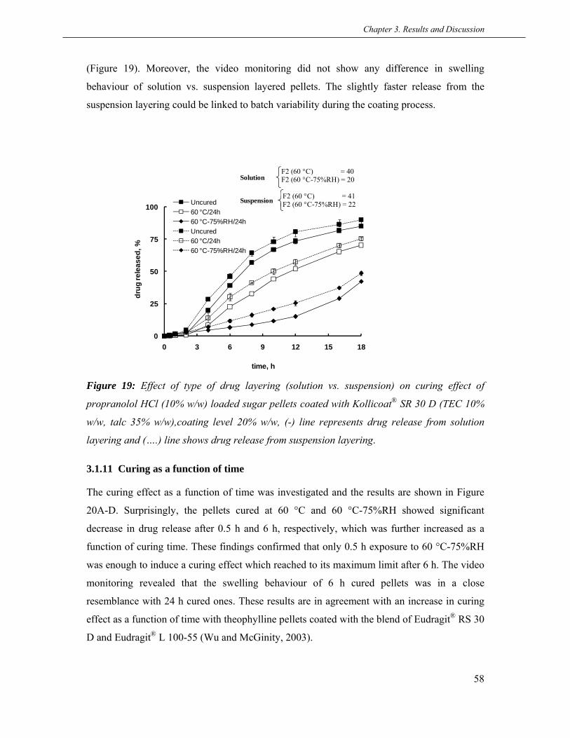
Chapter 3. Results and Discussion
58
0
25
50
75
100
0 3 6 9 12 15 18
drug
rele
ased
, %
time, h
Uncured60 °C/24h60 °C-75%RH/24hUncured60 °C/24h60 °C-75%RH/24h
F2 (60 °C) = 40F2 (60 °C-75%RH) = 20
F2 (60 °C) = 41F2 (60 °C-75%RH) = 22
Solution
Suspension
(Figure 19). Moreover, the video monitoring did not show any difference in swelling
behaviour of solution vs. suspension layered pellets. The slightly faster release from the
suspension layering could be linked to batch variability during the coating process.
Figure 19: Effect of type of drug layering (solution vs. suspension) on curing effect of
propranolol HCl (10% w/w) loaded sugar pellets coated with Kollicoat® SR 30 D (TEC 10%
w/w, talc 35% w/w),coating level 20% w/w, (-) line represents drug release from solution
layering and (….) line shows drug release from suspension layering.
3.1.11 Curing as a function of time
The curing effect as a function of time was investigated and the results are shown in Figure
20A-D. Surprisingly, the pellets cured at 60 °C and 60 °C-75%RH showed significant
decrease in drug release after 0.5 h and 6 h, respectively, which was further increased as a
function of curing time. These findings confirmed that only 0.5 h exposure to 60 °C-75%RH
was enough to induce a curing effect which reached to its maximum limit after 6 h. The video
monitoring revealed that the swelling behaviour of 6 h cured pellets was in a close
resemblance with 24 h cured ones. These results are in agreement with an increase in curing
effect as a function of time with theophylline pellets coated with the blend of Eudragit® RS 30
D and Eudragit® L 100-55 (Wu and McGinity, 2003).

Chapter 3. Results and Discussion
59
0
25
50
75
100
0 3 6 9 12 15 18
drug
rele
ased
, %
time, h
Curing time 0.5 h
Uncured
60 °C/0.5h
60 °C-75%RH/0.5h
F2 (60 °C) = 84F2 (60 °C-75%RH) = 44
0
25
50
75
100
0 3 6 9 12 15 18
drug
rele
ased
, %
time, h
Curing time 2 h
Uncured
60 °C/2h
60 °C-75%RH/2h
F2 (60 °C) = 54F2 (60 °C-75%RH) = 32
0
25
50
75
100
0 3 6 9 12 15 18
drug
rele
ased
, %
time, h
Curing time 6 h
Uncured
60 °C/6h
60 °C-75%RH/6h
F2 (60 °C) = 42F2 (60 °C-75%RH) = 28
0
25
50
75
100
0 3 6 9 12 15 18
drug
rele
ased
, %
time, h
Curing time 24 h
Uncured
60 °C/24h
60 °C-75%RH/24h
F2 (60 °C) = 40F2 (60 °C-75%RH) = 20
A) B)
C) D)
Figure 20: Effect of curing on propranolol HCl (10% w/w) loaded sugar pellets coated with
Kollicoat® SR 30D as a function of curing time (TEC 10% w/w, talc 35% w/w), coating level
20% w/w, A) 0.5 h, B) 2 h, C) 6 h and D) 24 h.
Chapter 3. Results and Discussion
60
3.1.12 Conclusions
The Kollicoat® RS 30 D coated pellets showed a strong curing effect within a drug loading
range of 10%-20%. Importantly, the curing effect was not associated with a conventionally
known cause for curing “further gradual coalescence of the polymeric films”. This was
confirmed by no changes in the permeability (complete film formation) and in the mechanical
properties of the films upon curing. The main reason for the observed curing effect was found
to be a completely different swelling behaviour of the uncured and cured pellets during
release. The uncured pellets had a localized swelling whereas the cured pellets showed an
extensive uniform swelling caused by the decrease in adhesive force of the coating upon
curing. Additionally, an osmotically driven drug release mechanism from the coated pellets
was predicted.

Chapter 3. Results and Discussion
61
3.2 Effect of starter core, drug solubility and drug loading on the curing
effect of Kollicoat® SR 30 D coatings
Drug release from polymer coated pellets is often occurred by three mechanisms: i) diffusion
through intact polymeric film, ii) through water-filled channels and iii) through micro/macro
cracks within polymeric film. The type of involved drug release mechanism is mainly
dependent on the properties of starter cores (e.g. osmotic activity, water uptake and swelling),
and drug solubility/loading. Accordingly, the type of starter core and drug solubility could
also play a critical role on the curing mechanism of aqueous polymer coatings especially
when the further gradual coalescence of films is not the reason for curing. In this regard,
either no or very little work without explanations has been done up-to-date. In previous
section, the strong curing effect of Kollicoat® SR 30 D coated pellets was observed by using
osmotically active sugar cores and propranolol HCl (212 mg/ml) as a model drug within 10%-
20% drug loadings Therefore, the aim of this part of work was to further investigate in detail
the effect of starter core, drug solubility and drug loading on the curing effect of Kollicoat®
SR 30 D coated pellets.
3.2.1 Effect of starter core and drug solubility
To explain the effect of starter core on the curing effect of Kollicoat® SR 30 D coated pellets,
osmotically active sugar cores (called as SC cores) and osmotically inactive microcrystalline
cellulose cores (called as MCC cores) of the same size were used. In order to investigate the
effect of drug solubility, the following drugs were used: a poorly soluble (carbamazepine-0.2
mg/ml), a drug with intermediate solubility (theophylline-12 mg/ml) and two freely soluble
drugs (tramadol HCl-500 mg/ml and metoprolol tartrate->1000mg/ml). Both the SC and MCC
cores were loaded with 10% of different drugs and were coated 20% with Kollicoat® SR 30
D. The curing effect was evaluated by comparing the release profiles of uncured and cured
pellets using f2 similarity factor. The dissolution studies were carried out till 48 h due to the
two main reasons: i) to observe the complete shape of release profiles and ii) to get an
appropriate release profile for poorly and intermediate soluble drugs.
The results depicted that the SC cores had a strong curing effect with carbamazepine and
theophylline (Figure 21A & B). However, tramadol HCl and metoprolol tartrate did not show
curing effect (Figure 21C & D). Interestingly, the drug release was increased with

Chapter 3. Results and Discussion
62
0
25
50
75
100
0 8 16 24 32 40 48
drug
rele
ased
, %
time, h
MCC - Carbamazepine 10% dl
Uncured60 °C/24h60 °C-75%RH/24h
F2 (60 °C) = 82F2 (60 °C-75%RH) = 53
0
25
50
75
100
0 8 16 24 32 40 48
drug
rele
ased
, %
time, h
MCC - Theophylline 10%dl
Uncured
60 °C/24h
60 °C-75%RH/24h
F2 (60 °C) = 65F2 (60 °C-75%RH) = 53
0
25
50
75
100
0 8 16 24 32 40 48
drug
rele
ased
, %
time, h
MCC - Tramadol HCl 10% dl
Uncured60 °C/24h60 °C-75%RH/24h
F2 (60 °C) = 50F2 (60 °C-75%RH) = 66
0
25
50
75
100
0 8 16 24 32 40 48
drug
rele
ased
, %
time, h
MCC - Metoprolol tartrate 10%dl
Uncured60 °C/24h60 °C-75%RH/24h
F2 (60 °C) = 50F2 (60 °C-75%RH) = 51
0
25
50
75
100
0 6 12 18 24 30 36 42 48
drug
rele
ased
, %
time, h
SC- Carbamazepine 10% dl
Uncured
60 °C/24h
60 °C-75%RH/24h
F2 (60 °C) = 62F2 (60 °C-75%RH) = 31
0
25
50
75
100
0 8 16 24 32 40 48
drug
rele
ased
, %
time, h
SC - Theophylline 10%dl
Uncured
60 °C/24h
60 °C-75%RH/24h
F2 (60 °C) = 64F2 (60 °C-75%RH) = 45
0
25
50
75
100
0 8 16 24 32 40 48
drug
rele
ased
, %
time, h
SC - Metoprolol tartrate 10%dl
Uncured
60 °C/24h
60 °C-75%RH/24h
F2 (60 °C) = 61F2 (60 °C-75%RH) = 69
0
25
50
75
100
0 8 16 24 32 40 48
drug
rele
ased
, %
time, h
SC - Tramadol HCl 10%dl
Uncured
60 °C/24h
60 °C-75%RH/24h
F2 (60 °C) = 58F2 (60 °C-75%RH) = 66
A) E)
B) F)
C) G)
D) H)
Figure 21: Effect of starter core and drug solubility on the curing effect of Kollicoat® SR 30
D coated pellets (TEC 10% w/w, talc 35% w/w), drug loading 10% w/w, coating level 20%
w/w, A) SC-carbamazepine, B) SC-theophylline, C) SC-tramadol HCl, D) SC-metoprolol
tartrate, E) MCC-carbamazepine, F) MCC-theophylline, G) MCC-tramadol HCl and H)
MCC-metoprolol tartrate.
Chapter 3. Results and Discussion
63
carbamazepine and theophylline after curing compared to propranolol HCl (shown earlier in
section 3.1.1). In addition, the shape of release profiles with SC cores was strongly influenced
by drug solubility. For example, carbamazepine followed a zero order release due to its very
poor solubility while a sigmoidal release pattern was obtained with theophylline, tramadol
HCl and metoprolol tartrate.
The coated pellets using MCC as a starter cores did not exhibit any curing effect irrespective
of drug solubility (Figure 21E-H). However, a high variability in the release rates is an
indication of strong effect of drug solubility. Almost zero order release patterns were obtained
with carbamazepine, theophylline and metoprolol tartrate while tramadol HCl revealed a
pulsatile type release profile. The investigation of the exact reasons for the different release
patterns was beyond the scope of study. The further supportive studies like water uptake-
weight loss and swelling behaviour during dissolution were carried out for the better
understanding of curing mechanism.
3.2.2 Water uptake and weight loss of coated pellets
The water uptake and weight loss results of SC cores are shown below in Figure 22A-C.
Clearly, the water uptake of the coated pellets increased significantly after curing at 60 °C-
75%RH and an obvious correlation was found between water uptake and drug solubility. For
instance, the water uptake of the carbamazepine pellets cured at 60 °C-75%RH was 326%
after 18 h in comparison with 424% of theopylline and 772% of metoprolol tartrate. The
lower water uptake of carbamazepine pellets could be attributed to its poor solubility and
higher lipophilicity that hindered the penetration of medium into the SC cores whereas the
higher water uptake of metoprolol tartrate pellets could be linked to its higher solubility and
hydrophilicity that permitted the free ingress of medium. In comparison, the decreased weight
loss of metoprolol tartrate pellets was in accordance with the decreased release rates upon
curing. However, the decreased weight loss of carbamazepine and theophylline up to 18 h was
not in agreement with the increased release rates after curing. This could be explained as
follows: with uncured pellets, probably sugar released faster than the poorly soluble drugs
through macro cracks formed in coatings due to rapid penetration of medium to cores at weak
points of drug layer. However, with pellets cured at 60 °C-75%RH, the penetration of
medium to the core was hindered by poorly soluble drug layer due to uniform swelling of
coatings caused by decrease in adhesive force upon curing, as a result, mainly drug released
via micro cracks till 18 h. This also explains increase in drug release of carbamazepine and

Chapter 3. Results and Discussion
64
0
100
200
300
400
500
0 8 16 24 32 40 48
wat
er u
ptak
e, %
time, h
Water uptakeSC- Carbamazepine 10%dl
Uncured60 °C/24h60 °C-75%RH/24h
0
25
50
75
100
0 8 16 24 32 40 48
wei
ght l
oss,
%
time, h
Weight lossSC- Carbamazepine 10%dl
Uncured60 °C/24h60 °C-75%RH/24h
0
100
200
300
400
500
600
700
0 8 16 24 32 40 48
wat
er u
ptak
e, %
time, h
Water uptakeSC- Theophylline 10%dl
Uncured60 °C/24h60 °C-75%RH/24h
0
200
400
600
800
1000
0 8 16 24 32 40 48
wat
er u
ptak
e, %
time, h
Water uptakeSC- Metoprolol tartrate 10%dl
Uncured60 °C/24h60 °C-75%RH/24h
0
25
50
75
100
0 8 16 24 32 40 48
wei
ght l
oss,
%
time, h
Weight lossSC- Metoprolol tartrate 10%dl
Uncured
60 °C/24h
60 °C-75%RH/24h
A)
B)
C)
Figure 22: Effect of drug solubility on the water uptake and weight loss of Kollicoat® SR 30
D coated uncured and cured sugar pellets (TEC 10% w/w, talc 35% w/w), drug loading 10%
w/w, coating level 20% w/w, A) carbamazepine, B) theophylline and C) metoprolol tartrate.
0
25
50
75
100
0 8 16 24 32 40 48
wei
ght l
oss,
%
time, h
Weight lossSC- Theophylline 10%dl
Uncured60 °C/24h60 °C-75%RH/24h

Chapter 3. Results and Discussion
65
0
5
10
15
20
25
30
0 8 16 24 32 40 48
wei
ght l
oss,
%
time, h
Weight lossMCC- Carbamazepine 10%dl
Uncured60 °C/24h60 °C-75%RH/24h
0
5
10
15
20
25
30
0 8 16 24 32 40 48
wei
ght l
oss,
%
time, h
Weight lossMCC- Theophylline 10%dl
Uncured60 °C/24h
60 °C-75%RH/24h
0
5
10
15
20
25
30
0 8 16 24 32 40 48
wei
ght l
oss,
%
time, h
Weight lossMCC- Metoprolol tartrate 10%dl
Uncured60 °C/24h
60 °C-75%RH/24h
0
25
50
75
100
125
150
175
200
0 8 16 24 32 40 48
wat
er u
ptak
e, %
time, h
Water uptakeMCC- Theophylline 10%dl
Uncured60 °C/24h60 °C-75%RH/24h
0
25
50
75
100
125
150
175
200
0 8 16 24 32 40 48
wat
er u
ptak
e, %
time, h
Water uptakeMCC- Metoprolol tartrate 10%dl
Uncured60 °C/24h60 °C-75%RH/24h
0
25
50
75
100
125
150
175
200
0 8 16 24 32 40 48
wat
er u
ptak
e, %
time, h
Water uptakeMCC- Carbamazepine 10%dl
Uncured60 °C/24h60 °C-75%RH/24h
A)
B)
C)
Figure 23: Effect of drug solubility on the water uptake and weight loss of Kollicoat® SR 30
D coated uncured and cured MCC pellets (TEC 10% w/w, talc 35% w/w), drug loading 10%
w/w, coating level 20%, A) carbamazepine, B) theophylline and C) metoprolol tartrate.
Chapter 3. Results and Discussion
66
theophylline after curing in contrast to propranolol HCl.
The water uptake of MCC pellets was significantly lower than the SC pellets which might be
due to the insoluble nature of MCC cores (Figure 23A-C). However, a small increase in water
uptake of the pellets cured at 60 °C-75%RH was observed as a function of drug solubility. For
example, the water uptake of carbamazepine pellets after 24 h was 89% compared to 123% of
metoprolol tartrate which could be due to difference in drug solubility and lipophilicity.
However, the weight loss of uncured and cured MCC pellets was in close agreement with the
drug release results irrespective of drug solubility.
3.2.3 Video monitoring of coated pellets during drug release
The video monitoring of the uncured and cured SC and MCC pellets during dissolution were
performed to see the effect of starter core and drug solubility on the swelling behaviour. The
description of video analysis is as under:
SC cores: The video monitoring of the uncured pellets of carbamazepine and theophylline
revealed a localized swelling of the coating caused by the rapid ingress of medium at the point
where drug layer was weak. In contrary, the pellets cured at 60 °C-75%RH exhibited a
uniform swelling due to decrease in the adhesive force of coatings. However, the pellets cured
at 60 °C showed a mixed type of swelling behaviour (localized and low uniform) of the
coating (All three types of swelling behaviour have been described in detail for propranolol
HCl in section 3.1.4.). The selective pictures at definite time points from the video monitoring
of carbamazepine are presented in Figure 24A as an example.
However, both the uncured and cured pellets of tramadol HCl and metoprolol tartrate with
10% drug loading clearly displayed an extensive uniform swelling (Figure 24B.) This could
be explained by the higher solubilities of these drugs that resulted in a rapid dissolution of the
drug layer, as a result, the medium reached to the core immediately from all sides causing an
equal distribution of osmotic pressure towards the whole coating and eventually uniform
swelling. This is why with both uncured and cured pellets the drug released through micro
cracks in the coating without any curing effect.
Furthermore, in order to confirm the importance of effect of drug solubility on swelling
behaviour and extent, SC cores were directly coated with Kollicoat® SR 30 D. As expected,
both of the uncured and cured pellets displayed an extensive uniform swelling similar to the
metoprolol tartrate pellets. This confirmed the effect of drug solubility on swelling behaviour

Chapter 3. Results and Discussion
67
A) Carbamazepine Uncured 60 °C-75%RH/24 h
1 h
2 h
4 h
B) Metoprolol tartrate Uncured 60 °C-75%RH/24 h
1 h
2 h
4 h
Figure 24: Pictures from video monitoring of the uncured and cured SC pellets coated with
Kollicoat® SR 30 D (TEC 10% w/w, talc 35% w/w) during dissolution, drug loading 10%
w/w, coating level 20% w/w, A) carbamazepine and B)metoprolol tartrate.
Collapse
Local swelling/rupture
Uniform swelling
Uniform swelling
Uniform swelling

Chapter 3. Results and Discussion
68
and in turn on curing effect. Clearly, the overall swelling of the pellets was increased by
increasing drug solubility (data not shown).
MCC cores: The video monitoring of MCC pellets did not show a big difference between the
swelling behaviour of the uncured and cured pellets. Neither the uncured pellets underwent a
localized swelling nor the cured ones showed an extensive uniform swelling like of the SC
pellets. The selective pictures at definite time points from the video recordings of
carbamazepine are shown in Figure 25. However, a few tiny micro cracks were noticed in the
coating of uncured pellets whereas the cured pellets as well showed some evidence of
expulsion of the drug via micro cracks.
Uncured 60 °C-75%RH/24 h
Figure 25: Pictures from video monitoring of the uncured and cured carbamazepine (10%
w/w) loaded MCC pellets coated with Kollicoat® SR 30 D (TEC 10% w/w, talc 35% w/w)
during dissolution, coating level 20% w/w.
Micro cracks
Low uniform swelling
Chapter 3. Results and Discussion
69
Importantly, the SC pellets had a curing effect when both the uncured and cured pellets
followed entirely a different swelling behaviour which, in turn, depends on the drug solubility
and loading. In comparison, the no significant difference in swelling of the uncured and cured
pellets, independent of drug solubility and loading, with same release mechanism could be the
main reason for no curing effect with MCC cores.
3.2.4 Effect of drug loading
The effect of drug solubility in combination with drug loading on curing was also evaluated.
In this regards, the drugs with different solubilities were layered onto SC cores in different
loadings (2%, 10%, 20%, 30% and 50%). The drug layered cores were coated with 20% of
Kollicoat® SR 30 D and the presence of curing effect, the swelling extent and the behaviour
of pellets during dissolution are enlisted in Table 4.
The results depicted that the curing effect was strongly dependent on drug solubility and drug
loading. The curing effect was seen with drug solubility ranging from 0.2 mg/ml
(carbamazepine) to 212 mg/ml (propranolol HCl) (data for propranolol HCl has shown
earlier) in a drug loading range of 10%-20%. Moreover, no curing effect was observed with
freely soluble drugs (tramadol HCl-500 mg/ml and metoprolol tartrate-> 1000 mg/ml) in all
the investigated loadings. The presence or absence of curing effect can be explained as
following:
Curing effect: As it has been concluded earlier that the curing effect occurred only when
uncured pellets showed a localized swelling whereas cured pellets followed a uniform
swelling. The present results also depict that the curing effect of carbamazepine, theophylline
and propranolol HCl pellets in a drug loading range of 10%-20% is associated with their
different swelling behaviour during dissolution before and after curing. In all these cases, the
video monitoring revealed the localized swelling of the uncured pellets in comparison with
the extensive uniform swelling of the cured pellets (which has been described earlier).
However, outside this range like 2%, 30% and 50%, similar swelling behaviour of the
uncured and cured pellets is an indicative of no curing effect. This could be due to that 2%
drug loading formed a thin layer while 30% and 50% made a thick layer around the pellets,
thus, either permitting immediate or delaying the penetration of medium to the core and
eventually preventing the localized swelling of uncured pellets. Furthermore, the low uniform
swelling of carbamazepine and theophylline in 30% and 50% drug loadings might be
associated with their low drug solubility and high lipophilicity.
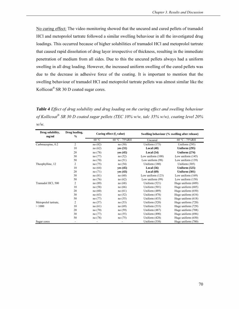
Chapter 3. Results and Discussion
70
Drug solubility, Drug loading,mg/ml %
60 °C 60 °C - 75%RH Uncured 60 °C - 75%RHCarbmazepine, 0.2 2 no (82) no (50) Uniform (175) Uniform (295)
10 no (62) yes (31) Local (40) Uniform (295)20 no (78) yes (45) Local (54) Uniform (274)30 no (77) no (52) Low uniform (100) Low uniform (145)50 no (70) no (51) Low uniform (98) Low uniform (139)
Theophylline, 12 2 no (75) no (54) Uniform (180) Uniform (305)10 no (64) yes (45) Local (56) Uniform (325)20 no (71) yes (43) Local (69) Uniform (301)30 no (81) no (60) Low uniform (123) Low uniform (169)50 no (76) no (62) Low uniform (99) Low uniform (158)
Tramadol HCl, 500 2 no (89) no (68) Uniform (521) Huge uniform (689)10 no (58) no (66) Uniform (501) Huge uniform (685)20 no (60) no (61) Uniform (489) Huge uniform (650)30 no (63) no (52) Uniform (478) Huge uniform (634)50 no (77) no (55) Uniform (435) Huge uniform (618)
Metoprolol tartrate, 2 no (57) no (53) Uniform (520) Huge uniform (720)> 1000 10 no (61) no (69) Uniform (533) Huge uniform (729)
20 no (70) no (59) Uniform (487) Huge uniform (708)30 no (77) no (55) Uniform (490) Huge uniform (696)50 no (78) no (75) Uniform (428) Huge uniform (650)
Sugar cores ‐ ‐ ‐ Uniform (538) Huge uniform (780)
Swelling behaviour (% swelling after release)Curing effect (f2 value)
No curing effect: The video monitoring showed that the uncured and cured pellets of tramadol
HCl and metoprolol tartrate followed a similar swelling behaviour in all the investigated drug
loadings. This occurred because of higher solubilities of tramadol HCl and metoprolol tartrate
that caused rapid dissolution of drug layer irrespective of thickness, resulting in the immediate
penetration of medium from all sides. Due to this the uncured pellets always had a uniform
swelling in all drug loading. However, the increased uniform swelling of the cured pellets was
due to the decrease in adhesive force of the coating. It is important to mention that the
swelling behaviour of tramadol HCl and metoprolol tartrate pellets was almost similar like the
Kollicoat® SR 30 D coated sugar cores.
Table 4 Effect of drug solubility and drug loading on the curing effect and swelling behaviour
of Kollicoat® SR 30 D coated sugar pellets (TEC 10% w/w, talc 35% w/w), coating level 20%
w/w.

Chapter 3. Results and Discussion
71
3.2.5 Conclusions
The pellets with MCC cores did not show curing effect irrespective of drug solubility while
SC pellets revealed a strong curing effect depending on drug solubility and drug loading. The
curing effect with SC pellets was observed in those cases when both the uncured and cured
pellets exhibited a completely different swelling behaviour during dissolution which was
potentially influenced by drug solubility and drug loading. In addition, the drug solubility also
had a pronounce influence on the swelling of SC pellets.
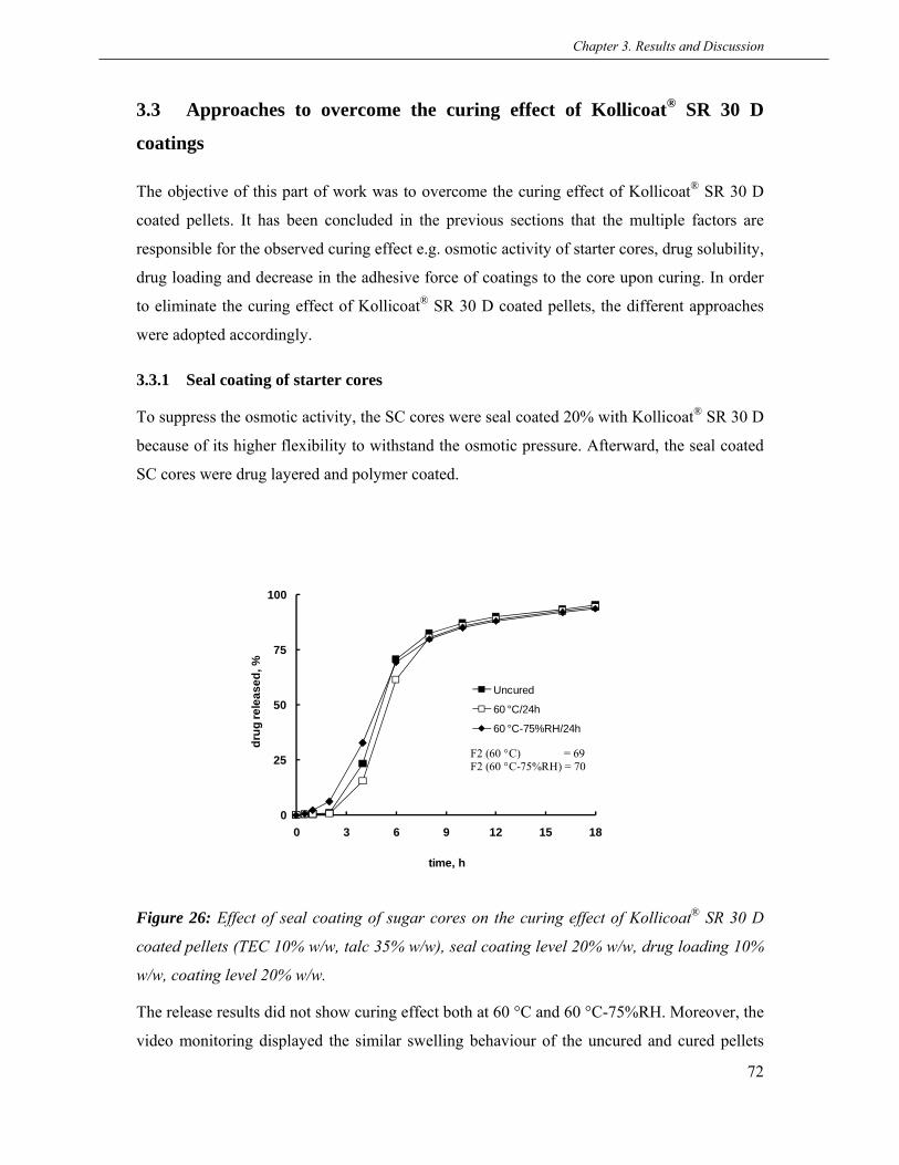
Chapter 3. Results and Discussion
72
0
25
50
75
100
0 3 6 9 12 15 18
drug
rele
ased
, %
time, h
Uncured
60 °C/24h
60 °C-75%RH/24h
F2 (60 °C) = 69F2 (60 °C-75%RH) = 70
3.3 Approaches to overcome the curing effect of Kollicoat® SR 30 D
coatings
The objective of this part of work was to overcome the curing effect of Kollicoat® SR 30 D
coated pellets. It has been concluded in the previous sections that the multiple factors are
responsible for the observed curing effect e.g. osmotic activity of starter cores, drug solubility,
drug loading and decrease in the adhesive force of coatings to the core upon curing. In order
to eliminate the curing effect of Kollicoat® SR 30 D coated pellets, the different approaches
were adopted accordingly.
3.3.1 Seal coating of starter cores
To suppress the osmotic activity, the SC cores were seal coated 20% with Kollicoat® SR 30 D
because of its higher flexibility to withstand the osmotic pressure. Afterward, the seal coated
SC cores were drug layered and polymer coated.
Figure 26: Effect of seal coating of sugar cores on the curing effect of Kollicoat® SR 30 D
coated pellets (TEC 10% w/w, talc 35% w/w), seal coating level 20% w/w, drug loading 10%
w/w, coating level 20% w/w.
The release results did not show curing effect both at 60 °C and 60 °C-75%RH. Moreover, the
video monitoring displayed the similar swelling behaviour of the uncured and cured pellets

Chapter 3. Results and Discussion
73
which reflects that the localized swelling of the uncured pellets was restricted after the seal
coating of SC cores. In contrast, the curing effect could not be successfully eliminated when
SC cores were seal coated 20% using Aquacoat® ECD (data not shown). This could be
possibly related to the brittle coatings of Aquacoat® ECD which might not withstand the
osmotic pressure. Clearly, the presence of localized swelling in uncured pellets suggested the
rupturing of Aquacoat® ECD seal coatings.
3.3.2 Improving the adhesion of coatings
The decrease in adhesion of the coating to the core upon curing was found to be one of
another cause of the observed curing effect. In order to hinder the decrease in adhesion of
coating upon curing, the drug to binder ratio was increased from 9:1 to 3:1. Alternatively, a
10% sub coating of HPMC was also applied.
The release results depicted that the curing effect was successfully overcome by increasing
the drug to binder ratio or by applying a 10% sub coating of HPMC (Figure 27A & B). The
video monitoring revealed that uncured pellets showed a localized swelling and rupturing of
the coatings but the extensive uniform swelling of the cured pellets at 60 °C-75%RH was
reduced significantly. Furthermore, the water uptake and the extent of swelling of the cured
pellets came closer to the uncured ones (data not shown).

Chapter 3. Results and Discussion
74
0
25
50
75
100
0 3 6 9 12 15 18
drug
rele
ased
, %
time, h
HPMC 10% sub coating
Uncured
60 °C/24h
60 °C-75%RH/24h
F2 (60 °C) = 82F2 (60 °C-75%RH) = 63
0
25
50
75
100
0 3 6 9 12 15 18
drug
rele
ased
, %
time, h
Increasing drug:binder ratio (3:1)
Uncured
60 °C/24h
60 °C-75%RH/24h
F2 (60 °C) = 97F2 (60 °C-75%RH) = 73
A)
B)
Figure 27: Effect of A) increased drug:binder ratio (3:1) and B) a 10% sub coating of HPMC
on the curing effect of propranolol HCl (10% w/w loaded) sugar pellets coated with
Kollicoat® SR 30 D (TEC 10% w/w, talc 35% w/w), coating level 20% w/w.
In addition, the adhesion of the coating was determined by preparing the model tablets. The
results showed that the adhesion of coating was decreased to a small extent upon curing and,
the uncured tablet also exhibited an increase in the adhesive force which is a representative of

Chapter 3. Results and Discussion
75
0
5
10
15
20
25
30
35
Uncured 60 °C-24h 60 °C-75%RH-24h
Forc
e, N
Increasing drug:binder ratio (3:1)
10% HPMC sub coating
a strong binding of the coating to the core (Figure 28). This could be due to increase in
interaction between the primary and secondary hydroxyl groups of HPMC with the polymer
coatings. The increased concentrations of HPMC phthalate has been found to adhere strongly
to MCC tablets (Missaghi et al., 2004; Lehtola et al., 1995; Row, 1981)
Figure 28: Effect of curing on the adhesive force of Kollicoat® SR 30 D coatings after
increasing drug to binder ratio (3:1) and applying a 10% w/w sub coating of HPMC (TEC
10% w/w, talc 35% w/w),coating level 20% w/w, the test was performed using model tablets
(n=9).
3.3.3 Making the coatings brittle
This approach was adopted to reduce the flexibility of Kollicoat® SR 30 D coatings by
incorporating the higher amount of talc and using titanium dioxide. Interestingly, by adding
75% of talc to the coating formulations, the curing effect was eliminated at 60 °C but not at
60 °C-75%RH (Figure 29A). However, when the amount of talc was further increased to
100%, a stable release profiles indicated the complete absence of curing effect (Figure 29B).
The difference in the water uptake of the uncured and cured pellets was minimized
significantly (data not shown). Moreover, the video monitoring revealed that the extent of
swelling of the cured pellets decreased effectively which indicates that the presence of higher
amount of talc made the coating brittle and rough. The decrease in flexibility of coatings with

Chapter 3. Results and Discussion
76
0
25
50
75
100
0 3 6 9 12 15 18
drug
rele
ased
, %
time, h
Talc 75%
Uncured
60 °C/24h
60 °C-75%RH/24h
F2 (60 °C) = 85F2 (60 °C-75%RH) = 46
0
25
50
75
100
0 3 6 9 12 15 18
drug
rele
ased
, %
time, h
Talc 100%
Uncured
60 °C/24h
60 °C-75%RH/24h
F2 (60 °C) = 65F2 (60 °C-75%RH) = 68
0
25
50
75
100
0 3 6 9 12 15 18
drug
rele
ased
, %
time, h
TiO2 35%
Uncured
60 °C/24h
60 °C-75%RH/24h
F2 (60 °C) = 79F2 (60 °C-75%RH) = 70
0
25
50
75
100
0 3 6 9 12 15 18
drug
rele
ased
, %
time, h
TiO2 20%
Uncured
60 °C/24h
60 °C-75%RH/24h
F2 (60 °C) = 74F2 (60 °C-75%RH) = 60
100% addition of talc was also confirmed by the mechanical properties of free films (data not
shown). With 100% addition of talc, since the coating mainly consisted of talc and polymer
A) B)
C) D)
Figure 29: Effect of incorporating higher amount of talc (w/w) and using TiO2(w/w) as an
anti-tacking agent on curing effect of propranolol HCl (10% w/w) loaded sugar pellets coated
with Kollicoat® SR 30 D (TEC 10% w/w), coating level 20% w/w, A) Talc 75%, B) Talc
100%, C) TiO2 20% and D) TiO2 35%.
latex and the particle size of the polymer latex was much smaller than that of talc, the talc
would form the skeleton of the controlled release coating and the polymer latex would be
distributed within the talc during coating process. During the curing, the polymer particles
present within the talc matrix were more densified around talc particles resulting in more
porous coating (Maejima and McGinity, 2001). It has also been reported that the surface of

Chapter 3. Results and Discussion
77
polymer coating became rough after curing by using higher amount of talc. No curing effect
suggested that the release rates through the micro cracks of the cured and via the macro cracks
of the uncured coatings were equal.
Furthermore, the effect of higher amount of talc 100% on the adhesive force of the coatings
was determined using model tablets and a decrease in overall adhesive force of the coating
was found. This happened because talc is a hydrophobic substance that is generally added to
the coating formulations to reduce the stickiness. The hydrophobic talc particles become
embedded within the polymeric film and interfere with hydrogen bond formation between the
tablet surface and the film coating. Additionally, talc causes stiffening of the film and
increases the internal stresses within the polymer, as has been evidenced by an increase in the
Tg of the polymer (Okhamafe and York, 1984; Okhamafe and York, 1985). However, the
adhesion of the coating was not affected upon curing (Figure 30) which could be due to that
the hydrophobic skeleton of talc hindered the penetration of water. As a result, the interfacial
cleavage of bonding between the polymer and the tablet surface could be prevented and no
significant decrease in adhesion occured.
Another strategy was adopted to replace hydrophobic talc with hydrophilic small sized
titanium dioxide (TiO2) due to its better wetting and dispersibility. The curing effect was
completely eliminated by 20% addition of TiO2 to the coating formulations and by further
increasing the concentration to 35%, the release profile remained almost unchanged. In
addition, the video monitoring expressed a low uniform swelling of the uncured and cured
pellets suggesting a pronounced reduction in the flexibility of coatings. This reflects that the
elimination of curing effect with smaller quantities of TiO2 could be linked to its better
covering of polymeric particles that resulted in reduction of flexibility.

Chapter 3. Results and Discussion
78
0
2
4
6
8
10
12
14
Uncured 60 °C-24h 60 °C-75%RH-24h
Forc
e, N
Figure 30: Effect of curing on the adhesive force of Kollicoat® SR 30 D coatings after
incorporating talc 100% w/w in the coating (TEC 10% w/w), coating level 20% w/w, the test
was performed using model tablets (n=9).
3.3.4 Use of pore-formers in coatings
This strategy was applied to make the coating porous in order to avoid the localized swelling
of the uncured pellets upon leaching out of water soluble additives. Commonly used water
soluble additives are low-molecular weight materials (e.g., sucrose, lactose, sodium chloride
and calcium phosphate etc) and hydrophilic polymers including polyethylene glycol,
polyvinylpyrrolidone and cellulose ethers such as HPMC. However, HPMC (Pharmacoat®
606) and another hydrophilic polymer [poly(vinyl alcohol)-poly(ethylene glycol) (PVA-PEG)
graft copolymer, the trade name Kollicoat® IR] were added in different concentrations (5%,
7.5% and 10%) to coating formulations. Clearly, the coated pellets did not show curing effect
by the addition of Pharmacoat® 606 to the coating formulations in all concentrations (Figure
31A-C). However, f2 values represented the most stable release profiles with 7.5% addition of
Pharmacoat® 606. The curing effect of carbamazepine pellets was not eliminated by 7.5% and
10% addition of Pharmacoat® 606. However, 20% of Pharmacoat® 606 addition provided a
stable release profile. No elimination of the curing effect with 7.5% and 10% addition of

Chapter 3. Results and Discussion
79
0
25
50
75
100
0 3 6 9 12 15 18
drug
rele
ased
, %
time, h
Pharmacoat 606 5%
Uncured
60 °C/24h
60 °C-75%RH/24h
F2 (60 °C) = 89F2 (60 °C-75%RH) = 55
0
25
50
75
100
0 3 6 9 12 15 18
drug
rele
ased
, %
time, h
Pharmacoat 606 7.5%
Uncured
60 °C/24h
60 °C-75%RH/24h
F2 (60 °C) = 70F2 (60 °C-75%RH) = 83
0
25
50
75
100
0 3 6 9 12 15 18
drug
rele
ased
, %
time, h
Pharmacoat 606 10%
Uncured
60 °C/24h
60 °C-75%RH/24h
F2 (60 °C) = 66F2 (60 °C-75%RH) = 60
0
25
50
75
100
0 3 6 9 12 15 18
drug
rele
ased
, %
time, h
Pharmacoat 606 20%Carbamazepine
Uncured60 °C/24h60 °C-75%RH/24h
F2 (60 °C) = 68F2 (60 °C-75%RH) = 69
A) B)
C) D)
Figure 31: Effect of addition of pore former on the curing effect of propranolol HCl and
carbamazepine loaded sugar cores coated with Kollicoat® SR 30D, drug loading 10%,
coating level 20%, A) propranolol HCl-Pharmacoat® 606 5%, B) propranolol HCl-
Pharmacoat® 606 7.5%, C) propranolol HCl-Pharmacoat® 606 10% and D) carbamazepine-
Pharmacoat® 606 20%.
Pharmacoat® 606 could be due to the poor solubility of carbamazepine. The drug release rates
were increased by adding pore-formers due to an increase in the permeability of coatings. In
addition, the video monitoring showed a low uniform swelling of the uncured and cured
pellets due to the porous coatings. An increase in permeability of ethylcellulose coatings by
HPMC addition has been shown (Lindstedt et al., 1989; Wong et al., 1991). Also, the release
of theophylline from matrix pellets coated with Aquacoat® ECD was shown to be stable by

Chapter 3. Results and Discussion
80
10% addition of Kollicoat® IR to the coating formulations (Siepmann et al., 2007; Siepmann
2008; Muschert et al., 2009) In comparison, the drug release was not stabilized by the
addition of Kollicoat® IR to the coating formulations and only the release rates were changed
as a function of Kollicoat® IR concentration.
3.3.5 Long term storage stability
The long term storage stability of Kollicoat® SR 30 D coated pellets, which did not exhibit
curing effect, were evaluated. In this regard, two batches i) in which drug to binder ratio was
increased from 9:1 to 3:1 and ii) in which higher amount of talc 100% was incorporated in the
coating, were selected and the stability studies were continued up to 12 months under stress
conditions.
The results showed that both the batches were stable at 40 °C and 40 °C-75%RH till 12
months (Figure 32A & B) which confirms the right selection of approaches in accordance
with the identified reasons for the curing mechanism. This shows that Kollicoat® SR 30 D
coatings are potentially stable over long term storage under stress conditions once the short
term curing effect is eliminated.

Chapter 3. Results and Discussion
81
0
25
50
75
100
0 3 6 9 12 15 18
drug
rele
ased
, %
time, h
Drug:binder ratio (3:1)40 °C
Uncured40 °C/14d40 °C/1m40 °C/3m40 °C/6m40 °C/12m
0
25
50
75
100
0 3 6 9 12 15 18
drug
rele
ased
, %
time, h
Drug:binder ratio (3:1)40 °C-75%RH
Uncured
40 °C-75%RH/14d
40 °C-75%RH/1m
40 °C-75%RH/3m
40 °C-75%RH/6m
40 °C-75%RH/12m
0
25
50
75
100
0 3 6 9 12 15 18
drug
rele
ased
, %
time, h
Talc 100%40 °C
Uncured40 °C/14d40 °C/1m40 °C/3m40 °C/6m40 °C/12m
0
25
50
75
100
0 3 6 9 12 15 18
drug
rele
ased
, %
time, h
Talc 100%40 °C-75%RH
Uncured
40 °C-75%RH/14d
40 °C-75%RH/1m
40° C-75%RH/3m
40 °C-75%RH/6m
40 °C-75%RH/12m
A)
B)
Figure 32: Effect of long term stress storage conditions on the stability of propranolol HCl
(10% w/w) loaded sugar pellets coated with Kollicoat® SR 30 D (TEC 10% w/w), coating
level 20% w/w, A) Increasing drug:binder ratio (3:1), B) Incorporating 100% w/w talc in the
coating.

Chapter 3. Results and Discussion
82
3.3.6 Conclusions
The curing effect of Kollicoat® SR 30 D coated sugar pellets was successfully eliminated by
seal coating of cores to suppress their osmotic function, increasing drug to binder ratio or
applying a sub coating to resist a decrease in the adhesion of coating upon curing, reducing
flexibility of coatings by using higher amount of talc or alternatively by small sized
hydrophililc TiO2 and by adding hydrophilic polymers to make the coatings porous. In
addition, the coated pellets were stable over long term storage under stress conditions.

Chapter 3. Results and Discussion
83
3.4 Curing mechanism of Eudragit® NE 30 D coatings
The objective of this part of work was to investigate the curing mechanism of Eudragit® NE
30 D, another flexible polymer with a low Tg, coatings. The curing effect of Eudragit® NE 30
D coated pellets has been reported in literature but only a few aspects like curing time, curing
temperature and crystallization of nonoxynol, a stabilizer in dispersion, has been elaborated
without a clear understanding (Angela et al., 2003; Lin et al, 2001; Lin et al., 2003; Kucera et
al., 2009; Bajdik et al., 2003). Therefore, the curing mechanism of Eudragit® NE 30 D coated
sugar pellets was investigated for better understanding using propranolol HCl as a model
drug.
3.4.1 Drug release and supportive studies
The drug release from Eudragit® NE 30 D coated pellets was decreased significantly after
curing (Figure 33A). The f2 values of 35 and 30 represented a pronounced curing effect at 60
°C and 60 °C-75%RH, respectively. The supportive studies like water uptake-weight loss,
swelling studies and video monitoring were further carried out.
The water uptake of the uncured and cured pellets was nearly similar till 2 h and, after that,
the water uptake of the cured pellets increased gradually as a function of time (Figure 22B). In
comparison, the increased weight loss of the uncured pellets and decreased weight loss of the
cured pellets was in accordance with the release results. However, the difference in the water
uptake of the uncured and cured pellets was not that big like observed for the Kollicoat® SR
30 D coated pellets. The water uptake of Eudragit® NE 30 D pellets cured at 60 °C-75%RH
after 18 h was approximately 200% compared to 800% of the Kollicoat® SR 30 D coated
pellets. Although, Eudragit® NE 30 D is even more flexible than Kollicoat® SR 30 D, the
difference in water uptake could be attributed to completely different chemical composition of
the polymer dispersions. However, the uncured and cured coated pellets of Eudragit® NE 30
D followed a completely different swelling behaviour during video monitoring like the
Kollicoat® SR 30 D coated pellets. The local swelling of the uncured pellets with small visible
cracks was seen whereas the pellets cured at 60 °C-75%RH exhibited low uniform swelling
(Figure 34). Nevertheless, the overall swelling of Eudragit® NE 30 D coated pellets was
significantly lower than Kollicoat® SR 30 D coated pellets.
Furthermore, the drug release was performed in a high osmolality medium (1500 mOsmol/kg)
to hinder the localized swelling. The decreased release rates with no curing effect suggest the
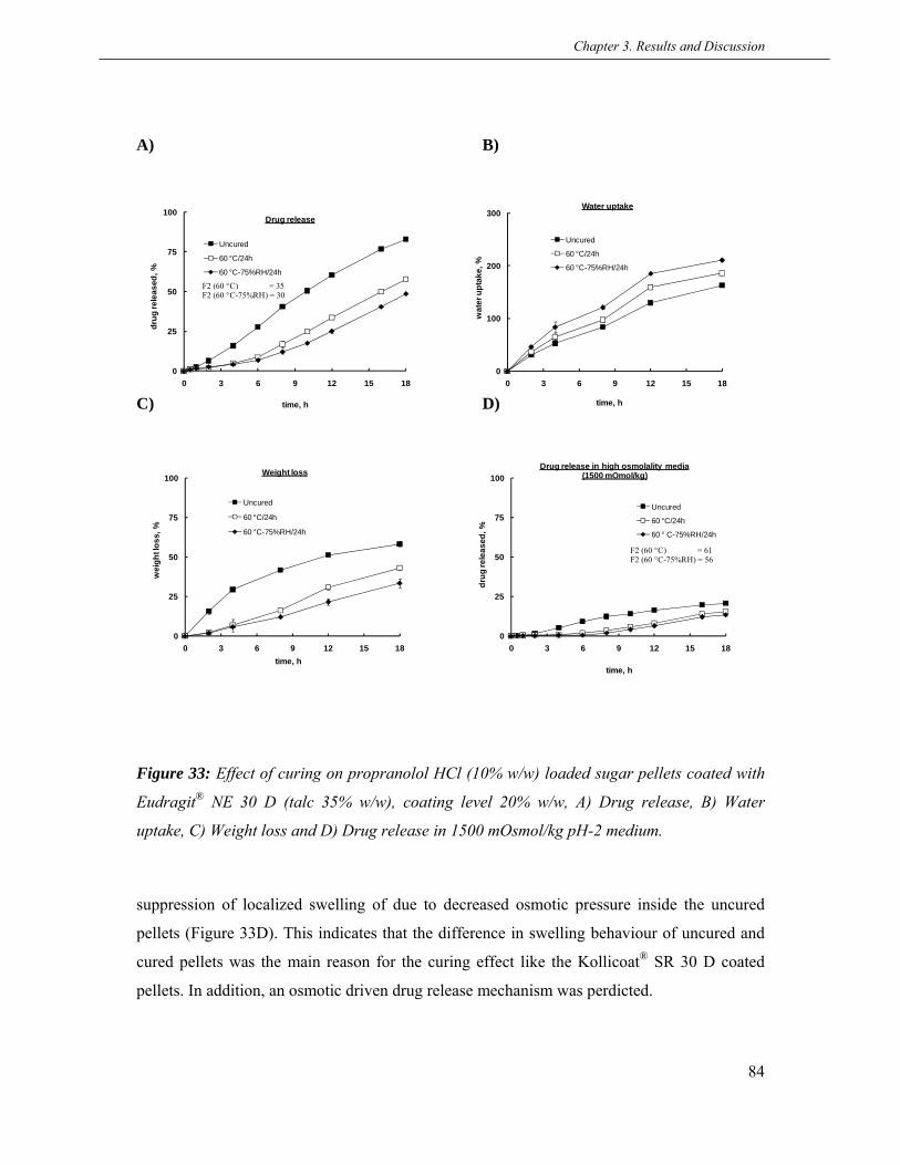
Chapter 3. Results and Discussion
84
0
25
50
75
100
0 3 6 9 12 15 18
wei
ght l
oss,
%
time, h
Weight loss
Uncured
60 °C/24h
60 °C-75%RH/24h
0
25
50
75
100
0 3 6 9 12 15 18
drug
rele
ased
, %
time, h
Drug release in high osmolality media (1500 mOmol/kg)
Uncured
60 °C/24h
60 ° C-75%RH/24h
F2 (60 °C) = 61F2 (60 °C-75%RH) = 56
0
25
50
75
100
0 3 6 9 12 15 18
drug
rele
ased
, %
time, h
Drug release
Uncured
60 °C/24h
60 °C-75%RH/24h
F2 (60 °C) = 35F2 (60 °C-75%RH) = 30
0
100
200
300
0 3 6 9 12 15 18
wat
er u
ptak
e, %
time, h
Water uptake
Uncured
60 °C/24h
60 °C-75%RH/24h
A) B)
C) D)
Figure 33: Effect of curing on propranolol HCl (10% w/w) loaded sugar pellets coated with
Eudragit® NE 30 D (talc 35% w/w), coating level 20% w/w, A) Drug release, B) Water
uptake, C) Weight loss and D) Drug release in 1500 mOsmol/kg pH-2 medium.
suppression of localized swelling of due to decreased osmotic pressure inside the uncured
pellets (Figure 33D). This indicates that the difference in swelling behaviour of uncured and
cured pellets was the main reason for the curing effect like the Kollicoat® SR 30 D coated
pellets. In addition, an osmotic driven drug release mechanism was perdicted.
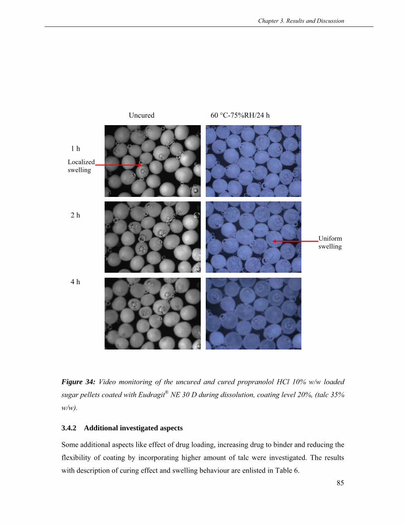
Chapter 3. Results and Discussion
85
Localized swelling
Uniform swelling
Uncured 60 °C-75%RH/24 h
1 h
2 h
4 h
Figure 34: Video monitoring of the uncured and cured propranolol HCl 10% w/w loaded
sugar pellets coated with Eudragit® NE 30 D during dissolution, coating level 20%, (talc 35%
w/w).
3.4.2 Additional investigated aspects
Some additional aspects like effect of drug loading, increasing drug to binder and reducing the
flexibility of coating by incorporating higher amount of talc were investigated. The results
with description of curing effect and swelling behaviour are enlisted in Table 6.

Chapter 3. Results and Discussion
86
Parameter Curing Drug:binder ratio (3:1) Talc 100% in coating2 30
Curing effect, f2 value 60 °C no (73) no (56) no (57) no (88)
60 °C-75%RH no (54) no (51) no (59) no (93)
Swelling behaviour Uncured Uniform Uniform Local Low uniform
60 °C-75%RH Extensive uniform Extensive uniform Low uniform Low uniform
Drug loading, %Description of experiments
The results showed that no curing effect was seen with a low (2%) or with a high (30%) drug
loading. In addition, the video monitoring during dissolution revealed the similar swelling
behaviour of the uncured and cured pellets in both drug loadings like the Kollicoat® SR 30 D
coated pellets.
Two approaches, i) increasing drug to binder ratio to resist decrease in adhesive force of the
coating and ii) incorporating higher amount of talc (100%) to reduce the flexibility of the
coatings, were successfully applied to eliminate the curing effect. Moreover, the long term
storage of the coated pellets with talc 100% in the coating was carried out under stress
conditions for 6 months and the drug release was found to be well stable (Figure 35 ).
Table 5 Effect of drug loading, drug to binder ratio and higher amount of talc in coating, on
the curing effect of propranolol HCl (10% w/w) loaded sugar pellets coated with Eudrgait®
NE 30 D, coating level 20% w/w.
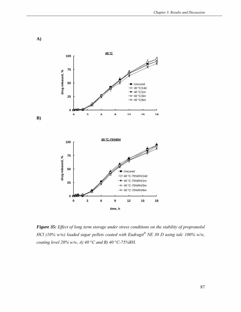
Chapter 3. Results and Discussion
87
0
25
50
75
100
0 3 6 9 12 15 18
drug
rele
ased
, %
time, h
40 °C
Uncured40 °C/14d40 °C/1m40 °C/3m40 °C/6m
0
25
50
75
100
0 3 6 9 12 15 18
drug
rele
ased
, %
time, h
40 °C-75%RH
Uncured40 °C-75%RH/14d40 °C-75%RH/1m40 °C-75%RH/3m40 °C-75%RH/6m
A)
B)
Figure 35: Effect of long term storage under stress conditions on the stability of propranolol
HCl (10% w/w) loaded sugar pellets coated with Eudragit® NE 30 D using talc 100% w/w,
coating level 20% w/w, A) 40 °C and B) 40 °C-75%RH.
Chapter 3. Results and Discussion
88
3.4.3 Conclusions
The Eudragit® NE 30 D coated sugar pellets showed a strong curing effect by following the
similar mechanism like the Kollicoat® SR 30 D coated pellets. The observed curing effect was
successfully eliminated by adopting different approaches in accordance with the identified
reasons. Importantly, the Eudragit® NE 30 D coated pellets without curing effect were storage
stable under stress conditions till 6 months.

Chapter 3. Results and Discussion
89
3.5 Evaluation of montmorillonites as anti-tacking agents
The objective of this part of work was to investigate anti-tacking properties of
montmorillonites (MMTs) in comparison with talc, a conventionally used anti-tacking agent,
using aqueous polymer dispersions. MMTs have a large surface area and a high aspect ratio;
exhibit good adsorption ability, cation exchange capacity and drug carrying capability. MMTs
are mostly used to enhance mechanical properties, reduce flammability and improve gas
barrier properties in plastic and paper industry (Majumdara et al., 2003). MMTs have been
used for different purposes in pharmaceutical field such as for the formation of drug-clay and
polymer-clay intercalated nanocomposites to attain controlled release (Menga et al., 2009;
Malhotra et al., 2006; Siew-Yoong and Milford, 2008; Joshi et al., 2010; Brostow et al., 2010;
Pongjanyakul and Rongthong, 2010), and to small extent as a filler, glidant and lubricant
(McGinity and Lach, 1975). However, no work so far has been done to investigate the anti-
tacking properties of MMTs.
3.5.1 Stability of the mixed dispersions
The stability of aqueous polymer dispersions is often disturbed upon addition of additives.
Addition of pigments to latex dispersions seldom affects their stability due to latex-pigment
interactions by disturbing electrical double layer balance (Derjaguin et al., 1941; Verway et
al., 1948; Hunter, 1989; Isrelachvili, 1991; Lyklema, 1995). Therefore, the stability of the
polymer dispersions resulted by the addition of different concentrations (10%, 20% and 35%
w/w) of the MMTs and talc was examined for 7 days. All polymer dispersions were found to
be stable by the addition of talc. However, Eudragit® RS 30 D and Eudragit® RL 30 D were
stable with both of used MMTs; Cloisite® Ca++ and Nanofill® 116. In contrast, Eudragit® NE
30 D and Eudragit® NM 30 D dispersions were stable with Nanofill® 116 and irreversible
flocculation occurred with Cloisite® Ca++, while Kollicoat® SR 30D was stable with Cloisite®
Ca++ and not with Nanofill® 116. The instability could be caused by the change in pH and
viscosity of the dispersions, surface charge of the colloidal particles and interaction of
pigments with the polymeric particles (Nyamweya et al., 2001; Dangel et al., 2000; Garcia-
Garcia et al., 2009; Ishikawa et al., 2005). The investigation of the exact reason for the
unstable dispersions was beyond the scope of this study. Both instability and stability of the
used polymer dispersions were observed independent of the MMTs concentrations. The stable
dispersions were used for further experiments. The solid contents of stable dispersions with

Chapter 3. Results and Discussion
90
Polymer/water Talc
Deionized water 104 ± 2.1
Kollicoat® SR 30 D 126 ± 1.5
Eudragit® RS 30 D 110 ± 2.8
Eudragit® NE 30 D 114 ± 0.9
Contact angle, θ° (n = 3)
0 19 ± 2.7
0 24 ± 0.7
0 18 ± 3.1
Cloisite® Ca++ Nanofill® 116
0 22 ± 1.6
35% addition of Nanofill® 116 were adjusted to 5% w/w due to its high swelling properties
for further studies.
3.5.2 Wettability of the MMTs and talc
The uniform distribution of additives in aqueous polymer dispersion is largely dependent on
their wettability. The hydrophilic substances have higher wettability and can be uniformly
distributed in the formulations whereas hydrophobic substances exhibit the opposite
behaviour due to their low wettability (Grundke et al., 2008). In order to check wettability, the
contact angle on the surfaces of the MMTs and talc was measured using deionized water and
the polymer dispersions (Table 6). Talc had the contact angle values over 100 with deioinzed
water and the polymer dispersions which showed its strong hydrophobic characteristics. In
contrary, the droplet on the surface of Cloisite® Ca++ spread very quickly and no value of
contact angle was recorded whereas Nanofill 116 exhibited small values due to its high
swelling properties. The lower contact values proved the higher wettability of the MMTs. The
increase in wettability of the agar based nanocomposite films by adding Cloisite® Na+ has also
been reported (Rhim, 2009).
Table 6 Contact angles of the MMTs and talc using deionized water and different aqueous
polymer dispersions.
3.5.3 Tackiness of various polymers
Tackiness of various aqueous polymer dispersions, which generally cause sticking problem
during coatings, was evaluated. Eudragit® NE 30 D, Eudragit® NM 30 D and Kollicoat® SR
30 D were selected as the flexible polymers, while Eudragit® RS 30 D and Eudragit® RL 30 D

Chapter 3. Results and Discussion
91
were used as an example of the sticky brittle polymers. The obtained tackiness force was of
the following order: Eudragit® NE 30 D ≥ Eudragit® NM 30 D > Eudragit® RS 30 D >
Kollicoat® SR 30 D > Eudragit® RL 30 D (Figure 36). The films of Eudragit® NE 30 D and
Eudragit® NM 30 D exhibited the maximum tackiness which could be due to their low glass
transition temperature and high flexibility. Even though the product temperature is usually
adjusted between 16–20 °C for this polymer, the agglomeration problem during coating
cannot be overcome completely (Bodmeier and Paeratakul, 1994b; Wesseling et al., 1999).
Interestingly, the films of Kollicoat® SR 30 D, another flexible polymer known to cause an
agglomeration problem during coating, did not show higher tackiness like Eudragit® NE 30 D.
This could be related to the presence of polyvinyl alcohol, a water soluble polymer, in
Kollicoat® SR 30 D dispersion (Kollicoat® SR 30 D technical information, 2007). The films
of Eudragit® RS 30 D and Eudragit® RL 30 D displayed tackiness behaviour almost similar to
Kollicoat® SR 30 D. Although, often strong electrostatic interaction and agglomeration
problem are evident with these polymers if use without anti-tacking agents. The lower
tackiness with these polymers could be linked to their high glass transition temperature. The
slightly lower tackiness of Eudragit® RL 30 D compared to Eudragit® RS 30 D could be
possibly explained by the presence of more quaternary ammonium groups in the polymer
(Wesseling et al., 1999).
Figure :36 Tackiness of films prepared from different aqueous polymer dispersions
(Eudragit® RS & RL 30 D and Kollicoat® SR 30 D were plasticized with 20% w/w and 10%
w/w TEC respectively, while Eudragit® NE & NM 30 D films were plasticizer free).
In addition, the detachment force required to separate the polymeric films was increased after
inducing tackiness at 40 °C-75% RH for 2 h. This could be due to the exposure of the films to
0
10
20
30
40
Force, N
40 °C‐2h
40 °C‐75%RH ‐2h

Chapter 3. Results and Discussion
92
more humid conditions where water acted as a plasticizer and increased the flexibility of
polymer particles and consequently increased tackiness.
3.5.4 Effect of the MMTs and talc on tackiness
The tackiness of the different polymeric films (Eudragit® NE 30 D, Kollicoat® SR 30D and
Eudragit® RS 30 D) using various concentrations (10%, 20% and 35%) of the MMTs
(Cloisite® Ca++ and Nanofill® 116) and talc are shown in Figure (37A-C). Interestingly, the
MMTs reduced the tackiness of polymeric films more effectively than talc in all used
concentrations and at both tackiness inducing conditions (40 °C-2 h and 40 °C-75%RH/2 h).
Eudragit® NE 30 D films with 20% of Nanofill® 116 had lower detachment force when
compared with 20% and 35% of talc (Figure 37A). Also Eudragit® NM 30 D films had the
same trend (data not shown). Similarly, the films of Kollicoat® SR 30 D with 20% of
Cloisite® Ca++ required lower force to separate the films than those with 20% and 35% of talc
(Figure 37B). The films of Eudragit® RS 30 D with 10% of cloisite® Ca++ and Nanofill® 116
exhibited a lower detachment force in comparison with 10%, 20% and 35% of talc. The
Eudragit® RL 30 D films had the same trend (data not shown). The lower detachment force of
films with the MMTs could be attributed to their uniform distribution in the polymer
dispersions and then in casted films (Figure 38A-C). In comparison, talc was not uniformly
distributed and a few talc free small patches were observed in the films (Figure 38A-C). The
irregular distribution of talc might be related to its high hydrophobicity, which can be
explained by the absence of Al and OH in the octahedral and basal surface of elementary
sheets, respectively. On the other hand, the MMTs, being hydrophilic in nature, were
homogenously dispersed in the formulations and as a result had a uniform and smooth
distribution in the casted films. This led to a better covering of polymeric particles by the
MMTs and eventually reduced the tackiness more effectively.

Chapter 3. Results and Discussion
93
0
5
10
15
20
25
30
35
0 10 20 35
force, N
conc., %
40 ºC ‐ 2 h
No additives
Nanofill 116
Talc
0
5
10
15
20
25
30
35
0 10 20 35
force, N
conc., %
40 ºC ‐ 75%RH ‐ 2 h
No additives
Nanofill 116
Talc
0
2
4
6
8
10
0 10 20 35
force, N
conc., %
40 ºC ‐ 2 h
No additives
Cloisite Ca++
Talc
0
2
4
6
8
10
0 10 20 35
force, N
conc., %
40 ºC ‐ 75%RH ‐ 2 h
No additives
Cloisite Ca++
Talc
0
2
4
6
8
10
0 10 20 35
force, N
conc., %
40 ºC ‐ 2 h
No additives
Cloisite Ca++
Nanofill 116
Talc
0
2
4
6
8
10
0 10 20 35
force, N
conc., %
40 ºC ‐ 75%RH ‐ 2 h
No additives
Cloisite Ca++
Nanofill 116
Talc
A) Eudragit® NE 30D
B) Kollicoat® SR 30D
C) Eudragit® RS 30D
Figure 37: Effect of different concentrations of the MMTs and talc on the tackiness of various
polymeric films A) Eudragit® NE 30 D, B) Kollicoat® SR 30 D and C) Eudragit® RS 30 D.

Chapter 3. Results and Discussion
94
A) Eudragit® NE 30 D
No additive Nanofill® 116 Talc
B) Kollicoat® SR 30 D
No additive Cloisite® Ca++ Talc
C) Eudragit® RS 30 D
No additive Cloisite® Ca++ Talc
Figure 38: Macroscopic images of the polymeric films containing 20% w/w of the MMTs and
talc A) Eudragit® NE 30 D, B) Kollicoat® SR 30 D and C) Eudragit® RS 30 D.

Chapter 3. Results and Discussion
95
40 °C /2h 40 °C - 75%RH /2h 40 °C /2h 40 °C - 75%RH /2h
Kollicoat® SR 30 D* – 5.20 ± 0.27 8.32 ± 0.47 7.15 ± 0.48 10.54 ± 0.49Cloisite® Ca++, 10 0.71 ± 0.10 1.21 ± 0.05 1.53 ± 0.26 2.28 ± 0.16Cloisite® Ca++, 20 0.21 ± 0.06 0.40 ± 0.04 0.79 ± 0.06 1.21 ± 0.07Cloisite® Ca++, 35 0.10 ± 0.01 0.20 ± 0.01 0.35 ± 0.05 0.63± 0.09
Talc, 10 1.38 ± 0.01 2.07 ± 0.20 2.07 ± 0.20 2.29 ± 0.07Talc, 20 0.95 ± 0.05 1.05 ± 0.16 1.05 ± 0.16 1.92 ± 0.05Talc, 35 0.63 ± 0.04 0.79 ± 0.03 0.79 ± 0.03 1.05 ± 0.06
Eudragit® RS 30 D** – 5.58 ± 0.22 8.93 ± 0.31 11.46 ± 0.70 15.69 ± 0.47Cloisite® Ca++, 10 0.84 ± 0.06 1.42 ± 0.15 2.46 ± 0.32 3.98 ± 0.20Cloisite® Ca++, 20 0.26 ± 0.03 0.91 ± 0.04 1.19 ± 0.16 1.96 ± 0.23Cloisite® Ca++, 35 0.07 ± 0.01 0.23 ± 0.02 0.58 ± 0.05 1.09 ± 0.08
Talc, 10 2.30 ± 0.15 3.52 ± 0.14 5.79 ± 0.10 8.31 ± 0.59Talc, 20 1.31 ± 0.09 2.36 ± 0.11 3.51 ± 0.07 5.46 ± 0.11Talc, 35 0.88 ± 0.08 1.53 ± 0.08 2.54 ± 0.18 4.03 ± 0.27
*Kollicoat® SR 30 D-TEC concentration increased from 10% to 20% w/w**Eudragit® RS 30 D-TEC concentration increased from 20% to 30% w/w
Polymer Anti-tacking agent, %
Detachment force (N), M. V. ± S. D., n = 6
Lower plasticizer concentations Higher plasticizer concentrations
3.5.5 Effect of plasticizer concentration on tackiness
Higher amounts of plasticizers are added into coating formulations for many reasons, for
instance, making the coatings flexible to avoid any damages during the compression of
pellets, to reduce stability problems especially with brittle coatings and in some cases to
withstand the mechanical agitation of gastrointestinal tract on thin coated dosage forms.
Therefore, the effect of higher concentrations of plasticizer (TEC) on anti-tacking effect of the
MMTs and talc was determined and the results are enlisted in Table 7. The tackiness of
Kollicoat® SR 30 D and Eudragit® RS 30 D films was increased as a function of TEC
concentration but the effect was more pronounced with Eudragit® RS 30 D films which could
probably due to the sticky nature of the polymer.
Table 7 Effect of plasticizer concentration on the tackiness of Kollicoat® SR 30 D and
Eudragit® RS 30 D casted polymeric films.
The glass transition temperature (Tg) and minimum film formation temperature (MFT)
decreases with an increase in TEC concentration and it correlates with the tackiness of the
dried films (Wesseling et al., 1999). A correlation between hydrophilic plasticizers and the
adhesion of polymeric film has also been reported (Felton and McGinity, 1997). However, the
increased concentrations of TEC did not affect the anti-tacking properties of the MMTs and
talc noticeably.

Chapter 3. Results and Discussion
96
Kollicoat® SR 30 D* –Cloisite® Ca++, 10Cloisite® Ca++, 20Cloisite® Ca++, 35
Talc, 35Eudragit® RS 30 D** –
Cloisite® Ca++, 10Cloisite® Ca++, 20Cloisite® Ca++, 35
Talc, 35*Kollicoat® SR 30 D plasticized with TEC 10% w/w
27
232425
**Eudragit® RS 30 D plasticized with TEC 20% w/w
Anti-tacking agent, %Polymer Tg, °C
25
2833
242025
3.5.6 Effect on Tg of polymers
The DSC studies of the polymeric casted films were performed to see the effect of the MMTs
and talc on the Tg of the polymers. The results showed that 10% addition of Cloisite® Ca++
induced a small increase in Tg of Kollicoat® SR 30 D which was gradually increased as a
function of concentration whereas 35% addition of talc did not bring any change in Tg of
Kollicoat® SR 30 D. In comparison, a pronounced increase in Tg of Eudragit® RS 30 D was
observed by adding Cloisite® Ca++ For example, 10% addition of Cloisite® Ca++ increased the
Tg of the Eudragit® RS 30 D from 20 to 25 ºC which was further raised to 33 ºC by
increasing concentration to 35%. However, 35% addition of talc also increased the Tg of
Eudragit® RS 30 D from. 20 to 25 ºC (Table 8). The increase in Tg could be related to
multiple factors such as reduction of polymer free volume, interaction between the polar
groups of the polymer and the MMTs, and physical stiffening resulting from the presence of
the rigid antiplasticizer molecules adjascent to the polar group of the polymer (Jackson et al.,
1967). The increase in Tg of the amylose by polymer-nanoclay interaction has been reported
(Liua et al., 2011). The more increase in Tg of Eudragit® RS 30 D could be due to interaction
of positively charged groups of Eudragit® RS 30 D with negatively charged ions on the
surface of the MMTs.
Table 8 Effect of the MMTs and talc on the Tg of Kollicoat® SR 30 D and Eudragit® RS 30 D.

Chapter 3. Results and Discussion
97
3.5.7 Coating processability of polymer dispersions
The coating processability of the different aqueous polymer dispersions with the addition of
the MMTs was evaluated in order to check their practical advantage over talc. A good coating
quality with high efficiency and short processing time has always been of a great importance
for pharmaceutical industries. For this purpose, three different concentrations of the MMTs
(10 %, 20% and 35%) were compared with 35% of talc. Cloisite® Ca++ was used with
Kollicoat® SR 30 D and Eudragit® RS 30 D, while Nanofill® 116 was investigated with
Eudragit® NE 30 D and Eudragit® RS 30 D.
The coating processability was analyzed on a small scale fluidized bed coater (Mini-Glatt 4,
Glatt GmbH, Binzen, Germany) equipped with a bottom spary (Wurster system) using 70 g of
sugar pellets loaded with propranolol HCl 10% as cores. The performance of coating was
rated into three zones of excellent, good and poor quality based on spray rate, processing
time, flow of pellets, yield and nozzle clogging (Cervera et al., 2004).
The coating processability of Eudragit® NE 30 D is depicted in Figure 39A-D. The addition of
Nanofill® 116 to Eudragit® NE 30 D formulations resulted in better coating processability in
comparison with talc. A 10% addition of Nanofill® 116 provided a wider zone of excellent
processability compared to 35% of talc (Figure 39A & D). However, by further increasing
Nanofill® 116 concentrations to 20% and 35%, the zones of excellent and good processability
were widened to a small extent. This can be explained becuase Eudragit® NE 30 D has a low
glass transition temperature of - 8 ºC and to use a product temperature over 23 ºC is not
possible.
The coating processability of Kollicoat® SR 30 D was improved effectively by the addition of
different concentrations of Cloisite® Ca++ (Figure 40A-D). An addition of 10% Cloisite® Ca++
to coating formulation provided much broader zone of excellent processability in comparison
with 35% of talc (Figure 40A & D). With the addition of 10% of Cloisite® Ca++, a spray rate
of up to 2.7 g/min was achieved without any signs of bed formation, nozzle clogging or
sticking of pellets using a product temperature range of 28–36 ºC. Importantly, by further
increasing the concentration of Cloisite® Ca++ to 20% and 35%, the zones of excellent and
good processability were widened correspondingly. For example, a maximum spray rate of
3.3 g/min with good flowability of pellets could be achieved with 35% addition of Cloisite®

Chapter 3. Results and Discussion
98
12
15
18
21
24
27
30
33
36
39
0.5 0.9 1.3 1.7 2.1 2.5 2.9 3.3 3.7 4.1
Prod
uct t
empe
ratu
re, °
C
Spray rate, g/min
12
15
18
21
24
27
30
33
36
39
0.5 0.9 1.3 1.7 2.1 2.5 2.9 3.3 3.7 4.1
Prod
uct t
empe
ratu
re, °
C
Spray rate, g/min
A) B)
C) D)
Figure 39: Coating processability of Eudragit® NE 30 D formulations containing A)
Nanofill® 116 10% w/w, B) Nanofill® 116 20% w/w, C) Nanofill® 116 35% w/w and D) Talc
35% w/w, (■) excellent, (■) good and (□) poor.
12
15
18
21
24
27
30
33
36
39
0.5 0.9 1.3 1.7 2.1 2.5 2.9 3.3 3.7 4.1Pr
oduc
t tem
pera
ture
, °C
Spray rate, g/min
12
15
18
21
24
27
30
33
36
39
0.5 0.9 1.3 1.7 2.1 2.5 2.9 3.3 3.7 4.1
Prod
uct t
empe
ratu
re, °
C
Spray rate, g/min

Chapter 3. Results and Discussion
99
13
16
19
22
25
28
31
34
37
40
0.5 0.9 1.3 1.7 2.1 2.5 2.9 3.3 3.7 4.1
Prod
uct t
empe
ratu
re, °
C
Spray rate, g/min
13
16
19
22
25
28
31
34
37
40
0.5 0.9 1.3 1.7 2.1 2.5 2.9 3.3 3.7 4.1
Prod
uct t
empe
ratu
re, °
C
Spray rate, g/min
A) B)
C) D)
Figure 40: Coating processability of Kollicoat® SR 30 D formulations containing A)
Cloisite® Ca++ 10%, B) Cloisite® Ca++ 20%, C) Cloisite® Ca++ 35% and D) Talc 35%, (■)
excellent, (■) good and (□) poor
13
16
19
22
25
28
31
34
37
40
0.5 0.9 1.3 1.7 2.1 2.5 2.9 3.3 3.7 4.1
Prod
uct t
empe
ratu
re, °
C
Spray rate, g/min
15
17
19
21
23
25
27
29
31
33
35
37
39
0.5 0.9 1.3 1.7 2.1 2.5 2.9 3.3 3.7 4.1
Prod
uct t
empe
ratu
re, °
C
Spray rate, g/min

Chapter 3. Results and Discussion
100
Ca++ which is almost twice as high as of 35% talc. Moreover, the number of pellet doubles
after coating process was significantly lower with Cloisite® Ca++ coated formulations. In
contrast, an addition of 35% talc could attain maximum 1.7 g/min of spray rate using a
product temperature range of 28–32 ºC with a few times nozzle clogging.
The better coating processability of formulations with Nanofill® and Cloisite® Ca++could be
attributed to their smaller particle size and better wettability over talc that led to their stable
uniform distribution in the coating formulations. However, the lower wettability of talc
particles due to its high hydrophobicity could be the reason for unstable non-uniform
distribution which resulted in low spray rate and nozzle clogging (Lin et al., 1994).
Eudragit® RS 30 D, a charged cationic polymer, is widely used for the controlled release
coatings. The tendency of sticking during coating process and upon curing has been widely
reported with this polymer (Maejima et al., 2000; Pearnchob and Bodmeier, 2003).
The coating processability of Eudragit® RS 30 D formulations using different concentrations
of Cloisite® Ca++ (10%, 20% and 35%) and talc (35%) is shown in Figure 41A-D. The coating
processability of Eudragit® RS 30 D formulations was improved to a large extent by the
addition of Cloisite® Ca++ than talc. For example, a formulation with 10% addition of
Cloisite® Ca++ achieved a maximum spray rate of 3.1 g/min with a higher product temperature
range in comparison to 2.1 g/min of talc 35% with a lower product temperature range (Figure
41A & D). Moreover, the spray rate was further increased to 3.5 g/min and 3.9 g/min by
increasing the concentration of Cloisite® Ca++ to 20% and 35%, respectively (Figure 41B &
C). The improved coating processability of Eudragit® RS 30 D with the addition of Cloisite®
Ca++could also be linked to its stable and uniform distribution in the coating formulations.
The extent of electrostatic interaction during the coating process was decreased with Cloisite®
Ca++ formulations which might be due to the neutralization of positively charged -NH3 groups
of Eudragit® RS 30 D by negatively charged ion on the surface of Cloisite® Ca++. The coating
processability of Eudragit® RS 30 D using Nanofill® 116 was also effectively improved (data
not shown).
The uniform distribution of the MMTs was confirmed from the SEM images. Figure 42A-B
showed the SEM micrographs of cross section of the Eudragit® RS 30 D coated pellets with
Cloisite® Ca++ and Nanofill® 116 and talc distribution in the coatings. The order of
distribution was ranked as Nanofill® 116 > Cloisite® Ca++ > talc.

Chapter 3. Results and Discussion
101
Another advantage of Cloisite® Ca++ addition to the coating formulation was the pale yellow
appearance of the final coated pellets which could be beneficial to enhance the patient
compliance. The quality of product and coating process could also be reflected by the colour
of the product since it is an easily observable feature of the drug product (Chan et al., 2001).
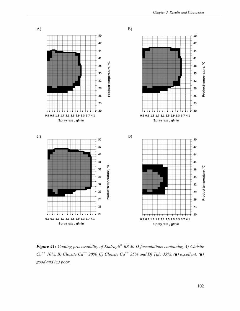
Chapter 3. Results and Discussion
102
20
23
26
29
32
35
38
41
44
47
50
0.5 0.9 1.3 1.7 2.1 2.5 2.9 3.3 3.7 4.1
Prod
uct t
empe
ratu
re, °
C
Spray rate , g/min
20
23
26
29
32
35
38
41
44
47
50
0.5 0.9 1.3 1.7 2.1 2.5 2.9 3.3 3.7 4.1
Prod
uct t
empe
ratu
re, °
C
Spray rate , g/min
A) B)
C) D)
Figure 41: Coating processability of Eudragit® RS 30 D formulations containing A) Cloisite
Ca++ 10%, B) Cloisite Ca++ 20%, C) Cloisite Ca++ 35% and D) Talc 35%, (■) excellent, (■)
good and (□) poor.
20
23
26
29
32
35
38
41
44
47
50
0.5 0.9 1.3 1.7 2.1 2.5 2.9 3.3 3.7 4.1
Prod
uct t
empe
ratu
re, °
C
Spray rate , g/min
20
23
26
29
32
35
38
41
44
47
50
0.5 0.9 1.3 1.7 2.1 2.5 2.9 3.3 3.7 4.1
Prod
uct t
empe
ratu
re, °
C
Spray rate , g/min

Chapter 3. Results and Discussion
103
A) Cloisite® Ca++ Nanofill® 116 Talc
B)
C)
Figure 42: SEM images of the cross section of pellets showing the distribution of the MMTs
and talc (35% w/w) in Eudragit® RS 30 D coatings, TEC 20% w/w, coating level 20% w/w, A)
cross-section of the whole pellets (lower magnification,) B) cross-section of one segment
(higher magnification) and C) mapping of Mg.

Chapter 3. Results and Discussion
104
200 ml, pH-2 40 °C 40 °C - 75%RH 60 °C 60 °C - 75%RH 37 ± 0.5 °C
Kollicoat® SR 30 D* – 99.8 99.8 99.9 99.9 99.9Cloisite® Ca++, 10 2.5 60.1 4.2 64.4 60.1Cloisite® Ca++, 20 0.15 22.6 1.4 24.6 29.6Cloisite® Ca++, 35 0.07 1.7 0.4 4.1 2.2
Talc, 35 0.7 99.3 7.1 99.3 88.1Eudragit® RS 30 D** – 97.6 98.8 97.5 98.3 49.5
Cloisite® Ca++, 10 8.6 1.44 3.5 2.56 2.44Cloisite® Ca++, 20 6.5 0.98 2.6 0.51 1.33Cloisite® Ca++, 35 0.86 0.32 0.8 1.38 0.32
Talc, 35 18.5 8.2 2.8 89.9 2.8Eudragit® NE 30 D – 99.8 99.9 99.9 99.9 99.9
Nanofill® 116, 10 1.17 13.8 0.99 24.2 24.9Nanofill® 116, 20 0.98 6.8 0.6 7.9 5.55Nanofill® 116, 35 0.67 3.4 0.52 4.2 4.52
Talc, 35 0.67 24.1 0.67 90.5 90.5*Kollicoat® SR 30 D plasticized with 10% TEC**Eudragit® RS 30 D plasticized with 20% TEC
% agglomeration after 24 hCuring / Storage Anti-tacking agent, %Polymer
3.5.8 Tackiness of the coated pellets during curing
To evaluate the anti-tacking performance during curing, the coated pellets using different
concentrations of the MMTs and talc were stored at different curing conditions without
adding any external anti-tacking agent. The tackiness was characterized by % agglomeration
(Table 9).
The coated pellets without adding anti-tacking agent in the coating formulations were
irreversibly agglomerated during curing. In comparison, the Kollicoat® SR 30 D coated
pellets with 10% addition of Cloisite® Ca++ did not stick at elevated temperature but showed
60% of agglomeration at elevated temperature/humidity which was reduced to 30% and 4%
by increasing concentration to 20% and 35%, respectively. In contrary, the pellets with 35%
addition of talc in the coating were completely agglomerated at elevated temperatures and
humidity.
Table 9 Effect of the MMTs and talc on the tackiness of coated pellets during curing at
different temperature and humidity.
The Eudragit® RS 30 D pellets with Cloisite® Ca++ in the coatings did not stick irrespective of
concentration and curing conditions. However, the pellets with 35% of talc in the coating did

Chapter 3. Results and Discussion
105
not agglomerate under low temperature and humidity conditions but had a 89 % of
agglomeration at higher temperature and humidity. The Eudragit® NE 30 D coated pellets
with 10% of and 20% of Nanofill® 116 showed about 24% and 7.9% of agglomeration,
respectively, under elevated temperatures and humidities which was limited to 4.5% with
35% addition of Nanofill® 116. In comparison, the pellets with 35% talc in the coating did not
stick at elevated temperatures but had a 90% agglomeration at elevated temperatures and
humidities.
The difference in the agglomeration tendency of various polymers at different elevated
temperatures and humidities could be related to their variable sensitivity towards the exposed
conditions. A clear correlation was found between the tackiness of the films, coating
processability and sticking of the pellets upon curing.
3.5.9 Mechanical properties of polymeric films
The type and the amount of the additives in the film can significantly affect the mechanical
properties which in turn can have a big influence on the drug release mechanism from the
coated drug product. Hence, the effect of different conentrations of the MMTs and talc on the
mechanical properties of casted films in a dry state was evaluated (Table 10).
The Cloisite® Ca++ addition decreased the flexibility of the Kollicoat® SR 30 D films more
effectively than talc while the puncture strength was less affected which is an indicative of
better mechanical strength of films. For instance, a film with of 10% of Cloisite® Ca++ had
less % elongation and more puncture strength when compared with 35% addition of talc. In
contrast, the addition of Cloisite® Ca++ to Eudragit® RS 30 D films decreased both flexibility
and puncture strength significantly more than talc which is suggestive of hard and brittle
films. This could be due to interaction of positively charged –NH3 groups of the polymer with
negatively charged ions of the Cloisite® Ca++. The decrease in tensile strength and elongation
of poly-L-lactide composites in presence of Cloisite Ca++ has been reported (Rhim et al.,
2009).
However, the interesting results were found with Eudragit® NE 30 D films. The addition of
Nanofill® 116 reduced the flexibility of the films higher than talc. However, the puncture
strength was increased as a function of Nanofill® 116 concentration which was even more
than with the pure films. The increase in puncture strength occured by the stronger interfacial
interaction through hydrogen or ionic bonds between polymer and intercalated layered silicate

Chapter 3. Results and Discussion
106
with vast interfacial area (Alamsi et al., 2010). A similar behaviour of increase in puncture
strength with increase in the clay content have been observed in various biopolymer-based
nanocomposite films such as starch (Alamsi et al., 2010; Avella et al., 2005; Cyras et al.,
2008; Chrissafis et al., 2007; Huang et al., 2006; Jong-Whan Rhim, 2009), chitosan
(Casariego et al., 2009), and soy protein (Kumar et al., 2010b).
Table 10 Effect of the MMTs and talc on the mechanical properties of different polymeric
casted films in dry state (thickness = 33 –350 µm).
It can be concluded that the MMTs decreased the flexibility of polymers higher than talc.
However, the MMTs reduced the puncture strength of the charged polymers (Eudragit® RS 30
D) more and less of the neutral polymers (Kollicoat® SR 30D, Eudragit® NE 30 D) when
compared with talc. The pronounced reduction in the puncture strength of Eudragit® RS 30 D
films could also be due to increase in Tg (Table 8).
Kollicoat® SR 30 D* –Cloisite® Ca++, 10Cloisite® Ca++, 20Cloisite® Ca++, 35
Talc, 10Talc, 20Talc, 35
Eudragit® RS 30 D** –Cloisite® Ca++, 10Cloisite® Ca++, 20Cloisite® Ca++, 35
Talc, 10Talc, 20Talc, 35
Eudragit® NE 30 D –Nanofill® 116, 10Nanofill® 116, 20Nanofill® 116, 35
Talc, 10Talc, 20Talc, 35
*Kollicoat® SR 30 D plasticized with TEC 10%
Polymer Anti-tacking agent, %Mechanical properties, n = 9
M. V. ± S. D. M. V. ± S. D.Puncture Strength, N/mm2 Elongation, %
17.7 ± 0.913.4 ± 0.913.1 ± 1.212.6 ± 0.812.7 ± 0.211.9 ± 1.211.6 ± 0.610.7 ± 0.73.9 ± 0.10.7 ± 0.10.6 ± 0.19.7 ± 0.88.2 ± 0.26.7 ± 0.812.6 ± 0.615.3 ± 0.425.3 ± 0.5
8.4 ± 0.67.9 ± 0.37.2 ± 0.4
176.4 ± 1.992.5 ± 1.473.8 ± 0.137.7 ± 0.1132.5 ± 0.8122.0 ± 0.1
157.7 ± 0.273.9 ± 0.4
338.2 ± 0.5
114.2 ± 0.273.7 ± 0.237.5 ± 0.214.7 ± 0.210.6 ± 0.145.5 ± 0.2
26.2 ± 0.7 63.9 ± 0.9
**Eudragit® RS 30 D plasticized with TEC 20%
276.0 ± 0.1246.2 ± 0.1
40.2 ± 0.437.7 ± 0.1431.1 ± 0.7

Chapter 3. Results and Discussion
107
3.5.10 Drug release from coated pellets
The drug release from the coated pellets was performed to compare the effect of the MMTs
and talc on release rates. In addition, the pellets were cured to elucidate the effect of curing.
Some of the drug release results are shown in Figure 43A-E comparing 35% addition of both
MMTs and talc to coating formulations.
The drug release from Kollicoat® SR 30 D coated pellets shows that Cloisite® Ca++ addition
did not bring unwanted change in release rates before and after curing (Figure 43A & B).
Positively, the curing effect was eliminated at elevated temperature which could be due to
reduction in the flexibility of the coatings (shown earlier). The elimination of curing effect of
Kollicoat® SR 30 D coated pellets has been shown by decreasing the flexibility of coatings
(Ahmed et al., 2008). Moreover, the 20% addition of Cloisite® Ca++ to coatings almost had the
same results (data not shown).
In comparison with talc addition, the slightly faster drug release from Eudragit® RS 30 D
coated pellets was observed with Cloisite® Ca++ (Figure 43C & D). This could be due to
pronounced decrease in the flexibility of coatings by Cloisite® Ca++. In addition, 10% and
20% addition of Cloisite® Ca++ almost had the same results (data not shown). The drug
release was remained unaffected after curing from pellets having Cloisite® Ca++ or talc in the
coatings.
Nanofill® 116 addition as well did not show unexpected effect on the drug release from
Eudragit® NE 30 D coated pellets in comparison with talc (Figure 43E & F). However,
likewise Kollicoat® SR 30 D, the curing effect was minimized due to the decrease in
flexibility of the coatings.

Chapter 3. Results and Discussion
108
0
25
50
75
100
0 3 6 9 12 15 18
drug
rele
ased
, %
time, h
Uncured60 °C/24h60 °C-75%RH/24h
0
25
50
75
100
0 3 6 9 12 15 18
drug
rele
ased
, %
time, h
Uncured
60 °C/24h
60 °C-75%RH/24h
0
25
50
75
100
0 3 6 9 12 15 18
drug
rele
ased
, %
time, h
Uncured
60 °C/24h
60 °C-75%RH/24h
0
25
50
75
100
0 3 6 9 12 15 18
drug
rele
ased
, %
time, h
Uncured
60 °C/24h
60 °C-75%RH/24h
0
25
50
75
100
0 3 6 9 12 15 18
drug
rele
ased
, %
time, h
Uncured
60 °C/24h
60 °C-75%RH/24h
0
25
50
75
100
0 3 6 9 12 15 18
drug
rele
ased
, %
time, h
Uncured
60 °C/24h
60 °C-75%RH/24h
A) B)
C) D)
E) F)
Figure 43: Propranolol HCl release from pellets coated with different polymers using 35%
w/w of the MMTs and talc, coating level 20% w/w, A) Kollicoat® RS 30 D-Cloisite® Ca++, B)
Kollicoat® RS 30 D-talc, C) Eudragit® RS 30 D-Cloisite® Ca++, D) Eudragit® RS 30 D-talc,
E) Eudragit® NE 30 D-Nanofill® 116 and D) Eudragit® NE 30 D-talc.

Chapter 3. Results and Discussion
109
0
25
50
75
100
0 3 6 9 12 15 18
drug
rele
ased
, %
time, h
Uncured60°C-24h without AA60°C-24h 1% Closiite Ca++60°C-24h 1% Talc60°C-24h 1% SiO2
3.5.11 MMTs as external anti-tacking agent
The performance of the MMTs as external anti-tacking agent was evaluated. In this regard,
1% addition of Cloisite® Ca++, talc and Aerosil® to Eudragit® RS 30 D coated pellets were
compared during curing at 60 °C. The agglomerated pellets after curing were separated by
applying mechanical force which could damage the coating depending on the extent of
sticking. The difference in damage was compared from release results of the uncured and
cured pellets and thus related with anti-tacking effect.
Figure 44: Theophylline release Eudragit® RS 30 D coated pellets showing the anti-tacking
effect of different anti-tacking agents during curing (TEC 20% w/w), coating level 20 % w/w.
The pellets cured without adding anti-tacking agent were irreversibly sticked together and a
strong mechanical force was applied to separate them. In comparison, the pellets cured using
1% of talc, Aerosil® and Cloisite® Ca++ showed partial reversible sticking of various extents.
The pellets were separated by applying a mild mechanical force.
Theophylline release from the uncured pellets followed almost zero order patterns due to its
low solubility. However, the pellets cured without anti-tacking agent revealed a very fast
release profile with 80% release in 1 h (Figure 44). This was due to the breaking of coating
during separation of the pellets, which was seen in macroscopic pictures after the release

Chapter 3. Results and Discussion
110
(Figure 45B). In contrast, the pellets cured using 1% of talc and aerosil showed a small
increase in release profile which is an indication of tiny cracks formed in coatings while
separating the pellets (Figure 45C & D). However, the drug release from pellets cured using
Cloisite® Ca++ 1% exhibited a small decrease and no visible cracks were observed in coatings
from the macroscopic picture after release. This indicates that pellets cured with Cloisite®
Ca++ had less sticking and a gentle mechanical force was enough to separate them. As a result,
the coating was not damaged and the drug release decreased due to better film formation upon
curing.
A) B) C)
D) E)
Figure 45: Macroscopic pictures of the Eudragit® RS 30 D coated pellets after release (TEC
20% w/w), coating level 20% w/w, A) uncured, B) 60 °C/24 h cured without anti-tacking
agent, C) 60 °C/24 h cured using 1% w/w of aerosil, D) 60 °C/24 h cured in presence of 1%
w/w of talc and E) 60 °C/24 h cured in presence of 1% w/w of Cloisite® Ca++.
Crack/rupture
Crack/rupture

Chapter 3. Results and Discussion
111
3.5.12 Conclusions
Two MMTs, Cloisite® Ca++ and Nanofill® 116, were found to be the promising anti-tacking
agents for aqueous polymer coatings. The MMTs in lower concentrations reduced the
tackiness of casted polymeric films more effectively comparing with a higher concentration of
talc. Practically, the MMTs improved the coating processability of different aqueous polymer
dispersions to a large extent. In addition, the MMTs exhibited better anti-tacking properties
during curing of coated pellets. The MMTs affected the mechanical properties varyingly
depending on the nature of polymer (charged vs. neutral) but did not bring any unexpected
change on the drug release from coated pellets before and after curing. Positively, in some
case, the elimination of curing effect was observed due to decrease in flexibility of the
coatings.


Chapter 4. Summary
112
4 Summary
Curing mechanism of flexible aqueous polymer coatings
The main objective of this part of work was to investigate and understand the curing
mechanism of Kollicoat® SR 30 D coated pellets. In this regard, effect of starter cores, drug
solubility, drug loading, adhesion of coating to core and flexibility of coating on curing was
evaluated in detail.
A strong curing effect (change in drug release) was observed with propranolol HCl loaded
sugar pellets coated with Kollicoat® SR 30 D within a drug loading range of 10%-20% at
elevated temperature (60 °C) and elevated temperature/humidity (60 °C-75%RH). The curing
effect was more pronounced at 60 °C-75%RH. Importantly, a complete film formation from
Kollicoat® SR 30 D coatings was confirmed by unchanged properties (water uptake, weight
loss, mechanical properties in wet state and permeability) of free films after curing. Thus, the
observed curing effect could not be explained by well-known hydroplasticization or further
coalescence of polymeric particles. However, a significant difference between water uptake
and swelling of uncured and cured pellets was observed during dissolution. The curing effect
was explained by a completely different swelling behaviour of uncured and pellets cured at 60
°C-75%RH observed from video monitoring during dissolution. This is described in the
following two scenarios:
First scenario (observed for uncured pellets): Upon medium penetration into pellets, coating
detached from the drug layer only locally at “weak point”. Dissolution of the sugar core
resulted in a rapid arising of osmotic pressure directed towards the coating only at that “weak
point” (localized swelling). Because of high flexibility, coating elongated at this point
extensively (approx. 300% as estimated from the pictures) until rupturing by exceeding the
elongation limit of the coating. After rupturing flexible coating collapsed, therefore pellets
after drug release appeared moderately swollen. Drug release occurred through visible macro
cracks formed after rupturing of coatings and followed a typical sigmoidal release pattern.

Chapter 4. Summary
113
Second scenario (observed for pellets cured at 60 °C-75 %RH): Swelling of the pellets
occurred uniformly along the drug layer/coating interface due to decrease in the adhesive
force of coatings upon curing. In this case, osmotic pressure was directed towards the whole
inner surface of the coating resulting in a maximal extension of up to 150 % without visible
rupturing (extensive uniform swelling). Drug release occurred slowly after a certain lag time
necessary for the thinning of the coating and the formation of non-visible micro pores.
Coating extended until equilibrium of inner/outer liquids and did not collapse. Therefore,
pellets appeared extremely swollen (5 times) after release.
In case of pellets cured at 60 °C, some of the pellets exhibited a localized swelling while the
others followed a low uniform, thus had a release profile faster than those cured at 60 °C-
75%RH but slower than uncured pellets.
Importantly, the presence of a curing effect was only observed when uncured and cured
pellets followed 1st and 2nd scenario (completely different swelling behaviour), respectively.
No curing effect was observed either in low (2%-5%) or in high (30%-50%) loadings of
propranolol HCl. In these cases, the video monitoring revealed that both uncured and cured
pellets followed 2nd scenario during dissolution. This could be explained as follow: 2%-5%
drug loading formed a thin layer which was rapidly dissolved upon medium penetration
whereas the thicker 30%-50% layer delayed the penetration of medium. As a result, medium
reached to the cores equally from all sides. Hence, uncured and cured pellets followed the 2nd
scenario (uniform swelling) and eventually showed no curing effect.
The curing effect of Kollicoat® SR 30 D coated pellets was found to be strongly dependent on
drug solubility. Likewise propranolol HCl, a strong curing effect was seen by following the
above mentioned mechanism with a poorly soluble drug (carbamazepine) and a drug with
intermediate solubility (theophylline) within a drug loading range of 10%-20%, but not for
2%-5% and for 30%-50% drug loadings. In comparison, freely soluble drugs (tramadol HCl
and metoprolol tartrate) did not show curing effect irrespective of drug loading. Clearly,
uncured and cured pellets of these drugs exhibited an extensive uniform swelling by
following the 2nd scenario which could be due to rapid dissolving of drug layer caused by
their higher solubilities.
Osmotically inactive MCC cores did not show curing effect irrespective of drug solubility and
drug loading. Like the sugar pellets, water uptake and swelling of MCC coated pellets did not
change significantly upon curing.

Chapter 4. Summary
114
The curing effect of Kollicoat® SR 30 D coated pellets was successfully eliminated by
adopting different approaches in accordance with the identified reasons i) a seal coating of
sugar cores to suppress their osmotic activity ii) an approach to hinder a decrease in adhesion
of coating upon curing by increasing drug to binder ratio (3:1) or by applying a 10% sub
coating of HPMC iii) incorporating a higher amount of talc (100%) to make coatings brittle or
using small sized hydrophilic titanium dioxide and iv) making the coatings porous by adding
pore-formers.
The Kollicoat® SR 30 D coated pellets were well storage stable under stress conditions for 12
months.
In addition, Eudragit® NE 30 D coated sugar pellets showed a strong curing effect by
following similar mechanism like Kollicoat® SR 30 D pellets.
Montmorillonites as anti-tacking agents
The objective of this part of work was to investigate and evaluate the anti-tacking effect of
two naturally occurring montmorillonites (MMTs), marketed as Cloisite® Ca++ and Nanofill®
116, in comparison with talc, a conventionally used anti-tacking agent, using different
aqueous polymer dispersions. In this regard, stability of mixed dispersions, tackiness and
mechanical properties of casted films and coating processability of different polymer
dispersions was determined using different concentrations of MMTs (10%, 20% and 35%) in
comparison with 35 % of talc.
Eudragit® NE/NM 30 D were not stable upon Cloisite® Ca++ addition whereas Kollicoat® SR
30 D was not stable upon Nanofill® 116 addition. In comparison, Eudragit® RL/RS 30 D were
well stable with both MMTs. The exact reason for the instability of the dispersions was
beyond the scope of the present study. The stable dispersions were used for the further
studies.
The MMTs reduced the tackiness of casted polymeric films significantly more than talc.
Clearly, an addition of 10% MMTs to the polymer dispersions resulted in a lower detachment
force to separate tacky films than with 35% of talc. This was explained by a more
homogenous distribution of MMTs in polymer films due to their smaller particle size and
higher hydrophilicity. As a result, polymeric particles were covered better by MMTs and
tackiness was effectively decreased. In contrary, talc was not homogenously distributed due to
its higher hydrophobicity and a few talc free patches in the films were observed which

Chapter 4. Summary
115
resulted in less effective reduction of tackiness. An increase in plasticizer concentration did
not affect significantly anti-tacking properties of the MMTs.
The coating processability (spray rate and yield) of different aqueous polymer dispersions was
effectively improved with lower concentrations of MMTs than with a higher concentration of
talc. As an example, Eudragit® RS 30 D formulations with 10% addition of Cloisite® Ca++
could achieve a maximum spray rate of 3.1 g/min in comparison to 2.1 g/min with 35%
addition of talc, which was increased to 3.5 g/min by further increasing the concentration to
20%. Moreover, broader ranges of product temperatures could be used during coatings with
MMTs containing formulations in comparison with talc. This was attributed to an increase in
the polymer Tg by addition of MMTs confirmed by DSC studies. In addition, MMTs reduced
the tackiness of coated pellets significantly better than talc during curing.
Furthermore, MMTs decreased the flexibility of polymers more than talc. However, reduction
in puncture strength of charged polymers (Eudragit® RS/RL 30 D) by MMTs was more
pronounced than for neutral polymers (Kollicoat® SR 30D and Eudragit® NE/NM 30 D) in
comparison with talc. This could be due to the interaction between the positively charged
groups of Eudragit® RS/RL 30 D and the negatively charged groups on the surface of MMTs.
Clearly, MMTs addition to the coating formulations did not bring unwanted changes in drug
release from coated pellets. In some cases, the elimination of curing effect could be linked to
decrease in the flexibility of coatings.

Chapter 5. Zusammenfassung
116
5 Zusammenfassung
Curing Mechanismus von flexiblen wässrigen Überzügen
Das Hauptziel dieses Teils der Arbeit war den Curing-Mechanismus von mit Kollicoat® SR
30 D überzogenen Pellets zu untersuchen und zu verstehen. Im Hinblick darauf wurde im
Detail der Effekt des Starterkerns, der Wirkstofflöslichkeit, Wirkstoffbeladung, Adhäsion des
Überzuges an den Kern und die Flexibilität des Überzugs auf das Curing bewertet.
Die Wirkstofffreisetzung von Kollicoat® SR 30 D überzogenen mit Propranolol HCl
beschichteten Zuckerkernen wurde durch das Curing innerhalb einer Wirkstoffbeladung von
10-20% gesenkt und dieser Effekt war am stärksten ausgeprägt bei 60°C-75% RH (erhöhte
Temperatur/ Feuchtigkeit). Eine komplette Filmausbildung des Kollicoat® SR 30 D Überzugs
konnte anhand von unveränderten Eigenschaften des freien Filmes nach dem Curing
(Wasseraufnahmefähigkeit, Gewichtsverlust, mechanische Eigenschaften im nassen Zustand
und Permeabilität) bestätigt werden. Folglich konnte der beobachtete Curing-Effekt nicht
erklärt werden mit dem bereits bekannten hydroplasticization oder einer weitergehenden
Koaleszenz der Polymerpartikel. Es konnte allerdings ein signifikanter Unterschied zwischen
der Wasseraufnahmefähigkeit und dem Schwellen von nicht nachbehandelten und
nachbehandelten Pellets beobachtet werden. Der signifikante Unterschied in der
Freisetzungsrate (Curing-Effekt) von nicht nachbehandelten und nachbehandelten Pellets
wurde basierend auf Videoaufnahmen während der Auflösung anhand der zwei folgenden
Szenarien erklärt:
Erstes Szenario (beobachtet bei nicht nachbehandelten Pellets): Bei der Penetration des
Mediums in die Pellets löste sich der Überzug von der Wirkstoffschicht nur stellenweise an
„Schwachstellen“ ab. Die Auflösung des Zuckerkernes resultierte in einer schnellen Erhöhung
des osmotischen Druckes gegen den Überzug nur an den „Schwachstellen“ (lokalisiertes

Chapter 5. Zusammenfassung
117
Schwellen). Aufgrund der hohen Flexibilität dehnte sich der Überzug an diesen Stellen stark
(annähernd 300% wie aus den Bildern geschlossen werden kann) bis zum Reißen durch
Überschreiten des Dehnungslimits des Überzugs. Nach dem Reißen kollabiert der flexible
Überzug, daher erscheinen die Pellets nach der Wirkstofffreisetzung moderate geschwollen.
Die Wirkstofffreisetzung erfolgte nur durch die sichtbaren Makrorisse, gebildet durch das
Reißen des Überzugs, und folgte einem typischen sigmoidalen Freisetzungsprofil.
Zweites Szenario (beobachtet für nachbehandelte Pellets bei 60°C-75% RH): Das Schwellen
der Pellets erfolgte gleichmäßig entlang der Wirkstoffschicht/Überzugs-Grenzfläche aufgrund
einer Erniedrigung der adhäsiven Kräfte des Überzugs durch das Curing. In diesem Fall war
der osmotische Druck gegen die gesamte innere Oberfläche des Überzugs gerichtet und
resultierte in einer maximalen Ausdehnung von bis zu 150% ohne sichtbare Risse (starkes,
einheitliches Schwellen). Die Wirkstofffreisetzung erfolgte nach einer gewissen
Verzögerungszeit, die nötig war zum Verdünnen des Überzugs und der Ausbildung nicht
sichtbarer Mikroporen. Der Überzug dehnte sich aus bis zu einem Gleichgewicht zwischen
innerer/äußerer Flüssigkeit und kollabierte nicht. Daher erschienen die Pellets nach der
Freisetzung extrem geschwollen (5 mal).
Im Falle der bei 60°C nachbehandelten Pellets wiesen einige lokalisiertes Schwellen auf,
während die anderen ein geringes einheitliches aufwiesen. Demzufolge hatten sie ein
schnelleres Freisetzungsprofil als diejenigen, die bei 60°C-75% RH nachbehandelt wurden
aber langsamer als die nicht nachbehandelten Pellets.
Wichtig anzumerken ist, dass der Curing-Effekt nur auftrat, wenn die nicht nachbehandelten
und nachbehandelten Pellets dem ersten und zweiten Szenario folgten (komplett
unterschiedliches Schwellungsverhalten). Kein Curing-Effekt war zu beobachten bei niedriger
(2-5%) oder hoher (30-50%) Beladung mit Propranolol HCl. In diesen Fällen zeigten die
Videoaufnahmen, dass beide , nicht nachbehandelte und nachbehandelte Pellets, dem zweiten
Szenario während der Auflösung folgten. Das könnte wie folgt erklärt werden: die
Wirkstoffbeladung von 2-5% bildete nur eine dünne Schicht, die sich schnell auflöste bei der
Mediumpenetration, wohingegen die dickere 30-50% Schicht die Penetration des Medium

Chapter 5. Zusammenfassung
118
verlangsamte. Infolgedessen erreichte das Medium die Kerne gleichmäßig von allen Seiten.
Daher folgten nicht nachbehandelte und nachbehandelte Pellets dem zweiten Szenario
(gleichmäßiges Schwellen) und zeigten eventuell keinen Curing-Effekt.
Der Curing-Effekt von Kollicoat® SR 30 D überzogenen Pellets zeigte sich als stark abhängig
von der Wirkstofflöslichkeit. Gleichermaßen wie bei Propranolol HCl, konnte mit einem
schwerlöslichen Wirkstoff (Carbamazepin) und einem Wirkstoff mittlerer Löslichkeit
(Theophyllin) innerhalb einer Wirkstoffbeladung von 10-20%, aber nicht für 2-5% und 30-
50%, ein starker Curing-Effekt, dem oben genannten Mechanismus entsprechend, beobachtet
werden. Im Vergleich dazu, zeigten Wirkstoffe mit sehr hoher Löslichkeit (Tramadol HCl und
Metoprololtartrat) keinen Curing-Effekt über den gesamten untersuchten
Wirkstoffbeladungsbereich. Nicht nachbehandelte und nachbehandelte Pellets dieser
Wirkstoffe wiesen eindeutig ein starkes, dem zweiten Szenario folgend ein einheitliches
Schwellen auf, was durch das schnelle Auflösen der Wirkstoffschicht aufgrund der hohen
Löslichkeit verursacht sein könnte.
Osmotisch unaktive MCC-Kerne zeigten keinerlei Curing-Effekt unabhängig von der
Wirkstofflöslichkeit und Wirkstoffbeladung. Ähnlich wie bei den Zuckerpellets änderte sich
die Wasseraufnahmefähigkeit sowie das Schwellen bei den überzogenen MCC-Pellets nicht
wesentlich.
Der Curing-Effekt von Kollicoat® SR 30 D überzogenen Pellets wurde erfolgreich beseitigt
durch verschiedene Ansätze entsprechend der vorher erkannte kritischen Gründe i) ein
Schutzüberzug des Zuckerkerns um die osmotische Aktivität zu unterdrücken ii) einen Ansatz
die Herabsetzung der Adhäsion durch das Curing zu verhindern, indem das Verhältnis von
Wirkstoff zu Bindemittel erhöht wurde (3:1) oder durch Aufbringen eines 10%igen
Unterüberzugs aus HPMC iii) Einarbeitung einer größeren Menge Talk (100%) um den
Überzug spröde zu machen oder durch das Ersetzen von Talk mit einem anderen hydrophilen
Antiklebmittel mit kleiner Partikelgröße (TiO2) und iv) den Überzug porös machen durch
Zusatz von Porenformern.
Chapter 5. Zusammenfassung
119
Die mit Kollicoat® SR 30 D überzogenen Pellets waren unter Stressbedingungen für 12
Monate gut lagerstabil.
Außerdem wurde mit einem anderen flexiblen Polymer ebenfalls ein starker Curing-Effekt
beobachtet; Eudragit® NE 30 D überzogene Zuckerpellets folgten beinahe dem selben
Mechanismus.
Montmorillonite als Antiklebmittel
Ziel dieses Teils der Arbeit war es den Antiklebeffekt von zwei natürlich vorkommenden
Montmorilloniten (MMTs), vermarktet als Cloisite® Ca++ und Nanofill® 116, zu untersuchen
und im Vergleich zu Talk als konventionelles Antiklebmittel in verschiedenen wäßrigen
Polymerdispersionen zu bewerten. Im Hinblick darauf wurden die Stabilität,
Verarbeitungsfähigkeit des Überzugs aus gemischten Polymerdispersionen, Klebrigkeit und
mechanische Eigenschaften der gegossenen Polymerfilme und Wirkstofffreisetzung der
überzogenen Pellets unter Verwendung verschiedener MMT-Konzentrationen (10%, 20% und
30%) bestimmt und mit Talk (35%) verglichen.
Eudragit® NE/NM 30 D war instabil bei Zusatz von Cloisite® Ca++, wohingegen Kollicoat®
SR 30 D instabil bei Zusatz von Nanofill® 116 war. Im Vergleich dazu war Eudragit®
NE/NM 30 D gut stabil mit beiden MMTs. Der genaue Grund für die Instabilität der
Dispersionen ging über den Rahmen der hier vorgestellten Studie hinaus. Die stabilen
Dispersionen wurden für die weitergehenden Untersuchungen benutzt.
Die MMTs reduzierten die Klebrigkeit der gegossenen Polymerfilme signifikant stärker als
Talk. Ein Zusatz von 10% MMTs zu den Polymerdispersionen resultierte eindeutig in einer
geringeren Ablösekraft zum Trennen der klebrigen Filme als mit 35% Talk. Das erklärt sich
durch die homogenere Verteilung der MMTs in den Polymerfilmen begründet in ihrer
kleineren Partikelgröße und höheren Hydrophilie. Daraus resultierend sind die
Polymerpartikel besser durch die MMTs bedeckt und die Klebrigkeit ist effektiv verringert.
Im Gegensatz hierzu war Talk nicht homogen verteilt wegen seiner höheren Hydrophobie und
es wurden sogar einige wenige talklose Stellen innerhalb der Filme beobachtet, was zu einer

Chapter 5. Zusammenfassung
120
weniger effektiven Reduktion der Klebrigkeit führte. Eine Erhöhung der
Weichmacherkonzentration hatte einen geringen Effekt auf den Antiklebeffekt der MMTs.
Wichtig anzumerken ist, dass die Verarbeitungsfähigkeit der Überzüge (Sprührate und –
ergiebigkeit) der verschiedenen wässrigen Polymerdispersionen mit kleineren MMT-
Konzentrationen viel besser war als mit hohen Konzentrationen von Talk. Als Beispiel
hierfür: Formulierungen mit Eudragit® RS 30 D unter Zusatz von 10% Cloisite® Ca++ hatten
ein Maximum der Sprührate bei 3,1 g/min im Vergleich zu 2,1 g/min bei einem Zusatz von
35% Talk, das konnte erhöht werden auf 3,5 g/min durch die Erhöhung der Cloisite® Ca++-
Konzentration auf 20%. Außerdem konnte während dem Coating mit MMT-enthaltenden
Formulierungen ein breiterer Bereich an Produkttemperatur verwendet werden. Das rührt von
der Erhöhung der Polymer-Tg durch Zusatz von MMTs und wurde mit DSC-Untersuchungen
bestätigt. MMTs reduzierten auch die Klebrigkeit der überzogenen Pellets während der
Nachbehandlung signifikant besser als Talk.
Zudem senkten MMTs die Flexibilität aller benutzten Polymere stärker als Talk. Jedoch war
die Verringerung der Spannungsfestigkeit geladener Polymere (Eudragit® RS/RL 30 D) durch
MMTs ausgeprägter als bei den neutralen Polymeren (Kollicoat® SR 30 D und Eudragit®
NE/NM 30 D), wenn man sie beide mit Talk vergleicht. Das könnte an der Wechselwirkung
der positiv geladenen Gruppen von Eudragit® RS/RL 30 D und der negativ geladenen
Gruppen auf der Oberfläche der MMTs liegen. Der Zusatz von MMTs zur
Überzugsformulierung beeinflusste eindeutig nicht die Wirkstofffreisetzung aus den
überzogenen Pellets. In einigen Fällen konnte die Eliminierung des Curing-Effekts mit der
verringerten Flexibilität der Überzüge in Verbindung gebracht werden.


Chapter 6. References
121
6 References
A)
Achanta, A.S., Adusumilli, P.S., James, K.W., Rhodes, C.T., 1997. Development of hot melt
coating methods. Drug Dev. Ind. Pharm. 23, 441-449.
Ahmed, A.R., Irfan, M., Dashevsky, A., Kolter, K., Bodmeier, R., 2008. Elimination of curing
effect of coatings with the aqueous dispersion of Kollicoat® SR 30D. AAPS. Atlanta, GA,
USA.
Alamsi, H., Ghanbarzadeh, B., Entezami, A.A., 2010. Physicochemical properties of starch-
CMC-nanoclay biodegradable films. Int. J. Biol. Macromol. 46, 1–5.
Amighi, K., Moes, A.J., 1996b. Influence of plasticizer concentration and storage conditions
on the drug release rate from Eudragit RS 30D film-coated sustained-release theophylline
pellets. Eur. J. Pharm. Biopharm. 42, 29-35.
Amighi, K., Moes, A.J., 1997. Influence of curing conditions on the drug release rate from
Eudragit NE 30D film coated sustained-release theophylline pellets. STP Pharm. Sci. 7, 141-
147.
Angela, Y.L., Nouman, A.M., Pope, D., Larry, L.A., 2003. A Study of the Effects of Curing
and Storage Conditions on Controlled Release Diphenhydramine HCl Pellets Coated with
Eudragit NE 30 D. Pharm. Dev.Technol. 8, 277–287.
Appel, L.E., Zentre, G., 1991. Pharm. Res. 8, 600.
Aquacoat® ECD technical information. 2009. FMC, USA.
Augsburger, M.E., 1995. Surface effects in film coatings. In: Cole, G., Hogan, J., Aulton, M.
(Eds.), Pharmaceutical coating technology. Taylor & Francis Books, Ltd, pp.118-151.
Avella, M., De Vlieger, J.J., Errico, M.E., Fischer, S., Vacca, P.,Volpe, M.G., Biodegradable
starch/clay nanocomposite films for food packaging applications. Food Chem. 93, 467–474.

Chapter 6. References
122
B)
Bajdik, J., Pintye-Hódi, K., Regdon, G., Fazekas, P., Szabó-Révész, P., ErÞs, I., 2003. The
effect of storage on the behaviour of Eudragit® NE 30 D free film. J. Therm. Anal. Calorim.
73, 607-613.
Bando, H., McGinity, J. W., 2006. Relationship between drug dissolution and leaching of
plasticizer for pellets coated with an aqueous Eudragit® S100:L100 dispersion. Int. J. Pharm.
323, 11-17.
Bandelin, F.J., 1989. Compressed tablets by wet granulation. In: Lieberman, H.A., Lachman,
L., Schwarts, J.B. (2ndEd.), Pharmaceutical dosage forms. Marcel Dekker, New York, pp. 131-
193.
Bess, W.S., Kulkarni, N., Ambike, S.H., Ramsay, M.P., 2001. Fast dissolving orally
consumable film containing an ion exchange resin as a taste masking agent. WO patent
01/70194, 27 September.
Bianchini, R., Bruni, G., Gazzaniga, A., Vecchio, C., 1993. D-Indobufen extended-release
pellets prepared by coating with aqueous polymer dispersions. Drug Dev. Ind. Pharm. 19,
2021–2041.
Bindschaedler, C., Gurny, R., Doelker, E., 1983. Labo. Pharma. Probl. Tech, 31, 389.
Bodmeier, R., Paeratakul, O., 1990. Theophylline tablets coated with aqueous latexes
containing dispersed pore-formers. J. Pharm. Sci. 79, 925-928.
Bodmeier, R., Paeratakul, O., 1991. Process and formulation variables affecting the drug
release from chlorpheniramine maleate loaded beads coated with commercial and self-
prepared aqueous ethyl cellulose pseudo latexes. Int. J. Pharm. 70, 59–68.
Bodmeier, R., Paeratakul, O., 1991. Constant potassium chloride release from microporous
membrane-coated tablets prepared with aqueous colloidal polymer dispersions, Pharm. Res. 8,
355-359.
Bodmeier, R., Paeratakul, O., 1994b. The effect of curing on drug release and morphological
properties of ethyl cellulose pseudolatex-coated beads. Drug Dev. Ind. Pharm. 20, 1517-1533.
Bodmeier, R., 1997. Tabletting of coated pellets. Eur. J. Pharm. Biopharm. 43, 1-8.
Bodmeier, R., Pareatakul, O., 1997. Pasticizer uptake by aqueous colloidal polymer
dispersions used for coating of dosage forms. Int. J. Pharm. 152, 17-26.

Chapter 6. References
123
Bodmeier, R., Siepmann, J., 1999. Nondegradable polymers for drug delivery. In :
Mathiowitz, E. (2ndEd.), Encyclopedia of controlled drug delivery. John Wiley & Sons, New
York, pp. 664-689.
Brostow, W., Dutta, M., Ricardo de Souza, J., Rusek, P., Marcos de Medeiros, A., Ito E.N.,
2010. Nanocomposites of poly(methyl methacrylate) (PMMA) and montmorillonite (MMT)
Brazilian clay: A tribological study. Poly. Lett. 4, 570–575.
Budavari, S., O’Neil, M.J., Smith, A., Heckelman, P.E., Kinneary, J.F., 1996. The Mereck
Index. (12thEd.), Merck Research Laboratories Rahway, NJ, pp.1741.
C)
Casariego, A., Souza, B.W.S., Cerqueira, M.A., Teixeira, J.A., Cruz, L., Díaz, R., 2009.
Chitosan/clay film’s properties as affected by biopolymer and clay micro/nanoparticles’
concentrations. Food Hydrocolloid. 23, 1895–1902.
Cervera, M.F., Heinämäki. J., Räsänen, E., Antikainen, O., Nieto, O.M., Colarte, A.I.,
Yliruusi, J., 2004. Determination of tackiness of chitosan film-coated pellets exploiting
minimum fluidization velocity. Int. J. Pharm. 281, 119–127.
Chan, L.W., Chan, W.Y., Heng, P.W., 2001. An improved method for the measurement of
colour uniformity in pellet coating. Int. J. Pharm. 213, 63-74.
Cheng, X.X., Turton, R., 2000. The prediction of variability occurring in fluidized bed
coating equipment. II. The measurement of particle circulation rates in a bottom-spray
fluidized bed coater. Pharm. Dev. Technol. 5, 323-332.
Chien, Y.W., 1985. The use of biocompatible polymers in rate-controlled drug delivery
systems. Pharm. Technol. 5, 50-66.
Chowhan, Z.T., Amaro, A.A., Chi, L-H., 1982. Comparative evaluations of aqueous film
coated tablet formulations by high humidity aging. Drug Dev. Ind. Pharm. 8, 713-737.
Chrissafis, K., Antoniadis, G., Paraskevopoulos, K.M., Vassiliou, A., Bikiaris, D.N., 2007.
Comparative study of the effect of different nanoparticles on the mechanical properties and
thermal degradation mechanism of in situ prepared poly(e-caprolactone) nanocomposites.
Compos. Sci. Technol. 67, 2165–2174.

Chapter 6. References
124
Christensen, F.N., Bertelsen, P., 1997. Qualitative description of the Wurster-based fluid-bed
coating process. Drug Dev. Ind. Pharm. 23, 451-463.
Coswar, D.R., 1974. Introduction to controlled release. In: Tanquary, A.C., Lacey, R. E.
(Eds.), Controlled Release of Biologically Active Agents. Plenum Press, New York, pp.1-13.
Cyras, V.P., Manfredi, L.B., Ton-That, M.T.,Vázquez, A., 2008. Physical and mechanical
properties of thermoplastic starch/montmorillonite nanocomposite films. Carbohyd. Polym.
73, 55–63.
D)
Dangel, C., Schepky, G., Reich, H.B., 2000. Comparative studies with Kollicoat® MAE 30D
and Kollicoat® MAE 30DP in aqueous spray dispersions and enteric coatings on highly
swellable caffeine cores. Drug Dev. Ind. Pharm. 26, 415-421.
Dashvesky, A., Wagner, K., Krause, A., Kolter, K., Bodmeier, R., 1999. Coating of pellets
with a new aqueous polymer dispersion, Kollicoat SR 30D. Pharm. Sci. Suppl. pp. 5311.
Dashevsky, A., Wagner, K., Kolter, K., Bodmeier, R., 2005. Physicochemical and release
properties of pellets coated with Kollicoat® SR 30 D, a new aqueous polyvinyl acetate
dispersion for extended release. Int. J. Pharm. 290, 15-23.
Dashevsky, A., Ahmed, A.R., Mota, J., Irfan, M., Kolter, K., Bodmeier, R.A., 2010. Effect of
water-soluble polymers on the physical stability of aqueous polymeric dispersions and their
implications on the drug release from coated pellets. Drug Dev. Ind. Pharm. 36, 152-160.
Daumesnil, R., 1994. Marketing considerations for multiparticulate drug delivery systems. In:
Ghebre-Sellassie, I. (Ed.), Multiparticulate Oral Drug Delivery. Marcel Dekker, New York,
pp. 457-474.
Deasy, P.B., 1991. Microencpasulation of drugs by pan and air suspension techniques. Crit.
Rev. Ther. Drug 8, 39-89.
Derjaguin, B. V., Laudau, L. D., 1941. Act. Phy. 14, 633.
Dressman, J.B., Amidon, G.L., Reppas, C., Shah, V.P., 1998. Dissolution testing as a
prognostic tool for oral drug absorption: Immediate release dosage forms. Pharm. Res. 15, 11-
22.
Chapter 6. References
125
Dressman, J.B., Palsson, B., 1995. Mechanism of release from coated pellets. In: Ghebre-
Selassie, I. (Eds.), Multiparticulate Oral Drug Delivery, Macerl Dekker, New York, pp. 285-
306.
Dressman, J.B., Derbin, G.M., Ismailos, G., Jarvis, C., Ozturk, A., Palsoon, B.O., Wheatly, T.
A., 1995. Circumvention of pH-dependent release from ethylcellulose-coated pellets. J.
Control. Release 36, 251-260.
Du, J., Hoag, S.W., 2001. The influence of excipients on the moisture sensitive drugs aspirin
and niacinamide: Comparison of tablets containing lactose monohydrate with tablets
containing anhydrous lactose. Pharm. Dev. Technol. 6, 159-166.
E)
Eckersley, S.T., Rudin, A., 1990. Mechansim of film formation from polymer latexes. J. Coat.
Technol. 62, 89-100.
Ensslin, S., Moll, K.P., Mäder, K., 2008. New insight into modified release pellets–Internal
structure and drug release mechanism. J. Control. Release 128, 149–156.
Erdmann, H., Scheiffele, S., Schepky, G., Kolter, K., 2000. Coatings of different drugs with
optimized Kollicoat EMM 30D coatings. Proc. 3rd Worlds Meeting APV/APGI 3,131-132.
Eudragit® technical information, 2010. Evonik, Darmstadt, Germany.
European medicine Agency. ICH, guidelines.
F)
Felton. L.A., McGinity, J.W., 1997. Influence of plasticizers on the adhesive properties of an
acrylic resin copolymer to hydrophilic and hydrophobic tablet compacts. Int. J. Pharm. 154,
167-178.
Felton, L.A., McGinity, J.W., 1999. Adhesion of polymeric films to pharmaceutical solids.
Eur. J. Pharm. Biopharm. 47, 1-14.
Felton, L.A., McGinity, J.W., 1999. Influence of pigment concentration and particle size on
adhesion of an acrylic resin copolymer. Drug Dev. Ind. Pharm. 25, 597-604.

Chapter 6. References
126
Felton, L.A., McGinity, J.W., 1996. Influence of tablets hardness and hydrophobicity on the
adhesive properties of an acrylic resin copolymer. Pharm. Dev. Technol. 1, 381-389.
Felton, L.A., McGinity, J.W., 1997. Influence of plasticizers on the adhesive properties of an
acrylic resin copolymer to hydrophilic and hydrophobic tablet compact. Int. J. Pharm. 154,
167-178.
Fisher, D.G., Rowe, R.C., 1976. The adhesion of film coatings to tablet surfaces-
instrumentation and preliminary evaluation. J. Pharm. Pharmacol. 28, 886-889.
Flöβer, A., Kolter, K., Reich, H.B., Schepky, G., 2000. Variation of composition of an enteric
formulation based on Kollicoat MAE 30 D. Drug Dev. Ind. Pharm. 26, 177-187.
Fukumori, Y., Ichikawa, H., Jono, K., Fukuda, T., 1993. Effect of additives on agglomeration
in aqueous coating with hydroxypropylcellulose. Chem. Pharm. Bull. 41, 725-730.
G)
García-García, S., Wold, S., Jonsson, M., 2009. Effects of temperature on the stability of
colloidal montmorillonite particles at different pH and ionic strength. Appl. Clay Sci. 43, 21–
26.
Ghebre-Sellassie, I., 1989. Pellets-A general overview. In: Ghebre-Sellassie, I. (Ed.),
Pharmaceutical pelletization technology. Marcel Dekker, New York, pp.1-13.
Gilligan, C. A., Wan Po, A.L., 1991. Factors affecting drug release from a pellet system
coated with an aqueous colloidal dispersion. Int. J. Pharm. 73, 51-68.
Goodhart, F.W., Harris, M.R., Murthy, K.S., Nesbitt, R.U., 1984. An evaluation of aqueous
film-forming dispersions for controlled release. Pharm. Technol. 8, 64-71.
Grundke, K., Michela, S., Knispelb, G., Grundler, A., 2008. Wettability of silicone and
polyether impression materials: Characterization by surface tension and contact angle
measurements. Colloid. Surface A. 317, 598-609.
Gundert-Remy, U., Moller, H. (Eds.), Oral controlled release products, WVG, Stuttgart, 1990.
Grunnerson, R.E., Bruno, R.C., 1990. US Patent 4,946 694, 7 August.

Chapter 6. References
127
Guo, J.H., Robertson, R.E., Amidon, G.L., 1991. Influence of physical aging on mechanical
properties of polymer free films: the prediction of long–term aging effects on the water
permeability and dissolution rate of polymer film-coated tablets. Pharm. Res. 8, 1500-1504.
Guo, J.H., Robertson, R.E., Amidon, G.L., 1993. An investigation into mechanical and
transport properties of aqueous latex films: A new hypothesis for film-forming mechanism of
aqueous dispersion system. Pharm. Res. 10, 405-410.
Guo, J.H., 1994. A theoretical and experimental study of additive effects of physical aging
and antiplasticization on the water permeability of polymer film coatings. J. Pharm. Sci. 83,
447-449.
Guo, J.H., Robertson, R.E., Amidon, G.L., 1992. Thermodynamic aspects of the
disappearance of antiplasticization in slightly plasticized polymer films. J. Pharm. Sci., 82,
1229-1230.
Gutierrez-Rocca, J.C., McGinity, J.W., 1993. Influence of Aging on the Physical-Mechanical
Properties of Acrylic Resin Films Cast from Aqueous Dispersions and Organic Solutions
Drug Dev. Ind. Pharm. 19, 315-332.
H)
Hamed, E., Sakr, A., 2003. Effect of curing conditions and plasticizer level on the release of
higly lipophilic drug from coated multiparticulate drug delivery system. Pharm. Dev. Tech. 8,
397-407.
Harris, M.R., Ghebre-Sellassie, I., 1986. A water based coating process for sustained release.
Pharm. Technol. 10, 102-107.
Harris, M.R., Ghebre-Sellassie, I., 1997. Aqueous Polymeric Coating for Modified Release
Dosage Forms. In: McGinity, J. W. (2ndEd.), Aqueous Polymeric Coating for Pharmaceutical
Dosage Forms. Marcel Dekker, New York, pp. 81-100.
Higuchi, T., 1961. Rate of release of medicaments from ointment bases containing drugs in
suspension, J. Pharm. Sci. 50, 874.
Higuchi, T., 1963. Mechanism of sustained–action medication, J. Pharma. Sci. 52, 1145-1149.
Hjartstam, J., Hjertberg, J., 1998. Swelling of pellets coated with a composite film containing
ethylcellulose and hydroxypropyl methylcellulose, Int. J. Pharm. 161, 23-28.

Chapter 6. References
128
Hoffman, A., Donbrow, M., Benita, S., 1986. Direct measurement of individual microcapsule
dissolution as a tool for determination of release mechanism. J. Pharm. Pharmacol. 38, 764-
766.
Hogan, J.E., 1995a. Film Coating Materials and Their Properties. In: Cole, G., Hogan, J.,
Aulton, M. (Ed.), Pharamceutical Coating Technology. Taylor & Francis Books, London, 6-
52.
Huang, M., Yu, J., Ma, X., 2006. High mechanical performance MMT-urea and formamide-
plasticized thermoplastic cornstarch biodegradable nanocompos-ites. Carbohyd. Polym. 63,
393–399.
Hunter, R.J., 1989. Foundations of Colloid Surface. Clarendon. 1 and 2, Press, Oxford.
I)
Ishikawa, Y., Katoh, Y., Ohshima, H., 2005. Colloidal stability of aqueous polymeric
dispersions: Effect of pH and salt concentration. Colloid. Surface B. 42, 53–58.
Isrelachvili, J.N., 1991. Intermolecular and Surface Forces. Second. Academic Press, New
York.
Joshi, G.V., Kevadiya, B.D., Patel, H.A., Bajaj, H.C., Jasra R.V., 2009. Montmorillonite as a
drug delivery system: Intercalation and in vitro release of timolol maleate. Int. J. Pharm. 374,
53–57.
Ito, R., Golman, B., Shinohara, K., 2005. Design of multi-layer coated particles with
sigmoidal release pattern. Chem. Eng. Sci. 60, 5415–5424.
Iyer, U., Hong, W.H., Das, N., Ghebre-Sellassie, I., 1990. Comparative evaluation of three
organic solvent and dispersion-based ethylcellulose coating formulations. Pharm. Tech. 14,
68-86.
Iyer, R.M., Augsburger, L.L., Parikh, D.M., 1993. Evaluation of drug layering and coating:
effect of process mode and binder level. Drug Dev. Ind. Pharm. 19, 981-998.
J)

Chapter 6. References
129
Jackson, W.J., Caldwell, J.R., 1967. Antiplasticization III Characteristics and properties of
antiplaticizable polymers. J. Appl. Polymer. Sci. 11, 227-224.
Johnson, B.A., Zografi, G., 1986. Adhesion of hydroxypropyl cellulose films to low energy
solid substrates. J. Pharm. Sci. 75, 529-533.
Johnson, J.L., 2007. Pharmaceutical tablet coating. In: Arthur, A. (Ed.), Coatings Materials
and Surface Coatings. CRC press, USA, pp. 67.
Joshi, G.V., Kevadiya, B.D., Patel, H.A., Bajaj, H.C., Jasra R.V., 2010. Design and evaluation
of controlled drug delivery system of buspirone using inorganic layered clay mineral. Rev.
Mineral. Geochem. 132, 526–530.
Jones, D.M., 1988. Air suspension coating. Pharm. Tech.
Jones, D., 1994. Air suspension coating for multiparticulates. Drug Dev. Ind. Pharm. 20,
3175-3206.
Jozwiakowski, M.J., Franz, R.M., Jones, D.M., 1990. Characterization of a hot- melt fluid bed
coating process for fine granules. Pharm. Res. 7, 1119-1126.
K)
Keshikawa, T., Nakagami, H., 1994. Film formation with coating systems of aqueous
suspensions and latex dispersions of ethylcellulose. Chem. Pharm. Bull. 42, 656-662.
Khan, H., Fell, J.T., Macleod, G.S., 2001. The influence of additives on the spreading
coefficient and adhesion of a film coating formulation to a model tablet surface. Int. J. Pharm.
227, 113-119.
Khalil, H., Mahajan, D., Rafailovich, M., 2005. Polymer–montmorillonite clay
nanocomposites. Part 1: complexation of montmorillonite clay with a vinyl monomer. Polym.
Int. 54, 423–427.
Khosla, R., Davis, S.S., 1990. The effect of tablet size on the gastric emptying of non-
disintegrating tablets. Int. J. Pharm. 62, R9-R11.
Kollicoat® SR 30D Technical information, 2007. BASF, Ludwigshafen, Germany.
Kokubo, H., Tanno, F., Nishiyama, Y., 2001. Aqueous film coating agent and oral solid
preparation. EP Patent 1103 253, 30 May.

Chapter 6. References
130
Kumar, P., Sandeep, K.P., Alavi, S., Truong, V.D., Gorga, R.E., 2010b. Effect of type and
content of modified montmorillonite on the structure and properties of bio-nanocomposite
films based on soy protein isolate and montmorillonite. J. Food Sci. 75, N46–N56.
Kolter, K., Ruchatz, F., 1999. Influence of plasticizers on the physic-chemical properties of
Kollicoat SR 30D-films. Pharm. Sci. I. Suppl. 4225.
Körber, M., Hoffart, V., Walther, M., Macrae, R.J., Bodmeier, R., 2010. Effect of
unconventional curing conditions and storage on pellets coated with Aquacoat ECD. Drug
Dev. Ind. Pharm. 36, 190-199.
Kranz, H., Gutsche, S., 2009. Evaluation of the drug release patterns and long term stability of
aqueous and organic coated pellets by using blends of enteric and gastrointestinal insoluble
polymers. Int. J. Pharm. 380, 112-119.
Kucera, S., Shah, N.H., Malick, W., Infeld, M.H., McGinity, J.W., 2009. Influence of an
Acrylic Polymer Blend on the Physical Stability of Film-Coated Theophylline Pellets. AAPS
Pharm. Sci. Tech. Article 10.
Kucera, S.A., Shah, N.H., Malick, A.W., Infeld, M.A., 2007. The use of proteins to minimize
the physical aging of Eudragit sustained release films. Drug Dev. Ind. Pharm. 33, 717-726.
L)
Lagaly, G., Ziesmer, S., 2003. Colloid chemistry of clay minerals: The coagulation of
montmorillonite dispersions. Adv. Colloid Interfac. 100, 105-128.
Laicher, A., Lorck, C.A., Grunenberg, P.C., Klemm, H., Stanislaus, F., 1993. Aqueous
coating of pellets to sustained-release dosage forms in a fluid-bed coater. Pharm. Ind. 55,
1113-1116.
Lyklema, J., 1995. Fundamentals of Interface and Colloid Surface: Solid–Liquid Interfaces.
Academic Press, New York, 1.
Lehmann, K., 1989. Polymers for coating pharmaceutical preparations. In: Lehmann, K.,
Practical course in lacquer coating. Röhm GmbH, Darmstadt, pp. 2-4.
Lehmann, K., 1994. Coating of multiparticulate using polymeric solutions. In: Ghebre-
Sellassie, I. (Ed.), Multiparticulate Oral Drug Delivery. Marcel Dekker, New York, pp. 51-78.

Chapter 6. References
131
Lehmann, K., 1997. Chemistry and application properties of polymethacrylate coating
systems. In: McGinity, J.W. (2ndEd.), Aqueous polymeric coating for pharmaceutical dosage
forms. Marcel Dekker, New York, pp. 101-176.
Lehtola, V.M., Heinamaki, J.T., Nikupaavo, P., Yliruusi, J.K., 1995. Effect of some
excipients and compression pressure on the adhesion of aqueous-based hydroxypropyl
methylcellulose film coatings to the tablet surface. Drug Dev. Ind. Pharm. 21, 1365-1375.
Leong, K.C., Lu, G.Q., Rudolph, V.A., 1999. Comparative study of the fluidized bed coating
of cylinderical metal surfaces with various thermoplastic polymer powders. J. Mater. Proc.
Tech. 89, 354-360.
Leszczyńskaa, A., Njugunab, J., Pielichowskia, K., Banerjeec, J.R., 2007.
Polymer/montmorillonite nanocomposites with improved thermal properties. Part I: Factors
influencing thermal stability and mechanisms of thermal stability improvement. Therm. Acta.
453, 1-55.
Lin, K., Peck, G., 1994. Characterization of talc samples from different sources. Drug Dev.
Ind. Pharm. 20, 2993-3003.
Lin, A.Y., Muhammad, N.A., Pope, D., Augsburger, L.L., 2003. A Study of the Effects of
Curing and Storage Conditions on Controlled Release Diphenhydramine HCl Pellets Coated
with Eudragit® NE30D. Pharm. Dev. Technol. 8, 277–287.
Liua, H., Chaudharya, D., Yusab, S., Tadéa, M.O., 2011. Preparation and characterization of
sorbitol modified nanoclay with high amylose bionanocomposites. Carbohyd. Polym. 85, 97-
104.
Liu, J., Williams, R.O., 2002b. Properties of heat-humidity cured cellulose acetate phthalate
free films. Eur. J. Pharm. Sci. 17, 31-41.
Li, S.P., Mehta, G.N., Buehler, J.D., Grrim, W.M., Harwood, R.J., 1990. The effect of film-
coating additives on the in vitro dissolution release rate of ethyl cellulose-caoted Theophylline
granules. Pharm. Technol. 23, 21-24.
Lippold, B.H., Sutter, B.K., Lippod, B.C., 1989. Parameters controlling drug release from
pellets coated with aqueous ethylcellulose dispersions. Int. J. Pharm. 54, 15-25.

Chapter 6. References
132
Lin, A.Y., Augsburger, L.L., 2001. Study of crystallization of endogenous surfactants in
Eudragit NE 30D-free films and its influence on drug-release properties of controlled-release
diphenhydramine HCl pellets coated with Eudragit NE 30D. AAPS Pharm Sci. 3, 14-20.
Lin, A.Y., Muhammad, N.A., Pope, D., Augsburger, L.L., 2003. A Study of the Effects of
Curing and Storage Conditions on Controlled Release Diphenhydramine HCl Pellets Coated
with Eudragit® NE30D. Pharm. Dev. Tech. 8, 277–287.
Lindstedt, B., Ragnarsson, G., Hajartstam, J., 1989. Osmotic pumping as a release mechanism
for membrane-coated formulations. Int. J. Pharm. 56, 261-268.
Lorck, C.A., Gruneberg, P.C., Junger, H., Laicher, A., 1997. Influence of process parameter
on sustained–release Theophylline pellets coated with aqueous polymer disprersions and
organic solvents-based polymer solution. Eur. J. Pharm. Biopharm. 43, 149-157.
M)
Maejima, T., McGinity, J.W., 2001. Influence of film additives on stabilizing drug release
from pellets coated with acrylic polymers. Pharm. Dev. Technol. 6, 211-221.
Majumdara, D., Blantona, T.N., Schwarkb, D.W., 2003. Clay–polymer nanocomposite
coatings for imaging application. Appl. Clay Sci. 23, 265–273.
Marucci, M., Ragnarsson, G., Nilsson, B., Axelsson, A., 2010. Osmotic pumping release from
ethyl–hydroxypropyl–cellulose-coated pellets: A new mechanistic model. J. Control. Release
142, 53–60.
Massimo, G., Catellani, P.L., Santi, P., Bettini, R., Vaona, G., Bonfanti, A., Maggi, L.,
Colombo, P., 2000. Disintegration propensity of tablets evaluated by means of disintegrating
forces kinetics. Pharm. Dev. Technol. 5, 163-169.
Menga, N., Zhoua, N-L., Zhang, S-Q., Jian, S., 2009. Controlled release and antibacterial
activity chlorhexidine acetate (CA) intercalated in montmorillonite. Int. J. Pharm. 382, 45–49.
McTeigue, D., Parikh, N., Wynn, D.W., Pillai, R.S. 2002. Taste masked Pharmaceutical
particles. EP Patent 1166 777, 2 January.
Mehta, A.M., 1989. Processing and equipment considerations for aqueous coatings. In:
McGinity, J.W. (Ed.), Aqueous polymeric coatings for pharmaceutical dosage forms, Marcel
Dekker, New York, pp. 267-302.

Chapter 6. References
133
Mehta, A.M., Valazza, M.J., Ebele, S.E., 1986. Evaluation of fluid bed process for enteric
coating systems. Pharm. Technol. 10, 46-56.
Mesiha, M.S., Valles, J., 1993. A screening study of lubricants in wet powder masses suitable
for extrusion–spheronization. Drug Dev. Ind. Pharm. 19, 943-959.
Missaghi, S., Fassihi, R., 2004. A novel approach in the assessment of polymeric film
formation and film adhesion on different pharmaceutical solid substrates. AAPS Pharm. Sci.
Tech. 5, Article 29.
Mittal, K.L., 1980. Interfacial chemistry and adhesion: Developments and prospects. Pure
App. Chem. 52, 1295-1305.
Moore, J.W., Flanner, H.H., 1996. Mathematical comparison of curves with an emphasis on
in-vitro dissolution profiles. Pharm. Tech. 20, 64-74.
Muhammad, N.A., Boisvert, W., Harris, M.R., Weiss, J., 1992. Modifying the release
properties of Eudragit L 30 D. Drug Dev. Ind. Pharm. 18, 519-538.
Muschert, S., Siepmann, F., Leclercq, B., Carlin, B., Siepmann, J., 2009. Prediction of drug
release from ethylcellulose coated pellets. J. Control. Release 135, 71-79.
Muschert, S., Siepmann, F., Cuppok, Y., Leclercq, B., Carlin, B., Siepmann, J., 2009.
Improved long term stability of aqueous ethylcellulose film coatings: Importance of the type
of drug and starter core. Int. J. Pharm. 368, 138-145.
N)
Nadkarni, P.D., Kildsig, D.O., Kramer, P.A., Banker, G.S., 1975. Effect of surface roughness
and coating solvent on film adhesion to tablets. J. Pharm. Sci. 64, pp. 1554-1557.
Narisawa, S., Nagata, M., Hirakawa, Y., Kobayashi, M., Yoshino, H., 1997. An organic acid-
induced sigmoidal release system for oral controlled-release preparations. III. Elucidation of
the anomalous drug release behavior through osmotic pumping mechanism. Int. J. Pharm. 14,
85-91.
Nyamweya, N., Mehta, K.A., Hoag, S.W., 2001. Characterization of the interactions between
polymethacrylate-based aqueous polymeric dispersions and aluminum lakes. J. Pharm. Sci.
90, 1937-1947.

Chapter 6. References
134
O)
Obara, S., Maruyama, N., Nishiyama, Y., Kokubo, H., 1999. Dry coating: An innovative
enteric coating emthod using a cellulose derivative. Eur. J. Pharm. Biopharm. 47, 51-59.
Obara, S., McGinity, J.W., 1995. Influence of processing variables on the properties of free
films prepared from a aqueous polymeric dispersions by a spary technique. Int. J. Pharm. 126,
1-10.
O’ Donnel, P.B., McGinity, J.W., 1997. Mechanical properties of polymeric films prepared
from aqueous polymeric dispersion. In: McGinity J.W. (2ndEd.), Aqueous Polymeric Coating
for Pharmaceutical Dosage Forms. Marcel Dekker, New York, pp. 517-548.
Okhamafe, A.O., York, P., 1985. The adhesion characteristics of some pigmented and
unpigmented aqueous-based film coatings applied to aspirin tablets. J. Pharm. Pharmacol. 37,
849-853.
Okhamafe, A.O., York, P., 1985. The glass transition in some pigmented polymer systems
used for tablet coating. J. Macromol. Sci. Phys. B23, 373-382.
Okhamafe, A.O., York, P., 1985. Relationship between stress, interaction and the mechanical
properties of some pigmented tablet coating films. Drug Dev. Ind. Pharm. 11, 131-146.
Okhamafe, A.O., York, P., 1984. Interaction phenomenn in some aqueous-based tablet
coating polymer systems. Pharm. Res. 2, 19-23.
Onion, A., 1986a, Film from water-based colloidal dispersions. Manuf. Chem. 12, 55-59.
Osterwald, H.P., 1985. Properties of film-formers and their use in aqueous systems. Pharm.
Res. 2, 14-18.
Ozturk, A., Ozturk, S., Palsson, B.O., Wheatley, T.A., Dressman, J.B., 1990. Mechanism of
release from pellets coated with an ethylcellulose-based films. J. Control. Release 14, 203-
213.
P)
Parikh, N.H., Porter, S.C., Rohera, B.D., 1993. Tensile properties of free films cast from
aqueous ethylcellulose dispersion. Pharm. Res. 10, 810-815.

Chapter 6. References
135
Patton, T.C., 1979. In: Paint flow and pigment Dispersion-A Rheological Approach to
Coating and Ink Technolog. John Wiley & Sons, New York, pp. 423-424.
Patwardhan, S.A., Das, K.G., 1983. An overview of controlled–release technology. In: Das,
K.G. (Ed.), Controlled-Release Technology-Bioengineering Aspects. John Wiley & Sons,
New York, pp. 1-14.
Pearnchob, N., Bodmeier, R., 2003. Dry Powder Coating of Pellets with Micronized
Eudragit® RS for Extended Drug Release. Pharm. Res. Article 20.
Petereit, H.U., Aβmus, M., Lehmann, K., 1999. Glyceryl monostearate as aglidant in aqueous
film-caoting formulations. Eur. J. Pharm. Biopharm. 47, 73-78.
Pfeffer, R., Dave, R.N., Wie, D., Ramlakhan, M., 2001. Synthesis of engineered particulates
with tailored properties using dry particle coating. Powder technol. 117, 40-67.
Plaizier-Vercammen, J.A., DE Nerve, R.E., 1993. Evaluation of water and organic coating
formulations for the properties of tablets against humidity. Pharmazie 48, 441-446.
Polli, J.E., Rekhi, G.S., Augsburger, L.L., Shah, V.P., 1997. Methods to Compare Dissolution
Profiles and a Rationale for Wide Dissolution Specifications for Metoprolol Tartrate Tablets.
J. Pharm. Sci. 86, 690-700.
Pongjanyakul, T., Rongthong, T., 2010. Enhanced entrapment efficiency and modulated drug
release of alginate beads loaded with drug–clay intercalated complexes as microreservoirs.
Carbohyd. Polym. 81, 409–419.
Pospısil, M., Capkova, P., Merinska. D., Malac, Z., Simonik, J., 2001. Structure Analysis of
Montmorillonite Intercalated with Cetylpyridinium and Cetyltrimethylammonium: Molecular
Simulations and XRD Analysis. J. Colloid Int. Sci. 236, 127–131.
R)
Rangarsson, G., Johansson, M.O., 1988. Coated drug cores in multiple unit preparations
influence of particle size. Drug Dev. Ind. Pharm. 14, 2285-2297.
Rekhi, G.S., Porter, S.C., Jambhekar, S.S., 1995. Factors affecting the release of propranolol
hydrochloride from beads coated with aqueous polymeric dispersions. Drug Dev. Ind. Pharm.
21, 709-729.
Chapter 6. References
136
Reynolds, A.D., 1970. A new technique for the production of spherical particles. Chem.
Aerosil News 41, 40-43.
Rowe, R.C., 1977. Rate effects in the measurement of the adhesion of film coatings to tablet
surfaces. J. Pharm. Pharmacol. 29, 58-59.
Rowe, R. C., 1980. The adhesion of film coatings to tablet surfaces-measurement on
biconcovex tablets. J. Pharm. Pharmacol. 32, 214-215.
Rowe, R.C., 1981. The adhesion of film coatings to tablet surface-the effect of some direct
compression excipients and lubricant. J. Pharm. Pharmacoal. 33, 1-4.
Rowe, R.C., 1977. The adhesion of film coatings to tablet surfaces-the effect of some direct
compression excipients and lubricants. J. Pharm. Pharmacol. 29, 723-726.
Rhim, J-W., 2009. Effect of clay contents on mechanical and water vapor barrier properties of
agar-based nanocomposite films. Carbohyd. Polym. 86, 691-199.
Rock, T.C., Grellmann, I., Schepky, G., Kolter, K., 2000. A new in-vitro test model to predict
the suitability of films for colon targeting. Proc. 3rd World Meeting APV/APGI 3, 33–34.
Cloisite® Ca++ and Nanofill® 116 technical information. Rockwood additives, 2010.
Moosburg, Germany.
Roy, G.M., 1994. Taste masking in oral Pharamaceuticals. Pharm. Technol. 18, 84-99.
Rudnic, E.M., Kottke, M.K., 1996. Tablets dosage forms. In: Banker, G.S., Rhodes, C.T.
(3rdEds.), Modern pharmaceutics. Marcel Dekker, New York, pp. 333-394.
S)
Samani, M.S., Adrangui, M., Farid, D.J., Nokhodchi, A., 1999. Effect of polysorbates on
atenolol release from film-coated tablets. Drug Dev. Ind. Pharm. 25, 513-516.
Sarisuta, N., Lawanprasert, P., Puttipipatachorn, S., Srikummoon, K., 2006. The influence of
drug-excipient and drug-polymer interactions on butt adhesive strength of ranitidine
hydrochloride film-coated tablets. Drug Dev. Ind. Pharm. 32, 463-471.
Schmid, S., Muller-Goymann, C.C., Schmidt, P.C., 2000. Interactions during aqueous film
coating of ibuprofen with Aquacoat ECD. Int. J. Pharm. 197, 35-39.

Chapter 6. References
137
Shah, N.H., Stiel, D., Infeld, M.H., Railkar, A.S., Malick, A.W., Patrawala, M., 1992.
Elasticity of soft gelatin capsules containing polyethylene glycol 400-quantitation and
resolution. Pharm. Technol. 16, 126-131.
Shao, Z.J., Morales, I.L., Diaz, S., Muhammad, N.A., 2002. Drug Release from Kollicoat SR
30D-Coated Nonpareil Beads: Evaluation of Coating Level, Plasticizer Type, and Curing
Condition. AAPS Pharm. Sci. Tech. Article 3.
Siepmann, F., Muschert, S., Leclercq, B., Carlin, B., Siepmann, J., 2008. How to improve the
storage stability of aqueous polymeric film coatings. J. Control. Release 126, 26-33.
Siepmann, F., Hoffmann, A., Leclercq, B., Carlin, B., Siepmann, J., 2007. How to adjust
desired drug release patterns from ethylcellulose-coated dosage forms. J. Control. Release
119, 182–189.
Spitael, J., Kinget, R., 1977. Preparation and evaluation of free-films: Influence of method of
preparation and solvent composition upon permeability. Pharm. Acta. Helv. 52, 47-50.
Spitael, J., Kinget, R., 1980. Influence of solvent composition upon film-coating. Pharma.
Acta Helv. 55, 157-160.
Sriwongjanyna, M., 1996. Pharmaceutical application of ion-exchange resins. Ph.D. thesis,
University of Texas at Austin, Austin, USA.
Steward, P.A., Hearn, J., Wilkinson, M.C., 2000. An Overview of polymer latex film
formation and properties. Adv. Colloid Interfac. 86, 195-267.
Strong, J., 1935. On the cleaning of surfaces. Rev. Scient. Instrum. 6, 97-98.
Sun, Y.M., Huang, W.F., Chang, C.C., 1999. Spray–coated and solution–cast ethylcellulose
pseudolatexes membranes. J. Membrane Sci. 157, 159-170.
T)
Theeuwes, F., 1985. A physical approach to drug delivery. In: Borchardt, R.T., Repta, A.J.,
Stella, V. J. (Eds.), Directed drug delivery. Humana Press, Clifton.
Tonnesen, H.H., 2001. Formulation and stability testing of photolabile drugs. Int. J. Pharm.
225, 1-14.

Chapter 6. References
138
U)
Urquhart, J., 1981. Controlled Release Pharmaceuticals. Washington, DC, American
Pharmaceutical Association.
V)
Vanderfoff, J.W., 1970. Mechanism of film Formation of Latices. Br. Polym. J. 2, 161-173.
Vanderhoff, J.W., El-Asser, M.S., 1979. Patent 4, 177, 177.
Verway, E.J.W., Overbeek, J.T.G., 1948. Theory of the Stability of Lyophobic Colloids.
Elsvier Pub.Co. New York.
Voyutskii, S.S., 1971. Interfacial contact and bonding in autohesion. Com. J. Adh. 3, 69.
Vecchio, C., Fabiani, F., Sangalli, M. E., Zema, L., Grazzaniga, A., 1998. Rotary tangential
spray technique for aqueous film coating of ibuprofen pellets. Drug Dev. Ind. Pharm. 24, 269-
274.
Vecchio, C., Fabiani, F., Gazzaniga, A., 1995. Use of colloidal silica as a separating agent in
film forming process performed with aqueous dispersions of acrylic resins. Drug Dev. Ind.
Pharm. 21, 1781-1787.
W)
Wan, L.S.C., Lai, W.F., 1993. The influence of anti-tack additives on drug release from film-
caoted granules. Int. J. Pharm. 94, 39-47.
Wang, J.Z.Y., Bogner, R.H., 1995. Solvent-free film coating using a novel photocurable
polymer. Int. J. Pharm. 119, 81-89.
Wetzel, F.H., ASTM Bull. 64, 221, 1957.
Wei, X., Suna, N., Wu, B., Yin, C.,Wua, W., 2006. Sigmoidal release of indomethacin from
pectin matrix tablets: Effect of in situ crosslinking by calcium cations. Int. J. Pharm. 318,
132–138.
Wesseling, M., Kuppler, F., Bodmeier, R., 1999. Tackiness of acrylic and cellulosic polymer
films used in the coating of solid dosage forms. Eur. J. Pharm. Biopharm. 47, 73–78.

Chapter 6. References
139
Wesseling, M., Bodmeier, R., 2001. Influence of plasticization time, curing conditions,
storage time, and core properties on the drug release from aquacoat-coated pellets. Pharm.
Dev. Tech. 6, 325-331.
Wheatley, T.A., Steuernagel, C.R., 1997. Latex Emulsions for controlled Drug Delivery. In:
McGinity, J.W. (3rdEd.), Aqueous Polymeric Coatings for Pharmaceutical Dosage Forms.
Marcel Dekker, New York, pp. 1-54.
Williams III, R.O., Liu, J., 2000. Influence of processing and curing conditions on beads
coated with an aqueous dispersion of cellulose acetate phthalate. Eur. J. Pharm. Biopharm. 49,
243-252.
Wong, D., Paeratakul, O., Bodmeier, R., 1991. Combination of hydroxypropyl methyl
cellulose (HPMC) and aqueous latexes for coating puposes. Pharm. Res. 10, S-116.
Wulf, M., Uhlmann, P., Michel, S., Grundke, K., 2000. Surface tension studies of levelling
additives in powder coatings. Prog. Org. Coat. 38, 59-66.
Wu, C., McGinity, J.W., 2003. Influence of an enteric polymer on drug release rates of
Theophylline from pellets coated with Eudragit RS 30 D. Pharm. Dev. Tech. 8, 103-110.
Wu, C., McGinity, J.W., 2000. Influence of relative humidity on the mechanical and drug
release properties of theophylline pellets coated with an acrylic polymer containing
methylparaben as a non-traditional plasticizer. Eur. J. Pharma. Biopharma. 50, 277-284.
Y)
Yang, Q.W., Flament, M.P., Siepmann, F., Busignies, V., Leclerc, B., Herry, C., Tchoreloff,
P., Siepmann, J., 2010. Curing of aqueous polymeric film coatings: Importance of the coating
level and type of plasticizer. Eur. J. Pharm. Biopharm. 74, 362-70.
Yang, S.T., Van Savage, G., Weiss, J., Ghebre-Selassie, I., 1992. The effect of spray mode
and chamber geometry of fliud-bed coating equipment and other parameters on an aqueous–
based ethylcellulose coating. Int. J. Pharm. 86, 247-257.
Yuen, K.H., Deshmukh, A.A., Newton, J.M., 1993. Development and in-vitro evaluation of a
multiparticulate sustained release theophylline formulation. Drug Dev. Ind. Pharm. 19, 855-
874.

Chapter 6. References
140
Yuasa, H., Nakano, T., Kanaya, Y., 1997. Suppression of agglomeration in a fluidized bed
coating I. Suppression of agglomeration by adding Na Cl. Int. J. Pharm. 158, 195-201.
Z)
Zheng, W., McGinity, J.W., 2003. Influence of Eudragit NE 30D blended with Eudragit L 30
D-55 on the release of phenylpropanolamine hydrochloride from coated pellets. Drug Dev.
Ind. Pharm. 29, 357-366.
Zheng, W., Sauer, D., McGinity, J.W., 2005., Influence of hydroxyethyl cellulose on the drug
release properties of Theophylline pellets coated with Eudragit RS 30D. Eur. J. Pharm.
Biopharm. 59, 147-154.
Zhang, F., McGinity, J.W., 2000. Properties of hot-melt extruded Theophylline tablets
containing poly(vinyl acteate). Drug Dev. Ind. Pharm. 26, 931-942.

Chapter 7. Publications
141
7 Publications
Research articles
Dashevsky, A., Ahmed, A.R., Mota, J., Irfan, M., Kolter, K., Bodmeier, R., Effect of water-
soluble polymers on the physical stability of aqueous polymeric dispersions and their
implications on the drug release from coated pellets. Drug Dev. Ind. Pharm. 36, 152-160,
2010.
Irfan, M., Ahmed, A.R., Dashevsky, A., Kolter, K., Bodmeier, R., A new insight of curing
mechanism of flexible aqueous polymer coatings. (under preparation)
Irfan, M., Ahmed, A.R., Dashevsky, A., Bodmeier, R., Reduction in tackiness and improved
coating processability of aqueous polymer coatings using montmorillonites. (under
preparation)
Poster presentations
Ahmed, A.R., Irfan, M., Dashevsky, A., Kolter, K., Bodmeier, R., 2008. Elimination of
curing effect of coatings with the aqueous dispersion Kollicoat® SR 30 D. Annual AAPS
Meeting, Atlanta, GA, USA.
Irfan, M., Ahmed, A.R., Dashevsky, A., Kolter, K., Bodmeier, R., 2009. Formulation
parameters affecting the adhesion of Kollicoat® SR 30D coatings to the drug layer in coated
pellets and their implications on curing phenomena. Annual CRS meeting, Copenhagen,
Denmark.
Irfan, M., Ahmed, A.R., Dashevsky, A., Kolter, K., Bodmeier, R., 2010. Curing and aging
phenomena of polyvinyl acetate (Kollicoat® SR 30 D) coatings. Biannual Meeting of the
GDCh-Division of "Macromolecular Chemistry" and Polydays. Berlin-Dahlem, Germany.
Irfan, M., Ahmed, A.R., Dashevsky, A., Kolter, K., Bodmeier, R., Effect of starter core, drug
solubility and loading on curing and aging phenomena of polyvinyl acetate (Kollicoat® SR 30
D) coatings (abstract accepted for Annual AAPS Meeting 2011, Washington DC, USA).


Chapter 8. Curriculum Vitae
142
8 Curriculum Vitae
For reasons of data protection, the Curriculum Vitae is not included in the online version.