Embed Size (px)
Citation preview

,-j-y л V \ ^ 7 '^ W r ^ '¡ ?*-/.-;* '^1'· .7 T" Л ^ С"* Τ ,·ν
./^3 а Λ>3 7 · - - f .? . " ··· :■.
^•5 . - i ^ - ^ - j r · - η ,. LJ
W C53£
/ 9 9 8
GENETIC ANALYSIS OF SMAD2 GENE
IN
HEPATOCELLULAR CARCINOMA
A THESIS SUBMITTED TO
THE DEPARTMENT OF MOLECULAR BIOLOGY AND GENETICS
AND
THE INSTITUTE OF ENGINEERING AND SCIENCE OF
BILKENT UNIVERSITY
IN PARTIAL FULFILLMENT OF THE REQUIREMENTS
FOR THE DEGREE OF MASTER OF SCIENCE
By ALPER ROMANO
July, 1998

л /с536
-R66^908
f.’'О ώ U í j

T U M
SEVDIKLEMME

Alper Romano
I certify that I read this thesis and that in my opinion it is fully adequate, in scope and in quality, as thesis for the degree of Master of Science.
í É ^
Dr.M.Cenwrc Yakicier
I certify that I read this thesis and that in my opinion it is fully adequate, in scope and in quality, as thesis for the degree of Master of Science.
<r
Prof. Dr. Mehmet Oztüfk'
1 certify that I read this thesis and that in my opinion it is fully adequate, in scope and in quality, as thesis for the degree of Master of Science.
Approved for Institute of Engineering and Science.
Prof.DT. Mehmet Bar^}^Director of Institute of Enginefenng and Science
ABSTRACT
Genetic Analysis of Smad2 gene in Hepatocellular Carcinoma
Alper Romano
M.S. in Molecular Biology and Genetics
Supervisor: Dr. Cengiz Yakicier
July 1998, 58 pages
Hepatocellular carcinoma is one of the most malignant cancers and is the
most frequent one in some regions in the world. Although it is a multistage disease,
its genetic composition is not well understood. TGF(3 is shown to be a strong
inhibitor of cell growth and during hepatocellular carcinogenesis there is an escape
from the anti-proliferative effect of TGF(3. Smad2 protein is the mediator of response
to TGFP and its gene is mutated in several cancers. To clarify the role of Smad2 in
TGFP signalling in hepatocellular carcinoma we performed single-strand-
conformation-polymorphism (SSCP) analysis in five exons of Smad2 for 35 tumor
samples and in C-terminal region for five hepatoma cell lines. Two alterations were
found out of 35 samples and no abnormal expression or big deletions were observed
in cell lines. Thus Smad2 might be involved at least a part of hepatocellular
carcinomas.
Ill

ÖZET
Hepatoselüler karsinomlarda Smad2 geninin genetik analizi
Alper Romano
Moleküler Biyoloji ve Genetik Bölümü Yüksek Lisansı
Tez Danışmanı: Dr. Cengiz Yakıcıer
Temmuz 1998, 58 sayfa
Hepatoselüler karsinom en kötü huylu kanserlerden biri olup, dünyanın bazı
yörelerinde en sık rastlanan kanser türüdür. Çok aşamalı bir hastalık
olmasına rağmen genetik içeriği pek anlaşılamamıştır. TGF|3, karaciğer hücre
büyümesinin güçlü bir engelleyicisi olup, hepatoselüler karsinom sırasında
TGFP’nın anti-proliferatif etkisinden bir kaçış söz konusudur. Smad2, TGF(3
sinyalinin hücre dışından hücre çekirdiğine taşınmasından sorumlu bir
proteindir ve bu genin bazı kanserlerde mute olduğu gösterilmiştir. Smad2
geninin hepatoselüler karsinomlardaki TGFp sinyalindeki rolünü açığa
kavuşturmak için beş exon için 35 tümör örneğinde ve C-ucu bölgesi için beş
hepatom hücre hattında (celi lines) SSCP analizi uyguladık. 35 örnekte iki
değişiklik bulundu ve hücre hatlarında ifade bozukluğu ya da büyük
delesyonlar gözlemlenmedi. Sonuç olarak, Smad2, hepatoselüler
karsinomların en azından bir kısmında rol oynuyor olabilir.
IV

ACKNOWLEDGEMENTS
First of all, I would like to thank Dr. Cengiz Yakicier for choosing me to work with . He cared for the experiments and the project as much as possible and had always creative solutions and useful suggestions to problems we encountered.
Secondly, I have my gratitudes to Burcu Irmak. We became colleagues perhaps by chance but I think we were able to built a good communication and mutuality. She showed me that during stressful periods, hot discussions are the best way to regain the emotional balance.
Prof Dr. Mehmet Öztürk supported me with all his heart. Wherever he was present, it was impossible not to feel his aura. I learned much from him in a positive way and I deeply appreciate his attitude toward the students.
I would like to thank Assoc. Prof Dr. Tayfun Özçelik for teaching me that the beauty of order is hidden in the details of reality.
Gökçe and Korkut were my compañeros during my time in Bilkent. Both of them had always a new idea in mind and their opinions were always important for me. 1 think we were able to form an hetero-trimer based on intelligence, curiosity and humour.
Hilal tried anything to improve the atmosphere of the lab. Beside being an excellent organizer of the lab, she showed also to be a gifted DJ and a socially conscious person.
I would like to thank to Liitfiye Mesci for her assistance. She is indispensable in the department, especially as a source of materials and non-technical information.
them.Birsen and Marie helped me a lot to get better sequencing results and interpret
My friends in MBG (including Burçak Vural) and in EPT deserve my appreciation for their patience and for standing my cruel jokes, cynicism and insolence.
I would like to thank to Happiness, sine qua non.
My family, as being a part of my concerns, successes and problems during these years is the most deserving of my gratitude.

TABLE OF CONTENTS
Page
Title i
Signature Page ii
Abstract Hi
Özet iv
Acknowledgment V
Table of contents vi
List of tables ix
List of figures X
Abbreviations xi
VI

l.INTRODUCTION
1.1-Hepatocellular Carcinoma
1.1.1- Hepatic fibrosis and cirrhosis
1.1.2- HBVandHCV
1.1.3- Aflatoxin
1.1.4- Genetics of Hepatocellular Carcinoma
1.1.4.1 - Allelotyping studies
1.1.4.2- Mutations
1.1.4.2- Other alterations
1
2
3
3
4
4
6
7
Page
1.2- Transforming growth factor P (TGFp) 8
1.2.1- Biological effects of TGpp 8
1.2.2- TGPP signalling pathway 9
1.2.2.1- TGPP receptors 10
1.2.2.2- Smad proteins 11
1.2.2.3- Transcriptional activation by Smads 13
1.3- TGFß signalling and cancer
1.3.1- M6P/IGP2 Receptor
1.3.2- TGPß receptors
1.3.3- Smads
1.3.3.1- Allelotyping studies
1.3.3.2- Mutational analysis
1.3.3.3- Knockout and transgenic mouse models
2. AIM
14
15
15
15
16
17
20
VI1

3. MATERIALS AND METHODS 21
3.1- Tumour specimens and cell lines 21
3.2- Solutions 21
3.3- Expression of cell lines 24
3.3.1-PCR 24
3.3.2- Agarose gel electrophoresis 25
3.4- SSCP analysis 25
3.4.1- PCR conditions 25
3.4.2- Polyacrylamide gel electrophoresis 27
3.4.3- Exposure and development 28
3.4.4- Automated Sequencing 28
4. RESULTS
4.1- Smad2 expression in hepatoma cell lines
4.2- SSCP analysis of hepatoma cell lines
4.3- Sequencing analysis of hepatoma cell lines
4.4- SSCP analysis of tumour samples
29
29
30
31
31
5. DISCUSSION 36.
6.REFERENCES 47.
7.APPENDICES 48
VllI

LIST OF TABLES Page
Table 1. LOH regions, frequently observed in HCC 5
Table 2. New LOH regions 6
Tables. Mutations in HCC 7
Table 4. Other Alterations in HCC 7
Table 5. Smad2 mutations in cancers 17
Table 6. Characteristics of tissue samples 22
IX
LIST OF FIGURES page
Figure 1. Expression of Smad2 in hepatoma cell lines 29
Figure 2. SSCP analysis of C-term Smad2 in hepatoma cell lines 30
Figure 3. SSCP analysis of wild-type and mutant53 30
Figure 4. SSCP analysis of normal samples 32
Figure 5. SSCP analysis of T49 in exon 10 33
Figure 6. SSCP analysis of T37 in exon 11 34
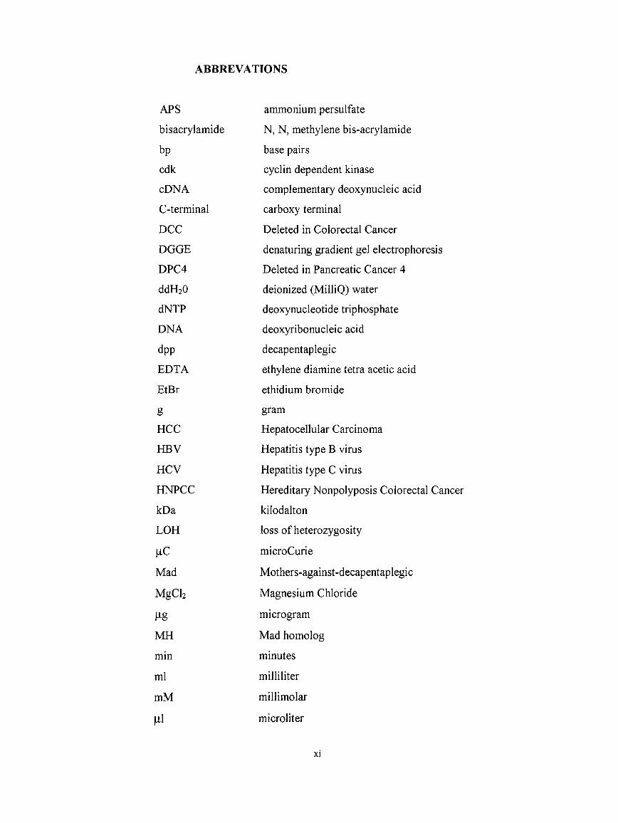
ABBREVATIONS
APS ammonium persulfate
bisacrylamide N, N, methylene bis-acrylamide
bp base pairs
cdk cyclin dependent kinase
cDNA complementary deoxynucleic acid
C-terminal carboxy terminal
DCC Deleted in Colorectal Cancer
DGGE denaturing gradient gel electrophoresis
DPC4 Deleted in Pancreatic Cancer 4
ddH20 deionized (MilliQ) water
dNTP deoxynucleotide triphosphate
DNA deoxyribonucleic acid
dpp decapentaplegic
EDTA ethylene diamine tetra acetic acid
EtBr ethidium bromide
g gram
HCC Hepatocellular Carcinoma
HBV Hepatitis type B virus
HCV Hepatitis type C virus
HNPCC Hereditary Nonpolyposis Colorectal Cancer
kDa kilodalton
LOH loss of heterozygosity
|iC microCurie
Mad Mothers-against-decapentaplegic
MgCb Magnesium Chloride
l^g microgram
MH Mad homolog
min minutes
ml milliliter
mM millimolar
microliter
XI

MMTV mouse mammary tumour virus
MSI microsatellite instability
N-terminal amino terminal
PAGE polyacrylamide gel electrophoresis
PCR polymerase chain reaction
RNA ribonucleic acid
s e e squamous cell carcinoma
sec seconds
Smad Sma- and Mad related gene
s s e p single strand conformation polymorphism
TBE tris-boric acid-EDTA
TEMED N,N,N,N-tetramethyl- 1,2 diaminoethane
TGFp Transforming growth factor p
TPR Transforming growth factor P receptor
Tris tris (hydroxymethyl)-methylamine
UV ultraviolet
Xll

The liver is a large digestive gland where nutrients are processed. It comprises a
mixture of cell types, although most prevelant cells are the epithelial liver cells , so
called hepatocytes. Hepatocytes perform a broad range of metabolic and secretory
tasks. Even though hepatocytes derive from gut epithelium, they have a different life
style compared to other gut epithelial cells. Under normal physiological conditions,
adult hepatocytes are non-dividing cells but they retain their capacity to proliferate.
Proliferation is seen only either as a response to massive hepatocyte loss ( partial
hepatectomy, hepatotoxic agent intake, acute and chronic hepatitis, cirrhosis) or as a
result of loss of antiproliferative control ( hepatocellular carcinoma) (Ozturk 1994).
Therefore, cell proliferation plays a key rol during tumorigenesis, as observed in the
rat model (Grisham 1996).
1.1 Hepatocellular Carcinoma
1. INTRODUCTION
More than 80 percent of primary liver cancers are primary hepatocellular carcinoma
(HCC) which derive from hepatocytes. It is the 7th most common cancer in males and
males develop HCC 2-3 times more than women. HCC carries a poor prognosis, with
survival times from diagnosis measured in months. Although surgical resection by
partial or total hepatectomy is available, partial hepatectomy is associated with a very
high recurrence rate ( 25 percent per year) (Isselbacher et al. 1991). HCC is the 4th
most common cause of death from cancer, responsible for 300000 deaths each year.

While hepatitis B and hepatitis C viral infections are considered major etiological
factors, poor nutrition, alcohol and cigarette consumption, food and water
contaminants, natural plant toxicants and oral contraceptives may also play a role. The
incidence of HCC is generally low in Western countries ( 1-7/100000 ) and high in
South-east Asia and sub-Saharan Africa ( 25-75/100000) (Bosch and Munoz 1990).
1.1.1 Hepatic cirrhosis and fibrosis
The most frequent form of liver injury (hepatocyte destruction) occurs during chronic
HBV and HCV infections. These infections cause decades of inflammation that ends
up with fibrosis. Hepatic fibrosis is a response to liver injury by recruitment of
inflammatory cells, activation of mesenchymal (stellate) cells and release of
cytokines. An important feature of hepatic fibrosis is that it is a reversible
accumulation of extracellular matrix in response to chronic injury in which nodules
have not yet developed, whereas cirrhosis implies an irreversible process in which
thick bands fully encircle the parenchyma, forming regenerating nodules (Burt 1993).
80% of HCC develop a cirrhotic liver, mostly due to chronic viral infections.
Furthermore, about 38 percent of patients with HBV- associated cirrhosis had HCC
at autopsy. It should be noted that although presence of cirrhosis increases the risk for
HCC it is not a requirement (Craig et al. 1990).
1.1.2 HBV and HCV
HCV infection is a major risk factor for the development of HCC worldwide. Two
facts are employed as evidence : 1) 50- 75 percent of HCC patients have anti-HCV or
HCV RNA detectable in serum in southern Europe and Japan. 2) In patients with
chronic HCV infection, progression can be noted from milder forms of hepatitis to
cirrhosis, and eventually, to HCC. Unlike the HBV, HCV is not a DNA virus and
doesn't become integrated into host genome. It is likely that HCV has an indirect
effect by causing liver injury. After 20 years of infection, 6-8 percent of patients with
chronic hepatitis C can be expected to have developed HCC (Di Bisceglie 1998).
Hepatitis B surface antigen is common in countries where HCC is also common.
HBsAg carriers are 40-100 times higher at risk to develop HCC than the non-carriers.
HBV is a DNA virus and after infection it becomes integrated into host genome. But
the location of integration was shown to be random and probably has no effect on
pathogenity. HBV doesn't possess any gene product with transforming activity. Its X
gene product which acts as transcription factor both for the virus and the host genome,
was demonstrated to bind to p53 and cause its inactivation. Thus, this can be a
mechanism of tumour progression (Feitelson 1993, Ueda et al. 1995).
1.1.3 Aflatoxin
Aflatoxin is a myotoxin produced by some species of Aspargillus. In the regions were
HCC is epidemic, aflatoxin is taken in as a food contaminant. It is the only etiological
factor that was shown directly to cause p53 mutation. It targets the codon 249 of p53
genes, and impairs its activity. This mutation is a hotspot where aflatoxin exposure is
high but low in other areas (Ozturk et al. 1991, Ozturk et al. 1994)
1.1.4 Genetics of HCC
Like many other cancer types HCC is a multistage disease while taking decades to
develop it. But the genes which are inactivated during tumourigenesis are poorly
identified.
It is known that overexpression and/or activation of cellular protooncogenes and
deletion or inactivation of tumour suppressor genes occur during multi-step
carcinogenesis through either point mutations, gene amplification, gene
rearrangement, alteration in gene méthylation pattern, or a change in transcriptional
regulation. These genes encode proteins of the cellular signal transduction system,
which function to regulate gene expression, cellular growth and differentiation
(Alberts et al. 1994).
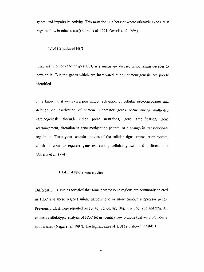
1.1.4.1 Allelotyping studies
Different LOH studies revealed that some chromosome regions are commonly deleted
in HCC and these regions might harbour one or more tumour suppressor genes.
Previously LOH were reported on Ip, 4q, 5q, 6q, 8p, lOq, 1 Ip, 16p, 16q and 22q. An
extensive allelotypic analysis of HCC let us identify new regions that were previously
not detected (Nagai et al. 1997). The highest rates of LOH are shown in table 1.

Table 1 High Frequence of Loss of Heterozygosity (LOH) in HCC
Chr. Location LOH rate Candidate
(%) Genes
2q36-37 29%
4q35 40%
6q27 36% M6P/IGF2R
7pl5 30%
8p23 42%
13ql2-ql3 30-32% RB, BRCA2
16q23-q24 28%
17pl3 33% p53
Other regions which were shown also to be deleted for the first time are in table 2.

Table 2 Other chromosomal regions lost during MCC
Chr. Location LOH rate
(%)
Iq22-q23 20%
Iq42-q43 24%
7q33-q34 20%
8q23-q24 23%
9pl2-pl4 21%
9q34-qter 20%
14q32 23%
17q24 21%
1.1.4.2 Mutations
During HCC, candidate genes are not commonly inactivated. Most of the tumor
suppressor genes are rarely mutated. Additionally, mutations in oncogenes are also not
common ( table 3 ). Some alterations which change the expression or the activity of
the protein have been observed in HCC (table 4).

Table 3: Mutations in tumoursuppressors and oncogenes
Genes LOH% Mutation References
P53 51% 30-55% Puisieux and Ozturk 1997
M6P/IGF2R 70% 25% De Souza et al. 1995
61% 55% Yamada et al. 1997
P16 4/26 germline Chaubert et al. 1997
%41 1/23 Bidenetal. 1997
yes 3/62 Kita et al. 1996
RB Rare Zhang X. et al. 1994
BRCA2 1/60 somatic Katagiri et al. 1996
2/60 germline
DLC-1 44% Yuanetal. 1998
PRLTS 2/48 Fujiwara et al. 1995
Ras Rare Shen and Ong 1996
1.1.4.3 Other alterations
Table 4: Alterations other than mutations in tumour suppressors and oncogenes
in HCC
pl6 48% de novo meth. Chaubert et al. 1997
IGF2 20% overexpression Li et al. 1997
Uchida K. et al. 1997
CyclinDl 4/30 amplifications Zhang Y. J. et al. 1993
p53 inactivation by HBx Feitelson et al. 1993

1.2 Transforming growth factor P (TGFp)
1.2.1 Biological effects of TGFP
TGpp is a 25 kDa homodimeric peptide which is produced by nonparenchymal liver
cells and secreted in a latent form. It appears to be an important cytokine both in
normal and diseased liver. Because it is able to increase the levels of many
extracellular proteins, it has been closely associated with the promotion of fibrosis and
progression of cirrhosis. In normal human hepatocytes no TGPP messenger or protein
is detectable. In cirrhotic livers although hepatocytes in nodules continue not to
express TGpp, some cells in fibrous septa expressed it. In hepatocellular carcinomas,
clusters of tumoural hepatocytes express large amounts of TGpp. Interestingly normal
and cirrhotic liver cells express the TGFp receptors but they disappear in plasma
membrane of tumour hepatocytes though they were in cytoplasm (Bedossa et al. 1995).
The apparent paradox of how hepatocytes proliferate despite of production of
elevated TGpp production by nonparenchymal cells is best clarified in a rat model.
Accordingly, TGppi is expressed only in nonparenchymal cells, but TGpp receptors
were expressed in both cell types. As the liver regeneration reach its peak, TGppi
mRNA levels are elevated and there is a significant depression of TGpp receptor
mRNA levels only in hepatocytes (Date et al. 1998).
Transforming growth factors (TGF-P) are potent inhibitors of proliferation of most
cell types in culture and in vivo. However many tumorigenic cell lines have lost
response to negative growth-regulatory effects of TGppi which is the most commonly
studied TGPp molecule. Thus, it has been thought that the understanding of the
8
molecular mechanism of the TGpp will reveal much about the tumorigenesis.
Although many cell cycle proteins have been thought as potential targets of TGF|3
such as cyclins, cyclin dependent kinases (Cdks) and Cdk inhibitors, it is not clear yet
whether these targets are directly involved or the effect on these targets are simply a
consequence of the TGppi induced growth arrest (Alexandrow and Harold 1995),
There has been conflicting results about the role of TGppi at early and late stages of
carcinogenesis due to in vitro studies and different approaches. Accordingly, TGP(3l
acts either as a tumour promoter or as a suppressor at early stages and either
stimulates or suppresses the malignant progression during the later stages (Cui et al.
1996).
1.2.2 TGPP signalling pathway
Although transforming growth factor (3 (TGPP) was discovered as a peptide which
stimulates the proliferation of rat kidney fibroblasts, it acts as a potent growth
inhibitor of many normal cell types, including epithelial cells. It has been shown that
it also induces cell differentiation and production of extracellular matrix proteins.
TGP(3 belongs to a large superfamily of peptides which are structurally conserved
throughout the evolution and includes members such as activins and bone
morphogenic factors (BMPs) and they play important roles in differentiation and
morphogenesis. TGPP has three different isoforms in humans (TGP(3l-3), encoded by
different genes (Hoodless and Wrana 1994). It is secreted in a latent form from the
cells and they become active by proteolysis in which IGF2R plays an important role
(De Souza et al. 1995).
9
It has now been firmly established that most of these factors, including TGPP, signal
through type I and II serine/threonine kinase receptors. Another receptor type called
XPR-III is a proteoglycan with a short cytoplasmic domain. It is not required for the
biological activity of TGppi and TGPPS but may have an accessory function for
TGPP2. Type II and type I receptors are the main mediators of the TGPP signalling,
they contain a cytoplasmic kinase domain and extracellular cystein-rich regions. In
addition, type I receptor has a highly conserved cytoplasmic glycine- and serine-rich
segment (GS domain). In the absence of ligand, XPR-I and XPR-II exist as homomers
in the cell surface. In addition, XpR-II is constitutively phosphorylated by cellular
kinases and by itself (Derynck 1994). Upon ligand binding by XPR-II, XPR-I which
alone is not able to recognize free XGPP is recruited to form a heterotetrameric
terniary complex. Then, XPR-I is phosphorylated by the kinase activity of XPR-II at
its threonine and serine residues of GS domain. XPR-II itself is not a substrate of XPR-
I and its phosphorylation is not altered as a consequence of ligand-induced complex
formation. Thus, it can be concluded that XPR-I is downstream of XpR-II in the
XGPp signalling and its kinase activity is essential for the phosphorylation of
downstream substrates.( Wrana J. L. et al. 1994) (for figure see Appendix).
1.2.2.1 TGFP receptors
10

1.2.2.2 Smad proteins
The receptors for TGPP are quite specific and no other ligand has been shown capable
of binding to them. But it is also clear that identification of downstream targets of
TPR-I would reveal much information about the specific effects of TGFp. Smad
proteins were identified as human homologues of Mad protein which mediates TGpp
-like dpp signalling in Drosophila. At present, at least eight distinct Smad proteins
were identified in humans. They are highly conserved at their amino-terminal MHl
and carboxy-terminal MH2 domain among themselves and other vertebrates. These
two domains are connected by a linker region which is less well conserved. It is
extremely rich in serine and proline residues. Structural analysis showed that no
known structural motifs exists in these genes. However sequence comparisons have
revealed that Smadl, SmadS are highly homologous and form a group. Indeed
functional and biochemical analysis have proved that they specifically carry BMP
signalling while Smad2 and Smad3 form another group and are involved in
activin/TGpp signalling. Thus, different signals recruit distinct Smads. These Smads
are called receptor-regulated Smads. Smad4 is more distantly related to the other
Smads and is a central protein for both pathways. Smad6 and Smad? thought to act as
inhibitors of these signals (Kretzschmar and Massague 1998). It has been established
that the MH2 domain of receptor-regulated Smads is responsible for most of its
functions. The function of MHl domain is mere to inhibit the biological activities of
MH2 domains by a direct physical interaction between MHl and MH2 domains
(Kretzschmar et al. 1997). The carboxy-terminal of the receptor-regulated Smads
contain a SS(V/M)S motif which is phosporylated by their type I receptors (Abdollah
11

et al. 1997). This phosphorylation event was shown to be essential for the function of
Smads. Upon phosphorylation, the inhibition of MHl domain is releaved and they
form heteromeric complexes with Smad4 to enter into the nucleus (Macias-Silva et al.
1996). Again, it is the MH2 domain of Smad2 and Smadl which interacts physically
with Smad4. In nucleus these complexes are able to interact with other proteins and
act as regulators of gene expression (Lagna et al. 1996).
As inhibitors of TGpp/BMP signalling, Smad6 and Smad7 constitute a distinct class
of Smads. They lack the SSXS motif at their carboxy terminal. Smad7 is able to block
the activation of target genes, induced by TGF(3, probably by association with TGFb
receptor complexes (Hayashi et al. 1997, Nakao et al. 1997). Smad6 has also an
inhibitory effect, but whether its function is specific for BMP signalling or it is a
general inhibitor has yet to be clarified (Imamura et al. 1997).
Are the Smad proteins targets of other kinases and signalling pathways? The cellular
signalling pathways are complex and dynamic processes. They may recruit the same
substrates and have synergistic or antagonistic effects. Interestingly, Smadl is shown
to be phosphorylated at serine residues in the linker region by Erk MAP kinases in
vitro and in vivo. This phosphorylation inhibits the nuclear accumulation of Smadl.
Thus Erk kinase pathway can have an antagonistic effect on BMP induced signalling
(Kretzschmar et al. 1997). There is also evidence for a similar mechanism for Smad2
(De Caestecker et al. 1998).
12
1.2.2.3 Transcriptional activation by Smads
One of the first evidence came from the fact that when fused to GAL4 DNA-binding
domain, C-terminals of Smadl, Smad2 and Smad4 are able to act as transcriptional
activators (Liu et al. 1996). A study in Xenopus embryos have shown this hypothesis
to be true also in vivo. In response to activin or TGF[3, a complex called activin-
response factor (ARF) is formed, and binds to the promoter region of Mix2, an
immediate-early activin response gene. This complex contains both FASTI, a winged-
helix transcription factor and XSmad2. The MH2 domain of XSmad2 interacts with
FASTI (Chen et al 1996). The third component of ARF is identified as Smad4 and
both its MHl and MH2 domains are essential for the stimulation of Mix2 reporter,
contributing to DNA-binding and transactivation functions respectively (Chen et al.
1997). A similar example is activation of Xenopus forehead gene directly by C-
terminal of XSmad2 via defined regulatory regions (Howell and Hill 1997). The
dissimilarity between these regulatory sequences can be best expained that either
Smads interact with different transciption factors upon nucleus entry or there is dose-
dependent action of activin. While these target genes are restricted to Xenopus,
Smads regulate directly the TGFP inducible human type VII collagene gene
expression (Vindevoghel et al. 1998).
A recent study (Moustakas and Kardassis 1998) have shown that in HepG2 hepatoma
cell lines which are responsive to TGF|3 , Smad proteins are able to regulate and
increase the hepatic activity of p21/WAFl expression. It is suggested that Smad3 and
Smad4 probably exert this effect by functionally interacting with transcription factor
13

Spl rather than directly binding to DNA. Another study shows that Smad3 and Smad4
are part of a nuclear complex that directly binds CAGA boxes in the promoter of
PAI-1 upon TGPP induction. But Smad2 neither participates in it nor binds to these
elements (Dennler et al. 1998).
1.3 TGFP Signalling and Cancer
The resistance to TGpp induced growth inhibition in tumours probably results from
disruption of one of the components of TGpp signalling pathway. As the
understanding of this pathway increased, several proteins shown to be mutated in
various cancers and they became strong candidates of tumour suppressor genes.
1.3.1 M6P/IGF2 Receptor
M6P/IGP2 Receptor has distinct activities which are important for the regulation of
different molecules such as mannose 6 phospate and IGP2 . IGP2 is a growth and
survival factor which is degraded by this receptor. Interestingly, the inactive form of
TGPp also binds to IGF2R and is activated by plasmin with the help of the receptor.
Thus, inactivation of this receptor will have two effects. First, it will increase the level
of IGF2. Second, it will decrease the active TGPP levels which may help the
hepatocytes to escape from negative-growth effect of TGpp. Frequent loss of
heterozygosity and mutations of IGF2R gene have been reported in HCC ( De Souza
et al. 1995, Yamada et al. 1997).
14

1.3.2 TGFP Receptors
Many cancer cell lines are known to acquire TGPP resistance. Inactivating mutations
in TGpp receptor type II and type I, can be a cause for TGpp resistance in cancer.
Mutations have been identified in TPR-II gene, in two head and neck squamous cell
carcinoma (HNSCC) cell lines (Garrigue-Antar et al. 1995), in cutaneous T-Cell
lymphoma cell lines (Knaus et al.l996), in colon and gastric cell lines (Vincent et al.
1997) and in colon cancer cells with MSI (Markowitz et al. 1995). These mutations
can reflect genetic changes aquired in culture rather than the real situation in primary
cancers. Indeed, until now, XPR-II was shown to be mutated only in sporadic colon
(Takenoshita et al. 1995) and endometrial (Lu et al. 1996) cancers with microsatellite
instability (MSI), or in hereditary non-polyposis colorectal cancer (HNPCC) cases (Lu
et al. 1996) and not in cancers of breast, pancreas and liver (Vincent et al. 1996). Thus
TPR-II acts as a tumor suppressor mainly in cancers with MSI, although a very recent
study indicates that a HNPCC patient without MSI has a germline mutation in TPR-II
gene outside the (A) 10 tract (Lu et al. 1998).
1.3.3 Smads
1.3.3.1 Allelotyping studies
Even before the identification of Smad4, 18q loss was seen frequently in many tumour
types. But many of these studies are quite confusing because of discrepancies on the
percantage and the location of LOH in a tumour type. An extensive study on 12
15
tumour types concentrated on 18q21.1 using polymorphic markers near Smad4 and
Smad2. Significant losses ( >20%) were observed in HNSCCs, melanomas,
osteosarcomas, in breast, lung, hepatocellular, ovarian, pancreatic and prostatic
cancers (Schutte et al. 1996).
1.3.3.2 Mutational analysis
Most cell lines, who are insensitive to TGF|3, express intact TGpp receptors in their
cell surface (Vincent et al. 1996). Thus, Smad proteins as mediators of TGF(3
signalling are candidate tumor suppressors. Smad4 was first identified as a candidate
tumor suppressor gene involved in pancreatic cancer (Hahn et al. 1996). Later on, it
was shown to be mutated frequently also in colorectal cancers (Thiagalingam et al.
1996, Takagi et al. 1996), but infrequently in ovarian cancers (Schutte et al. 1996), in
lung cancers (Nagatake et al. 1996), in gastric carcinomas (Powell et al. 1997) and
recently in biliary tract carcinoma (Hahn et al. 1998). Smad2 mutations are observed
only in colorectal (Riggins et al. 1997, Eppert et al. 1996) and in lung (Uchida et
al.l997) cancers (Table 5). Recently germline mutations were found in Smad4 in
families with juvenile polyposis syndrom (Howe et al. 1998).
Majority of the mutations are harboured within the carboxy terminal MH2 domain.
The crystal structure of C- terminal domain of Smad4 revealed that it forms a trimer
in crystal. Most of the missense mutations are in trimer-interface and disrupt the
homo-oligomerization. These mutation also prevented formation of heteromeric
complexes between Smad4 and Smad2. It can be concluded that homomerization of
Smad4 is prerequisite for heteromerization and signal transduction (Shi et al. 1997).16

A conserved Arginine residue in the N terminal of is shown to be mutated both in
Smad4 and Smad2 . N-terminal of the Smads have an inhibitory effect on Smad
function by interacting with its C-terminal. It was shown that these mutations increase
the affinity of N-terminal on C-terminal and create an autoinhibitory effect. Such
mutations abolish the function of phosphorylation and thus formation of heteromeric
complexes ( Hata et al. 1997),
Table 5: Somatic mutations in Smad2 gene.
Mutation Codon Exon Tumour type Reference
Deletion 345-358 9 Colorectal RIGGINS et al.
CTT- CGT L440R 11 Colorectal
c c
c c
c c
EPPERT et al.
c c
c c
c c
CCT- CAT P445H 11
GAC-GAG D450E 11
CGC-TGC R133C 4
GAC-CAC D450H 11 Lung
c c
UCHIDA et al.
c cDeletion 434-436 11
17

1.3.3.3 Knockout and Transgenic Mouse Models
Knockout mice for Smad4 die before day 7.5 of embryogenesis. The growth
retardation, lack of gastrulation and impaired differentiation is similar to the
phenotype of Bmp4 mutant embryos. Additionally, the anterior-posterior axis is also
disrupted in rescued embryos. These data suggests that a central signalling molecule
of TGPP related pathways, Smad4 is essential for embryogenesis (Sirard et al. 1998).
Similarly Smad2 mutant embryos demonstrated that Smad2 function is essential for
the establishment of anterior-posterior polarity, and development of the primitive
endoderm lineage. These mutants entirely lack embryonic germ layer tissues (Waldrip
et al. 1998). Interestingly, TGF|3l-3 or activin deficient mice are normal at birth
(Schull et al. 1992). This fact can be explained either by redundancy of these
molecules or that Smad2 acts downstream of other yet unidentified ligands.
Mice which are transgenic for TGF-a under the MMTV promoter develop mammary
tumours. In mice transgenic both for TGF-a and TGF-P there is resistance to
mammary tumour formation, probably by reduction of epithelial mass (Pierce et al.
1995)
A transgenic mouse model with keratinocyte-targeted expression of TGF^l suggests
that TGFpi has biphasic effects during multistage carcinogenesis. At early tumor
promotion stages of carcinogenesis, TGFpi acts as a growth inhibitor of kératinocytes
and inhibits benign tumour outgrowth. At later stages the cells that were able to
escape this inhibition through genetic alterations in TGFP signalling, have an elevated
18
level of TGFßl which enhances their malignant conversion. The mouse model clearly
shows that the TGFßl effects the malignant phenotype by altering the expression of
extracellular matrix and cell adhesion molecules rather than by evasion of host
immune surveillance, enhancement of angiogenesis or elevated apoptosis (Cui et al.
1996).
Compound heterozygote mice , carrying both mutant Smad4 and APC genes were
viable and they lost the wild-type allele in tumour tissue. Interestingly, the intestinal
tumours progressed into a malignant phenotype at a much earlier stage than the Apc-
knockout mice. Thus, Smad4 mutations are sufficient to bring the polyp adenomas
induced by APC mutations , in a malignant stage without any other genetic changes.
(Takaku et al. 1998).
19
As discussed before, TGPP has a strong anti-proliferative effect on normal
hepatocytes. Nevertheless, the malignant hepatocytes express TGF|3 and continue to
proliferate. As observed in other cancers and some hepatoma cell lines escape from
anti-proliferative effect of TGpp might be an important step during hepatocellular
carcinogenesis.
Therefore, we think that TGP(3 signalling pathway is involved in hepatocellular
carcinogenesis. Smad2 is a direct mediator of TGpp response and was shown to be
mutated in several cancers. So we concentrate our efforts on the genetic alterations of
Smad2 in liver cancers. Smad2 is a 467 aa protein coded by 11 exons and is located
near Smad4 on 18q21.1 where LOH was observed in some cancers (Takenoshita et al
1998).
The aim of this study is to check whether the genetic abnormalities of Smad2 gene are
involved in hepatocellular carcinoma.
2. AIM
20

3. MATERIALS AND METHODS
3.1 Tumour Specimens and cell lines
Genomic DNA of 35 tumours were chosen from a collection as published in (Unsal et
al. 1994). The patients were from South Africa(n=l 1), China (n=8),
Mozambique(n=7), Japan (n=6), and Germany (n=3). 80 % percent of the samples are
HBV positive and 20 % had p53 mutations. From the informative cases(n=20), 20%
had cirrhosis (table 6).
Six hepatoma cell lines were used in this study are HepG2 (Aden et al. 1979), Hep3B
(Aden et al. 1979), HuH7 (Nakabayashi et al. 1982), Mahlavu (Alexander et al. 1984),
FOCUS (He et a l l 984) and PLC/PRF/5 (Alexander et al. 1976).
3.2 Solutions:
lOxTBE Buffer Solution
108 g Tris
55 g Boric acid
8.3 g EDTA
are dissolved in 1 It of deionized water until its pH is 8.3
21

Table 6: Characteristics of tumour samples
IUM(JKS P53 HBV Sin;ul2 1 Stfitlc < arrliosis Metiislii.sis
T8 i SA-1.7 WT N1i Hiüly Ní> No
TI7,ÇSA'T) ! WT - N Lalo Yes Ycsr is (M) 249 Mul + N 1 Lalo Ni> No
I WT - [ N ! Laic No NoT19 SA T ) ! WT - N Lalo No No121 (SA-T) I w f {'.s-v) + N i I..aic Nt) Yes121 (M) ' 249 Mul •T N 1 -au; Yes YcsT29 <;M) ! 249 Mul - N i barí y No Nt>
'П1 (SA-’i'i ! WT + N Laic Yes Ycs'1M (SA-T) i WT + i1 1 \л\\с No Ycs
I WT ...._ + ..... ... [ 1.Л1С Yes Vos1A7ÍM) + ! AU. Exil I..íUto Yes Ycs
T39 (SA-S) Wr ! -h K i r.alc i..........Yes NoT43 Î S A -T) 286-Sbp-<l(;l ' + N Late· No Nu Inior.
T47(M)..... Г 157 Mul I + ! N Lato No NoT49 (SA -6 ! WT i 1 ' A il. Ex K) I alo i No No
T5l (M) ' WT + 1 N Late ' No 1 _ YcsT53 (M) i WT I + ; N Late , Yes NoT73 (G) WT (Ex 7-9) i ■+· 1 N l.ato ! YesT80 (G) ! W'I' + i N ! No íníor. i Yes No init>r.C2(C) I WT !
Í+ 1 N No Inlor. i No íníor. i No lniv>r .
C3 (C) i WT !I
1 ! N No Inlor, Nt) Inlor. ! NolniorC6 (C) I WT i ·+ ! N No liUoi. No íníor. No Inior.07 (C> i WT I i+ i N i No Iníor. No íníor. !! .No Inior.
CIO(C) j 249 Mul Í 4· i N i Nt) íníor. No Iníor. No Inior.C12 iC) j WT i + i N 1 No Iníor. No Iníor. No Inior.( : i 5 c c ) i WT j + N No inlor. No Iníor. No iniV)r
....... Ç23(C) j WT + N i No iníor. No Iníor. No in loi. j
JITÜ) w r + N ' No Jnlor. No Iníor. No inior.J3T (J) i W'T (Hx 5-7) + N 1 No Iníor. No Iníor. Nt) Infor.J4T (J) WT (Fix 7-9) -1- N : No Iníor. No Iníor. Nt) lnit)i.m (i) WT(Fîx7;9) - N 1 No lnh>r. No Inlor. Nt) Inior.J9T(J) j WT (bx 7) - N ! No Inlor. No ínít)r. Ni.) Init)r.
л о т (J) i W'T (Ex 7) : N 1 No Iníor. No Iníor. No hiior.G5 (G) ; WTiHxS-7) N 1 No Iníor. No Iníor. No hilor.
The samples were from South Africa-Lesotho (SA-L), South Africa-Transkei (SA-T),
South Africa-Swaziland (SA-S), South Africa-Caucasian population (SA-C), from
Mozambique (M), Germany (G), Japan (J) and China (C).
22

SSCP Loading Buffer Solution
95 % Formamide
0.04 % Bromphenol blue
0.04 % Xylene Cyanol
lOmM NaOH
6 X Loading Buffer Solution
30 % Glycerol
0.04 % Bromphenolblue
0.04 % Xylene Cyanol
AdH20
Agarose Gel Solution for PCR
30 ml lx TBE
0.6 g Agarose
heated in microwave for 1 min. and added 1 ml of EtBr
Polyacrylamide Gel Solution for SSCP
the stock solution is 50 %
49.5 g Acrylamide
0.5 g Bisacrylamide
Add ddHaOto 100 ml
23

3.3 Expression of cell lines
3.3.1 PCR
Primers: The forward (F) and reverse (R) primers for the amplification of Smad2
coding region are as follows (Riggins et al.) :
TC 104 (F ): 5' GG A TCC T AA TAC GAC TC A CTA TAG GG A G AC CAC CAT
GGG TAA GAA CAT GTC GTC CAT C 3'
TC 103 (R ): 5' TTT CCA TGG GAC TTG ATT GG 3'
TC 104 (F) includes signals for transcription and translation
Polymerase Chain Reaction (PCR) conditions:
Reaction mixture: for each exon the PCR reaction was performed in 25 ml of mixture
containing:
1 X Buffer
1.5mMMgCl2
0.8 mM dNTP mix
20 pmol F primer
20 pmol R primer
50 ng DNA
1 u Taq polymerase
A H2O
24

PCR conditions for amplification consisted of a 3-min dénaturation step at 95°C,
followed by 35 cycles of 40 sec at 95°C, 40 sec at 56° and 90 sec at 72°C. After 35
cycles a last step at 72°C for 7 min was added.
3.3.2 Agarose gel electrophoresis
5 ml of PCR product was mixed with 1 ml of 6 x loading buffer and loaded into 1.5 %
agarose gel electrophoresis. They were run 80V for 40 min and then visualised under
UV light.
3.4 Single-Strand Conformation Polymorphism (SSCP) Analysis
3.4.1 PCR conditions:
Primers: Intronic or partly intronic primers were designed by myself. The exact
positions of the primers can be found in Appendix A. The forward(F) and reverse (R)
primers for the amplification of each exon are as follows:
exon 4 : F: 5' TCTGATAGTGGTAAGGGTTT 3'
R: 5' GTCTCTTGATGGTCGTCTC 3'
exon 8 : F: 5' CAGTTACTTACTCAGAACCT 3'
R: 5' GCCTACATTATGAGTATACAG 3'
25
exon 9: F: 5 'CCAAAGTCACACTGAAATAG 3
R: 5' AGCAAGTTGACATGATAGG
exon 10: F: 5' GCATGCTCATATTCTAAAAC 3'
R: 5' ACTGTGGAAATTTAAGAACC 3'
exon 11: F: 5' GCCTGTGGACTTGAATTTCAT 3'
R: 5' GGACTTGATTGGTGAAGCTTT 3'
C-term : F: 5' CAGGGTTTTGAAGCCGTCTA 3'
R: 5' CTTGAGCAACGCACTGAAGG 3'
Polymerase Chain Reaction (PCR) conditions:
Reaction mixture: for each exon the PCR reaction was performed in 25 ml of mixture
containing:
1 X PCR Buffer
1.5 mM MgC12
0.04 mM dNTP mix
[33P] 0.5 |iCi dCTP
20 pmol F primer
20 pmol R primer
50 ng DNA
1 u Taq polymerase
A H2O26

Reaction conditions: the reaction condition is the same for each exon except the
anneling temparature (Ta). The Ta for each exon is as follows:
exon 4: 56°C
exon 10: 50°C
exon 8: 53°C
exon 11: 56°C
exon 9: 57°C
C-term: 60°C
PCR conditions for amplification consisted of a 3-min dénaturation step at 95°C,
followed by 30 cycles of 30 sec at 95°C, 30 sec at Ta and 30 sec at 72°C. After 30
cycles an additional step at 72°C for 5 min was performed.
3.4.2 Polyacrylamide gel electrophoresis:
The mixture contains:
I xTBE
10 % Glycerol
20 % polyacrylamide gel solution
add ddH2 0 to 100 ml
After mixed precisely and degased, 400 ml of 10% Ammonium persulfate (APS) and
40 ml of Temed is added for a mixture volume of 100 ml. This solution is poured into
gel apparatus and left for polymerization for 2 hours.
27
SS CP reaction:
Mixture:
2 ml of PCR product was mixed with 8ml of SSCP loading buffer in an Eppendorf ®
tube.
Dénaturation:
The tubes were denatured at 95°C for 2 min and then put immediatly into ice.
Loading:
The samples were loaded into an electrophoresis apparatus and electrophoresed at
4°C , at constant 45 W for 14 hours.
3.4.3 Exposure and development
Then the gel was dried on a 3M Wattman® paper and exposed to X-ray film for 3
days. The exposed film was developed in developer machine.
3.4.4 Automated sequencing
The sequencing reactions were performed and analysed in ABI Prism 377 Sequencer,
using the same primer as used for SSCP analysis.
28

4. RESULTS
4.1 Smad2 expression in hepatoma cell lines
Xrt>“OOK)
<G OS
55
<1oosC/) n
Smad2
iSiiiSiiiipi»B»aipillSE:»£
i:.|(;;ii. :-..’t.;,j,"' " ‘ ' ■ . ‘ li' ' i i ' ’%> > ^ '
Figure 1: Expression of hepatoma cell lines
Smad2 expression was not exactly known in the hepatoma cell lines. The whole
coding region of six hepatoma cell lines are amplified by PCR (conditions described
above). Single band was observed with expected size for all cell lines tested. All the
six cell lines express Smad2. There are no large deletions in any of the cell lines.
Although four of them had similar amounts of amplified products, a nested PCR from
these products still shows that Hep3B and Huh? are less amplifyible (Figure 1).
29

4.2 SSCP analysis of hepatoma cell lines
3 3"D1 (Ji
00
M a■o -a
WB·
sr ^ ZL oOsC
« fe ijw ;;..; .;■,;.
Figure 2; SSCP analysis of Smad2(C-term) Figure 3; SSCP analysis of
for hepatoma cell lines in 10% Acrylamide gel wildtype and mutant p53
SSCP analysis is performed for a part of the conserved C-terminal domain of Smad2
in five hepatoma cell lines. The upper and lower bands correspond to each single-
stranded DNA of the amplified region. No different migration pattern (eg. no shifted
30

bands) were observed (Figure 2). SSCP analysis for exon 7 of wildtype and mutant
(p53248 and p53249 ) p53 genes are used as a control for the efficiency of SSCP
conditions. Both mutants had a distinct migration pattern from the wild-type exon of
the gene.
4.3 Sequencing analysis of hepatoma cell lines.
As to confirm the SSCP results, the PCR products of Smad2 (C-term) was
sequenced. No alteration was detectable by sequencing data, confirming the SSCP
results in Figure 1.
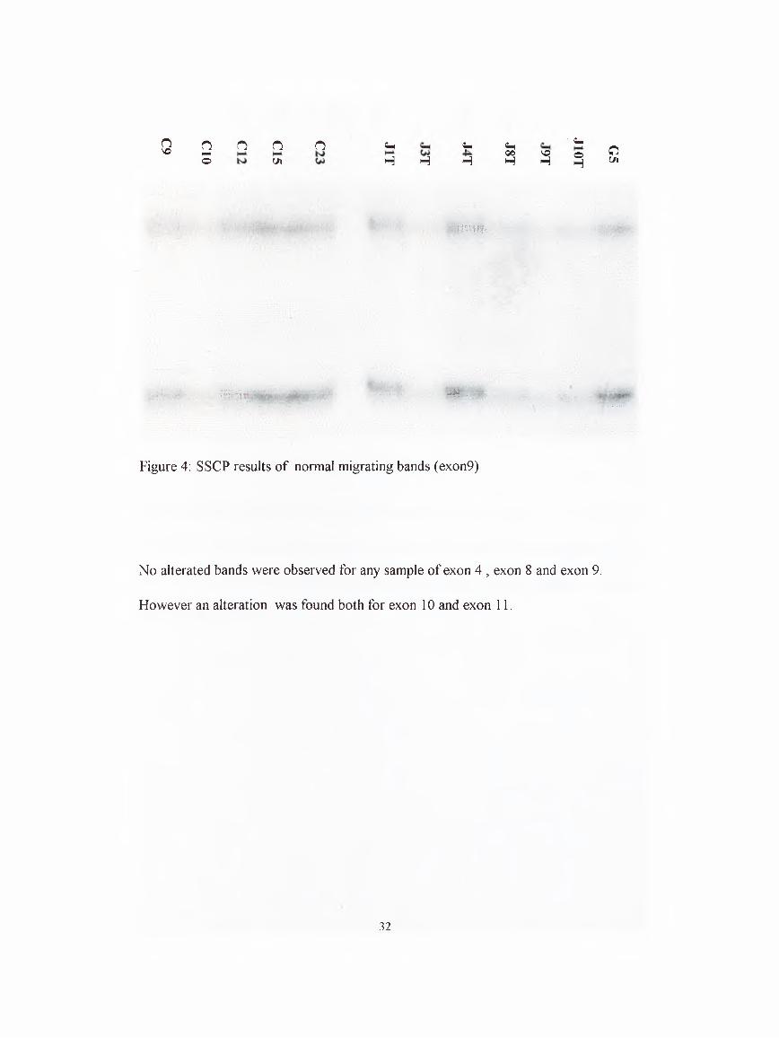
4.4 SSCP analysis of tumour samples.
Exons 4,8,9,10 and 11 of Smad2 gene were analysed by SSCP analysis in 35 samples.
The results gave us three distinct bands, one far below of the g e l, and the other two
in upper side of the gel relatively. The intensity of the lowest band was higher, and
due to its migration pattern and intensity this band corresponds to the double-stranded
product. The two upper ones migrate according to their confirmation and they
correspond to the single strands (Figure 4)
31
oVO o n o n Ch in Ch ■ ^
H-‘ 00 VO oO U l H H H H H HO(Jl
iSS!'№,
i i i i i 4H; -
Figure 4: SSCP results of normal migrating bands (exon9)
No alterated bands were observed for any sample of exon 4 , exon 8 and exon 9.
However an alteration was found both for exon 10 and exon 11.
.32
H H H H H Hin in 4 4-4 VO
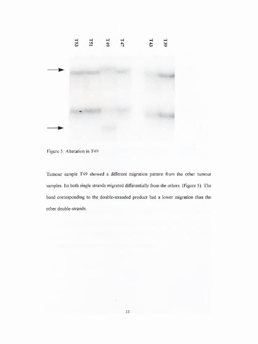
Figure 5: Alteration in T49
Tumour sample T49 showed a different migration pattern from the other tumour
samples. Its both single strands migrated differentially from the others. (Figure 5). The
band corresponding to the double-stranded product had a lower migration than the
other double-strands.
33
H H H H H H Hu 4in -4H
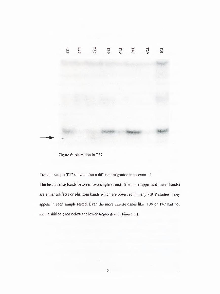
Figure 6: Alteration in T37
Tumour sample T37 showed also a different migration in its exon 11.
The less intense bands between two single strands (the most upper and lower bands)
are either artifacts or phantom bands which are observed in many SSCP studies. They
appear in each sample tested. Even the more intense bands like T39 or T47 had not
such a shifted band below the lower single-strand (Figure 5 ).
34

5. DISCUSSION
Members of TGF-P superfamily have very important effects on cell proliferation,
differentiation, organ development and morphogenesis. TGF-P 1 itself is involved
during embryogenesis and differentiation. It is known to be a potent inhibitor of most
of the normal cells, mostly of epithelial cells. However, during tumourigenesis the
malignant cells continue to proliferate although the TGFP levels in these tissues
increase. It is even suggested that TGFpi is a enhancer of malignancy in
carcinogenesis (Cui et al. 1996).
This paradox can be best explained that the cancer cells acquire mutations which
enable them to escape from negative growth effect of TGFp. The last three years gave
us many evidences that this might be true for many types of cancer. With the
identification of TGFP receptors, especially one of them, TPR-II was shown to be
mutated or inactivated. But, unfortunately these mutation were only observed in
cancers with microsatellite instability, such as HNPCC and gastric cancers where a
(A) 10 TpR-II is the target (Vincent et al. 1997). We can even suggest logically; as
they are the only known receptors for TGFp, the effect of TGFP as an enhancer of
malignancy can be accomplished if they remain intact. Smad2, 3 and 4 proteins are
shown to be the sole mediators of TGFP until today. Together with the other Smads
they have highly conserved amino- and carboxy-terminal domains. The C-terminal
domain was shown to mediate the activity of TGFP both in invertebrates and in
vertebrates, including mammalians ( Kretzschmar and Massague 1998 ). Smad4
35

became the focus of cancer research when it was shown highly mutated in pancreatic
cancers. It is located near to in 18q21.1 a region which shows a high rate of LOH in
some cancer types ( Schutte et al. 1996 ). DCC gene which was thought to be the
main target of inactivation in this region, is not considered as a tumour suppressor
anymore. Interestingly, Smad2 ,the main partner of DPC4 in TGpp pathway, is also
located very near to DPC4. They are inactivated in some cancer types (Riggins et al.
1996, Eppert et al. 1996, Uchida et al. 1997). It is clear that such an inactivation will
abolish the effects of TGpp. It is demonstrated that they are essential for the
signalling.
It was long known that both during fibrosis, cirrhosis and during hepatocellular
carcinoma there is an increase in TGpp levels, although the source of the production
is still an unsolved question (Bedossa et al. 1995). Although there is one report
showing microsatellite instability in hepatocellular carcinoma, the target gene
mutations like XPR-II have not been studied (Salvucci et al. 1996). Purthermore, it
was shown that they are intact and expressed (Vincent et al. 1996). Some has
suggested that the receptors can be downregulated during tumourigenesis, but there is
not enough evidence for it (Sue et al. 1995). Therefore, TGpp related Smads are
potential targets of inactivation as the hepatocytes escape from inhibitory effect of
TGPp.
The expression of Smad2 in liver cell lines were not clear and was not studied
previously. The amplification of Smad2 cDNA in all six hepatoma cell lines clearly
reveales that Smad2 is expressed in them, including Mahlavu cell line which is
36

unresponsive to TGFpl (Ito et al. 1990). We have observed lesser amount of
amplification for some cell lines. Studies have to be done in order to clarify Smad2
protein expression in these cell lines. In addition, the SSCP analysis of the most
conserved C-terminal region (codons 400-464) from cDNA have demonstrated that
there is no alteration in this region of these cell lines. Automatic sequencing data of
this region of five cell lines, including Mahlavu confirmed this information.
To detect the significance of Smad2 mutations in liver cancer, we performed SSCP
analysis for five exons of Smad2 gene. The four exons cover all the conserved
carboxy-terminal region (MH2 domain). This region alone is shown to be sufficient to
mediate the TGFp signalling. Furthermore, all the mutations that are found until
today, except one, are located in this domain. In structural analysis, these mutations
are shown to impair the homomerization and heteromerization which are essential for
transcriptional activity. Other developmental mutations of invertebrates are also
located here. The other mutation is found in exon 4, in conserved N-terminal which is
thought to have a negative-regulatory effect on C-terminus. Therefore, we chose this
exon as the fifth exon for SSCP analysis.
SSCP analysis is more frequently used than the other mutation detection methods like
hetero-duplex analysis or DGGE. It is an established technique and shown to detect
about 80% of the actual mutations. All the mutational analysis of Smad genes on
gene level are carried out by this technique. Furthermore, the shifted single strands
give us an advantage to isolate them from the gel, and sequence them after
amplification which is difficult for the heteroduplex technique because the
heteroduplexes contain normal and mutated strands. Additionally, Smad mutations are37

mainly missense mutations for which SSCP is shown to be powerful. Smad2 exons
are small exons ideal for SSCP analysis which has the highest resolution between 100-
250 base pairs.
SSCP analysis of two patients have shown to have alterations. Interestingly these
belong to the last two exons of Smad2 in which 90 % of the mutations are located.
One sample was from Mozambique (T37) and the other one (T49) from South Africa.
It looks like that no correlation exists between their p53 status because one patient has
p53 249 mutation whereas the other one has an intact p53. Both were infected by
HBV. We used intronic primers to amplify genomic DNA of the patients. Therefore
there is a chance that these alterations are mere polymorphisms in the introns. There is
also a probability that these mutations are polymorphisms in the coding regions which
seem highly improbable because according to the data published until now, there is
no polymorphism in this region. One of the alteration had a lower shift in its
doublestrand band which raises the question whether it can be a small deletion. For
the other alteration (T37), when the sample which derived from non-tumour tissue
was analysed by SSCP , it had a similar abnormal migration pattern like T37. So we
may conclude that it is either a polymorphism or a germline mutation.
This is the first study showing a Smad2 alteration in liver cancer. It can be speculated
that even though mutations of Smad2 and/or Smad4 (MS thesis of Burcu Irmak) are
not frequent, this can be considered first study showing mutations of TGpp pathway
in HCC.
38

As future perspectives, the mRNA level of Smad2 in different hepatoma cell lines
should be confirmed by using the expression level of a house-keeping gene as an
internal control. Naturally, the protein expression levels of Smad2 also should be
checked in cell lines , to have a final conclusion. The molecular cause for the altered
bands have to be determined by sequencing analysis to find out the exact nature of the
shift.
39

6. REFERENCES
Abdollah S., Macias-Silva M., Tsukazaki T., Hayashi H., Attisano L., Wrana J. L. (1997) TbetaRI phosphorylation of Smad2 on Ser465 and Ser467 is required for Smad2- Smad4 complex formation and signaling. J Biol Chem IIT. 27678- 27685
Aden D. P., Fopel A,, Plotkin S., Damjanov I , Knowles B. B. (1979) Controlled synthesis of HBsAg in a differentiated human liver carcinoma cell line. Nature 282: 615-616
Alberts B., Bray D., Lewis I , Raff M., Roberts K., Watson J. D. (1994) Ch. 24 in Molecular biology of the cell. Garland Publishing, INC
Alexander J. J., Bey E. M., Geddes E. W., Lecatsas G. (1976) Establishment of a continously prowing cell line from primary carcinoma of the liver. S Afr M ed J 50; 2124-2128
Alexander J. J. (1984) In vitro studies of human hepatocellular carcinoma cell lines. Adv H epatitis Res 190-195
Alexandrow M. G. ,and Harold M. L. (1995) Transforming growth factor P and cell cycle regulation. Cancer Res 55: 1452-1457
Bedossa P., Peltier E., Terris B., Franco D., Poynard T. (1995) Transforming growth factor-beta (TGF-Pl) and TGF-Pl receptors in normal, cirrhotic, and neoplastic human livers. Hepatology 21; 460-466
Biden K., Young J., Buttenshaw R., Searle J., Cooksley G., Xu D. B., Leggett (1997) Frequency of mutation and deletion of thetumor suppressor gene CDKN2A (MTSl/pl6)in hepatocellular carcinoma from an Australian population. Hepatology 25:593-597
Bosch F. X. and Munoz N. (1990) Etiology, pathology, and treatment of hepatocellular carcinoma in north America, eds. Tabor E., Di Bisceglie A. M., Purcell R. H. pp. 35-54
Burt A. D. (1993) C. L. Oakley lecture (1993) cellular and molecular aspects of hepatic fibrosis. J Pathol 170: 105-114
40

Chaubert P., Gayer R., Zimmermann A., Fontolliet C., Stamm B.,Bosman F., Shaw P. (1997) Germ-line mutations of the pl6INK4 (MTSl) gene occur in a subset of patients with hepatocellular carcinoma. Hepatology 25:1376-1381
Chen X.,Rubbock M. J., Whitman M. (1996) A transcriptional partner for MAD protein in TGF-P signalling. Nature 383: 691-696
Chen X., Weisberg E., Fridmacher V., Watanabe M., Naco G., Whitman M. (1997) Smad4 and FAST-1 in the assembly of activin-responsive factor. Nature 389:85- 89
Craig J. R., Klatt E. C., Yu M. (1990) Etiology, pathology, and treatment of hepatocellular carcinoma in north america. eds. Tabor E., Di Bisceglie A. M., Purcell R. H. pp. 177-190
Cui W., Fowlis D. J., Bryson S., Duffie E., Ireland H., Balmain A., Akhurst R. J. (1996) TGF beta 1 inhibits the formation of benign skin tumors, but enhances progression to invasive spindle carcinomas in transgenic mice. Cell 86:531-542
Date M., Matsuzaki K., Matsushita M., Sakitani K., Shibano K., Okajima A., Yamamoto C., Ogata N., Okumura T., Seki T., Kubota Y., Kan M., McKeehan W. L., Inoue K. (1998) Differential expression of transforming growth factor-beta and its receptors in hepatocytes and nonparenchymal cells of rat liver after CC14 administration. .7//epurto/ 28: 572-581
De Caestecker M. P., Parks W. T., Frank C. J., Castagnino P., Bottaro D. P., Roberts A. B., Lechleider R. J. (1998) Smad2 transduces common signals from receptor serine-threonine and tyrosine kinases. Genes D ev 12:1587-1592
De Souza A. T., Hankins G. R., Washington M. K., Orton T. C., Jirtle R. L. (1995) M6P/IGF2R gene is mutated in human hepatocellular carcinomas with loss of heterozygosity. Nat Genet 11:447-449
Dennler S., Itoh S., Vivien D., ten Dijke P., Huet S., Gauthier J. M. (1998) Direct binding of smad3 and smad4 to critical TGF beta-inducible elements in the promoter of human plasminogen activator inhibitor-type 1 gene. EMBO J 17: 3091-3100
DerynckR. (1994) TGF-P-receptor-mediated signalling. TIBS 19: 548-553
Di Bisceglie A. M. (1998) Hepatitis C and hepatocellular carcinoma. Hepatology 26: 34S-38S
41

Eppert K., Scherer S. W., Ozcelik H., Pirone R., Hoodless P., Kim H., Tsui L. C., Bapat B.,Gallinger S., Andrulis I. L., Thomsen G. H., Wrana J. L., Attisano L. (1996) MADR2 maps to 18q21 and encodes a TGFbeta-regulated MAD-related protein that is functionally mutated incolorectal carcinoma. Cell 86:543-552
Feitelson M. A., Zhu M., Duan L. X., London W. T. (1993) Hepatitis B x antigen and p53 are associated in vitro and in liver tissues from patients with primary hepatocellular carcinoma. Oncogene 8: 1109-1117
Fujiwara Y., Ohata H., Kuroki T., Koyama K., Tsuchiya E., Monden M., Nakamura (1995) Isolation of a candidate tumor suppressor gene on chromosome 8p21.3- p22 that is homologous to an extracellular domain of the PDGF receptor beta gene. Oncogene 10: 891-895
Garrigue-Antar L., Munoz-Antonia T., Antonia S. J., Gesmonde J.,Vellucci V. F., Reiss M. (1995) Missense mutations of the transforming growth factor beta type II receptor in human head and neck squamous carcinoma cells. Cancer Res 55: 3982-3987
Grisham J. W. (1996) Interspecies comparison of liver carcinogenesis: implications for cancer risk assessment. Carcinogenesis 18:59-81
Hahn S. A., Schutte M., Hoque A. T., Moskaluk C. A., da Costa L. T., Rozenblum E., Weinstein C. L., Fischer A., Yeo C. J., Hruban R. H., Kern S. E. (1996) DPC4, a candidate tumor suppressor gene at human chromosome 18q21.1. Science 271: 350-353
Hahn S. A., Bartsch D., Schroers A., Galehdari H., Becker M., Ramaswamy A., Schwarte-Waldhoff I., Maschek H., Schmiegel W. (1998) Mutations of the DPC4/Smad4 gene in biliary tract carcinoma. Cancer Res 58:1124-1126
Hayashi H., Abdollah S., Qiu Y., Cai J., Xu Y. Y., Grinnell B. W., Richardson M. A., Topper J. N., Gimbrone M. A. Jr, Wrana J. L., Falb D. (1997)The MAD-related protein Smad7 associates with the TGFbeta receptor and functions as an antagonist of TGFbeta signaling. Cell 89: 1165-1173
Hata A., Lo R.. S., Wotton D., Lagna G , Massague J. (1997) Mutations increasing autoinhibition inactivate tumour suppressors Smad2 and Smad4. Nature 388 :82- 87
He L., Isselbacher K. J., Wands J. R., Goodman H. M., Shih C., Quaroni A. (1984) Establishment and characterization of a new human hepatocellular carcinoma cell line. In Vitro 20: 493-504
42
Hoodless P. A. and Wrana J. L. (1994) Mechanism and function of signaling by the TGPP superfamily. Curr Top M icrobiol Immunol 228: 235-212
Howe J. R., Roth S., Ringold J. C., Summers R. W., Jarvinen H. J., Sistonen P., Tomlinson I. P., Houlston R. S., Bevan S., Mitros F. A., Stone E. M., Aaltonen L. A. (1998) Mutations in the SMAD4/DPC4 gene injuvenile polyposis. Science 280: 1086- 1088
Howell M. and Hill C. S. (1997) XSmad2 directly activates the activin-inducible, dorsal mesoderm gene XFKHl in Xenopus embryos. EMBO J 16: 7411-7421
Imamura T., Takase M., Nishihara A., Oeda E., Hanai J., Kawabata M., Miyazono K. (1997) Smad6 inhibits signalling by the TGF-beta superfamily. Nature 389: 622- 626
Isselbacher K. J., Wands J. R. (1991) in Harrison’s principles of internal medicine, pp 1350-1353
Ito N., Kawata S., Tamura S., Takaishi K., Saitoh R., Tarui S. (1990) Modulation of c- myc expression by transforming growth factor beta 1 in human hepatoma cell lines. Jpn J Cancer Res 81:216-219
Katagiri T., Nakamura Y., Miki Y. (1996) Mutations in the BRCA2 gene in hepatocellular carcinomas. Cancer Res 56: 4575-4577
Kita R., Nishida N., Fukuda Y., Azechi H., Matsuoka Y., Komeda T., Sando T., Nakao K., Ishizaki K. (1996) Infrequent alterations of the pl6INK4a gene in liver cancer. Int. J. Cancer 67: 176-180
Knaus P. I., Lindemann D., DeCoteau J. F., Perlman R., Yankelev H., Hille M., Kadin M. E., Lodish H. F. (1996) A dominant inhibitory mutant of the type II transforming growth factor betareceptor in the malignant progression of a cutaneous T-cell lymphoma. M ol Cell B iol 16: 3480-3489
Kretzschmar M., Doody J., Massague J. (1997) Opposing BMP and EGF signalling pathways converge on the TGF-beta family mediator Smadl. Nature 389: 618- 622
Kretzschmar M., Massague J. (1998) SMADs: mediators and regulators of TGF-beta signaling. Curr Opin Genet D ev 8: 103-111
Lagna G., Hata A., Hemmati-Brivanlou A., Massague J. (1996) Partnership between DPC4 and SMAD proteins in TGF-beta signalling pathways. Nature 383: 832- 836
43

Li X., Nong Z., Ekstrom C., Larsson E., Nordlinder H., Hofmann W. J., Trautwein C., Odenthal M., Dienes H. P., Ekstrom T. J., Schirmacher P. (1997) Disrupted IGF2 promoter control by silencing of promoter PI in human hepatocellular carcinoma. Cancer Res 51·. 2048-2054
Liu F., Hata A., Baker J. C., Doody J., Carcamo J., Harland R. M., Massague J. (1996) A human Mad protein acting as a BMP-regulated transcriptional activator. Nature 381: 620-623
Lu S. L., Zhang W. C., Akiyama Y., Nomizu T., Yuasa Y. (1996) Genomic structure of the transforming growth factor beta type II receptor gene and its mutations in hereditary nonpolyposis colorectal cancers. Cancer Res 56: 4595-4598
Lu S. L., Kawabata M., Imamura T., Akiyama Y., Nomizu T., Miyazono K., Yuasa Y. (1998) HNPCC associated with germline mutation in the TGF-beta type II receptor gene. Nat Genet 19: 17-18
Macias-Silva M., Abdollah S., Hoodless P. A., Pirone R., Attisano L., Wrana J. L. (1996) MADR2 is a substrate of the TGFbeta receptor and its phosphorylation is requiredfornuclearaccumulationandsignaling.ee// 87: 1215-1224
Markowitz S., Wang J., Myeroff L., Parsons R., Sun L., Lutterbaugh J., Fan R. S., Zborowska E., Kinzler K. W., Vogelstein B. (1995) Inactivation of the type II TGF-beta receptor in colon cancer cells with microsatellite instability. Science 268: 1336-1338
Moustakas A., Kardassis D. (1998) Regulation of the human p21/WAFl/Cipl promoter in hepatic cells by functional interactions between Spl and smad family members. Proc N atl A cad Sci U S A 95: 6733-6738
Nagatake M., Takagi Y., Osada H., Uchida K., Mitsudomi T., Saji S., Shimokata K.,Takahashi T., Takahashi T. (1996) Somatic in vivo alterations of the DPC4 gene at 18q21 in human lung cancers. Cancer Res 56: 2718-2720
Nakabayashi H., Taketa K., Miyano K., Yamane T., Sato J. (1982) Growth of human hepatoma cell lines with differentiated functions in chemically defined medium. Cancer Res 42'. 3858-3863
Nagai H., Pineau P., Tiollais P., Buendia M. A., Dejean A. (1997) Comprehensive allelotyping of human hepatocellular carcinoma. Oncogene 14: 2927-2933
Nakao A., Afrakhte M., Moren A., Nakayama T., Christian J. L., Heuchel R., Itoh S., Kawabata M., Heldin N. E., Heldin C. H., ten Dijke P. (1997) Identification of Smad7, a TGFbeta-inducible antagonist of TGF-beta signalling. Nature 389:631- 635
44

Ozturk М. (1994) Primary liver cancer, etiological and progression factors ed. Brechot C. pp. 269-281, CRC Bota Raton, FL.
Ozturk M., Bressac B., Puisieux A., Kew M., Volkmann M. (1991) p53 mutation in hepatocellular carcinoma after aflatoxin exposure. Lancet 338: 1356-1359
Pierce D. F. Jr, Gorska A. E., Chytil A., Meise K. S., Page D. L., Coffey R. J. Jr, Moses H. L. (1995) Mammary tumor suppression by transforming growth factor beta 1 transgene expression. Proc N atl A cad Set U S A 92: 4254-4258
Powell S. M., Harper J. C., Hamilton S. R., Robinson C. R., Cummings O. W. (1997) Inactivation of Smad4 in gastric carcinomas. Cancer Res 57: 4221-4224
Puisieux A., Ozturk M. (1997) TP53 and hepatocellular carcinoma. Path B iol 10: 864- 870
Riggins G. J., Kinzler K. W., Vogelstein B., Thiagalingam S. (1997) Frequency of Smad gene mutations in human cancers. Cancer Res 57: 2578-2580
Salvucci M., Lemoine A., Azoulay D., Sebagh M., Bismuth H., Reynes M., May E., Debuire B. (1996) Frequent microsatellite instability in post hepatitis В viral cirrhosis. Oncogene 13: 2681-2685
Schutte M., Hruban R. H., Hedrick L., Cho K. R., Nadasdy G. M., Weinstein C. L., Bova G. S., Isaacs W. B., Cairns P., Nawroz H., Sidransky D., Casero R. A. Jr, Meltzer P. S., Hahn S. A., Kern S. E. (1996) DPC4 gene in various tumor types. Cancer Res 56: 2527-2530
Shen H. M., Ong C. N. (1996) Mutations of the p53 tumor suppressor gene and ras oncogenes in aflatoxin hepatocarcinogenesis. Mutation Research 366: 23-44
Shi Y., Hata A., Lo R. S., Massague J., Pavletich N. P. (1997) A structural basis for mutational inactivation of the tumour suppressor Smad4. Nature 388: 87-93
Shull M. M., Ormsby I., Kier A. B., Powlowski S., Diebold R. J., Yin M., Allen R., Sidman C., Proetzel G., Calvin D. (1992) Targeted disruption of the mouse transforming growth factor-beta 1 results in multifocal inflammatory disease. Nature 359: 693-699
Sirard C., de la Pompa J. L., Elia A., Itie A., Mirtsos C., Cheung A., Hahn S., Wakeham A., Schwartz L., Kern S. E., Rossant J., Mak T. W. (1998) The tumor suppressor gene Smad4/Dpc4 is required for gastrulation and later for anterior development of the mouse embryo. Genes D ev 12: 107-119
45

Sue S. R., Chari R. S., Kong F. M., Mills J. J., Fine R. L., Jirtle R. L., Meyers W. C.(1995) Transforming growth factor-beta receptors and mannose 6- phosphate/insulin-like growth factor-II receptor expression in human hepatocellular carcinoma. Ann Surg 222: 171-178
Takagi Y., Kohmura H., Futamura M., Kida H., Tanemura H., Shimokawa K., Saji S.(1996) Somatic alterations of the DPC4 gene in human colorectal cancers in vivo. Gastroenterology 111: 1369-1372
Takaku K., Oshima M., Miyoshi H., Matsui M., Seldin M. F., Taketo M. M. (1998) Intestinal tumorigenesis in compound mutant mice of both Dpc4 (Smad4) and Ape genes. Cell 92: 645-656
Takenoshita S., Tani M., Mogi A., Nagashima M., Hagiwara K., Bennett W. P., Yokota J . , Harris C. C. (1996) Mutation analysis of the entire transforming growth factor beta type II receptor gene in sporadic human colon cancer using genomic DNA and intron primers. Oncogene 14: 1255-1258
Takenoshita S., Mogi A., Nagashima M., Yang K., Yagi K., Hanyu A., Nagamachi Y.,Miyazono K., Hagiwara. (1998) Characterization of the MADH2/Smad2 gene, a human Mad homolog responsible for the transforming growth factor-beta and activin signal transduction pathway. Genomics 48:1-11
Thiagalingam S., Lengauer C., Leach F. S., Schutte M., Hahn S. A., Overhauser J., Willson J. K., Markowitz S., Hamilton S. R., Kern S. E., Kinzler K. W., Vogelstein B. (1996) Evaluation of candidate tumour suppressor genes on chromosome 18 in colorectal cancers. Nat Genet 13: 343-346
Uchida K., Nagatake M., Osada H., Yatabe Y., Kondo M., Mitsudomi T., Masuda A.,Takahashi T., Takahashi T. (1996) Somatic in vivo alterations of the JV18-1 gene at 18q21 in human lung cancers. Cancer Res 56: 5583-5585
Uchida K., Kondo M., Takeda S., Osada H., Takahashi H., Nakao A. (1997) Altered trancriptional regulation of the insulin-like growth factor 2 gene in human hepatocellular carcinoma. M olecular Carcinogenesis 18: 193-198
Ueda H., Ullrich S. J., Gangemi J. D., Kappel C. A., Ngo L., Feitelson M. A.,Jay G. (1995) Functional inactivation but not structural utation of p53 causes liver cancer. Nat Genet 9: 41-47
Unsal H., Yakicier C., Marcais C., Kew M., Volkmann M., Zentgraf H., Isselbacher K. J., Ozturk M. (1994) Genetic heterogeneity of hepatocellular carcinoma. Proc N atl A cad Sci USA 91:822-826
46
Vincent F., Hagiwara K., Ke Y., Stoner G. D., Demetrick D. J., Bennett W. P. (1996) Mutational analysis of the transforming growth factor P type II receptor in sporadic human cancers of the pancreas, liver, and breast. Biochem Biophys Res Com m unTiy. 561-564
Vincent F., Nagashima M., Takenoshita S., Khan M. A., Gemma A ,Hagiwara K., Bennett W. P. (1997) Mutation analysis of the transforming growth factor-beta type II receptor in human cell lines resistant to growth inhibition by transforming growth factor-beta. Oncogene 15; 117-122
Vindevoghel L., Kon A., Lechleider R. J., Uitto J., Roberts A. B., Mauviel A. (1998) Smad-dependent transcriptional activation of human type VII collagen gene (COL7A1) promoter by transforming growth factor-beta. J B iol Chem 273: 13053-13057
Waldrip W. R., BikoflfE. K., Hoodless P. A., Wrana J. L., Robertson E. J. (1998) Smad2 signaling in extraembryonic tissues determines anterior-posterior polarity of the early mouse embryo. Cell 92: 797-808
Wrana J. L., Attisano L., Wieser R., Ventura F., Massague J. (1994) Mechanism of activation of the TGFP receptor. Nature 370: 341-347
Yamada T., De Souza A. T., Finkelstein S., Jirtle R. L. (1997) Loss of gene encoding mannose 6-phosphate/insulin-like growth factor II receptor is an early event in Xwex ceercmog&nesis. Proc N atl A cad Sci USA 94: 10351-10355
Yuan B. Z., Miller M. J., Keck C. L., Zimonjic D. B., Thorgeirsson S. S.,Popescu N. C. (1998) Cloning, characterization, and chromosomal localization of a gene frequently deleted in human liver cancer (DLC-1) homologous to rat RhoGAP. Cancer Res \ 2196-2199
Zhang X., Xu H. J., Murakami Y., Sachse R., Yashima K., Hirohashi S., Hu S. X., Benedict W. F., Sekiya T. (1994) Deletions of chromosome 13q, mutations in Retinoblastoma 1, and retinoblastoma protein state in human hepatocellular czxcmom2L. Cancer Res 54:4177-4182
Zhang Y. J., Jiang W., Chen C. J., Lee C. S., Kahn S. M., Santella R. M., Weinstein I. B. (1993) Amplification and overexpression of cyclin D1 in human hepatocellular carcinoma. Biochem Biophys Res Commun 196: 1010-1016
47

о"Π■СЭ
(Л(Q‘Dш
3(Q
■σω
ωч:

Q.О3шіз'
1\)Q .О3ωd '
CD
ооо .
13CÛ
CDCQ
оD
СПСл)
ГО"Nj
ÏNJ00CD
CDО
Сл)СЛ
-sjСП
госо
со
н73
CDXо3
ш
ІЧ)
(А)
СП
σ>
00
ш
глглdО
( ΛΗ7 3(Zо73Г Л
( / )Z>DГЧ)

SMAD2 gene C-term cDNA
PCR size: 198 bp Primers : TC 147-148 Accession: AF027964
1197 1394
198 bp
SMAD2 gene EXON 4
Exon size: 204 bp PCR size: 189 bp Primers : GA 325-326 Accession: U78728
317 520
* Mutation at codon 133

SMAD2 gene EXON 8
Exon size: 213 bp PCR size: 269 bp Primers : GA 437-438 Accession: U78731
785 997 + 56
intron 8
138 bp
■ 260 bp
SMAD2 gene EXON 9
Exon size: 138 bp PCR size: 242 bp Primers : GA 370-371 Accession: U78732
998 1135 + 44
intron 8 intron 9
138 bp
242 bp

SMAD2 gene EXON 10
Exon size: 145 bp PCR size: 275 bp Primers : GA 435-436 Accession: U78732
1136 1280 + 37
intron 9
- 9 3 145 bp
275 bp —
SMAD2 gene EXON 11
Exon size: 124 bp PCR size: 192 bp Primers : GA 368-369 Accession: U78733
1281 1404 + 19
intron 10
- 49 124 bp
192 bp —


Model 377 Version 2.1.1
RISM17«3B 148
3B 148 Lane 17
Signal G:842 A:1394 T;1039 C:701 DT47oAc(A Set-Any Primer}SN 569Points 840 to 2368 Base 1: 840
Page 1 of 1Fri, Oct 10, 1997 10:22 Thu, Oct 09, 1997 18:50 Spacing: 0.0


![[Л] авазы һәм Л,л хәрефләре](https://img.pdfslide.tips/doc/110x75/55b78535bb61eb44268b45aa/-55b78535bb61eb44268b45aa.jpg)