Embed Size (px)
Citation preview
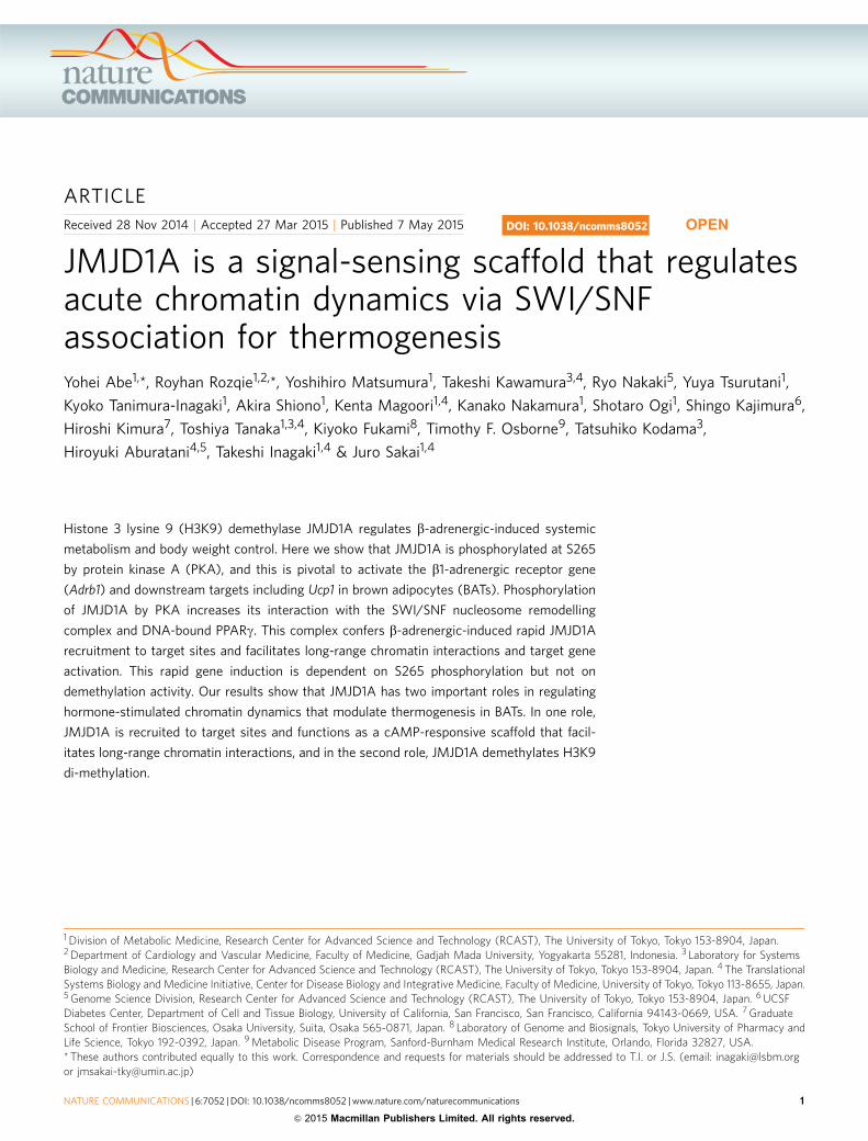
ARTICLE
Received 28 Nov 2014 | Accepted 27 Mar 2015 | Published 7 May 2015
JMJD1A is a signal-sensing scaffold that regulatesacute chromatin dynamics via SWI/SNFassociation for thermogenesisYohei Abe1,*, Royhan Rozqie1,2,*, Yoshihiro Matsumura1, Takeshi Kawamura3,4, Ryo Nakaki5, Yuya Tsurutani1,
Kyoko Tanimura-Inagaki1, Akira Shiono1, Kenta Magoori1,4, Kanako Nakamura1, Shotaro Ogi1, Shingo Kajimura6,
Hiroshi Kimura7, Toshiya Tanaka1,3,4, Kiyoko Fukami8, Timothy F. Osborne9, Tatsuhiko Kodama3,
Hiroyuki Aburatani4,5, Takeshi Inagaki1,4 & Juro Sakai1,4
Histone 3 lysine 9 (H3K9) demethylase JMJD1A regulates b-adrenergic-induced systemic
metabolism and body weight control. Here we show that JMJD1A is phosphorylated at S265
by protein kinase A (PKA), and this is pivotal to activate the b1-adrenergic receptor gene
(Adrb1) and downstream targets including Ucp1 in brown adipocytes (BATs). Phosphorylation
of JMJD1A by PKA increases its interaction with the SWI/SNF nucleosome remodelling
complex and DNA-bound PPARg. This complex confers b-adrenergic-induced rapid JMJD1A
recruitment to target sites and facilitates long-range chromatin interactions and target gene
activation. This rapid gene induction is dependent on S265 phosphorylation but not on
demethylation activity. Our results show that JMJD1A has two important roles in regulating
hormone-stimulated chromatin dynamics that modulate thermogenesis in BATs. In one role,
JMJD1A is recruited to target sites and functions as a cAMP-responsive scaffold that facil-
itates long-range chromatin interactions, and in the second role, JMJD1A demethylates H3K9
di-methylation.
DOI: 10.1038/ncomms8052 OPEN
1 Division of Metabolic Medicine, Research Center for Advanced Science and Technology (RCAST), The University of Tokyo, Tokyo 153-8904, Japan.2 Department of Cardiology and Vascular Medicine, Faculty of Medicine, Gadjah Mada University, Yogyakarta 55281, Indonesia. 3 Laboratory for SystemsBiology and Medicine, Research Center for Advanced Science and Technology (RCAST), The University of Tokyo, Tokyo 153-8904, Japan. 4 The TranslationalSystems Biology and Medicine Initiative, Center for Disease Biology and Integrative Medicine, Faculty of Medicine, University of Tokyo, Tokyo 113-8655, Japan.5 Genome Science Division, Research Center for Advanced Science and Technology (RCAST), The University of Tokyo, Tokyo 153-8904, Japan. 6 UCSFDiabetes Center, Department of Cell and Tissue Biology, University of California, San Francisco, San Francisco, California 94143-0669, USA. 7 GraduateSchool of Frontier Biosciences, Osaka University, Suita, Osaka 565-0871, Japan. 8 Laboratory of Genome and Biosignals, Tokyo University of Pharmacy andLife Science, Tokyo 192-0392, Japan. 9 Metabolic Disease Program, Sanford-Burnham Medical Research Institute, Orlando, Florida 32827, USA.* These authors contributed equally to this work. Correspondence and requests for materials should be addressed to T.I. or J.S. (email: [email protected] [email protected])
NATURE COMMUNICATIONS | 6:7052 | DOI: 10.1038/ncomms8052 | www.nature.com/naturecommunications 1
& 2015 Macmillan Publishers Limited. All rights reserved.

The coupling of environmental cues with the regulation ofgene transcription is a very important step in all cellularadaptive responses. For example, catecholamines released
from the sympathetic nervous system in response to coldexposure bind to b-adrenergic receptors (b-ARs), and triggerthe downstream cyclic AMP (cAMP) signalling cascade. ThecAMP-dependent protein kinase A (PKA) phosphorylates avariety of downstream target substrates (for example, cAMP-responsive element binding protein (reviewed in ref. 1)) totranscriptionally upregulate energy expenditure genes2,3. Recentevidence suggests that in addition to transcription factors (TFs),histone modification enzymes such as histone methyltransferasesand demethylases play essential roles in gene transcription andadaptive responses4.
JMJD1A (Jumonji domain containing 1A, also referred to asKDM3A or JHDM2A), a member of the Jumonji C-domaincontaining histone demethylase family, catalyses removal ofH3K9 mono- and di-methylation (H3K9me1 and H3K9me2;ref. 5) and functions as a co-activator for androgen receptor, aswell as a crucial regulator in spermatogenesis, germ celldevelopment, sex determination, tumorigenesis and hypoxia-inducing factor-1a-mediated gene transcription5–12. AlthoughJMJD1A regulates a wide array of appropriate gene targets indifferent settings, this enzyme lacks intrinsic DNA sequencespecificity. Therefore, how JMJD1A is targeted to specific genes inresponse to given environmental stimuli was largely unknownand of current interest.
We and another group reported that JMJD1A deficiency resultsin obesity with defects in brown adipose tissue functions that leadto cold intolerance and decreased oxygen consumption13,14. Atthe molecular level, b-adrenergic stimulation induces binding ofJMJD1A to the uncoupling protein 1 gene (Ucp1) whose product ispivotal for heat generation. This b-adrenergic-induced binding ofJMJD1A to a proximal Ucp1 enhancer region is a critical step forsubsequent gene activation; however, how b-adrenergic stimulationtriggers JMJD1A recruitment to Ucp1 and other genes involved inenergy expenditure in BATs has remained elusive.
The chromatin remodelling SWI/SNF (SWItch/Sucrose Non-Fermentable) complex couples the perturbation of histone–DNAcontacts with promoter access by TFs to their cognate DNAelements15. SWI/SNF reportedly has a potential role in long-range genomic interactions (reviewed in ref. 16); however,whether rapid environmental changes that alter cell activity inresponse to hormone signalling (that is, catecholamines)contribute to higher-order chromatin conformational changesand whether SWI/SNF is involved in such rapid action have notbeen reported.
Post-translational modifications allow proteins to play multipleroles in different physiological contexts. Thus, histone modifica-tion enzymes are feasible targets of post-translational modifica-tions that enable cells to adopt various environmental changes. Inthe current study, we show that JMJD1A is phosphorylated atserine 265 by PKA downstream from b-adrenergic stimulation.
This modification facilitates JMJD1A interaction with SWI/SNFand DNA-bound peroxisome proliferator-activated receptor-g(PPARg). This phosphorylation switch in JMJD1A is indepen-dent of its demethylase activity, suggesting that it plays ascaffolding role to mediate long-range chromatin interactionsthat position distal enhancers in close proximity to target genepromoters for key thermogenic genes.
Resultsb-Adrenergic-dependent genomic localization of JMJD1A.To analyse the JMJD1A-dependent transcriptional programmeduring b-adrenergic stimulation, we combined chromatinimmunoprecipitation (ChIP)-seq and global gene expressionanalyses. Immortalized pre-BATs (namely, pre-iBATs) were dif-ferentiated and ChIP-seq was conducted using a newly generatedmonoclonal anti-mouse JMJD1A antibody at 0 time and 2 hfollowing treatment with the b-AR pan-agonist isoproterenol(ISO). ChIP-seq peak calling by SICER identified 27,397 genomicregions as significant binding sites of JMJD1A in ISO-treatediBATs. JMJD1A localized on proximal promoters (B13%),intragenic (B52%) and intergenic regions (B24%; Fig. 1a). Thesequencing tag density was concentrated within proximal regionsof transcription start sites (TSSs; Supplementary Fig. 1a). JMJD1Apeaks were significantly enriched for clusters of sequencemotifs bound by PPAR with the highest Z-score of 62.7 in theJMJD1A-associated regions with the 10,000 highest SICERscores (Fig. 1b).
Global gene expression analysis in iBATs following the sameISO stimulation treatment identified 67 genes that were inducedby 20.8-fold after 1 h ISO treatment (that is, ISO dependent).Compared with the top 2,000 ISO-induced JMJD1A binding sites,which were annotated to 1,551 proximal genes, 39 out of 67 ISO-induced genes were found to be occupied by JMJD1A (Fig. 1c,d).These genes included Ucp1, Ppargc1a, Pdk4, Pck1 and Adrb1,which are all important metabolic factors induced followingb-adrenergic stimulation17,18 (Fig. 1d,e and SupplementaryFig. 1b). The sequence tag-mapping in Fig. 1e and Supple-mentary Fig. 1b revealed that JMJD1A was significantly enrichedat the genomic loci for these key metabolic genes.
JMJD1A is phosphorylated by PKA at serine 265. WhetherJMJD1A might be phosphorylated following ISO treatment wasevaluated by using liquid chromatography-tandem mass spec-trometry (LC/MS/MS) analysis. The immunoprecipitatedJMJD1A from HeLa cells treated with ISO for 1 h was excisedfrom an SDS–PAGE gel and subjected to LC/MS/MS (Fig. 2a).This analysis revealed that JMJD1A was phosphorylated at twoconsecutive residues on amino acids S264 and S265. These resi-dues were found as part of a putative consensus sequence forPKA phosphorylation (Fig. 2b). In vitro phosphorylation assaysdemonstrated that PKA phosphorylated recombinant humanJMJD1A (hJMJD1A; amino acids (a.a.) 1–300) at S265 (Fig. 2c).
Figure 1 | b-Adrenergic-dependent genomic localization of JMJD1A. (a) Genome-wide distribution of JMJD1A binding sites in ISO (1mM for 2 h) treated
iBATs. Ups, upstream; dws, downstream; ISO, isoproterenol. (b) Table depicting TF binding motifs enriched at constituent enhancers within JMJD1A binding
regions relative to genomic background and associated Z-scores. (c,d) JMJD1A ChIP-seq and transcriptional microarray analysis performed in iBATs at day
8 of differentiation (day 8). Heatmap represents top 10,000 high SICER scored JMJD1A binding sites under ISO-plus condition (1mM for 2 h). Colour
intensity represents Z-score of JMJD1A binding sites under ISO-minus versus those under ISO-plus condition. The higher the (red/yellow) contrast it
becomes, the higher ISO-induced JMJD1A recruitment to the given binding sites relative to ISO-minus. For reference, a colour intensity scale is included
(c, left panel). Venn diagram showing the top 2,000 sites annotated ISO-induced JMJD1A binding and the number of ISO-induced genes 420.8-fold by ISO
(1mM for 1 h; c, right panel). The overlapping genes were listed in d. (e) Genome browser shots showing the ISO-induced JMJD1A recruitments on selected
genomic regions analysed by ChIP-seq in iBATs (day 8) treated with ISO (1 mM) or vehicle for 2 h (left panel). mRNA levels of Adrb1 and Ucp1 in iBATs (day
8) after ISO (1 mM) treatment at the indicated time points. Data were presented as fold change relative to 0 h (mean±s.e.m.) of three technical replicates
(error bars are too tiny to see; right panel).
ARTICLE NATURE COMMUNICATIONS | DOI: 10.1038/ncomms8052
2 NATURE COMMUNICATIONS | 6:7052 | DOI: 10.1038/ncomms8052 | www.nature.com/naturecommunications
& 2015 Macmillan Publishers Limited. All rights reserved.

Approximately 50% of the S265A mutant protein was notphosphorylated and the S264/265A double mutant was no longerphosphorylated by PKA, while PKA phosphorylation wasretained in S264A mutant (Fig. 2c). These data suggest that S265is likely the major PKA phosphorylation site.
Immunoblot analysis with a newly generated phospho-specificantibody against phospho-S265-JMJD1A detected WT-JMJD1Atransiently expressed in iBATs cultured under ISO-plus condi-tions; however, this antibody failed to detect the S265A-JMJD1Amutant (Fig. 2d). Immunoprecipitated JMJD1A from lysates of
PPAR, RXR 62.73
EBF 56.46
AP2 54.44
NR2F 54.18
ER 53.86
Transcription factor Motif Z-score
ISO 1 h induction
39 genes
2,000 binding sites
67 genes
ISO (–) ISO (+)
10,0
00 b
indi
ng s
ites
by J
MJD
1A
Value
Color key
10
JMJD1A ISO (+)
genomic distribution
700
700
40
0
40
0
Time after ISOstimulation
Rel
ativ
e ex
pres
sion
00.81.62.43.2
0 1 2
Adrb1
02.5
57.510
0 1 2
Ucp1
(h)
(h)
27 9430023L20Rik
28 Ptp4a1
29 Rgs2
30 Adrb1
31 Ppargc1a
32 Tob1
33 Ier3
34 Cebpd
35 Jun
36 Ddit4
37 Atf3
38 Arrdc3
39 Ugdh
1 Pdk4
2 Sash1
3 Bag3
4 Pim3
5 Uap1
6 Pck1
7 Fosl2
8 Ucp1
9 Phlda1
10 Cxcl1
11 Ier5
12 Ccrn4l
13 Irf2bp2
14 Nfkbiz
15 Socs2
16 Thbs1
17 Ccnl1
18 Maff
19 Mat2a
20 Lpin1
21 Vegfa
22 Scd1
23 Gadd45g
24 Nfil3
25 Rasd1
26 Rhob
5,000–2,000 ups2,000–0 upsIntragenic2,000–0 dws5,000–2,000 dws Others
Value
10 kb
10 kb
Adrb1
Ucp1
Ucp1
Adrb1
ISO (–)
ISO (+)
ISO (–)
ISO (+)
40
40
10
10
30
30
10
10
2
Bits 1
05’
2
Bits 1
5’
5’
5’
5’
0
2
Bits 1
0
2
Bits 1
0
2
Bits 1
0
3’
3’
3’
3’
3’
1 2 3 4 5 6 7 8 9 10
1 2 3 4 5 6 7 8 9 10
11 12 13
1 2 3 4 5 6 7 8 9 10 11 12 13 14
11
2
2
3
3
4
4
5
5
6
6
7
7
8
8
9 10 11 12 13 14 15 16 17 18 19 20
weblogo.berkeley.edu
weblogo.berkeley.edu
weblogo.berkeley.edu
weblogo.berkeley.edu
weblogo.berkeley.edu
NATURE COMMUNICATIONS | DOI: 10.1038/ncomms8052 ARTICLE
NATURE COMMUNICATIONS | 6:7052 | DOI: 10.1038/ncomms8052 | www.nature.com/naturecommunications 3
& 2015 Macmillan Publishers Limited. All rights reserved.

iBATs pretreated with ISO was detected by anti-P-S265-JMJD1Aantibody but the antibody failed to recognize JMJD1A in cellspretreated with the PKA inhibitor H89 (Fig. 2e).
Phospho-S265 is dispensable for enzymatic activity in vitro.To examine whether S265 phosphorylation might alter JMJD1Ademethylase activity, we performed an in vitro demethylationassay. H3K9me2 demethylation activity of JMJD1A did not differbetween phosphorylated and non-phosphorylated full-lengthrecombinant JMJD1A (Supplementary Fig. 2a). In addition,mutation of S265 to alanine (S265A) did not alter the substratespecificity or demethylase activity (Supplementary Fig. 2b). Bothwild-type (WT) and S265A recombinant full-length JMJD1Acatalysed the removal of mono- and di-methylation of H3K9 butnot tri-methylation (Supplementary Fig. 2c). Immunohis-tochemistry and subcellular fractionation showed that both WTand S265A mutant JMJD1A predominantly localized in thenucleus with lesser staining in the cytosol of iBATs(Supplementary Fig. 2d,e).
Gene activation by JMJD1A requires phosphorylation at S265.To determine the impact of S265 phosphorylation on genetranscription, we retrovirally transduced either a V5-tagged WT-hJMJD1A, a mutant S265A human version of JMJD1A (S265A-hJMJD1A) or the control Zeor-empty vector into iBATs where wealso knocked down expression of the endogenous mouse JMJD1Aby short hairpin RNA (shRNA; iBATshs). Human Jmjd1a isresistant to this shRNA designed against mouse Jmjd1a and theretroviral vector is driven by a very weak long terminal repeat
promoter. Immunoblot analysis showed that protein level of V5-hJMJD1A was equivalent to that of native JMJD1A(Supplementary Fig. 3a). Reverse transcription–quantitative real-time PCR (qPCR) analyses revealed that transduction of WT-hJMJD1A into iBATshs upregulates ISO-induced expression ofkey metabolic genes such as Ucp1 and Adrb1; however, S265A-hJMJD1A was incapable of stimulating gene expression eventhough it was expressed at levels similar to the WT protein(Fig. 3a and Supplementary Fig. 3a). In contrast, expression of awhite adipocyte marker gene, Hk1, did not differ significantlybetween WT- and S265A-hJMJD1A-iBATshs (Fig. 3a). Theposition of S265 was highly specific: because substitution of otherserine residues nearby such as S264 or S341 to alanine (S264Aand S341A, respectively) did not perturb ISO-induced Adrb1 andUcp1 expression, meanwhile S265A and S264/265/341A (3SAmutant) reduced expression (Fig. 3b). It is also notable thatmutation of key residues required for demethylase activity,H1120Y or H1120F, (ref. 5) did not affect the ISO-inducedexpression of Adrb1 and Ucp1 (Fig. 3c,d). ChIP–qPCR analysisrevealed that ISO treatment of iBATs did not affect H3K9me2levels at Adrb1 gene locus (Supplementary Fig. 3b). These resultsindicate that phospho-S265-JMJD1A (P-JMJD1A)-induced genetranscription appears to be independent of H3K9 demethylaseactivity at Adrb1 locus.
PKA-induced P-JMJD1A–SWI/SNF-PPARc protein complex.To identify co-regulator complexes for P-JMJD1A, nuclearextracts of ISO-treated 3T3-L1 adipocytes were immunoprecipi-tated with anti-mJMJD1A antibody. Putative interacting partners
parent+2H-98precursor ion m/z 672.312 (z=2)
K S pS E N N G T L V S Ky7
b5
y2
b10
y11 y3
b9
y4
b8
y5
b7
y6
b6 b11
y8
b4
y9
b3
y10
b2
50
75100
150
250
JMJD1AIg
Gα-
JMJD
1A
kDa
IB:
Empty WT S265A
α-V5
ISO – +
α-β-Actin
α-p-JMJD1A(pSer265)
– + – +
PKA (–) PKA (+)
S26
4/26
5A
WT
S26
4AS
265A
S26
4/26
5A
WT
S26
4AS
265A
GST-hJMJD1A (a.a. 1–300)
m/Z
0
20
40
60
80
100672.31 m/z, 2+, 1,342.61 Da, (parent error: –0.084 ppm)
y6y7b6b2 y2 y3 b3 y4 b4
y5b5 b7
y8b8
y9b9
b10y10
b11 y11
K S S+80 E N N G T L V S K
K S V L T G N N E S+80 S K
0 200 400 600 800 1,000 1,200
Rel
ativ
e in
tens
ity (
%)
JMJD1A
256 65275
PKA consensus sequence(K/R)-K/R-X-S*/T*
Human RIGAV KRKSSENNGT LVSKQBovine RMGAV KRKSSENNGN LVSKQMouse RTGAV KRKSSENNGS SVSKQRat RIGAV KRKSSENNGS SVSKQ
Chicken RIGGV KRKPSENSGS VDAKHXenopus KPRAP KRKSQDTESE DQTEL
264 265
IB:
α-p-JMJD1A(pSer265)
α-JMJD1A
α-TBP
H89
ISO
Veh
icle
ISO
H89
+IS
O
IgGIP: α-JMJD1A
Input
JMJD1A(a.a. 1–300)
p-JMJD1A(a.a. 1–300)
150
150
150
37
kDa
150
37
kDa
Figure 2 | JMJD1A is phosphorylated at serine 265 by PKA. (a) Post-translational modifications of JMJD1A identified by mass spectrometry. JMJD1A
protein in ISO-treated HeLa cells were immunoprecipitated with anti-hJMJD1A antibody (IgG-F0026), separated by SDS–PAGE gel, stained with SYPRO
Ruby and then subjected to in-gel digestion for mass spectrometry (left panel). MS/MS spectrum of the P-JMJD1A fragment from K263 to K274,
m/z¼ 672.312 (Z¼ 2) is shown in the right panel. (b) The PKA consensus site is conserved in various species. (c) In vitro PKA kinase assay. WT, S264,
S265A or S264A/S265A mutated JMJD1A (a.a. 1–300) recombinant GST-fusion proteins were PKA-treated and subjected to Phos-tag SDS–PAGE followed
by immunoblot (IB) analysis with anti-GST antibody. (d) ISO-induced JMJD1A phosphorylation at S265. Whole-cell lysates from WT- or S265A-hJMJD1A
expressing iBATshs (day 8) treated with ISO (10mM for 1 h) were subjected to immunoprecipitation (IP) using anti-mJMJD1A (IgG-F0618) followed by IB
analysis with anti-P-S265-JMJD1A. (e) IB analysis showing PKA-mediated phosphorylation of native JMJD1A at S265. iBATs (day 8) were pretreated with
PKA inhibitor H89 (20 mM) for 20 min and then treated with ISO (10mM) for 1 h. Whole-cell lysates were subjected to IP using anti-mJMJD1A (IgG-F0618)
and IB analysis with anti-P-S265-JMJD1A. Uncropped images of the blots (c–e) are shown in Supplementary Fig. 11.
ARTICLE NATURE COMMUNICATIONS | DOI: 10.1038/ncomms8052
4 NATURE COMMUNICATIONS | 6:7052 | DOI: 10.1038/ncomms8052 | www.nature.com/naturecommunications
& 2015 Macmillan Publishers Limited. All rights reserved.

were separated by SDS–PAGE followed by SYPRO Ruby stain(Fig. 4a). LC/MS/MS analysis led to the identification of threesubunits of the SWI/SNF chromatin remodelling complex withinthe JMJD1A immunoprecipitation complex: AT-rich interactivedomain containing protein 1A (ARID1A), BRG1 (also referred toas SMARCA4) and BRG1-associated factor of 60 kDa, subunit b(BAF60b) (Fig. 4b and Supplementary Fig. 4a). The numbers ofpeptides identified in the immunoprecipitates were significantlyincreased when the cells were pretreated with ISO (Fig. 4b).BRG1, ARID1A and BAF60b were also detected by immunoblotanalysis of the immunoprecipitates of V5-tagged WT-hJMJD1Aexpressing iBATshs lysates confirming the LC/MS/MS analysis.Interestingly, the interaction of these SWI/SNF components withJMJD1A was increased by ISO treatment (Fig. 4c, compare lanes4 and 5). By contrast, SWI/SNF components were recovered atvery low levels from the co-immunoprecipitates of S265A-
hJMJD1A-iBATshs lysates (Fig. 4c, compare lanes 9 and 10). Thetotal cellular levels of the SWI/SNF factors were not clearlyaffected by ISO (Fig. 4c, compare lanes 1 and 2). An interactionbetween PPARg and JMJD1A, which was inducible upon ISOtreatment of iBATs, was detected as well (Fig. 4d, compare lanes 2and 7). Following transfection of short interfering RNAs speci-fically targeting Arid1a, Brg1, or Baf60b, co-immunoprecipitation(co-IP) of JMJD1A with PPARg was no longer observed (Fig. 4d),demonstrating that the association between JMJD1A and PPARgrequires SWI/SNF. We next clarified whether or not only phos-phorylated JMJD1A (P-JMJD1A) is able to associate with SWI/SNF complex and PPARg. Because fetal bovine serum used forcell culture already contains non-negligible levels of catechola-mines, we pre-cultured differentiated iBATs for 6 h in 0.1%bovine serum albumin, then added ISO for 1 h and performed aco-IP assay. The result in Fig. 4e showed that PPARg wasco-immunoprecipitated with JMJD1A only under the ISO-pluscondition (Fig. 4e, compare lanes 2 and 3). Non-phosphorylatedform S265A JMJD1A was not co-immunoprecipitated, indicatingthat this interaction was dependent on phosphorylation of S265(Fig. 4e, lanes 5 and 6). JMJD1A was also co-immunoprecipitatedwith an antibody to PPARg but only when ISO was included. TheJMJD1A bound to PPARg was indeed phosphorylated because itwas detected by the anti-P-JMJD1A antibody (Fig. 4f, comparelanes 2 and 3). These data demonstrate that only P-JMJD1A isable to associates with SWI/SNF complex and PPARg.
Crosstalk between SWI/SNF and P-JMJD1A in gene regulation.We next examined the role of SWI/SNF interaction withP-JMJD1A in activation of gene expression for Adrb1 and Ucp1.The basal and ISO-induced levels of Adrb1 and Ucp1 were sig-nificantly reduced following knockdown of Arid1a, Brg1 orBaf60b (Fig. 4g,h). Specificity of the knockdown was validated byqPCR and immunoblot analyses (Supplementary Fig. 4b,c). Thesedata support the model in Fig. 4I, predicting that SWI/SNF isinvolved in the phosphorylation-dependent activation of geneexpression by JMJD1A.
Co-localization of JMJD1A and SWI/SNF on PPARc targets.Next, we used ChIP-seq for two major components of the
Time after ISO stimulation (h)
Rel
ativ
eex
pres
sion
02468
0 0.5 1 2
Adrb1
060
120180240
0 0.5 1 2
Ucp1
00.40.81.21.6
0 0.5 1 2
Hk1
EmptyWTS265A
S265A
Empt
y
WT
S264A
Em
pty
WT
S26
4A
S26
5A
IB:
α-V5JMJD1A
α-β-Actin
S34
1A
3SA
S341A 3S
A
Rel
ativ
e e
xpre
ssio
n
0
0.5
1
1.5Adrb1
Empt
yW
T
S264A
S265A
S341A 3S
A0
0.5
1
1.5Ucp1
Empt
yW
T
S264A
S265A
S341A 3S
A
Em
pty
WT
H11
20Y
α-β-Actin
IB:Empty WT
H1120Y
α-V5JMJD1A
H11
20F
H1120F
Rel
ativ
eex
pres
sion
0
0.5
1
1.5Adrb1
0
0.5
1
1.5Ucp1
Empt
yW
T
H1120
Y
H1120
F
Empt
yW
T
H1120
Y
H1120
F
1,321
NH2 COOH
885 –889LXXLL
1,058 –1,281JmjC
S265
*
H1120
1 P
150
37
kDa
150
37
kDa
662 –687Zn finger
Figure 3 | Phosphorylation of JMJD1A at S265 is crucial for
b-adrenergic-induced gene transcriptions. (a) ISO-induced Adrb1 and Ucp1
mRNA levels in iBATshs stably expressing WT- or S265A-hJMJD1A or
empty vector were measured by RT-qPCR. The mRNA values are depicted
relative to mRNA in iBATshs transduced with empty vector on day 8 of
differentiation before ISO treatment (0 h), which are arbitrarily defined as 1.
(b) Adrb1 and Ucp1 mRNA levels in WT or serine to alanine mutants
(S264A, S265A, S341A or, 3SA) JMJD1A expressing iBATshs measured by
RT-qPCR after 1 h ISO treatment (top panel). 3SA represents all three
mutations of S264A, S265A and S341A. Data were presented as fold
change relative to WT-hJMJD1A-iBATshs after normalized to cyclophilin.
Immunoblot (IB) analysis for WT and various mutant JMJD1A proteins and
Oil Red O (ORO) staining (bottom panel). (c) Schematic representation of
the domain architecture of hJMJD1A. Phosphorylation site at S265 and
Fe(II) binding site at H1120 are shown. (d) Comparable ISO-induced gene
expressions of Adrb1 and Ucp1 in WT and demethylase dead JMJD1A
mutants expressing iBATshs. RT-qPCR was performed to quantify mRNA
levels of Adrb1 and Ucp1 genes in WT-, H1120Y- or H1120F-hJMJD1A-
iBATshs treated with ISO for 1 h (top panel). Data were presented as fold
change relative to WT-hJMJD1A-iBATshs. IB analysis and ORO in the
indicated iBATs (bottom panel). Data are presented as mean±s.e.m. of
three technical replicates (a,b,d) (error bars are too tiny to see in some
figures). Uncropped images of the blots (b,d) are shown in Supplementary
Fig. 11.
NATURE COMMUNICATIONS | DOI: 10.1038/ncomms8052 ARTICLE
NATURE COMMUNICATIONS | 6:7052 | DOI: 10.1038/ncomms8052 | www.nature.com/naturecommunications 5
& 2015 Macmillan Publishers Limited. All rights reserved.

SWI/SNF complex (BRG1 and ARID1A; Fig. 5a) and comparedthe results with our JMJD1A ChIP-seq (Fig. 1c,e and Supplemen-tary Fig. 1b). We also included formaldehyde-assisted isolation ofregulatory element-seq (FAIRE-seq) and ChIP-seq for histone H3Lys-4 tri-methylation (H3K4me3) and histone H3 Lys-27 acet-ylation (H3K27ac) in control and ISO-treated iBATs (Fig. 5a).The co-localization of JMJD1A, BRG1, ARID1A and PPARg wasrevealed by the significant overlap in sequence tag-density profilescomparing the corresponding genome browser tracks shown inFig. 5a with our ChIP-seq data sets for PPARg and JMJD1A(Fig. 1c,e and Supplementary Fig. 1b). We also directly comparedthe binding signals of ARID1A, BRG1, PPARg and JMJD1A
obtained from ChIP-seq data within the top 10,000 binding sitesof JMJD1A in iBATs. The heatmap in Supplementary Fig. 5ashows co-localization of ARID1A, BRG1 and PPARg withJMJD1A on the 39 ISO-stimulated JMJD1A target genes selectedin Fig. 1d. Several representative ChIP-seq profiles of their targetsand ISO-induced gene expression profiles are also shown inSupplementary Fig. 6. These results strongly suggest that for-mation of this higher-order complex is a key step for b-adre-nergic-induced JMJD1A recruitment to its target genes, which isalso a prerequisite for subsequent H3K9me2 demethylation ofchromatin at the Ucp1 enhancer (B2 kb) as previously shown14
(Fig. 4i).
ARID1A
IB:
RPB1BRG1
250
150
100
75
kDa
Phos-tagBiotin HRP
α-JMJD1A
α-β-Actin
Veh
icle
ISO
ISO
IP : α-JM
JD1A
Ctrl Ig
G
Sypro rubystain:
JMJD1A
1 2 3Lane
BAF60b
Vehicle ISO
ARID1A/BAF250a 242 15 42
SMRCA4/BRG1 181 5 15
RPB1/POLR2A 217 13 21
JMJD1A 148 21 14
Numbers of peptideIdentified protein
MW
(kDa)
SMRD2/BAF60b 59 0 5
S265 P
PPARγ
ARID1A
BRG1(ATPase)
JMJD1A
SWI/SNF
BAF60b
Adrb1, Ucp1
PPRE
Inpu
t
IB: ISO 1h
IP:
α-PPARγ
α-PPARγ
IgG
IgG
WT
α-V
5 JM
JD1A
α-V
5 JM
JD1A
α-V5JMJD1A
α-V5JMJD1AIP
pel
let
1 2 3 4 5 6Lane
+ – + + – +
S265A
150
37
kDa
150
α-BRG1
α-V5JMJD1A
Inpu
tIB: ISO 1h +
IgG
Inpu
t
IgG
IP
WT S265A
IP
α-V
5 (J
MJD
1A)
α-V
5 (J
MJD
1A)
– +– +– +–+ +
250
250
50150
kDa
IP:
α-V
5JM
JD1A
α-V
5JM
JD1A
IgG
IgG
ISO 1h (+)
IB:
1 2 3 5 6 7 8Lane 4
Inpu
tIP
pel
let
α-PPARγ
α-PPARγ
α-V5JMJD1A
α-V5JMJD1A
si-B
af60
b
si-A
rid1a
si-C
tl
si-B
rg1
si-C
tlsi
-Baf
60b
si-A
rid1a
si-C
tl
si-B
rg1
si-C
tl
9 10
(–)
150
50
kDa
150
50
150
50
150
50
kDa
Adrb1
2
2.5
00.5
11.5
22.5
3
Rel
ativ
e ex
pres
sion
0
0.5
1
1.5
si-B
rg1
#1 #2si
-Ctl
si-B
rg1
#1 #2
si-C
tl
si-A
rid1a
#1 #2si
-Ctl
si-A
rid1a
#1 #2
si-C
tl
si-B
af60
b#1 #2
si-C
tl
si-B
af60
b#1 #2
si-C
tl
0
0.5
1
1.5
2
2.5(–)
(+)
ISO 1h
02468
Rel
ativ
e ex
pres
sion
0
2
4
6
8
Ucp1
10
0
1
2
3
4
5(–)
(+)
ISO 1h
si-B
rg1
#1 #2si
-Ctl
si-B
rg1
#1 #2
si-C
tl
si-A
rid1a
#1 #2si
-Ctl
si-A
rid1a
#1 #2
si-C
tl
si-B
af60
b#1 #2
si-C
tl
si-B
af60
b#1 #2
si-C
tl
1 2 3 5 6 7 8Lane 4 9 10
α-ARID1A
α-BAF60b
Inpu
t
IB:ISO 1h
IP:
α-PPARγ
α-PPARγ
IgG
IgG
WT
α-P
PA
Rγ
α-V5JMJD1A
α-V5JMJD1A
IP p
elle
t
1 2 3 4 5 6Lane
+ – + + – +
S265A
α-P
PA
Rγ
50
150
150
50
kDa
(pSer265)α-p-JMJD1A
150
ARTICLE NATURE COMMUNICATIONS | DOI: 10.1038/ncomms8052
6 NATURE COMMUNICATIONS | 6:7052 | DOI: 10.1038/ncomms8052 | www.nature.com/naturecommunications
& 2015 Macmillan Publishers Limited. All rights reserved.

JMJD1A occupies lineage-specific distal enhancers. Recentstudies have shown that SWI/SNF-mediated higher-order chro-matin interactions play significant roles in gene transcription16.In our studies, ChIP-seq analysis showed that B24% of JMJD1Aoccupied sites are intergenic regions (Fig. 1a). Comparing theJMJD1A localization to H3K27ac, an epigenetic mark of activeenhancers revealed that JMJD1A occupied H3K27ac-enrichedremote enhancers following ISO treatment. In addition, the ChIP-seq comparison in Fig. 5a showed that PPARg, BRG1 and ARID1Awere also localized to these chromatin regions. These observationsprompted us to propose that a JMJD1A–SWI/SNF-PPARg complexplays a significant role in controlling higher-order chromatininteractions to regulate gene transcription in response to b-adrenergic stimulation. The potential remote enhancers marked byH3K27ac located 20–50 kb upstream of Adrb1 (Fig. 5a), which is anideal distance to examine a long-range looping model bychromosome conformation capture (3C) analysis19.
A survey of the extended Adrb1 locus revealed JMJD1Aoccupancy in the vicinity of Adrb1 gene body and also tochromatin domains located 44 and 22 kb upstream (Fig. 5a,b, leftpanel). We refer to 44 and 22 kb elements here as E1 and E2enhancers, respectively, because they both displayed H3K27acenrichment and only low levels of H3K4me3 (Fig. 5a), suggestingthat they contain active enhancers. An evaluation of publishedChIP-seq data obtained from normal mouse tissues also revealedthat H3K27ac was present at E1 and E2 regions specifically inbrown adipose tissue (Supplementary Fig. 5b, left panel). Furtherquantification by ChIP–qPCR showed that recruitment ofJMJD1A, ARID1A and BRG1 to these two enhancers wasincreased by ISO treatment (Fig. 5b–d, left panels). By contrast,PPARg was recruited to these enhancers of Adrb1 gene regardlessof ISO stimulation, indicating that PPARg is bound beforestimulation (Fig. 5e, left panel). ISO treatment did not grosslyperturb levels of H3K27ac at E1 and E2 (Fig. 5a, left panel;Fig. 5f,g). Since CCAAT/enhancer binding protein-a (C/EBPa)and C/EBPb associate with SWI/SNF20, we postulated that C/EBPfamily members may associate with E1 and E2. ChIP–qPCRshowed that C/EBPa and C/EBPb also occupy the E1 and E2enhancers (Fig. 5h, left panel).
Additional analysis of ChIP-seq and ChIP–qPCR on the Ucp1locus showed co-localization of ARID1A, BRG1, PPARg, C/EBPaand C/EBPb with JMJD1A, as well as enrichment of H3K27ac atthe enhancer region located 13, 5 and 2 kb upstream of thetranslation initiation site (Fig. 5a–e,h, right panels). An evaluationof published ChIP-seq data obtained from normal mouse tissuesalso revealed that H3K27ac was present at -13, -5 and -2 kbregions (Supplementary Fig. 5b, right panel).
P-JMJD1A mediates Adrb1 enhancer–promoter interaction.The above observations raised the intriguing hypothesis that ISOfacilitates long-range DNA looping such that TFs (PPARg,CEBPa and b) and the SWI/SNF chromatin remodelling complexthat are located at distal E1 and/or E2 enhancers are brought inproximity to the TSS of Adrb1, facilitating the recruitment ofRNA polymerase and a subsequent increase in Adrb1 genetranscription. To evaluate this, we performed a 3C analysisquantified by qPCR using an anchor point fixed near Adrb1 orE1. 3C-qPCR using the Adrb1 anchor point revealed that E1made contact with Adrb1 and its contact frequency was inducibleby ISO treatment (Fig. 6a). 3C-qPCR using an anchor point at E1showed that both E1-E2 proximity and E1-Adrb1 promoterproximity were ISO dependent (Supplementary Fig. 7a). Toge-ther, these data indicate that E1 is brought in close proximity tothe E2-Adrb1 promoter contact via DNA long-range looping inan ISO-inducible manner as schematically illustrated in Fig. 6e.The looping interactions between E1 and Adrb1 promoter weredetected as early as 15 min and reached a maximum at 60 minand reduced by 60% after 120 min following ISO treatment andthen declined (Fig. 6b, top panel). This time course was closelymatched by the timing of PKA-dependent phosphorylation ofJMJD1A that was detected as early as 5–15 min, peaked at 60 minand was reduced to 60% after 120 min (Fig. 6b, bottom panel).This was also correlated with the pattern of Adrb1 mRNAaccumulation (Fig. 1e). Similar results were obtained followingforskolin (FSK) activation of cAMP signalling (SupplementaryFig. 7b).
Interestingly, the S265A mutation significantly reduced thelong-range looping signal in response to FSK treatment (Fig. 6c).Similarly, knockdown of Brg1 abrogated the FSK-dependentchromatin looping between E1 and Adrb1 promoter as well(Fig. 6d). Collectively, we conclude that PKA-dependent JMJD1Aphosphorylation at S265 facilitates higher-order chromatinconformation changes that require SWI/SNF.
Enhancer–promoter interactions increase Adrb1 expression.To examine the combined roles for E1 and E2, and looping inAdrb1 gene expression, the promoter region was cloned with oneor both of the two enhancer regions into pGL3 basic luciferasevector and transfected into iBATs (Fig. 7a, left panel) that werethen cultured in the absence or presence of a differentiationcocktail. The experiments demonstrated that the promoter hadweak activity, and that each of the two enhancers only con-tributed a modest increase in luciferase reporter expression (B2-fold) (Fig. 7a). Intriguingly, the combination of both E1 and E2
Figure 4 | Phosphorylation of JMJD1A triggers the interaction with SWI/SNF and PPARc. (a,b) JMJD1A-associated proteins were immunoprecipitated
with anti-mJMJD1A antibody (IgG-F0231) from 3T3-L1 cells treated with ISO (10mM for 1 h), separated by SDS–PAGE gel, stained with SYPRO Ruby and
then subjected to in-gel digestion for mass spectrometry (a, top panel). Identified proteins were shown in b and Supplementary Fig. 4a. P-JMJD1A protein
was demonstrated by immunoblot (IB) analysis using anti-phospho-S265-JMJD1A antibody (a, bottom panel). (c) Nuclear extracts from either WT- or
S265A-hJMJD1A-iBATshs were treated with either ISO (10 mM for 1 h) or vehicle and subjected to immunoprecipitation (IP) with anti-V5 antibody and
followed by IB analysis with anti-BRG1, anti-ARID1A or anti-BAF60b. (d) ISO-dependent JMJD1A association with PPARg via ARID1A, BRG1 and BAF60b.
WT-hJMJD1A-iBATshs were subjected to IP with anti-V5 and followed by IB with anti-PPARg antibody (IgG-A3409). (e,f) S265 phosphorylation is crucial
for JMJD1A binding to PPARg. WT or S265A-hJMJD1A-iBATshs were pre-cultured in 0.1% bovine serum albumin containing DMEM for 6 h then treated
with ISO (10 mM for 1 h) or vehicle and nuclear extracts from each cells were subjected IP with anti-V5 antibody followed by IB with anti-PPARg antibody
(e). The same extracts were also subjected IP with anti-PPARg antibody followed by IB with either anti-V5 antibody or anti-P-S265-JMJD1A antibody (f).
(g,h) JMJD1A and SWI/SNF complex interaction was functionally linked to gene expressions. Adrb1 and Ucp1 mRNA levels were quantified by RT-qPCR
in WT-hJMJD1A-iBATshs transfected with control short interfering RNA (siRNA) or two independent siRNAs specifically targeting Arid1a, Brg1 or Baf60b
under either ISO-plus (1 mM for 1 h) or minus condition. Data were presented as fold change relative to control siRNA transfected cells under ISO-minus
condition. Error bars represent mean±s.e.m. of three technical replicates. The experiments were performed at least three times and the most
representative one is shown. (i) Schematic drawing of JMJD1A–SWI/SNF–PPARg complex. P-JMJD1A at S265 induces forming a complex with SWI/SNF
chromatin remodeler and TF PPARg recruited to PPRE (Fig. 1b). PPRE, PPAR-responsive element. Uncropped images of the blots (a,c–f) are shown in
Supplementary Figs 11 and 12.
NATURE COMMUNICATIONS | DOI: 10.1038/ncomms8052 ARTICLE
NATURE COMMUNICATIONS | 6:7052 | DOI: 10.1038/ncomms8052 | www.nature.com/naturecommunications 7
& 2015 Macmillan Publishers Limited. All rights reserved.

enhancers synergistically drove luciferase gene expression(eightfold; Fig. 7a). ChIP–qPCR by RNA polymerase II (Pol II)showed that Pol II recruitment at TSS and across Adrb1 genebody was induced by twofold at 1 h ISO treatment (Fig. 7b,bottom panel). This was accompanied by hyper-acetylation ofhistone H3 at Adrb1 gene body (Fig. 7c,d). The induction of Pol IIrecruitment was also observed on the Ucp1 gene body (20-fold inFig. 7b). Agreement with previous study, these data indicate thatmRNA levels are due to increased transcriptions rather thanmRNA stability under b-adrenergic stimulation21.
Luciferase reporter analysis on the Ucp1 locus showed thatpGL3 luciferase reporter construct that harbours the proximal(� 2 kb) enhancer–promoter drove appreciable level of luciferasegene expression and the additional two distal enhancers (� 13
and � 5 kb regions) plus proximal (� 2 kb) enhancer–promoterto this construct drove much higher luciferase gene expression(Supplementary Fig. 8). This result suggests that distal enhancersare important in Ucp1 gene expression. Since the S265A mutationin JMJD1A severely reduced Ucp1 expression (Fig. 3a), it suggeststhat P-JMJD1A–SWI/SNF-PPARg may also contribute to long-range DNA looping at Ucp1 to facilitate enhancer–promoterinteractions.
P-S265-JMJD1A regulates thermogenesis. Among P-JMJD1Atargets, ADRB1 is the most upstream component in the b-adre-nergic signalling pathway. As reported previously21, the ISO-induced Adrb1 gene expression reached a peak at 1 h (Fig. 1e).
Adrb1
Adrb1 5 kbE1 E2
H3K4me3
H3K27ac
H3K27ac
ARID1A
PPARγ
ARID1A
JMJD1A
JMJD1A
(+)
(+)
(–)
(+)
(–)
(+)
(–)
(–)
BRG1
BRG1 (–)
(+)
1200
70107010
50105010250
250
250
450
450
Ucp15 kb
Ucp1
1200
505
505
4510451025
225
2250
350
350
–44 kb –22 kb +0.7 kbPrimers position –13 kb –5 –2
FAIRE (–) 15 0
20 0
–13 kb –5 kb –2 kb +0.6 kb
Ucp1
(–) (+) (–) (+) (–) (+) (–) (+)0
0.51
1.52
C/EBPα, β, δ ChIPIgGC/EBPαC/EBPβC/EBPδ
IgGC/EBPαC/EBPβC/EBPδ
00.01
0.04
0.020.03
PPARγ ChIP
% In
put
00.01
0.04
0.020.03
Hox
C8
Adrb1
–44
–22
+0.
7
–13 –5 –2
+0.
6 (kb)
Ucp1
(–)(+)
ISO 2h
ISO 2h
(–)(–)(–)(–)
ISO 2h(+)
(+)(+)(+)
Cyc
lo
Adrb1
–44
–22
+0.
7 (kb)
% In
put
H3K27ac ChIP
00.20.40.60.8
1(–)(+)
ISO 2h
Cyc
lo
Adrb1
–44
–22
+0.
7 (kb)
% In
put
H3ac ChIP
00.050.100.150.200.250.30
(–) (+)
ISO 2h
00.020.040.060.080.1
0
0.02
0.04
0.06
0.08C
yclo
Adrb1
–44
–22
+0.
7
–13 –5 –2
+0.
6 (kb)
Ucp1
% In
put
BRG1 ChIP
(–)(+)
ISO 2h
00.010.020.030.040.05
0.010.020.030.04
0
% In
put
Cyc
lo
Adrb1
–44
–22
+0.
7
–13 –5 –2
+0.
6 (kb)
Ucp1
ARID1A ChIP
(–)(+)
ISO 2h
% In
put
Cyc
lo
Adrb1
–44
–22
+0.
7
–13 –5 –2 (kb)
Ucp1
JMJD1A ChIP
00.20.40.60.8
00.20.40.60.8
(–)(+)
ISO 2h
ISO2h
01234
(–) (+)ISO
Cyclo –44 kb –22 kb +0.7 kb
Adrb1
(–) (+) (–) (+) (–) (+)
% In
put
H3K4me3
H3K27ac
H3K27ac
ARID1A
PPARγ
ARID1A
JMJD1A
JMJD1A
(+)
(+)
(–)
(+)
(–)
(+)
(–)
(–)
BRG1
BRG1 (–)
(+)
Primers position
FAIRE (–)
ISO 2h
PPARγ (+) 250
250
PPARγ (+)
+0.6 kb
10060
20
128
4
30
10
30
10
15
5
15
5
15
15
30
10
30
10
56750 kb 56760 kb 56770 kb 56780 kb 56790 kb 56800 kb
5
5
503010
503010
1006020
15
5
4030201040302025
15
5
15
25
5
15
15
35251553525155
85800 kb 85805 kb 85810 kb 85815 kb
5
5
50
30
1050
30
10
Figure 5 | Co-localization of JMJD1A–SWI/SNF-PPARc across Adrb1 and Ucp1 genomic regions. (a) ChIP-seq profiles for H3K4me3, H3K27ac, BRG1,
ARID1A, PPARg and JMJD1A and formaldehyde-assisted isolation of regulatory element (FAIRE)-seq open chromatin profile on Adrb1 and Ucp1 genomic
regions. iBATs (day 8) were treated with 1 mM ISO or vehicle for 2 h and subjected to ChIP-seq or FAIRE-seq analysis. Light pink shadows highlight the
enhancers from H3K27ac ChIP-seq data. JMJD1A, SWI/SNF components (ARID1A and BRG1) and PPARg co-localized at distal enhances of Adrb1 and Ucp1.
Scale bars, 5 kb. (b–h) ChIP–qPCR of ISO-induced binding of JMJD1A (b), ARID1A (c), BRG1 (d), PPARg (e), H3ac (f), H3K27ac (g), C/EPBa (h), C/EBPb (h) or
C/EBPd (h) on enhancers each of Adrb1 and Ucp1. Vertical axis represents % input. The experiments in b–h were performed at least three times and the
representative one is shown. Error bars represent mean±s.e.m. of three technical replicates.
ARTICLE NATURE COMMUNICATIONS | DOI: 10.1038/ncomms8052
8 NATURE COMMUNICATIONS | 6:7052 | DOI: 10.1038/ncomms8052 | www.nature.com/naturecommunications
& 2015 Macmillan Publishers Limited. All rights reserved.

This was also reflected at ADRB1 protein levels (SupplementaryFig. 9a). We also showed that expression of Ucp1, anotherP-JMJD1A target, was also reduced at the mRNA and proteinlevels in S265A-hJMJD1A-iBATshs (Fig. 3a and SupplementaryFig. 9a). Maximum cAMP production following treatment withthe ADRB1 selective agonist dobutamine (DOB) in S265A-hJMJD1A-iBATshs was reduced relative to identically treatedWT-hJMJD1A-iBATshs (Supplementary Fig. 9b), which isconsistent with reduced ADRB1 protein in the S265A-hJMJD1A-iBATshs (Supplementary Fig. 9a). Also, DOB-stimulated glycerol release was blunted in S265A-hJMJD1A-iBATshs (Supplementary Fig. 9c). To examine further whetherphosphorylation of S265 is critical for BATs function, wemeasured respiratory activity using the extracellular fluxanalyser. The mitochondrial respiration of WT-hJMJD1A-iBATshs was markedly induced (approximately double) by DOBstimulation, while this induction was markedly blunted in S265A-hJMJD1A-iBATshs (Supplementary Fig. 9d). This DOB-inducedrespiration in WT-hJMJD1A-iBATshs was largely insensitive tooligomycin and was thus uncoupled (Supplementary Fig. 9d, toppanels). The characteristic proton leakage profile in BATs wasindeed reduced in S265A-hJMJD1A-iBATshs (SupplementaryFig. 9d). These additional observations demonstrate that thekey cellular responses mediated by b1-AR signalling are alsoreduced in response to the S265A-JMJD1A mutation.
P-S265 JMJD1A in brown adipose tissue of mice in vivo. Toanalyse the physiological relevance of the above observations, weevaluated the phosphorylation switch model for JMJD1A in vivo.First, using the anti-P-S265-JMJD1A antibody, we showed thatJMJD1A was phosphorylated in brown adipose tissue of micein vivo in response to both ISO injection and following coldexposure (Fig. 8a,b). Jmjd1a� /� mice are cold intolerant(Supplementary Fig. 10a and ref. 14) and exhibit lower oxygenconsumption relative to Jmjd1aþ /þ mice under cold exposure(4 �C) (Supplementary Fig. 10b). We next used the 3C methodand showed that the enhancer–promoter proximity between E1enhancer and Adrb1 promoter was significantly increased inbrown adipose tissue following ISO treatment and cold exposurein Jmjd1aþ /þ mice but not in identically treated Jmjd1a� /�mice (Fig. 8c–e). We also measured Adrb1 protein levels andshowed that ISO and cold exposure selectively increased ADRB1expression that was selectively increased in the Jmjd1aþ /þmice, whereas ADRB3 was constitutively elevated in Jmjd1a� /� mice but was unaffected by ISO (Fig. 8f–h). This reciprocaleffect is consistent with previous reports21. These results indicatethat JMJD1A is required for enhancer E1-Adrb1 promoterproximity in b-adrenergic stimulation and validated theobservations in iBATs culture system.
Taken together, these results provide strong evidence that S265of JMJD1A is indeed phosphorylated in vivo by b-adrenergic
0
10
20
30
0–10–20–30–40–50–60Distance relative to Adrb1 (kb)
E1 E2R
elat
ive
cros
slin
king
freq
uenc
ies
TSS
Ser265
cAMP-PKA
Phosphorylated JMJD1A-dependentcomplex formation
TSS
Enhancer 2
Ser265
P
JMJD1A
Pol II
Enhancer 1
SWI/SNFEnhancer 1
Pro Coding region
Pol II
Pro Coding regionEnhancer 2
β-Adrenergicstimulation
(cold exposure)
SWI/SNF
P
JMJD1APPARγ
PPARγ
******
***
Distance relative to Adrb1 (kb)
Rel
ativ
e cr
ossl
inki
ngfr
eque
ncie
s
WT FSK (–)WT FSK (+)S265A FSK (–)S265A FSK (+)
05
10
20
3025
15
WT
S26
5A WT
S26
5A WT
S26
5A WT
S26
5A WT
S26
5A
–33 –26 –24–41–51(E1) (E2)
10
20
30
40
50
0
Distance relative to Adrb1 (kb)
Rel
ativ
e cr
ossl
inki
ngfr
eque
ncie
s
***
si-Ctl FSK (–)si-Ctl FSK (+)
si-Brg1 FSK (–)si-Brg1 FSK (+)
si-C
tl#1 #2
si-B
rg1
–33 –26 –24–41–51
si-C
tl#1 #2
si-B
rg1 si
-Ctl
#1 #2si
-Brg
1 si-C
tl#1 #2
si-B
rg1si
-Ctl
#1 #2si
-Brg
1
(E2)(E1)
(–)(+)
ISO 1h
0
10
20
30
40
50
–41 –33 –26 –24
0153060120240
ISO (Min)
Rel
ativ
e cr
ossl
inki
ng
freq
uenc
ies
Distance relative to Adrb1 (kb)
*
(E1) (E2)
IP:
IB: 0 0 5 1530 60120
α-JMJD1A
ISO
IgG α-JMJD1A
(Min)
150
150p-JMJD1A
(e.g., Adrb1)
PPARγ
PPARγα-P-Ser/ThrPKA substrate
Anchorpoint
*
Figure 6 | P-JMJD1A mediates PKA-induced enhancer–promoter interaction at the Adrb1 locus. (a–d) 3C-qPCR analysis of the interaction frequency of
the restriction fragments with the anchor point fixed near the Adrb1 gene. The grey shadows in a highlight the regions containing E1 and E2 enhancer
elements and anchor point. Crosslinked chromatin samples were prepared from differentiated iBATs (day 8) treated with 1 mM ISO or vehicle for 1 h (a),
treated with 1mM ISO for the indicated time periods (b), from WT- and S265A-hJMJD1A iBATshs treated with 20mM FSK or vehicle for 20 min (c) or from
differentiated iBATs transfected with control or two independent Brg1 siRNA treated with 20mM FSK or vehicle for 20 min (d). Time course of ISO-induced
JMJD1A phosphorylation was determined by immunoprecipitation (IP) followed by immunoblot (IB) analysis (b, bottom panel). Uncropped images of the
blots are shown in Supplementary Fig. 13. Error bars represent±s.e.m. of three independent experiments. Student’s t-test was performed for comparisons
in a and analysis of variance were performed followed by Tukey’s post hoc comparison in b–d. *Po0.05 and ***Po0.001 were considered statistically
significant. (e) A schematic model. See the discussion for details.
NATURE COMMUNICATIONS | DOI: 10.1038/ncomms8052 ARTICLE
NATURE COMMUNICATIONS | 6:7052 | DOI: 10.1038/ncomms8052 | www.nature.com/naturecommunications 9
& 2015 Macmillan Publishers Limited. All rights reserved.

stimulation and participates in enhancer–promoter looping in aphysiologically relevant context.
DiscussionIn the current study, we have identified JMJD1A as a signal-sensing scaffold that transduces thermogenic cAMP signalling toactivation of target gene expression through rapid changes inhigher-order chromatin organization. The molecular basis ofsignal sensing by JMJD1A is attributable to direct PKA-dependent phosphorylation at S265. Using an antibody thatspecifically recognizes the S265 phosphorylation, we showedthat JMJD1A was phosphorylated at this site in response to
b-adrenergic stimulation in cultured iBATs and in brown adiposetissue in vivo. Mechanistically, we showed that assembly of theJMJD1A–SWI/SNF complex, recruitment of signature enhancersto a target promoter and transcriptional activation were alldependent on PKA phosphorylation of S265 in JMJD1A. As aresult of this PKA-dependent switch in JMJD1A–SWI/SNFinteraction, a PPARg-bound enhancer is brought in proximityof the responsive TSS, which accelerates RNA polymeraserecruitment and transcription (Fig. 6e). Thus, a JMJD1A–SWI/SNF–PPARg complex serves as a cAMP-sensing epigeneticdeterminant that couples DNA long-range looping and transcrip-tion of thermogenic genes. Whereas PKA-dependent phosphor-ylation was required for JMJD1A to interact with SWI/SNF, itsdemethylase activity was not affected by phosphorylation and amutation that destroyed demethylase activity did not affectJMJD1A functional interaction with SWI/SNF and PPARg.Therefore, the role of JMJD1A as a regulator of enhancer–promoter interactions at Adrb1 is separate from its role in H3K9demethylation.
The Adrb1 gene product (b1-AR) is the most upstreamcomponent of b-adrenergic signalling pathway that directlycouples the cAMP-PKA-mediated phosphorylation cascade withdownstream cellular events. We identified Adrb1 as a bona fidetarget of cAMP-sensing through JMJD1A. Adrb1 is under positiveb-adrenergic control and is thus self-inducing under thecondition of increased sympathetic tone21,22. Importantly, theresponse to norepinephrine through increased Adrb1 isinherently transient because its transcript quickly decays withan mRNA half-life of 17 min (ref. 21). This rapid ‘On-Off’regulation by extracellular cues must be flexible and adaptive tobe physiologically relevant to responses such as thermogenesis,which requires immediate and robust fuel mobilization followingacute cold exposure. It is equally important for this positivefeedback control to be promptly cancelled once the cAMP levelsreaches higher unfavourable levels in the target cells23.
One important characteristic of the machinery revealed in thecurrent study is the PKA-dependent ‘spatial regulation’ of theremote enhancer and associated TFs, which conferred the rapid‘On-Off’ transcriptional switch. Once the extracellular signal issensed, transcriptionally competent TFs (for example, PPARg,CEBPa and CEBPb) waiting at remote enhancers are immedi-ately recruited to the promoter/TSS via long-range looping toturn transcription ‘On’. On the flip side, enhanced transcriptioncan also be quickly turned ‘Off’ via the rapid dephosphorylation
Adrb1
048
121620
ISO 0 hISO 1 hISO 2 h
05
101520253035
Cyc
lo
–0.6
+0.
4
+0.
7
+1.
4 (kb)
Cyc
lo
–0.6
+0.
4
+0.
7
+1.
4 (kb)
ISO 0 hISO 1 hISO 2 h
..GAAAGTGGCGCA A ATGTCACCAGTGA..Mouse
..GAATGTGGCACA A ATGTCACCAATGA..Rat
..GAATGCGGCACA A ATGTTAC---TGA..Human
..************ * *************..chr19: 56,753,230 56,753,255
Consensus PPRE
5’-GGGGCA A AGGTCA-3’a t g g
Ind(–) Ind(+)
Adrb1 Pro+E1Adrb1 Pro+E2
Adrb1 Pro+mut E1
PPRE
Adrb1 Pro+E1+E2
Empty
pGL3-luc vector
LUC
LUCLUCLUCLUCPro
Pro
Adrb1 ProLUCPro
ProProE1
E1E1
E2E2
–44 kb –22 kb
5 kb
Adrb1
–0.6 kb +0.4 kb +1.4 kb +0.6 kb
Pol II ChIP
–0.6 +0.4 +1.4 (kb) from TSS
Adrb1
H3K4me3 ISO (+)
JMJD1A ISO (–)
JMJD1A ISO (+)
ARID1A ISO (–)
PPARγ ISO (–)
PPARγ ISO (+)
ARID1A ISO (+)
BRG1 ISO (–)
BRG1 ISO (+)
IgGPol II
Ucp1
02
304560
0 0.5 1 2 0 0.5 1 2048
1216
0 0.5 1 20369
12
02
304560
0 0.5 1 2 (h)
120 100
60
20
40
302010
40
3020
05010501025
025
025
0
450
450
56790kb 56792kb 56794kb 56796kb 56798kb 56800kb 56802kb 56804kb
Normalized luciferase activity(RLU Firefly/Renilla)
25
0
* ************
**
***
LUCProE1 E2
250
PPARγ ChIP-seq
0 1 1.50.5
Fol
d en
richm
ent
Adrb1
Fol
d en
richm
ent
H3K27ac ChIP
Adrb1
25
15
525
15
5
20
105
20
105
40302010
40302010
H3ac ChIP
Fol
d en
richm
ent
25
15
5
Figure 7 | Looping of two enhancers to the Adrb1 promoter
synergistically enhances Adrb1 gene expression. (a) Luciferase reporter
activity driven by Adrb1 promoter and two enhancer elements. iBATs were
transfected with the indicated luciferase reporter plasmids (left panel) and
cultured with differentiation medium containing 5 mg ml� 1 insulin plus
125mM indomethacin or only insulin for 2 days, then the luciferase activity
was measured (right bottom panel). Mutated PPRE consensus sequence in
Proþmut E1 plasmid is shown (top right panel). Data were normalized to
Renilla internal control. Error bars represent±s.e.m. of three independent
experiments. Analysis of variance were performed followed by Tukey’s test,
and *Po0.05, **Po0.01 and ***Po0.001 were considered statistically
significant. (b–d) ISO-induced RNA Pol II recruitment and histone
acetylation mark at the vicinity of Adrb1 and Ucp1 genes. ChIP–qPCR
analysis of Pol II (b), H3ac (c) and H3K27ac (d) in iBATs at day 8 of
differentiation treated with 1mM ISO for 0, 1 or 2 h at Adrb1 gene (b, top
panel). Genome browser shot of the Adrb1 gene from Fig. 5a and the
positions of the sets of primers used for the Pol II ChIP–qPCR are denoted
(b, top panel). Data are normalized to precipitated DNA (fold enrichment).
Error bars represent±s.e.m. of three technical replicates. PPRE, PPAR-
responsive element; RLU, relative light unit.
ARTICLE NATURE COMMUNICATIONS | DOI: 10.1038/ncomms8052
10 NATURE COMMUNICATIONS | 6:7052 | DOI: 10.1038/ncomms8052 | www.nature.com/naturecommunications
& 2015 Macmillan Publishers Limited. All rights reserved.

of JMJD1A and the disassembly of the JMJD1A–SWI/SNF–PPARg complex. This spatial regulation of remote enhancer andassociated competent proteins (TFs and chromatin remodelers)may have profound merit for a rapid on-off gene transcriptionalswitch.
It has been postulated that b3-AR plays a leading role inactivation of brown adipose tissue24. However, Bengtssonet al.21,25 reported that acute cold exposure of mice led to fullcessation of expression of Adrb3 but a reciprocal significantincrease in Adrb1. Our results show a similar reciprocalexpression pattern for the two different b-AR proteins inresponse to cold in WT mice (Fig. 8g). Mice with a globalb3-AR knockout (b3KO) have no significant defect in adaptiveresponse to cold exposure26,27. These animals do exhibitupregulation of b1-AR (not b2), suggesting that its absence iscompensated by b1-AR. In addition, b1-AR KO (b1KO) alonereduced brown adipose tissue mass and thermogenicresponsiveness28. Taken together, all of the available dataindicate that b1-AR is important for thermogenesis and thenew results of the current study show that JMJD1A is pivotal forb-adrenergic signalling, activation of Adrb1 expression andthermogenesis.
Ucp1 gene expression was severely reduced in iBATs thatexpress the S265A mutant JMJD1A. Upstream of Ucp1 gene, we
found three distinct enhancers that exhibited an increase inH3K27ac and binding by PPARg after ISO treatment. Recruit-ment of both JMJD1A and also SWI/SNF proteins were alsoinduced by ISO.
The PKA-dependent scaffold function of JMJD1A is indepen-dent of its lysine demethylase function. Previous study showedthat JMJD1A is recruited to the proximal enhancer (B2 kb) ofUcp1 gene and decreases levels of H3K9me2 in cultured BATs14.However, the mechanism for the b-adrenergic-dependentrecruitment of JMJD1A to the Ucp1 enhancers was elusive. Inthe current study, we show that association of P-JMJD1A withSWI/SNF and PPARg conferred b-adrenergic-induced JMJD1Arecruitment to target sites. Thus, the S265 phosphorylation onJMJD1A is possibly a prerequisite to recruit the enzyme togenomic target sites where the local chromatin serves as asubstrate for demethylation of H3K9me2. Chronic conditionssuch as prolonged cold exposure (for example, several days toweeks exposure) or cold exposure after acclimating atthermoneutral conditions may lead to more drastic H3K9me2changes at enhancers in brown fat as a consequence of thechronic adaption to cold environment.
Overall, the current studies combined with previous observa-tions suggest that JMJD1A has dual roles for the adaptation toenvironmental cues (for example, catecholamine): one for
IgGIP: α-JMJD1A
(pSer265)
IB:α-p-JMJD1A
α-JMJD1A
(°C)25 4 425
0 1
+/+
4 0 1 4
–/–
ISO
Jmjd1a
α-ADRB1
α-ADRB3
α-β-Actin
α-JMJD1A
(h)
IgGIP: α-JMJD1A
(pSer265)
IB:α-p-JMJD1A
α-JMJD1A
ISO 0 4 40 (h)
E1*
0
5
10
15
20
25
30
Rel
ativ
e cr
ossl
inki
ngfr
eque
ncie
s
Distance relative to Adrb1 (kb)
(–) (+)ISO 4 h
E2
–60 –50 –40 –30 –20 0
E1 E2
05
101520253035
Rel
ativ
e cr
ossl
inki
ngfr
eque
ncie
s
Distance relative to Adrb1 (kb)
28°C 4°C
–60 –50 –40 –30 –20 0
28 4
α-ADRB1
α-ADRB3
α-β-Actin
(°C)+/+ –/–
Jmjd1a (4°C)
α-ADRB1
α-ADRB3
α-β-Actin
α-JMJD1A
Rel
ativ
e cr
ossl
inki
ngfr
eque
ncie
s
0
5
10
15
20
Jmjd1a+/+ 4°CJmjd1a–/– 28°CJmjd1a–/– 4°C
Distance relative to Adrb1 (kb)
(E1) (E2)
Jmjd1a+/+ 28°C
–51 –33 –26 –24–41
150
150kDa
150
150kDa
75
kDa
50150
37
75
kDa37
50
75
kDa37
50150
**
*
Anchorpoint
Anchorpoint
Figure 8 | P-S265-JMJD1A induces enhancer–promoter interaction in response to b-adrenergic signalling in brown adipose tissue of mice in vivo.
(a,b) Immunoblot (IB) analyses for P-JMJD1A proteins in the brown adipose tissue from 14-week-old C57BL/6J mice treated with ISO (10 mg kg� 1, by
subcutaneous (s.c.) injection) for 4 h (a) or 12-week-old C57BL/6J mice placed at 25 or 4 �C for 6 h (b). Whole-cell extracts from brown adipose tissue
were subjected to immunoprecipitation (IP) followed by IB analysis. (c–e) Dynamic changes in higher-order chromatin conformation of the Adrb1 locus in
brown adipose tissue of ISO-induced and cold-exposed mice. 3C-qPCR analysis was performed with the anchor point fixed near the Adrb1 gene in brown
adipose tissue of Jmjd1aþ /þ mice injected with ISO (10 mg kg� 1, by s.c. injection) for 4 h (c) or exposed to 28 or 4 �C for 6 h (d), or Jmjd1aþ /þ and
Jmjd1a� /� mice exposed to 28 or 4 �C for 6 h (e) as described in Fig. 6a–d. Error bars represent±s.e.m. of three independent experiments. Student’s
t-test was performed for comparisons in c and d, and analysis of variance were performed followed by Tukey’s post hoc comparison in e. *Po0.05 was
considered statistically significant. (f–h) IB analysis for ADRB1 and ADRB3 proteins in brown adipose tissue from Jmjd1aþ /þ or Jmjd1a� /� mice
injected with ISO (10 mg kg� 1, by s.c. injection) for indicated hours (f), from WT mice placed at 28 or 4 �C for 6 h (n¼ 3 mice per group; g), and brown
adipose tissue from Jmjd1aþ /þ and Jmjd1a� /� mice exposed to 4 �C for 6 h (n¼ 3 mice per group; h). Uncropped images of the blots (a,b,f–h) are
shown in Supplementary Fig. 13.
NATURE COMMUNICATIONS | DOI: 10.1038/ncomms8052 ARTICLE
NATURE COMMUNICATIONS | 6:7052 | DOI: 10.1038/ncomms8052 | www.nature.com/naturecommunications 11
& 2015 Macmillan Publishers Limited. All rights reserved.

hormone-induced dynamic chromatin remodelling and the otherfor demethylation that ensures a long-term stable pattern of geneexpression. Our data suggest that PKA-dependent DNA loopingplays a more dominant role for the acute and dynamic response,while H3K9 demethylation subsequently plays a key role for amore stable long-term change in gene expression.
MethodsAntibodies. Mouse monoclonal antibodies immunoglobulin G F0618 (IgG-F0618)and IgG-F0231 against mouse JMJD1A (a.a. 843–893) were produced by immu-nizing separate mice with gp64 fusion protein expressing baculovirus. Mousemonoclonal antibodies IgG-F0026 and IgG-F1628 against hJMJD1A were pro-duced by immunizing mice with affinity-purified recombinant hJMJD1A protein.A rabbit polyclonal phospho-specific antibody against JMJD1A-Ser265 was pro-duced by Operon Biotechnologies with a synthetic phosphopeptide correspondingto residues surrounding Ser265 JMJD1A (Ac- C-KRKS(pS)ENNGS-amide). A listof the other antibodies used in this article is shown in the Supplementary Table 1.
Immunoprecipitation and immunoblotting. Whole-cell extracts were separatedinto soluble supernatant, nuclear extracts and the cytosolic fractions from the cellsthat were prepared as previously described29 with slight modifications: we usedboth protease inhibitors (5 mg ml� 1 pepstatin A, 10 mg ml� 1 leupeptin,2.8 mg ml� 1 aprotinin, 1 mM dithiothreitol (DTT) and 0.5 mMphenylmethylsulfonyl fluoride (PMSF)) and phosphatase inhibitors (40 mM NaFand 1 mM Na3VO4). For immunoprecipitation, whole-cell lysates or nuclearextracts were immunoprecipitated in cell lysis buffer (50 mM HEPES-KOH, pH7.9, 150 mM NaCl, 1.5 mM MgCl2 and 1% NP-40) containing both proteaseinhibitors (5 mg ml� 1 pepstatin A, 10 mg ml� 1 leupeptin, 2.8 mg ml� 1 aprotinin,1 mM DTT and 0.5 mM PMSF) and phosphatase inhibitors (40 mM NaF and1 mM Na3VO4) for 6 h wheel rotating at 4 �C as described, using the antibodiesdescribed in the legend to figures. For immunoblot analysis, aliquots of proteinswere separated by SDS–PAGE and transferred to nitrocellulose membrane (Bio-Rad Laboratories). Immunodetection was carried out with the indicated antibodies(Supplementary Table 1) and bound antibodies were visualized with peroxidase-conjugated affinity-purified donkey anti-mouse or anti-rabbit IgG using ECL Plus(Amersham Biosciences), and luminescence images were analysed by ImageQuantLAS mini (GE Healthcare). Uncropped images of blots are shown inSupplementary Figs 11–15.
Chromatin immunoprecipitation. For ChIP using JMJD1A, ARID1A, BRG1,C/EBPa, C/EBPb and C/EBPd antibodies, iBATs were crosslinked with 1.5 mMethylene glycol bis(succinimidylsuccinate) (Thermo Scientific) for 30 min at roomtemperature (RT) and then directly a second crosslinking was performed byaddition of 1% formaldehyde for 10 min. Fixation was stopped by adding 0.2 Mglycine. After crosslinking, nuclear pellets were prepared for ChIP as describedpreviously with the following modifications as described before8,29,30. Nuclearpellets were re-suspended in 2 ml lysis buffer (25 mM Tris-HCl, pH 8.0, 3 mMEDTA, 0.2% SDS, 0.9% Triton X-100 and 133 mM NaCl) and chromatin DNA wassheared to 2 kb average in size through sonication. Pre-washed magneticDynabeads (Life Technologies) were incubated with a specific antibody listed inSupplementary Table 1 in 500 ml of buffer A (PBS containing 0.01% Tween 20) for1 h by wheel rotating at RT. Subsequently, sonicated crosslinked nuclear lysates(350 mg in 200ml buffer A) were added and incubated overnight at 4 �C by wheelrotating. The beads were washed several times and eluted with elution buffer (1%SDS and 100 mM NaHCO3), the eluent was incubated with pronase (1 mg ml� 1) at42 �C for 2 h and then incubated at 65 �C overnight to reverse the crosslinks. DNAwas purified using QIA quick PCR purification kit (Qiagen) and the concentrationwas measured by Qubit double-stranded DNA High Sensitivity assay kit(Invitrogen). For ChIP using the other antibodies (anti-H3K4me3, anti-H3K9me2,anti-H3K27ac, anti-H3ac, anti-Pol II and anti-PPARg), iBATs at day 8 ofdifferentiation were crosslinked with 0.5% formaldehyde for 10 min and chromatinDNA was sonicated to be 0.5 kb average in size. ChIP-sequencing was done with anIllumina/Solexa sequencer as previously described8,29,30.
ChIP–qPCR analysis. ChIP samples were also analysed by gene-specific qPCRanalyses. The results were presented as input per cent or normalized to precipitatedDNA (fold enrichment). All primer sequences used in this article are listed inSupplementary Table 2.
Real-time qPCR. The method for qPCR has been described31,32. PCR was carriedout in 384-well plates using the ABI PRISM 7900HT sequence detection system(Applied Biosystems). All reactions were done in triplicate. All primer sequencesused in this article are listed in Supplementary Tables 2–4.
Formaldehyde-assisted isolation of regulatory elements. Formaldehyde-assis-ted isolation of regulatory elements was performed as described previously33,34
Briefly, formalin-crosslinked cell pellets were lysed in SDS lysis buffer (50 mM Tris-HCl, pH 8.0, 10 mM ETDA, 1% SDS and proteinase inhibitor cocktail (NacalaiTesque)) and sonicated by ultrasound homogenizer (Bioruptor UCD-200TM,Cosmo Bio). Samples were subjected to phenol/chloroform extraction twicefollowed by ethanol precipitation and subsequently purified using QIAquick PCRpurification kit (Qiagen).
ChIP-seq data processing. All bound DNA fragments were mapped to UCSCbuild mm9 (NCBI Build 37) assembly of the mouse genome by a mapping pro-gramme ELAND (http://support.illumina.com/sequencing/sequencing_software/casava.html) based on the 50-side 36 bp sequences.
Identifying ChIP-seq-enriched regions. Regions of ChIP-seq that were enrichedover the background were identified with SICER35,36 using the default values forthe parameters: Window Size, 200 bp; Gap Size, 400 and E-value threshold, 100.Through this analysis, 27,396 genomic regions were picked up from JMJD1AChIP-seq in iBATs at day 8 of differentiation under ISO-plus condition and weredetermined to be ‘significant binding sites’ and were used for the followingclustering analysis.
Annotation of the JMJD1A binding sites. The JMJD1A binding sites wereannotated as follows: the intergenic binding sites were assigned to the closest genesand the intragenic bindings sites were assigned to those genes.
Clustering analysis of JMJD1A binding sites. To adjust the distribution of thenumber of DNA sequence tags mapped in the top 10,000 ‘significant binding sites’of JMJD1A, Z-scores of either JMJD1A, ARID1A, BRG1 or PPARg ChIP-seq werecalculated along JMJD1A ChIP-seq in the iBATs under ISO-plus condition. Sub-sequently, the JMJD1A binding sites were classified by a hierarchical clusteringprogram (HOPACH, http://stat-www.berkeley.edu/Blaan/Research/Research_-subpages/Papers/hopach.pdf) based on the Z-scores.
Analysis of TF binding motifs. To gain insights into the mechanism by whichJMJD1A recognizes is recruited to its cognate DNA sequences, we studied thefrequency of known TF binding motifs in the JMJD1A-associated regions with thetop 10,000 highest JMJD1A-associated regions with higher SICER scores. DNAsequences that JMJD1A bound to were examined for motifs that are known to bebinding sites of TFs using Genomatix (http://www.genomatix.de/). The top 10known TF binding motifs were selected as the putative binding motifs based on theZ-scores representing scores of motif enrichment on the basis of random promotersequences. To reconstruct exact binding motifs, all sequences corresponding to theidentified putative motifs were assembled using a scoring method named matrixsimilarity score (MSS)37.
Transcriptional microarray analysis. Genome-wide transcriptome analysis wasperformed using the Affymetrix GeneChip system according to the manufacturer’sinstructions as we previously described29. Labelled cRNA probes were hybridizedto Mouse Genome 430 2.0 array (Affymetrix) and scanned by AffymetrixGeneChip scanner 3000 (Affymetrix). To calculate the average difference for eachgene probe, GeneChip Analysis Suite software version 5.0 was used (Affymetrix).All transcripts with a detective call of absent across all time points as well as thesewith average difference call below 100 were excluded from the study. Total RNAwas extracted from iBATs at day 8 of differentiation at the indicated time points ofISO treatment (0, 1 and 2 h).
Database search for identifying kinase consensus sequences. Putative PKAphosphorylation sequences located in JMJD1A were surveyed by the Scansite MotifScanner (http://scansite.mit.edu/motifscan_seq.phtml).
In-gel digestion and LC/MS/MS. JMJD1A and/or JMJD1A-associated proteinsthat were immunoprecipitated from HeLa cells or 3T3-L1 cells treated with ISO(10 mM for 1 h) with anti-hJMJD1A (IgG-F0026) or anti-mJMJD1A (IgG-F0231),respectively, were separated on SDS–PAGE, stained by SYPRO Ruby (Invitrogen),and the excised band was digested in-gel as previously described38. LC/MS/MS wasperformed using an LTQ orbitrap XL ETD mass spectrometer (Thermo FisherScientific). The methods for LC/MS/MS were as we described previously39, and thesubsequent database search for peptide identification was slightly modified fromthose described previously39. Tandem mass (MS/MS) spectra were extracted usingProteome Discoverer version 1.3. All MS/MS samples were analysed using Mascot(Matrix Science, version 2.4.1). Mascot was set up to search Sprot_2013_11.fasta(selected for Mus, 16,693 entries) with the digestion enzyme trypsin, the maximumnumber of missed cleavage sites of three, fragment ion mass tolerance of 0.60 Daand a precursor ion tolerance of 5.0 p.p.m. Carbamidomethyl of cysteine wasspecified as a fixed modification and acetylation of the protein N terminus,pyroglutamation of glutamine and phosphorylation of tyrosine, serine andthreonine were specified as variable modifications. Scaffold software (version
ARTICLE NATURE COMMUNICATIONS | DOI: 10.1038/ncomms8052
12 NATURE COMMUNICATIONS | 6:7052 | DOI: 10.1038/ncomms8052 | www.nature.com/naturecommunications
& 2015 Macmillan Publishers Limited. All rights reserved.

Scaffold 4.2.1, Proteome Software Inc.) was used for peptide/protein identification.Peptide identifications were accepted if they could be established at 495.0%probability, and protein identifications were accepted if they could be established at499.0% probability and contained at least two identified peptides.
Establishment of a BAT line and Oil red O staining. Stromal vascular fractions(SVFs) were isolated from interscapular BATs of C57BL/6 mouse at P3 by digestingthe brown adipose tissue depots with collagenase D (Roche) and dispase II (Roche)in PBS supplemented with 10 mM CaCl2 according to the method described pre-viously40. SVFs were subsequently infected with retrovirus expressing large Tantigen pBabe SV40 Large T antigen from Addgene (no. 13970) for 12 h. After 3days of infection, the infected SVFs were selected by puromycin at 2 mg ml� 1. Theimmortalized cell line (pre-iBATs) was tested for differentiation capacity andexpression levels of Ucp1 as described40. Differentiation into iBATs was induced bythe treatment of subconfluent cell first for 2 days insulin (5 mg ml� 1) andindomethacin (125 mM) in basal medium. The cells were then returned to the basalmedium containing insulin (5 mg ml� 1; maintenance medium), which wasreplenished every other day. The cells were stained with Oil Red O asdescribed29,30.
To establish shRNA-mediated JMJD1A knockdown iBATs (iBATshs), iBATswere transduced with retroviral vector expressing shRNA targeting mouse Jmjd1aand were selected by G418 (1 mg ml� 1) in basal medium for 6 days. To establishiBATshs ectopically expressing V5-tagged WT or mutants JMJD1A, iBATshs wereinfected with retrovirus expressing WT, S264A, S265A, S341A, 3SA, H1120Y,H1120F -hJMJD1A or empty vector followed by Zeocin selection (0.1 mg ml� 1)for 12 days. The resultant iBATs transformants were designated WT, S264A,S265A, S341A, 3SA, H1120Y, H1120F -hJMJD1A-iBATshs and empty -hJMJD1A-iBATshs, respectively.
Other cell culture. 3T3-L1 pre-adipocytes and HeLa cells obtained from Amer-ican Type Culture Collection and Plat E packaging cells were maintained in basalmedium.
3C assay. 3C assay was performed following a protocol as described previously41
with some modification. Briefly, cells were fixed in 1% formaldehyde for 10 min atRT. The crosslinking reaction was quenched with 0.125 M glycine and cells werecollected by centrifugation at 400g for 5 min at 4 �C. The pellet was suspended in1 ml of ice-cold lysis buffer (10 mM Tris-HCl, pH 8.0, 10 mM NaCl and 0.2% NP-40) containing protease inhibitors (5 mg ml� 1 pepstatin A, 10 mg ml� 1 leupeptin,2.8 mg ml� 1 aprotinin, 1 mM DTT and 0.5 mM PMSF) and phosphatase inhibitors(40 mM NaF and 1 mM Na3VO4), and incubated for 90 min at 4 �C. The swollencells were centrifuged at 3,300g for 1 min at 4 �C in microcentrifuge. The pellet wasdissolved in digestion buffer containing 0.3% SDS, incubated for 1 h at 37 �C,digested with 600U Hind III (Takara) containing 1.8% Triton X-100 overnight at37 �C, followed by random re-ligation with 350U T4 DNA ligase (Takara). Afterreversing crosslinks by incubation with proteinase K (0.1 mg ml� 1) at 65 �C for4 h, genomic DNA was purified by phenol/chloroform extraction and ethanolprecipitation. The ligated DNA (50 ng) was assessed by qPCR using ABI 7900HTsystem (Applied Biosystems) and TaqMan Universal PCR Master Mix (AppliedBiosystems). All primer sequences used in this article are listed in SupplementaryTable 4. A mouse BAC clones (RP23-469J23) was used to normalize the levels of3C products that may have been PCR amplified with different primer efficiencies aspreviously described42. For 3C assay using the mouse BATs, fresh tissues wereminced in PBS, crosslinked with 1% formaldehyde, homogenized with Dounce-type homogenizer containing 1 ml of ice-cold lysis buffer containing proteaseinhibitors and phosphatase inhibitors, incubated for 90 min at 4 �C and werecentrifuged at 3,300g for 1 min at 4 �C. The pellet was processed for Hind IIIdigestion as described above.
Statistics. All data are presented as mean±s.e.m. Student’s t-test was performedfor the comparison of two groups. For multiple comparisons, analysis of variancewere performed followed by Tukey’s post hoc comparison. *Po0.05, **Po0.01 and***Po0.001.
References1. Altarejos, J. Y. & Montminy, M. CREB and the CRTC co-activators: sensors for
hormonal and metabolic signals. Nat. Rev. Mol. Cell Biol. 12, 141–151 (2011).2. Kozak, U. C. et al. An upstream enhancer regulating brown-fat-specific
expression of the mitochondrial uncoupling protein gene. Mol. Cell. Biol. 14,59–67 (1994).
3. Nedergaard, J. et al. UCP1: the only protein able to mediate adaptive non-shivering thermogenesis and metabolic inefficiency. Biochim. Biophys. Acta1504, 82–106 (2001).
4. Kooistra, S. M. & Helin, K. Molecular mechanisms and potential functions ofhistone demethylases. Nat. Rev. Mol. Cell Biol. 13, 297–311 (2012).
5. Yamane, K. et al. JHDM2A, a JmjC-containing H3K9 demethylase, facilitatestranscription activation by androgen receptor. Cell 125, 483–495 (2006).
6. Okada, Y., Scott, G., Ray, M. K., Mishina, Y. & Zhang, Y. Histone demethylaseJHDM2A is critical for Tnp1 and Prm1 transcription and spermatogenesis.Nature 450, 119–123 (2007).
7. Kuroki, S. et al. Epigenetic regulation of mouse sex determination by thehistone demethylase Jmjd1a. Science 341, 1106–1109 (2013).
8. Mimura, I. et al. Dynamic change of chromatin conformation in response tohypoxia enhances the expression of GLUT3 (SLC2A3) by cooperativeinteraction of hypoxia-inducible factor 1 and KDM3A. Mol. Cell. Biol. 32,3018–3032 (2012).
9. Cho, H. S. et al. The JmjC domain-containing histone demethylaseKDM3A is a positive regulator of the G1/S transition in cancer cells viatranscriptional regulation of the HOXA1 gene. Int. J. Cancer 131,E179–E189 (2012).
10. Krieg, A. J. et al. Regulation of the histone demethylase JMJD1A by hypoxia-inducible factor 1 alpha enhances hypoxic gene expression and tumor growth.Mol. Cell. Biol. 30, 344–353 (2010).
11. Osawa, T. et al. Inhibition of histone demethylase JMJD1A improves anti-angiogenic therapy and reduces tumor-associated macrophages. Cancer Res. 73,3019–3028 (2013).
12. Parrish, J. K., Sechler, M., Winn, R. A. & Jedlicka, P. The histone demethylaseKDM3A is a microRNA-22-regulated tumor promoter in Ewing Sarcoma.Oncogene 34, 257–262 (2015).
13. Inagaki, T. et al. Obesity and metabolic syndrome in histone demethylaseJHDM2a-deficient mice. Genes Cells 14, 991–1001 (2009).
14. Tateishi, K., Okada, Y., Kallin, E. M. & Zhang, Y. Role of Jhdm2a in regulatingmetabolic gene expression and obesity resistance. Nature 458, 757–761 (2009).
15. Wang, W. et al. Diversity and specialization of mammalian SWI/SNFcomplexes. Genes Dev. 10, 2117–2130 (1996).
16. Euskirchen, G., Auerbach, R. K. & Snyder, M. SWI/SNF chromatin-remodelingfactors: multiscale analyses and diverse functions. J. Biol. Chem. 287,30897–30905 (2012).
17. Myers, S. A., Eriksson, N., Burow, R., Wang, S. C. & Muscat, G. E. Beta-adrenergic signaling regulates NR4A nuclear receptor and metabolic geneexpression in multiple tissues. Mol. Cell. Endocrinol. 309, 101–108 (2009).
18. Lowell, B. B. & Spiegelman, B. M. Towards a molecular understanding ofadaptive thermogenesis. Nature 404, 652–660 (2000).
19. Naumova, N., Smith, E. M., Zhan, Y. & Dekker, J. Analysis of long-rangechromatin interactions using chromosome conformation capture. Methods 58,192–203 (2012).
20. Kowenz-Leutz, E. & Leutz, A. A C/EBP beta isoform recruits the SWI/SNFcomplex to activate myeloid genes. Mol. Cell 4, 735–743 (1999).
21. Bengtsson, T., Cannon, B. & Nedergaard, J. Differential adrenergic regulation ofthe gene expression of the beta-adrenoceptor subtypes beta1, beta2 and beta3 inbrown adipocytes. Biochem. J. 347(Pt 3): 643–651 (2000).
22. Cannon, B. & Nedergaard, J. Brown adipose tissue: function and physiologicalsignificance. Physiol. Rev. 84, 277–359 (2004).
23. Hausdorff, W. P., Caron, M. G. & Lefkowitz, R. J. Turning off the signal:desensitization of beta-adrenergic receptor function. FASEB J. 4, 2881–2889(1990).
24. Zhao, J., Cannon, B. & Nedergaard, J. Thermogenesis is beta3- but not beta1-adrenergically mediated in rat brown fat cells, even after cold acclimation. Am.J. Physiol. 275, R2002–R2011 (1998).
25. Bengtsson, T., Redegren, K., Strosberg, A. D., Nedergaard, J. & Cannon, B.Down-regulation of beta3 adrenoreceptor gene expression in brown fat cells istransient and recovery is dependent upon a short-lived protein factor. J. Biol.Chem. 271, 33366–33375 (1996).
26. Susulic, V. S. et al. Targeted disruption of the beta 3-adrenergic receptor gene.J. Biol. Chem. 270, 29483–29492 (1995).
27. Revelli, J. P. et al. Targeted gene disruption reveals a leptin-independent role forthe mouse beta3-adrenoceptor in the regulation of body composition. J. Clin.Invest. 100, 1098–1106 (1997).
28. Ueta, C. B. et al. beta(1) Adrenergic receptor is key to cold- and diet-inducedthermogenesis in mice. J. Endocrinol. 214, 359–365 (2012).
29. Okamura, M. et al. COUP-TFII acts downstream of Wnt/beta-catenin signal tosilence PPARgamma gene expression and repress adipogenesis. Proc. NatlAcad. Sci. USA 106, 5819–5824 (2009).
30. Wakabayashi, K. et al. The peroxisome proliferator-activated receptor gamma/retinoid X receptor alpha heterodimer targets the histone modification enzymePR-Set7/Setd8 gene and regulates adipogenesis through a positive feedbackloop. Mol. Cell. Biol. 29, 3544–3555 (2009).
31. Tanaka, T. et al. PPARbeta/delta activation of CD300a controls intestinalimmunity. Sci. Rep. 4, 5412 (2014).
32. Liang, G. et al. Diminished hepatic response to fasting/refeeding and liver Xreceptor agonists in mice with selective deficiency of sterol regulatory element-binding protein-1c. J. Biol. Chem. 277, 9520–9528 (2002).
33. Giresi, P. G., Kim, J., McDaniell, R. M., Iyer, V. R. & Lieb, J. D. FAIRE(Formaldehyde-Assisted Isolation of Regulatory Elements) isolates activeregulatory elements from human chromatin. Genome Res. 17, 877–885 (2007).
NATURE COMMUNICATIONS | DOI: 10.1038/ncomms8052 ARTICLE
NATURE COMMUNICATIONS | 6:7052 | DOI: 10.1038/ncomms8052 | www.nature.com/naturecommunications 13
& 2015 Macmillan Publishers Limited. All rights reserved.

34. Waki, H. et al. Global mapping of cell type-specific open chromatin byFAIRE-seq reveals the regulatory role of the NFI family in adipocytedifferentiation. PLoS Genet. 7, e1002311 (2011).
35. Zang, C. et al. A clustering approach for identification of enriched domainsfrom histone modification ChIP-Seq data. Bioinformatics 25, 1952–1958(2009).
36. Hoang, S. A., Xu, X. & Bekiranov, S. Quantification of histone modificationChIP-seq enrichment for data mining and machine learning applications. BMCRes. Notes 4, 288 (2011).
37. Kel, A. E. et al. MATCH: A tool for searching transcription factor binding sitesin DNA sequences. Nucleic Acids Res. 31, 3576–3579 (2003).
38. Fujinoki, M. et al. Identification of 36 kDa phosphoprotein in fibrous sheath ofhamster spermatozoa. Comp. Biochem. Physiol. B Biochem. Mol. Biol. 137,509–520 (2004).
39. Daigo, K. et al. Proteomic analysis of native hepatocyte nuclear factor-4alpha(HNF4alpha) isoforms, phosphorylation status, and interactive cofactors.J. Biol. Chem. 286, 674–686 (2011).
40. Aune, U. L., Ruiz, L. & Kajimura, S. Isolation and differentiation of stromalvascular cells to beige/brite cells. J. Vis. Exp. e50191 (2013).
41. Watanabe, T. et al. Higher-order chromatin regulation and differential geneexpression in the human tumor necrosis factor/lymphotoxin locus inhepatocellular carcinoma cells. Mol. Cell. Biol. 32, 1529–1541 (2012).
42. Hagege, H. et al. Quantitative analysis of chromosome conformation captureassays (3C-qPCR). Nat. Protoc. 2, 1722–1733 (2007).
AcknowledgementsWe thank Dr Mitsuyoshi Nakao for helpful discussion, Dr Tadashi Shiraishi for readingthe manuscript, Drs Yoichi Shinkai and Makoto Tachibana for Jmjd1a� /�mice,Noriko Nagao, Aoi Uchida, Saki Kawashima, Reiko Kuwahara, Akashi Taguchi-Izumiand Ayano Yoshida for their technical assistance, and other members of the Sakailaboratory for their helpful discussion. This work was supported in Grant-in-Aid for
Scientific Research (S) (grant number 22229009) and (B) (grant number 25291002), theProject for Developing Innovation Systems of MEXT and, in part, by Grants-in-Aid forScientific Research on Priority Areas (17054004) from the Ministry of Education,Culture, Sports, Science and Technology.
Author contributionsT.I. and J.S. designed the study and wrote the paper. Y.A., R.R., Y.M., T.K., Y.T, K.T.-I.,A.S., K.M., K.N. and S.O. carried out experiments. R.N carried out analyses. S.K. andH.K. provided materials.
Additional informationAccession codes: The GEO accession numbers for gene expression microarray data andChIP-seq data reported in this paper is GSE58936 and GSE67586, respectively.
Supplementary Information accompanies this paper at http://www.nature.com/naturecommunications
Competing financial interests: The authors declare no competing financial interests.
Reprints and permission information is available online at http://npg.nature.com/reprintsandpermissions/
How to cite this article: Abe, Y. et al. JMJD1A is a signal-sensing scaffold that regulatesacute chromatin dynamics via SWI/SNF association for thermogenesis. Nat. Commun.6:7052 doi: 10.1038/ncomms8052 (2015).
This work is licensed under a Creative Commons Attribution 4.0International License. The images or other third party material in this
article are included in the article’s Creative Commons license, unless indicated otherwisein the credit line; if the material is not included under the Creative Commons license,users will need to obtain permission from the license holder to reproduce the material.To view a copy of this license, visit http://creativecommons.org/licenses/by/4.0/
ARTICLE NATURE COMMUNICATIONS | DOI: 10.1038/ncomms8052
14 NATURE COMMUNICATIONS | 6:7052 | DOI: 10.1038/ncomms8052 | www.nature.com/naturecommunications
& 2015 Macmillan Publishers Limited. All rights reserved.