Embed Size (px)
Citation preview

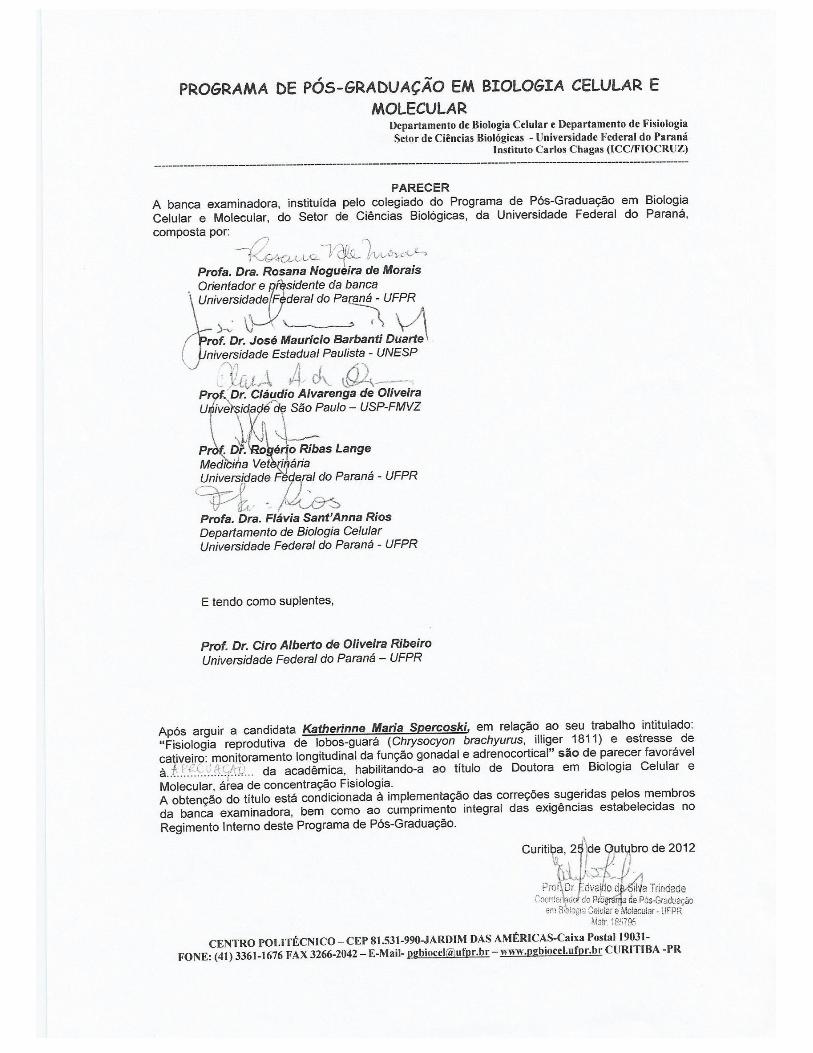
KATHERINNE MARIA SPERCOSKI
FISIOLOGIA REPRODUTIVA DE LOBOS-GUARÁ (Chrysocyon
brachyurus, Illiger 1811) E ESTRESSE DE CATIVEIRO:
MONITORAMENTO EM LONGO PRAZO DA FUNÇÃO GONADAL E
ADRENOCORTICAL
CURITIBA - PR
2013
KATHERINNE MARIA SPERCOSKI
FISIOLOGIA REPRODUTIVA DE LOBOS-GUARÁ (Chrysocyon
brachyurus, Illiger 1811) E ESTRESSE DE CATIVEIRO:
MONITORAMENTO EM LONGO PRAZO DA FUNÇÃO GONADAL E
ADRENOCORTICAL
Tese apresentada como requisito parcial à obtenção do grau de Doutor em Biologia Celular e Molecular, área de concentração em Fisiologia, Programa de Pós-Graduação em Biologia Celular e Molecular, Setor de Ciências Biológicas da Universidade Federal do Paraná.
Orientadora: Profa. Dra. Rosana Nogueira de
Morais. Co-orientador: Prof. Dr. Anderson J. Martino
Andrade.
CURITIBA - PR
2013

Universidade Federal do Paraná Sistema de Bibliotecas
Spercoscki, Katherinne Maria Fisiologia reprodutiva de lobos-guará (chrysocyon brachyurus, illiger 1811) e estresse de cativeiro: monitoramento em longo prazo da função gonadal e adrenocortical. / Katherinne Maria Spercoski. – Curitiba, 2013. 124 f.: il., color. ; 30cm.
Orientador: Rosana Nogueira de Morais Co-orientador: Anderson J. Martino de Andrade
Tese (doutorado) - Universidade Federal do Paraná, Setor de Ciências Biológicas. Programa de Pós-Graduação em Biologia Celular e Molecular.
1. Logo-guará 2. Canídeos - metabolismo I. Título II. Morais, Rosana Nogueira de III. Andrade, Anderson J. Martino de IV. Universidade Federal do Paraná. Setor de Ciências Biológicas. Programa de Pós-Graduação em Biologia Celular e Molecular.
CDD (20. ed.) 599.7444

AGRADECIMENTOS
Ao programa de Pós-Graduação, pela oportunidade e pelo carinho com que todos
os professores sempre me trataram;
À minha orientadora, professora, companheira e grande amiga Profa. Dra. Rosana
Nogueira de Morais. Teacher amada, obrigada por me ensinar, acompanhar e
ajudar a trilhar por um caminho que eu desconhecia, que é a vida científica. Pelas
oportunidades que abriu, por ter sempre acreditado no meu trabalho e nos meus
esforços, desde minha iniciação científica, há 11 anos;
Ao Prof. Dr. Anderson J. Martino-Andrade por ter assumido minha co-orientação.
Obrigada pela enorme paciência comigo;
Ao Prof. Dr. Edvaldo Trindade, coordenador do programa de Pós-Graduação e
amigo de muitas horas. Professor querido, muito obrigada por tudo que o senhor fez
por mim, por todo seu carinho e paciência, pela sua forma de ser e lidar com todas
as pessoas;
À Marlene, secretária do programa de Pós-Graduação, por todo carinho, atenção,
apoio, favores e por gentileza que sempre teve comigo, desde os tempos do
mestrado;
À CAPES pela bolsa concedida;
Ao Instituto para Conservação dos Carnívoros Neotropicais, pela oportunidade
de participar do Projeto Lobo-guará, por toda ajuda durante a coleta de amostras e
contatos com as instituições;
Às instituições Zoológicos de Americana (SP), de Ilha Solteira (SP), Companhia
Brasileira de Metalurgia e Mineração (MG), pela participação no projeto, por todo
apoio técnico e logístico;
Aos grandes amigos Nuch e Ronaldo, obrigado por ter permitido a execução desse
trabalho, pela ajuda, pelo carinho, por toda paciência e compreensão nas minhas
faltas;
Aos amigos do laboratório: Katlyn, Marina e Evaldo. Obrigada por tudo que fizeram
por mim. Katlyn querida, te amo amiga “bruxa”. Marininha, o que seria de mim sem
você? Evaldo, você chegou ao final de tudo isso, mas o carinho e alegria que gerou
em todos nós foram enormes. Muito obrigada a todos vocês;
Aos amigos queridos Daiam e Andrei, pela verdadeira amizade e imensa ajuda,
tenham certeza de que grande parte desse trabalho não teria sido possível sem o
apoio que me deram;
À amiga Drihele, pela ajuda nos trabalhos de campo em Araxá, obrigada amiga;
Aos amigos queridos do Criadouro Científico da CBMM, Dra. Laura, Anésio, Jairo,
Donizete, Alonso, Eduardo, Hebert e Leonardo. Obrigada pela receptividade e
apoio durante os trabalhos de campo, pelo carinho e pelas boas risadas;
À amiga queridíssima Birgit, pela ajuda nas horas felizes, mas principalmente nos
momentos mais difíceis de minha vida, em todos você estava lá, apoiando,
ajudando, parando suas coisas para me ouvir e enxugar minhas as lágrimas;
À amiga querida Camila, muito obrigada pela imensa ajuda durante todo esse
processo. Desde nossa viajem para Canastra (nosso lugar Sagrado) até agora na
correria da escrita e revisão do inglês;
Ao meu indescritível amigo e verdadeiro “irmão” Deni. De, obrigada por todas as
horas ao meu lado, me apoiando sempre;
Aos amigos Carol e Robson, vocês são super especiais, obrigada por todo apoio
emocional e espiritual que me deram;
Às amigas e “irmãs” Ana Flávia e Fernanda, obrigada por todo apoio, pela nossa
amizade especial e verdadeira (na alegria e na tristeza). Aninha, obrigada também
pelas drenagens que me desinchavam nos momentos mais estressantes. Fer, não
tem como estudar tanto sobre lobos-guará e não ter você sempre em mente e no
coração;
Aos animaizinhos do projeto, queridos lobos-guarás que estiveram, mesmo sem
querer, fazendo parte da minha vida e nos ajudando a trabalhar pela conservação da
vida selvagem que ainda resta no planeta;
A Equipe Espiritual que sempre me assistiu. Obrigada pela presença constante,
principalmente na fase de escrita que foi, vocês bem sabem, o meu maior desafio;
A todos vocês, amigos recentes e não tão recentes, quero agradecer por serem
parte da importante de mim, por todos os momentos de união e também pelos de
desunião, porque todos me ajudaram a ser quem eu sou.
Finalmente, agradeço à Deus, Força Maior do Universo, pela Vida, pela existência
do Universo, por todas as oportunidades que me ofereceu e oferece, por toda
execução desse trabalho junto comigo e, à minha família Sagrada, imprescindível e
de suma importância em minha vida. Meu Amor por vocês será sempre infinito e
incondicional.
Muito obrigada!
“(...) São as nossas escolhas, Harry, que revelam quem realmente somos,
muito mais do que as nossas qualidades.”
Alvo Dumbledore
Em Harry Potter e a Câmara Secreta, por J. K. Rowling

RESUMO
O lobo-guará (Chrysocyon brachyurus, Illiger 1811) é uma das espécies mais conhecidas de canídeos que habitam as áreas de cerrado. A espécie é classificada pela IUCN – International Union for Conservation of Nature – na categoria "quase ameaçada". Esforços para a conservação de lobos-guará têm sido feitos, visando manter populações de cativeiro viáveis. Infelizmente, a população em cativeiro não é auto-sustentável devido à baixa taxa de prenhez e alta mortalidade neonatal. Especula-se que essa baixa eficiência reprodutiva possa estar associada com distúrbios endócrinos gonadais, como resultado do estresse crônico de cativeiro. Desta forma, nossos objetivos foram gerar dados básicos sobre a função gonadal e adrenocortical de lobos guará cativos em condições normais e submetidos a estimulação adrenocortical aguda e crônica. Foram coletadas amostras de fezes de lobos-guará e os metabólitos de corticóides (MCF), progestágenos (MPF), androgênios (MAF) e estrogênios (MEF) foram extraídos das fezes e quantificados por enzima imunoensaio. No primeiro manuscrito desse estudo, foram caracterizados os padrões de excreção de metabólitos hormonais fecais ao longo do ciclo reprodutivo normal de lobos-guará. Fezes de 9 lobos-guará adultos foram coletadas de 2-5 dias / semana durante período de 5-12 meses. Foram avaliadas as concentrações médias totais obtidas para todos os metabolitos hormonais fecais. As concentrações de todos os metabolitos gonadais fecais apresentaram-se baixas no anestro para ambos os gêneros. Nas fêmeas, durante o proestro observou-se aumento nas concentrações de MEF e MAF. Já as concentrações de MPF tiveram elevação no período peri-ovulatório, mantendo-se elevada durante todo o diestro. Os machos apresentaram aumento nas concentrações de todos os metabólitos gonadais fecais apenas no período referente à lactação e cuidado parental. Em relação à função adrenocortical, as concentrações de metabólitos de corticóides fecais nas fêmeas foram mais altas nos períodos de proestro, lactação e cuidado parental; enquanto que nos machos houve elevação de MCF apenas no período de cuidado parental, quando os mesmos participam do cuidado com os filhotes. No segundo manuscrito desse estudo, foram avaliados os efeitos da estimulação adrenocortical crônica e aguda nas concentrações de metabólitos gonadais fecais. Para melhor entendimento desse manuscrito, o mesmo foi subdividido em três partes, cujos objetivos foram: parte 1) avaliar como a estimulação adrenocortical crônica afeta a função gonadal; parte 2) analisar como a estimulação adrenocortical aguda, por meio da administração de hormônio adrenocorticotrópico (ACTH), afeta a função gonadal; e parte 3) avaliar as funções adrenocortical e gonadal de dois casais cativos de lobos-guará mantidos em recintos menores e com exposição à visitação pública. Nas partes 1 e 3 desse manuscrito, amostras fecais de 5 casais de lobos-guará foram coletadas de 2-5 dias / semana durante 10-21 meses. Na parte 2 foram analisadas amostras fecais de 11 lobos-guará foram coletadas por 3 -7 dias antes até 7-14 dias após os estímulos adrenocorticais agudos, pela administração do ACTH sintético de curta duração. Os resultados obtidos na parte 1 mostraram que machos e fêmeas tiveram evidências endócrinas de estimulação adrenocortical crônica. Os perfis longitudinais para metabólitos gonadais fecais mostraram padrões acíclicos. Na parte 2, o desafio ao ACTH mostrou diminuição nas concentrações de androgênios (MAF) e aumento em estrogênios (MEF) nos machos. Houve correlação positiva (r2 = 0,750, P <0,01) entre as concentrações de MCF e MPF em situações de estímulo agudo em ambos os gêneros. Na parte 3, os perfis

longitudinais de metabólitos de corticóides fecais (MCF) não demonstraram claramente características de estresse crônico nos casais mantidos em recintos menores e sujeitos à visitação pública, no entanto todos os animais apresentaram os elevados níveis de metabólitos gonadais fecais. Em conclusão, o primeiro manuscrito (estudo de caracterização) demonstrou que fêmeas de lobos-guará apresentam flutuações hormonais típicas do ciclo reprodutivo normal de canídeos, enquanto que os machos apresentam aumento nas concentrações de androgênios durante o período de acasalamento e cuidado parental. Aumentos fisiológicos nos corticóides ocorrem na fase de proestro em fêmeas e no período de lactação e cuidado parental em ambos os gêneros. O manuscrito 2 (análise de estresse) demonstrou perda dos padrões hormonais normais durante o ciclo reprodutivo dos animais quando os mesmos estão em condições de hiperestimulação adrenocortical prolongada e, ainda, alterações nas concentrações de metabólitos gonadais fecais nas condições de estimulação adrenocortical aguda, confirmando que tanto a estimulação aguda como, e principalmente, crônica do eixo HHA pode comprometer a eficiência reprodutiva em lobos-guará. Palavras-chaves: Lobos-guará. Cão doméstico. Metabólitos esteroidais fecais. Ciclo reprodutivo. Função gonadal. Função adrenocortical. Estresse crônico. Estresse agudo. Desafio ao ACTH.
ABSTRACT
The maned wolf (Chrysocyon brachyurus, Illiger 1811) is one of the most typical canid species that inhabits the Brazilian grassland areas (known as Cerrado). This species is recognized by the IUCN – International Union for Conservation of Nature - as ‘nearly threatened’. Efforts for conservation of maned wolves have been done in order to maintain a viable and self-sustaining captive population. Unfortunately, the captive population is not self-sustained due to low pregnancy success and high neonatal mortality. It is speculated that their low reproductive efficiency may be associated with endocrine gonadal disorders as a result of chronic captivity stress. Thus, our aims were to generate basic data about gonadal and adrenocortical function in captive maned wolves under normal conditions and subjected to chronic and acute adrenocortical stimuli. Fecal samples of captive maned wolves were collected and metabolites of corticoid (FCM), progestagens (FPM), androgens (FAM) and estrogens (FEM) were extracted from feces and quantified by enzyme immunoassay. In the first manuscript of this study, excretion patterns of fecal hormones metabolites throughout normal reproductive cycle in maned wolves were investigated. Feces from 9 adult animals were collected 2–5 days/week for 2-12 months. The overall concentration mean of fecal hormones metabolites was analyzed. Anestrus showed lower concentration of fecal gonadal metabolites in both genders. At proestrus there was an increase on the level of FAM and FEM in female. FPM concentration begins to rise at the periovulatory period, maintaining a high level during diestrus. In males, after the pups’ birth, during lactation and parental care period, fecal gonadal metabolites means have a significant increase. Regarding adrenocortical function, fecal corticoid metabolites on females showed higher concentration during proestrus and lactation and parental care period, while in males this elevation was observed only during parental care, as they participate in the care of the pups. In the second manuscript of this study, effects of chronic and acute adrenocortical stimulation on fecal gonadal metabolites were evaluated. To better understand this manuscript it was subdivided in three parts, whose the aims were: part 1) evaluate how a chronic stimulation on adrenocortical activity affects the gonadal function; part 2) analyze how the acute adrenocortical stimulation (by administration of adrenocorticotropic hormone - ACTH) affects the excretion of fecal sex steroid metabolites; and part 3) evaluate fecal adrenocortical and gonadal metabolites concentrations in two couples captive maned wolves kept in smaller enclosures and exposure to public visitation. In parts 1 and 3 of this manuscript, fecal samples of five maned wolf couples were collected 2-5 days/week for 10-21 months. On part 2, fecal samples of 11 maned wolves were collected during 3-7 days before up to 7-14 days after adrenocortical stimuli, by the administration of a short-acting synthetic ACTH. On part 1, male and female demonstrated endocrine evidences of adrenocortical chronic stimulation and gonadal longitudinal profiles showed an acycling pattern. On part 2, the ACTH challenge led to a decrease of androgens and increase of estrogens levels in male. There were positive correlations (r2=0.750; P<0.01) between FCM and FPM concentration on acute stress for both genders. In part 3, longitudinal profiles did not clearly demonstrate characteristics of chronic stress on couples kept in smaller enclosures subject to public visitation, however all animals presented higher fecal gonadal metabolites levels. In conclusion, the first manuscript (characterization study) demonstrated that female maned wolves present typical hormonal fluctuations of a normal canine reproductive cycle, while male show

increased androgens concentrations during mating and parental care periods. Corticoids normally rise during proestrus in female and lactation / parental care period in both genders. The manuscript 2 (study of stress) demonstrated loss of normal reproductive cycle profiles, when the animals were in conditions of prolonged adrenocortical hyperstimulation; and also, changes on fecal gonadal metabolites concentration in conditions of acute adrenocortical stimulation; confirming that both chronic and acute stimulation of the HPA axis can compromise reproductive efficiency in captive maned wolves. Keywords: Maned wolves. Fecal steroids metabolites. Reproductive cycle. Gonadal function. Adrenocortical function. Chronic stress. Acute stress. ACTH challenge.

APRESENTAÇÃO
Esta tese está apresentada na forma de dois manuscritos:
−−−− Characterization of fecal adrenocortical and gonadal metabolites profiles
in captive maned wolves (Chrysocyon brachyurus) throughout
reproductive cycle;
−−−− Effects of chronic and acute adrenocortical stimuli on fecal gonadal
metabolites concentrations and reproductive cycle in captive maned
wolves (Chrysocyon brachyurus).
Nos capítulos 1 e 2 são apresentadas a Introdução geral da tese e a Revisão
de Literatura, respectivamente. O capítulo 3 apresenta as Hipóteses e Predições que
levaram a execução do presente estudo, sendo que os Objetivos, gerais e
específicos, estão apresentados no capítulo 4.
No capítulo 5 os Materiais e metodologia utilizados, resultados, discussão,
conclusões e referências encontram-se em cada manuscrito e representam a íntegra
desse trabalho.
O capítulo 6 apresenta as Conclusões gerais da tese. As referências referem-
se ao conteúdo apresentado na Introdução geral (capítulo 1) e na Revisão de
Literatura (capítulo 2).
LISTA DE FIGURAS
FIGURA 1 Espécime de lobo-guará (Chrysocyon brachyurus) adulto,
fêmea................................................................................................
22
FIGURA 2 Mapa de distribuição do lobo-guará na américa latina .................... 23
FIGURA 3 Esquema das principais vias mediadoras da resposta endócrina
do eixo HHG ao estímulo estressor..................................................
32
MANUSCRITO 1
FIGURE 1 Overall concentration mean (ng/g and µg/g wet feces; mean ±
EPM) of fecal gonadal and adrenocortical metabolites of captive
female (A) and male (B) maned wolves throughout reproductive
cycle phases. Males’ data were also grouped based on females’
oestrus cycle[[[[[[[[[[[[[[[[[[[[[[[..
64
FIGURE 2 Longitudinal profiles of FEM (ng/g wet feces; triangles, grey lines)
and FPM (µg/g wet feces; circles, black lines) in 3-year-old (A)
and 6-year-old (B) female maned wolf[[[[[[[[[[[[..
65
FIGURE 3 Overall concentration mean (ng/g and µg/g wet feces; mean ±
EPM) of fecal gonadal and adrenocortical metabolites in captive
female (smooth columns) and male (diagonal hatched columns)
maned wolves during reproductive and non-reproductive seasons.
66
MANUSCRITO 2
FIGURE 1 Graphs on left show FCM concentration mean (ng/g wet feces;
mean ± EPM) pre (smooth columns) and post (hatched
columns) semen collection for the first ten and from the eleventh
to 44th procedures in female (A) and male (B) captive maned
wolves. Graphs on right show % increase of FCM concentration
mean in relation to the pre procedure mean found in female (C)
and male (D)[[[[[[[[[[[[[[[[[[[[[[...
108
FIGURE 2 FCM concentration mean (ng/g wet feces; mean ± EPM) during
the period of semen collection and from 30 days after the last
procedures to end of the monitoring period in female (smooth

columns) and male (diagonal hatched columns) captive maned
wolves[[[[[[[[[[[[[[[[[[[[[[[[[..
109
FIGURE 3 Longitudinal profile of FCM concentration (ng/g wet feces) of
representative female (triangles; discontinuous lines) and male
(circles; solid line) captive maned wolves in which the male was
submitted to semen collection[[[[[[[[[[[[[[[.
110
FIGURE 4 Longitudinal profile of fecal progestagens (µg/g wet feces;
triangles, grey solid line) and estrogens (ng/g wet feces; circles,
black discontinuous line) of representative female (A) and fecal
androgens (ng/g wet feces; losanges, black discontinuous line)
of representative male (B) captive maned wolves during
andrological study[[[[[[[[[[[[[[[[[[[[..
111
FIGURE 5 Overall mean concentration (ng/g and µg/g wet feces; mean ±
EPM) of fecal gonadal and adrenocortical metabolites in captive
female (smooth columns) and male (diagonal hatched columns)
maned wolves during reproductive and non-reproductive
seasons[[[[[[[[[[[[[[[[[[[[[[[[...
112
FIGURE 6 Pre-challenge FCM concentration means (ng/g dry feces; mean
± EPM) in responsive and non-responsive maned wolves to the
ACTH stimuli. Females: smooth columns; males: diagonal
hatched columns[[[[[[[[[[[[[[[[[[[[[
112
FIGURE 7 Profiles of adrenocortical and fecal gonadal metabolites
concentrations (ng/g wet feces, except for FPM shown in µg/g)
in captive maned wolves submitted to the ACTH challenge
experiment[[[[[[[[[[[[[[[[[[[[[[[..
113
FIGURE 8 % increase on fecal corticoid (smooth columns), androgens
(vertical hatched columns), progestagens (horizontal hatched
columns) and estrogens (crosshatch grid columns) metabolites
concentrations in captive maned wolves on the day of the
corticoid peak in relation to the day prior the ACTH challenge[..
113
FIGURE 9 Longitudinal profile of FCM concentration (ng/g wet feces) in
female (triangles; discontinuous lines) and male (circles; solid
line) captive maned wolves from Americana (A) and Ilha Solteira

(B) Zoos[[[.............................................................................. 114
FIGURE 10 FCM concentration mean (ng/g wet feces; mean ± EPM) in
female (smooth columns) and male (diagonal hatched columns)
captive maned wolves from Americana (A) and Ilha Solteira (B)
Zoos during reproductive and non-reproductive seasons[[[...
114
FIGURE 11 Longitudinal profile of fecal androgens (A; ng/g wet feces),
estrogens (B; ng/g wet feces) and progestagens (C; µg/g wet
feces) concentrations from a female (triangles, discontinuous
line) and male (circles, solid line) captive maned wolves[[[[
115
FIGURE 12 Concentration mean (ng/g and µg/g wet feces; mean ± EPM) of
fecal gonadal metabolites in female (white columns) and male
(grey columns) captive maned wolves during reproductive and
non-reproductive seasons[[[[[[[[[[[[[[[[[
116

LISTA DE TABELAS
MANUSCRITO 1
TABLE 1 Maned wolves’ couple analyzed, age (years), monitoring period
(months) and enclosure area (m2)...................................................
63
MANUSCRITO 2
TABLE 1 Brazilian institutions, animals, age (years), enclosure area (m2),
monitoring period (months) and part of the study which animals
were used[[[[[[[[[[[[[[[[[[[[[[[[..
107
LISTA DE ABREVIATURAS
ACTH Adrenocorticotropic hormone (hormônio adrenocorticotrófico)
ADH Antidiuretic hormone (hormônio antidiurético)
ANH Atrial natriuretic hormone (hormônio natriurético atrial)
AZA American Zoo and Aquarium Association
CBMM Companhia Brasileira de Metalurgia e Mineração
CITES Convetion on International Trade in Endangered Species of Wild Fauna
and Flora
CL Corpo lúteo
CRH Corticotropin release hormone (hormônio liberador de corticotropina)
CV Coeficient of variation (coeficiente de variação)
D2 Dopamine receptor (receptor de dopamine)
DHEA Dehidroxiepiandrosterona
EIA Enzyme immunoassay
FAM Fecal androgens metabolites
FCM Fecal corticoids metabolites
FEM Fecal estrogens metabolites
FPM Fecal progestagens metabolites
FSH Follicle stimulating hormone (hormônio folículo estimulante)
GnRH Gonadotropin releasing hormone (hormônio liberador de gonadotrofina)
HHA Hipotálamo-hipófise-adrenal
HHG Hipotálamo-hipófise-gônadas
HPA Hipotálamo-pituitária-adrenal
HPG Hipotálamo-pituitária-gônadas
IUCN International Union for Conservation of Nature
LH Luteinizing hormone (hormônio luteinizante)
MAF Metabólitos de androgênios fecais
MCF Metabólitos de corticoids fecais
MEF Metabólitos de estrogênios fecais
MMA Ministério do Meio Ambiente
MPF Metabólitos de progestágenos fecais
MWSSP Maned Wolf Species Survival Plan
NRS Non reproductive season
NT Near to threatened
PRL Prolactin (prolactina)
RS Reproductive season
UC Unidade de conservação

SUMÁRIO
1 INTRODUÇÃO....................................................................................... 20
2 REVISÃO DE LITERATURA................................................................. 22
2.1 LOBO-GUARÁ....................................................................................... 22
2.2 ESTRESSE E MECANISMOS FISIOLÓGICOS DE RESPOSTA AO
ESTRESSE............................................................................................
25
2.3 CARACTERÍSTICAS HORMONAIS DO CICLO OVARIANO DE
CADELAS..............................................................................................
28
2.4 ATIVIDADE ADRENOCORTICAL X FUNÇÃO REPRODUTIVA........... 30
3 HIPÓTESES E PREDIÇÕES................................................................. 33
4 OBJETIVOS.......................................................................................... 34
4.1 OBJETIVO GERAL................................................................................ 34
4.2 OBJETIVOS ESPECÍFICOS.................................................................. 34
5 MATERIAIS, MÉTODOS E RESULTADOS.......................................... 36
5.1 CONSIDERAÇÕES GERAIS................................................................. 36
5.2 MANUSCRITO 1.................................................................................... 37
ABSTRACT............................................................................................ 39
1. INTRODUCTION............................................................................... 41
2. METHODS......................................................................................... 42
2.1. Animals and sample collection........................................................ 42
2.1. Fecal extraction and analysis.......................................................... 44
2.1.1. Fecal extraction............................................................................ 44
2.1.2. Fecal enzyme immunoassay....................................................... 44
2.3. Data analysis.................................................................................. 45
2.3.1. Oestrus cycle phases................................................................... 45
2.3.2. Reproductive season profiles....................................................... 46
3. RESULTS.......................................................................................... 46
3.1. Hormone data grouped based on females’ reproductive cycle[[ 46
3.1.1. Females[[[[[[[[[[[[[[[[[[[[[[[[. 47
3.1.2. Males[[[[[[[[[[[[[[[[[[[[[[[[[. 47
3.1.3. Gender differences[[[[[[[[[[[[[[[[[[[.. 48
3.1.4. Proestrus length in young and adult female maned wolves[[. 49
3.2. Reproductive season[[[[[[[[[[[[[[[[[[[.. 49
4. DISCUSSION..................................................................................... 50
5. ACKNOWLEDGEMENTS.................................................................. 58
6. DECLARATION OF INTEREST......................................................... 58
7. REFERENCES.................................................................................. 58
8. WEB REFERENCES......................................................................... 62
5.3 MANUSCRITO 2.................................................................................... 67
ABSTRACT............................................................................................ 69
1. INTRODUCTION............................................................................... 71
2. METHODS......................................................................................... 74
2.1. Animals........................................................................................... 74
2.1.1. Part 1........................................................................................... 75
2.1.2. Part 2........................................................................................... 76
2.1.3. Part 3[[[[[[[[[[[[[[[[[[[[[[[[[. 77
2.2. Fecal sample collection[[[[[[[[[[[[[[[[[[.. 78
2.3. Fecal extraction and analysis[[[[[[[[[[[[[[[[ 80
2.3.1. Fecal extraction[[[[[[[[[[[[[[[[[[[[[ 80
2.3.2. Enzyme immunoassay (EIA)[[[[[[[[[[[[............ 81
2.4. Data analysis.................................................................................. 82
3. RESULTS.......................................................................................... 82
3.1. Part 1[[[[[[[[[[[[[[[[[[[[[[[[[[. 82
3.1.1. Effects of semen collection procedures on adrenocortical
function[[[[[[[[[[[[[[[[[[[[[[[[[[[.
82
3.1.2. Effects of chronic adrenocortical stimuli on fecal gonadal
metabolites concentration[[[[[[[[[[[[[[[[[[[..
84
3.2. Part 2[[[[[[[[[[[[[[[[[[[[[[[[[[. 85
3.3. Part 3[[[[[[[[[[[[[[[[[[[[[[[[[[. 87
4. DISCUSSION..................................................................................... 88
5. ACKNOWLEDGEMENTS.................................................................. 100
6. DECLARATION OF INTEREST......................................................... 100
7. REFERENCES.................................................................................. 100
6 CONCLUSÕES..................................................................................... 117

REFERÊNCIAS..................................................................................... 118

20
1 INTRODUÇÃO
O lobo-guará (Chrysocyon brachyurus, Illiger 1811) é o maior canídeo sul-
americano, ocorrendo nos campos abertos e cerrados da América do Sul
(RODRIGUES, 2002). Apesar de sua ampla distribuição, a espécie encontra-se
ameaçada de extinção (MMA, 2003; IUCN, 2012), principalmente devido à redução e
fragmentação de seu habitat natural, o bioma do cerrado, o qual é considerado um
dos mais ameaçados do planeta (MYERS et al., 2000). O isolamento da espécie em
áreas reduzidas afeta gravemente o lobo-guará, pela sua necessidade de grandes
áreas de vida (AZEVEDO,2008; RODRIGUES, 2002), além de, em longo prazo,
reduzir a variabilidade genética nestas subpopulações, afetando seu sucesso
reprodutivo (DE PAULA et al., 2008). Estratégias de conservação têm sido
executadas na tentativa de assegurar a sobrevivência desta espécie, incluindo o
manejo integrado de populações in situ e ex situ (PRIMACK e RODRIGUES, 2001;
RODDEN et al., 1996; DE PAULA et al., 2008).
A manutenção de populações viáveis em cativeiro permite a obtenção de
informações importantes sobre a biologia da espécie, porém a informação disponível
sobre a biologia reprodutiva de lobos-guarás ainda é limitada. Além disto, os dados
disponíveis indicam que as populações de cativeiro não são “auto-sustentáveis”
(SONGSASEN et al., 2006; PRIMACK e RODRIGUES, 2001; VELLOSO et al., 1998;
CUMMINGS et al., 2007) e apresentam baixa eficiência reprodutiva (MAIA e
GOUVEIA, 2002; VANSTRELLS e PESSUTI, 2010), a qual pode estar associada a
distúrbios endócrinos associados ao estresse crônico de cativeiro. Dados do nosso
laboratório demonstraram maior concentração basal de metabólitos de
glicocorticóides fecais em lobos-guarás cativos quando comparados a animais de
vida livre (SPERCOSKI, 2007).
Sabe-se que a condição de estresse pode alterar a função gonadal em muitas
espécies, já que níveis plasmáticos elevados de glicocorticóides podem inibir a
secreção do hormônio liberador de gonadotropinas (GnRH) (Ferin, 2006; Berne et
al., 2004). Além deste efeito direto sobre a adeno-hipófise, estudos evidenciam que
a ativação do córtex da adrenal, por meio do desafio com hormônio
adrenocorticotrópico (ACTH), também provoca aumento nas concentrações
plasmáticas de hormônios sexuais de origem adrenal (MWANZA et. al., 2000;
CHATDARONG et al., 2006; TSUMA et al., 1998; HAUNC; HALTMEYER, 1975;

21
FENSKE, 1997; WILLARD et al., 2005; BOLANOS et al., 1997; YOSHIDA; NAKAO,
2006; VAN LIER et al., 1999; HEDBERG et al., 2007; GINEL et al., 2012; FRANK et
al., 2004).
Esse aumento de progestágenos circulantes pode interferir na regulação
endócrina do ciclo reprodutivo de fêmeas, já que a ovulação e formação do corpo
lúteo para manutenção da gestação dependem de um balanço hormonal muito
preciso.
Fêmeas de cães domésticos (FELDMAN e NELSON, 2004) e de lobo-guará
(SONGSASEN et al., 2006) apresentam perfil hormonal semelhante de controle do
ciclo ovariano e, se o aumento de esteróides sexuais adrenais em situações de
estresse também for comprovado em canídeos, possivelmente o eixo hipotálamo-
hipofisário-gonadal será afetado.
Dados consistentes sobre função gonadal e adrenocortical em lobos-guará
ainda são escassos, sendo que questões importantes sobre o impacto da qualidade
do ambiente e práticas de manejo sobre a eficiência reprodutiva de lobos-guarás
ainda estão em aberto.

22
2 REVISÃO DE LITERATURA
2.1 LOBO-GUARÁ
A ordem Carnivora é formada por 7 famílias, 92 gêneros e 240 espécies de
ocorrência mundial (NOWAK, 1991). O lobo-guará, dentro da Ordem Carnivora, está
inserido na família Canidae, que engloba 16 gêneros e 36 espécies, sendo uma
única espécie pertencente ao gênero Chrysocyon, não havendo ainda subespécies
reconhecidas (SHELDON, 1992). O lobo-guará é o maior canídeo da América do Sul
(DIETZ, 1984; SONGSASEN et al., 2006; AZEVEDO, 2008), medindo entre 95 e 115
cm de comprimento (mais 38 a 50 cm de cauda) e pesando entre 20 e 30 kg
(RODRIGUES, 2002). Sua aparência física difere significativamente da de outros
canídeos, principalmente devido às pernas longas e magras. Possui orelhas
grandes, pêlos longos de coloração laranja–avermelhado na maior parte do corpo,
crina negra no dorso, focinho, patas dianteiras e mais da metade distal das patas
traseiras de coloração negra (SHELDON, 1992; Rodrigues, 2002) (FIGURA 1).
FIGURA 1: ESPÉCIME DE LOBO-GUARÁ (Chrysocyon brachyurus) ADULTO, FÊMEA. FONTE: Acervo pessoal, Araxá - MG (2011).

23
A espécie habita áreas de campos abertos, cerrados e matas de capoeira na
região central da América do Sul. A área de distribuição cobre cerca de 5 milhões de
km2 em seis países: Argentina, Bolívia, Brasil, Paraguai, Peru e Uruguai (DIETZ,
1984; RODRIGUES, 2002; DE PAULA et al., 2008; AZEVEDO, 2009) (FIGURA 2).
FIGURA 2: MAPA DE DISTRIBUIÇÃO DO LOBO-GUARÁ NA AMÉRICA LATINA. AS ÁREAS HACHURADAS MOSTRAM A OCORRÊNCIA DA ESPÉCIE. FONTE: IUCN, on line (2012).
Atualmente o lobo-guará está listado entre as espécies ameaçadas de extinção
no Brasil, na categoria Vulnerável (MMA, 2003). Na classificação da International
Union for Conservation of Nature (IUCN) encontra-se na categoria próximo de
ameaçado (NT – “near to threatened”) (IUCN, 2012) e na classificação do
Convention on International Trade in Endangered Species of Wild Fauna and Flora
(CITES) está listado no apêndice 2. Esse apêndice não proíbe o comercio
internacional da espécie, entretanto o mesmo é estritamente controlado, numa
tentativa de impedir que a espécie possa vir a se tornar ameaçadas de extinção e
assim passar ao apêndice 1 (CITES, 2012).
A ameaça mais significativa para espécie é a redução e fragmentação de
habitat, porém outras fontes de ameaças, como aumento da mortalidade de

24
indivíduos por atropelamentos, caça, captura de filhotes pelo comércio ilegal,
aumento da incidência de doenças e mortalidade de filhotes devido à interação com
cães domésticos também acabam comprometendo, em menor escala, a situação da
espécie (RODRIGUES, 2002; DE PAULA et al., 2008).
Todos esses fatores servem para reduzir o tamanho populacional, gerando
instabilidade demográfica e genética e, consequentemente, aumentando a
probabilidade de extinções locais, como têm ocorrido em algumas regiões de
campos aberto do Estado do Paraná (comunicação pessoal com Azevedo, F. C..
Coordenadora do Projeto Mamíferos do Cerrado). Estima-se que a atual população
brasileira de lobos-guarás em vida livre seja de aproximadamente 20.000 indivíduos,
entretanto apesar do número total de indivíduos indicar uma população mínima
viável, esses indivíduos estão dispersos, formando subpopulações (DE PAULA et
al., 2008).
O bioma do cerrado é, desde 2000, considerado como um dos mais
ameaçados do mundo (MYERS et al., 2000), com cerca de 25% de sua área original
preservada (Machado et al., 2004). Infelizmente, apenas 2% da área do cerrado
encontra-se hoje protegida e manejada em Unidades de Conservação (UC) (KLINK
e MACHADO, 2005), sendo que, em sua maioria, essas áreas não possuem
tamanho suficiente para manter populações viáveis de grandes predadores, como o
lobo-guará (RODRIGUES, 2002).
O lobo-guará é encontrado em quase todas as UCs deste bioma, mas
populações viáveis, considerando no mínimo 500 animais, são estimadas em
apenas três: Parque Nacional do Araguaia – TO; Complexo Parque Nacional das
Nascentes do Rio Parnaíba – Estação Ecológica da Serra Geral do Tocantins - PI e
Parque Estadual do Mirador – MA. Desta forma, apenas poucas áreas, isoladas, têm
a capacidade de manter populações viáveis de lobos-guará (RODRIGUES, 2002).
A espécie é solitária e territorialista, as áreas de vida são fixas e podem variar
de 25 e 132 km2, um tamanho consideravelmente grande que, em geral, não são
ocupadas por outros lobos que não o casal (AZEVEDO, 2008). Na maioria dos
casos as limitações das áreas são lugares fisicamente identificáveis como rochas ou
estradas, sendo a demarcação feita normalmente com urina e fezes (DIETZ, 1984;
AZEVEDO, 2008).
Os machos demarcam inicialmente suas áreas sugerindo um sistema onde o
macho determina sua área antes da formação do casal. Novas adições no território

25
podem ser feitas pelo casal, em períodos de estação reprodutiva (DIETZ, 1984).
Tanto pares de machos quanto pares de fêmeas já foram observados em confronto
físico direto, demonstrando a natureza territorialista da espécie (Rodrigues, 2002).
A formação do casal normalmente se dá na estação reprodutiva, que inicia no
outono (março até julho). A espécie é monoéstrica anual, apresentando
características de monogamia facultativa, podendo permanecer com o mesmo par
por muito tempo (DIETZ, 1984). A gestação é em média de 65 dias, com a maioria
dos nascimentos ocorrendo de maio a setembro, durante a estação seca. O número
de filhotes varia, na natureza, de dois a quatro filhotes, os quais permanecem na
área de vida da mãe durante aproximadamente um ano, quando começam a
dispersar (RODRIGUES, 2002).
Em animais cativos o desmame completo ocorre ao redor de 15 semanas, os
filhotes começam a ingerir sólidos regurgitados pelos pais depois de quatro semanas
de idade. A maturidade sexual ocorre por volta de um ano, mas normalmente não se
reproduzem até o segundo ano. Em cativeiro os lobos-guarás podem viver até 16
anos, mas informações precisas em situação natural são escassas (RODDEN et al.,
1996).
Sua dieta é considerada onívora, sendo constituída basicamente de frutos e
pequenos vertebrados, em proporção aproximada de 50% para cada categoria. A
maioria dos estudos indica os frutos de lobeira (Solanum lycocarpum) como a
categoria alimentar mais freqüente. A lobeira é particularmente importante por estar
disponível o ano todo, garantindo suprimento de frutos na estação seca (inverno),
quando a maioria das outras espécies não está com frutos (DIETZ, 1984;
RODRIGUES, 2002; SILVA; TALAMONI, 2003; QUEIROLO; MOTTA-JUNIOR,
2007). Os animais consumidos por lobos-guarás são na maioria de pequeno a médio
porte, como pequenos mamíferos (roedores, lagoformos e marsupiais), répteis,
aves, peixes e anfíbios (MORATÓ, 2001; QUEIROLO; MOTTA-JUNIOR, 2007).
2.2 ESTRESSE E MECANISMOS FISIOLÓGICOS DE RESPOSTA AO ESTRESSE
A condição de estresse tem sido, nos últimos anos, amplamente discutida e
utilizada para definir o grau de qualidade de vida dos animais, tanto de espécies
domésticas como de selvagens (MILLSPAUGH; WASHBURN, 2004; MOSTL;
PALME, 2002; SHERIFF et al., 2011).

26
O termo estresse (do Inglês “stress”) foi usado inicialmente na física para
traduzir o grau de deformidade de um material quando submetido a um esforço ou
tensão. Hans Selye (1907 – 1982) foi o primeiro pesquisador que utilizou este termo
na medicina e biologia, para traduzir o esforço de adaptação do organismo frente a
mudanças consideradas ameaçadoras ao seu bem estar ou à sua vida (KORTE et
al., 2005). De forma geral, a palavra tem sido associada com eventos negativos e
suas conseqüências são conhecidas como “resposta ao estresse” (MORGAN;
TROMBORG, 2007). Entretanto, como um mecanismo fisiológico, a resposta ao
estresse per se não é deletéria, ao contrário, melhora a capacidade de mobilização
energética do organismo, adaptando-o para reagir ou fugir do estímulo estressor
(MOSTL; PALME, 2005).
Uma série de eventos neuro-endócrinos estão envolvidos com esta resposta
orgânica e dentre os hormônios envolvidos, os mais utilizados como indicadores de
estresse têm sido os glicocorticóides (BREUNER; HAHN, 2003; MILLSPAUGH;
WASHBURN, 2004; KORTE et al., 2005; KEAY et al., 2006; MORGAN;
TROMBORG, 2007; SHERIFF et al., 2011; BUIJS et al., 2011).
As glândulas adrenais são órgãos complexos e multifuncionais e que,
juntamente com o sistema nervoso autonômico, têm papel-chave nos processos
fisiológicos de adaptação a mudanças (YOUNG et al., 2004; PALME et al., 2005).
A glândula apresenta duas partes estrutural e funcionalmente bem distintas: o
córtex, onde são produzidos e secretados hormônios esteroidais importantes, como
glico e mineralocorticóides; e a medula, responsável pela síntese e secreção dos
hormônios catecolaminérgicos. Entretanto, células do córtex podem estar presentes
na medula, ao mesmo tempo em que células medulares podem estar presentes no
córtex, permitindo a influência direta de uma região glandular sobre a outra. Essa
relação íntima entre o córtex e a medula da adrenal é similar à relação anátomo-
funcional entre o sistema nervoso adrenérgico e o eixo hipotálamo–hipófise–adrenal
(HHA) (BERNE et al., 2004; MCNICOL, 1992).
Nos mecanismos fisiológicos de resposta ao estresse, o estímulo estressor é
percebido por diversas áreas do sistema nervoso central, que ativam tanto neurônios
adrenérgicos, que secretam adrenalina e noradrenalina; quanto neurônios
hipotalâmicos, que secretam os hormônios liberador de corticotrofina (CRH) e
antidiurético (ADH). A ativação destes neurônios é mutuamente reforçada, pois a
noradrenalina aumenta a liberação de CRH, enquanto este, por sua vez, eleva a

27
descarga de noradrenalina. (BERNE et al., 2004; GUYTON, 2006; MOSTL ; PALME,
2002; SUTHERLAND et al., 2009).
A liberação dos hormônios hipotalâmicos CRH e ADH estimula a liberação do
hormônio adrenocorticotrófico (ACTH) na hipófise anterior, o qual, por sua vez,
estimula a síntese e secreção de glicocorticóides adrenais, elevando seus níveis
plasmáticos. Ao mesmo tempo, o estímulo adrenérgico direto sobre a medula
provoca a elevação dos níveis plasmáticos de adrenalina e noradrenalina. Juntos, os
sistemas elevam a produção de glicose e priorizam a utilização deste substrato para
o sistema nervoso, disponibilizando outros substratos metabólicos para os demais
tecidos (BERNE et al., 2004; GUYTON, 2006).
Esta etapa inicial da resposta ao estresse pode ser chamada de reação de
alarme, onde todas as respostas corporais entram em estado de prontidão geral,
sem envolvimento específico ou exclusivo de um órgão em particular (BERNE et al.,
2004; GUYTON, 2006).
Se o estímulo estressor continua por períodos mais longos, sobrevém uma
segunda etapa chamada fase de resistência, que se caracteriza pela hiperatividade
da glândula adrenal. A adrenalina e o CRH produzem um estado geral de vigilância,
atenção focalizada e ativação de comportamento defensivo e/ou agressivo. O CRH
inibe a liberação do hormônio do crescimento e de gonadotropinas, podendo inibir a
atividade sexual, ao mesmo tempo em que os altos níveis plasmáticos de cortisol
podem suprimir a ovulação (BERNE et al., 2004; FERIN, 2006). Neste estágio, o
organismo começa a ajustar-se aos estímulos, e entra num processo de adaptação
para poder suportar a condição por mais tempo.
Caso os estímulos continuem, tornando-se crônicos e repetitivos, as respostas
metabólicas adversas tornam-se mais evidentes, podendo ocasionar modificações
físicas ou psicológicas como comportamento estereotipado (WURBELL;
STAUFFACHER, 1996), fraqueza, perda de peso, tendências anti-sociais, baixa
capacidade reprodutiva dentre outras (CHAND; LOVEJOY, 2011; CHARBONNEl et
al., 2008; CYR; ROMERO, 2007; DALEY et al., 2000; DOBSON; SMITH, 2000;
FARSTAD, 1998; PEREIRA et al., 2006; MCCONNACHIE et al., 2012a, 2012b;
MOORE; JESSOP, 2003; TURNER et al., 2005; YOUNG et al., 2006). O organismo
entra em estado de exaustão, com queda da capacidade adaptativa e falha nos
mecanismos de ajuste e redução das reservas de energia.

28
Esse estado de exaustão está relacionado com a própria regulação do eixo
HHA, cuja principal forma de regulação se dá por meio da retroalimentação negativa.
A ativação do eixo aumenta a síntese e secreção de glicocorticóides, que por sua
vez, inibem a secreção de CRH hipotalâmico e ACTH hipofisário, diminuindo a
atividade do eixo. Os efeitos da retroalimetação negativa, pelos glicocorticóides, na
liberação de ACTH também podem ser indiretamente moduladas por meio de
informações neurais de outras áreas do sistema nervoso central para os neurônios
do CRH no hipotálamo. Além disso, os glicocorticóides ativam o gene que codifica o
hormônio natriurético atrial (ANH), que também inibe a liberação basal de CRH e
ACTH. A própria regulação na tradução, transcrição e exposição dos receptores de
glicocorticóides pode ser modulada na exposição a concentrações elevadas destas
substâncias (fenômeno de “down regulation”). Os principais tipos de receptores
assim modulados são os receptores genômicos de baixa afinidade (BERNE et al.,
2004; GUYTON, 2006).
Dessa forma, a ação supressiva dos glicocorticóides pode perdurar mesmo
após cessar a exposição a estas moléculas. A hipersecreção crônica de
glicocorticóides leva a atrofia funcional do eixo HHA e a sua recuperação completa,
após a retirada da influencia supressiva, pode levar até um ano, durante esse tempo
a resposta normal da glândula adrenal ao estresse não pode ser assegurada
(BERNE et al., 2004).
2.3 CARACTERÍSTICAS HORMONAIS DO CICLO OVARIANO DE CADELAS
A fisiologia reprodutiva da fêmea de cão doméstico possui particularidades
únicas, distinguindo-se de outras espécies. Normalmente o ciclo ovariano da cadela
pode ocorrer de 1 a 3 vezes ao ano, com intervalo de 5 a 12 meses, dependendo da
raça, e não apresenta características sazonais (CONCANNON et al., 1989;
FELDMAN; NELSON, 2004).
Normalmente o ciclo é dividido em quatro fases distintas: proestro, estro,
diestro e anestro. O Proestro é o período de crescimento folicular. Ao final do
anestro, sinalizações ovarianas e principalmente extra-ovarianas atuam sobre o
hipotálamo, aumentando a atividade do eixo hipotálamo-hipófise-gônadas (HHG)
(CONCANNON, 2009). O aumentando a concentrações de gonadotrofinas

29
(hormônios folículo estimulante (FSH) e luteinizante (LH)) atuam sobre os ovários,
estimulando o crescimento folicular e, consequentemente, a produção e secreção de
estrogênios (CONCANNON, 2009; FELDMAN; NELSON, 2004). Desta forma,
hormonalmente o proestro é caracterizado pelo aumento nos níveis plasmáticos de
estrogênios que resulta na ocorrência de descarga vaginal sanguinolenta, atração de
machos e preparação uterina para possível gestação (FELDMAN; NELSON, 2004).
As concentrações plasmáticas de estrogênios continuam aumentando e
alcançam o pico máximo (de até 4,6 vezes o valor dos níveis observados durante o
anestro) em 24 – 48 horas antes do pico ovulatório de LH, quando passam a decair
(FELDMAN; NELSON, 2004).
Concentrações plasmáticas de progestágenos, por sua vez, são baixas durante
quase todo proestro, elevando-se rapidamente nas últimas 24 a 72 horas desta fase,
antes do pico ovulatório de LH, sendo essa uma das características marcantes do
ciclo ovulatório de cadelas. Esse aumento nas concentrações de progestágenos
parece estar associado à prévia luteinização do folículo, antes da ovulação
(CONCANNON, 2009; VERSTEGEN-ONCLIN; VERSTEGEN, 2008).
Além disso, também ocorre aumento nos níveis sanguineos de testosterona,
que alcançam o pico máximo muito próximo ou ao mesmo tempo que o pico
ovulatório de LH (FELDMAN; NELSON, 2004; CONCANNON, 2009).
Sendo assim, o período de transição da fase do proestro para o estro é
hormonalmente caracterizado pelo declínio nas concentrações de estrogênios ao
mesmo tempo em que os níveis de progestágenos aumentam. Esse balanço
hormonal estimula dois importantes eventos: 1) mudanças no comportamento sexual
da fêmea, que passa a aceitar a monta pelo macho; 2) esse balanço sinaliza o
hipotálamo e a hipófise para liberação do pico ovulatório de LH (FELDMAN;
NELSON, 2004).
No estro, as concentrações de estrogênios continuam a decair enquanto que as
de progestágenos progressivamente aumentam e se mantém alta durante o restante
da fase e todo o diestro (FELDMAN; NELSON, 2004; SONGSASEN; WILDT, 2007).
Hormonalmente, o diestro é caracterizado pela predominância de
progestágenos. Após a ovulação, o corpo lúteo (CL) é capaz de sintetizar e secretar
progesterona por todo período de gestação e além, em fêmeas não gestantes. Desta
forma, outra característica do ciclo reprodutivo de cadelas é a ocorrência de
pseudogestação fisiológica, onde o corpo lúteo permanece ativo,

30
independentemente do fato de ocorrer gestação, pois nessa espécie não há nenhum
mecanismo luteolítico conhecido e sendo assim o CL pode permanecer ativo por até
55 – 75 dias (VERSTEGEN-ONCLIN; VERSTEGEN, 2008; FELDMAN; NELSON,
2004; SONGSASEN; WILDT, 2007; CONCANNON, 2009).
Outra característica importante é que durante a gestação não há gonadotrofina
placentária, secreção de progesterona placentária ou atividade de aromatase
placentária e a produção e secreção de esteróides sexuais são inteiramente de
origem ovariana (CONCANNON, 2009).
O anestro é a fase do ciclo reprodutivo onde ocorre a involução uterina,
caracterizado pela quiescência de atividade ovariana e redução nas concentrações
de progesterona e outros hormônios esteróides ovarianos. Em fêmeas pseudo-
gestantes o início dessa fase não é facilmente perceptível, não havendo uma
demarcação clínica óbvia (FELDMAN; NELSON, 2004; SONGSASEN; WILDT,
2007).
2.4 ATIVIDADE ADRENOCORTICAL X FUNÇÃO REPRODUTIVA
A função gonadal é regulada pelo eixo hipotálamo-hipófise-gônadas (HHG).
Resumidamente, o hipotálamo, por meio da liberação do hormônio liberador de
gonadotrofinas (GnRH), comanda a porção hipófise-gonadal do eixo. Vários núcleos
hipotalâmicos liberam, de forma pulsátil, GnRH que por sua vez estimula a liberação
das gonadotrofinas hormônio folículo estimulante (FSH) e hormônio luteinizante
(LH). Essas gonadotrofinas agem nos ovários e testículos, estimulando a atividade
gonadal e assim a secreção de esteróides gonadais (progestágenos, estrogênios,
androgênios) (BERNE et al., 2004; GUYTON, 2006; FERIN, 2006).
A natureza pulsátil desse hormônio é importante sinalizadora para os
gonadotropos na produção e secreção de FSH e/ou LH. Desta forma, condições que
interfiram na geração dos pulsos de GnRH irão perturbar o eixo HHG. A completa
ausência de pulsos resulta em inatividade total do eixo, enquanto que anormalidades
menores, tais como, redução da freqüência de pulsos de GnRH, como normalmente
ocorre durante situações de estresse, alteram a produção e liberação de
31
gonadotrofinas, interferindo na função reprodutiva em maior ou menor grau (FERIN,
2006).
Os mecanismos, centrais ou periféricos, pelos quais os estímulos estressores
podem interferir com a função reprodutiva normal são inúmeros e intrincados e
podem influenciar no eixo HHG em qualquer nível de controle, entretanto acredita-se
que o impacto inicial e predominante seja no sistema nervoso central,
especificamente sobre o controle da geração de pulsos de GnRH com conseqüência
na liberação de LH, já que grande parte dos estímulos estressores conhecidos e
estudados atua inibindo a secreção de LH (FERIN, 2006).
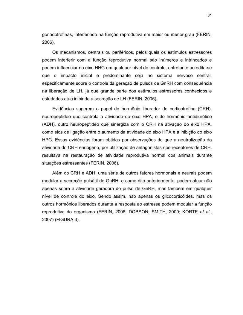
Evidências sugerem o papel do hormônio liberador de corticotrofina (CRH),
neuropeptideo que controla a atividade do eixo HPA, e do hormônio antidiurético
(ADH), outro neuropeptideo que sinergiza com o CRH na ativação do eixo HPA,
como elos de ligação entre o aumento da atividade do eixo HPA e a inibição do eixo
HPG. Essas evidências foram obtidas por observações de que a neutralização da
atividade do CRH endógeno, por utilização de antagonistas dos receptores de CRH,
resultava na restauração de atividade reprodutiva normal dos animais durante
situações estressantes (FERIN, 2006).
Além do CRH e ADH, uma série de outros fatores hormonais e neurais podem
modular a secreção pulsátil de GnRH, e como dito anteriormente, podem atuar não
apenas sobre a atividade geradora do pulso de GnRH, mas também em qualquer
nível de controle do eixo. Sendo assim, não apenas os glicocorticóides, mas os
outros hormônios liberados durante a resposta ao estresse podem modular a função
reprodutiva do organismo (FERIN, 2006; DOBSON; SMITH, 2000; KORTE et al.,
2007) (FIGURA 3).

32
FIGURA 3: ESQUEMA DAS PRINCIPAIS VIAS MEDIADORAS DA RESPOSTA ENDÓCRINA DO EIXO HHG AO ESTÍMULO ESTRESSOR. Linhas continuas: estimulação; linhas tracejadas: inibição. FONTE: Ferin, 2006. Stress and the reproductive system. In: Knobill and Neill’s Physiology of Reproduction, 3º Edição, Elsevier, p. 2656.

33
3. HIPÓTESES E PREDIÇÕES
Nós propomos que, estimulações agudas e/ou crônicas na função
adrenocortical de lobos-guará cativos possam desencadear alterações na função
gonadal e, com isso, levar a falhas no ciclo reprodutivo dos animais, principalmente
em fêmeas. Se essa hipótese for verdadeira, esperamos encontrar, nos animais
submetidos a hiperestimulação prolongada (estimulação crônica), alterações nos
perfis hormonais de esteróides gonadais (metabólitos de estrogênios, progestágenos
e androgênios fecais) que demonstrem / indiquem falhas no ciclo reprodutivo.
Para tanto, devido à escassez de informações acerca da função
adrenocortical de lobos-guará, torna-se primeiramente necessário caracterizar o
perfil de atividade adrenocortical, por meio da quantificação de metabólitos de
corticóides fecais (MCF), normal durante as diferentes fases do ciclo reprodutivo da
espécie.
Além disso, em relação ao estímulo adrenocortical agudo, esperamos que,
nos animais que foram submetidos ao desafio hormonal por meio da administração
do hormônio adrenocorticotrópico (ACTH), ocorram alterações significativas nas
concentrações desses mesmos metabólitos fecais (estrogênios, progestágenos e
androgênios), sugerindo que o córtex da glândula adrenal também possa secretar
hormônios sexuais em resposta ao estímulo pelo ACTH.

34
4. OBJETIVOS
4.1 Objetivo Geral
O presente estudo tem por objetivo gerar dados básicos sobre a função
adrenocortical e gonadal de lobos-guará cativos em condições normais e quando
submetidos à hiperestimulação adrenocortical prolongada e aguda.
4.2 Objetivos Específicos
• Caracterizar os perfis de atividade adrenocortical, por meio da quantificação de
metabólitos de corticóides fecais, de machos e fêmeas ao longo do ciclo
reprodutivo normal da espécie.
• Caracterizar os perfis de atividade gonadal, por meio da quantificação de
metabólitos gonadais fecais (estrogênios, progestágenos e androgênios), de
machos e fêmeas ao longo do ciclo reprodutivo normal da espécie.
• Comparar os achados por fases do ciclo reprodutivo de todos os metabólitos
hormonais fecais em fêmeas, machos e entre os gêneros.
• Avaliar os dados obtidos de todos os metabólitos hormonais fecais com base na
estação reprodutiva da espécie.
• Analisar os efeitos da hiperestimulação adrenocortical prolongada nos perfis
individuais e nas concentrações médias totais de cada metabólito gonadal fecal.
• Analisar os efeitos da hiperestimulação adrenocortical aguda, por meio da
administração do hormônio adrenocorticotrópico (ACTH), nas concentrações de
metabólitos gonadais fecais.
• Comparar as possíveis diferenças encontradas entre os gêneros.

35
• Analisar as funções adrenocortical e gonadal de dois casais de lobos-guará
cativos mantidos em recintos menores e sujeitos à visitação pública.

36
5 MATERIAIS, MÉTODOS E RESULTADOS
5.1 CONSIDERAÇÕES GERAIS
Todo o material, a metodologia e as técnicas empregados, bem como os
resultados e a discussão específica de cada um dos dois estudos que compõem
esta tese estão descritos nos manuscritos apresentados a seguir (manuscrito 1 e 2).
Todos os estudos foram aprovados pela Comissão de Ética no Uso de Animais do
Setor de Ciências Biológicas da Universidade Federal do Paraná (CEUA
23075.031103/2012-23) e tiveram licença do Instituto Brasileiro do Meio Ambiente e
dos Recursos Naturais Renováveis – IBAMA (22610-SisBio).
37
5.2 MANUSCRITO 1
Characterization of fecal adrenocortical and gonadal metabolites profiles in
captive maned wolves (Chrysocyon brachyurus) throughout normal
reproductive cycle.
Katherinne M. Spercoskia*, Katlyn B. Meyera, Marina Heuschkela, Laura T. Oliveirab,
Ronaldo G. Moratoc, Nucharin Songsasend; Anderson J. M. Andradea, Rosana N.
Moraisa
aUniversidade Federal do Paraná, Setor de Ciências Biológicas, Departamento de
Fisiologia, Centro Politécnico, CEP 81531-990, Postal Box 19031, Curitiba, PR,
Brazil. E-mail addresses: [email protected]; [email protected];
[email protected]; [email protected]; [email protected]
bCriadouro Científico de Fauna Silvestre para Fins de Conservação, Companhia
Brasileira de Metalurgia e Mineração - CBMM, CEP 38183-970, Postal Box 08,
Araxá, MG, Brazil. E-mail address: [email protected]
cCentro Nacional de Pesquisa e Conservação de mamíferos Carnívoros, Instituto
Chico Mendes de Conservação da Biodiversidade(ICM – Bio), CEP 12941-680,
Atibaia, SP, Brazil. E-mail address: [email protected]
dCenter for Species Survival, Smithsonian Conservation Biology Institute, National
Zoological Park, Front Royal, VA 22630, United States of America. E-mail address:
* Corresponding author: Universidade Federal do Paraná, Setor de Ciências
Biológicas, Departamento de Fisiologia, Centro Politécnico, CEP 81531-990, Postal

38
Box 19031, Curitiba, PR, Brazil. E-mail address: [email protected], Tel:+55
41 3361-1719, Fax: +55 41 3361-1714.

39
ABSTRACT
Patterns of fecal adrenocortical and gonadal hormones metabolites excretion
were investigated throughout normal reproductive cycle in captive maned wolves
(Chrysocyon brachyurus). Fecal samples of 9 adult maned wolves (4 females and 5
males) were collected 2–5 days/week for 4-11 months. Hormone metabolites of
estrogens (FEM), progestagens (FPM), androgens (FAM) and corticoid (FCM) were
extracted from feces and quantified by enzyme immunoassay. The overall
concentration means, including the findings in males, were analyzed considering: 1)
the female reproductive phases; and 2) based on the reproductive season of the
specie (from February to July). The concentration of fecal gonadal metabolites is
lower during the anestrus period for both genders. On females, at the proestrus there
is an increase on the FAM and FEM levels, while the FPM concentration begins to
rise only at the periovulatory period, maintaining a high level during diestrus and then
decaying on lactation and parental care period. On males, the FAM level begins to
rise on females’ proestrus and periovulatory period and decrease on diestrus phase.
After the pups’ birth, during lactation and parental care period, means concentration
of FAM, FEM and FPM presented significant increase. The pattern of fecal corticoid
metabolites excretion shows a higher concentration during proestrus and lactation
and parental care period on females. Males show no variation of FCM during most
part of the female’ reproductive cycle, although a significant increase is observed
during parental care, when the males participate in the care of the pups. When data
are grouped based on the breeding season, many previously observed differences
disappear, mainly in the profiles of androgens and corticoids in males.

40
KEY WORDS: maned wolf, reproductive cycle, gonadal function, adrenal activity,
fecal steroids metabolites, captive breeding.

41
1. Introduction
The maned wolf (Chrysocyon brachyurus, Illiger 1811) is one of the most typical
canid species that inhabits the Brazilian grassland areas (known as Cerrado) (Dietz
1984). The maned wolf is recognized by the IUCN – World Conservation Union - as a
‘nearly threatened’ species (IUCN 2012), mainly due to the reduction and
fragmentation of its natural habitat for agricultural development and cattle farms
(Silveira and Jácomo 2003). The Brazilian Cerrado is comprised of unique fauna and
flora, ranking it one of the world’s 25 biodiversity hotspots (Myers et al. 2000).
Unfortunately the fragmentation of this biome is an ongoing process (Carvalho et. al.
2009) and only 2% of it is government-protected and managed in units of
conservation (UC) (Klink and Machado 2005). This reality seriously affects the
maned wolves since they demand a significant sized home range (averaging 50.9
km2/547 ft2) (Azevedo 2008). Maned Wolves’ Population and Habitat Viability
Assessment estimated that nearly 20,000 wolves are still in nature, mostly in Brazil
(de Paula et. al. 2008), but the exact number of individuals remains unknown and
wild populations are increasingly at risk.
Since 1984, in response to the uncertain future of wild populations, the American
Zoo and Aquarium Association (AZA) created the Maned Wolf Species Survival Plan
(MWSSP) and it had one critical goal: to maintain a viable and self-sustaining captive
population (Rodden et. al. 1996). Since then, together with an integrated
management of in situ populations, efforts for conservation of maned wolves have
been done in order to develop policies to optimize captive population management
(Maia and Gouveia 2002). Maintaining viable ex situ populations is considered an
important element against extinction, besides being a great source for researches
that try to understand better the biology of this species.

42
Although the reproduction of captive maned wolves has improved throughout the
past few years, this population is still not self-sustained due to low pregnancy
success and high neonatal mortality (Songsasen et al. 2006; Maia and Gouveia
2002; Vanstreels and Pessutti 2010). Basic information on gonadal endocrine profiles
of captive maned wolves has been reported (Velloso et al. 1998; Songsasen et al.
2006; Costa et al. 2008) and it is speculated that their low reproductive efficiency
may be associated with endocrine gonadal disorders as a result of chronic captivity
stress (Songsasen et al. 2006; Cummings et al. 2007). Unfortunately, there are few
studies in maned wolves involving adrenocortical function and no reports are found
describing how the glucocorticoids concentration varies along a normal reproductive
cycle, not even in couples who have had reproductive success. Based on this
information more studies may be designed to assess the real incidence of chronic
stress in captive maned wolves’ couples and how the increase of adrenocortical
activity is influencing their reproductive cycles.
Therefore, the aims of this study were to characterize fecal adrenocortical and
gonadal hormone metabolites profiles in captive male and female maned wolves
based on females’ normal reproductive cycle and also on the known reproductive
season of the species.
2. Methods
2.1. Animals and sample collection
Fecal samples were collected from 9 adult maned wolves (4 females and 5
males; age range 2-9 years) maintained in a Brazilian conservation breeding center
(Criadouro Científico de Fauna Silvestre para Fins de Conservação, Companhia
43
Brasileira de Metalurgia e Mineração, Araxá-MG) (Table 1). During the breeding
season (March – June) the feces of females were collected 3 times/week and males’
1-2 times/week. Out of the breeding season (July – February) the samples of females
were collected 1-2 times/week and males’ 1 time/week. One female was monitored
for 2 successive breeding season (16 months) and two couples were monitored
during the first semester of the year (4-6 months), totalizing 10 endocrine profiles.
All animals were exposed to natural photoperiod, housed in pairs and were
subject to the public. Their management was done according to institution’s routine.
The size range of the enclosures varied from 2000 to 5000 m2 (10,760 to 53,820 ft2)
and the wolves’ diet was compound of approximately 40% commercial dog food
and/or minced beef, 40% fruit and 20% vegetables supplemented with vitamins.
Additionally, the wolves received a freshly killed white rat (raised at the institution)
and one boiled egg 3 times a week. All the animals had access to fresh water ad
libitum.
The fecal samples of females and males were differentiated by the presence of
seeds and papaya peel (Carica papaya) in the males’ scats. The institution’s handlers
offered papaya to the males at one end of the enclosure, over the screen fence, while
the females received banana (Musa spp.) at the other end. This management
ensured that the papaya seeds and peel were eaten only by males.
The samples were collected non-invasively, directly from the enclosures’ floor, by
morning (7:00-8:00 a.m.), as part of the handling routine, were placed in plastic bags
(labeled with date and animal’s identification) and stored at -20ºC (-4 ºF) before being
transported in ice for analysis.

44
2.2. Fecal extraction and analysis
2.2.1. Fecal extraction
Fecal extraction and hormone quantification were performed in the Laboratory of
Reproductive Physiology, Universidade Federal do Paraná, Curitiba, PR. All
reagents (except when specified) were purchased from Sigma-Aldrich (Sigma-Aldrich
Brasil Ltda, São Paulo, Brazil) and all solutions prepared with Milli-Q water. Fecal
extraction was performed by the methods of Spercoski et. al. (2012) with slight
modifications (Anexo 1). Briefly, an aliquot of ~0.5 g of wet, well-mixed, fecal sample
was placed in a glass tube containing 5 ml of 80% ethanol:20% distilled water and
was vigorously shaken for 30 min using a Multi-Pulse vortexer (Glass-Col, Terre
Haute, IN). Each sample was centrifuged (1,000xg, 15 min) and the supernatant was
recovered. The mean (± SEM) of extraction efficiency, by the addition of labeled H3+-
cortisol, was 86.2 ± 0.5% with a coefficient of variation (CV) of 9.1%.
2.2.2. Fecal enzyme immunoassays (EIA)
Fecal extracts were quantified by enzyme immunoassay (described by Brown et.
al. (2004) for fecal estrogens (FEM), progestagens (FPM), androgens (FAM) and
corticoids (FCM) metabolites. The polyclonal antiserum used in the study were
conjugate estrone (R522-2; 1:20,000 dilution; FEM), pregnane (CL425; 1:10,000
dilution; FPM), testosterone (R156/7; 1:10,000 dilution; FAM) and cortisol (R4866;
1:8,500 dilution; FCM) provided by Coralie Munro (University of California–Davis,
Davis, CA).

45
Validation assays for all hormones had already been described (Songsasen et al.
2006; Spercoski et al. 2012). Intra- and inter assay coefficients of variation were <3
% and <15%, respectively, for all EIAs.
2.3. Data analysis
The overall data of the individuals were combined, divided in different groups for
statistical analysis and checked for normality. Parametric data were statistically
analyzed using one way ANOVA followed by Tukey’ multiple comparison procedure
or t-test. Non-parametric data were analyzed using Kruskal-Wallis one way ANOVA
on ranks followed by a Dunn’s multiple comparison procedure or Mann-Whitney rank
sum test. All analysis were considered significant when P<0.05.
The results are presented in ng/g of wet feces, except for FPM where the
concentration is presented in µg/g.
2.3.1. Oestrus cycle phases
The data, both female and male, was divided in 5 different reproductive cycle
phases based on fecal hormone concentrations of the females: 1) anestrus phase –
data of samples obtained before the proestrus phase and after diestrus phase for
pseudo-pregnant or after lactation and parental care phase for pregnant females; 2)
proestrus phase - from the increase of FEM concentration to the day of the FEM
peak; 3) peri-ovulatory phase - comprising the 2 days after the FEM peak (late
proestrus) and the first 7 days of oestrus. This phase was determined based on the
FEM peak that happens simultaneously with an increase on FPM concentration and
the FAM peak (Feldman and Nelson 2004; Concannon 2009); 4) diestrus phase –
from 17 days after the FEM peak to the following 65 days for pseudo-pregnant or
46
until one day before parturition for pregnant females; 5) lactation and parental care
phase – from the day of parturition to the following 60 days. To better understand this
division see figure 2.
Overall data were separated by gender and each fecal gonadal and
adrenocortical metabolite was analyzed for differences between the reproductive
phases and for differences between genders in each of the reproductive phases.
2.3.2. Reproductive season profiles
The hormonal data of maned wolves were also analyzed based on reproductive
season. Data of both genders were divided in two groups: 1) fecal samples obtained
during the reproductive season (RS) and 2) fecal samples obtained during non-
reproductive season (NRS). All samples collected between February 25 and July 15
(date range from the proestrus onset to the end of the periovulatory period) were
considered RS. All fecal gonadal and adrenocortical metabolites were analyzed for
differences between RS and NRS and between genders.
3. Results
3.1. Hormone data grouped based on females’ reproductive cycle
Overall concentration mean of fecal metabolites (estrogens, FEM; progestagens,
FPM; androgens, FAM; corticoids, FCM) of captive maned wolves, for each phase of
the reproductive cycle, are shown in figure 1.
47
3.1.1. Females
FEM concentration was lower during anestrus (4.3 ± 0.3 ng/g; n=82; P<0.05) and
rose from proestrus (9.4 ± 0.7 ng/g; n=152) to diestrus (15.7 ± 0.7 ng/g; n=63).
During the proestrus period a high frequency of FEM peaks (average 5.0 ± 2.3; mean
highest value 23.7 ± 7.3 ng/g) was observed. Similarly, during diestrus the level of
FPM (2.6 ± 0.3 µg/g; n=53; P<0.05) was elevated above anestrus (0.3 ± 0.04 µg/g;
n=66) and proestrus (0.3 ± 0.02 µg/g; n=130). Lower FAM concentration was found
during anestrus (22.9 ± 2.6 ng/g; n=66; P<0.05) compared to proestrus (39.8 ± 3.5
ng/g; n=130) with a progressive return to the values observed at the anestrus phase,
during lactation and parental care period (24.4 ± 3.9 ng/g; n=38). As observed in
estrogens metabolites, FAM peaks (average 3.3 ± 0.9; mean highest value 138.3 ±
31.0 ng/g) were more frequent during the proestrus phase.
As for fecal adrenocortical patterns, FCM concentration was high during
proestrus (190.1 ± 13.5 ng/g; n=90) and lactation / parental care (230.7 ± 35.5 ng/g;
n=31; P<0.05) when compared to the anestrus phase (140.2 ± 16.0 ng/g; n=62). At
the other periods no significant variation on the level of FCM was observed.
3.1.2. Males
The results of males were also divided and analyzed based on females’
reproductive phases, in attempt to verify how the females’ hormonal fluctuations
through oestrus cycle affect males’ hormonal profiles.
The FEM concentration in males was high only during parental care period (12.9
± 0.6 ng/g; n=31; P<0.05). From proestrus (8.8 ± 0.8 ng/g; n=91) to diestrus (9.3 ±
1.8 ng/g; n=9) no differences were found. The anestrus overall concentration mean

48
was 5.1 ± 0.6 ng/g (n=57) and it did not show differences when compared to
proestrus, periovulatory and diestrus phases.
During parental care FPM presented higher concentration (0.7 ± 0.1 µg/g; n=31;
P<0.05) and lower concentration was found during proestrus (0.2 ± 0.01 µg/g; n=91;
P<0.05).
Higher concentration of FAM was observed at proestrus (42.7 ± 4.2 ng/g; n=90;
P<0.05), periovulatory (58.2 ± 21.2 ng/g; n=6) and parental care phases (47.2 ± 6.0
ng/g; n=31). Anestrus (25.9 ± 2.9 ng/g; n=57) and diestrus (25.4 ± 3.6 ng/g; n=33)
levels were significantly low (P<0.05). The fecal corticoid metabolites concentration
differed during parental care (229.7 ± 34.0 ng/g; n=31; P<0.05) with a higher level
when compared to anestrus (134.1 ± 18.3 ng/g; n=50). From proestrus (135.5 ± 14.0
ng/g; n=87) to diestrus (132.2 ± 18.6 ng/g; n=32) no differences were found.
3.1.3. Gender differences
FEM concentration showed differences in gender during diestrus, when females
had higher values (P<0.05) (Fig. 1). Females also showed higher baseline FPM
concentration at proestrus, periovulatory, diestrus and lactation / parental care
phases (P<0.05). Males presented a higher FAM level at lactation / parental care
period (P<0.05).
Overall FCM concentration only showed differences during the proestrus phase,
when the values in females were higher than in males (P<0.05).

49
3.1.4. Proestrus length in young and adult female maned wolves
In this study the onset of the reproductive season of female maned wolves was
identified by an increase on the FEM concentration that marked the first day of
proestrus. The reproductive season was from February 25 to July 15, the date range
from the proestrus’ onset to the end of the periovulatory period. The average length
of the proestrus was 75 days. Longitudinal profiles of FEM and FPM showed that the
ovulatory cycle, marked by an FEM peak simultaneous to an increase on FPM
concentration, can occur early in breeding season in adult females (6-7 year-old; Fig
2B) or extend for up to 85-92 days from the onset in younger females (2-3 years-old;
Fig 2A).
3.2. Reproductive season
Overall concentration means of fecal gonadal and adrenocortical metabolites,
grouped in reproductive and non-reproductive seasons (RS and NRS, respectively)
are shown in figure 3.
Female maned wolves in RS presented higher concentrations for all fecal
metabolites when compared to the NRS. In males, high concentration was found only
for FEM. Unlike the females, the FPM level of males was higher during NRS. The
FCM concentration in males did not differ between seasons. There were differences
associated with gender during RS in FEM, FPM and FCM concentrations where
females had a higher overall mean value. There were no differences between
genders during NRS.

50
4. Discussion
This study characterizes fecal adrenocortical and gonadal hormones metabolites
profiles along all reproductive cycle in captive maned wolves, generating baseline
data, mainly for corticoids overall concentration, of non-invasive monitoring of this
species. It also shows the fluctuations on corticoids levels throughout a reproductive
cycle.
The proestrus phase in female maned wolves is marked by increased
concentrations and frequency of peaks of fecal estrogens and androgens
metabolites. Later, during late proestrus and the periovulatory period, fecal
progestagens metabolites concentrations rise. Altogether these hormones profiles
identify the increase on activity of the hypothalamic-pituitary-gonadal axis (HPG).
These hormones profiles observed on female maned wolves are very similar with the
hormonal events that occur on normal domestic dog reproductive cycle (Feldman
and Nelson 2004; Songsasen et al. 2006).
In female domestic dog at late anestrus and onset of proestrus, progressive
changes in the ovary and, more importantly, in extra-ovarian tissues produce factors
that release the hypothalamus and pituitary from suppressive effects and allow an
increase of GnRH and LH pulse frequency, which act on the ovaries, promoting
follicular development and consequently increased estrogens and androgens levels
(Concannon 2009; Rajkovic et a. 2006; Hunzicker-Dunn and Mayo 2006).
Many features of the canine reproductive cycle are unique when compared to
other species, including a periovulatory rise in serum progesterone that stays
elevated for as long as 100 days in non-pregnant females, due to the absence of
luteolytic mechanism; delay in oocyte maturation after ovulation; delay in implantation
of the embryo; during pregnancy, there is no placental gonadotrophin, no placental
51
progesterone secretion or aromatase, and sex steroid production is entirely of
ovarian origin (Verstegen-Onclin and Verstegen 2008; Concannon 2009).
Hormone-wise, the normal onset of a canine ovarian cycle is characterized by
elevated plasma levels of estrogens that rise from 5-15 pg/mL at late anestrus up to
60-70 pg/mL at late proestrus. The peak of plasma estrogens is reached 24-48 hours
before the end of proestrus, marked by an LH surge. Serum progestagens
concentration is low (<0.5 ng/mL) throughout most of proestrus, rising on the last 6-3
days of this phase. This raise is a striking feature of the canid ovarian cycle, as
mentioned above, and is associated with pre-ovulatory luteinization of follicles, as
early as 6-3 days before the LH surge (Verstegen-Onclin and Verstegen 2008;
Concannon 2009; Feldman and Nelson 2004).
Plasma concentration of androgens also increases at late proestrus, however in
smaller magnitude, reaching higher levels near the LH peak, which occurs in early
estrus (Feldman and Nelson 2004). This increase on androgens level is probably
associated to estradiol production when androgens are the substrate for the
aromatase enzyme complex (Guthermuth et al. 1998; Concannon 2009).
Thus, the periovulatory period (from late proestrus to early estrus) is marked by
an increase of plasma progesterone and androgens concentrations associated with a
decrease on estrogens levels, reflecting the final maturation process of ovarian
follicles. This endocrine pattern of sex steroids at this phase leads to two important
events: changes on the female’s sexual behavior, which starts to accept mating; and
gives a strong feedback to the hypothalamus and pituitary, resulting on the LH surge
(Feldman and Nelson 2004).
During diestrus, progestagens plasma concentration continues to grow and
remains high throughout the entire phase due to the corpus luteum (CL) activity, for

52
the start and maintenance of pregnancy. Normally, plasma concentration of
estrogens is lower at diestrus, although variations within individual bitches have been
reported (Feldman and Nelson 2004).
Fecal progestagens and estrogens metabolites profiles found in this study are
similar to serum endocrine profiles reported for canine reproductive cycle (Feldman
and Nelson 2004). FPM concentration remains elevated during all pregnancy in
maned wolves. Although FEM concentration is higher on diestrus phase, this finding
was already reported for fecal metabolites profiles in female domestic dogs
(Guthermuth et al. 1998) and individual variations in serum profiles of estrogens on
diestrus have also been found (Olson et al. 1982).
It is believed that estrogens may play a role in sustaining corpus luteum (CL)
function, but this has not been clearly identified. Luteal regulation in canine diestrus
appears to be a complex and dynamic process in which several complementary
hormones interplay, such as progesterone, prolactin, LH, relaxin and estrogens
(Verstegen-Onclin and Verstegen 2008).
Some authors report that the increase of estradiol levels in pregnant bitches, from
10 days of the onset of diestrus, can simply reflect luteal stimulation by an increased
prolactin (PRL) level (Concannon 2009), as it is suggested that estradiol is entirely of
ovarian origin by the absence of detectable placental aromatase in domestic dogs
(Nishiyama et al. 1999). The two main luteotrophic hormones are LH and PRL,
whereas prolactin seems to be the main and essential support for canine CL
(Kooistra and Okkens 2002).
Endocrine gonadal profiles showed that the maned wolves’ reproductive season
can start as early as the end of February and extend up to mid July. The average of
proestrus length found in this study is very different from those reported for maned
53
wolves (Songsasen et al. 2006; Velloso et al. 1998; Rodden et al. 1996) and for
domestic dogs (Feldman and Nelson 2004; Concannon 2009). Two endocrine
profiles of females wolves that raised pups showed 13-15 days of proestrus length,
which is considered normal, however the other three profiles, being one of a female
that also conceived, showed prolonged proestrus (range 85-107 days). This
phenomenon seems to be associated with age, since the prolonged proestrus
profiles are from 2-3-year-old females while the others are from 6-7-year-old females.
The individual profiles of those females (see figure 2A) can reveal possible
anovulatory cycles where FEM peaks were not coincident with an increase of FPM
concentration.
As previously described, female domestic dog’s luteinization occurs before
ovulation, with rapid and generalized proliferation of luteinizing cells accompanied by
an increase of serum progesterone concentration (Feldman and Nelson 2004;
Verstegen-Onclin and Verstegen 2008). Irregularities in the LH surge and
consequently on the ovulation can occur as a result of abnormal early luteinization.
Clinically this can be recognized as false estrus, anovulatory cycles and/or prolonged
proestrus (Verstegen-Onclin and Verstegen 2008).
Fecal adrenocortical patterns of female maned wolves show higher fecal corticoid
concentration at proestrus and lactation / parental care period. Relative to proestrus,
a similar result is reported in female rats when the circadian peak in serum corticoid
at proestrus is twice higher than at estrus or metaestrus (diestrus) (Cavigelli et al.
2005).
In maned wolves the females’ proestrus marks the onset of the reproductive
season and it is possible that this higher concentration is associated with two
different factors: 1) increase of adrenocortical activity, due to higher energy demand

54
for this period (male approximation and breeding); 2) release of corticoids from sex
steroid-binding globulins.
It is known that circulating corticoids in females can also bind with gonadal
steroid-binding globulins to a considerable extent (Touma and Palme 2005). With the
onset of a new ovarian cycle, increases in concentration of estrogens and
progestagens can influence the occupancy of plasma gonadal steroid-binding
globulins, increasing the serum concentration of free corticoids and their metabolism,
thereby causing an increase of fecal metabolites.
As for the increase of adrenocortical activity mentioned above, many studies
report that the adrenocortical activity, as well as the reactivity of the hypothalamic-
pituitary-adrenal axis (HPA), can be seasonally modulated according to different
requirements throughout a life cycle, such as growth, pregnancy and lactation
(Touma and Palme 2005; Romero 2002).
So it is more likely that the increase on FCM levels during proestrus is associated
with a higher production of sex steroids, while during lactation and parental care it is
linked to modulation of adrenocortical activity.
In females, pregnancy itself induces a series of hormonal changes and almost all
endocrine glands have their activity increased, especially near the parturition, when
corticoid levels rise due to a release of fetal corticosteroids. During the lactation
period, the metabolic demand of the females is even higher, and the high levels of
corticoids can lead to amino acids, glucose and fatty acids mobilization from the diet,
as well as from maternal tissues, for milk production (Feldman and Nelson 2004;
Berne et al. 2006).
Fecal gonadal metabolites profiles of male maned wolves show higher
concentrations of androgens precisely at the female’s proestrus and peri-ovulatory

55
phase. Behavioral and ecological studies have already indicated that the maned wolf
is a seasonal breeder (Dietz 1984; Rodrigues 2002), therefore these results in fecal
androgens pattern are in agreement with other studies conducted with male maned
wolves that reported higher concentrations of serum testosterone (Maia et al. 2008),
better sperm quality and increased testicular size (Teodoro et al. 2012) during the
reproductive season.
Seasonal changes in spermatogenesis, morphology of the testis and peripheral
serum testosterone concentration have been reported in other canid species, such as
the red and blue foxes (Farstad 1998), African wild dogs (Newell-Fugate et al. 2012),
raccoon dogs (Qiang et al. 2003), Ethiopian wolves (Van Kesteren et. al. 2012),
Iberian wolves (Barja et al. 2008) and coyotes (Minter and DeLiberto 2008).
To cope with the changes of environmental seasonal fluctuation most species
exhibit seasonal cycles of physiological adaptation, such as the ability to restrict
breeding activity to a specific time of year, meeting the highest energy demanding
stages of reproduction (late pregnancy, birth, lactation, weaning of the young) with
annual peaks of food availability and more propitious conditions for survival of the
young. The annual cycle of photoperiod is the major source of environmental
information in control of a great variety of seasonal physiological activities, through a
rhythmic secretion of melatonin. It can modulate reproductive activity by regulating
the HPG axis so gametogenesis, sexual behavior and pregnancy are timed
accordingly (Malpaux 2006).
Another increase on fecal androgens levels occurs during parental care period.
At this phase, male maned wolves also show increased levels of estrogens,
progestagens and corticoids metabolites. Studies involving both captive and free-
living male maned wolves report that the males participate in parental care by

56
protecting and providing food to their offspring and mates (Dietz 1984; De Melo et al.
2009; Veado 1997, apud De Melo et al. 2009). So it is possible that these changes in
the hormonal profiles are associated with male parental behavior.
Among mammalian species, in which fathers contribute to the care of the youngs,
distinct relationships exist between hormones and parental behavior; and prolactin
has received most attention. Males in a variety of mammalian species experience
elevations in prolactin levels during periods in which they care for the young,
suggesting that prolactin commonly promotes paternal behavior (Nunes et al. 2001;
Ziegler 2000).
It is known that estrogens can regulate prolactin release in mammals, promoting
the stimulation of lactotrophs by activating classic nuclear receptors, which leads to
an increased expression of prolactin. Estrogens can also modify the responsiveness
of lactotrophs to other molecules and at high concentrations (e.g. late proestrus)
decrease the density of dopamine receptors (D2). Additionally, estrogens also alter
the density of the voltage-dependent calcium channel and electrical excitability of
lactotrophs, facilitating the process of exocytosis and increasing both basal activity
and responsiveness of these cells (Gregerson 2006).
Therefore it is possible that high levels of estrogens in male maned wolves during
parental care leads to a rise on prolactin levels. No studies were found reporting
patterns of prolactin levels in maned wolves during this phase.
The increase of estrogens during parental care suggests rise on the aromatase
enzyme complex activity. A recent study with ACTH challenge in male maned wolves
reveals that adrenocortical stimuli also increase FEM concentration (Spercoski 2012;
2th chapter). This report is in agreement with our findings, since the concentration of
fecal corticoids metabolites is also higher for this same reproductive phase.

57
The rise on corticoid levels observed after the pup’s birth can be linked with the
important role of these molecules on energy mobilization during costly periods, as
already mentioned for female maned wolves. In males it may also be true due to
energy costs associated with testosterone levels and territorial defense (Romero
2002; Ketterson et al. 1991; Sands and Crell 2004).
It has been suggested that male maned wolves play an important role in rearing
their offspring, but the partnership between male and female was not equal, with the
female spending much more time in the nest while the male brings preys and
performs territorial marking, other necessary and important roles for the survival of
the offspring (De Melo et al. 2009). Thus, higher corticoids and androgens levels
during parental care period found in this study are in agreement with behavior
observation studies in male maned wolves. Additionally, it is also known that
adrenocortical activity can be seasonally modulated in free-ranging maned wolves,
when higher levels of FCM were found during spring, when wolf pairs were raising
pups (Spercoski et. al. 2012).
Data analysis based on reproductive season also shows that within the season
there is an increase on fecal metabolites patterns; however some hormonal
fluctuations, such as the rise of androgens during mating, are not possible to be
observed due to the way the data is grouped.
In conclusion, female maned wolves present similar hormonal fluctuations of
normal canine reproductive cycle. The anestrus phase is marked by lower levels of
gonadal hormones, proestrus is characterized by an increased level and frequency of
estrogens peaks; during the periovulatory phase there is an increase of
progestagens that remains higher until the end of pregnancy. Males show increase of
androgens concentration during females’ proestrus and periovulatory phases.

58
Corticoids normally rise during proestrus and lactation / parental care period in
females. Finally, after the birth of the pups and during all parental care period the
males show increased fecal gonadal and adrenocortical metabolites levels.
5. Acknowledgements
This study was partially supported by Morris Animal Foundation (grant#
D04ZO-77). A scholarship to K. Spercoski was provided by the Coordenação de
Aperfeiçoamento de Pessoal de Nível Superior (CAPES) – Brazil. The authors thank
Companhia Brasileira de Metalurgia e Mineração Scientific Breeder. This research
was conducted within legal requirements (licenses # 22610 SisBio - Instituto
Brasileiro do Meio Ambiente e dos Recursos Naturais Renováveis;
23075.031103/2010-23 Comitê de Ética em Experimentação Animal, Setor de
Ciências Biológicas, Universidade Federal do Paraná, PR, Brazil).
6. Declaration of Interest
The authors have no conflicts of interest. The authors alone are responsible
for the content and writing of this paper.
7. References
Azevedo, F.C. 2008. Área de vida e organização espacial de lobos-guará (Chrysocyon brachyurus) na região do Parque Nacional da Serra da Canastra, Minas Gerais. Masters dissertation. Federal University of Minas Gerais, MG, Brazil.
Barja, I.; Silván, G.; Illera, J.C. 2008. Relationships between sex and stress hormone
levels in feces and marking behavior in a wild population of Iberian wolves (Canis lupus signatus). Journal of chemical ecology 34:697–701.
59
Berne, R. M.; Levy, M. N.; Koeppen, B. M.; Stanton, B. A. 2004. Fisiologia, 5th Edition, Elsevier Rio de Janeiro, RJ, Brazil.
Brown, J.; Walker, S. E Steinmain, K. 2004. Endocrine manual for the reproductive
assessment of domestic and non-domestics species. Conservation and Research Center, Smithsonian´s National Zoological Park. Front Royal, VA, EUA.
Carvalho, F.M.V.; De Marco Júnior, P.; Ferreira, L.G. 2009. The Cerrado into-pieces:
Habitat fragmentation as a function of landscape use in the savannas of central Brazil. Biological Conservation 142: 1392–1403.
Cavigelli, S. A,; Monfort, S. L.; Whitney, T. K.; Mechref, Y. S.; Novotny, N.;
McClintock, M. K. 2005. Frequent serial fecal corticoid measures from rats reflect circadian and ovarian corticosterone rhythms. The Journal of endocrinology 184:153–63.
Concannon, P.W. 2009. Endocrinologic control of normal canine ovarian function.
Reproduction in domestic animals 44 (2): 3–15. Cummings, O. D.; Brown, J. L.; Rodden, M.D. 2007. Behavioral and Physiologic
Responses to Environmental Enrichment in the Maned Wolf (Chrysocyon brachyurus). Zoo Biology 343: 331–343.
de Paula, R. C.; Medici, P.; Morato, R. G. 2008. Plano de Ação para Conservação do
Lobo- Guará: Analise de Viabilidade Populacional e de Habitat, Institute Brasileiro do Meio Ambiente e dos Recursos Naturais Renováveis (IBAMA), Brasilia, Brazil.
De Melo, L.F.B.; Sábato, M. A. L.; Vaz Magni, E. M.; Young, R. J.; Coelho, C. M.
2009. First observations of nest attendance behavior by wild maned wolves, Chrysocyon brachyurus. Zoo biology 28: 69–74.
Dietz, J. M., 1984. Ecology and social organization of maned wolves, Chrysocyon
brachyurus. Washington DC: Smithsonian Institution press. Farstad, W. 1998. Reproduction in foxes: current research and future challenges.
Animal reproduction science 53: 35–42. Fedlman, E. C.; Nelson, R. W. 2004. Canine and feline endocrinology and
reproduction, 3 ed., California, Saunders, 1099p. Gregerson, K. A. 2006. Prolactin: structure, function, and regulation of secretion. In:
Knobil and Neill’s Physiology of Reproduction, Neill, J. D., 3th edition, Elsevier Academic Press.
Guthermuth, D.F.; Concannon, P. W.; Daels, P. F.; Lasley, B.L. 1998. Pregnancy-
specific elevations in fecal concentrations of estradiol, testosterone and progesterone in the domestic dog (Canis familiaris). Theriogenology 50: 237–248.

60
Hunzicker-Dunn, M.; Mayo, K. 2006. Gonadotropin Signaling in the Ovary. In: Knobil
and Neill’s Physiology of Reproduction, Neill, J. D., 3th edition, Elsevier Academic Press.
Ketterson, E.D.; Nolan, Jr., V.; Wolf, L.; Ziegenfus, C.; Dufty Jr., A.M.; Ball, G.F.;
Johnsen, T. 1991. Testosterone and avian life histories: the effect of experimentally elevated testosterone on corticosterone and body mass in dark-eyed juncos. Hormones and Behavior 25: 489–503.
Klink, C. A.; Machado, R. B. 2005. Conservation of the Brazilian Cerrado, Conserv.
Biol. 19: 707–713. Kooistra, H.S.; Okkens, A.C. 2002. Secretion of growth hormone and prolactin during
progression of the luteal phase in healthy dogs: a review. Mol Cell Endoc 197:167–72.
Maia, O.B.; Gouveia, M.G. 2002. Birth and mortality of maned wolves Chrysocyon
brachyurus (Illiger, 1811) in captivity. Brazilian journal of biology 62: 25–32. Maia, O. B.; Jácomo, A. T. A.; Bringel, B. A.; Kashivakura, C. K.; Oliveira, C. A.;
Teodoro, L. O. F.; Silveira, L.; Costa, M. E. L. T.; Malta, M. C. C.; Furtado, M. M.; Torres, N. M.; Mattos, P. S. R.; Viau, P.; Lima, T. G. F.; Morato, R. G. 2008. Comparison of serum hormone levels of captive and free-living maned wolves Chrysocyon brachyurus. Brazilian Journal of Medical and Biological Research 41: 176-179.
Malpaux, B., 2006. Seasonal Regulation of Reproduction in Mammals. In: Knobil and
Neill’s Physiology of Reproduction, Neill, J. D., 3th edition, Elsevier Academic Press.
Minter, J. L.; DeLiberto, T. J. 2008. Seasonal variation in serum testosterone,
testicular volume, and semen characteristics in the coyote (Canis latrans). Theriogenology 69:946-952.
Myers, N.; Mittermeier, R. A; Mittermeier, C.G.; Fonseca, G.A B.; Kent, J. 2000.
Biodiversity hotspots for conservation priorities. Nature 403:853-858. Newell-Fugate, A.E.; Nöthling, J.O.; Bertschinger, H.J. 2012. Seasonal changes in
steroid hormone profiles, body weight, semen quality, and the reproductive tract in captive African wild dogs (Lycaon pictus) in South Africa. General and comparative endocrinology 178: 272–81.
Nishiyama, T.; Tsumagari, S.; Ito, M.; Kimura, J.; Watanabe, G.; Taya, K.; Takeishi,
M. 1999: Immunohistochemical study of steroidogenic enzymes in the ovary and placenta dur- ing pregnancy in the dog. Anat Histol Embryol 28: 125–129.
Nunes, S.; Fite, J. E.; Patera, K. J.; Frenchet, J. A. 2001. Interactions among paternal
behavior, steroid hormones, and parental experience in male marmosets (Callithrix kuhlii). Hormones and behavior 39: 70–82.

61
Olson, P. N.; Bowen, R. A.; Behrendt, M. D.; Olson, J. D.; Nett, T. M. 1982.
Concentrations of reproductive hormones in canine serum during late anestru, proestrus and estrus. Biology of reproduction 27:1196-1206.
Qiang, W.; Murase, T.; Tsubota, T. 2003. Seasonal changes in spermatogenesis and
testicular steroidogenesis in wild male raccoon dogs (Nyctereutes procynoides). The Journal of veterinary medical science 65: 1087–92.
Rajkovic, A,; Pangas, S. A.; Matzuk, M. M. 2006. Follicular Development: Mouse,
Sheep, and Human Models. In: Knobil and Neill’s Physiology of Reproduction, Neill, J. D., 3th edition, Elsevier Academic Press.
Rodden, M.D.; Sorenson, L. G.; Sherr, A ; Kleiman, D. G. 1996. Use of behavioral
measures to assess reproductive status in maned wolves (Chrysocyon brachyurus). Zoo Bilogy 15: 565 – 585.
Rodrigues, F.H.G. 2002. Biologia e conservação do lobo-guará (Chrysocyon
brachyurus) na Estação Ecológica de Águas Emendadas, DF. Thesis. Campinas State University, SP, Brazil.
Romero, L.M., 2002. Seasonal changes in plasma glucocorticoid concentrations in
free-living vertebrates. General and comparative endocrinology 128: 1–24. Sands, J.; Crell, S. 2004. Social dominance, aggression and faecal glucocorticoid
levels in a wild population of wolves, Canis lupus. Animal Behaviour 67:387–396.
Silveira, L.; Jácomo, A. T. A. 2003.Carnivore distribution and abundance patterns
along the Cerrado-Pantanal corridor, southwestern Brazil, in: Anais do I Workshop Sobre Pesquisa e Conservação de Carnívoros Neotropicais, São Paulo, Brazil.
Songsasen, N.; Rodden, M.; Brown, J. L.; Wildt, D. E. 2006. Patterns of fecal
gonadal hormone metabolites in the maned wolf (Chrysocyon brachyurus). Theriogenology 66:1743–1750.
Spercoski, K. M.; Morais, R. N.; Morato, R. G.; de Paula, R. C.; Azevedo, F. C.; May-
Junior, J. A.; Santos, J. P.; Reghelin, A. L.; Wildt, D. E.; Songsasen, N. 2012. Adrenal activity in maned wolves is higher on farmland and park boundaries than within protected areas. General and comparative endocrinology 179 (2):232-240.
Teodoro, L. O.; Melo-Junior, A. A.; Spercoski, K. M.; Morais, R. N.; Souza, F. F.
2012. Seasonal aspects of reproductive physiology in captive male maned wolves (Chrysocyon brachyurus, Illiger 1815). Reprod. Dom. Anim. 47 (Suppl. 6): 250–255.
62
Touma, C.; Palme, R. 2005. Measuring fecal glucocorticoid metabolites in mammals and birds: the importance of validation. Ann. NewYork Academy Science 1046: 54–74.
Van Kesteren, F.; Sillro-Zubiri, C.; Millar, R.; Argaw, K.; Macdonald, D. W.; Paris, M.
2012. Sex, stress and social status: Patterns in fecal testosterone and glucocorticoid metabolites in male Ethiopian wolves. General and Comparative Endocrinology 179:30-37.
Vanstreels, R. E. T.; Pessutti, C. 2010. Analysis and discussion of maned wolf
Chrysocyon brachyurus population trends in Brazilian institutions: lessons from the Brazilian studbook, 1969-2006. International Zoo Yearbook 44:1–15.
Veado, B.V. 1997. Parental behaviour in maned wolf Chrysocyon brachyurus at Belo
Horizonte Zoo. Int Zoo Year 35: 279–286. Velloso, A. L.; Wasser, S. K.; Monfort, S. L.; Dietz, J. M. 1998. Longitudinal fecal
steroid excretion in maned wolves (Chrysocyon brachyurus). General and comparative endocrinology 112:96–107.
Verstegen-Onclin, K.; Verstegen, J. 2008. Endocrinology of pregnancy in the dog: a
review. Theriogenology 70: 291–9. Ziegler, T.E. 2000. Hormones associated with non-maternal infant care: a review of mammalian and avian studies. Folia. Primatol 71 (1-2):6-21.
8. Web References
IUCN, 2012. IUCN Red List of Threatened Species. URL: www.iucnredlist.org. Downloaded on 6 September 2012.

63
Tables
Table 1 – Maned wolves’ couple analyzed, age (years), monitoring period (months) and
enclosure area (m2).
Animal Couple Age Enclosure area Monitoring period
Male 1
Female 1
1 2
2
2000 6
6
Male 2
Female 2
2 3
3
2500 4
4
Male 3
Female 3
3 3
3
2000 11
11
Male 4
Female 4
4 6
7
5000 11
11 (first breeding season)
Male 5
Female 4
5 4
8
5000 4
5 (second breeding season)

64
Figures
Figure 1 – Overall concentration mean (ng/g and µg/g wet feces; mean ± EPM) of fecal gonadal and adrenocortical metabolites of captive female (A) and male (B) maned wolves throughout reproductive cycle phases. Males’ data were also grouped based on females’ oestrus cycle. a,b,c Different letters within the same graph indicate differences (P<0.05; one way ANOVA, followed by Tukey’ multiple comparison procedure and Kruskal-Wallis one way ANOVA on ranks followed by Dunn’s multiple
0
5
10
15
20
a
ab abc b#
c
B
0
5
10
15
20
a
b
bc
c*c
AFEM ng/g wet feces
0
1
2
3
4
a a*
b*
b*
b*
FPM µg/g wet feces
0.0
0.5
1.02
3
4
ab
a#ab#
ab#b#
0
20
40
60
80
100
a
bab
ab a*
FAM ng/g wet feces
0
20
40
60
80
100
a
bc
abc
ab
c#
Anestrus
Proestrus
Periovulatory
Diestrus
Lactation / Parental care
0
50
100
150
200
250
300
a
b*ab ab
b
FCM ng/g wet feces
Anestrus
Proestrus
Periovulatory
Diestrus
Lactation / Parental care
0
50
100
150
200
250
300
a a# ab a
b

65
comparison procedure) between phases; *,# indicate differences (P<0.05; non pared t-test and Mann-Whitney rank sum test) between gender for each fecal metabolite.
Figure 2 – Longitudinal profiles of FEM (ng/g wet feces; triangles, grey lines) and FPM (µg/g wet feces; circles, black lines) in 3-year-old (A) and 6-year-old (B) female maned wolf. The underlines indicate the reproductive phases: anestrus (An), proestrus (Pr), periovulatory (PO), diestrus and lacation / parental care. The arrows indicate the day pups were born.
0
20
40
60
80
0
2
4
6
8
10
J F M A M J J A S O N D nada
PoProestrus
A
Anestrus Diestrus Lactation /Parental care
Anestrus
FEM ng/g wet feces
FPM µg/g wet feces
0
20
40
60
80
0
2
4
6
8
10
J F M A M J J A S O N D nada
PoPr
B
An Diestrus Lactation /Parental care
An
FEM ng/g wet feces
FPM µg/g wet feces

66
Figure 3 – Overall concentration mean (ng/g and µg/g wet feces; mean ± EPM) of fecal gonadal and adrenocortical metabolites in captive female (smooth columns) and male (diagonal hatched columns) maned wolves during reproductive and non-reproductive seasons. a,b,c Different letters within the same graph indicate differences (P<0.05; non pared t-test and Mann-Whitney rank sum test) between phases and genders.
0
50
100
150
200
250
Reproductiveseason
Non-reproductiveseason
a
b b b
FCM ng/g wet feces
0
10
20
30
40
50
aab
b*
ab*
FAM ng/g wet feces
0.0
0.5
1.0
1.5
Reproductiveseason
Non-reproductiveseason
a*
b*
c* c
FPM µg/g wet feces
0
5
10
15
20
ab
c*c*
FEM ng/g wet feces

67
5.3 MANUSCRITO 2 1
Effects of chronic and acute adrenocortical stimuli on fecal gonadal 2
metabolites concentrations and reproductive cycle in captive maned 3
wolves (Chrysocyon brachyurus) 4
5
Katherinne M. Spercoskia*, Laura O. Teodorob, Anésio A. Melo-Juniorb, Katlyn 6
B. Meyera, Andrei S. Santosa, Marina Heuschkela, Daiam L. Kampac, Drihele 7
R.S. Limad, Nucharin Songsasene; Ronaldo G. Moratof; Anderson J. M. 8
Andradea, Rosana N. Moraisa 9
10
aUniversidade Federal do Paraná, Setor de Ciências Biológicas, Departamento 11
de Fisiologia, Centro Politécnico, CEP 81531-990, Postal Box 19031, Curitiba, 12
PR, Brazil. E-mail addresses: [email protected]; [email protected]; 13
[email protected]; [email protected]; 14
[email protected]; [email protected] 15
bCriadouro Científico de Fauna Silvestre para Fins de Conservação, 16
Companhia Brasileira de Metalurgia e Mineração - CBMM, CEP 38183-970, 17
Postal Box 08, Araxá, MG, Brazil. E-mail address: [email protected]; 18
cClínica Veterinária Davet Saúde Animal, CEP 80320-040, Curitiba, PR, Brazil. 20
E-mail address: [email protected] 21
dFaculdades Integradas Espírita, CEP 82010-340, Curitiba, PR, Brazil E-mail 22
address: [email protected] 23

68
eCenter for Species Survival, Smithsonian Conservation Biology Institute, 24
National Zoological Park, Front Royal, VA 22630, United States of America. E-25
mail addres: [email protected]. 26
fCentro Nacional de Pesquisa e Conservação de mamíferos Carnívoros, 27
Instituto Chico Mendes de Conservação da Biodiversidade(ICM – Bio), CEP 28
12941-680, Atibaia, SP, Brazil. E-mail address: [email protected] 29
30
31
* Corresponding author: Universidade Federal do Paraná, Setor de Ciências 32
Biológicas, Departamento de Fisiologia, Centro Politécnico, CEP 81531-990, 33
Postal Box 19031, Curitiba, PR, Brazil. E-mail address: 34
[email protected], Tel:+55 41 3361-1719, Fax: +55 41 3361-1714. 35
36
37
38
39
40
41
42
43
44
45
46
47
69
ABSTRACT 48
Despite conservation efforts, captive maned wolves (Chrysocyon 49
brachyurus) still have low reproductive efficiency that may be related to the 50
stressful captive environment. To analyze how chronic or acute stimuli in 51
adrenocortical function can affect hormonal sex concentrations, and 52
consequently, the reproductive cycle in this species, we evaluated three 53
different situations. To better understand our work, this study were subdivided in 54
three parts, whose the aims were: Part 1) evaluate how a chronic stimulation on 55
adrenocortical activity on captive maned wolves affects their gonadal function; 56
Part 2) analyze how the acute adrenocortical stimulation (by administration of 57
adrenocorticotropic hormone - ACTH) on captive maned wolves affects their 58
excretion of fecal sex steroid metabolites; and Part 3) evaluate fecal 59
adrenocortical and gonadal metabolites concentrations in two couples captive 60
maned wolves kept in smaller enclosures and exposure to public visitation. For 61
parts 1 and 3, fecal samples of five couples of captive maned wolves were 62
collected 2-5 days/week during 10-21 months for longitudinal monitoring. For 63
part 2, fecal samples of 11 maned wolves (5 males; 6 females) were collected 64
during 3-7 days before adrenocorticotropic hormone (short-acting synthetic 65
ACTH; Synacthen® Depot; 0.25mg/mL; Novartis) administration and up to 7-14 66
days after it. Hormone metabolites of corticoid (FCM), progestagens (FPM), 67
androgens (FAM) and estrogens (FEM) were extracted from feces and 68
quantified by enzyme immunoassay. On first part, males were submitted to 69
semen collection procedures, as a part of andrological study conducted in 70
parallel with this work. Males and females demonstrated chronic adrenocortical 71
70
stimulation, although the females were not manipulated at any time. Gonadal 72
longitudinal profiles of theses animals, especially females, showed no pattern, 73
with profiles similar to those already reported for acycling female maned wolves. 74
On second part of this study, acute stimuli of adrenocortical gland, induced by 75
ACTH challenge, led to a decrease on androgens and increase on estrogens 76
levels in male maned wolves while in females there was an apparent increase 77
of androgens concentration. No changes were found on progestagens levels in 78
this experiment. On third, with maned wolves couples kept in smaller 79
enclosures (range 516 to 540 m2/ 5,554 to 5.812 ft2) and subjected to public 80
exposure (from 2 to 6 days/week, during day time - 8:00 a.m. – 5:00 p.m.), 81
longitudinal profiles of fecal corticoid metabolites presented lower 82
concentrations, while fecal gonadal metabolites levels were higher, mainly for 83
androgens and estrogens. Taken together all parts, this work demonstrated loss 84
of normal reproductive cycle profiles of fecal gonadal metabolites, mainly on 85
females, when occur chronic adrenocortical stimuli and further, the acute 86
stimulation of the HPA axis changed fecal gonadal metabolites concentrations, 87
which can compromise reproductive efficiency in captive maned wolves. 88
89
KEY WORDS: maned wolf, adrenocortical stimuli, reproductive cycle, fecal 90
steroids metabolites, ACTH challenge. 91
92
93
94
95
71
1. Introduction 96
It is known that the reproductive success is poor among captive maned 97
wolves (Chrysocyon brachyurus) (Maia and Gouveia 2002; Songsasen et al. 98
2006; Vanstreels and Pessutti 2010). A recent report based on the Brazilian 99
Studbook for Maned Wolf (BSMW) reveals that the breeding population 100
corresponds to only 15% of the overall captive population and only 36% of 101
those breeding captive animals have successfully bred over history (Vanstreels 102
and Pessutti 2010). 103
Similarly to what has been observed in North America, the Brazilian captive 104
population presents low reproductive efficiency for two main reasons: high 105
neonatal mortality that was reported in 79% of the animals before one year of 106
age and 42% in the first month of life (Vanstreels and Pessutti 2010); and also 107
low pregnancy rate (Songsasen et al. 2006). Thus, the captive population of 108
maned wolf is considered not self-sustained (Primack and Rodrigues 109
2001;Vanstreels and Pessuti 2010) and it is believed that this low efficiency can 110
be associated with endocrine disorders associated to chronic captivity stress 111
(Cummings et. al. 2007; Songsasen et al. 2006). 112
It is known that animals housed in artificial habitats are confronted by a wide 113
range of environmental challenges that lead them to physiological, 114
morphological and behavioral adaptation. While some of these situations can be 115
predictable, others are not. The unpredictable components cause an 116
"emergency state", which results in changes on both endocrine and metabolic 117
systems of the organism (Möstl and Palme 2002). 118

72
In an attempt to translate that effort to adaptation, Hans Selye (1907 - 1982) 119
used, for the first time in medicine and biology, the term stress; and since then 120
the word has been associated with adverse events, and their consequences are 121
known as "stress responses" (Korte et al. 2005; Morgan and Tromborg 2007). 122
A variety of hormones (adrenocorticotropic hormone (ACTH), corticoids, 123
catecholamines, prolactin, etc.) is involved in the stress response. The adrenal 124
glands have a key-role in hormonal reactions to stress as they are involved both 125
in the hypothalamic–pituitary–adrenocortical (HPA) axis and the symphatho-126
adreno- medullary system (Möstl and Palme 2002). Adverse situations trigger 127
responses of the adrenals, which result in an increase of corticoid and 128
catecholamine secretion. Short-term elevated corticoids concentrations enable 129
animals to escape from life-threatening situations (Möstl and Palme 2002; 130
Sheriff et al. 2011; Young et al. 2004). 131
The variety of unnatural stressors that captive animals are submitted to may 132
have a strong effect on the function of the HPA axis. Reduced space and 133
restricted movement, forced proximity to humans, reduced feeding 134
opportunities, maintenance in abnormal social groups, exposure to loud or 135
aversive sounds, arousing odors, and other restrictions of behavioral 136
opportunity can lead to a prolonged and sustained corticoid elevation to a 137
sufficient magnitude that can disrupts the homeostatic mechanism of animals, 138
effect known as chronic captivity stress (Morgan and Tromborg 2007). 139
This prolonged and sustained activation of the HPA axis has large 140
deleterious effects, among others, on the reproductive system, being usually 141
inhibitory, which may increase infertility rates (Ferin, 2006; Mostl and Palme 142

73
2002). There are numerous and intricate pathways and mechanisms, central or 143
peripheral, through which stressors may interfere with the normal reproductive 144
function. Most stressors inhibit tonic pulsatile luteinizing hormone (LH) 145
secretion. However, it is still uncertain if this is a result of direct inhibition of the 146
gonadotropin-releasing hormone (GnRH) pulse generator or of change in the 147
sensitivity of the gonadotropes to GnRH stimulation (Ferin, 2006). 148
In addition to this stressor effect on GnRH release from the brain, many 149
studies report that the activation of the HPA axis can induce secretion of 150
progesterone from the adrenal gland (Mwanza et. al., 2000; Chatdarong et al. 151
2006; Tsuma et. al. 1998). This increase in circulating progestagens is another 152
factor that may interfere on the endocrine regulation of females’ reproductive 153
cycles, since ovulation and corpus luteum formation depend on a very precise 154
hormonal balance. In heifers, an increase on progesterone concentration 155
around the estrus phase promoted alterations on estradiol concentrations with 156
delayed LH surge, which consequently extended the oestrus cycle (Duchens et. 157
al. 1994). 158
If this increase is also demonstrated in wild canids, such as the maned wolf, 159
which presents a hormonal control of the ovarian cycle similar to domestic dogs 160
(Songsasen et al. 2006; Velloso et al. 1998; Spercoski 2012, 1th chapter), the 161
raise on adrenal sex steroids induced by stress may alter the signaling that 162
promotes the LH surge by the hypothalamus and pituitary. 163
Endocrine information about the HPA axis activity, through non-invasive 164
methods, already demonstrated higher basal concentration of fecal corticoid 165
metabolites in captive maned wolves when compared to free-living animals 166

74
(Spercoski 2007). Nevertheless, important questions on how chronic 167
adrenocortex stimuli on captive can influence the reproductive cycle of maned 168
wolves are still unclear. 169
Thus, in attempt to answer part of these important questions, this work 170
analyzed and compared, at the same time, adrenocortical and gonadal function 171
on captive non-breeding maned wolves. Our study was conducted in three 172
different situations. To better understand, we divided this study in three parts, 173
whose the aims were: Part 1) evaluate how chronic increased on adrenocortical 174
activity on captive maned wolves affects their gonadal function; Part 2) analyze 175
how the acute adrenocortical stimulation (by administration of 176
adrenocorticotropic hormone - ACTH) on captive maned wolves affects their 177
excretion of fecal sex steroid metabolites; and Part 3) evaluate fecal 178
adrenocortical and gonadal metabolites concentrations in captive non-breeding 179
couples kept in smaller enclosures and subjected to public exposure. 180
181
182
2. Methods 183
2.1. Animals 184
Table 1 summarizes information of animals used in this work, in each of the 185
three parts of this study. 186
On longitudinal studies (first and third parts) were used 10 adult maned 187
wolves (5 females and 5 males; age range 4-10 years), kept in three different 188
Brazilian institutions (Americana Zoo-SP; Ilha Solteira Zoo-SP and the 189
75
conservation breeding center Criadouro Científico de Fauna Silvestre para Fins 190
de Conservação da Companhia Brasileira de Metalurgia e Mineração-MG). 191
The wolves were exposed to natural photoperiod, housed in pairs and were 192
subject to the public. Management was made according to each institution’s 193
routine. The size range of enclosures varied from 512 to 5000 m2 (5,511 to 194
53,819 ft2) and the wolves’ diet was composed of approximately 40% 195
commercial dogs food and minced beef meat, 40% fruit and 20% vegetables 196
with vitamin supplement and the animals had access to fresh water ad libitum. 197
On second part of this work (ACTH challenge) were used the same maned 198
wolves that were being monitored on first. 199
200
2.1.1. Part 1 201
At the same time that our work was being conducted, other research, about 202
seasonal aspects of reproductive physiology in captive male maned wolves 203
(Teodoro et al. 2012), was performed. The andrological study used the same 204
males that were being longitudinal monitored on this part of this work. 205
For a period of 13 months the males were submitted to semen collection 206
procedures by the method of digital manipulation of the penis, based on 207
domestic dogs protocols. Briefly, the wolves were handled in their own 208
enclosures, physically restrained using a catch pole and kept in quadruped 209
position. The abdominal region was cleaned before the digital stimulation of the 210
penis. After the semen collection, the animals were release and rewarded with 211

76
fruit and meat, aiming to condition them in a way they did not relate the 212
procedure to something unpleasant (Teodoro 2011; Teodoro et al. 2012). 213
Semen collection procedures ranged from 38-44 procedures in two males 214
and 15 procedures in a third male, and were performed between April/2010 and 215
May/2011, at a frequency of 1-3 times/ week, but with higher frequency during 216
the breeding season (March - June). 217
The female did not suffer any kind of manipulation and they remained loose 218
in the enclosure, however not kept close during the collection procedure. 219
In attempt to evaluate, at the same time, the adrenocortical and gonadal 220
function of the animals (males and females) this part of the work analyzed two 221
couples during nearly two years (21 months) and one for 9-10 months, totalizing 222
10 annual endocrine profiles. Data were also analyzed based on reproductive 223
season of the species (from February – July; range date obtained for maned 224
wolves breeding pairs, Spercoski 2012 – 1th chapter). 225
226
2.1.2. Part 2 227
In attempt to verify the effects of acute adrenocortical stimulation on 228
secretion of sex steroids in maned wolves, was performed the ACTH challenge 229
experiment. 230
The adrenocortical stimulus was made twice using different experimental 231
designs, where the first challenge totalized 21 days of experiment (from 7 days 232
before to 14 days after the stimuli) and the second 10 days (from 3 days before 233
to 7 days after). The difference between the experiments was relative to fecal 234
collection protocols (see bellow, section 2.2) and the stimuli were performed 235
77
equally in the two challenges. The first experiment was conducted between 236
December and January in 6 animals (3 females and 3 males) and the second 237
was done eight months after the first on the same animals, except for one male 238
that was submitted to clinical treatment. 239
The adrenocortical stimulation was performed by an intramuscular injection 240
of analogue synthetic of adrenocorticotropic hormone (tetracosatide acetate) of 241
short-action (Synacthen® Depot; 0.25mg/mL; Novartis) at a dose of an 242
ampoule/animal. The wolves’ dose was based on the recommended dose for 243
domestic dogs (0.25 mg/dog; Feldman and Nelson 2004). 244
The stimulations were conducted by morning (8:00 – 9:00 a.m.). The maned 245
wolves were captured, physically restrained and handled for the administration 246
of the hormone. The entire procedure, from the entrance of the handlers and 247
veterinarian in the enclosures until the release of the animals last for an 248
average of 5-10 minutes. After this, the wolves were observed during the first 249
hour after administration and then every hour until 4:00 p.m. (when the handlers 250
left the institution) in attempt to verify possible side effects. 251
No animals used in these experiments had clinical signs of adverse 252
reactions and all showed normal behavior after the administration of the 253
hormone and throughout the whole experiment. 254
255
2.1.3. Part 3 256
In attempt to evaluate fecal adrenocortical and gonadal metabolites 257
concentrations in captive maned wolves kept in smaller enclosures and 258
exposure to public visitation, two maned wolf couples from the Americana Zoo –259

78
SP (one couple) and Ilha Solteira Zoo –SP (one couple), not submitted to 260
andrological study, were non-invasively monitored. A longitudinal study was 261
performed during 11 months in one couple and for one year and a half (18 262
months) in the other, totalizing 6 annual endocrine profiles. Data were also 263
analyzed based on reproductive season following the same criteria used on part 264
1 of this study. 265
The animals lived in smaller enclosures (ranging from 516 to 540 m2 / 5,554 266
to 5,812 ft2) when compared to those of the conservation breeding center (from 267
1000 to 5000m2 / 10,764 to 53,816 ft2), and were exposed to public visitation 268
from 2 to 6 days a week, during day time (8:00 a.m. – 5:00 p.m.). Furthermore, 269
in one institution the enclosure of the wolves was located near to enclosures of 270
possible predators of the species. 271
272
273
2.2. Fecal sample collection 274
For longitudinal monitoring studies (parts 1 and 3) fecal samples were 275
collected by morning, as part of the handling’s routine, 3 times/week for females 276
and 2 times/week for males, within the breeding season (from March to June) 277
and 1-2 times/week for females and 1 time/week for males during non-breeding 278
season (from July to February). 279
Female and male fecal samples were differentiated by the males scats’ 280
color (due to the use of green food coloring, used in zoos) or by the presence of 281
papaya seeds and peel (Carica papaya) (used in conservation breeding center). 282

79
At the zoos, the animals were fed separately, while at conservation breeding 283
center the animals received fruits over the enclosures’ screen fence before their 284
daily diet. At the same time handlers offered papaya with seeds and peel to the 285
males, while the females received banana (Musa spp.) at the other end of the 286
enclosure. Papaya was also used the females’ diet, however only the fruit pulp. 287
This dietary management was done in the afternoon prior to the day the 288
feces were collected. The feeding time was always monitored to ensure that 289
both the food coloring and the papaya were eaten only by the males. 290
In the ACTH challenge (part 2), as mentioned above, two protocols of feces 291
collection were performed. On the first, samples were collected once a day, by 292
morning, during 7 days before the challenge and up to 14 days after it, totalizing 293
21 days of experiment. This protocol for sample collection was elaborated 294
based on the information that maned wolves normally defecate once or twice a 295
day (personal communication with others researchers; however after our two 296
experiments this information was documented by Vasconcellos et. al. 2011). 297
However due to the fact that 50% of the animals submitted to challenge did not 298
demonstrate adrenocortex stimuli, a new protocol was established, based on 299
Monfort et. al. (1998). 300
On the second challenge, all feces produced/day were collected during 3 301
days before the challenge and up to 7 days after it, totalizing 10 days of 302
experiment. Before starting the experiment it was observed that the wolves 303
defecate 2-3 times during the day (considering a day from 6:50 a.m. to 4:00 304
p.m.) and in the following morning there was on average of 4 stools / animal in 305
80
the enclosure. This demonstrated that these maned wolves presented more 306
frequency of defecation that only once or twice a day. 307
Based on this information, the feeding management of the wolves was 308
slightly modified to assure, as much as possible, the differentiation between the 309
male and female feces. The animals started receiving the fruits (papaya with 310
seeds and peel for males and banana for females) in the middle of the morning 311
(10:00 - 10:15 a.m.) and again in the afternoon, before their daily diet (3:00 - 312
3:30 p.m.), as described above. 313
The wolves were observed from 6:50 a.m. to 4:00 p.m. and fecal samples 314
were collected 10-15 minutes after each defecation and immediately stored at -315
20ºC (-4ºF). The feces produced between 4:00 p.m. and 6:50 a.m. of the next 316
day were collected immediately at the arrival of the handlers in the institution 317
and by 7:30 a.m. the enclosures were all clean. 318
The fecal collection for all experiments was done non-invasively, directly 319
from the enclosure’s floor, being placed in plastic bags (labeled with date, hour 320
and animal’s identification) and stored at -20ºC (-4ºF) before being transported 321
in ice to analysis. Fecal samples obtained in a 24-hour period, during the ACTH 322
challenge experiment, were pooled before extraction. 323
324
325
2.3. Fecal extraction and analysis 326
2.3.1. Fecal extraction 327
Fecal extraction and hormone quantification were performed in Laboratório 328
de Fisiologia da Reprodução, Universidade Federal do Paraná, Curitiba, 329

81
Paraná, Brazil. All reagents (except when specified) were purchased from 330
Sigma-Aldrich (Sigma-Aldrich Brasil Ltda, São Paulo, Brazil) and all solutions 331
prepared with Milli-Q water. Fecal extraction was performed by the methods of 332
Spercoski et. al. (2012), with slight modifications (Annex 1, thesis). The maned 333
wolves’ fecal sample for the longitudinal monitoring study were extracted wet 334
while the fecal samples of the ACTH challenge experiments were dried (24 335
hours at 70oC) and powdered. Briefly, an aliquot of ~0.5 g of the thawed or 336
powered, well-mixed, fecal sample was placed in a glass tube containing 5 ml of 337
80% ethanol:20% distilled water and vigorously shaken for 30 min using a Multi-338
Pulse vortexer (Glass-Col, Terre Haute, IN). Each sample was centrifuged 339
(1,000xg, 15 min) and the supernatant recovered. The mean (± SEM) of 340
extraction efficiency, by the addition of labeled H3+-cortisol, was 86.2 ± 0.5% 341
with a coefficient of variation (CV) of 9.1%. 342
343
2.3.2. Enzyme immunoassays (EIA) 344
Fecal extracts were quantified by enzyme immunoassay as described by 345
Brown et. al. (2004). The polyclonal antiserum used were pregnane (CL425; 346
1:10,000 dilution; FPM), conjugate estrone (R522-2; 1:20,000 dilution; FEM), 347
testosterone (R156/7; 1:10,000 dilution; FAM) and cortisol (R4866; 1:8,500 348
dilution; FCM), provided by Coralie Munro (University of California–Davis, 349
Davis, CA). 350
Validation assays for all hormones had already been described (Songsasen 351
et al. 2006; Spercoski et al. 2012). Intra- and inter assay coefficients of variation 352
were <3 % and <15%, respectively, for all EIAs. 353

82
2.4. Data analysis 354
The individuals’ overall data were divided in different groups that were 355
analyzed and checked for normality. Parametric data were statistically analyzed 356
using one way ANOVA (for comparisons of three or more groups), t-test and 357
paired t-test (for comparisons between two groups). When data did not pass on 358
normality, analysis was performed by Kruskall-Wallis (three or more groups), 359
Mann-Whitney or Wilcoxon tests (two groups), being considered significant 360
when P<0.05. 361
For the ACTH challenge profiles, the data were aligned according to the day 362
of the FCM peak, which was considered day zero. 363
Results are presented in ng/g or µg/g (for FPM concentrations) of wet feces 364
on longitudinal monitoring studies and dry feces on the ACTH challenge 365
experiment. 366
367
368
3. Results 369
3.1. Part 1 370
3.1.1. Effects of semen collection procedures on adrenocortical function 371
The acute response of the first semen collection was only observed in one 372
couple, where the increase of FCM concentration post-procedure was 1.8 fold 373
higher in the male and 17.7 fold higher in the female, although the female was 374
not manipulated at any time. 375
Considering the 2-1 days pre-procedure, FCM mean concentration found in 376
males, for the first ten semen collection, was 180.2 ± 45.4 ng/g (n=15). After 377

83
that, the pre-procedure mean increased 2.3 fold (423.5 ± 54.38 ng/g; n=55; 378
P<0.05) and remained high until the last semen collection (Fig. 1B). The same 379
result was found in females where the pre-procedure mean for the first ten 380
semen collection was 184.5 ± 43.8 ng/g (n=9) and increased up to 385.4 ± 75.2 381
ng/g (n=15; P<0.05; Fig. 1A). 382
Considering post-procedures days, FCM mean concentration for the first ten 383
semen collection was 303.5 ± 56.5 ng/g (n=10) and from the eleventh to 44th 384
was 709.1 ± 143.7 ng/g (n=23), demonstrating an increase (P<0.05) when 385
compared to the pre-procedures, respectively (Fig 1B). 386
In females, FCM post-procedures mean for the first ten semen collection 387
was 481.6 ± 67.3 ng/g (n=36) and showed a significant increase (P<0.05) 388
compared to pre-procedure mean. However, after ten semen collections the 389
FCM concentration mean did not differ between pre and post procedures (511.6 390
± 58.6 ng/g; n=24 on post procedure) (Fig. 1A). 391
The response amplitude, observed by % increase on the FCM concentration 392
mean relative to those found in pre-procedure, presented great individual 393
variability in males (Fig. 1D) for the first manipulations (range 10 – 723%), but 394
not for the following (range 14 – 74%). In females, the response amplitude was 395
diminished after the eleventh procedure (Fig. 1C). 396
After the end of the semen collection procedures a decrease on FCM mean 397
concentration was observed from mid June in females (417.7 ± 26.74 ng/g; 398
n=188 and 232.2 ± 24.3 ng/g; n=82, during procedures and 30 days after the 399
last semen collection respectively; P<0.05) and from mid June in males (496.3 ± 400
43.9 ng/g; n=164 and 353.7 ± 36.3 ng/g; n=121; P<0.05) (Fig. 2). 401

84
All these findings show endocrine evidences of chronic adrenocortical 402
stimulation from the tenth procedure up to 30 days after the last semen 403
collection. 404
Beside this, the wolves showed no behavioral changes. During the entire 405
period of study, the animals were regularly observed in attempt to check the 406
occurrence of any kind of behavioral change, including possible agonistic 407
interactions between the couple after collection and on subsequent days, which 408
did not happen. The wolves, both males and females, had behavioral 409
characteristics of the species including reproductive behavior and, during the 410
monitoring of the second breeding season, they were observed mating when 411
the semen collection procedures were still happening (Teodoro 2011). 412
413
3.1.2. Effects of chronic adrenocortical stimuli on fecal gonadal metabolites 414
concentration 415
The longitudinal profile of fecal adrenocortical and gonadal metabolites 416
concentrations from a representative maned wolf couple, in which the male was 417
submitted to semen collection, are shown in figures 3 and 4, respectively. 418
Female longitudinal profiles did not show any pattern of reproductive cycle 419
during first year of monitoring. In July of the second year, a month after the 420
FCM concentration had diminished (see Fig.4A), it was possible to notice a 421
possible ovulatory cycle by the increase on FPM levels with a sequent decrease 422
of FEM concentration. 423
Reproductive season profiles showed that FEM concentration mean of 424
females was higher during NRS (12.9 ± 0.6 ng/g; n=367; P<0.05) when 425

85
compared to RS (10.9 ± 0.5 ng/g; n=342). There were no differences in FEM 426
levels of males (9.2 ± 0.4 ng/g; n=217 and 7.9 ± 0.3 ng/g; n=229, on RS and 427
NRS respectively) (Fig. 5). 428
The FPM concentration mean differed with a higher concentration observed 429
during RS (1.5 ± 0.2 µg/g; n=238; P<0.05) than NRS (1.3 ± 0.1 µg/g; n=232) in 430
females. And as observed for FEM, males did not show differences between 431
seasons for FPM (0.52 ± 0.1 µg/g; n=215 and 0.54 ± 0.1 µg/g; n=228, on RS 432
and NRS respectively). 433
Androgens during NRS were higher in both female (56.8 ± 4.6 ng/g; n=228; 434
P<0.05) and male (42.5 ± 2.8 ng/g; n=229; P<0.05) in comparison to RS (30.1 ± 435
2.2 ng/g; n=229 and 36.9 ± 2.8 ng/g; n=212, in female and male respectively). 436
No differences were founded on FCM concentration mean between RS and 437
NRS females (329.1 ± 23.2 ng/g; n=163 and 331.1 ± 22.5 ng/g; n=226, on RS 438
and NRS respectively) and males (440.4 ± 35.29 ng/g; n=208 and 392.4 ± 439
29.25 ng/g; n=226) 440
There were gender differences between the seasons for FEM and FPM and 441
females had a higher value. 442
443
444
3.2. Part 2 445
Out of the 11 maned wolves that were submitted to the challenge, only 2 446
females and 3 males responded. The FCM concentration mean of the fifty days 447
prior to the ACTH challenge presented differences between non-responsive and 448
responsive females, with higher concentrations found in non-responsive 449

86
animals (938.9 ± 167.5 ng/g; n=17 and 388.4 ± 75.5 ng/g; n=14; P<0.05, for 450
non-responsive and responsive female, respectively) (Fig. 6). In males there 451
were no significant differences between the means (591.4 ± 95.3 ng/g; n=14 452
and 507.5 ± 59.2 ng/g; n=16, for non-responsive and responsive male, 453
respectively). 454
Fecal gonadal and adrenocortical metabolites profiles of the ACTH 455
challenge experiment in captive female and male maned wolves are shown in 456
Fig. 7. Increases on FCM after the challenge were found at 24h in males and 457
48h in females. 458
Male maned wolves showed a decrease of FAM concentrations and 459
increase on FEM levels after the challenge (P<0.05). FPM comparisons did not 460
show differences between pre and post concentrations. 461
The statistical analysis for female maned wolves was not performed due to 462
the low n of animals that responded to the challenge, besides this the two 463
females showed increase of 1.6-1.8 fold on androgens concentrations and 1.2-464
1.9 fold on progestagens levels. For FEM concentration it was not possible to 465
detect any tendency since an increase was presented by one female while the 466
other presented a decrease after the challenge. 467
Response amplitude, observed by % increase on fecal metabolites 468
concentration on the day of the corticoid peak in relation to the day prior the 469
ACTH challenge for both genders are shown in Fig. 8. 470
471
472
473

87
3.3. Part 3 474
Analysis of fecal adrenocortical and gonadal metabolites concentrations in 475
captive non-breeding couples kept in smaller enclosures and subjected to public 476
exposure showed that longitudinal profiles and analysis by reproductive season 477
did not clearly demonstrate characteristics of chronic adrenocortical stimuli 478
(Figs. 9, 10). Wolves from two institutions showed lower FCM concentration 479
mean during NRS compared to the reported for breeding pairs, and within the 480
breeding season only one female presented similar values to those of breeding 481
females (Spercoski 2012, 1th chapter). 482
Differently from FCM, all fecal gonadal metabolites were higher when 483
compared with those already reported for maned wolves breeding pairs 484
(Spercoski 2012, 1th chapter) and also from those reported on part 1 of this 485
work. Longitudinal profiles of fecal gonadal metabolites from a couple are 486
shown in Fig. 11. 487
Analysis by reproductive season showed no difference on the FEM 488
concentration mean (ng/g wet feces) between seasons for both females (20.5 ± 489
1.1; n=189 and 18.0 ± 1.5; n=90 on RS and NRS respectively) and males (17.8 490
± 1.6; n=85 and 18.2 ± 1.4; n=62) (Fig.12). 491
FPM values (µg/g wet feces), within RS females, were 6.9 ± 0.6 (n=131) 492
and 1.5 ± 0.2 (n=86) for males. During the months out of the season the 493
concentration means were 3.5 ± 0.3 (n=64) and 1.4 ± 0.2 (n=62) in females and 494
males, respectively. 495
For androgens (ng/g wet feces) 153.8 ± 24.1 (n=86) was found in males and 496
120.7 ± 11.3 (n=131) in females. The concentration mean out of the season for 497

88
males and females were 139.9 ± 18.8 (n=62) and 141.9 ± 16.4 (n=64), 498
respectively. 499
There were differences between seasons and genders only on FPM levels 500
mean, where RS showed higher values and, as for genders, males had lower 501
concentration mean during both seasons. 502
503
504
4. Discussion 505
This study evaluated the effects of chronic adrenocortical stimuli on the 506
gonadal function of captive maned wolves, showing that a sustained increase 507
on corticoids concentrations in great magnitude and for a prolonged time 508
causes abnormalities on reproductive cycles. It also shows that acute 509
adrenocortical stimuli, accessed by the ACTH challenge, alter fecal gonadal 510
metabolites concentrations in maned wolves. 511
Longitudinal profiles of the couples that were submitted to management 512
change for semen collection showed a persistent increase of adrenocortex 513
activity causing significant alterations on the reproductive cycle profile. FCM 514
concentration rose in both males and females, despite any kind of manipulation 515
on the female, and over the time the animals presented endocrine evidences of 516
chronic adrenocortical stimulation. 517
In canids, most commonly, semen is collected from ejaculates obtained by 518
digital manipulation or electroejaculation. Simple digital manipulation is 519
generally used in domestic dogs and is also reported as a method of semen 520
collection in farm foxes (Vulpes vulpes and Alopex lagopus), as the males are 521
89
trained to accept manipulation (Thomassen and Farstad 2009). For larger wild 522
canids, however, this method can be risky and major studies used 523
electroejaculation procedures (Zindl et. al. 2006; Cummings et al. 2007; 524
Johnston et al. 2007). Nevertheless, the digital manipulation method has also 525
been reported in maned wolves (Paula et al. 2002; Mascarenhas et al. 2002). 526
The protocol used for semen collection of monitored males in this study, as 527
mentioned above (see section 2.1.1 in methods), was done by digital 528
manipulation of the penis, based on defined procedures for domestic dogs and 529
was chosen to minimize the impacts of welfare caused by electroejaculation 530
after the procedure (personal communication Teodoro, L.O.). 531
Although the protocol did not require any chemical restraint or further 532
analgesia, the amplitude of the FCM peak in males for the first ten procedures 533
ranged from 2 to 8 folds, similar to those reported in jaguars after the first 534
semen collection using electroejaculation protocols (Morato et al. 2004). The 535
increase of corticoids concentration after electroejaculation has also been 536
reported in rams (Damian and Ungerfeld 2011), bulls (Whitlock et. al. 2012), 537
domestic cats (Carter et. al. 1984), cheetahs (Wildt et. al. 1984), clouded 538
leopards (Howard 1986) and others wild cats (Phillips et al. 1988). However, no 539
reports were found describing the impact of semen collection by digital 540
manipulation on the HPA axis. 541
The acute response observed since the first procedure was somewhat 542
expected, considering that under normal physiological conditions the HPA axis 543
activation occurs, causing increase on corticoids concentrations (Busch and 544
Hayward 2009; Möstl and Palme 2002; Young et al. 2004). This acute stress 545
90
response aims to promote physiological and behavioral adaptation to the 546
noxious stimulus, or ‘‘stressor”. Thus, the response is generally beneficial to the 547
animals (Sapolsky et. al. 2000). 548
In captivity, this occasional exposure to environmental or handling 549
challenges may be considered common as components of long-term 550
husbandry regimes and can occur in various situations, such as direct stare 551
from a more dominant individual (in the same or in the next enclosure), 552
approach of a human with restraining instruments, changing of enclosures, 553
transporting captive animals, etc. (Morgan and Tromborg 2007). 554
The changes on the animals’ management for semen collection altered their 555
routine and it is possible that each of the stressors (capturing, handling and 556
restraining the male) resulted in acute and/or persistent stress responses that, 557
whether mounted simultaneously or sequentially, can result in a state of chronic 558
adrenocortical stimulation (Dickens et. al. 2010). Furthermore, it is suggested 559
that the act of mating itself has positive effects on serum corticoid levels (Lane 560
2006; Veronesi et. al. 2011; Veronesi et. al. 2010) and fecal corticoid 561
metabolites (Sands and Crell 2004) in males. 562
So the acute response elicited by the first procedure, as mentioned above, 563
was already expected, however it was not expected that the procedures would 564
lead to a chronic condition, including the females. During semen collection the 565
males remained calm, allowed the manipulation and had normal ejaculation 566
(Teodoro 2011). In domestic dogs, fear and/or pain prevent males from 567
attaining a complete erection and ejaculating (Kutzler 2005), which was not 568
observed in the wolves. 569

91
Gonadal longitudinal profiles of theses animals, especially in females, 570
showed no pattern with large hormonal variation of the FEM and FPM 571
concentrations, presenting profiles similar to those already reported for acycling 572
female maned wolves (Songsasen et al. 2006; Velloso et al. 1998) and red 573
wolves (Canis rufus) (Walker et. al. 2002). Longitudinal profiles of normal 574
cycling female maned wolves generally show the onset of ovarian activity by an 575
increase of FEM concentration and frequency of peaks after a long anestrus 576
period. 577
The periovulatory phase is characterized by rising FPM levels, due to the 578
previous luteinization of follicles, and remains high for 60-100 days depending 579
whether the female is pregnant or pseudopregnant and, finally the ovarian 580
steroids return to anestrus values (Spercoski 2012, 1th chapter; Songsasen et 581
al. 2006; Velloso et al. 1998). Both longitudinal profiles and reproductive season 582
analysis show higher concentrations of fecal gonadal metabolites in females 583
when compared to breeding animals (Spercoski 2012, 1th chapter). 584
In this study, both acute and chronic HPA axis stimulation led to changes on 585
fecal gonadal metabolites concentrations. Unfortunately, our findings for the 586
ACTH challenge were too little, due to the small number of animals that 587
responded to the adrenocortex stimuli (range 2-3) and the great individual 588
variability observed. 589
Out of the 11 animals that were submitted to the challenge and only five 590
responded (2 females and 3 males). All maned wolves used in the experiment 591
belonged to conservation breeding center, where the males were also been 592
used in the andrological study. To conduct the first experiment, semen 593
92
collection was interrupted and the stimulus was made after one month in 3 594
males and 3 females, and only 1 male and 2 females were responsive. The 595
second challenge was conducted five months after the end of the semen 596
collection procedures in 2 males and 3 females, and only the males responded. 597
This low number of responsive animals is associated with higher FCM 598
concentration in the days prior the procedure, which is observed in females, 599
where the overall values mean of FCM in non-responsive animals were higher 600
than the responsive ones, within fifteen days prior to the challenge. This was 601
not verified for male maned wolves; however it is possible that all males could 602
have answered, because the supposedly "non-responsive" animals were going 603
through the first experiment, where the collection of fecal samples was 604
performed only once a day and maybe the FCM peak was lost. 605
Fecal progestagens metabolites did not change concentrations after the 606
adrenocortex stimuli in maned wolves as a response to the ACTH challenge. 607
Increases on serum concentration of progestagens stimulated by exogenous 608
ACTH have been reported in many species, such as rabbits (Haunc and 609
Haltmeyer 1975), guinea pigs (Fenske 1997), bovines (Willard et. al. 2005; 610
Bolanos et al. 1997; Yoshida and Nakao 2006), sows (Tsuma et al. 1998), 611
sheep (Van Lier et al. 1999), horses (Hedberg et al. 2007), domestic cats 612
(Chatdarong et al. 2006) and even in domestic dogs (Ginel et. al. 2012; Frank 613
et. al. 2004). 614
As mentioned above, our findings are from a very low n and there is great 615
individual variability, so we cannot infer with certainty that after stimulation of 616
the adrenal cortex no changes occur in the concentration of FPM. Despite these 617

93
results of the ACTH challenge, analysis of longitudinal profiles of FCM and FPM 618
show positive correlation (r2 = 0.750; P <0.05) between the concentration of 619
these hormones during the period of semen collection for both male and female, 620
showing that the activation of the HPA axis increase the FPM concentrations in 621
maned wolves. 622
The origin of these progestagens is probably associated with the process of 623
steroid biosynthesis in the adrenal gland where pregnenolone can be directed 624
to the synthesis of progesterone (via 3b-hydroxysteroid oxidoreductase and 625
∆5→4-3-oxosteroid isomerase) or 17- hydroxypregnenolone (via 17-626
hydroxylase). Either of these forms can be further be converted to 17-627
hydroxyprogesterone, which can then be directed toward the production of 628
corticosteroids (Fraser 1992). Thus, the production rate of theses precursors 629
may be greater than the conversion rate for forming corticoids, with a 630
consequent rise in the levels of serum progestagens and also FPM. 631
This data showing rising levels of FPM after induction of acute stress in 632
female maned wolves was of great importance for the characteristics of the 633
canids’ reproductive cycle, in which, at the beginning of proestrus, progesterone 634
concentrations are low, rising only at late proestrus, concomitantly with a 635
decline of estrogen levels. The combination of increasing progestagens 636
concentrations and decreasing estrogens levels in late proestrus stimulate two 637
major events: changes in the female sexual behavior, which comes to accept 638
the mount; and the strong positive feedback to the hypothalamus and the 639
pituitary, that results on the LH surge at the beginning of estrus (Feldman and 640
Nelson 2004; Concannon 2009). Thus, changes in progestagens concentrations 641
94
during this important period, related to acute stressful events, may be able to 642
prevent or alter the normal occurrence of these two events. This way the 643
possibility of failures on the ovulatory process is increased and consequently 644
the female cannot reach reproductive success. 645
For androgens the acute effects of the ACTH show decrease on FAM 646
concentration in males and an apparent increase in females. Decrease on 647
plasma testosterone after acute stress, by the ACTH challenge or after 648
electroejaculation procedures, has already been reported for bulls (Jonhson et. 649
al. 1982; Welsh and Jonhson 1981), rams (Damián and Ungerfeld 2011), rats 650
(Mann et. al. 1987) and domestic cats (Carter et. al. 1984). 651
The most biologically important androgen synthesized in the testis is the 652
testosterone, which is produced by interstitial Leydig cells under the control of 653
LH, which is under control of the GnRH pulse generator. So conditions that 654
interfere with the function of the GnRH pulse generator will disrupt the HPG 655
axis. Most studies show significant decreases in plasma LH by increases in 656
ACTH plasma levels following acute stress. And major evidence suggests that 657
this decrease in tonic pulsatile LH release by stressors is primarily related to an 658
inhibitory action on the hypothalamic GnRH pulse generator (Jeong and Kaiser 659
2006). Another study reported that ACTH could indirectly reduce the sensitivity 660
of the testis to LH, resulting in lower testosterone release (Mann et. al. 1987). 661
Finally, this decrease on androgens can also happen due to the conversion of 662
testosterone in estrogens by an increase on the aromatase enzyme complex 663
activity. 664

95
In vitro studies have shown that the aromatase activity can be stimulated by 665
corticoids, that induce the expression of components of the enzyme complex, 666
and by the ACTH, which besides stimulating the release of estrogen precursors, 667
can also directly signal the adipose tissue to increase aromatase activity 668
(Mendelson et. al. 1984; Simpson et al. 1981). 669
Therefore, lower FAM concentration after the challenge may be due to 670
reduced plasma levels of LH or by a possible decrease on sensitivity to 671
gonadotropin by the testis or an increase on aromatase activity or by the 672
association of all of these physiological events. 673
Although the results in females only suggest increased FAM concentration 674
after acute stress stimuli, it is possible that this increase is related to a higher 675
secretion of adrenal androgens and then, of FAM. Increases on DHEA serum 676
concentration stimulated by exogenous ACTH have been reported in others 677
species, such as red squirrels (Boonstra et al. 2008) and asian elephants (Yon 678
et al. 2007). 679
It is known that the ACTH also regulates the production and secretion of 680
adrenal androgens, thereby the ACTH stimulus of the zone fasciculata cells to 681
increase secretion of corticoids also acts on cells of the zona reticularis 682
increasing the secretion of DHEA (Vermeulen and Rubens 1992). 683
For estrogens, male maned wolves also presented increased FEM levels 684
after the challenge. In females, the effect was not clear due to the great 685
individual variability. A study on quantification of adrenal secretion rates of sex 686
steroids in male domestic dogs reports that during basal conditions 687
the adrenal produces larger amounts of androgens and estrogens, with 688

96
secretion rates 20 – 50.000 fold less than that of cortisol (Santen et. al. 1980) 689
and additionally, as also mentioned, corticoids and ACTH can possibly activate 690
the aromatase enzyme complex. 691
Therefore, this raise can be associated with increased adrenal secretion 692
rates of estrogens and/or androgens or with possible aromatase activation, and 693
maybe even by the association of these events. 694
Although the experiment in females was not conclusive, the analysis of 695
longitudinal profiles of FCM and FEM of animals monitored on part 1 of this 696
work show positive correlation (r2 = 0.600; P <0.05) between the concentration 697
of these hormones during the period of semen collection for both females and 698
males, suggesting that the activation of the HPA axis increases FEM 699
concentration in females. 700
Considering the couples that belong to the Zoos, although we cannot infer 701
that the animals were under chronic stress, it is possible that the lower levels 702
found, especially in one of the females, already demonstrate the beginning of a 703
state of exhaustion, a condition in which the adrenocortex, after a long time of 704
hyperstimulation, begins to fail regulation mechanisms of the HPA axis, starting 705
to suppress the release of corticoids (Berne et. al 2004). 706
The regulation system of the HPA axis happens through negative feedback, 707
where the activation of the axis increases synthesis and secretion of 708
glucocorticoids, which then inhibit the hypothalamic secretion of corticotrophin 709
release hormone (CRH) and of ACTH by the pituitary, decreasing the activity of 710
the axis. The effects of negative feedback on the pituitary by corticoids may also 711
be indirectly modulated by neural information to CRH neurons released in the 712
97
hypothalamus. Furthermore, corticoids activate the expression of atrial 713
natriuretic hormone (ANH), which also inhibits the basal release of CRH and 714
ACTH. The exposure to high concentrations of corticoids also causes down 715
regulation of its own classic receptor (genomic low affinity receptors) (Berne et 716
al. 2004). 717
Thus, the suppressive action of corticoids can last even after the exposure 718
to these molecules is ceased. The effects of corticoids upon an animal in an 719
exhaustion state lead to functional atrophy of the HPA axis and its full recovery 720
can take up to a year, during which the normal response to stress cannot be 721
assured (Berne et. al 2004). 722
The speculation that animals could be going under exhaustion could also 723
explain the high concentration of fecal gonadal metabolites, considering all 724
changes on sex hormones previously described. 725
On the other hand, it is also possible that these findings of low FCM level 726
and high concentration of fecal gonadal metabolites in all animals are related to 727
technical problems during the procedures of quantification of fecal metabolites, 728
from the collection of fecal samples. 729
An important factor that may have occurred is the delay in collecting the 730
fecal samples. In personal communication with the staff members responsible 731
for each institution, it was reported that the collections were made in the 732
morning, though the exact time has not been determined. Thus, the feces could 733
have been exposed to the environment for up to 19 hours (considering the 734
animal has defecated shortly after the handlers work shift - 5:00 pm - and the 735
collections were only made at 12: 00pm the following day). 736

98
After defecation, several factors, such as temperature, humidity, and other 737
environmental conditions may influence on the concentration of steroids 738
metabolites, mainly FCM. Additionally, microorganisms can alter these 739
molecules by enzymatic cleavage (Millspaugh and Washburn 2004; Palme 740
2005; Touma and Palme 2005). 741
The metabolism of corticoids generally includes seven different types of 742
reactions generating different groups of metabolites. Corticoid with α-ketol or 743
glycol side chains at C-17 (e.g. cortisol, cortisone) can undergo side-chain 744
cleavage forming 17-oxosteroids groups. The 11β-hydroxyandrostenedione, a 745
17-oxosteroid, is the product of cortisol side-chain cleavage, a type of 746
androstane that can be futher metabolized (Brownie 1992; Möstl and Palme 747
2002; Möstl et al 2005); and it is possible that these metabolites can cross-react 748
with the antiserum used for androgens quantification in the study, for the 749
similarity between the immunoreactive portion of these molecules. 750
Therefore, bacterial enzymes can alter all fecal metabolites after a 751
prolonged exposure to fecal samples, diminishing the concentration of FCM and 752
increasing the concentration of molecules which can be “read” as gonadal 753
metabolites in the sample of these animals; however, this is only a speculation. 754
The best way to investigate the metabolism and excretion pattern of steroids is 755
by using radiolabeled (14C/3H) hormones, although this approach is not always 756
possible due to economic and/or welfare restrictions, mainly when involving wild 757
animals (Möstl et al. 2005). 758
Another possibility, possibly simpler, is by exposing fresh fecal samples 759
(aliquots) to the environment, for a determined period of time and then 760
99
quantifying the metabolites to check if any change in fecal metabolites occurs. 761
Futures experiments will investigate this in maned wolves’ fecal samples. 762
In conclusion, this study demonstrates for the first time the loss of normal 763
reproductive cycle profiles of fecal gonadal metabolites on animals exposed to 764
chronic adrenocortical stimuli, confirming that chronic stimulation of the HPA 765
axis can leads to lower reproductive efficiency in captive maned wolves. We 766
have shown that fecal progestagens and estrogens levels increase under acute 767
stimulation of the HPA axis in both genders, while androgens metabolites 768
decrease in males. 769
It’s important to note the importance of andrological studies for generating 770
basic information that may be useful to future management of captive maned 771
wolves, however clarify that even the use of low invasive techniques for semen 772
collection, such as digital manipulation, the HPA axis can positively activate. 773
And considering the acute effects on fecal gonadal metabolites concentrations, 774
these stressful events can also lead to low reproductive efficiency, by increasing 775
the possibility of failures on the ovulatory process or disrupting sexual behavior 776
in the female depending on the reproductive stage in which their occur. 777
This study provided important information that help better understanding the 778
consequences and some mechanisms whereby chronically elevated corticoids 779
levels can compromise reproduction performance of captive maned wolves and 780
possibly free-living animals, since it is known that free-living maned wolves that 781
inhabit farmland regions, where fragmentation and conversion of natural areas 782
is the most apparent, also present high FCM concentration (Spercoski et al. 783
2012). Altogether, our current findings and a growing number of publications 784

100
from other researchers can be useful to improve the management of both 785
captive and free-living populations aiming the conservation of such a 786
charismatic species. 787
788
5. Acknowledgements 789
A scholarship to K. Spercoski was provided by the Coordenação de 790
Aperfeiçoamento de Pessoal de Nível Superior (CAPES) – Brazil. The authors 791
thank Companhia Brasileira de Metalurgia e Mineração Scientific Breeder, 792
Americana and Ilha Solteira Zoos. This research was conducted within legal 793
requirements (licenses # 22610 SisBio - Instituto Brasileiro do Meio Ambiente e 794
dos Recursos Naturais Renováveis; 23075.031103/2010-23 Comitê de Ética 795
em Experimentação Animal, Setor de Ciências Biológicas, Universidade 796
Federal do Paraná, PR, Brasil). 797
798
6. Declaration of Interest 799
The authors have no conflicts of interest. The authors alone are 800
responsible for the content and writing of this paper. 801
802
7. References 803
Berne, R. M.; Levy, M. N.; Koeppen, B. M.; Stanton, B. A. 2004. Fisiologia, 5th 804
Edition, Elsevier Rio de Janeiro, RJ, Brazil. 805
806
Bolanos, J. M.; Molina, J. R.; Forsberg, M. 1997. Effect of blood sampling and 807
administration of ACTH on cortisol and progesterone levels in 808
ovariectomized zebu cows. Acta Vet Scand 38:1–7. 809
810
Boonstra, R.; Lane, J. E.; Boutin, S.; Bradley, A.; Desantis, L.; Newman, A. E. 811
M.; Soma, K. K. 2008. Plasma DHEA levels in wild, territorial red squirrels: 812

101
seasonal variation and effect of ACTH. General and comparative 813
endocrinology 158(1):61–7. 814
815
Brown, J.; Walker, S. E Steinmain, K. 2004. Endocrine manual for the 816
reproductive assessment of domestic and non-domestics species. 817
Conservation and Research Center, Smithsonian´s National Zoological 818
Park. Front Royal, VA, EUA. 819
820
Brownie, A. C. 1992. The metabolism of adrenal cortical steroids. In: The 821
adrenal gland, James, V. H. T., 2th edition, Raven Press, New York, EUA. 822
823
Busch, D. S.; Hayward, L. S. 2009. Stress in a conservation context: A 824
discussion of glucocorticoid actions and how levels change with 825
conservation-relevant variables. Biological Conservation 142(12):2844–826
2853. 827
828
Carter, K. K.; Chakraborty, P. K.; Bush, M.; Wildt, D. E. 1984. Effects serum of 829
electroejaculation and ketamine-HCl on serum cortisol, progesterone and 830
testosterone male cat. Journal of andrology 5: 431-437. 831
832
Chatdarong, K. ; Ponglowhapan, S.; Karlsson, A.; Linde-Forsberg, C. 2006. The 833
effect of ACTH stimulation on cortisol and progesterone concentrations in 834
intact and ovariohysterectomized domestic cats. Theriogenology 66(6-835
7):1482–7. 836
837
Concannon, P W. 2009. Endocrinologic control of normal canine ovarian 838
function. Reproduction in domestic animals 44 Suppl 2:3–15. 839
840
Cummings, D.; Brown, J. L.; Rodden, M. D. 2007. Behavioral and physiologic 841
responses to environmental enrichment in the maned wolf (Chrysocyon 842
brachyurus). Zoo Biology 343(August):331–343. 843
844
Damián, J P.; Ungerfeld, R. 2011. The stress response of frequently 845
electroejaculated rams to electroejaculation: hormonal, physiological, 846
biochemical, haematological and behavioural parameters. Reproduction in 847
domestic animals 46(4):646–50. 848
849
Dickens, M. J.; Delehanty, D. J.; Romero, L. M. 2010. Stress: an inevitable 850
component of animal translocation. Biological Conservation 143(6):1329–851
1341. 852
853
Duchens, M.; Forsberg, M.; Edqvist, L.; Gustafsson, H.; Rodriguez-Martinez, H. 854
1994. Effect of induced suprabasal progesterone levels around estrus on 855
plasma concentrations of progesterone, estradiol-17b and LH in heifers. 856
Theriogenology 42:1159–69. 857
858
Fedlman, E. C.; Nelson, R. W. 2004. Canine and feline endocrinology and 859
reproduction, 3 ed., California, Saunders, 1099p. 860

102
861
Fenske, M. 1997. Role of cortisol in the ACTH-induced suppression of testicular 862
steroidogenesis in guinea pigs. The Journal of endocrinology 154(3):407–863
14. 864
865
Ferin, M. 2006. Stress and reproductive system. In: Knobill and Neill’s 866
physiology of reproduction, Neil, J. D., 3th edition, Elsevier. 867
868
Frank, L. A.; Davis, J. A.; Oliver, J. W. 2004. Serum concentrations of cortisol, 869
sex hormones of adrenal origin, and adrenocortical steroid intermediates in 870
healthy dogs following stimulation with two doses of cosyntropin. Am. J. 871
Vet. Res. 65(12):1631-1633. 872
873
Fraser, R. 1992. Biosynthesis of adrenocortical steroids. In: The adrenal gland, 874
James, V. H. T., 2th edition, Raven Press, New York, EUA. 875
876
Ginel, P. J.; Sileo, M. T.; Blanco, B.; Garfia, B.; Quintavalla, F. 2012. Evaluation 877
of serum concentrations of cortisol and sex hormones of adrenal gland 878
origin after stimulation with two synthetic ACTH preparations in clinically 879
normal dogs. Am. J. Vet. Res. 73(2):237-241. 880
881
Haun, C. K.; Haltmeyer, G. C. 1975. Effects of an intraventricular injection of 882
synthetic ACTH on plasma testosterone, progesterone and LH levels and 883
on sexual behavior in male and female rabbits. Neuroendocrinology 884
19(3):201-213. 885
886
Hedberg, Y.; Dalin, A. M.; Forsberg, M.; Lundeheim, N.; Hoffmann, B. Ludwig, 887
C.; Kindahl, H. Effect of ACTH (tetracosactide) on steroid hormone levels 888
in the mare. Part A: effect in intact normal mares and mares with possible 889
estrous related behavioral abnormalities. Animal reproduction science 890
100(1-2):73–91. 891
892
Jeong, K. H.; Kaiser, U. B. 2006. Gonadotropin-Releasing Hormone Regulation 893
of Gonadotropin Biosynthesis and Secretion. In: Knobill and Neill’s 894
physiology of reproduction, Neil, J. D., 3th edition, Elsevier. 895
896
Johnson, B. H.; Welsh, T. H.; Juniewicz, P. E. 1982. Suppression of luteinizing 897
hormone and testosterone secretion in bulls following adrenocorticotropin 898
hormone treatment. Biology of reproduction 26(2): 305-310. 899
900
Johnston, S. D.; Ward, D.; Lemon, J.; Gunn, I.; MacCallum, C. A.; Keeley, T.; 901
Blyde, D. 2007. Studies of male reproduction in captive African wild dogs 902
(Lycaon pictus). Animal Reproduction Science 100:338–355. 903
904
Korte, S. M.; Koolhaas, J. M.; Wingfield, J. C.; McEwen, B. S. 2005. The 905
Darwinian concept of stress: benefits of allostasis and costs of allostatic 906
load and the trade-offs in health and disease. Neuroscience and 907
biobehavioral reviews 29(1):3–38. 908

103
Kutzler, M. A. 2005. Semen collection in the dog. Theriogenology 64(3):747–54. 909
910
Lane, J. 2006. Can non-invasive glucocorticoid measures be used as reliable 911
indicators of stress in animals? Animal Welfare 15:331–342. 912
913
Maia, O. B.; Gouveia, M. G. 2002. Birth and mortality of maned wolves 914
Chrysocyon brachyurus (Illiger, 1811) in captivity. Brazilian journal of 915
biology 62(1):25–32. 916
917
Mann, D. R.; Free, C.; Nelson, C.; Scott, C.; Collins, D. C. 1987. Mutually 918
independent effects of adrenocorticotropin on luteinizing hormone and 919
testosterone secretion. Endocrinology 120(4):1542-1550. 920
921
Mascarenhas, R. M.; Júnior, M. C.; Borboleta, L. R.; Ribeiro, E. C. S.; 922
Bittencourt, V. L.; Paula, T. A. R. 2002. Condicionamento para coleta de 923
sêmen através de manipulação digital em lobo-guará (Chrysocyon 924
brachyurus). In: VI Congresso e XI encontro da ABRAVAS, Anais. 925
Guarapari, ES, Brazil. 926
927
Mendelson, C. R.; Smith, M. E.; Cleland, W. H.; Simpson, E. R. 1984. 928
Regulation of aromatase activity of cultured adipose stromal cells by 929
catecholamines and adrenocorticotropin. Mol. Cell. Endocrinology 37:61-930
72. 931
932
Millspaugh, J. J.; Washburn, B. E. 2004. Use of fecal glucocorticoid metabolite 933
measures in conservation biology research: considerations for application 934
and interpretation. General and comparative endocrinology 138:189–99. 935
936
Monfort, S. L.; Mashburn, K. L.; Brewer, B. A.; Creel, S. R. 1998. Evaluating 937
adrenal activity in African wild dogs (Lycaon pictus) by fecal corticosteroid 938
analysis. J Zoo Wildl Med. 29 :129-33. 939
940
Morato, R. G.; Bueno, M. G.; Malmheister, P.; Verreschi, I. T. N.; Barnabe, R. 941
C. 2004. Changes in the fecal concentrations of cortisol and androgen 942
metabolites in captive male jaguars (Panthera onca) in response to stress. 943
Brazilian journal of medical and biological research 37:1903–1907. 944
945
Morgan, K. N.; Tromborg, C. T. 2007. Sources of stress in captivity☆. Applied 946
Animal Behaviour Science 102:262–302. 947
948
Möstl, E.; Palme, R. 2002. Hormones as indicators of stress. Domestic animal 949
endocrinology 23:67–74. 950
951
Möstl, E.; Rettenbacher, S.; Palme, R. 2005. Measurement of corticosterone 952
metabolites in birds’ droppings: an analytical approach. Ann. New York 953
Academy Science 1046: 17–34. 954
955

104
Mwanza, A. M.; Madej, A.; Kindah, I. H.; Lundeheim, N.; Einarsson, S. 2000. 956
Plasma level of cortisol, progesterone, oestradiol-17 beta and protaglandin 957
F2 alfa metabolite after ACTH (Synacthen Depot) administration in 958
ovariectomized gilts. J. Vet. Med. A Physiology Pathology Clin. Med. 47(4): 959
193-200. 960
961
Oliveira, L. T. 2011. Análise andrológica e colheita de sêmen: aspectos 962
sazonais da fisiologia da reprodução do macho de lobo-guará 963
(Chrysocyon brachyurus, illiger 1811) de cativeiro. Master Dissertation. 964
University of Franca, SP, Brazil. 965
966
Palme, R. 2005. Measuring fecal steroids guidelines for practical application. 967
Ann. New York Academy Science 1046: 75–80. 968
969
Paula, T. A. R.; Bittencourt, V. L.; Santos, A. D. F.; Leite, F. L. G.; Costa, D. S.; 970
da Costa, M. E. L. T.; Malta, M. C. C.; Bastos, J. A. B. 2002. 971
Acompanhamento anual da concentração e patologias espermáticas de 972
lobo-guará adulto (Chrysocyon brachyurus). In: VI Congresso e XI 973
encontro da ABRAVAS. Anais. Guarapari, ES, Brazil. 974
975
Primack, R. B.; Rodrigues, E. 2001. Biologia da conservação. 1 Ed., Londrina-976
PR, Brazil, 328p. 977
978
979
Sands, J.; Crell, S. 2004. Social dominance, aggression and faecal 980
glucocorticoid levels in a wild population of wolves, Canis lupus. Animal 981
Behaviour 67:387–396. 982
983
Santen, R. J.; Samojlik, E.; Demers, L.; Badder, E. 1980. Adrenal of male dog 984
secretes androgens and estrogens. Am. J. Physiol. 239(2):109-112. 985
986
Sapolsky, R.M; Romero, L. M; Munck, A. U. 2000. How do glucocorticoids 987
influence stress responses? Integrating permissive, suppressive, 988
stimulatory, and preparative actions. Endocrine Reviews 21(1): 55–89. 989
990
Sheriff, M. J.; Dantzer, B.; Delehanty, B.; Palme, R.; Boonstra, R. 2011. 991
Measuring stress in wildlife: techniques for quantifying glucocorticoids. 992
Oecologia 166:869–87. 993
994
Simpson, E. R.; Ackerman, G. E.; Smith, M. E.; Mendelson, C. R. 1981. 995
Estrogen formation in stromal cells of adipose tissue of women: induction 996
by glucocorticosteroids. Proceedings of the National Academy of Sciences 997
of the United States of America 78:5690–5694. 998
999
Songsasen, N.; Rodden, M.; Brown, J. L.; Wildt, D. E. 2006. Patterns of fecal 1000
gonadal hormone metabolites in the maned wolf (Chrysocyon brachyurus). 1001
Theriogenology 66:1743–1750. 1002
1003

105
Spercoski, K. M. 2007. Monitoramento a longo prazo da excreção de 1004
corticóides fecias em lobos-guarás (Chrysocyon brachyurus, Illiger 1811) 1005
de cativeiro e vida livre: uma contribuição para o manejo e a conservação 1006
da espécie. Master Dissertation, Federal University of Paraná, PR, Brazil. 1007
1008
Spercoski, K. M.; Morais, R. N.; Morato, R. G.; de Paula, R. C.; Azevedo, F. C.; 1009
May-Junior, J. A.; Santos, J. P.; Reghelin, A. L.; Wildt, D. E.; Songsasen, 1010
N. 2012. Adrenal activity in maned wolves is higher on farmland and park 1011
boundaries than within protected areas. General and comparative 1012
endocrinology 179 (2):232-240. 1013
1014
Thomassen, R.; Farstad, W. 2009. Artificial insemination in canids: a useful tool 1015
in breeding and conservation. Theriogenology 71:190–199. 1016
1017
Touma, C.; Palme, R. 2005. Measuring fecal glucocorticoid metabolites in 1018
mammals and birds: the importance of validation. Ann. NewYork Academy 1019
Science 1046: 54–74. 1020
1021
Tsuma, V.T.; Einarsoon, S.; Madej, A.; Forsberg, M.; Lundeheim, N. 1998. 1022
Plasma levels of progesterone and cortisol after ACTH admin istration in 1023
lactating primiparous sows. Acta Vet Scand 39:71–76. 1024
1025
Van Lier, E.; Regueiro, M.; Pérez-Clariget, R.; Andersonn, H.; Kindahl, H.; 1026
Forsberg, M. 1999. Effects of adrenocorticotrophin (ACTH) and 1027
progesterone on luteinising hormone (LH) secretion in recently castrated 1028
rams. Animal reproduction science 55:115–26. 1029
1030
Vanstreels, R. E. T.; Pessutti, C. 2010. Analysis and discussion of maned wolf 1031
Chrysocyon brachyurus population trends in Brazilian institutions: lessons 1032
from the Brazilian studbook, 1969-2006. International Zoo Yearbook 44:1–1033
15. 1034
1035
Vasconcellos, A. S.; Chelini, M. O.; Palme, R.; Guimarães, M. A. B. V.; Oliveira, 1036
C. A.; Ades, C. 2011. Comparison of two methods for glucocorticoid 1037
evaluation in maned wolves. Pesq. Vet. Bras. 31:79-83. 1038
1039
Velloso, A. L.; Wasser, S. K.; Monfort, S. L.; Dietz, J. M. 1998. Longitudinal 1040
fecal steroid excretion in maned wolves (Chrysocyon brachyurus). General 1041
and comparative endocrinology 112:96–107. 1042
1043
Vermeulen, A.; Rubens, R. 1992. Adrenal virilism. In: The adrenal gland, 1044
James, V. H. T., 2th edition, Raven Press, New York, EUA. 1045
1046
Veronesi, M. C.; Tosi, U.; Villani, M.; Govoni, N.; Faustini, M.; Kindahl, H.; 1047
Madej, A.; Carluccio, A. 2010. Oxytocin, vasopressin, prostaglandin F(2α), 1048
luteinizing hormone, testosterone, estrone sulfate, and cortisol plasma 1049
concentrations after sexual stimulation in stallions. Theriogenology 73: 1050
460–467. 1051

106
1052
Veronesi, M. C.; De Amicis, I.; Panzani, S.; Kindahl, H.; Govoni, N.; Probo, M.; 1053
Carluccio, A. 2011. PGF(2α), LH, testosterone, oestrone sulphate, and 1054
cortisol plasma concentrations around sexual stimulation in jackass. 1055
Theriogenology 75(8):1489-98 1056
1057
Walker, S. L.; Waddell, W. T.; Goodrowe, K. L. 2002. Reproductive endocrine 1058
patterns in captive female and male red wolves (Canis rufus) assessed by 1059
fecal and serum hormone analysis. Zoo Biology 21:321–335. 1060
1061
Welsh, T. H.; Johnson, B. H. 1981. Influence of electroejaculation on peripheral 1062
blood concentrations of corticosteroids, progesterone, LH, and 1063
testosterone in bulls Arch. Androl. 7(3):245-250. 1064
1065
Wildt, D. E.; Meltzer, D.; Chakraborty, P. K.; Bush, M. 1984. Adrenal-testicular-1066
pituitary relationships in the cheetah subjected to anesthesia / 1067
electroejaculation. Biology of reproduction 30:665-672. 1068
1069
Wildt, D. E.; Howard, J G.; Chakraborty, P. K.; Bush, M. 1986. Reproductive 1070
physiology of the clouded leopard: II . A circannual analysis of adrenal-1071
pituitary-testicular relationships during electroejaculation or after 1072
adrenocorticotropin hormone challenge. Biology of reproduction 34: 949–1073
959. 1074
1075
Wildt, D. E.; Phillips, L. G., Simmons, L. G.; Chakraborty, P. K.; Brown, J. L.; 1076
Howard, J. G.; Teare, A.; Bush, M. 1988. A comparative analysis of 1077
ejaculate and hormonal characteristics of the captive male cheetah, tiger, 1078
leopard and puma. Biology of reproduction 38:245–255. 1079
1080
Willard, S. T.; Lay Jr, D. C.; Friend, T. H.; Neuendorff, D. A.; Randel, R. D. 1081
2005. Plasma progesterone response following ACTH administration 1082
during mid-gestation in the pregnant Brahman heifer. Theriogenology 1083
63:1061–1069. 1084
1085
Whitlock, B. K.; Coffman, E. A.; Coetzee, J. F.; Daniel, J. A. 2012. 1086
Electroejaculation increased vocalization and plasma concentrations of 1087
cortisol and progesterone, but not substance P, in beef bulls. 1088
Theriogenology 78(4):737-746. 1089
1090
Yon, L.; Kanchanapangka, S.; Chaiyabutr, N.; Stanczyk, F.; Meepan, S.; Lasley, 1091
B. 2007. ACTH stimulation in four asian bull elephants (Elephas 1092
maximus): an investigation of androgen sources in bull elephants. General 1093
and comparative endocrinology 151:246–51. 1094
1095
Yoshida, C.; Nakao, T. 2006. Plasma cortisol and progesterone responses to 1096
low doses of adrenocorticotropic hormone in ovariectmized lactating cows. 1097
The Journal of reproduction and development 52:797–803. 1098
1099
107
Young, K. M.; Walker, S. L.; Lanthier, C.; Waddel, W. T.; Monfort, S. L.; Brown, 1100
J. L. 2004. Noninvasive monitoring of adrenocortical activity in carnivores 1101
by fecal glucocorticoid analyses. General and Comparative Endocrinology 1102
137:148–165. 1103
1104
Zindl, A.; Asa, C. S.; Gunzel Apel, A. R. 2006. Influence of cooling rates and 1105
addition of frozen–thawed semen of generic gray (Canis lupus) and 1106
mexican gray wolf (C.l. baileyi). Theriogenology 66:1797–802. 1107
1108
1109
Tables 1110
Table 1 – Brazilian institutions, animals, age (years), enclosure area (m2), monitoring period
(months) and part of the study which animals were used.
Institution Animal Age Enclosure area Monitoring
period
Part
Male 1 9 516 17 3 Americana Zoo – SP
Female 1 4 17 3
Male 2 9 540 12 3 Ilha Solteira Zoo – SP
Female 2 9 12 3
Male 3 5 5000 21 1 and 2
Female 3 3 21 1 and 2
Male 4 3 2500 21 1 and 2
Female 4 6 21 1 and 2
Male 5 3 2000 10 1 and 2
CBMM conservation
breeding center– MG
Female 5 10 9 1 and 2
1111
1112

108
Figures1113
1114
Figure 1 – Graphs on left show FCM concentration mean (ng/g wet feces; mean ± EPM) pre 1115
(smooth columns) and post (hatched columns) semen collection for the first ten and from the 1116
eleventh to 44th procedures in female (A) and male (B) captive maned wolves. Graphs on right 1117
show % increase of FCM concentration mean in relation to the pre procedure mean found in 1118
female (C) and male (D). a,b,c Different letters indicate differences (P<0.05; non pared t-test and 1119
Mann-Whitney rank sum test) between groups. 1120
1121
1122
1123
1124
1125
0
50
100
150
200
250 C
% of increse relative
to previous day
0
200
400
600
800
1000
a
bb b
A
bla
FCM ng/g wet feces
0
200
400
600
800
1000
a
c
b
c
bla
B
First tenprocedures
From 11th to 44th
procedures
Pre Post Pre Post
FCM ng/g wet feces
0
200
400
600
800D
First tenprocedures
From 11th to 44th
procedures
% of increse relative
to previous day

109
0
200
400
600
a
b
Duringprocedures
30 days afterlast procedure
a
b
FemaleMale
FCM ng/g wet feces
1126
Figure 2 – FCM concentration mean (ng/g wet feces; mean ± EPM) during the period of semen 1127
collection and from 30 days after the last procedures to end of the monitoring period in female 1128
(smooth columns) and male (diagonal hatched columns) captive maned wolves. a,b Different 1129
letters indicate differences (P<0.05; non pared t-test and Mann-Whitney rank sum test) between 1130
groups. 1131
1132
1133
1134
1135
1136
1137
1138
1139
1140
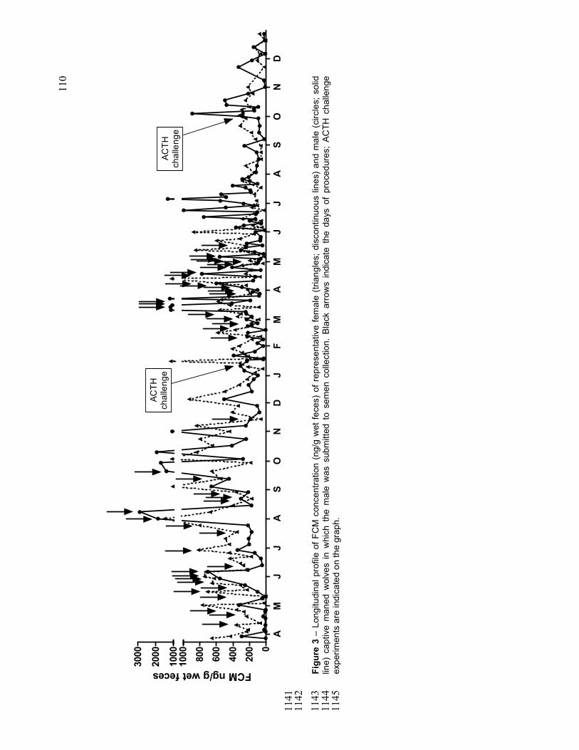
110
0
200
400
600
800
1000
1000
2000
3000
AM
JJ
AS
ON
DJ
FM
AM
JJ
AS
ON
Dnada
ACTH
challenge
ACTH
challenge
FCM ng/g wet feces
1141
1142
Figure 3 – Longitudinal profile of FCM concentration (ng/g wet feces) of representative female (triangles; discontinuous lines) and male (circles; solid
1143
line) captive maned wolves in which the male was submitted to semen collection. Black arrows indicate the days of procedures; ACTH challenge
1144
experiments are indicated on the graph.
1145

111
1146
Figure 4 – Longitudinal profile of fecal progestagens (µg/g wet feces; triangles, grey solid line) and estrogens (ng/g wet feces; circles, black
1147
discontinuous line) of representative female (A) and fecal androgens (ng/g wet feces; losanges, black discontinuous line) of representative male (B)
1148
captive maned wolves during andrological study. Black arrow indicates possible ovulatory cycle.
1149
1150
020406080
0246810
AM
JJ
AS
ON
DJ
FM
AM
JJ
AS
ON
Dnada
A
FEM ng/g wet feces
FPM µg/g wet feces
0
100
200
300
400
AM
JJ
AS
ON
DJ
FM
AM
JJ
AS
ON
Dnada
B
FAM ng/g wet feces

112
Figure 5 – Overall mean concentration (ng/g and µg/g wet feces; mean ± EPM) of fecal gonadal and adrenocortical metabolites in captive female (smooth columns) and male (diagonal hatched columns) maned wolves during reproductive and non-reproductive seasons. a,b,c Different letters within the same graph indicate differences (P<0.05; non pared t-test and Mann-Whitney rank sum test) between seasons and genders.
Figure 6 – Pre-challenge FCM concentration means (ng/g dry feces; mean ± EPM) in responsive and non-responsive maned wolves to the ACTH stimuli. Females: smooth columns; males: diagonal hatched columns. a,b Different letters within the same graph indicate differences (P<0.05; non pared t-test and Mann-Whitney rank sum test) between groups and genders.
0
500
1000
1500
ab
a
abb
Non-responsive Responsive
Female
Male
FCM ng/g dry feces
0
5
10
15
20
a
b
c
b
FEM ng/g wet feces
0.0
0.5
1.0
1.5
2.0
Reproductiveseason
Non-reproductiveseason
a
b
c
b
FPM µg/g wet feces
0
20
40
60
80
aa
b
b
FAM ng/g wet feces
0
200
400
600
Reproductiveseason
Non-reproductiveseason
FCM ng/g wet feces
a
a
aa
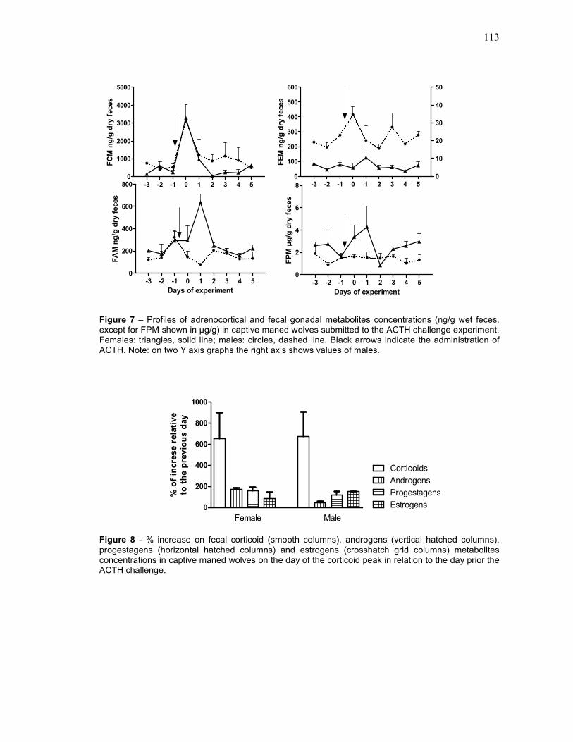
113
Figure 7 – Profiles of adrenocortical and fecal gonadal metabolites concentrations (ng/g wet feces, except for FPM shown in µg/g) in captive maned wolves submitted to the ACTH challenge experiment. Females: triangles, solid line; males: circles, dashed line. Black arrows indicate the administration of ACTH. Note: on two Y axis graphs the right axis shows values of males.
Figure 8 - % increase on fecal corticoid (smooth columns), androgens (vertical hatched columns), progestagens (horizontal hatched columns) and estrogens (crosshatch grid columns) metabolites concentrations in captive maned wolves on the day of the corticoid peak in relation to the day prior the ACTH challenge.
0
100
200
300
400
500
600
0
10
20
30
40
50
-3 -2 -1 0 1 2 3 4 5 nada
FEM ng/g dry feces
0
2
4
6
8
-3 -2 -1 0 1 2 3 4 5 nada
Days of experimentFPM µg/g dry feces
0
200
400
600
800
-3 -2 -1 0 1 2 3 4 5 nada
Days of experiment
FAM ng/g dry feces
0
1000
2000
3000
4000
5000
-3 -2 -1 0 1 2 3 4 5 nada
FCM ng/g dry feces
0
200
400
600
800
1000
Female Male
Corticoids
Androgens
Progestagens
Estrogens
% of increse relative
to the previous day

114
Figure 9 – Longitudinal profile of FCM concentration (ng/g wet feces) in female (triangles; discontinuous lines) and male (circles; solid line) captive maned wolves from Americana (A) and Ilha Solteira (B) Zoos.
Figure 10 – FCM concentration mean (ng/g wet feces; mean ± EPM) in female (smooth columns) and male (diagonal hatched columns) captive maned wolves from Americana (A) and Ilha Solteira (B) Zoos during reproductive and non-reproductive seasons. a,b Different letters within the same graph indicate differences (P<0.05) between seasons and genders.
0
50
100
150
200
250
Reproductiveseason
Non-reproductiveseason
A
FCM ng/g wet feces
aa
a
a
0
50
100
150
200
250
Reproductiveseason
Non-reproductiveseason
a
b
ab
b
B
FCM ng/g wet feces
0
200
400
600
800
1000
J F M A M J J A S O N D J F M A M JNADA
Lab blabla blB
AFCM ng/g wet feces
0
200
400
600
800
1000
J F M A M J J A S O N D nada
B
FCM ng/g wet feces

115
Figure 11 – Longitudinal profile of fecal androgens (A; ng/g wet feces), estrogens (B; ng/g wet feces) and progestagens (C; µg/g wet feces) concentrations from a female (triangles, discontinuous line) and male (circles, solid line) captive maned wolves.
0
100
200
300
400
500500
1000
1500
J F M A M J J A S O N D nada
AFAM ng/g wet feces
0
10
20
30
40
50
60
70
80
J F M A M J J A S O N D nada
B
FEM ng/g wet feces
0
2
4
6
8
1010203040
J F M A M J J A S O N D nada
C
FPM µg/g wet feces
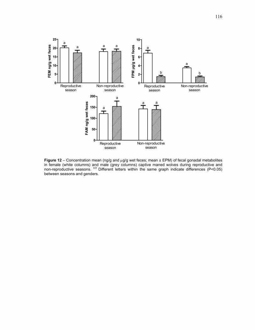
116
Figure 12 – Concentration mean (ng/g and µg/g wet feces; mean ± EPM) of fecal gonadal metabolites in female (white columns) and male (grey columns) captive maned wolves during reproductive and non-reproductive seasons. a,b Different letters within the same graph indicate differences (P<0.05) between seasons and genders.
0
5
10
15
20
25
Reproductiveseason
Non-reproductiveseason
FEM ng/g wet feces
aa a a
0
2
4
6
8
10
Reproductiveseason
Non-reproductiveseason
a
b
a
b
FPM µg/g wet feces
0
50
100
150
200
Reproductiveseason
Non-reproductiveseason
FAM ng/g wet feces
a
a
a a

117
6. CONCLUSÕES
Com base nas condições experimentais aqui descritas e nos resultados
obtidos, pode-se concluir que:
− As fêmeas de lobo-guará apresentam variações hormonais típicas do ciclo
ovariano de cadelas. A fase de anestro é marcada por reduzidas concentrações
nos metabólitos gonadais fecais. O proestro é caracterizado pelo aumento nas
concentrações e freqüência de picos de estrogênios. No período peri-ovulatório
ocorre aumento nas concentrações de metabólitos de progestágenos fecais que
se mantém altas até o final da gestação.
− Os machos de lobo-guará apresentam aumento nas concentrações de
metabólitos de androgênios fecais durante a fase de proestro e período peri-
ovulatório das fêmeas.
− As concentrações de corticóides aumentam normalmente durante o proestro e
fase de lactação e cuidado parental nas fêmeas.
− Após o nascimento dos filhotes os machos apresentam aumento nas
concentrações de todos os metabólitos gonadais e corticóides fecais.
− O perfil normal de metabólitos gonadais fecais foi alterado devido à estimulação
crônica do eixo hipotálamo-hipófise-adrenal, mostrando que o estresse crônico
ocasiona baixa eficiência reprodutiva.
− O estímulo adrenocortical agudo promovido pela administração de ACTH
aumentou as concentrações de metabólitos de progestágenos e estrogênios
fecais em fêmeas de lobos-guará. Nos machos os androgênios fecais foram
reduzidos pelo estímulo.

118
REFERÊNCIAS
AZEVEDO, F. C. Área de vida e organizaçao espacial de Lobos-Guará (Chrysocyon brachyurus) na região do Parque Nacional da Serra da Canastra, Minas Gerais, Brasil. Dissertação (mestrado) - Ecologia, Conservação e Manejo de Vida Silvestre, Instituto de Ciências Biológicas, Universidade Federal de Minas Gerais, Belo Horizonte, 2008. BERNE, R. M. et al. Fisiologia, 5a.ed., Rio de Janeiro: Elsevier, 2004. BOLANOS, J. M.; MOLINA, J. R.; FORSBERG, M. Effect of blood sampling and administration of ACTH on cortisol and progesterone levels in ovariectomized zebu cows (Bos indicus). Acta Veterinaria Scandinavica, v. 38, p. 01-07, 1997. BREUNER, C. W.; HAHN, T. P. Integrating stress physiology, environmental change, and behavior in free-living sparrows. Hormones and Behavior, v.43, p. 115-123, 2003. BUIJS, S. et al. Glucocorticoid metabolites in rabbit faeces--influence of environmental enrichment and cage size. Physiology & behavior, v. 04, p. 469-73, 2011. CHAND, D.; LOVEJOY, D. A. Stress and reproduction: controversies and challenges. General and comparative endocrinology, v.171, p. 253-257, 2011. CHARBONNEL, N.; CHAVAL, Y.; BERTHIER, K. Stress and demographic decline: a potential effect mediated by impairment of reproduction and immune function in cyclic vole populations. Physiological and biochemical zoology, v.81, p. 63-73, 2008. CHATDARONG, K. et al.. The effect of ACTH stimulation on cortisol and progesterone concentrations in intact and ovariohysterectomized domestic cats. Theriogenology, v.66, p.1482-1487, 2006. CITES, Convention on International Trade in Endangered Species of Wild Fauna and Flora. Disponível em: <http://www.cites.org/eng/app/index.php>. Acesso em 21/08/2012. CONCANNON, P.W. Endocrinologic control of normal canine ovarian function. Reproduction in domestic animals, v.44, n.2, p.03–15, 2009. CUMMINGS, O. D.; BROWN, J. L.; RODDEN, M.D.. Behavioral and Physiologic Responses to Environmental Enrichment in the Maned Wolf (Chrysocyon brachyurus). Zoo Biology, v. 343, p.331–343, 2007. CYR, N. E.; ROMERO, L. M. Chronic stress in free-living European starlings reduces corticosterone concentrations and reproductive success. General and comparative endocrinology, v.151, p.82-89, 2007. DALEY, C. A. et al. Effect of stress-like concentrations of cortisol on the feedback potency of oestradiol in orchidectomized sheep. Animal reproduction science, v.59, p.167-178, 2000. DE PAULA, R. C.; MÉDICI, P.; MORATO, R. G. Plano de ação para conservação do lobo-guará, análise de viabilidade populacional e de habitat (PHVA). Brasília: IBAMA, 2008. DIETZ, J. M. Ecology and Social Organization of the Maned Wolf (Chrysocyon brachyurus). Smithsonian Contributions to Zoology, v. 392, p. 1-51, 1984.
119
DOBSON, H.; SMITH, R. F.. What is stress, and how does it affect reproduction? Animal reproduction science, v.60-61, p.743-752, 2000. FARSTAD, W. Reproduction in foxes: current research and future challenges. Animal reproduction science, v.53, p.35-42, 1998. FEDLMAN, E. C.; NELSON, R. W. Canine and feline endocrinology and reproduction, 3. ed., California: Saunders, 2004. 1099p. FENSKE, M. Role of cortisol in the ACTH-induced suppression of testicular steroidogenesis in guinea pigs. The Journal of endocrinology, v.154, n.3, p.407–14, 1997. FERIN, M. Stress and reproductive system. In: Neil, J. D. Knobill and Neill’s physiology of reproduction. 3. ed. Philadelphia: Elsevier, 2006. FRANK, L. A.; DAVIS, J. A.; OLIVER, J. W. Serum concentrations of cortisol, sex hormones of adrenal origin, and adrenocortical steroid intermediates in healthy dogs following stimulation with two doses of cosyntropin. Am. J. Vet. Res., v.65, n.12, p.1631-1633, 2004. GINEL, P. J. et al. Evaluation of serum concentrations of cortisol and sex hormones of adrenal gland origin after stimulation with two synthetic ACTH preparations in clinically normal dogs. Am. J. Vet. Res., v.73, n.2, p.237-241, 2012. GUYTON, A. C.; HALL, J. E. Tratado de Fisiologia Médica. 11. ed., Rio de Janeiro: Elsevier, 2006. HAUN, C. K.; HALTMEYER, G. C. Effects of an intraventricular injection of synthetic ACTH on plasma testosterone, progesterone and LH levels and on sexual behavior in male and female rabbits. Neuroendocrinology, v.19, n.3, p.201-213, 1975. HEDBERG, Y. et al. Effect of ACTH (tetracosactide) on steroid hormone levels in the mare. Part A: effect in intact normal mares and mares with possible estrous related behavioral abnormalities. Animal reproduction science, v.100, n.1-2, p.73–91, 1975. IUCN, 2012. IUCN Red List of Threatened Species. Disponível em:<http:// www.iucnredlist.org.>. Acesso em: 06/09/2012. KEAY, J. M. Fecal glucocorticoids and their metabolites as indicators of stress in vaious mammalian species: A literature review. Journal of Zoo and Wildlife Medicine, v. 37, p.234-244, 2006. KLINK, C. A.; MACHADO, R. B.. Conservation of the Brazilian Cerrado. Conservation Biology, v.19, p.707-713, 2005. KORTE, S. M.; OLIVIER, B.; KOOLHAAS, J. M. A new animal welfare concept based on allostasis. Physiology & behavior, v. 92, p.422-428, 2007. MAIA, O. B.; GOUVEIA, M. G. Birth and mortality of maned wolves Chrysocyon brachyurus (Illiger, 1811) in captivity. Brazilian journal of biology, v.62, n.1, p.25–32, 2002. MACHADO, R. B.; RAMOS NETO M. B.; PEREIRA, P. G. P. Estimativas de perda da área do Cerrado brasileiro. Relatório técnico não publicado. Brasília, DF, Brasil, 2004. Disponível em: <http://www.conservation.org.br/arquivos/RelatDesmatamCerrado.pdf.> Acesso em: 13/09/2012.

120
MCCONNACHIE, S. H.; COOK, K. V.; PATTERSON, D. A. Consequences of acute stress and cortisol manipulation on the physiology, behavior , and reproductive outcome of female Pacific salmon on spawning grounds. Hormones and Behavior, v.62, p.67-76, 2012a. MCCONNACHIE, S. H.; COOK, K. V.; PATTERSON, D. A.. Consequences of acute stress and cortisol manipulation on the physiology, behavior, and reproductive outcome of female Pacific salmon on spawning grounds. Hormones and behavior, v.62, p.67-76, 2012b. MCNICOL, A. M. The human adrenal gland: aspects of structure, function and pathology. In: James, V. H. T. The adrenal gland. 2. ed., New York: Raven Press, 1992. MILLSPAUGH, J. J.; WASHBURN, B. E. Use of fecal glucocorticoid metabolite measures in conservation biology research: considerations for application and interpretation. General and comparative endocrinology, v.138 p.189-199, 2004. BRASIL. Ministério do Meio Ambiente (MMA). Instrução Normativa Nº 3, de 27 de maio de 2003. Diário Oficial da União, Brasília, DF, n.101, p.88-97, 27 mai. 2003, seção 1, pt.1. MOORE, I. T.; JESSOP, T. S. Stress, reproduction, and adrenocortical modulation in amphibians and reptiles. Hormones and Behavior 43:39-47, 2003. MORATÓ, D. Q. Seletividade e sazonalidade das presas consumidas pelo lobo guará (Chrysocyon brachyurus) no Parque Nacional da Serra da Canastra, Minas Gerais. 84f. Dissertação (Mestrado em Biociências) – Instituto de Biociências, Universidade de São Paulo, 2001. MORGAN, K. N.; TROMBORG, C. T. Sources of stress in captivity. Applied Animal Behaviour Science, v.102, p.262–302, 2007. MÖSTL, E; PALME, R.. Hormones as indicators of stress. Domestic animal endocrinology, v. 3, p.67-74, 2002. MYERS, N.; MITTERMEIER, R. A; MITTERMEIER, C. G.; FONSECA, G. A D. A; KENT, J. Biodiversity hotspots for conservation priorities. Nature, v.403, n. 6772, p. 853-858, 2000. MWANZA, A. M. Plasma level of cortisol, progesterone, oestradiol-17 beta and protaglandin F2 alfa metabolite after ACTH (Synacthen Depot) administration in ovariectomized gilts. J. Vet. Med. A Physiology Pathology Clin. Med., v.47, n.4, p.193-200, 2000. NOWAK, R. M. Walker’s Mammals of the World. 5 ed. Baltimore: The Johns Hopkins University Press, 1991. PEREIRA, R. J. G.; DUARTE, J. M. B.; NEGRÃO, J. A . Effects of environmental conditions, human activity, reproduction, antler cycle and grouping on fecal glucocorticoids of free-ranging Pampas deer stags (Ozotoceros bezoarticus bezoarticus). Hormones and Behavior, v.49, p.114 – 122, 2006. PRIMACK, R. B.; RODRIGUES, E. Biologia da conservação. Londrina: Efraim Rodrigues, 2001. 328p. QUEIROLO, D.; MOTTA-JUNIOR, J. C. Prey availability and diet of maned wolf in Serra da Canastra National Park, southeastern Brazil. Acta Theriologica, v.52, n.4, p.391–402, 2007 RODDEN, M.D. Use of behavioral measures to assess reproductive status in maned wolves (Chrysocyon brachyurus). Zoo Biology, v.15, p. 565-585, 1996.

121
RODRIGUES, F.H.G. Biologia e conservação do lobo-guará (Chrysocyon brachyurus) na Estação Ecológica de Águas Emendadas, DF. 2002, 96f. Tese (Doutorado) – Instituto de Biologia, Universidade de Campinas, SP, 2002. SHELDON, J.W. Wild dogs, the natural history of non domestic Canidae. San-Diego: Califórnia: Academic Press Inc., 1992. SHERIFF, M. J. et al. Measuring stress in wildlife: techniques for quantifying glucocorticoids. Oecologia, v.166, p.869–87, 2011. SILVA, J. A.; TALAMONI, S. A. Diet adjustments of maned wolves, Chrysocyon brachyurus (Illiger) (Mammalia, Canidae), subjected to supplemental feeding in a private natural reserve, Southeastern Brazil. Revista Brasileira de Zoologia, v.20, n.2, p.339–345, 2003. SONGSASEN, N. et al. Patterns of fecal gonadal hormone metabolites in the maned wolf (Chrysocyon brachyurus). Theriogenology, v.66, p.1743–1750, 2006. SONGSASEN, N.; WILDT, D. E. Oocyte biology and challenges in developing in vitro maturation systems in the domestic dog. Animal Reproduction Science, v.98, p.02–22, 2007. SPERCOSKI, K. M. Monitoramento a longo prazo da excreção de corticóides fecias em lobos-guarás (Chrysocyon brachyurus, Illiger 1811) de cativeiro e vida livre: uma contribuição para o manejo e a conservação da espécie. 2007, 55f. Dissertação (Mestrado) - Setor de Ciências Biológicas, Programa de Pós-graduação em Biologia Celular e Molecular, Universidade Federal do Paraná, Curitiba, 2007. SUTHERLAND, M. A.; SHOME, G. P.; HULBERT, L. E. 2009. Acute stress affects the physiology and behavior of allergic mice. Physiology & behavior, v.98, p.281-287, 2009. TSUMA, V.T. et al. Plasma levels of progesterone and cortisol after ACTH admin istration in lactating primiparous sows. Acta Vet Scand, v.39, p.71–76, 1998. TURNER, A. I.; HEMSWORTH, P. H.; TILBROOK, A. J. Susceptibility of reproduction in female pigs to impairment by stress or elevation of cortisol. Domestic animal endocrinology, v.29, p.398-410, 2005. VAN LIER, E. et al. Effects of adrenocorticotrophin (ACTH) and progesterone on luteinising hormone (LH) secretion in recently castrated rams. Animal reproduction science, v.55, p.115–26, 1999. VANSTREELS, R. E. T.; PESSUTTI, C. Analysis and discussion of maned wolf Chrysocyon brachyurus population trends in Brazilian institutions: lessons from the Brazilian studbook, 1969-2006. International Zoo Yearbook, v.44, p.01–15, 2010. VELLOSO, A. L. et al. Longitudinal fecal steroid excretion in maned wolves (Chrysocyon brachyurus). General and comparative endocrinology, v.112, p.96–107, 1998. VERSTEGEN-ONCLIN, K.; VERSTEGEN, J. Endocrinology of pregnancy in the dog: a review. Theriogenology, v.70, p. 291–299, 2008. WILLARD, S. T. et al.. Plasma progesterone response following ACTH administration during mid-gestation in the pregnant Brahman heifer. Theriogenology, v.63, p.1061–1069, 2005.

122
WURBEL, H.; STAUFFACHER, M. Prevention of stereotypy in laboratory mice: Effects on stress physiology and behaviour. Physiology & Behavior, v.59, p.1163-1170, 1996. YOSHIDA, C.; NAKAO, T. Plasma cortisol and progesterone responses to low doses of adrenocorticotropic hormone in ovariectmized lactating cows. The Journal of reproduction and development, v.52, p.797–803, 2006.


![[XLS] · Web viewAna Maria Caetano de Faria Ana Maria Caldeira Oliveira Ana Maria Covolam Ana Maria de Freitas Pinheiro Ana Maria Maniero Moreira Ana Maria Mouad Ana Maria Petraitis](https://img.pdfslide.tips/doc/110x75/5ad51e2a7f8b9a1a028cb048/xls-viewana-maria-caetano-de-faria-ana-maria-caldeira-oliveira-ana-maria-covolam.jpg)