Embed Size (px)
Citation preview
8/2/2019 Kk Dila Julnal Clab
http://slidepdf.com/reader/full/kk-dila-julnal-clab 1/15
R E S E A R C H A R T I C L E Open Access
Agrobacterium-mediated genetic transformationof Coffea arabica (L.) is greatly enhanced byusing established embryogenic callus culturesAlessandra F Ribas1, Eveline Dechamp1, Anthony Champion2, Benoît Bertrand1, Marie-Christine Combes2,
Jean-Luc Verdeil3, Fabienne Lapeyre3, Philippe Lashermes2 and Hervé Etienne1*
Abstract
Background: Following genome sequencing of crop plants, one of the main challenges today is determining the
function of all the predicted genes. When gene validation approaches are used for woody species, the mainobstacle is the low recovery rate of transgenic plants from elite or commercial cultivars. Embryogenic calli have
frequently been the target tissue for transformation, but the difficulty in producing or maintaining embryogenic
tissues is one of the main problems encountered in genetic transformation of many woody plants, including Coffea
arabica.
Results: We identified the conditions required for successful long-term proliferation of embryogenic cultures in C.
arabica and designed a highly efficient and reliable Agrobacterium tumefaciens-mediated transformation method
based on these conditions. The transformation protocol with LBA1119 harboring pBin 35S GFP was established by
evaluating the effect of different parameters on transformation efficiency by GFP detection. Using embryogenic
callus cultures, co-cultivation with LBA1119 OD600 = 0.6 for five days at 20 °C enabled reproducible transformation.
The maintenance conditions for the embryogenic callus cultures, particularly a high auxin to cytokinin ratio, the
age of the culture (optimum for 7-10 months of proliferation) and the use of a yellow callus phenotype, were the
most important factors for achieving highly efficient transformation (> 90%). At the histological level, successfultransformation was related to the number of proembryogenic masses present. All the selected plants were proved
to be transformed by PCR and Southern blot hybridization.
Conclusion: Most progress in increasing transformation efficiency in coffee has been achieved by optimizing the
production conditions of embryogenic cultures used as target tissues for transformation. This is the first time that a
strong positive effect of the age of the culture on transformation efficiency was demonstrated. Our results make
Agrobacterium-mediated transformation of embryogenic cultures a viable and useful tool both for coffee breeding
and for the functional analysis of agronomically important genes.
BackgroundGenome sequencing of important crop plants, such as
wheat, sugarcane, tomato, potato, banana, eucalyptus,
cacao and coffee, has already been completed or is inprogress [1]. The resulting information opens significant
new challenges in plant biology, including determining
the function of predicted genes and introducing new
desirable traits in pre-existing outstanding genotypes by
genetic engineering in a shorter time. Shortening the
time required is particularly attractive for the improve-ment of woody species [2]: pedigree selection programs
in Coffea arabica, a preferentially autogamous woody
species, often last 25 years. In response to an unex-
pected threat, genetically transformed plants could thus
provide a satisfactory solution, all the more since in a
few years, coffee genome sequencing [3] will enable
identification of genes coding for very important
* Correspondence: [email protected] de Coopération Internationale en Recherche Agronomique pour le
Développement - Département des Systèmes Biologiques (CIRAD-BIOS).
UMR-RPB (CIRAD, IRD, Université Montpellier II), 911 Avenue Agropolis, BP
64501, 34394 Montpellier, France
Full list of author information is available at the end of the article
Ribas et al . BMC Plant Biology 2011, 11:92
http://www.biomedcentral.com/1471-2229/11/92
© 2011 Ribas et al; licensee BioMed Central Ltd. This is an Open Access article distributed under the terms of the Creative CommonsAttribution License (http://creativecommons.org/licenses/by/2.0), which permits unrestricted use, distribution, and reproduction inany medium, provided the original work is properly cited.
8/2/2019 Kk Dila Julnal Clab
http://slidepdf.com/reader/full/kk-dila-julnal-clab 2/15
agronomical traits linked to disease resistance and/or
abiotic stresses.
Agrobacterium-mediated transformation is a widely
used and powerful tool for introducing foreign DNA
into many plant species. It has several advantages over
physical transformation methods including its tendency
to generate single or a low copy number of transgenes
with defined ends and preferential integration into tran-
scriptionally active regions of the chromosomes [4].
Agrobacterium-mediated DNA delivery has become a
powerful tool in functional genomics as it is the most
reliable way to assess gene function by generating gain-
of-function or loss-of-function mutants [5]. However,
for many important crop plants, including most woody
species, a method for genetic transformation has either
not yet been established or is still laborious and ineffi-
cient. For such species, higher throughput transforma-
tion systems are needed to be able to fully benefit fromthe rapid development of plant genomics for basic
research and for the design of new genetic improvement
strategies including both conventional breeding and
genetic transformation.
There are very few examples of the introduction of
genes of agronomic interest in Arabica coffee. Leroy et
al. [6] regenerated transgenic coffee plants carrying the
CRY1-AC gene from Bacillus thuringiensis, which is
effective against the coffee leaf miner. Ogita et al. [7]
obtained transgenic coffee plants with suppressed caf-
feine synthesis using RNA interference (RNAi) technol-
ogy through inhibition of a theobromine synthase gene
(CaMXMT1). Genetic transformation of coffee has been
achieved using several types of explants (leaves, embryo-
genic calli, somatic embryos, protoplasts) and different
approaches including A. tumefaciens-mediated transfor-
mation [6-9], A. rhizogenes-mediated transformation
[10,11] and biolistic gene delivery [12,13]. The recovery
of transgenic plants appears to be easier with Coffea
canephora species than with C. arabica [14-16]. How-
ever the protocols available so far, which mostly use A.
tumefaciens, are not reproducible and transformation
efficiency is very low (less than 1%), thus seriously limit-
ing their potential routine use. Today, embryogenic cal-
lus derived from leaf tissues is the most widely usedtarget tissue for the genetic transformation of C. ara-
bica [7,9,17]. However the induction of embryogenic
tissues in C. arabica takes longer and is more difficult
than in the other cultivated species C. canephora. The
limited availability of embryogenic tissues, together with
the low transformation efficiency of this type of tissue,
is one of the main limitations to genetic transformation
in coffee. Each round of a transformation experiment
requires a new and uncertain process of production and
selection of embryogenic calli that takes around eight
months.
For these reasons, defining the conditions for success-
ful long-term proliferation of embryogenic callus is a
decisive step towards the large-scale, continuous pro-
duction of target tissues for genetic transformation.
Embryogenic cultures have already been used to estab-
lish reliable and efficient genetic transformation proce-
dures for different woody species including Prunus
[18,19], grapevine [20,21], rubber tree [22] and chestnut
tree [23]. To date, embryogenic callus cultures main-tained in liquid or semi-solid medium have never been
used as target tissues for genetic transformation of the
two cultivated species C. arabica and C. canephora.
However, high-throughput methods for coffee propaga-
tion based on the use of established embryogenic cul-
tures have already been developed by our team [24,25]
and are currently used for the commercial diffusion of
elite F1 hybrid varieties [26,27]. Such systems could also
provide suitable support for the high-throughput regen-
eration of coffee transgenic varieties.
In the present work, we describe for the first time in
the C. arabica species a highly efficient and reliable
Agrobacterium -mediated transformation protocol that
allows the generation of thousands transgenic coffee
trees from multiple independent transformation events.
ResultsExplant and co-cultivation conditions
Different types of coffee embryogenic tissues were tested
under our transformation conditions; GFP detection was
performed 30 days after co-cultivation to confirm stable
transformation events (Table 1). In this study, the detec-
tion of GFP expression was successfully used to monitor
the transformation efficiency of all the parameters con-
cerned. A low GFP expression level was detected in co-cultivated embryogenic calli and almost no expression
was observed when embryogenic cell suspensions were
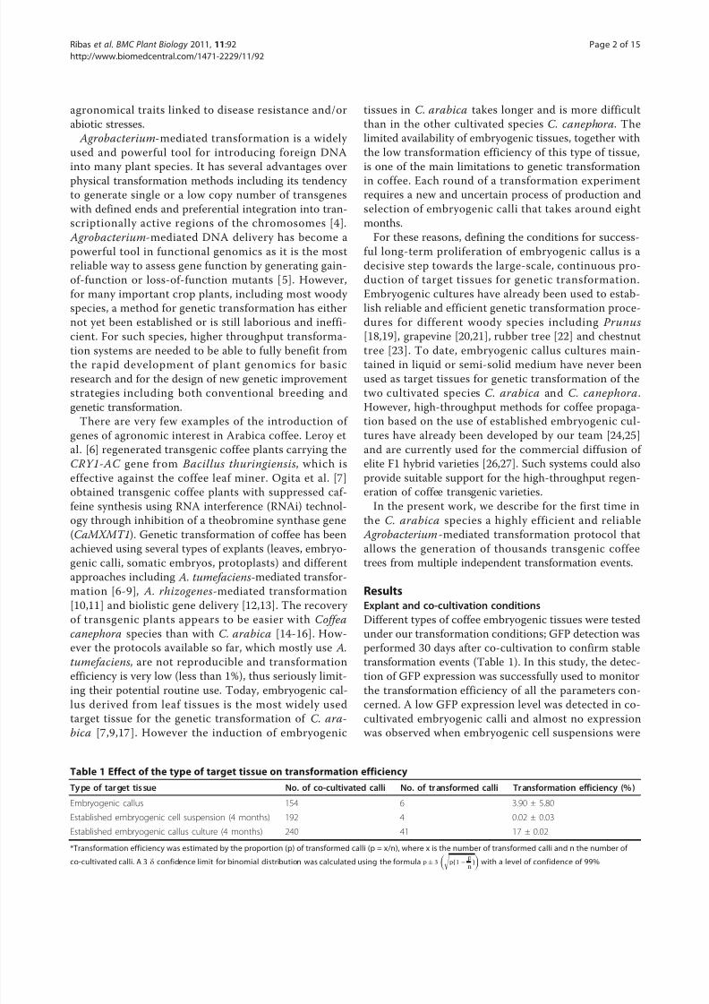
Table 1 Effect of the type of target tissue on transformation efficiency
Ty pe of target tis sue No. of co-cultivated calli No. of transformed calli Transformation efficiency (%)
Embryogenic callus 154 6 3.90 ± 5.80
Established embryogenic cell suspension (4 months) 192 4 0.02 ± 0.03
Established embryogenic callus culture (4 months) 240 41 17 ± 0.02
*Transformation efficiency was estimated by the proportion (p) of transformed calli (p = x/n), where x is the number of transformed calli and n the number of
co-cultivated calli. A 3 δ confidence limit for binomial distribution was calculated using the formula p± 3
p(1−
p
n)
with a level of confidence of 99%
Ribas et al . BMC Plant Biology 2011, 11:92
http://www.biomedcentral.com/1471-2229/11/92
Page 2 of 15
8/2/2019 Kk Dila Julnal Clab
http://slidepdf.com/reader/full/kk-dila-julnal-clab 3/15
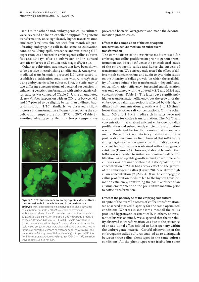
used. On the other hand, embryogenic callus cultures
were revealed to be an excellent support for genetic
transformation, since significantly higher transformation
efficiency (17%) was obtained with four-month-old pro-
liferating embryogenic calli in the same co-cultivation
conditions. Using epifluorescence analysis, strong GFP
expression was detected in embryogenic callus cultures
five and 30 days after co-cultivation and in derived
somatic embryos at all ontogenetic stages (Figure 1).
Other co-cultivation parameters that have been shown
to be decisive in establishing an efficient A. rhizogenes-
mediated transformation protocol [10] were tested to
establish co-cultivation conditions with A. tumefaciens
using embryogenic callus cultures. First, the efficiency of
two different concentrations of bacterial suspension in
enhancing genetic transformation with embryogenic cal-
lus cultures was compared (Table 2). Using an undiluted
A. tumefaciens suspension with an OD600 of between 0.6and 0.7 proved to be slightly better than a diluted bac-
terial solution (1/10). Similarly, we observed a slight
increase in transformation efficiency by reducing the co-
cultivation temperature from 27°C to 20°C (Table 2).
Another advantage is that the lower temperature
prevented bacterial overgrowth and made the deconta-
mination process easier.
Effect of the composition of the embryogenic
proliferation culture medium on subsequent
transformation
The composition of the nutritive medium used for
embryogenic callus proliferation prior to genetic trans-
formation can directly influence the physiological status
of the embryogenic callus and hence the success of
transformation. We consequently tested the effect of dif-
ferent salt concentrations and auxin to cytokinin ratios
on the intensity of callus growth (on which the availabil-
ity of tissues suitable for transformation depends) and
on transformation efficiency. Successful transformation
was only obtained with the diluted MS/2 and MS/4 salt
concentrations (Table 3). The latter gave significantly
higher transformation efficiency, but the growth of theembryogenic callus was seriously affected by this highly
diluted salt concentration: growth was 2 to 2.5 times
lower than at other salt concentrations. On the other
hand, MS and 1.5 MS media rich in salts were not
appropriate for coffee transformation. The MS/2 salt
concentration that enabled efficient embryogenic callus
proliferation and subsequently efficient transformation
was thus selected for further transformation experi-
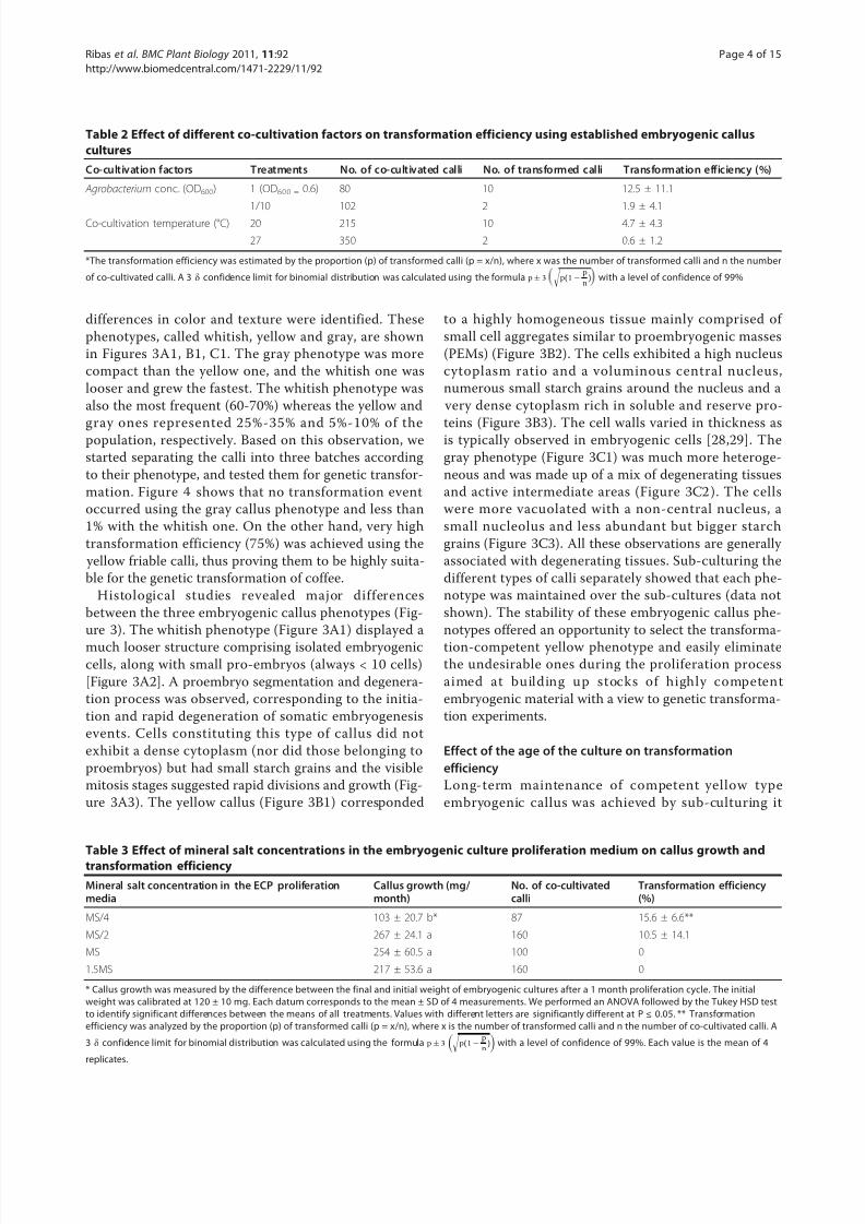
ments. Regarding the auxin to cytokinin ratio in the
proliferation medium, we first observed that 6-BA had a
strong negative effect on genetic transformation, as very
efficient transformation was obtained without exogenous
cytokinin (Figure 2A). However, it should be noted that
6-BA was not needed to sustain embryogenic callus pro-
liferation, as acceptable growth intensity over three sub-
cultures was obtained without it. Like cytokinin, the
concentration of 2,4-D had a weak effect on the growth
of the embryogenic callus (Figure 2B). A relatively high
auxin concentration (9 μM 2,4-D) in the embryogenic
callus proliferation medium led to the highest transfor-
mation efficiency, confirming the positive effect of an
auxinic environment on the pre-culture medium prior
to coffee transformation.
Effect of the phenotype of the embryogenic cultureIn spite of the overall success of coffee transformation,
we observed marked disparity for the same optimized
conditions. Whereas in some jars almost all the callus
produced hygromycin-resistant calli, in others, no resis-
tant callus was obtained. We suspected that the variabil-
ity observed in transformation was due to the existence
of an additional effect related to heterogeneity within
the embryogenic material. Careful observation of the
embryogenic callus cultures enabled us to distinguish
between three callus phenotypes in the same culture
conditions. All the phenotypes were friable but some
A
C
B
D
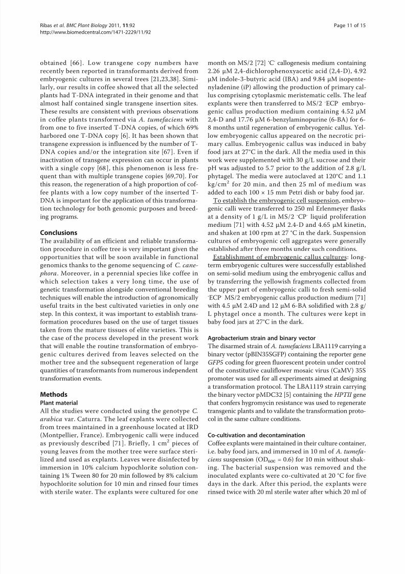
Figure 1 GFP fluorescence in embryogenic callus cultures
transformed with A. tumefaciens and in derived somatic
embryos. Transient expression in embryogenic callus 5 days after
co-cultivation, bar scale = 50 μM (A). Stable expression in
embryogenic callus culture 30 days after co-cultivation, bar scale =
50 μM (B). Stable expression in globular and heart stage 6 months
after co-cultivation, bar scale = 100 μM (C). Stable expression in
torpedo mature somatic embryos 7 months after co-cultivation, bar
scale = 500 μM (D). Images were obtained using a Leica MZ Fluo III
(optic 0.63 Zeiss) fluorescence microscope supplied with a DC 300F
camera (Leica Microsystems, Welzlar, Germany) with plant GFP filter
no. 3 from Leica: excitation wavelengths: 470-540 nm (BP), emission
wavelengths: 525-550 nm (BP).
Ribas et al . BMC Plant Biology 2011, 11:92
http://www.biomedcentral.com/1471-2229/11/92
Page 3 of 15
8/2/2019 Kk Dila Julnal Clab
http://slidepdf.com/reader/full/kk-dila-julnal-clab 4/15
differences in color and texture were identified. These
phenotypes, called whitish, yellow and gray, are shown
in Figures 3A1, B1, C1. The gray phenotype was more
compact than the yellow one, and the whitish one was
looser and grew the fastest. The whitish phenotype was
also the most frequent (60-70%) whereas the yellow and
gray ones represented 25%-35% and 5%-10% of thepopulation, respectively. Based on this observation, we
started separating the calli into three batches according
to their phenotype, and tested them for genetic transfor-
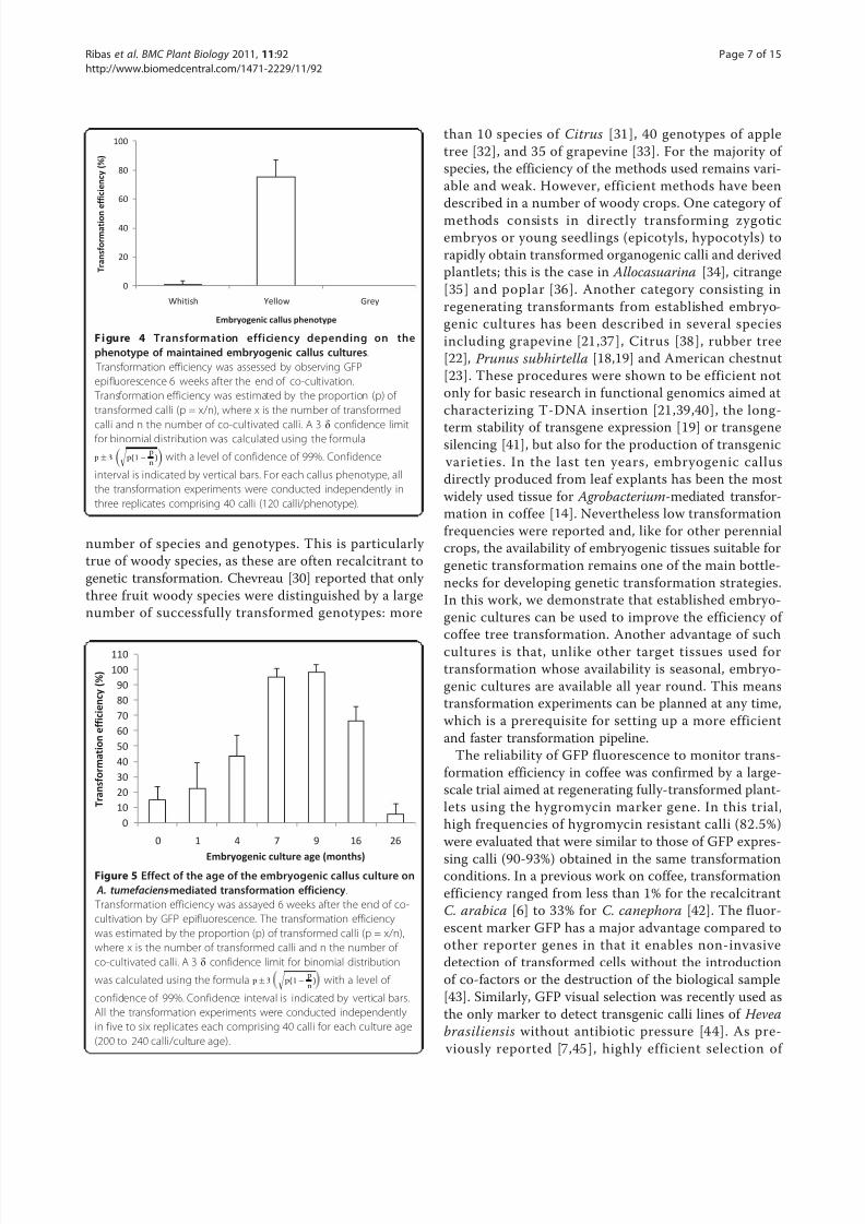
mation. Figure 4 shows that no transformation event
occurred using the gray callus phenotype and less than
1% with the whitish one. On the other hand, very high
transformation efficiency (75%) was achieved using the
yellow friable calli, thus proving them to be highly suita-
ble for the genetic transformation of coffee.
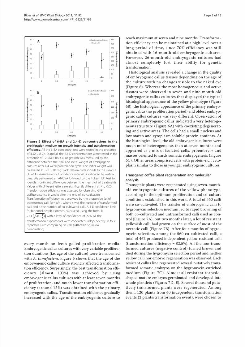
Histological studies revealed major differences
between the three embryogenic callus phenotypes (Fig-
ure 3). The whitish phenotype (Figure 3A1) displayed a
much looser structure comprising isolated embryogeniccells, along with small pro-embryos (always < 10 cells)
[Figure 3A2]. A proembryo segmentation and degenera-
tion process was observed, corresponding to the initia-
tion and rapid degeneration of somatic embryogenesis
events. Cells constituting this type of callus did not
exhibit a dense cytoplasm (nor did those belonging to
proembryos) but had small starch grains and the visible
mitosis stages suggested rapid divisions and growth (Fig-
ure 3A3). The yellow callus (Figure 3B1) corresponded
to a highly homogeneous tissue mainly comprised of
small cell aggregates similar to proembryogenic masses
(PEMs) (Figure 3B2). The cells exhibited a high nucleus
cytoplasm ratio and a voluminous central nucleus,
numerous small starch grains around the nucleus and a
very dense cytoplasm rich in soluble and reserve pro-
teins (Figure 3B3). The cell walls varied in thickness asis typically observed in embryogenic cells [28,29]. The
gray phenotype (Figure 3C1) was much more heteroge-
neous and was made up of a mix of degenerating tissues
and active intermediate areas (Figure 3C2 ). The cells
were more vacuolated with a non-central nucleus, a
small nucleolus and less abundant but bigger starch
grains (Figure 3C3). All these observations are generally
associated with degenerating tissues. Sub-culturing the
different types of calli separately showed that each phe-
notype was maintained over the sub-cultures (data not
shown). The stability of these embryogenic callus phe-
notypes offered an opportunity to select the transforma-
tion-competent yellow phenotype and easily eliminatethe undesirable ones during the proliferation process
aimed at building up stocks of highly competent
embryogenic material with a view to genetic transforma-
tion experiments.
Effect of the age of the culture on transformation
efficiency
Long-term maintenance of competent yellow type
embryogenic callus was achieved by sub-culturing it
Table 2 Effect of different co-cultivation factors on transformation efficiency using established embryogenic callus
cultures
Co-cultivation factors Treatments No. of co-cultivated calli No. of transformed calli Transformation efficiency (%)
Agrobacterium conc. (OD600) 1 (OD600 = 0.6) 80 10 12.5 ± 11.1
1/10 102 2 1.9 ± 4.1
Co-cultivation temperature (°C) 20 215 10 4.7 ± 4.3
27 350 2 0.6 ± 1.2
*The transformation efficiency was estimated by the proportion (p) of transformed calli (p = x/n), where x was the number of transformed calli and n the number
of co-cultivated calli. A 3 δ confidence limit for binomial distribution was calculated using the formula p± 3
p(1−
p
n) with a level of confidence of 99%
Table 3 Effect of mineral salt concentrations in the embryogenic culture proliferation medium on callus growth and
transformation efficiency
Mineral salt concentration in the ECP proliferationmedia
Callus growth (mg/ month)
No. of co-cultivatedcalli
Transformation efficiency(%)
MS/4 103 ± 20.7 b* 87 15.6 ± 6.6**
MS/2 267 ± 24.1 a 160 10.5 ± 14.1
MS 254 ± 60.5 a 100 0
1.5MS 217 ± 53.6 a 160 0
* Callus growth was measured by the difference between the final and initial weight of embryogenic cultures after a 1 month proliferation cycle. The initial
weight was calibrated at 120 ± 10 mg. Each datum corresponds to the mean ± SD of 4 measurements. We performed an ANOVA followed by the Tukey HSD test
to identify significant differences between the means of all treatments. Values with different letters are significantly different at P ≤ 0.05. ** Transformation
efficiency was analyzed by the proportion (p) of transformed calli (p = x/n), where x is the number of transformed calli and n the number of co-cultivated calli. A
3 δ confidence limit for binomial distribution was calculated using the formula p± 3
p(1−
p
n)
with a level of confidence of 99%. Each value is the mean of 4
replicates.
Ribas et al . BMC Plant Biology 2011, 11:92
http://www.biomedcentral.com/1471-2229/11/92
Page 4 of 15
8/2/2019 Kk Dila Julnal Clab
http://slidepdf.com/reader/full/kk-dila-julnal-clab 5/15
every month on fresh gelled proliferation media.Embryogenic callus cultures with very variable prolifera-
tion durations (i.e. age of the culture) were transformed
with A. tumefaciens. Figure 5 shows that the age of the
embryogenic callus culture strongly affected transforma-
tion efficiency. Surprisingly, the best transformation effi-
c ie nc y ( al mo st 1 00 %) w as a ch ie ve d b y u si ng
embryogenic callus cultures with at least seven months
of proliferation, and much lower transformation effi-
ciency (around 15%) was obtained with the primary
embryogenic callus. Transformation efficiency gradually
increased with the age of the embryogenic culture to
reach maximum at seven and nine months. Transforma-
tion efficiency can be maintained at a high level over a
long period of time, since 70% efficiency was still
obtained with 16-month-old embryogenic cultures.
However, 26-month-old embryogenic cultures had
almost completely lost their ability for genetic
transformation.
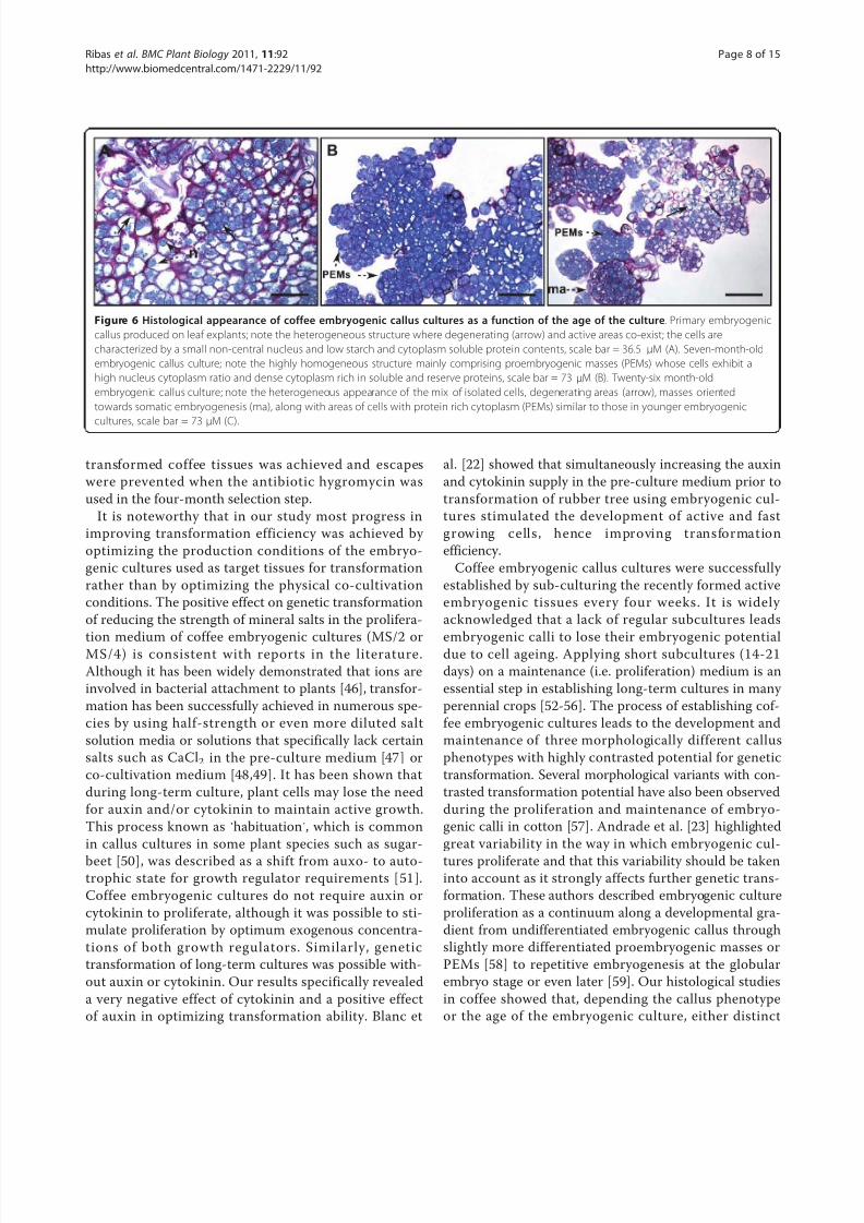
Histological analysis revealed a change in the quality
of embryogenic callus tissues depending on the age of
the culture with no changes visible to the naked eye
(Figure 6). Whereas the most homogeneous and active
tissues were observed in seven and nine-month old
embryogenic callus cultures that displayed the typical
histological appearance of the yellow phenotype (Figure
6B), the histological appearance of the primary embryo-
genic callus (no proliferation period) and oldest embryo-
genic callus cultures was very different. Observation of
primary embryogenic callus indicated a very heteroge-neous structure (Figure 6A) with coexisting degenerat-
ing and active areas. The cells had a small nucleus and
low starch and cytoplasm soluble protein contents. At
the histological level, the old embryogenic cultures were
much more heterogeneous than at seven months and
appeared as a mix of isolated cells, proembryos and
masses oriented towards somatic embryogenesis (Figure
6C). Other areas comprised cells with protein-rich cyto-
plasm similar to those in younger embryogenic cultures.
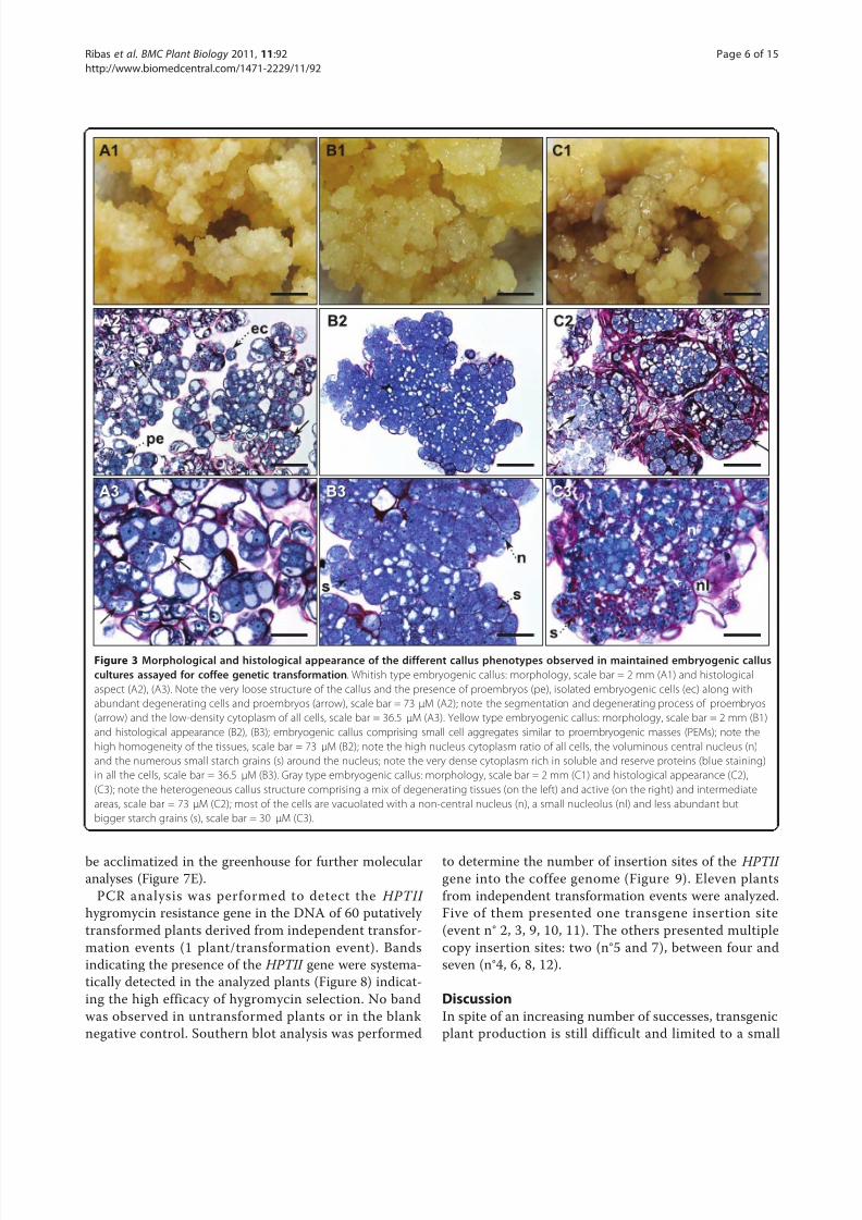
Transgenic coffee plant regeneration and molecular
analysis
Transgenic plants were regenerated using seven-month-
old embryogenic cultures of the yellow phenotype,
according to the optimum proliferation and co-culture
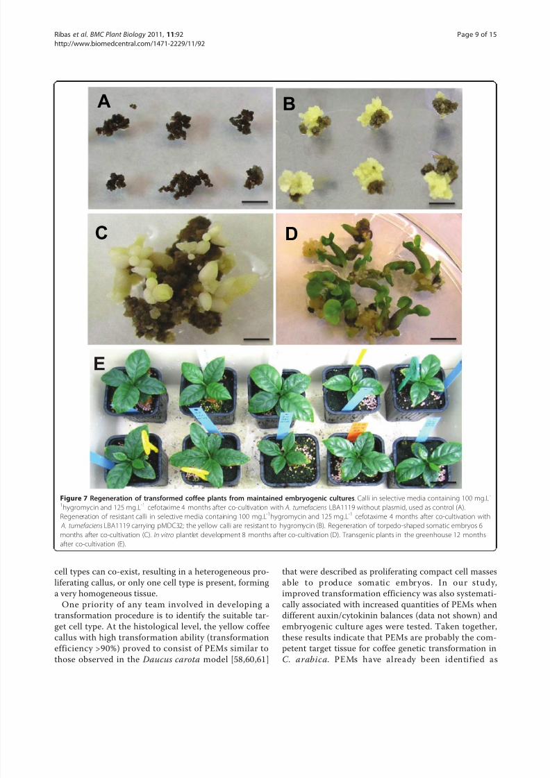
conditions established in this work. A total of 560 calli
were co-cultivated. The transfer of embryogenic calli to
hygromycin selection medium led to rapid browning of
both co-cultivated and untransformed calli used as con-
trol (Figure 7A), but two months later, a lot of resistant
yellowish calli had grown on the surface of most of the
necrotic calli (Figure 7B). After four months of hygro-
mycin selection, among the 560 co-cultivated calli, a
total of 462 produced independent yellow resistant calli
(transformation efficiency = 82.5%). All the non-trans-formed cultures (negative control) turned brown and
died during the hygromycin selection period and neither
yellow calli nor embryo regeneration was observed. Each
resistant callus line regenerated several putatively trans-
formed somatic embryos on the hygromycin-enriched
medium (Figure 7C ). Almost all resistant torpedo-
shaped mature embryos germinated and developed into
whole plantlets (Figures 7D, E). Several thousand puta-
tively transformed plants were regenerated. Among
them, 120 plants from 60 independent transformation
events (2 plants/transformation event), were chosen to
Aa
ab
bbb
6BA [μM]
B
a
aa a
a
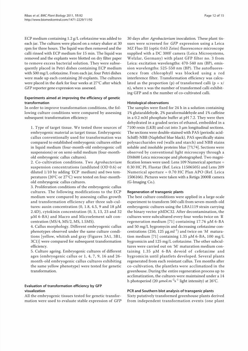
Figure 2 Effect of 6-BA and 2,4-D concentrations in the
proliferation medium on growth intensity and transformation
efficiency. All the 6-BA concentrations were tested in the presence
of 4.52 μM 2,4-D and all the 2,4-D concentrations were tested in the
presence of 12 μM 6-BA. Callus growth was measured by the
difference between the final and initial weight of embryogenic
cultures after a 4 week proliferation cycle. The initial weight was
calibrated at 120 ± 10 mg. Each datum corresponds to the mean ±
SD of 4 measurements. Confidence interval is indicated by vertical
bars. We performed an ANOVA followed by the Tukey HSD test to
identify significant differences between the means of all treatments.Values with different letters are significantly different at P ≤ 0.05.
Transformation efficiency was assessed by observing GFP
epifluorescence 6 weeks after the end of co-cultivation.
Transformation efficiency was analyzed by the proportion (p) of
transformed calli (p = x/n), where x was the number of transformed
calli and n the number of co-cultivated calli. A 3 δ confidence limit
for binomial distribution was calculated using the formula
p± 3
p(1−
p
n)
with a level of confidence of 99%. All the
transformation experiments were conducted independently in four
replicates each comprising 60 calli (240 calli/ hormonal
combination).
Ribas et al . BMC Plant Biology 2011, 11:92
http://www.biomedcentral.com/1471-2229/11/92
Page 5 of 15
8/2/2019 Kk Dila Julnal Clab
http://slidepdf.com/reader/full/kk-dila-julnal-clab 6/15
be acclimatized in the greenhouse for further molecular
analyses (Figure 7E).
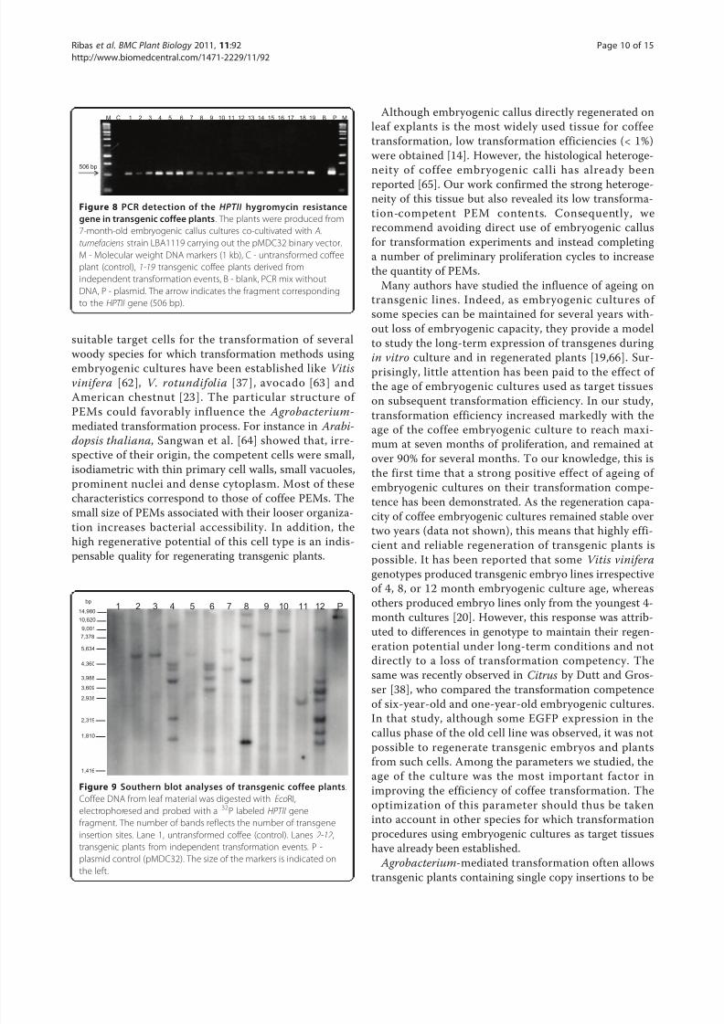
PCR analysis was performed to detect the HPTII
hygromycin resistance gene in the DNA of 60 putatively
transformed plants derived from independent transfor-
mation events (1 plant/transformation event). Bands
indicating the presence of the HPTII gene were systema-
tically detected in the analyzed plants (Figure 8) indicat-
ing the high efficacy of hygromycin selection. No band
was observed in untransformed plants or in the blank
negative control. Southern blot analysis was performed
to determine the number of insertion sites of the HPTII
gene into the coffee genome (Figure 9). Eleven plants
from independent transformation events were analyzed.
Five of them presented one transgene insertion site
(event n° 2, 3, 9, 10, 11). The others presented multiple
copy insertion sites: two (n°5 and 7), between four and
seven (n°4, 6, 8, 12).
DiscussionIn spite of an increasing number of successes, transgenic
plant production is still difficult and limited to a small
A
A
3
3
B
B
3
3
C
C
3
3
A 1 B 1 C 1
A
A
2
2 B 2 C 2
p e
e c
n
s
n l
n
s
s
Figure 3 Morphological and histological appearance of the different callus phenotypes observed in maintained embryogenic callus
cultures assayed for coffee genetic transformation. Whitish type embryogenic callus: morphology, scale bar = 2 mm (A1) and histological
aspect (A2), (A3). Note the very loose structure of the callus and the presence of proembryos (pe), isolated embryogenic cells (ec) along with
abundant degenerating cells and proembryos (arrow), scale bar = 73 μM (A2); note the segmentation and degenerating process of proembryos
(arrow) and the low-density cytoplasm of all cells, scale bar = 36.5 μM (A3). Yellow type embryogenic callus: morphology, scale bar = 2 mm (B1)
and histological appearance (B2), (B3); embryogenic callus comprising small cell aggregates similar to proembryogenic masses (PEMs); note the
high homogeneity of the tissues, scale bar = 73 μM (B2); note the high nucleus cytoplasm ratio of all cells, the voluminous central nucleus (n)
and the numerous small starch grains (s) around the nucleus; note the very dense cytoplasm rich in soluble and reserve proteins (blue staining)
in all the cells, scale bar = 36.5 μM (B3). Gray type embryogenic callus: morphology, scale bar = 2 mm (C1) and histological appearance (C2),
(C3); note the heterogeneous callus structure comprising a mix of degenerating tissues (on the left) and active (on the right) and intermediate
areas, scale bar = 73 μM (C2); most of the cells are vacuolated with a non-central nucleus (n), a small nucleolus (nl) and less abundant but
bigger starch grains (s), scale bar = 30 μM (C3).
Ribas et al . BMC Plant Biology 2011, 11:92
http://www.biomedcentral.com/1471-2229/11/92
Page 6 of 15
8/2/2019 Kk Dila Julnal Clab
http://slidepdf.com/reader/full/kk-dila-julnal-clab 7/15
number of species and genotypes. This is particularly
true of woody species, as these are often recalcitrant to
genetic transformation. Chevreau [30] reported that only
three fruit woody species were distinguished by a large
number of successfully transformed genotypes: more
than 10 species of Citrus [31], 40 genotypes of apple
tree [32], and 35 of grapevine [33]. For the majority of
species, the efficiency of the methods used remains vari-
able and weak. However, efficient methods have been
described in a number of woody crops. One category of
methods consists in directly transforming zygotic
embryos or young seedlings (epicotyls, hypocotyls) to
rapidly obtain transformed organogenic calli and derived
plantlets; this is the case in Allocasuarina [34], citrange
[35] and poplar [36]. Another category consisting in
regenerating transformants from established embryo-
genic cultures has been described in several species
including grapevine [21,37], Citrus [38], rubber tree
[22], Prunus subhirtella [18,19] and American chestnut
[23]. These procedures were shown to be efficient not
only for basic research in functional genomics aimed at
characterizing T-DNA insertion [21,39,40], the long-
term stability of transgene expression [19] or transgenesilencing [41], but also for the production of transgenic
varieties. In the last ten years, embryogenic callus
directly produced from leaf explants has been the most
widely used tissue for Agrobacterium-mediated transfor-
mation in coffee [14]. Nevertheless low transformation
frequencies were reported and, like for other perennial
crops, the availability of embryogenic tissues suitable for
genetic transformation remains one of the main bottle-
necks for developing genetic transformation strategies.
In this work, we demonstrate that established embryo-
genic cultures can be used to improve the efficiency of
coffee tree transformation. Another advantage of such
cultures is that, unlike other target tissues used for
transformation whose availability is seasonal, embryo-
genic cultures are available all year round. This means
transformation experiments can be planned at any time,
which is a prerequisite for setting up a more efficient
and faster transformation pipeline.
The reliability of GFP fluorescence to monitor trans-
formation efficiency in coffee was confirmed by a large-
scale trial aimed at regenerating fully-transformed plant-
lets using the hygromycin marker gene. In this trial,
high frequencies of hygromycin resistant calli (82.5%)
were evaluated that were similar to those of GFP expres-
sing calli (90-93%) obtained in the same transformationconditions. In a previous work on coffee, transformation
efficiency ranged from less than 1% for the recalcitrant
C. arabica [6] to 33% for C. canephora [42]. The fluor-
escent marker GFP has a major advantage compared to
other reporter genes in that it enables non-invasive
detection of transformed cells without the introduction
of co-factors or the destruction of the biological sample
[43]. Similarly, GFP visual selection was recently used as
the only marker to detect transgenic calli lines of Hevea
brasiliensis without antibiotic pressure [44]. As pre-
viously reported [7,45], highly efficient selection of
0
20
40
60
80
100
Whitish Yellow Grey
T r a n s f o r m a t i o n
e f f i c i e n c y ( % )
Embryogenic callus phenotype
Fi gu re 4 Transformation efficiency depending on the
phenotype of maintained embryogenic callus cultures.
Transformation efficiency was assessed by observing GFP
epifluorescence 6 weeks after the end of co-cultivation.
Transformation efficiency was estimated by the proportion (p) of
transformed calli (p = x/n), where x is the number of transformed
calli and n the number of co-cultivated calli. A 3δ
confidence limitfor binomial distribution was calculated using the formula
p± 3
p(1−
p
n)
with a level of confidence of 99%. Confidence
interval is indicated by vertical bars. For each callus phenotype, all
the transformation experiments were conducted independently in
three replicates comprising 40 calli (120 calli/phenotype).
0
10
20
30
40
50
60
70
80
90
100
110
0 1 4 7 9 16 26
T r a n s f o r m a t i o n e f f i c i e n c y ( % )
Embryogenic culture age (months)
Figure 5 Effect of the age of the embryogenic callus culture on
A. tumefaciens-mediated transformation efficiency.
Transformation efficiency was assayed 6 weeks after the end of co-
cultivation by GFP epifluorescence. The transformation efficiency
was estimated by the proportion (p) of transformed calli (p = x/n),
where x is the number of transformed calli and n the number of
co-cultivated calli. A 3 δ confidence limit for binomial distribution
was calculated using the formula p± 3
p(1−
p
n) with a level of
confidence of 99%. Confidence interval is indicated by vertical bars.
All the transformation experiments were conducted independently
in five to six replicates each comprising 40 calli for each culture age
(200 to 240 calli/culture age).
Ribas et al . BMC Plant Biology 2011, 11:92
http://www.biomedcentral.com/1471-2229/11/92
Page 7 of 15
8/2/2019 Kk Dila Julnal Clab
http://slidepdf.com/reader/full/kk-dila-julnal-clab 8/15
transformed coffee tissues was achieved and escapes
were prevented when the antibiotic hygromycin was
used in the four-month selection step.
It is noteworthy that in our study most progress in
improving transformation efficiency was achieved by
optimizing the production conditions of the embryo-
genic cultures used as target tissues for transformation
rather than by optimizing the physical co-cultivation
conditions. The positive effect on genetic transformation
of reducing the strength of mineral salts in the prolifera-
tion medium of coffee embryogenic cultures (MS/2 orMS/4) is consistent with reports in the literature.
Although it has been widely demonstrated that ions are
involved in bacterial attachment to plants [46], transfor-
mation has been successfully achieved in numerous spe-
cies by using half-strength or even more diluted salt
solution media or solutions that specifically lack certain
salts such as CaCl2 in the pre-culture medium [47] or
co-cultivation medium [48,49]. It has been shown that
during long-term culture, plant cells may lose the need
for auxin and/or cytokinin to maintain active growth.
This process known as ‘habituation’, which is common
in callus cultures in some plant species such as sugar-
beet [50], was described as a shift from auxo- to auto-trophic state for growth regulator requirements [51].
Coffee embryogenic cultures do not require auxin or
cytokinin to proliferate, although it was possible to sti-
mulate proliferation by optimum exogenous concentra-
tions of both growth regulators. Similarly, genetic
transformation of long-term cultures was possible with-
out auxin or cytokinin. Our results specifically revealed
a very negative effect of cytokinin and a positive effect
of auxin in optimizing transformation ability. Blanc et
al. [22] showed that simultaneously increasing the auxin
and cytokinin supply in the pre-culture medium prior to
transformation of rubber tree using embryogenic cul-
tures stimulated the development of active and fast
growing cells , hence improving transformation
efficiency.
Coffee embryogenic callus cultures were successfully
established by sub-culturing the recently formed active
embryogenic tissues every four weeks. It is widely
acknowledged that a lack of regular subcultures leads
embryogenic calli to lose their embryogenic potentialdue to cell ageing. Applying short subcultures (14-21
days) on a maintenance (i.e. proliferation) medium is an
essential step in establishing long-term cultures in many
perennial crops [52-56]. The process of establishing cof-
fee embryogenic cultures leads to the development and
maintenance of three morphologically different callus
phenotypes with highly contrasted potential for genetic
transformation. Several morphological variants with con-
trasted transformation potential have also been observed
during the proliferation and maintenance of embryo-
genic calli in cotton [57]. Andrade et al. [23] highlighted
great variability in the way in which embryogenic cul-
tures proliferate and that this variability should be takeninto account as it strongly affects further genetic trans-
formation. These authors described embryogenic culture
proliferation as a continuum along a developmental gra-
dient from undifferentiated embryogenic callus through
slightly more differentiated proembryogenic masses or
PEMs [58] to repetitive embryogenesis at the globular
embryo stage or even later [59]. Our histological studies
in coffee showed that, depending the callus phenotype
or the age of the embryogenic culture, either distinct
Figure 6 Histological appearance of coffee embryogenic callus cultures as a function of the age of the culture . Primary embryogenic
callus produced on leaf explants; note the heterogeneous structure where degenerating (arrow) and active areas co-exist; the cells are
characterized by a small non-central nucleus and low starch and cytoplasm soluble protein contents, scale bar = 36.5 μM (A). Seven-month-old
embryogenic callus culture; note the highly homogeneous structure mainly comprising proembryogenic masses (PEMs) whose cells exhibit a
high nucleus cytoplasm ratio and dense cytoplasm rich in soluble and reserve proteins, scale bar = 73 μM (B). Twenty-six month-old
embryogenic callus culture; note the heterogeneous appearance of the mix of isolated cells, degenerating areas (arrow), masses oriented
towards somatic embryogenesis (ma), along with areas of cells with protein rich cytoplasm (PEMs) similar to those in younger embryogenic
cultures, scale bar = 73 μM (C).
Ribas et al . BMC Plant Biology 2011, 11:92
http://www.biomedcentral.com/1471-2229/11/92
Page 8 of 15
8/2/2019 Kk Dila Julnal Clab
http://slidepdf.com/reader/full/kk-dila-julnal-clab 9/15
cell types can co-exist, resulting in a heterogeneous pro-
liferating callus, or only one cell type is present, forming
a very homogeneous tissue.
One priority of any team involved in developing a
transformation procedure is to identify the suitable tar-
get cell type. At the histological level, the yellow coffee
callus with high transformation ability (transformation
efficiency >90%) proved to consist of PEMs similar to
those observed in the Daucus carota model [58,60,61]
that were described as proliferating compact cell masses
able to produce somatic embryos. In our study,
improved transformation efficiency was also systemati-
cally associated with increased quantities of PEMs when
different auxin/cytokinin balances (data not shown) and
embryogenic culture ages were tested. Taken together,
these results indicate that PEMs are probably the com-
petent target tissue for coffee genetic transformation in
C. arabica. PEMs have already been identif ied as
A
C
B
D
E
Figure 7 Regeneration of transformed coffee plants from maintained embryogenic cultures. Calli in selective media containing 100 mg.L-
1hygromycin and 125 mg.L-1 cefotaxime 4 months after co-cultivation with A. tumefaciens LBA1119 without plasmid, used as control (A).
Regeneration of resistant calli in selective media containing 100 mg.L-1hygromycin and 125 mg.L-1 cefotaxime 4 months after co-cultivation with
A. tumefaciens LBA1119 carrying pMDC32; the yellow calli are resistant to hygromycin (B). Regeneration of torpedo-shaped somatic embryos 6
months after co-cultivation (C). In vitro plantlet development 8 months after co-cultivation (D). Transgenic plants in the greenhouse 12 months
after co-cultivation (E).
Ribas et al . BMC Plant Biology 2011, 11:92
http://www.biomedcentral.com/1471-2229/11/92
Page 9 of 15
8/2/2019 Kk Dila Julnal Clab
http://slidepdf.com/reader/full/kk-dila-julnal-clab 10/15
suitable target cells for the transformation of several
woody species for which transformation methods usingembryogenic cultures have been established like Vitis
vinifera [62], V. rotundifolia [37], avocado [63] and
American chestnut [23]. The particular structure of
PEMs could favorably influence the Agrobacterium-
mediated transformation process. For instance in Arabi-
dopsis thaliana, Sangwan et al. [64] showed that, irre-
spective of their origin, the competent cells were small,
isodiametric with thin primary cell walls, small vacuoles,
prominent nuclei and dense cytoplasm. Most of these
characteristics correspond to those of coffee PEMs. The
small size of PEMs associated with their looser organiza-
tion increases bacterial accessibility. In addition, the
high regenerative potential of this cell type is an indis-pensable quality for regenerating transgenic plants.
Although embryogenic callus directly regenerated on
leaf explants is the most widely used tissue for coffee
transformation, low transformation efficiencies (< 1%)
were obtained [14]. However, the histological heteroge-
neity of coffee embryogenic calli has already been
reported [65]. Our work confirmed the strong heteroge-
neity of this tissue but also revealed its low transforma-
tion-competent PEM contents. Consequently, we
recommend avoiding direct use of embryogenic callus
for transformation experiments and instead completing
a number of preliminary proliferation cycles to increase
the quantity of PEMs.
Many authors have studied the influence of ageing on
transgenic lines. Indeed, as embryogenic cultures of
some species can be maintained for several years with-
out loss of embryogenic capacity, they provide a model
to study the long-term expression of transgenes during
in vitro culture and in regenerated plants [19,66]. Sur-prisingly, little attention has been paid to the effect of
the age of embryogenic cultures used as target tissues
on subsequent transformation efficiency. In our study,
transformation efficiency increased markedly with the
age of the coffee embryogenic culture to reach maxi-
mum at seven months of proliferation, and remained at
over 90% for several months. To our knowledge, this is
the first time that a strong positive effect of ageing of
embryogenic cultures on their transformation compe-
tence has been demonstrated. As the regeneration capa-
city of coffee embryogenic cultures remained stable over
two years (data not shown), this means that highly effi-
cient and reliable regeneration of transgenic plants is
possible. It has been reported that some Vitis vinifera
genotypes produced transgenic embryo lines irrespective
of 4, 8, or 12 month embryogenic culture age, whereas
others produced embryo lines only from the youngest 4-
month cultures [20]. However, this response was attrib-
uted to differences in genotype to maintain their regen-
eration potential under long-term conditions and not
directly to a loss of transformation competency. The
same was recently observed in Citrus by Dutt and Gros-
ser [38], who compared the transformation competence
of six-year-old and one-year-old embryogenic cultures.
In that study, although some EGFP expression in thecallus phase of the old cell line was observed, it was not
possible to regenerate transgenic embryos and plants
from such cells. Among the parameters we studied, the
age of the culture was the most important factor in
improving the efficiency of coffee transformation. The
optimization of this parameter should thus be taken
into account in other species for which transformation
procedures using embryogenic cultures as target tissues
have already been established.
Agrobacterium-mediated transformation often allows
transgenic plants containing single copy insertions to be
M C 1 2 3 4 5 6 7 8 9 10 11 12 13 14 15 16 17 18 19 B P M
506 bp
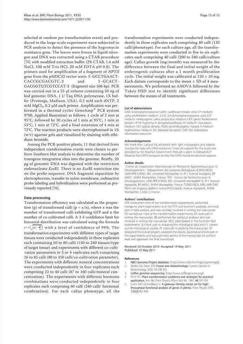
Figure 8 PCR detection of the HPTII hygromycin resistance
gene in transgenic coffee plants. The plants were produced from
7-month-old embryogenic callus cultures co-cultivated with A.
tumefaciens strain LBA1119 carrying out the pMDC32 binary vector.
M - Molecular weight DNA markers (1 kb), C - untransformed coffee
plant (control), 1-19 transgenic coffee plants derived from
independent transformation events, B - blank, PCR mix without
DNA, P - plasmid. The arrow indicates the fragment corresponding
to the HPTII gene (506 bp).
1 2 3 4 5 6 7 8 9 10 11 12 P
7,378
3,988
3,609
2,938
2,319
1,810
1,416
5,634
4,360
bp
9,001
10,620
14,980
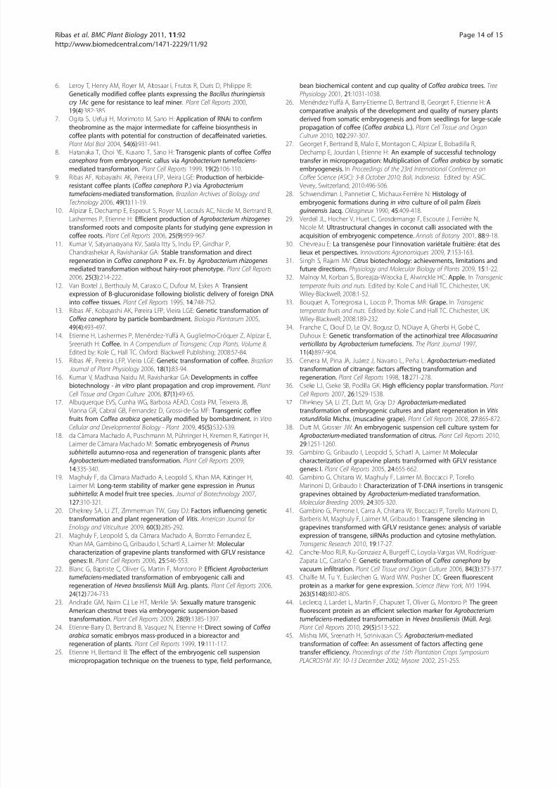
Figure 9 Southern blot analyses of transgenic coffee plants.
Coffee DNA from leaf material was digested with EcoRI,
electrophoresed and probed with a 32P labeled HPTII gene
fragment. The number of bands reflects the number of transgene
insertion sites. Lane 1, untransformed coffee (control). Lanes 2-12,
transgenic plants from independent transformation events. P -
plasmid control (pMDC32). The size of the markers is indicated on
the left.
Ribas et al . BMC Plant Biology 2011, 11:92
http://www.biomedcentral.com/1471-2229/11/92
Page 10 of 15
8/2/2019 Kk Dila Julnal Clab
http://slidepdf.com/reader/full/kk-dila-julnal-clab 11/15
obtained [66]. Low transgene copy numbers have
recently been reported in transformants derived from
embryogenic cultures in several trees [21,23,38]. Simi-
larly, our results in coffee showed that all the selected
plants had T-DNA integrated in their genome and that
almost half contained single transgene insertion sites.
These results are consistent with previous observations
in coffee plants transformed via A. tumefaciens with
from one to five inserted T-DNA copies, of which 69%
harbored one T-DNA copy [6]. It has been shown that
transgene expression is influenced by the number of T-
DNA copies and/or the integration site [67]. Even if
inactivation of transgene expression can occur in plants
with a single copy [68 ], this phenomenon is less fre-
quent than with multiple transgene copies [69,70]. For
this reason, the regeneration of a high proportion of cof-
fee plants with a low copy number of the inserted T-
DNA is important for the application of this transforma-tion technology for both genomic purposes and breed-
ing programs.
ConclusionsThe availability of an efficient and reliable transforma-
tion procedure in coffee tree is very important given the
opportunities that will be soon available in functional
genomics thanks to the genome sequencing of C. cane-
phora. Moreover, in a perennial species like coffee in
which selection takes a very long time, the use of
genetic transformation alongside conventional breeding
techniques will enable the introduction of agronomically
useful traits in the best cultivated varieties in only one
step. In this context, it was important to establish trans-
formation procedures based on the use of target tissues
taken from the mature tissues of elite varieties. This is
the case of the process developed in the present work
that will enable the routine transformation of embryo-
genic cultures derived from leaves selected on the
mother tree and the subsequent regeneration of large
quantities of transformants from numerous independent
transformation events.
Methods
Plant materialAll the studies were conducted using the genotype C.
arabica var. Caturra. The leaf explants were collected
from trees maintained in a greenhouse located at IRD
(Montpellier, France). Embryogenic calli were induced
as previously described [71]. Briefly, 1 cm2 pieces of
young leaves from the mother tree were surface steri-
lized and used as explants. Leaves were disinfected by
immersion in 10% calcium hypochlorite solution con-
taining 1% Tween 80 for 20 min followed by 8% calcium
hypochlorite solution for 10 min and rinsed four times
with sterile water. The explants were cultured for one
month on MS/2 [72] ‘C’ callogenesis medium containing
2.26 μM 2,4-dichlorophenoxyacetic acid (2,4-D), 4.92
μM indole-3-butyric acid (IBA) and 9.84 μM isopente-
nyladenine (iP) allowing the production of primary cal-
lus comprising cytoplasmic meristematic cells. The leaf
explants were then transferred to MS/2 ‘ECP’ embryo-
genic callus production medium containing 4.52 μM
2,4-D and 17.76 μM 6-benzylaminopurine (6-BA) for 6-
8 months until regeneration of embryogenic callus. Yel-
low embryogenic callus appeared on the necrotic pri-
mary callus. Embryogenic callus was induced in baby
food jars at 27°C in the dark. All the media used in this
work were supplemented with 30 g/L sucrose and their
pH was adjusted to 5.7 prior to the addition of 2.8 g/L
phytagel. The media were autoclaved at 120°C and 1.1
kg/cm2 for 20 min, and then 25 ml of medium was
added to each 100 × 15 mm Petri dish or baby food jar.
To establish the embryogenic cell suspension, embryo-genic calli were transferred to 250 ml Erlenmeyer flasks
at a density of 1 g/L in MS/2 ‘CP’ liquid proliferation
medium [71] with 4.52 μM 2.4-D and 4.65 μM kinetin,
and shaken at 100 rpm at 27 °C in the dark. Suspension
cultures of embryogenic cell aggregates were generally
established after three months under such conditions.
Establishment of embryogenic callus cultures: long-
term embryogenic cultures were successfully established
on semi-solid medium using the embryogenic callus and
by transferring the yellowish fragments collected from
the upper part of embryogenic calli to fresh semi-solid
‘ECP’ MS/2 embryogenic callus production medium [71]
with 4.5 μM 2.4D and 12 μM 6-BA solidified with 2.8 g/
L phytagel once a month. The cultures were kept in
baby food jars at 27°C in the dark.
Agrobacterium strain and binary vector
The disarmed strain of A. tumefaciens LBA1119 carrying a
binary vector (pBIN35SGFP) containing the reporter gene
GFP5 coding for green fluorescent protein under control
of the constitutive cauliflower mosaic virus (CaMV) 35S
promoter was used for all experiments aimed at designing
a transformation protocol. The LBA1119 strain carrying
the binary vector pMDC32 [5] containing the HPTII gene
that confers hygromycin resistance was used to regeneratetransgenic plants and to validate the transformation proto-
col in the same culture conditions.
Co-cultivation and decontamination
Coffee explants were maintained in their culture container,
i.e. baby food jars, and immersed in 10 ml of A. tumefa-
ciens suspension (OD600 = 0.6) for 10 min without shak-
ing. The bacterial suspension was removed and the
inoculated explants were co-cultivated at 20 °C for five
days in the dark. After this period, the explants were
rinsed twice with 20 ml sterile water after which 20 ml of
Ribas et al . BMC Plant Biology 2011, 11:92
http://www.biomedcentral.com/1471-2229/11/92
Page 11 of 15
8/2/2019 Kk Dila Julnal Clab
http://slidepdf.com/reader/full/kk-dila-julnal-clab 12/15
ECP medium containing 1.2 g/L cefotaxime was added to
each jar. The cultures were placed on a rotary shaker at 30
rpm for three hours. The liquid was then removed and the
calli rinsed with ECP medium for 15 min. The liquid was
removed and the explants were blotted on dry filter paper
to remove excess bacterial solution. They were subse-
quently placed in Petri dishes containing ECP medium
with 500 mg/L cefotaxime. From each jar, four Petri dishes
were made up each containing 20 explants. The cultures
were placed in the dark for four weeks at 27°C after which
GFP reporter gene expression was assessed.
Experiments aimed at improving the efficiency of genetic
transformation
In order to improve transformation conditions, the fol-
lowing culture conditions were compared by assessing
subsequent transformation efficiency:
1. Type of target tissue. We tested three sources of
embryogenic material as target tissue. Embryogenic
callus conventionally used for transformation was
compared to established embryogenic cultures either
in liquid medium (four-month-old embryogenic cell
suspensions) or on semi-solid medium (four-month-
old embryogenic callus cultures).
2. Co-cultivation conditions. Two Agrobacterium
suspension concentrations (undiluted (OD 0.6) or
diluted 1/10 by adding ‘ECP’ medium) and two tem-
peratures (20°C or 27°C) were tested on four-month-
old embryogenic callus cultures.
3. Proliferation conditions of the embryogenic callus
cultures. The following modifications to the ECP
medium were compared by assessing callus growth
and transformation efficiency after three sub-cul-
tures: auxin concentration (0, 1.8, 4.5, 9 and 18 μM
2.4D), cytokinin concentration (0, 3, 13, 23 and 32
μM 6-BA) and Macro and Microelement salt con-
centration (MS/4, MS/2, MS, 1.5MS).
4. Callus morphology. Different embryogenic callus
phenotypes observed under the same culture condi-
tions [yellow, whitish and gray (Figures 3A1, 3B1,
3C1)] were compared for subsequent transformation
efficiency.5. Culture ageing. Embryogenic cultures of different
ages (embryogenic callus or 1, 4, 7, 9, 16 and 26-
month-old embryogenic callus cultures exhibiting
the same yellow phenotype) were tested for genetic
transformation.
Evaluation of transformation efficiency by GFP
visualization
All the embryogenic tissues tested for genetic transfor-
mation were used to evaluate stable expression of GFP
30 days after Agrobacterium inoculation. These plant tis-
sues were screened for GFP expression using a Leica
MZ Fluo III (optic 0.63 Zeiss) fluorescence microscope
supplied with a DC 300F camera (Leica Microsystems,
Welzlar, Germany) with plant GFP filter no. 3 from
Leica: excitation wavelengths: 470-540 nm (BP), emis-
sion wavelengths: 525-550 nm (BP). The autofluores-
cence from chlorophyll was blocked using a red
interference filter. Transformation efficiency was calcu-
lated as the proportion (p) of transformed calli (p = x/
n), where x was the number of transformed calli exhibit-
ing GFP and n the number of co-cultivated calli.
Histological observations
The samples were fixed for 24 h in a solution containing
1% glutaraldehyde, 2% paraformaldehyde and 1% caffeine
in a 0.2 mM phosphate buffer at pH 7.2. They were then
dehydrated in a graded series of ethanol, embedded in a7100 resin (LKB) and cut into 3 μm longitudinal sections.
The sections were double-stained with PAS (periodic acid-
Schiff)-NBB (Naphthol blue black). PAS specifically stains
polysaccharides red (walls and starch) and NBB stains
soluble and insoluble proteins blue [73,74]. Sections were
observed by conventional light microscopy through a
DM600 Leica microscope and photographed. Two magni-
fication lenses were used: Lens 109 Numerical aperture =
0.30 HC PL Fluotar (Ref. Leica 11506505) and Lens 209
Numerical aperture = 0.70 HC Plan APO (Ref. Leica
1506166). Pictures were taken with a Retiga 2000R camera
(G-Imaging Co.).
Regeneration of transgenic plants
The best culture conditions were applied in a large-scale
experiment to transform 560 calli from seven-month-old
embryogenic cultures using the LBA1119 strain carrying
the binary vector pMDC32. After decontamination, the
cultures were subcultured every four weeks twice on ‘R’
regeneration medium [71] containing 17.76 μM 6-BA
and 50 mg/L hygromycin and decreasing cefotaxime con-
centrations (250, 125 μg.ml-1) and twice on ‘M’ matura-
tion medium [71] containing 1.35 μM 6-BA, 100 mg/L
hygromicin and 125 mg/L cefotaxime. The other subcul-
tures were carried out on ‘M’ maturation medium con-taining 1.35 μM 6-BA devoid of cefotaxime and
hygromicin until plantlets developed. Several plants
regenerated from each resistant callus. Ten months after
co-cultivation, the plantlets were acclimatized in the
greenhouse. During the entire regeneration process up to
acclimatization, the cultures were maintained under a 14
h photoperiod (20 μmol.m-2s-1 light intensity) at 26°C.
PCR and Southern blot analysis of transgenic plants
Sixty putatively transformed greenhouse plants derived
from independent transformation events (one plant
Ribas et al . BMC Plant Biology 2011, 11:92
http://www.biomedcentral.com/1471-2229/11/92
Page 12 of 15
8/2/2019 Kk Dila Julnal Clab
http://slidepdf.com/reader/full/kk-dila-julnal-clab 13/15
selected at random per transformation event) and pro-
duced in the large-scale experiment were subjected to
PCR analysis to detect the presence of the hygromycin
resistance gene. The leaves were frozen in liquid nitro-
gen and DNA was extracted using a CTAB procedure
[75] with modified extraction buffer (2% CTAB, 1.4 mM
NaCl, 100 mM Tris HCl, 20 mM EDTA pH 0.8). The
primers used for amplification of a fragment of HPTII
gene from the pMDC32 vector were: 5’-GCCTGAACT-
CACCGCGACGTC-3’ and 5’-GCACT-
GACGGTGTCGTCCAT-3’ (fragment size 506 bp). PCR
was carried out in a 25 μl volume containing 50 ng of
leaf genomic DNA, 1 U Taq DNA polymerase, 1X buf-
fer (Promega, Madison, USA), 0.2 mM each dNTP, 2
mM MgCl2, 0.2 μM each primer. Amplification was per-
formed in a thermal cycler GeneAmp® PCR system
9700, Applied Biosystem) as follows: 1 cycle of 2 min at
95°C, followed by 30 cycles of 1 min at 95°C, 1 min at55°C, 1 min at 72°C, and a final extension of 4 min at
72°C. The reaction products were electrophoresed in 1%
(w/v) agarose gels and visualized by staining with ethi-
dium bromide.
Among the PCR-positive plants, 11 that derived from
independent transformation events were chosen to per-
form Southern blot analysis to determine the number of
transgene integration sites into the genome. Briefly, 20
μg of genomic DNA was digested with the restriction
endonuclease EcoRI. There is no EcoRI restriction site
on the probe sequence. DNA fragment separation by
electrophoresis, transfer to nylon membrane, radioactive
probe labeling and hybridization were performed as pre-
viously reported [76].
Data processing
Transformation efficiency was calculated as the propor-
tion (p) of transformed calli (p = x/n), where x was the
number of transformed calli exhibiting GFP and n the
number of co-cultivated calli. A 3 δ confidence limit for
binomial distribution was calculated using the formula
p± 3
p(1−
p
n) with a level of confidence of 99%. The
transformation experiments with different types of target
tissues were conducted independently in three replicates
each containing 50 to 80 calli (150 to 240 tissues/typeof target tissue) and experiments with different co-culti-
vation parameters in 3 or 4 replicates each comprising
26 to 85 calli (80 to 350 calli/co-cultivation parameter).
The experiments with different mineral concentrations
were conducted independently in four replicates each
comprising 22 to 40 calli (87 to 160 calli/mineral con-
centration). The experiments with different hormone
combinations were conducted independently in four
replicates each comprising 60 calli (240 calli/ hormonal
combination). For each callus phenotype, all the
transformation experiments were conducted indepen-
dently in three replicates each comprising 40 calli (120
calli/phenotype). For each culture age, all the transfor-
mation experiments were conducted in five to six repli-
cates each comprising 40 calli (200 to 240 calli/culture
age). Callus growth (mg/month) was measured by the
difference between the final and initial weight of the
embryogenic cultures after a 1 month proliferation
cycle. The initial weight was calibrated at 120 ± 10 mg.
Each datum corresponds to the mean ± SD of 4 mea-
surements. We performed an ANOVA followed by the
Tukey HSD test to identify significant differences
between the means of all treatments.
List of abbreviations
6-BA: 6-benzylaminopurine; CaMV: cauliflower mosaic virus; CP medium:
callus proliferation medium; 2,4-D: 2,4-dichlorophenoxyacetic acid; ECP
medium: embryogenic callus production medium; GFP: green fluorescenceprotein; HPTII: hygromycin phosphotransferase; M medium: maturation
medium: OD: optical density; PEMs: proembryogenic masses; R medium:
regeneration medium; SD: standard deviation; TAIR: the Arabidopsis
information resources.
Acknowledgements
We thank Marc Lartaud for assistance with light micrographs and Isabelle
Jourdan for help with DNA extraction. Financial support for this study wasprovided by the Brazilian Government through a grant to Alessandra F.
Ribas by the CAPES program and by the CIRAD funds for doctoral support.
Author details1Centre de Coopération Internationale en Recherche Agronomique pour le
Développement - Département des Systèmes Biologiques (CIRAD-BIOS).
UMR-RPB (CIRAD, IRD, Université Montpellier II), 911 Avenue Agropolis, BP
64501, 34394 Montpellier, France. 2IRD - Institut de Recherche pour le
Développement, UMR RPB (CIRAD, IRD, Université Montpellier II), 911 AvenueAgropolis, BP 64501, 34394 Montpellier, France. 3CIRAD-BIOS, MRI, UMR-DAP,
Plant cell imaging platform (www.PHIV.cirad.fr), Avenue Agropolis, 34398Montpellier, Cedex 5, France.
Authors’ contributionsAFR conducted most of the transformation experiments, performed
transgenic plant regeneration and the PCR and Southern analyses, and took
part in data analysis, and was centrally involved in writing the manuscript.
ED carried out most of the transformation experiments. AC took part in
writing the manuscript. BB performed the statistical analyses and was
involved in writing the manuscript. MCC participated in the Southern blot
experiments. JLV took part in analyzing the histological data and FL carried
out the histological studies. PL took part in drafting the manuscript. HE
designed the overall project, analyzed the results, planned and took part in
the experiments, and was a primary author of the manuscript. All authors
read and approved the final manuscript.
Received: 20 October 2010 Accepted: 19 May 2011
Published: 19 May 2011
References
1. NBCI Genome Project database. [http://www.ncbi.nlm.nih.gov/genomeprj].
2. Merkle SA, Dean JFD: Forest tree biotechnology. Current Opinion in
Biotechnology 2000, 11:298-302.
3. Coffee genome sequencing. [http://www.coffeegenome.org/ ].
4. Birch RG: Plant transformation: problems and strategies for practical
application. Ann Rev Plant Physiol 0Plant Mol Bio 1997, 48:297-326.
5. Curtis MD, Grossniklaus U: A gateway cloning vector set for high-throughput functional analysis of genes in planta. Plant Physiol 2003,
133(2):462-469.
Ribas et al . BMC Plant Biology 2011, 11:92
http://www.biomedcentral.com/1471-2229/11/92
Page 13 of 15
8/2/2019 Kk Dila Julnal Clab
http://slidepdf.com/reader/full/kk-dila-julnal-clab 14/15
6. Leroy T, Henry AM, Royer M, Altosaar I, Frutos R, Duris D, Philippe R:
Genetically modified coffee plants expressing the Bacillus thuringiensis
cry 1Ac gene for resistance to leaf miner. Plant Cell Reports 2000,
19(4):382-385.7. Ogita S, Uefuji H, Morimoto M, Sano H: Application of RNAi to confirm
theobromine as the major intermediate for caffeine biosynthesis in
coffee plants with potential for construction of decaffeinated varieties.Plant Mol Biol 2004, 54(6):931-941.8. Hatanaka T, Choi YE, Kusano T, Sano H: Transgenic plants of coffee Coffea
canephora from embryogenic callus via Agrobacterium tumefaciens-
mediated transformation. Plant Cell Reports 1999, 19(2):106-110.
9. Ribas AF, Kobayashi AK, Pereira LFP, Vieira LGE: Production of herbicide-
resistant coffee plants (Coffea canephora P.) via Agrobacterium
tumefaciens-mediated transformation. Brazilian Archives of Biology and
Technology 2006, 49(1):11-19.
10. Alpizar E, Dechamp E, Espeout S, Royer M, Lecouls AC, Nicole M, Bertrand B,
Lashermes P, Etienne H: Efficient production of Agrobacterium rhizogenes-
transformed roots and composite plants for studying gene expression in
coffee roots. Plant Cell Reports 2006, 25(9):959-967.
11. Kumar V, Satyanarayana KV, Sarala Itty S, Indu EP, Giridhar P,
Chandrashekar A, Ravishankar GA: Stable transformation and direct
regeneration in Coffea canephora P ex. Fr. by Agrobacterium rhizogenesmediated transformation without hairy-root phenotype. Plant Cell Reports
2006, 25(3):214-222.12. Van Boxtel J, Berthouly M, Carasco C, Dufour M, Eskes A: Transient
expression of B-glucuronidase following biolistic delivery of foreign DNA
into coffee tissues. Plant Cell Reports 1995, 14:748-752.
13. Ribas AF, Kobayashi AK, Pereira LFP, Vieira LGE: Genetic transformation of
Coffea canephora by particle bombardment. Biologia Plantarum 2005,
49(4):493-497.
14. Etienne H, Lashermes P, Menéndez-Yuffá A, Guglielmo-Cróquer Z, Alpizar E,
Sreenath H: Coffee. In A Compendium of Transgenic Crop Plants. Volume 8.
Edited by: Kole C, Hall TC. Oxford: Blackwell Publishing; 2008:57-84.
15. Ribas AF, Pereira LFP, Vieira LGE: Genetic transformation of coffee. Brazilian
Journal of Plant Physiology 2006, 18(1):83-94.
16. Kumar V, Madhava Naidu M, Ravishankar GA: Developments in coffeebiotechnology - in vitro plant propagation and crop improvement. Plant
Cell Tissue and Organ Culture 2006, 87(1):49-65.
17. Albuquerque EVS, Cunha WG, Barbosa AEAD, Costa PM, Teixeira JB,
Vianna GR, Cabral GB, Fernandez D, Grossi-de-Sa MF: Transgenic coffeefruits from Coffea arabica genetically modified by bombardment. In Vitro
Cellular and Developmental Biology - Plant 2009, 45(5):532-539.
18. da Câmara Machado A, Puschmann M, Pühringer H, Kremen R, Katinger H,
Laimer de Câmara Machado M: Somatic embryogenesis of Prunus
subhirtella autumno-rosa and regeneration of transgenic plants after
Agrobacterium-mediated transformation. Plant Cell Reports 2009,
14:335-340.
19. Maghuly F, da Câmara Machado A, Leopold S, Khan MA, Katinger H,
Laimer M: Long-term stability of marker gene expression in Prunussubhirtella: A model fruit tree species. Journal of Biotechnology 2007,
127:310-321.
20. Dhekney SA, Li ZT, Zimmerman TW, Gray DJ: Factors influencing genetictransformation and plant regeneration of Vitis. American Journal for
Enology and Viticulture 2009, 60(3):285-292.
21. Maghuly F, Leopold S, da Câmara Machado A, Borroto Fernandez E,
Khan MA, Gambino G, Gribaudo I, Schartl A, Laimer M: Molecular
characterization of grapevine plants transformed with GFLV resistancegenes: II. Plant Cell Reports 2006, 25:546-553.
22. Blanc G, Baptiste C, Oliver G, Martin F, Montoro P: Efficient Agrobacterium
tumefaciens-mediated transformation of embryogenic calli and
regeneration of Hevea brasiliensis Müll Arg. plants. Plant Cell Reports 2006,
24(12):724-733.
23. Andrade GM, Nairn CJ, Le HT, Merkle SA: Sexually mature transgenic
American chestnut trees via embryogenic suspension-based
transformation. Plant Cell Reports 2009, 28(9):1385-1397.
24. Etienne-Barry D, Bertrand B, Vasquez N, Etienne H: Direct sowing of Coffeaarabica somatic embryos mass-produced in a bioreactor and
regeneration of plants. Plant Cell Reports 1999, 19:111-117.
25. Etienne H, Bertrand B: The effect of the embryogenic cell suspension
micropropagation technique on the trueness to type, field performance,
bean biochemical content and cup quality of Coffea arabica trees. Tree
Physiology 2001, 21:1031-1038.
26. Menéndez-Yuffá A, Barry-Etienne D, Bertrand B, Georget F, Etienne H: A
comparative analysis of the development and quality of nursery plants
derived from somatic embryogenesis and from seedlings for large-scale
propagation of coffee (Coffea arabica L.). Plant Cell Tissue and Organ
Culture 2010, 102:297-307.27. Georget F, Bertrand B, Malo E, Montagon C, Alpizar E, Bobadilla R,
Dechamp E, Jourdan I, Etienne H: An example of successful technology
transfer in micropropagation: Multiplication of Coffea arabica by somatic
embryogenesis. In Proceedings of the 23rd International Conference on
Coffee Science (ASIC): 3-8 October 2010; Bali, Indonesia. Edited by: ASIC.
Vevey, Switzerland; 2010:496-506.
28. Schwendiman J, Pannetier C, Michaux-Ferrière N: Histology of
embryogenic formations during in vitro culture of oil palm Elaeis
guineensis Jacq. Oléagineux 1990, 45:409-418.
29. Verdeil JL, Hocher V, Huet C, Grosdemange F, Escoute J, Ferrière N,
Nicole M: Ultrastructural changes in coconut calli associated with the
acquisition of embryogenic competence. Annals of Botany 2001, 88:9-18.
30. Chevreau E: La transgenèse pour l’innovation variétale fruitière: état des
lieux et perspectives. Innovations Agronomiques 2009, 7:153-163.
31. Singh S, Rajam MV: Citrus biotechnology: achievements, limitations andfuture directions. Physiology and Molecular Biology of Plants 2009, 15:1-22.
32. Malnoy M, Korban S, Boreajza-Wisocka E, Alwinckle HC: Apple. In Transgenic temperate fruits and nuts. Edited by: Kole C and Hall TC. Chichester, UK:Wiley-Blackwell; 2008:1-52.
33. Bouquet A, Torregrossa L, Locco P, Thomas MR: Grape. In Transgenic
temperate fruits and nuts. Edited by: Kole C and Hall TC. Chichester, UK:
Wiley-Blackwell; 2008:189-232.
34. Franche C, Diouf D, Le QV, Bogusz D, N’Diaye A, Gherbi H, Gobé C,
Duhoux E: Genetic transformation of the actinorhizal tree Allocasuarina
verticillata by Agrobacterium tumefaciens. The Plant Journal 1997,
11(4):897-904.
35. Cervera M, Pina JA, Juárez J, Navarro L, Peña L: Agrobacterium-mediated
transformation of citrange: factors affecting transformation and
regeneration. Plant Cell Reports 1998, 18:271-278.36. Cseke LJ, Cseke SB, Podilla GK: High efficiency poplar transformation. Plant
Cell Reports 2007, 26:1529-1538.
37. Dhekney SA, Li ZT, Dutt M, Gray DJ: Agrobacterium-mediated
transformation of embryogenic cultures and plant regeneration in Vitisrotundifolia Michx. (muscadine grape). Plant Cell Reports 2008, 27:865-872.
38. Dutt M, Grosser JW: An embryogenic suspension cell culture system for
Agrobacterium-mediated transformation of citrus. Plant Cell Reports 2010,
29:1251-1260.
39. Gambino G, Gribaudo I, Leopold S, Schartl A, Laimer M: Molecular
characterization of grapevine plants transformed with GFLV resistance
genes: I. Plant Cell Reports 2005, 24:655-662.
40. Gambino G, Chitarra W, Maghuly F, Laimer M, Boccacci P, Torello
Marinoni D, Gribaudo I: Characterization of T-DNA insertions in transgenicgrapevines obtained by Agrobacterium-mediated transformation.
Molecular Breeding 2009, 24:305-320.
41. Gambino G, Perrone I, Carra A, Chitarra W, Boccacci P, Torello Marinoni D,Barberis M, Maghuly F, Laimer M, Gribaudo I: Transgene silencing in
grapevines transformed with GFLV resistance genes: analysis of variable
expression of transgene, siRNAs production and cytosine methylation.
Transgenic Research 2010, 19:17-27.
42. Canche-Moo RLR, Ku-Gonzalez A, Burgeff C, Loyola-Vargas VM, Rodríguez-Zapata LC, Castaño E: Genetic transformation of Coffea canephora by
vacuum infiltration. Plant Cell Tissue and Organ Culture 2006, 84(3):373-377.
43. Chalfie M, Tu Y, Euskirchen G, Ward WW, Prasher DC: Green fluorescent
protein as a marker for gene expression. Science (New York, NY) 1994,
263(5148):802-805.
44. Leclercq J, Lardet L, Martin F, Chapuset T, Oliver G, Montoro P: The green
fluorescent protein as an efficient selection marker for Agrobacterium
tumefaciens-mediated transformation in Hevea brasiliensis (Müll. Arg).
Plant Cell Reports 2010, 29(5):513-522.45. Mishra MK, Sreenath H, Scrinivasan CS: Agrobacterium-mediated
transformation of coffee: An assessment of factors affecting gene
transfer efficiency. Proceedings of the 15th Plantation Crops Symposium
PLACROSYM XV: 10-13 December 2002; Mysore 2002, 251-255.
Ribas et al . BMC Plant Biology 2011, 11:92
http://www.biomedcentral.com/1471-2229/11/92
Page 14 of 15
8/2/2019 Kk Dila Julnal Clab
http://slidepdf.com/reader/full/kk-dila-julnal-clab 15/15
46. Romantschuk M: Attachment of plant pathogenic bacteria to plant
surfaces. Annual Review of Phytopathology 1992, 30(1):225-243.
47. Montoro P, Teinseree N, Rattana W, Kongsawadworakul P, Michaux-
Ferrière N: Effect of exogenous calcium on Agrobacterium tumefaciens-mediated gene transfer in Hevea brasiliensis (rubber tree) friable calli.
Plant Cell Reports 2000, 19(9):851-855.
48. Cheng M, Fry JE, Pang S, Zhou H, Hironaka CM, Duncan DR, Conner TW,Wan Y: Genetic transformation of wheat mediated by Agrobacteriumtumefaciens. Plant Physiol 1997, 115(3):971-980.
49. Azadi P, Chin DP, Kuroda K, Khan RS, Mii M: Macro elements in inoculation
and co-cultivation medium strongly affect the efficiency of
Agrobacterium-mediated transformation in Lilium. Plant Cell Tissue and
Organ Culture 2010, 101(2):201-209.
50. Van Geyt JPC, Jacobs M: Suspension culture of sugarbeet (Beta vulgaris
L.) induction and habituation of dedifferentiated and self regenerating
cell lines. Plant Cell Reports 1985, 4:66-69.
51. Meins F, Foster R: Reversible, cell-heritable changes during the
developement of tobacco pith tissues. Dev Biol 1985, 108:1-5.
52. Ara H, Jaiswal U, Jaiswal V: Mango (Mangifera indica L.). In Protocols for
somatic embryogenesis in woody plants. Volume 77. Edited by: Jain SM,
Gupta PK. The Netherlands: Springer; 2005:229-256.
53. Ford CS, Fischer LJ, Jones SA, Nigro S, Makunga NP, van Staden J: Somaticembryogenesis in Pinus patula. In Protocols for somatic embryogenesis in
woody plants. Volume 77. Edited by: Jain SM, Gupta PK. The Netherlands:Springer; 2005:121-139.
54. Tang W, Newton RJ: Transgenic christmas trees regenerated from
Agrobacterium tumefaciens-mediated transformation of zygotic embryos
using the green fluorescence protein as a reporter. Mol Breeding 2005,
16(3):235-246.
55. Tremblay FM, Iraqi D, El Meskaoui A: Protocol of somatic embryogenesis:
black spruce (Picea mariana (Mill.) B.S.P.). In Protocols for somatic
embryogenesis in woody plants. Volume 77. Edited by: Jain SM, Gupta PK.
The Netherlands: Springer; 2005:59-68.
56. Jain S: An updated overview of advances in somatic embryogenesis in
forest trees. In Plantation technology in tropical forest science. Edited by:
Suzuki K, Ishii K, Sakurai S, Sasaki S. Tokyo, Japan: Springer-Verlag;2006:113-122.
57. Wu SJ, Wang HH, Li FF, Chen TZ, Zhang J, Jiang YJ, Ding Y, Guo WZ,
Zhang TZ: Enhanced Agrobacterium-mediated transformation of
embryogenic calli of upland cotton via efficient selection and timelysubculture of somatic embryos. Plant Mol Biol Rep 2008, 26(3):174-185.
58. Halperin W: Alternative morphogenetic events in cell suspensions. Am J
Bot 1966, 53:443-453.
59. Merkle SA, Parrott WA, Flinn BS: Morphogenic aspects of somatic
embryogenesis. In In vitro embryogenesis in plants. Edited by: Thorpe TA.
Dordrecht: Kluwer; 1995:155-203.
60. Pierik RLM: In vitro culture of higher plant. Boston: Kluver academic
publisher; 1997.
61. Ibaraki Y, Kurata K: Automation of somatic embryo production. Plant Cell
Tissue and Organ Culture 2001, 65:179-199.
62. Mauro MC, Toutain S, Walter B, Pinck L, Otten L, Coutos-Thevenot P,
Deloire A, Barbier P: High efficiency regeneration of grapevine plants
transformed with the GFLV coat protein gene. Plant Sci 1995, 112:97-106.
63. Cruz-Hernandez A, Witjaksono , Litz RE, Gomez Lim M: Agrobacteriumtumefaciens - mediated transformation of embryogenic avocado cultures
and regeneration of somatic embryos. Plant Cell Reports 1998,
17(6):497-503.64. Sangwan RS, Bourgeois Y, Brown S, Vasseur G, Sangwan-Norreel B:
Characterization of competent cells and early events of Agrobacterium-
mediated genetic transformation in Arabidopsis thaliana. Planta 1992,
188:439-456.
65. Söndahl MR, Spahlinger D, Sharp WR: A histological study of high
frequency and low frequency induction of somatic embryos in cultured
leaf explants of Coffea arabica L. Z Pflanzenphysiol 1979, 94:101-108.
66. Find JI, Charity JA, Grace LJ, Kritensen MMMH, Krogstrup P, Walter C: Stable
genetic transformation of embryogenic cultures of Abies nordmanniana
(Nordmann fir.) and regeneration of transgenic plants. In Vitro Cellular
and Developmental Biology - Plant 2005, 41:725-730.67. Hansen G, Wright MS: Recent advances in the transformation of plants.
Trends Plant Sci 1999, 4:226-232.
68. Prols F, Meyer P: The methylation pattern of chromosomal integration
region influence gene activity of transferred DNA in petunia. Plant J
1992, 2:165-175.
69. Tingay S, McElroy D, Kalla R, Fleg S, Wang M, Thornton S, Brettell RIS:
Agrobacterium tumefaciens-mediated barley transformation. Plant J 1997,
11:1369-1376.
70. Flavell RB: Inactivation of gene expression in plants as a consequence of specific sequence duplication. Proc Natl Acad Sci USA 1994, 91:3490-3496.
71. Etienne H: Protocol of somatic embryogenesis: Coffee (Coffea arabica L.
and C. canephora P.). In Protocols for somatic embryogenesis in woody
plants. Volume 77. Edited by: Springer. the Netherlands: Jain SM and Gupta
PK; 2005:167-179.
72. Murashige T, Skoog F: A revised medium for rapid growth and bio assays
with tobacco tissue cultures. Physiol Plant 1962, 15(3):473-497.73. Fisher D: Protein staining of ribboned open sections for light
microscopy. Histochimie 1968, 16:92-96.
74. Buffard-Morel J, Verdeil JL, Pannetier C: Somatic embryogenesis of
coconut (Cocos nucifera L.) from leaf explants: histological study.
Canadian Journal of Botany 1992, 70:735-741.
75. Doyle JJ, Doyle JLA: A rapid DNA isolation procedure for small quantities
of fresh leaf tissue. Phytochemical Bulletin 1987, 19:11-15.
76. Noir S, Patheyron S, Combes MC, Lashermes P, Chalhoub B: Construction
and characterisation of a BAC library for genome analysis of the
allotetraploid coffee species (Coffea arabica L.). Theor Appl Genet 2004,109(1):225-230.
doi:10.1186/1471-2229-11-92Cite this article as: Ribas et al .: Agrobacterium-mediated genetictransformation of Coffea arabica (L.) is greatly enhanced by usingestablished embryogenic callus cultures. BMC Plant Biology 2011 11:92.
Submit your next manuscript to BioMed Centraland take full advantage of:
• Convenient online submission
• Thorough peer review
• No space constraints or color figure charges
• Immediate publication on acceptance
• Inclusion in PubMed, CAS, Scopus and Google Scholar
• Research which is freely available for redistribution
Submit your manuscript atwww.biomedcentral.com/submit
Ribas et al . BMC Plant Biology 2011, 11:92
http://www.biomedcentral.com/1471-2229/11/92
Page 15 of 15