Embed Size (px)
Citation preview

Liz ThomasJune 15, 2009

Definition: Absence of airflow for at least 10 seconds despite persistence of respiratory effort.
Severity of OSA can be classified based on the apnea-hypopnea index (AHI, number of apneas plus hypopneas per hour of sleep)
AHI 5-15 = mild AHI 16-30 = moderate AHI >30 = severe

Obstructive sleep apnea Inadequate sleep Poor sleep hygiene Chronic pain Shift work Medications Drug, ETOH abuse Depression Insomnia Limb movements Narcolepsy

Obesity Craniofacial and upper airway anatomic
abnormalities (enlarged tonsils) Increasing age Untreated hypothyroidism Male sex

Excessive daytime sleepiness Accounts by bed partner of witnessed
apneas and snoring Awakening with sensation of gasping or
choking Nocturnal diaphoresis Morning headaches Nocturia Alterations in mood
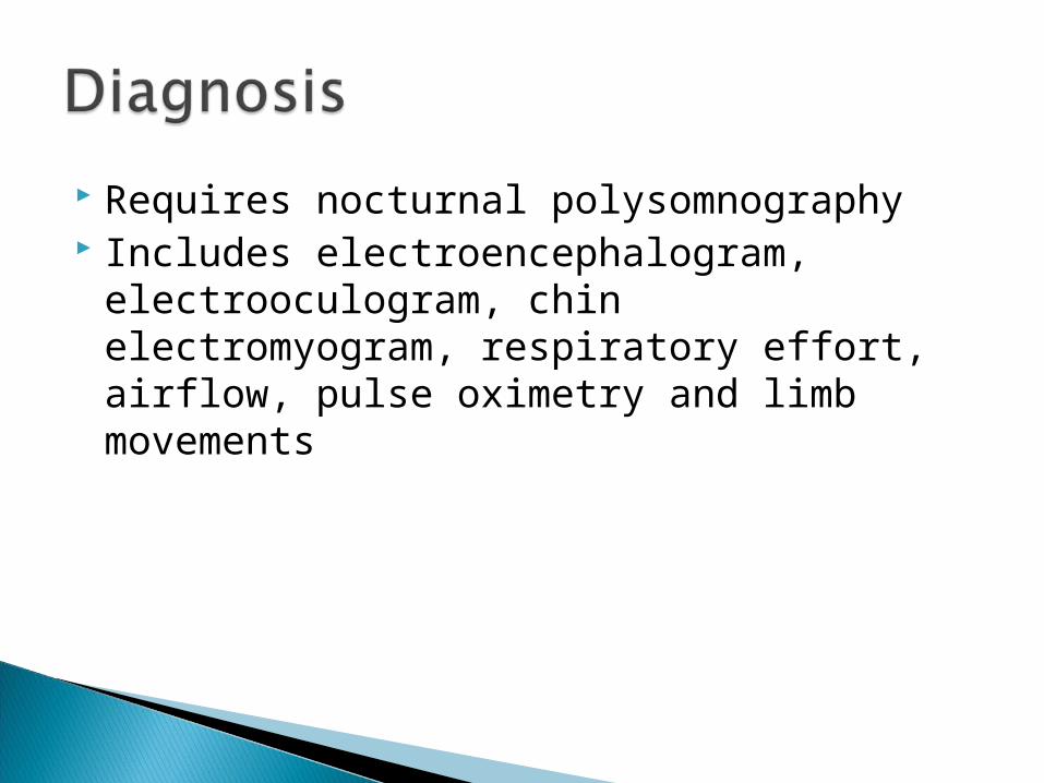
Requires nocturnal polysomnography Includes electroencephalogram,
electrooculogram, chin electromyogram, respiratory effort, airflow, pulse oximetry and limb movements

Conservative approach◦ Weight loss◦ Exercise◦ Improved sleep hygiene◦ Supine preclusion◦ Nasal decongestants ◦ Smoking cessation◦ Avoidance of sedating drugs◦ Avoidance of alcohol
Definitive approach◦ Nasal CPAP◦ Auto-CPAP◦ Bi-level ventilation◦ Oral appliances◦ Surgery: uvulopalatopharyhgoplasty, laser-assisted uvuloplasty,
improved nasal patency, maxillomandibular advancement

In patients with AHI >5 associated with complaints of daytime sleepiness, CPAP therapy has been shown to improve quality of life, cognitive function, and symptoms of daytime sleepiness. There are also beneficial effects on mortality, blood pressure and cardiac function.

A 53-year old man is evaluated for management of OSA which was diagnosed 2 months ago when he was evaluated for excessive sleepiness. Polysomnography performed at that time showed an apnea-hypopnea index of 55 events per hour. The index normalized to 4 events per hour with CPAP at 18cm H2), which was prescribed for his apnea.
He says now that he cannot tolerate nasal CPAP because of nasal congestion and he has not used CPAP for two weeks. He has continued daytime sleepiness that affects his performance at work.
Which of the following is the most appropriate management for this patient's condition at this time?
A) Refer for upper airway surgery for obstructive sleep apnea B) Change to an auto-titrating positive airway pressure device
(APAP) C) Prescribe an oral device to be worn during sleep D) Manage nasal congestion and stress regular use of CPAP

A 24 year old woman is evaluated for episodes of falling asleep at inappropriate times during the day. Sleepiness occurs most often after lunch or while driving a car. She goes to bed at midnight and gets up for work at 6am. Her only medication is oral contraceptives. She has no symptoms of depression, drug use, restless leg syndrome, hypothyroidism, or insomnia. She does not know whether she snores and no one in her family has sleep problems. Physical examination reveals normal weight and vital signs.
Which of the following is the most appropriate next step in the management of this patient?
A) Schedule polysomnography B) Counsel for proper sleep hygiene with increase in sleep time C) Schedule electroencephalography D) Prescribe modafinil

Type PaO2/FiO2 CXR Other
Acute Lung Injury
<300 Bilateral infiltrates
No CHF
Acute lung failure
<300 Any infiltrates No CHF
Acute respiratory distress syndrome
<200 Bilateral infiltrates
No CHF
Acute hypoxic respiratory failure
<200 Any finding No COPD

V/Q mismatch Shunt Diffusion abnormality Decreased mixed venous oxygen Greater acidity and higher temperature of
blood shift the oxyhemoglobin dissociation curve to the right, lowering the blood oxygen content for a given oxygen tension

Pulmonary◦ Acute bacterial or viral pneumonia◦ Gastric aspiration◦ Inhalation injury◦ Near drowning◦ Pulmonary contusion
Non-pulmonary causes◦ Severe sepsis◦ Hypertransfusion syndrome◦ Acute pancreatitis◦ Transfusion-related acute lung injury◦ Cardiopulmonary bypass

Failure of NPPV if patient is a candidate initially Severe dyspnea with use of accessory muscles and
paradoxical abdominal motion Respiration rate >35/min Life-threatening hypoxemia (PaO2/FiO2 <200) Severe acidosis (pH < 7.25) or hypercapnia (PaCo2
>60mm Hg) Respiratory arrest Impaired mental status Cardiovascular complications (hypotension, shock,
heart failure) Other complications (metabolic abnormalities,
sepsis, pneumonia, pulmonary embolism, barotrauma, massive pleural effusion)

Studied in ARMA trial Showed reduction in ARDS mortality from 40% to 30% with a
low (6ml/kg) rather than high (12ml/kg) tidal volume Established "lung protective" ventilator strategies to avoid
ventilator-associated lung injury resulting from excessive stretching of the lung during mechanical ventilation
ALVEOLI study showed no advantage of a higher PEEP compared to a lower PEEP, both adjusted to maintain adequate oxygenation
Current recommendation is to use either a volume- or pressure- limited mode with a low tidal volume (6ml/kg) while monitoring plateau pressure that should be kept <30cm H2O.
PaCO2 is allowed to rise if necessary to achieve these goals (permissive hypercapnea) and PEEP is adjusted to maintain FiO2 <60% with SaO2 >88%
If hypoxemia persists, prone positioning or high frequency oscillation are sometimes used, but no studies have yet demonstrated improved outcomes

Avoid excessive minute volume that contributes to dynamic hyperinflation (auto-PEEP) and alkalemia that results from the compensatory metabolic alkalosis for chornic hypercarbia
Keep tidal volume small (5-7ml/kg ideal body weight)
Backup respiratory rate 10-14/min Lower rate increases cycle time which permits
more time for exhalation and emptying of the lung
Shortening Inspiratory time is another way to increase expiratory time

Invasive mechanical ventilation should be avoided in patients with asthma if at all possible
Complications include pneumothorax and pneumomediastinum
In needed, the approach is similar to that used for COPD patients
Excessive respiratory rates and tidal volumes should be avoided, plateau pressures should be kept <30-35cm H2O and permissive hypercapnea is used
When ventilation remains difficult, heliox or even general anesthesia may be tried

Minimizing the duration of mechanical ventilation is desirable to reduce complications
When patients no longer require high levels of oxygen (SaO2 >89% with FiO2 ≤40%), are hemodynamically stable, and not excessively sedated, spontaneous breathing trials using a T-piece or low levels of CPAP or pressure support should be initiated
If the patient tolerates SBP for 30-120 minutes without excessive tachypnea, hemodynamic instability, or oxygen desaturation, extubation should be performed

A 67-year old man is receiving mechanical ventilation for ARDS. He underwent laparotomy and diverting colostomy for a ruptured diverticulum 72 hours ago, and now has a fever to 40.0C and has diffuse bilateral infiltrates that have been present for the past 1 day. Two deep tracheal suction specimens are sent for culture and a gram stain shows 4+ gram negative rods. The patient’s oxygen saturation is worsening, and his MAP has dropped to 58mm Hg despite three 1-L boluses of normal saline, with only 15mL of urine output in the past hour. He is also noted to have a lactic acidosis and thrombocytopenia, with a platelet count now falling to 42,000 in the absence of heparin or H2-antagonist therapy.
Which of the following would be appropriate management for this patient?
A) Start resuscitation with colloids B) Avoid activated protein C C) Start low-dose dopamin D) Adjust the ventilator with 6mL/kg of ideal body weight and a
plateau pressure <30cm H2O

A 77-year old man on chronic hemodialysis is evaluated in the emergency department for severe dyspnea. He is in respiratory distress, but is alert and responsive. His blood pressure is 216/92, pulse 122, RR 44. He is using accessory muscles to breath. He has JVD; lungs have bilateral crackles, and cardiac exam reveals a summation gallop with a 3/6 systolic murmur. There is no edema. ABG on 50% O2 by high-flow mask are pO2 64, pCO2 50, pH 7.24. EKG shows sinus tach with non-specific STT wave abnormalities and CXR is pending. He receives oxygen, nitroglycerine, furosemide, and small doses of morphine but remains very dyspneic.
Which of the following interventions would most likely avoid intubation in this patient?
A) Increasing the dose of morphine; continue nitroglycerine and furosemide
B) Starting noninvasive continuous airway pressure (4cm H2O) C) Starting noninvasively administered pressure support (8cm H2O)
and PEEP (4cm H2O) D) Increase the FiO2 via face mask

A 78 year old man is admitted to the ICU from the ED where he had presented with a respiratory arrest and was promptly intubated. The patient has a long history of smoking and of severe COPD on long-term O2 therapy at home and a previous measurement of ABG’s on 2L oxygen during a stable state revealed a pO2 of 92mm Hg, pCO2 58mmHg, and pH of 7.45.
Physical examination is notable for a barrel chest and fine expiratory wheezes. Heart sounds are barely audible. ABG just before intubation were pO2 of 220mmHg, pCO2 of 122mmHg, and pH of 7.04 while receiving 100% O2. In the ED , initial ventilator settings were assist/control mode with a rate of 20/min, tidal volume of 600mL, PEEP of 5cm H2O and FiO2 of 50%. CXR shows hyperinflation, extensive bullous emphysema, and a RLL infiltrate.
On arrival to the ICU, he is hypotensive with a systolic pressure of 80mmHg, unresponsive to an initial fluid bolus. A pulmonary artery catheter is inserted; right atrial pressure is 20cm H2O, pulmonary artery pressure is 66/25 mmHg, pulmonary capillary wedge pressure is 21mmHg, and cardiac index is 1.8 L/min/m2. EKG shows a rate of 122/min, sinus rhythm with multiple premature atrial beats, and nonspecific STT changes. During an accidental disconnection from the ventilator, the patient’s blood pressure and wedge pressure normalize but deteriorate when he is reconnected.
Which of the following is the most appropriate next step in the management of this patient? A) Administer more fluids B) Start dobutamine therapy C) Obtain a repeat STAT portable chest radiograph D) Lower the respiration rate and tidal volume

Heterogeneous disorder that includes:◦ Emphysema◦ Chronic Bronchitis◦ Obliterative Bronchiolitis◦ Asthma w/ bronchitis
GOLD: A disease states characterized by airflow limitation that is not fully reversible. Usually progressive and is associated with abnormal inflammatory response

Stage 0: Normal spirometry, chronic cough and sputum production
Stage I: Mild FEV1/FVC <70% predicted and FEV1 ≥ 80% predicted
Stage II: FEV1/FVC < 70% and FEV1 ≥ 50% and < 80% predicted
Stage III: FEV1/FVC < 70% and FEV1 ≥ 30% and < 50% predicted
Stage IV: FEV1/FVC < 70% and FEV1 < 30% predicted or FEV1 < 50% predicted and chronic respiratory symptoms

GOLD - a report produced by NHLBI & WHO defines COPD exacerbation as - acute increase in sx beyond normal daily variation
Includes one or more of the following cardinal symptoms (over 2 days):◦ Cough increases in frequency and severity◦ Sputum production increases in volume and/or
changes character◦ Dyspnea increases

It is estimated that:◦ 50-60% COPD exacerbations are due to
respiratory infections ◦ 10% are due to environmental pollution◦ 30% are of unknown etiology

Most infection are thought to be viral, and have been confirmed by viral culture or serology in 18 - 64% of exacerbation
The most common viruses are influenza, parainfluenza, coronavirus, and rhinovirus

Viral infections cause the majority of COPD exacerbations; bacterial infections also trigger exacerbations
Most common bacterial causes:◦ Haemophilus influenzae◦ Moraxella catarrhalis◦ Streptococcus pneumoniae
Less Common include◦ Kebsiella, Peduomonas◦ Rarely - Chlamydia and Legionella

Treatment of exacerbations of COPD includes antibiotics directed against H. influenzae, M. catarrhalis, and S. pneumoniae ◦ The use of antibiotics in exacerbations of COPD
is based on placebo-controlled trials that found that antibiotics improve clinical outcomes in many patients with an exacerbation of COPD
Brochodilator, steroids, O2

"Standard"Doxycycline 100 mg BIDTrimethoprim-sulfamethoxazole DS BIDAmoxicillin 500 mg BID
"Modernized list"Amoxicillin-clavulanate (Augmentin) 875 mg BIDAzithromycin (Zithromax) Z pack (6-250
mg)Cefpodoxime (Vantin) 200 mg BIDCefuroxime (Ceftin) 250-500 mg BIDCefprozil (Cefzil) 500 mg BIDLoracarbef (Lorabid) 400 mg BIDLevofloxacin (Levaquin) 500 mg QDCiprofloxacin (Cipro) 500 mg BID

Stable COPD:◦ Bronchodilaters◦ Anticholinergics◦ Steroids◦ (No mucolytics or leukotrienes)

Bullectomy may reduce sx in pt w/ bulla causing compression of adjoining lung tissue -> improve lung function
Lung Volume Reduction surgery - FEV <20% and diffusion capacity <20% and people with upper lobe disease had better outcomes
Lung Transplant - FEV1 <35, PaO2 55-60, PaCO2 >50

A 72-year-old man is evaluated for progressive dyspnea on exertion and a morning cough productive of thick white sputum. The patient is a life-long cigarette smoker and was diagnosed with chronic obstructive pulmonary disease 4 years ago; he has had two unscheduled office visits in the past 6 months for bronchitis.
On physical examination, he is thin (BMI 20), his chest is hyperinflated, breath sounds are diminished, he has 1+ ankle edema. Spirometry shows an FEV1 35% of predicted which improves 5% with albuterol. Lung volume measurement shows a total lung capacity of 140% and residual volume of 130%; the DLco is 55% of predicted.
Which of the following is the most appropriate therapy for this patient?
(A) Albuterol, tiotropium, and inhaled corticosteroids (B) Ipratropium bromide and tiotropium (C) Albuterol/ipratropium bromide inhaler, a long-acting beta-agonist,
and oral corticosteroids (D) Ipratropium bromide and montelukast

A 45-year-old man is evaluated for mild dyspnea on exertion. He has smoked 1.5 packs of cigarettes a day for 30 years. His personal and family medical history is unremarkable.
On physical examination, the chest is clear; cardiac examination and chest radiograph are normal. Spirometry shows the FEV1 of 70%, FVC of 75%, FEV1/FVC of 70%. After administration of a bronchodilator, the FEV1 rises to 80% and the FVC to 85%; the FEV1/FVC ratio is 75%. Ther serum IgE concentration is normal, and there are no eosinophils on the peripheral blood smear.
Which of the following is the most likely diagnosis? (A) Chronic obstructive pulmonary disease, stage 0 (B) Chronic obstructive pulmonary disease, stage 1 (C) Moderate persistent asthma (D) Restrictive lung disease