Embed Size (px)
Citation preview
Luminescence of a new class of UV–blue-emitting phosphorsMSi2O22dN2+2/3d:Ce3+ (M 5 Ca, Sr, Ba){
Y. Q. Li,* G. de With and H. T. Hintzen
Received 1st June 2005, Accepted 11th August 2005
First published as an Advance Article on the web 7th September 2005
DOI: 10.1039/b507735d
The luminescence properties of Ce3+,Na+-codoped MSi2O22dN2+2/3d (M 5 Ca, Sr, Ba) are reported.
The undoped and Ce3+,Na+-codoped MSi2O22dN2+2/3d powders were prepared by a solid-state
reaction at temperatures between 1300–1400 uC under N2–H2 (10%) atmosphere in the system
MO–SiO2–Si3N4 (M 5 Ca, Sr, Ba). MSi2O22dN2+2/3d (M 5 Ca, Sr, Ba) crystallizes in the
monoclinic system with different crystal structures. For excitation in the 300–360 nm range,
MSi2O22dN2+2/3d:Ce3+ shows typical broad emission bands peaking at about 392, 473 and 396 nm
for M 5 Ca, Sr and Ba, respectively. In particular, CaSi2O22dN2+2/3d:Ce3+ shows an unusual short-
wavelength emission (y392 nm) with a very small Stokes shift of 2200 cm21; BaSi2O2N2:Ce3+ shows
an interesting white-light emission in the visible range 350–600 nm for excitation at 365 nm.
1. Introduction
The Ce3+ ion has a 4f1 electronic ground state configuration. The
luminescence of the Ce3+ ion originates from a transition from
the lowest 5d level to the ground states which is split by spin–
orbit coupling into two components, 2F5/2 and 2F7/2, separated
by y2000 cm21.1 Since the position of the lowest 5d levels is
strongly influenced by the local coordination, the emission
wavelength of Ce3+ varies with different host lattices from UV to
the visible range corresponding to the emission colors from blue
to red.1 In oxide host lattices, the emission of Ce3+ generally is
located in the UV to blue (300–500 nm) spectral range.1 An
exception is the yellow-emitting YAG:Ce3+ due to its large
crystal field splitting.1,2 A large crystal field splitting can also be
realized by N32 replacement of O22. In addition, nitride-based
host lattices provide more covalent bonding (like in sulfides)
resulting in the 5d band shifting to lower energy.1,3–5 Indeed,
long-wavelength emission in Ce3+-doped rare-earth-(oxy)nitride
and alkaline-earth silicon nitride materials is observed.4,6
In comparison with oxides, nitride and oxynitride-based
materials can give some surprises not only in structure (like an
unusual motif) but also in physical characteristics which are
reflected by their unique mechanical, electrical, thermal and
optical properties.7–13 Definitely, the nitrogen atom is believed
to play a key role due to its high formal charge and large
covalent character in nitride-based materials.7–9
In the system M–Si–O–N (M 5 Ca, Sr, Ba), alkaline-earth
silicon oxynitride compounds with composition MSi2O2N2
(M 5 Ca, Sr, Ba) are known.14–18 This kind of oxynitride is of
interest for luminescent materials because its composition is
situated between the oxide compound M2SiO4 and the pure
nitride compound M2Si5N8 (M 5 Ca, Sr, Ba). Eu2+-doped
M2SiO4 phosphor materials are well-known green (M 5 Ca,
Ba) and yellow (M 5 Sr) emitting phosphors,19–22 while
M2Si5N8:Eu2+ (M 5 Ca, Sr, Ba) is a new family of red-emitting
phosphors showing excellent luminescence properties for
white-light LED applications.23,24 Recently, we have reported
on Eu2+-doped MSi2O22dN2+2/3d phosphor materials with
yellow (M 5 Ca), green to yellow (M 5 Sr) and blue-green
(M 5 Ba) emission colors,17 which are also promising
candidates for use as conversion phosphors for white-light
LED applications.25 In contrast, the luminescence properties
of Ce3+-activated alkaline-earth silicates in the BaO–SrO–SiO2
system have been reported in an earlier work which revealed
that M2SiO4:Ce3+ and MSiO3:Ce3+ (M 5 Sr, Ba) exhibited a
peak emission wavelength at about 390 nm with slight
variations resulting from compositional changes.26 Most
recently, we have reported the luminescence properties of
Ce3+-activated M2Si5N8 (M 5 Ca, Sr, Ba) using Li or Na as a
charge compensator.6 Especially, Sr2Si5N8:Ce3+ turns out to be
a very attractive green-emitting phosphor for use in white-light
LEDs owing to its high conversion efficiency in the UV blue
range (370–450 nm). These peculiar behaviors inspired us to
extend our study to the Ce3+-doped MSi2O22dN2+2/3d system
(M 5 Ca, Sr, Ba). In this study, undoped and Ce3+-doped
MSi2O22dN2+2/3d (M 5 Ca, Sr, Ba) compounds were
synthesized by a solid-state reaction using Na+ as charge-
compensator. Furthermore, new X-ray powder diffraction
data and the lattice parameters of MSi2O22dN2+2/3d (M 5 Ca,
Sr) are presented as we have found that previous studies on
these compounds are imprecise.15,16 Finally, the unconven-
tional luminescence properties of MSi2O22dN2+2/3d:Ce3+
(M 5 Ca, Sr, Ba) are reported.
2. Experimental
2.1. Synthesis
All powder samples of undoped and Ce3+,Na+-codoped
MSi2O22dN2+2/3d (M 5 Ca, Sr, Ba) were prepared by a solid-
state reaction at high temperatures using Na+ as a charge com-
pensator. As found in our previous study,17 the approximate d
Laboratory of Materials and Interface Chemistry, Eindhoven Universityof Technology, P.O. Box 513, 5600 MB, Eindhoven, The Netherlands{ Electronic supplementary information (ESI) available: measured andcalculated XRD powder diffraction data for MSi2O22dN2+2/3d
(M 5 Ca, Sr). See http://dx.doi.org/10.1039/b507735d
PAPER www.rsc.org/materials | Journal of Materials Chemistry
4492 | J. Mater. Chem., 2005, 15, 4492–4496 This journal is � The Royal Society of Chemistry 2005
Publ
ishe
d on
07
Sept
embe
r 20
05. D
ownl
oade
d by
Pur
due
Uni
vers
ity o
n 28
/08/
2014
15:
20:1
0.
View Article Online / Journal Homepage / Table of Contents for this issue
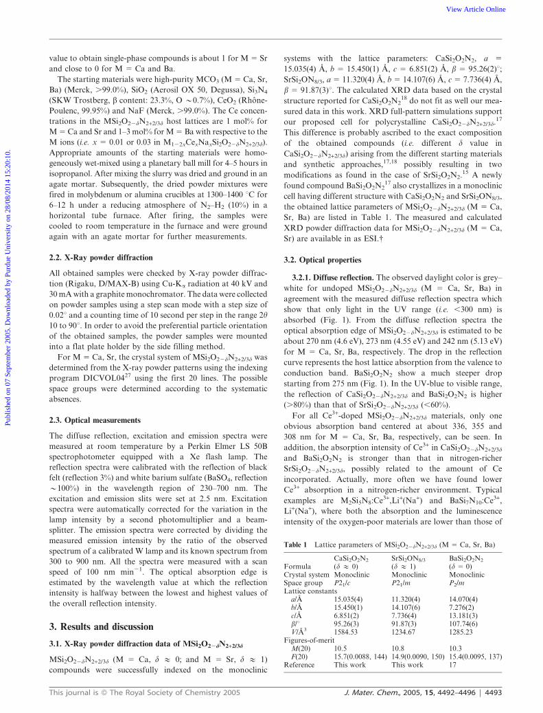
value to obtain single-phase compounds is about 1 for M 5 Sr
and close to 0 for M 5 Ca and Ba.
The starting materials were high-purity MCO3 (M 5 Ca, Sr,
Ba) (Merck, .99.0%), SiO2 (Aerosil OX 50, Degussa), Si3N4
(SKW Trostberg, b content: 23.3%, O y0.7%), CeO2 (Rhone-
Poulenc, 99.95%) and NaF (Merck, .99.0%). The Ce concen-
trations in the MSi2O22dN2+2/3d host lattices are 1 mol% for
M 5 Ca and Sr and 1–3 mol% for M 5 Ba with respective to the
M ions (i.e. x 5 0.01 or 0.03 in M122xCexNaxSi2O22dN2+2/3d).
Appropriate amounts of the starting materials were homo-
geneously wet-mixed using a planetary ball mill for 4–5 hours in
isopropanol. After mixing the slurry was dried and ground in an
agate mortar. Subsequently, the dried powder mixtures were
fired in molybdenum or alumina crucibles at 1300–1400 uC for
6–12 h under a reducing atmosphere of N2–H2 (10%) in a
horizontal tube furnace. After firing, the samples were
cooled to room temperature in the furnace and were ground
again with an agate mortar for further measurements.
2.2. X-Ray powder diffraction
All obtained samples were checked by X-ray powder diffrac-
tion (Rigaku, D/MAX-B) using Cu-Ka radiation at 40 kV and
30 mA with a graphite monochromator. The data were collected
on powder samples using a step scan mode with a step size of
0.02u and a counting time of 10 second per step in the range 2h
10 to 90u. In order to avoid the preferential particle orientation
of the obtained samples, the powder samples were mounted
into a flat plate holder by the side filling method.
For M 5 Ca, Sr, the crystal system of MSi2O22dN2+2/3d was
determined from the X-ray powder patterns using the indexing
program DICVOL0427 using the first 20 lines. The possible
space groups were determined according to the systematic
absences.
2.3. Optical measurements
The diffuse reflection, excitation and emission spectra were
measured at room temperature by a Perkin Elmer LS 50B
spectrophotometer equipped with a Xe flash lamp. The
reflection spectra were calibrated with the reflection of black
felt (reflection 3%) and white barium sulfate (BaSO4, reflection
y100%) in the wavelength region of 230–700 nm. The
excitation and emission slits were set at 2.5 nm. Excitation
spectra were automatically corrected for the variation in the
lamp intensity by a second photomultiplier and a beam-
splitter. The emission spectra were corrected by dividing the
measured emission intensity by the ratio of the observed
spectrum of a calibrated W lamp and its known spectrum from
300 to 900 nm. All the spectra were measured with a scan
speed of 100 nm min21. The optical absorption edge is
estimated by the wavelength value at which the reflection
intensity is halfway between the lowest and highest values of
the overall reflection intensity.
3. Results and discussion
3.1. X-Ray powder diffraction data of MSi2O22dN2+2/3d
MSi2O22dN2+2/3d (M 5 Ca, d # 0; and M 5 Sr, d # 1)
compounds were successfully indexed on the monoclinic
systems with the lattice parameters: CaSi2O2N2, a 5
15.035(4) A, b 5 15.450(1) A, c 5 6.851(2) A, b 5 95.26(2)u;SrSi2ON8/3, a 5 11.320(4) A, b 5 14.107(6) A, c 5 7.736(4) A,
b 5 91.87(3)u. The calculated XRD data based on the crystal
structure reported for CaSi2O2N218 do not fit as well our mea-
sured data in this work. XRD full-pattern simulations support
our proposed cell for polycrystalline CaSi2O22dN2+2/3d.17
This difference is probably ascribed to the exact composition
of the obtained compounds (i.e. different d value in
CaSi2O22dN2+2/3d) arising from the different starting materials
and synthetic approaches,17,18 possibly resulting in two
modifications as found in the case of SrSi2O2N2.15 A newly
found compound BaSi2O2N217 also crystallizes in a monoclinic
cell having different structure with CaSi2O2N2 and SrSi2ON8/3,
the obtained lattice parameters of MSi2O22dN2+2/3d (M 5 Ca,
Sr, Ba) are listed in Table 1. The measured and calculated
XRD powder diffraction data for MSi2O22dN2+2/3d (M 5 Ca,
Sr) are available in as ESI.{
3.2. Optical properties
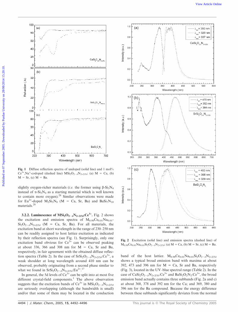
3.2.1. Diffuse reflection. The observed daylight color is grey–
white for undoped MSi2O22dN2+2/3d (M 5 Ca, Sr, Ba) in
agreement with the measured diffuse reflection spectra which
show that only light in the UV range (i.e. ,300 nm) is
absorbed (Fig. 1). From the diffuse reflection spectra the
optical absorption edge of MSi2O22dN2+2/3d is estimated to be
about 270 nm (4.6 eV), 273 nm (4.55 eV) and 242 nm (5.13 eV)
for M 5 Ca, Sr, Ba, respectively. The drop in the reflection
curve represents the host lattice absorption from the valence to
conduction band. BaSi2O2N2 show a much steeper drop
starting from 275 nm (Fig. 1). In the UV-blue to visible range,
the reflection of CaSi2O22dN2+2/3d and BaSi2O2N2 is higher
(.80%) than that of SrSi2O22dN2+2/3d (,60%).
For all Ce3+-doped MSi2O22dN2+2/3d materials, only one
obvious absorption band centered at about 336, 355 and
308 nm for M 5 Ca, Sr, Ba, respectively, can be seen. In
addition, the absorption intensity of Ce3+ in CaSi2O22dN2+2/3d
and BaSi2O2N2 is stronger than that in nitrogen-richer
SrSi2O22dN2+2/3d, possibly related to the amount of Ce
incorporated. Actually, more often we have found lower
Ce3+ absorption in a nitrogen-richer environment. Typical
examples are M2Si5N8:Ce3+,Li+(Na+) and BaSi7N10:Ce3+,
Li+(Na+), where both the absorption and the luminescence
intensity of the oxygen-poor materials are lower than those of
Table 1 Lattice parameters of MSi2O22dN2+2/3d (M 5 Ca, Sr, Ba)
FormulaCaSi2O2N2
(d # 0)SrSi2ON8/3
(d # 1)BaSi2O2N2
(d 5 0)Crystal system Monoclinic Monoclinic MonoclinicSpace group P21/c P21/m P2/mLattice constants
a/A 15.035(4) 11.320(4) 14.070(4)b/A 15.450(1) 14.107(6) 7.276(2)c/A 6.851(2) 7.736(4) 13.181(3)b/u 95.26(3) 91.87(3) 107.74(6)V/A3 1584.53 1234.67 1285.23
Figures-of-meritM(20) 10.5 10.8 10.3F(20) 15.7(0.0088, 144) 14.9(0.0090, 150) 15.4(0.0095, 137)
Reference This work This work 17
This journal is � The Royal Society of Chemistry 2005 J. Mater. Chem., 2005, 15, 4492–4496 | 4493
Publ
ishe
d on
07
Sept
embe
r 20
05. D
ownl
oade
d by
Pur
due
Uni
vers
ity o
n 28
/08/
2014
15:
20:1
0.
View Article Online
slightly oxygen-richer materials (i.e. the former using b-Si3N4
instead of a-Si3N4 as a starting material which is well known
to contain more oxygen).28 Similar observations were made
for Eu2+-doped M2Si5N8 (M 5 Ca, Sr, Ba) and BaSi7N10
materials.28
3.2.2. Luminescence of MSi2O22dN2+2/3d:Ce3+. Fig. 2 shows
the excitation and emission spectra of M0.98Ce0.01Na0.01-
Si2O22dN2+2/3d (M 5 Ca, Sr, Ba). For all materials, the
excitation band at short wavelength in the range of 230–250 nm
can be readily assigned to host lattice excitation as indicated
by their reflection spectra (see Fig. 1). Surprisingly, only one
excitation band obvious for Ce3+ can be observed peaking
at about 336, 366 and 308 nm for M 5 Ca, Sr and Ba,
respectively, in fair agreement with the obtained diffuse reflec-
tion spectra (Table 2). In the case of SrSi2O22dN2+2/3d:Ce3+, a
weak shoulder at long wavelength around 410 nm can be
observed, probably originating from a second phase similar to
what we found in SrSi2O22dN2+2/3d:Eu2+.17
In general, the 5d levels of Ce3+ can be split into at most five
different crystal-field components.1 The above observation
suggests that the excitation bands of Ce3+ in MSi2O22dN2+2/3d
are seriously overlapping (although the bandwidth is small)
and/or that some of them may be located in the conduction
band of the host lattice. M0.98Ce0.01Na0.01Si2O22dN2+2/3d
shows a typical broad emission band with maxima at about
392, 473 and 396 nm for M 5 Ca, Sr and Ba, respectively
(Fig. 3), located in the UV–blue spectral range (Table 2). In the
case of CaSi2O22dN2+2/3d:Ce3+ and BaSi2O2N2:Ce3+, the broad
emission band actually contains three subbands (Fig. 2a and c)
at about 368, 378 and 392 nm for the Ca; and 369, 380 and
396 nm for the Ba compound. Because the energy difference
between these subbands significantly deviates from the normal
Fig. 1 Diffuse reflection spectra of undoped (solid line) and 1 mol%
Ce3+,Na+-codoped (dashed line) MSi2O22dN2+2/3d: (a) M 5 Ca, (b)
M 5 Sr, (c) M 5 Ba.
Fig. 2 Excitation (solid line) and emission spectra (dashed line) of
M0.98Ce0.01Na0.01Si2O22dN2+2/3d: (a) M 5 Ca, (b) M 5 Sr, (c) M 5 Ba.
4494 | J. Mater. Chem., 2005, 15, 4492–4496 This journal is � The Royal Society of Chemistry 2005
Publ
ishe
d on
07
Sept
embe
r 20
05. D
ownl
oade
d by
Pur
due
Uni
vers
ity o
n 28
/08/
2014
15:
20:1
0.
View Article Online
value between the ground states of 2F5/2 and 2F7/2
(y2000 cm21),1 these subbands suggest the presence of
multi-emission centers of the Ce3+ ions. This hypothesis is
confirmed by the fact that the shape (for M 5 Ca) and position
(for M 5 Ba) of the emission subbands can be changed
by varying the excitation wavelength (Fig. 3). In agreement
with this observation, for CaSi2O2N2 the large number of six
crystallographic Ca sites in a unit cell was reported,18 which
could also apply to BaSi2O2N2.
As mentioned before, from the compositional point of
view, MSi2O22dN2+2/3d apparently lies between alkaline-earth
silicates and alkaline-earth silicon nitrides. Normally, a lower
energy 5d Ce3+ excitation band together with a longer
wavelength emission band is expected due to a highly covalent
bonding and a large crystal field splitting in the nitride or
oxynitride compounds.4 Therefore, the local coordination
around M ions in MSi2O22dN2+2/3d can be probed by
luminescent ions, such as Ce3+ and Eu2+. With respect to the
luminescence properties, both the excitation and emission
spectra of MSi2O22dN2+2/3d:Ce3+ are more like those of Ce3+-
doped alkaline-earth silicates26 rather than those of
M2Si5N8:Ce3+.6 For example, for MSiO3:Ce3+ (M 5 Sr, Ba)
and M2SiO4:Ce3+ (M 5 Sr, Ba), the main excitation band is
around 300–335 nm and the emission band is around 390 nm,26
while for M2Si5N8:Ce3+ the principle excitation band is
around 400 nm and the emission band is found at about
470–560 nm depending on the type of M.6 For Eu2+-doped
MSi2O22dN2+2/3d (M 5 Ca, Sr, Ba),17 the luminescence
properties are also close to those of Eu2+-doped alkaline-earth
silicates19–22 while significantly different from M2Si5N8:Eu2+
(M 5 Ca, Sr, Ba).23,24 Therefore it can be concluded that the
M ions in MSi2O22dN2+2/3d are dominantly coordinated by
O atoms, in agreement with the structure elucidation of
CaSi2O2N2, which is a layer silicon oxynitride in which Ca2+
ions are connected by six O atoms and one N atom in
the range of 2.28–2.79 A.18 In addition, based on the fact
that these alkaline-earth silicates consist of layers (i.e. SrSiO3),
chains (i.e. BaSiO3) and isolated [SiO4] tetrahedra,29
MSi2O22dN2+2/3d (M 5 Sr, Ba) is possibly also composed of
layers of [Si(O,N)4] tetrahedral groups similar to the reported
CaSi2O2N2 structure.18
The estimated Stokes shifts are about 2200, 6500 and
5000 cm21 for M 5 Ca, Sr and Ba, respectively (Table 2).
These results are completely contrary to what we have found
for Eu-doped MSi2O22dN2+2/3d (M 5 Ca, Sr, Ba), where
BaSi2O2N2:Eu2+ has the smallest Stokes shift y1700 cm21
while CaSi2O22dN2+2/3d:Eu2+ has a significantly larger Stokes
shift (y5100 cm21).17 Exactly similar opposing trends for Ce3+
and Eu2+ were also found for M2SiO4, i.e., 2900 (M 5 Sr) vs.
4800 cm21 (M 5 Ba) for Ce3+;26 and 5500–6000 (M 5 Sr) vs.
5000 cm21 (M 5 Ba) for Eu2+.19–21 In general, with the ionic
radius of M increasing going from Ca, Sr to Ba the Stokes shift
is expected to decrease for isostructural compounds as we
indeed have found in MYSi4N7:Ce3+ (M 5 Sr, Ba)30,31 with
homovalent Ce3+/Y3+substitution and M2Si5N8:Ce3+,Li+(Na+)
(M 5 Sr, Ba)6 with heterovalent Ce3+/M2+substitution. The
reason for the deviation of the Ca . Sr . Ba sequence
evidently is the fact that the MSi2O22dN2+2/3d compounds have
different crystal structures. In the case of different behaviors of
the Ce3+ and Eu2+ ions, this is possibly related to their different
site preferences as influenced by charge compensation neces-
sary for Ce3+ in contrast to Eu2+ in MSi2O22dN2+2/3d,
eventually resulting in significantly different trends in lumines-
cence properties.
Finally, it is worth noting that CaSi2O22dN2+2/3d:Ce3+ is a
high potential UV–blue-emitting phosphor material with high
efficiency and low thermal quenching (i.e. a small Stokes shift).
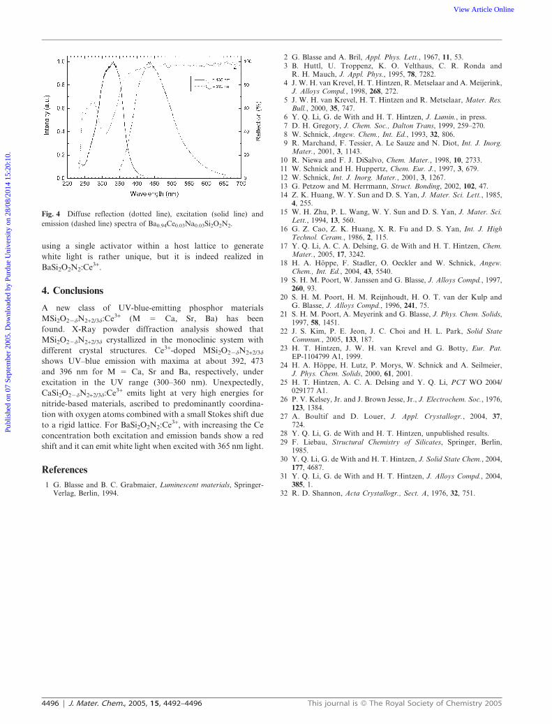
With respective to Ce3+-doped BaSi2O2N2, first, when the Ce
concentration increases from 1 to 3 mol% both the excitation
and emission bands show significant shifts to long-wavelength
(Fig. 4). As there is no significant change in the Stokes
shift (,200 cm21), this red-shift is mainly attributed to the
BaSi2O2N2 lattice shrinkage caused by the replacement of the
large Ba2+ ion (1.35 A, CN 5 6) by the smaller Ce3+ (1.01 A,
CN 5 6) and Na+ (1.02 A, CN 5 6) ions.32 Correspondingly,
the BaCe–O(N) distances become shorter which leads to the
increase in the crystal-field splitting. As a consequence, the
lowest 5d level shifts to lower energy. Second, an attractive
feature of BaSi2O2N2:Ce3+ is that it shows white light for
excitation under 365 nm, especially for high Ce concentrations.
As far as we know, no such studies have been reported. Just
Table 2 Optical properties of MSi2O22dN2+2/3d:Ce3+,Na+ (1 mol%) (M 5 Ca, Sr, Ba)
M d Absorption band/nm 5d excitation band/nm Emission band/nm CIE coordinates (x, y) Stokes shift/cm21
Ca 0 336 336 392 (0.165, 0.061) y2200Sr 1 355 366 473 (0.197, 0.263) y6500Ba 0 308 308 396 (0.161, 0.096) y5000
Fig. 3 Color coordinates deduced from the emission band of
M0.98Ce0.01Na0.01Si2O22dN2+2/3d, (m) M 5 Ca, (&) M 5 Sr, ($)
M 5 Ba (lexc 5 337, 364, 308 nm for M 5 Ca, Sr and Ba, respectively).
This journal is � The Royal Society of Chemistry 2005 J. Mater. Chem., 2005, 15, 4492–4496 | 4495
Publ
ishe
d on
07
Sept
embe
r 20
05. D
ownl
oade
d by
Pur
due
Uni
vers
ity o
n 28
/08/
2014
15:
20:1
0.
View Article Online
using a single activator within a host lattice to generate
white light is rather unique, but it is indeed realized in
BaSi2O2N2:Ce3+.
4. Conclusions
A new class of UV-blue-emitting phosphor materials
MSi2O22dN2+2/3d:Ce3+ (M 5 Ca, Sr, Ba) has been
found. X-Ray powder diffraction analysis showed that
MSi2O22dN2+2/3d crystallized in the monoclinic system with
different crystal structures. Ce3+-doped MSi2O22dN2+2/3d
shows UV–blue emission with maxima at about 392, 473
and 396 nm for M 5 Ca, Sr and Ba, respectively, under
excitation in the UV range (300–360 nm). Unexpectedly,
CaSi2O22dN2+2/3d:Ce3+ emits light at very high energies for
nitride-based materials, ascribed to predominantly coordina-
tion with oxygen atoms combined with a small Stokes shift due
to a rigid lattice. For BaSi2O2N2:Ce3+, with increasing the Ce
concentration both excitation and emission bands show a red
shift and it can emit white light when excited with 365 nm light.
References
1 G. Blasse and B. C. Grabmaier, Luminescent materials, Springer-Verlag, Berlin, 1994.
2 G. Blasse and A. Bril, Appl. Phys. Lett., 1967, 11, 53.3 B. Huttl, U. Troppenz, K. O. Velthaus, C. R. Ronda and
R. H. Mauch, J. Appl. Phys., 1995, 78, 7282.4 J. W. H. van Krevel, H. T. Hintzen, R. Metselaar and A. Meijerink,
J. Alloys Compd., 1998, 268, 272.5 J. W. H. van Krevel, H. T. Hintzen and R. Metselaar, Mater. Res.
Bull., 2000, 35, 747.6 Y. Q. Li, G. de With and H. T. Hintzen, J. Lumin., in press.7 D. H. Gregory, J. Chem. Soc., Dalton Trans, 1999, 259–270.8 W. Schnick, Angew. Chem., Int. Ed., 1993, 32, 806.9 R. Marchand, F. Tessier, A. Le Sauze and N. Diot, Int. J. Inorg.
Mater., 2001, 3, 1143.10 R. Niewa and F. J. DiSalvo, Chem. Mater., 1998, 10, 2733.11 W. Schnick and H. Huppertz, Chem. Eur. J., 1997, 3, 679.12 W. Schnick, Int. J. Inorg. Mater., 2001, 3, 1267.13 G. Petzow and M. Herrmann, Struct. Bonding, 2002, 102, 47.14 Z. K. Huang, W. Y. Sun and D. S. Yan, J. Mater. Sci. Lett., 1985,
4, 255.15 W. H. Zhu, P. L. Wang, W. Y. Sun and D. S. Yan, J. Mater. Sci.
Lett., 1994, 13, 560.16 G. Z. Cao, Z. K. Huang, X. R. Fu and D. S. Yan, Int. J. High
Technol. Ceram., 1986, 2, 115.17 Y. Q. Li, A. C. A. Delsing, G. de With and H. T. Hintzen, Chem.
Mater., 2005, 17, 3242.18 H. A. Hoppe, F. Stadler, O. Oeckler and W. Schnick, Angew.
Chem., Int. Ed., 2004, 43, 5540.19 S. H. M. Poort, W. Janssen and G. Blasse, J. Alloys Compd., 1997,
260, 93.20 S. H. M. Poort, H. M. Reijnhoudt, H. O. T. van der Kulp and
G. Blasse, J. Alloys Compd., 1996, 241, 75.21 S. H. M. Poort, A. Meyerink and G. Blasse, J. Phys. Chem. Solids,
1997, 58, 1451.22 J. S. Kim, P. E. Jeon, J. C. Choi and H. L. Park, Solid State
Commun., 2005, 133, 187.23 H. T. Hintzen, J. W. H. van Krevel and G. Botty, Eur. Pat.
EP-1104799 A1, 1999.24 H. A. Hoppe, H. Lutz, P. Morys, W. Schnick and A. Seilmeier,
J. Phys. Chem. Solids, 2000, 61, 2001.25 H. T. Hintzen, A. C. A. Delsing and Y. Q. Li, PCT WO 2004/
029177 A1.26 P. V. Kelsey, Jr. and J. Brown Jesse, Jr., J. Electrochem. Soc., 1976,
123, 1384.27 A. Boultif and D. Louer, J. Appl. Crystallogr., 2004, 37,
724.28 Y. Q. Li, G. de With and H. T. Hintzen, unpublished results.29 F. Liebau, Structural Chemistry of Silicates, Springer, Berlin,
1985.30 Y. Q. Li, G. de With and H. T. Hintzen, J. Solid State Chem., 2004,
177, 4687.31 Y. Q. Li, G. de With and H. T. Hintzen, J. Alloys Compd., 2004,
385, 1.32 R. D. Shannon, Acta Crystallogr., Sect. A, 1976, 32, 751.
Fig. 4 Diffuse reflection (dotted line), excitation (solid line) and
emission (dashed line) spectra of Ba0.94Ce0.03Na0.03Si2O2N2.
4496 | J. Mater. Chem., 2005, 15, 4492–4496 This journal is � The Royal Society of Chemistry 2005
Publ
ishe
d on
07
Sept
embe
r 20
05. D
ownl
oade
d by
Pur
due
Uni
vers
ity o
n 28
/08/
2014
15:
20:1
0.
View Article Online