Embed Size (px)
Citation preview

Lymph Node Status Assessment for GastricCarcinoma: Is the Number of Metastatic
Lymph Nodes Really Practical as aParameter for N Categories in the
TNM Classification?
YASUHIRO KODERA, MD,* YOSHITAKA YAMAMURA, MD, YASUHIRO SHIMIZU, MD,AKIHITO TORII, MD, TAKASHI HIRAI, MD, KENZO YASUI, MD, TAKESHI MORIMOTO, MD,
TOMOYUKI KATO, MD, AND TSUYOSHI KITO, MD
Department of Gastroenterological Surgery, Aichi Cancer Center, Nagoya, Japan
Background and Objectives:The anatomical extent of nodal metastasishas been replaced by the number of metastatic nodes as a key indicator ofprognosis (pN categories) in the fifth edition of Tumor Node MetastasisClassification for gastric carcinoma by the International Union AgainstCancer. The rate of metastatic nodes among all the nodes harvested is alsoa good prognostic factor. The object of this study is to evaluate which ofthe three parameters for the assessment of nodal status is the most appro-priate for use in the stage classification.Methods: Retrospective survival analyses were performed in 656 con-secutive patients with advanced gastric carcinoma who underwent D2lymphadenectomy and for whom histopathologic data of more than 20lymph nodes were available.Results: Although all three systems served well to classify the patientsinto distinct groups in terms of survival curves, stratification by the num-ber of metastatic nodes was vulnerable to stage migration because ofdifferences in the number of lymph nodes harvested. Such stage migrationwas not evident for the other two parameters.Conclusions: Lymph node metastatic rate can be recommended as aninternationally applicable parameter for lymph node involvement of gas-tric carcinoma.J. Surg. Oncol. 1998;69:15–20. © 1998 Wiley-Liss, Inc.
KEY WORDS: prognosis; survival curve; stage classification
INTRODUCTION
The absence or presence and anatomical extent oflymph node metastasis are significant prognostic factorsfor gastric carcinoma [1,2]. These factors, expressed interms of pN categories proposed by the InternationalUnion Against Cancer (UICC) [3] or n categories pro-posed by the Japanese Society for Gastric Cancer Studyin the Japanese Classification of Gastric Carcinoma(JCGC) [4], have served as an essential component of thewidely acclaimed stage classifications for gastric carci-noma. However, the anatomical extent of nodal metas-tasis cannot be assessed with accuracy unless sufficientsystematic lymphadenectomy is performed. In most
Western institutions where the extended lymphadenecto-my is not recognized as a standard treatment for gastriccarcinoma [5,6], the n or pN categories may not havebeen practical as a key parameter for a stage classifica-tion scheme.
Recently, the UICC decided to replace these with thenumber of metastatic lymph nodes, another promising
*Correspondence to: Y. Kodera, MD, Department of Gastroenterologi-cal Surgery, Aichi Cancer Center, 1-1, Kanokoden, Chikusa-ku,Nagoya, Aichi 464, Japan. Fax No.: (81)52-763-5233.Accepted 20 June 1998
Journal of Surgical Oncology 1998;69:15–20
© 1998 Wiley-Liss, Inc.

prognosticator [7,8] as new pN categories in their latestversion of the Tumor Node Metastasis (TNM) Classifi-cation [9]. The authors have recently shown that the rateof metastatic nodes among all the nodes resected is asignificant prognostic factor for stage IV gastric carci-noma [10]. In this retrospective study involving 656 pa-tients with advanced gastric carcinoma, we attempted toevaluate the feasibility of the three modes of lymph nodestatus assessment.
PATIENTS AND METHODS
During the 10-year period between 1983 and 1992,1913 patients with gastric carcinoma underwent surgeryat Aichi Cancer Center Hospital. Of these, 656 consecu-tive patients who fulfill the following criteria were se-lected: (1) carcinoma invaded beyond the muscularispropria (i.e., advanced carcinoma); (2) extended lymph-adenectomy had been performed; and (3) retrieval fol-lowed by histopathologic evaluation of no less than 20lymph nodes had been accomplished. The age of thepatients ranged from 24 to 91 years, with a mean andstandard deviation of 59.6 ± 11.2. Of these patients, 417(63.6%) were male. The surgical procedures performedwere total gastrectomy in 250, proximal gastrectomy in25, distal gastrectomy in 379, and pancreaticoduodenec-tomy in 2. The extent of lymphadenectomy was D2 in543 and more extensive in 113. Lymph nodes were im-mediately sent postoperatively for hematoxylin and eosinstaining and histopathological examination. Numbers ofresected and metastatic lymph nodes from each anatomi-cal region as defined by the JCGC were recorded.
On the basis of the data thus obtained, stratificationsaccording to three different modes of lymph node statusassessment were performed. The first of these was theabsence or presence and extent of nodal metastases (ncategories), defined by the JCGC [4]. In the n categories,n0 denotes absence of node metastasis, while n1 signifiesmetastasis mainly to the perigastric nodes and n2 denotesmore extensive metastasis within the regional lymphnodes. n3 and n4 that are collectively treated as > n3 inthe current study (n3 denotes involvement of hepatoduo-denal, mesenteric, or retropancreatic nodes, and n4 sig-nifies involvement of para-aortic nodes) have been de-fined as distant metastases (pM1) in the TNM classifi-cation, with the exception of metastatic nodes along thehepatoduodenal ligament (No.12 node), which is definedas one of the regional lymph nodes by the TNM classi-fication [11]. The stratification in terms of number ofmetastatic nodes was performed according to the fifthedition of TNM classification [5], in which carcinomawith no metastatic nodes is defined as pN0, those with1–6 metastatic nodes is defined as pN1, 7–15 nodes aspN2, and more than 15 nodes as pN3. Finally, stratifica-tion into four groups according to the lymph node meta-static rate (the number of metastatic lymph nodes divided
by the number of retrieved lymph nodes ×100) was per-formed as follows; carcinoma with a nodal metastaticrate of 0%, 1–19 %, 20–60%, and >60%.
In order to evaluate the influence of the number oflymph nodes harvested on each of the nodal status as-sessments, the patients were then classified into twogroups according to the nodal yields; the patients withnodal yields of less than 40 (group A, n4 327) and thosewith yields of 40 or more (group B, n4 329). There wasno significant difference (P = 0.2383 by chi-square test)in the frequency of noncurative operation (n4 39 forgroup A and n4 29 for group B) between the groups.The number of patients belonging to the clinical stagesdefined by the JCGC were also similarly distributed forthe two groups (69 stage I, 61 stage II, 125 stage III, and72 stage IV patients for group A and 43 stage I, 88 stageII, 132 stage III, and 66 stage IV patients for group B).The lack of difference in these background factors isreflected in the survival curves that were almost identicalfor groups A and B (Fig. 1). The survival curves forgroups A and B were then compared within several sub-groups as stratified according to the three differentmodes of lymph node status assessment described above.
Statistical Analysis
All the patients in this study were followed until April1, 1997, or until death. Deaths by causes other than gas-tric carcinoma recurrence are treated as censored cases.The Kaplan-Meier method was used to plot the survivalcurves, and the generalized Wilcoxon test was employedto evaluate the differences between these curves. Stu-dent’st-test was performed for comparison of number ofmetastatic lymph nodes between the two groups. Thecorrelation between the number of metastatic lymphnode and lymph node metastatic rate was assessed byPearson’s correlation coefficient.
Fig. 1. The patients were divided into two groups according to thenumber of lymph nodes resected. Nodal yields for group A patients (n4 327) ranged from 20 to 39, while that for group B patients (n4329) was 40 or more. Survival curves for the two groups were almostidentical.
16 Kodera et al.

RESULTS
A total of 27,396 lymph nodes with a mean nodal yieldof 41.8 ± 14.9 (mean ±SD) per specimen (range 20–113)were removed, of which 4,427 (16.2%) showed metas-tasis. Whether the number of resected nodes was below(group A) or above 40 (group B) did not affect survivalfor the patients in the current study (P 4 0.4307) (Fig.1). There was, however, an expected and significant dif-ference in the number of metastatic nodes between thetwo groups (5.44 ± 7.24 for group A and 8.05 ± 11.48 forgroup B,P 4 0.0006 by chi-square test). The correlationbetween the number of metastatic nodes and nodal meta-static rate was significant for both of the groups (P <0.0001) (Fig. 2).
There was a significant difference in survival curvesbetween each of the subgroups stratified by the anatomi-cal extent of metastatic nodes (n categories). Five-yearsurvival rates for each category were as follows: 78% (n4 196) for n0, 64% for n1 (n4 186) , 35% for n2 (n4
202), and 22% for greater than n3 (n4 72) (P < 0.0001).The same was true of the stratifications by the number ofmetastatic nodes (pN categories; 5-year survival rates of78% for pN0, 62% for pN1, 34% for pN2, 16% for pN3,and 19% for pM1) and nodal metastatic rate (Fig. 3) (P< 0.0001). These results imply that any one of theselymph node staging scheme is feasible, provided the his-topathologic evaluation is performed adequately.
The difference in survival curves between the groupsA and B were not significant among any subgroup ofpatients stratified according to the n-categories or the rateof metastatic nodes (Fig. 4). By contrast, a significantand striking difference in the survival curves between thegroups A and B was noted among the patients classifiedby the new TNM classification as pN2 (P = 0.0082) (Fig.
Fig. 2. The correlation between the number of metastatic nodes andlymph node metastatic rate was statistically significant (P < 0.0001)both for group A (top) (n4 327) and for group B (bottom) (n4 329)patients.
Fig. 3. Lymph node status assessment in terms of n categories by theJapanese Classification for Gastric Carcinoma (top), pN categories bythe TNM Classification (middle), and the rate of the metastatic lymphnodes (bottom). The differences in survival curves between the sub-groups were significant (P < 0.0001) in all three modes of classifica-tion.
N-Categories for Gastric Carcinoma 17
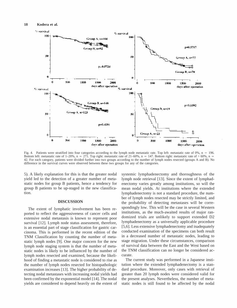
5). A likely explanation for this is that the greater nodalyield led to the detection of a greater number of meta-static nodes for group B patients, hence a tendency forgroup B patients to be up-staged in the new classifica-tion.
DISCUSSION
The extent of lymphatic involvement has been re-ported to reflect the aggressiveness of cancer cells andextensive nodal metastasis is known to represent poorsurvival [12]. Lymph node status assessment, therefore,is an essential part of stage classification for gastric car-cinoma. This is performed in the recent edition of theTNM Classification by counting the number of meta-static lymph nodes [9]. One major concern for the newlymph node staging system is that the number of meta-static nodes is likely to be influenced by the number oflymph nodes resected and examined, because the likeli-hood of finding a metastatic node is considered to rise asthe number of lymph nodes resected for histopathologicexamination increases [13]. The higher probability of de-tecting nodal metastases with increasing nodal yields hadbeen confirmed by the exponential model [14]. The nodalyields are considered to depend heavily on the extent of
systemic lymphadenectomy and thoroughness of thelymph node retrieval [13]. Since the extent of lymphad-enectomy varies greatly among institutions, so will themean nodal yields. At institutions where the extendedlymphadenectomy is not a standard procedure, the num-ber of lymph nodes resected may be strictly limited, andthe probability of detecting metastases will be corre-spondingly low. This will be the case in several Westerninstitutions, as the much-awaited results of major ran-domized trials are unlikely to support extended D2lymphadenectomy as a universally applicable procedure[5,6]. Less extensive lymphadenectomy and inadequatelyconducted examination of the specimens can both resultin a decreased number of metastatic nodes, leading tostage migration. Under these circumstances, comparisonof survival data between the East and the West based onthe TNM classification can no longer be considered ac-curate.
The current study was performed in a Japanese insti-tution where the extended lymphadenectomy is a stan-dard procedure. Moreover, only cases with retrieval ofgreater than 20 lymph nodes were considered valid forthe present analyses. Nevertheless, the number of meta-static nodes is still found to be affected by the nodal
Fig. 4. Patients were stratified into four categories according to the lymph node metastatic rate. Top left: metastatic rate of 0%, n4 196.Bottom left: metastatic rate of 1–20%, n4 271. Top right: metastatic rate of 21–60%, n4 147. Bottom right: metastatic rate of > 60%, n442. For each category, patients were divided further into two groups according to the number of lymph nodes resected (groups A and B). Nodifference in the survival curves were observed between these two groups for any of the categories.
18 Kodera et al.

yield. While the effort to increase the nodal yield did notseem to affect survival (no difference in survival betweenthe groups A and B,P 4 0.4307), it did increase theprobability to detect metastases, hence the significantlyincreased number of metastatic nodes for group B (P 40.0006). Consequently, a difference in the number oflymph nodes harvested induced stage migration; thegroup B patients with greater nodal yields were likely tobe up-staged when stratified according to the number ofmetastatic nodes. This was evident from the observationthat the group B patients who belong to pN2 (7–15 meta-static nodes) were found to have a significantly (P 40.0082) better survival than that of the group A patientswho fell within the same category. Stage migration in thenew TNM classification can thus result not only from thedeficient lymph node yields due to limited lymphadenec-tomy, but also from varying lymph node yields, the rea-son for which is multifactorial. Such stage migrationcould render the TNM classification unreliable unlesssome measures are taken to circumvent it. One way to dothis is to perform histologic examination of a fixed num-ber of nodes for standardization [13,14]. In this case, theclassification may become invalid for some patients
whose nodal yield is too small. Our solution to the prob-lem deduced from the current study is to make use of thelymph node metastatic rate, shown also to be a goodprognostic indicator [10], as a parameter for the lymphnode status assessment in the stage classification. Lymphnode metastatic rate correlated significantly (P < 0.0001)with the number of metastatic nodes, a current parameterfor pN categories in the new TNM classification. At thesame time, it was found not to be affected by the nodalyield, as evident from the fact that the survival curves forgroups A and B were almost identical for all the sub-groups stratified according to the lymph node metastaticrate. Therefore, lymph node metastatic rate can be pro-posed as a novel parameter for lymph node status assess-ment, and similar analyses are warranted in the institu-tions with a sufficient number of cases but with divergentpolicies towards the extent of systematic lymphadenec-tomy.
REFERENCES1. Kodera Y, Yamamura Y, Torii A, et al.: Postoperative staging of
gastric carcinoma. A comparison between the UICC Stage Clas-sification and the 12th Edition of the Japanese General Rules forGastric Cancer Study. Scand J Gastroenterol 1996;31:476–480.
Fig. 5. Patients were stratified into four categories according to the number of metastatic nodes as specified in the 5th edition of the TNMClassification. Top left: pN0, n4 196. Bottom left: pN1, n4 239). Top right: pN2, n4 105. Bottom right: pN3 and pM1, n4 116. Thepatients in each category were divided further into two groups according to the number of lymph nodes resected (groups A and B). For pN2 (7–15metastatic nodes), the difference in survival curves between groups A and B was statistically significant (P 4 0.0082).
N-Categories for Gastric Carcinoma 19

2. Kim JP, Kim YW, Yang HK, Noh D: Significant prognostic fac-tors by multivariate analysis of 3926 gastric cancer patients.World J Surg 1994;18:872–877.
3. Hermanek P, Sobin LH (eds): ‘‘UICC TNM Classification ofMalignant Tumours.’’ 4th Ed. New York: Springer-Verlag, 1992.
4. Japanese Research Society for Gastric Cancer (ed): ‘‘JapaneseClassification of Gastric Carcinoma.’’ Tokyo: Kanehara Shuppan,1995.
5. Bonenkamp JJ, Songun I, Hermans J, et al.: Randomised com-parison of morbidity after D1 and D2 dissection for gastric cancerin 996 Dutch patients. Lancet 1995;345:745–748.
6. Cuschieri A, Fayers P, Fielding J, et al.: Postoperative morbidityand mortality after D1 and D2 resections for gastric cancer: Pre-liminary results of the MRC randomised controlled surgical trial.Lancet 1996,347:995–999.
7. Adachi Y, Kamakura T, Mori M, et al.: Prognostic significance ofthe number of positive lymph nodes in gastric carcinoma. Br JSurg 1994;81:414–416.
8. Wu CW, Hsieh MC, Lo SS, et al.: Relation of number of positive
lymph nodes to the prognosis of patients with primary gastricadenocarcinoma. Gut 1996;38:525–527.
9. Sobin LH, Wittekind C (eds): ‘‘TNM Classification of MalignantTumours.’’ 5th Ed. New York: John Wiley & Sons, 1997.
10. Kodera Y, Yamamura, Y, Shimizu Y, et al.: Metastatic paragastriclymph node rate is a significant prognostic factor for resectablestage IV gastric cancer. J Am Coll Surg 1997;185:65–69.
11. Hermanek P, Hutter RVP, Sobin LH, et al. (eds): ‘‘TNM Atlas.Illustrated Guide to the TNM/pTNM Classification of MalignantTumours.’’ Berlin: Springer, 1997.
12. Baba H, Korenaga D, Okamura T, et al.: Prognostic factors ingastric cancer with serosal invasion. Arch Surg 1989;124:1061–1064.
13. Bunt AMG, Hermans J, van der Velde CJ, et al.: Lymph noderetrieval in a randomized trial on Western-type versus Japanese-type surgery in gastric cancer. J Clin Oncol 1996;14:2289–2294.
14. Bunt AM, Hogendoorn, PC, van de Velde CJ, et al.: Lymph nodestaging standards in gastric cancer. J Clin Oncol 1995;13:2309–2316.
20 Kodera et al.