Embed Size (px)
Citation preview

1
Maintenance of stereocilia and apical junctional complexes by Cdc42 in cochlear hair cells
Takehiko Ueyama1,*,†, Hirofumi Sakaguchi2,†, Takashi Nakamura1,2, Akihiro Goto3,
Shigefumi Morioka1,2, Aya Shimizu1, Kazuki Nakao4, Yoshitaka Hishikawa5, Yuzuru Ninoyu1,2,
Hidetoshi Kassai4, Shiro Suetsugu6, Takehiko Koji7, Bernd Fritzsch8, Shigenobu Yonemura9,
Yasuo Hisa2, Michiyuki Matsuda3, Atsu Aiba4, Naoaki Saito1,*
1Laboratory of Molecular Pharmacology, Biosignal Research Center, Kobe University, Kobe
657-8501, Japan; 2Dept. of Otolaryngology-Head and Neck Surgery, Kyoto Prefectural Univ. of Medicine, Kyoto
602-8566, Japan; 3Lab. of Bioimaging and Cell Signaling, Graduate School of Biostudies, Kyoto Univ., Kyoto
606-8315, Japan; 4Lab. of Animal Resources, Center for Disease Biology and Integrative Medicine, Faculty of
Medicine, Univ. of Tokyo, Tokyo 113-0033, Japan; 5Division of Histochemistry and Cell Biology, Department of Anatomy, Faculty of Medicine,
Univ. of Miyazaki, Miyazaki 889-1692, Japan; 6Lab. of Membrane and Cytoskeleton Dynamics, Institute of Molecular and Cellular Biosciences,
Univ. of Tokyo, Tokyo 113-0032, Japan; 7Dept. of Histology and Cell Biology, Nagasaki Univ. Graduate School of Biomedical Sciences,
Nagasaki 852-8523, Japan; 8Dept. of Biology, College of Liberal Arts and Sciences, Univ. of Iowa, Iowa, IA 52242, USA 9Electron Microscope Lab., Center for Developmental Biology, RIKEN, Kobe 650-0047, Japan
Running title: Hearing loss in Cdc42-KO mice
Keywords: deafness, stereocilia, apical junctional complexes, actin turnover, FRET
† These authors contributed equally to this work.
* Corresponding author: Takehiko UEYAMA and Naoaki SAITO
1-1 Rokkodai-cho, Nada-ku, Kobe 657-8501, Japan,
Tel: +81-78-803-5962, Fax: +81-78-803-5971
E-mails: [email protected] and [email protected]
This is an Open Access article distributed under the terms of the Creative Commons Attribution License(http://creativecommons.org/licenses/by/3.0), which permits unrestricted use, distribution and reproduction in any medium providedthat the original work is properly attributed.
© 2014. Published by The Company of Biologists Ltd.Jo
urna
l of C
ell S
cien
ceA
ccep
ted
man
uscr
ipt
JCS Advance Online Article. Posted on 14 March 2014

2
Abstract
Cdc42 is a key regulator of dynamic actin organization. However, little is known about how
Cdc42-dependent actin regulation influences steady-state actin structures in differentiated
epithelia. We employed inner ear hair cell (HC)-specific conditional knockout to analyze the role
of Cdc42 in HCs possessing highly elaborate stable actin protrusions (stereocilia). HCs of
Atoh1–Cre;Cdc42flox/flox mice developed normally but progressively degenerated after maturation,
resulting in progressive hearing loss particularly at high frequencies. Cochlear HC degeneration
was more robust in inner HCs than in outer HCs, and began as stereocilia fusion and depletion,
accompanied by a thinning and waving circumferential actin belt at apical junctional complexes
(AJCs). Adenovirus-encoded GFP-Cdc42 expression in HCs and fluorescence resonance energy
transfer (FRET) imaging of HCs from transgenic mice expressing Cdc42-FRET biosensor
indicated Cdc42 presence/activation at stereociliary membranes and AJCs in cochlear HCs.
Cdc42-knockdown in MDCK cells produced phenotypes similar to those of Cdc42-deleted HCs,
including abnormal microvilli, disrupted AJCs, and downregulated actin-turnover represented by
enhanced phospho-cofilin levels. Thus, Cdc42 influenced maintenance of stable actin structures
through elaborate tuning of actin-turnover and maintained function and viability of cochlear
HCs.
Jour
nal o
f Cel
l Sci
ence
Acc
epte
d m
anus
crip
t
3
Introduction
Dynamic actin turnover and rearrangement alter overall cell geometry and polarity as well as
local membrane topology to form distinct cellular structures (Campellone and Welch, 2010). In
some structures such as lamellipodia and the Listeria comet tail, actin filaments are highly
dynamic with the turnover rate measured in seconds (Ponti et al., 2004; Theriot et al., 1992),
whereas they are relatively stable in stereocilia (Schwander et al., 2010) and apical junctional
complexes (AJCs) (Ivanov et al., 2005).
Stereocilia and AJCs are present in inner ear hair cells (HCs), which are specialized sensory
epithelia detecting hearing and balance. In the cochlea, HCs are arranged in a single row of inner
HCs (IHCs) and three rows of outer HCs (OHCs). Although IHCs and OHCs are believed to
share common mechanotransduction machinery, they have distinct roles during sound detection:
IHCs are true sensors, whereas OHCs function as an amplifier through an active process that
involves stereociliary and somatic motility (Matsumoto et al., 2010; Schwander et al., 2010).
Stereocilia are actin-based protrusions and exquisitely organized microvilli/filopodia composed
of hundreds of parallel actin filaments with the plus ends at the distal tip, and are organized into
precise rows of graded height (Frolenkov et al., 2004). High sensitivity of HCs depends on the
coordinated movement of stereocilia upon mechanical stimulation; thus, the length and shape of
stereocilia are determined precisely. Moreover, as HCs are usually not replaced (Collado et al.,
2008), turnover of stereociliary components is a lifetime requirement. Although a recent model
suggested that stereocilia maintain their steady state through treadmill-like dynamic actin
turnover (Rzadzinska et al., 2004), others reported no such finding (Zhang et al., 2012), raising a
question about the actin turnover mechanism in stereocilia. AJCs are another actin structure
essential for HC function, such as maintenance of proper cell arrangement and epithelial barrier
(Collado et al., 2011). However, the mode and effect of actin turnover at AJCs remain unknown
(Nunes et al., 2006).
Jour
nal o
f Cel
l Sci
ence
Acc
epte
d m
anus
crip
t

4
Cdc42 is a key regulator of the actin cytoskeleton. However, the function of Cdc42 in HCs
has not been studied, particularly regarding the development and maintenance of stereocilia.
Cdc42, Rac1, and RhoA are the best characterized Rho-family small GTPases, and all are
expressed in HCs (Kalinec et al., 2000). Although they have common regulators and effectors,
their activity results in different effects. Cdc42 is essential for filopodia formation (Chen et al.,
2000; Yang et al., 2006), whereas Rac and RhoA induce lamellipodia and stress fiber formation,
respectively (Heasman and Ridley, 2008). Cdc42 induces actin polymerization through its
interaction with actin nucleators (Campellone and Welch, 2010), including the Arp2/3 complex
component N-WASP (Takenawa and Suetsugu, 2007), and promotes filopodia induction and
stabilization through the PAK/LIM kinase (LIMK) pathway and the cofilin phosphorylation cycle
(Matsumoto et al., 2010; Melendez et al., 2011). Cdc42 also regulates AJC formation in vitro
(Otani et al., 2006; Qin et al., 2010) and in vivo (Melendez et al., 2011).
Here, we show that Cdc42 is localized and it functions at stereociliary membranes and AJCs
for their maintenance in HCs. Cdc42 deletion resulted in abnormal stereocilia and AJC
morphology and was associated with downregulated actin turnover, leading to slowly progressive
cochlear HC loss. These data provide new insights into the function of Cdc42 at apical
protrusions and AJCs in differentiated epithelia in vivo.
Jour
nal o
f Cel
l Sci
ence
Acc
epte
d m
anus
crip
t

5
Results
Inactivation of the Cdc42 gene in inner ear HCs by Atoh1–Cre transgenic (TG) mice
We examined and confirmed expression of Cdc42 mRNA in cochlea using the reverse
transcription-polymerase chain reaction (RT-PCR; data not shown) and in situ hybridization
(ISH) (Fig. 1A). We used the Atoh1 promoter, a basic helix–loop–helix (bHLH) transcription
factor for HC-specific inactivation of Cdc42 (Chen et al., 2002; Jahan et al., 2013). The pattern of
Atoh1–Cre-directed recombination was assessed in the inner ear at embryonic day (E) 13.5 and
E15.5 as well as postnatal day 3 (P3) using -galactosidase activity of a rearranged
CAG-floxCATflox-LacZ reporter mice (hereafter referred to as Atoh1–Cre;LacZ). Consistent with a
previous report (Chen et al., 2002), X-gal staining was first observed at E15.5 in the cochlea and
vestibule of Atoh1–Cre;LacZ mice but not in control (CAG-floxCATflox-LacZ ) mice (data not
shown). Cochlear X-gal staining was restricted to IHCs and OHCs at P3 (Fig. S1A).
Immunostaining against Cre also showed a specific signal in IHCs and OHCs at P1 of Atoh1–
Cre+/- mice but not in littermate Atoh1–Cre-/- mice (Fig. S1B). We attempted to detect
endogenous Cdc42 using two different anti-Cdc42 antibodies (Abs) but could not detect a
specific signal in inner ear tissues with conventional fixation. Therefore, we used trichloroacetic
acid (TCA) fixation, known to be effective for several antibodies against actin-related proteins
(Hayashi et al., 1999). Cdc42 immunoreactivity was detected in TCA-fixed IHCs and OHCs at
P0, with particularly intense reactivity in stereocilia of Cdc42flox/flox mice (Fig. 1B), which was
consistent with a report stating that Cdc42 is a hair bundle (stereocilia) protein (Shin et al., 2013).
As expected, this reactivity was absent in the HCs of Atoh1–Cre;Cdc42flox/flox mice (Fig. 1B).
Progressive hearing loss and cochlear HC loss in Atoh1–Cre;Cdc42flox/flox mice
In order to assess cochlear function, we first used auditory brainstem response (ABR), an
electrophysiological hearing test that detects evoked potentials in the auditory pathway from the
Jour
nal o
f Cel
l Sci
ence
Acc
epte
d m
anus
crip
t
6
cochlea to the lower brainstem. We examined hearing in 2–8-week-old Atoh1–Cre;Cdc42flox/flox
mice using ABR with broadband click stimuli corresponding to the low frequencies at 2–4 kHz
(Fig. 2A). No differences were observed in hearing between age-matched Cdc42flox/flox mice and
Atoh1–Cre;Cdc42+/+ mice at 3 weeks or heterozygous Atoh1–Cre;Cdc42flox/+ mice at 8 weeks (n
≥ 6; 20.0 ± 3.2 vs. 18.3 ± 2.8 dB sound pressure level (SPL); n ≥ 6; 21.7 ± 4.8 vs. 25.0 ± 4.9 dB
SPL, respectively); Cdc42flox/flox mice were used as controls for all subsequent studies. At 2 weeks,
Atoh1–Cre;Cdc42flox/flox mice had a slightly elevated ABR threshold compared with that in
control mice (Fig. 2A, 35.0 ± 2.3 vs. 23.1 ± 2.2 dB SPL), progressing to 70.5 ± 3.2 dB SPL by 8
weeks (Fig. 2A). An 8–32 kHz tone-burst stimulation resulted in more severe hearing loss at high
frequencies during the early stage (Fig. 2B). We further confirmed hearing loss in Atoh1–
Cre;Cdc42flox/flox mice using the distortion product otoacoustic emission (DPOAE) response,
which detects a low-level sound generated by the active mechanism of OHCs and is emitted to
the ear canal. A significant decrease in the DPOAE level was detected at 4 weeks in response to
high frequencies and progressively deteriorated to encompass all frequencies by 8 weeks (Fig.
2C). Although Atoh1 also functions in vestibular HCs (Chen et al., 2002), Atoh1–Cre;Cdc42flox/flox
mice had no detectable balance impairment and exhibited normal gait and swimming ability
throughout their lives.
No obvious changes in gross tissue morphology were detected in either the cochlea or the
vestibule including their sensory epithelia [organ of Corti (OC), vestibular macula, and crista
ampularis] of Atoh1–Cre;Cdc42flox/flox mice at P0 and 2 weeks (Fig. S1C). The number of IHCs
and OHCs labeled with the HC-specific marker myosin VIIa in Atoh1–Cre;Cdc42flox/flox mice at 2
weeks was identical to that in control mice, suggesting normal development of IHCs and OHCs
(data not shown). However, losses of IHCs and OHCs were occasionally detected without any
changes in the spiral ganglion or stria vascularis in Atoh1–Cre;Cdc42flox/flox mice at 8 weeks (Fig.
S1C); therefore, we focused on HC viability. Phalloidin staining of the filamentous actin (F-actin)
Jour
nal o
f Cel
l Sci
ence
Acc
epte
d m
anus
crip
t

7
in Atoh1–Cre;Cdc42flox/flox mice indicated loss of HCs in the middle turn of the cochlea at 4
weeks (Fig. 2D) and extensive loss at 8 weeks, particularly in IHCs and in the basal turn (Fig. 2D,
E), which was consistent with reduced sensitivity to high frequencies. In contrast, the number of
HCs and the shapes of kinocilia and stereocilia in the vestibule were normal in Atoh1–
Cre;Cdc42flox/flox mice at 5 weeks (Fig. S1D, E).
Localization and activation of Cdc42 at stereocilia and AJCs in cochlear HCs
We next identified the site where Cdc42 has prominent functions in HCs. Because precise
structural features were lost in TCA-fixed immunostained samples, we expressed
adenovirus-encoded GFP-Cdc42 or GFP-Cdc42(T17N;4A), an inactive Cdc42 mutant lacking the
membrane-targeting motif, in organotypic cochlear explants. Intense GFP-Cdc42 fluorescence
was observed in confocal reconstructions at stereociliary membranes and AJCs (Fig. 3A). In
contrast, GFP-Cdc42(T17N;4A) was not localized to the stereocilia or AJCs (Fig. 3B). To
investigate whether Cdc42 is active and functioning at stereociliary membranes and AJCs, we
examined OCs harvested from P2 TG mice expressing Cdc42-fluorescence resonance energy
transfer (FRET) biosensor (Cdc42-FRET biosensor mice) using two-photon excitation
fluorescence microscopy (Fig. 3C). The FRET/CFP ratio was intense at stereociliary membranes
(Fig. 3D) and higher at the apical cell–cell junctions (apicolateral membranes) than that at the
basolateral membranes (Fig. 3D). Intriguingly, the FRET/CFP ratio was higher in the upper parts
than in the basal parts of stereocilia (Fig. 3D–F). These high FRET/CFP ratios decreased
significantly following treatment with the selective Cdc42 inhibitor ML141 (Fig. 3G). Moreover,
high FRET/CFP ratios at the stereocilia and AJCs were also observed at P9 when HCs are
functionally and structurally mature (Frolenkov et al., 2004) (data not shown).
Progressive degeneration and loss of cochlear HCs in Atoh1–Cre;Cdc42flox/flox mice
Jour
nal o
f Cel
l Sci
ence
Acc
epte
d m
anus
crip
t

8
We analyzed the ultrastructure of OCs, particularly stereocilia, in Atoh1–Cre;Cdc42flox/flox
mice using scanning electron microscopy (SEM). The regular arrangement of IHCs and OHCs
was maintained in control mice at 8 weeks (Fig. 4A). Stereocilia in IHCs were arranged into a
few gently curved rows with a moderately determined length in each row (Fig. 4G), whereas
OHC stereocilia were arranged in three rows with a distinct W-shape alignment and precise
length gradient among and within the rows (Fig. 4C).
The cellular arrangement and apical configuration of both IHCs and OHCs in Atoh1–
Cre;Cdc42flox/flox mice at P3 were identical to those in control mice (Fig. S1F). At P8, IHCs in
Atoh1–Cre;Cdc42flox/flox mice obtained a mature shape, in which kinocilia were ablated and
stereocilia were aligned in rows in a staircase pattern, which was the same as control IHCs (Fig.
4E). The shape and array of stereocilia in OHCs were also indistinguishable from those in control
mice (Fig. 4F). However, when hearing was fully mature at 2 weeks, occasional IHC
degeneration with partial stereocilia fusion was observed in Atoh1–Cre;Cdc42flox/flox mice (Fig.
4H). IHCs showed further degeneration and were randomly eliminated at 4 weeks (Fig. 4I), and
IHC loss and stereocilia fusion were frequently observed in the middle turn of the cochlea at 6
weeks (Fig. 4J). Finally, most IHCs were lost at 8 weeks, except for a small number of IHCs with
fused rod-like stereocilia in the middle turn (Fig. 4B). IHCs were more frequently present in the
apical turn at 8 weeks, but stereocilia were often fused (Fig. 4K–M). In the middle turn of Atoh1–
Cre;Cdc42flox/flox mice, the ratio of IHCs with fused stereocilia was 15.2, 31.4, 42.8, and 88.2% at
2, 4, 6, and 8 weeks, respectively.
The degeneration and loss of OHCs were much less pronounced during this period. OHCs
were occasionally lost at 8 weeks in Atoh1–Cre;Cdc42flox/flox mice (Fig. 4B), and the remaining
OHCs had scattered stereocilia bundles with a disrupted W-shaped profile; however, no
stereocilia fusion was observed (Fig. 4D).
Jour
nal o
f Cel
l Sci
ence
Acc
epte
d m
anus
crip
t

9
Disturbed ultrastructure of stereocilia and AJCs in Atoh1–Cre;Cdc42flox/flox mice
We used transmission electron microscopy (TEM) to further examine the structural changes
in stereocilia and AJCs using IHCs in the middle turn of the cochlea at 2 and 6 weeks. No
apparent changes were detected in AJCs of Atoh1–Cre;Cdc42flox/flox mice at 2 weeks (Fig. S4A,
B), whereas remarkable changes were observed in the ultrastructures of stereocilia and AJCs at 6
weeks. In contrast to regular alternation of rows of IHCs and supporting cells (SCs) in control
mice (Fig. 5A), IHCs in Atoh1–Cre;Cdc42flox/flox mice were often absent and replaced by SCs (Fig.
5B). IHCs in control mice were bordered by the arcuate-shaped apical junctional membrane and
the underlying thick perijunctional density of the circumferential actin belt (Fig. 5A). Stereocilia
in control IHCs were apically located and each had a single rootlet inserted into the cuticular
plate (Fig. 5A). In contrast, the membranes at the base of stereocilia in most IHCs of Atoh1–
Cre;Cdc42flox/flox mice were elevated and contained some actin cores with rootlets (Fig. 5B),
which penetrated a visibly normal cuticular plate and appeared to be normal in length, indicating
that these stereocilia were fused at the base. The vertical shape of AJCs was ruffled in Atoh1–
Cre;Cdc42flox/flox mice compared with that in control mice (Fig. 5C). At high magnification, the
circumferential actin belt in IHCs was thinner in Atoh1–Cre;Cdc42flox/flox mice than in control
mice (Fig. 5D).
Cdc42-KD in MDCK cells as a model of Cdc42-deleted cochlear HCs
To further confirm the effect of Cdc42 deletion and investigate the role of Cdc42 in
stereocilia and AJCs, we established an in vitro model using MDCK cells, which possess
microvilli (structures analogous to primordial stereocilia) and have been used as a model for
AJCs (Ben-Yosef et al., 2003; Nakano et al., 2009). First, we examined the subcellular
localization of Cdc42 in MDCK cells stably expressing GFP-Cdc42, MDCKGFP-Cdc42, plated on
Matrigel to produce cysts. GFP-Cdc42 fluorescence was intense at the apical membrane and
Jour
nal o
f Cel
l Sci
ence
Acc
epte
d m
anus
crip
t

10
weakly high at the lateral membrane of MDCKGFP-Cdc42 cells in these cysts (Fig. 6A), consistent
with a previous report (Qin et al., 2010). Next, we established MDCK cells with stable KD of
Cdc42 (MDCKCdc42-KD) using the most effective shRNA-plasmid sh197 (Fig. 6B, C). Using SEM,
we found that the number of microvilli was significantly reduced and the cell border was often
dentated at the cell junctions to partially overlie the surface of the neighboring cells in two
MDCKCdc42KD clones (MDCKCdc42-KDa and MDCKCdc42-KDg) in comparison with the control
MDCK cells (MDCKcont) (Fig. 6D). MDCKCdc42-KDg was used for all further studies. The reduced
number of microvilli and the dentated cell border in MDCKCdc42-KDg cells were almost completely
rescued by adenoviral-mediated expression of an shRNA-resistant form of GFP-Cdc42 but not an
shRNA-resistant form of the inactive GFP-Cdc42(T17N;4A) (Fig. 6E, F). High-resolution
morphological examination of MDCKCdc42-KDg cells by scanning helium-ion microscopy (SHIM)
showed that microvilli were scattered and had abnormally ragged, fused, short, or elongated
morphology (Fig. 6G). Finally, we examined the formation of TJs in MDCKCdc42-KDg cells.
Although the TJ marker ZO1 was localized correctly in MDCKcont cells (Fig. 7A), ZO1 was not
targeted at TJs in MDCKCdc42-KDg cells (Fig. 7B; similar results were obtained using
MDCKCdc42-KDa cells; data not shown). The localization of ZO1 at the cell junctions was
recovered by introducing shRNA-resistant GFP-Cdc42 (Fig. 7C) but not that of
GFP-Cdc42(T17N;4A) (Fig. 7D).
Altered actin-regulatory signaling in MDCKCdc42-KD cells and reduced actin turnover in
HCs of Atoh1–Cre;Cdc42flox/flox mice
The disturbed ultrastructures in the stereocilia and AJCs of Atoh1–Cre;Cdc42flox/flox mice,
together with the abnormalities in microvilli and TJs of MDCKCdc42-KD cells, suggested that
Cdc42 deletion compromises actin dynamics in stereocilia, microvilli, and AJCs. To understand
Cdc42 signaling in these stable actin structures, we examined several molecules associated with
Jour
nal o
f Cel
l Sci
ence
Acc
epte
d m
anus
crip
t
11
actin turnover in MDCKCdc42-KD cells.
N-WASP, which is a downstream target of Cdc42, has a closed conformation when in the
inactive state. N-WASP is activated by binding of Cdc42 to the Cdc42/Rac interactive binding
(CRIB) region, together with phosphorylation at Tyr256 (Suetsugu et al., 2002). The
phospho-N-WASP was localized at the apical surface and apicolateral membranes of MDCKcont,
but not MDCKCdc42-KD, cells plated on Matrigel (Fig. S2A). MDCK cells with stable KD of
N-WASP (MDCKNWASP-KD; Fig. S2B) plated on a filter insert had fused microvilli at their bases
and a small reduction in microvilli number (Fig. S2C, D). Furthermore, the wavy staining of ZO1
in MDCKNWASP-KD cells indicated that TJs were abnormal (Fig. S2F). Thus, MDCKNWASP-KD cells
showed similar but milder phenotypes than MDCKCdc42-KD cells.
PAKs are well known downstream targets of Cdc42, and phospho-PAK was reduced in
MDCKCdc42-KD cells (Fig. 8A), whereas phosphorylation of LIMK (Fig. 8A) and cofilin (Fig. 8B),
which are downstream targets of PAKs, was unexpectedly increased in both clones of
MDCKCdc42-KD cells, consistent with a previous report (Garvalov et al., 2007). Since LIMK is the
substrate of Rho-associated protein kinases (ROCKs) (Amano et al., 2010), we examined
phosphorylation of the ROCK substrate myosin phosphatase targeting subunit 1 (MYPT1) and
found that it was enhanced in MDCKCdc42-KD cells (Fig. 8C). After treatment with the ROCK
inhibitor Y27632, the enhanced phosphorylation of cofilin was moderately normalized and that of
MYPT1 was almost fully normalized (Fig. 8D). The activation of the RhoA–ROCK pathway in
MDCKCdc42-KD cells was confirmed using an Ab to active RhoA and an Ab to phospho-myosin
light chain 2 (MLC2) (Fig. S3A, B).
The dislocalized phospho-N-WASP and enhanced levels of phospho-cofilin suggested that
Cdc42 deletion leads to reduced actin turnover in MDCKCdc42-KD cells. To test if the actin
turnover is also reduced in HCs, we evaluated the actin depolymerization rate in the HCs of
Atoh1–Cre;Cdc42flox/flox mice. We added an actin polymerization inhibitor cytochalasin D (CD),
Jour
nal o
f Cel
l Sci
ence
Acc
epte
d m
anus
crip
t

12
which arrests actin polymerization and causes shortening of stereocilia by continuous
depolymerization at the minus ends of F-actin, to the explant culture of OC, as previously
described (Rzadzinska et al., 2004). The length of stereocilia in the areas of interest (Fig. S3D, E)
was comparable between control mice (1.47 ± 0.03–1.85 ± 0.04 μm) and Atoh1–Cre;Cdc42flox/flox
mice (1.46 ± 0.06–1.78 ± 0.05 μm) before treatment with CD, but was significantly reduced by
treatment with CD (Fig. S3F). As expected, stereocilia after treatment was slightly but
significantly longer in Atoh1-Cre;Cdc42flox/flox mice (1.11 ± 0.02–1.20 ± 0.04 μm) than in control
mice (0.99 ± 0.02–1.04 ± 0.02 μm) (Fig. S3F), i.e., the rate of stereociliary shortening was less in
Atoh1-Cre;Cdc42flox/flox mice (0.35–0.58 μm/32 h) than in control mice (0.48–0.81 μm/32 h) in all
the three areas observed (Fig. S3G).
Jour
nal o
f Cel
l Sci
ence
Acc
epte
d m
anus
crip
t
13
Discussion
We demonstrated for the first time the localization and activation of Cdc42 at the stereociliary
membranes and AJCs in cochlear HCs by the following two methods: exogenous expression of
adenovirus-encoded GFP-Cdc42 in HCs and FRET imaging of HCs in an explant OC from
Cdc42-FRET biosensor TG mice. This result was supported by a study that reported observing
the highest FRET/CFP ratio of Cdc42 at the apical surface of 3D-cultured MDCK cells, thereby
suggesting that Cdc42 plays a role in the microvilli (Yagi et al., 2012). Consistent with the
localization and activation profiles of Cdc42, prominent structural disturbance was observed in
stereocilia and AJCs of Atoh1–Cre;Cdc42flox/flox cochlear HCs. The phenotype observed in
cochlear HCs of Atoh1–Cre;Cdc42flox/flox mice may be a consequence of the direct influence of
Cdc42 deletion during actin turnover in stereocilia and AJCs. This speculation was further
supported by the enhanced phospho-cofilin levels in MDCKCdc42-KD cells and reduced actin
depolymerization rate in the stereocilia of Atoh1–Cre;Cdc42flox/flox cochlear HCs.
The mature stereocilia in Atoh1–Cre;Cdc42flox/flox mice showed a variety of degenerative
changes, such as loss, shortening, fusion, and elongation. Stereocilia loss and shortening are also
caused by the deletion of actin regulatory molecules located at the tip of stereocilia, e.g., espins,
Eps8, myosin XVa, and whirlin (Manor et al., 2011; Sekerkova et al., 2011; Zampini et al., 2011).
Eps8 is known to promote filopodial growth by forming the Eps8–IRSp53–Cdc42 complex
(Disanza et al., 2006), and myosin XVa is the carrier protein of whirlin and Eps8 to form the
myosin XVa–whirlin–Eps8 complex at the tip of stereocilia (Manor et al., 2011). The reduction of
stereocilia may be partly explained by the alteration of actin polymerization and stabilization at
the tip of stereocilia due to Cdc42 deficiency. Intense Cdc42 activity, particularly at the upper
parts of stereocilia in Cdc42-FRET biosensor mice, also supports this possibility. However, we
cannot exclude the possibile involvement of a hitherto undefined/unconventional
Cdc42-dependent pathway in stereocilia. Stereocilia fusion is another specific change seen in
Atoh1–Cre;Cdc42flox/flox mice that seemed to initiate at the base of stereocilia. Stereocilia fusion
Jour
nal o
f Cel
l Sci
ence
Acc
epte
d m
anus
crip
t

14
may be a consequence of the downregulation of actin depolymerization, which prevents shaping
of the tapered actin core at the base and results in the “zipping up” of the interstereociliary
membrane toward the tip (Sakaguchi et al., 2008; Self et al., 1999). On the other hand, stereocilia
fusion may be due to a dysfunction in the tethering of the plasma membrane to the actin
cytoskeleton at the base of stereocilia. The overlapping phenotype is found in several other KO or
mutant mouse lines considered to have disturbed membrane tethering, such as those of radixin
(Kitajiri et al., 2004), protein tyrosine phosphatase receptor Q (PTPRQ) (Goodyear et al., 2003),
and myosin VI (Self et al., 1999). PTPRQ and myosinVI cooperate at the base of stereocilia
(Sakaguchi et al., 2008). However, Atoh1–Cre;Cdc42flox/flox mice exhibited maintenance of
specific distribution of PTPRQ and phospho-ERM proteins (data not shown). KO mice of
TRIOBP, which maintains the stereocilia rootlet, also show fused stereocilia (Kitajiri et al.,
2010); however, the rootlet structure of Atoh1–Cre;Cdc42flox/flox stereocilia was unchanged (Fig.
5B). Finally, the presence of unidefined Cdc42 function in shaping of stereocilia bases cannot be
excluded.
Although Cdc42 activity in stereocilia was confirmed in both developing (P2) and mature
(P9; data not shown) stages in Cdc42-FRET biosensor mice, the late-onset stereocilia phenotype
observed in Atoh1–Cre;Cdc42flox/flox mice suggests the dominant involvement of Cdc42 in the
steady-state actin turnover in mature stereocilia. Late-onset progressive hearing loss associated
with impaired actin turnover also occurs in Atoh1–Cre-mediated β- or γ-actin single KO HCs at 6
weeks of age, whereas deletion of both β- and γ-actin leads to stereocilia loss by P5 (Perrin et al.,
2010). Thus, the common phenotype of late-onset progressive stereocilia disruption observed in
β-actin or γ-actin single KO and Atoh1–Cre;Cdc42flox/flox mice suggests that partial reduction of
actin polymerization affects only the maintenance of stable actin protrusions and not their
development. On the other hand, the late-onset phenotype may be explained as the compensation
of Cdc42 in developing stereocilia by other actin polymerization factors, such as RhoQ, which is
a small GTPase highly homologous to Cdc42 (Heasman and Ridley, 2008), and ELMOD1, which
Jour
nal o
f Cel
l Sci
ence
Acc
epte
d m
anus
crip
t

15
is a GTPase-activating protein of small GTPases that is temporarily expressed in HCs during
development and whose mutation results in HC phenotypes similar to those of Atoh1–
Cre;Cdc42flox/flox mice (Johnson et al., 2012).
We found increased levels of phospho(inactivated)-cofilin in MDCKCdc42-KD cells, consistent
with a study that reported that Cdc42-deficient neurons displaying increased inactivation of
cofilin and arrested filopodial dynamics (Garvalov et al., 2007). Moreover, neurons lacking both
the actin depolymerization factor (ADF) and cofilin (Flynn et al., 2012) exhibited reduced
filopodia. These data indicate that the formation/maintenance of actin protrusions requires the
coordinated action of actin polymerization factors (e.g., Cdc42 and N-WASP) and actin
depolymerization factors (e.g., ADF and cofilin), both of which are detected as stereociliary
proteins (Shin et al., 2013). The balance between actin polymerization and depolymerization
should be elaborately tuned in stable actin protrusions; thus, deletion of actin polymerization
factors or actin depolymerization factors or both may cause downregulation of net actin turnover,
which leads to structural disturbances. In fact, we confirmed the presence of a significantly, but
not drastically, reduced actin depolymerization rate in the stereocilia of Atoh1–Cre;Cdc42flox/flox
mice. The absence of a drastic effect may be attributed to the slow actin turnover rate in
stereocilia, with the exception of the distal part. The reduced length detected in CD-treated
stereocilia (0.48–0.81 μm in control mice and 0.35–0.58 μm in Atoh1–Cre;Cdc42flox/flox mice) was
within the range of the reported length (0.3–0.5 μm) of the distal part, in which rapid actin
turnover occurs (Zhang et al., 2012). It may have occurred because of the limited time scale (32
h) or explant culture at P5, a time at which the morphological abnormality of HCs had not yet
appeared in Atoh1–Cre;Cdc42flox/flox mice.
Cofilin phosphorylation is complex and tightly regulated by several competitive pathways,
including PAKs, LIMKs, myotonic dystrophy kinase-related Cdc42-binding protein kinase
(MRCK), and ROCKs. Using MDCKCdc42-KD cells, we found that Cdc42 regulates cofilin activity
by antagonizing the RhoA–ROCK pathway, based on the following observations: 1) increase of
Jour
nal o
f Cel
l Sci
ence
Acc
epte
d m
anus
crip
t
16
active RhoA levels (Fig. S3A) and 2) moderate recovery of enhanced phospho-cofilin by Y27632
(Fig. 8D). In accordance, Cdc42 suppressed RhoA signaling upstream of ROCK in
BDNF-stimulated neurons (Chen et al., 2006). A feedback mechanism from F-actin to RhoA for
sensing the actin polymerization status may be present to control the Cdc42-mediated antagonism
of RhoA signaling (Fig. S3C). In addition, although MDCKCdc42-KD cells exhibited phenotypes in
microvilli similar to those of Atoh1–Cre;Cdc42flox/flox stereocilia, MDCKNWASP-KD cells had a less
severe phenotype than those of MDCKCdc42-KD cells, suggesting the presence of other
Cdc42-mediated actin polymerization signaling mechanisms (Fig. S3C).
Persistent disruption of stereociliary structure and function may ultimately lead to HC death.
For instance, deficiency of molecules with specific functions in stereocilia, e.g., PTPRQ, myosin
XVa, and whirlin, are known to show extensive loss of both IHCs and OHCs by the age of 12
weeks (Gong et al., 2006; Goodyear et al., 2003; Mburu et al., 2003). Stereocilia degeneration in
Atoh1–Cre;Cdc42flox/flox mice began at 2 weeks when HC loss and AJC disruption were not
observed (Fig. S4A, B), further supporting this possibility. Cdc42-KD impaired AJCs in MDCK
cells, as indicated by disturbed ZO-1 localization in MDCKCdc42-KD cells, which is compatible
with the results of previous reports (Otani et al., 2006; Popoff and Geny, 2009). The
ultrastructural disturbances of AJCs in Atoh1–Cre;Cdc42flox/flox mice were observed at 6 weeks of
age (Fig. 5D) but not at 2 weeks (Fig. S4B), suggesting that Cdc42 is also involved in the
long-term maintenance of the circumferential actin belt of AJCs in cochlear HCs. The disturbance
of AJCs in Atoh1–Cre;Cdc42flox/flox mice was more mild than that observed in MDCKCdc42-KD
cells, probably because of the heterologous (only HC–SC but not HC–HC) cellular connection at
the level of AJCs in the OC (Fig. 3E). Cdc42–PARs–atypical PKC (aPKC) complex is a
well-known polarity regulator at the apical domain of cells (Ishiuchi and Takeichi, 2011; Suzuki
and Ohno, 2006). Immunohistological analysis showed impaired apical localization of aPKC in
both IHCs (Fig. S4C–F) and OHCs (data not shown) of Atoh1–Cre;Cdc42flox/flox mice, suggesting
that the Cdc42–PARs–aPKC complex may also be involved in the disturbance of apical domains
Jour
nal o
f Cel
l Sci
ence
Acc
epte
d m
anus
crip
t

17
in HCs, including stereocilia roots/bases and AJCs. Dysfunction of the epithelial barrier may
affect HC morphology and viability in Atoh1–Cre;Cdc42flox/flox mice, similar to that previously
reported in mice with mutations in the TJ proteins Claudin 14 and Claudin 9. However, because
Claudin-mutant mouse lines are severely deaf as early as P15–16 and HC loss is almost
exclusively limited to OHCs (Ben-Yosef et al., 2003; Nakano et al., 2009), it is unlikely that the
Cdc42-deficient phenotypes are primarily or solely caused by dysfunction of the epithelial barrier.
Finally, IHC-dominant loss is unique to Atoh1–Cre;Cdc42flox/flox mice. In general, IHCs are less
vulnerable than OHCs to various etiologies such as aging, noise damage, ototoxic agents, and
genetic disorders (Schacht et al., 2012). A simple explanation for the IHC predominance is
compensation by actin-regulatory molecules specific to OHCs, such as p55 (Mburu et al., 2006)
and gelsolin (Mburu et al., 2010), which may be required for the maintenance of a more
rigorously ordered alignment and length of stereocilia in OHCs than that in IHCs.
In summary, Cdc42 deletion caused the downregulation of actin turnover in cochlear HCs,
which resulted in morphological abnormalities in stereocilia, with additional disturbance in AJCs
after maturation, ultimately leading to slowly progressive cochlear HC loss. Further research is
required to explore further the Cdc42-signaling(s) associated with maintenance of stereocilia (Fig.
S3C) and to verify whether abnormal ciliation is directly related to HC viability.
Jour
nal o
f Cel
l Sci
ence
Acc
epte
d m
anus
crip
t

18
Methods
Animals
This study was approved by the Institutional Animal Care and Use Committee and carried out
according to the Kobe University Animal Experimentation Regulation. CAG-floxCATflox-LacZ mice
(Sakai and Miyazaki, 1997); Cdc42-FRET biosensor mice (Goto et al., 2013) are previously
described. Cdc42flox has loxP-sites flanking exon 2 (Aizawa et al., 2012). Atoh1–Cre;Cdc42flox/+
progeny of Cdc42flox/flox and Atoh1–Cre (Matei et al., 2005) mice were back-crossed with Cdc42flox/flox
to obtain Atoh1–Cre;Cdc42flox/flox mice, which were backcrossed to Cdc42flox/flox to generate
experimental animals. Offspring were genotyped by PCR using the following primer pairs: for
Atoh1–Cre, 5′-GCATACCTGGAAAATGCTTC-3′ and 5′-CCAGTGAAACAGCATTGCTG-3′; for
Cdc42flox, 5′-ATCGGTCACTGTTCTACTTTG-3′ and 5′-TACTGCTATGACTGAAAACCTC-3′.
Antibodies and Chemicals
The following specific Abs were used (polyclonal unless indicated): Cdc42, PAK1/2,
phospho-PAK1/2(Ser192/204), LIMK, phospho-LIMK(Thr508/508), phospho-cofilin(Ser3)(77G2),
cofilin(D3F9) monoclonal, MYPT1, MLC2, and phospho-MLC2 Abs were all obtained from CST;
Cdc42 monoclonal (BD Biosciences); N-WASP, phospho-MYPT1(Thr850), and Cre monoclonal
(Millipore); phospho-N-WASP (Try256; ECM Biosciences); ZO1 monoclonal (1A12; Invitrogen);
PKC (C-20, Santa Cruz), acetylated tubulin monoclonal (Sigma-Aldrich); myosin VIIa (Proteus
Bioscience); GFP and GADPH monoclonal Ab (MBL International). ML141, a selective inhibitor for
Cdc42 (Surviladze et al., 2010), and Y27632, a selective inhibitor of ROCK, were from MERCK
and Wako Pure Chemicals, respectively. Alexa488(or 568)-conjugated phalloidin and Alexa488(or
568)-conjugated secondary Abs were from Invitrogen.
Plasmids
Human Cdc42 was amplified by PCR using cDNA (MTC panel II; Clontech), and cloned into
pUC118. All mutations were introduced using QuickChange Lightning Site-Directed Mutagenesis kit
(Agilent Technologies) and subcloned into pEGFP(C1). Cdc42(T17N;4A) was introduced a
dominant-negative (T17N) and a membrane targeting defective (the polybasic motif [KKSRR, 183–
Jour
nal o
f Cel
l Sci
ence
Acc
epte
d m
anus
crip
t

19
187 aa] in Cdc42 C-terminal was changed into AASAA) mutations (Ueyama et al., 2006).
The short hairpin RNA (shRNA) expression plasmids containing a target sequence were made
using pSUPER(neo) (OrigoEngine). Three target sequences for Cdc42 knockdown (KD) tested were
sh197 (dog cording nucleotides 197–215, 5′-GATTACGACCGCTGAGTTA-3′, previously
established (Qin et al., 2010)), sh29 (5′-GCGATGGTGCCGTTGGTAA-3′), and sh333
(5′-GCTTGTTGGGACCCAAATTGA-3). Two target sequences for N-WASP KD tested were sh1396
(dog nucleotides 1396–1416, 5′-GGAATTGTGGGTGCATTAATG-3′) and sh1405 (5′-GGTGCAT TA
ATGGAAGTAATG-3′). sh197-resistant GFP-Cdc42 and GFP-Cdc42(T17N;4A) plasmids were made
by placing 6-base silent changes within the targeting sequence (5′-GcTTAaGgCCatTaAGTTA-3′).
Preparation of adenovirus solutions expressing GFP, GFP-Cdc42, and GFP-NWASP
GFP, GFP-Cdc42 and GFP-Cdc42(T17N;4A) resistant to sh197, were amplified by PCR and
cloned into the pDONR221 entry vector (Invitrogen). GFP, GFP-Cdc42, and GFP-Cdc42(T17N;4A)
in the pAd/CMV/V5-DEST destination vector (Invitrogen) were obtained using the pDONR221
constructs and Gateway technology (Invitrogen). Adenovirus solution was started by transfection of
HEK293-FT cells (Invitrogen) with PacI-digested destination plasmid using FuGENE HD (Promega).
The final high-titer virus solution was obtained by CsCl buoyant density centrifugation followed by
the dialysis. The final titer of the virus solution was ~1011 PFU/ml.
In situ hybridization
ISH was performed as described previously (Choijookhuu et al., 2012) using P5 WT mice. A
45-base DNA antisense (coding nucleotides 432-476 in mouse Cdc42;
5′-GCAGAGCACTCCACATACTTGACAGCCTTCAGATCCCGCGCCAGC-3′) and sense
(5′-GCTGGCGCGGGGATCTGAAGGCTGTCAAGTATGTGGAGTGCTCTGC-3′) probes were
labeled at their 5′-end with digoxigenin-11-dUTP.
X-gal and HE stainings
X-gal staining was used to detect β-galactosidase expression in whole embryos and dissected
inner ears, as previously described (Kassai et al., 2008). Samples for histology were embedded in
paraffin wax and 5 µm-sections were collected on glass slides, de-paraffinized, and stained using
Jour
nal o
f Cel
l Sci
ence
Acc
epte
d m
anus
crip
t
20
Myer Hematoxylin and Eosin (HE) solution (Muto Pure Chemicals). X-gal and HE stainings were
photographed under a light microscope (Axioplan II; Carl Zeiss) with a DP26 camera (Olympus).
ABR and DPOAE measurements
To assess hearing, Atoh1–Cre;Cdc42flox/flox and littermate control mice at the age of 2, 3, 4, 5, 6,
and 8 weeks were tested by auditory brainstem response (ABR) measurement in either an unilateral
or both ears separately. At least 4 animals were tested in each group, and the number of tested ears
was 13, 16, 22, 6, 14, 28 for control mice and 16, 12, 31, 8, 28, 40 for Atoh1–Cre;Cdc42flox/flox mice at
2, 3, 4, 5, 6, and 8 weeks, respectively. Mice were anesthetized with 50 mg/kg pentobarbital i.p. and
placed on a heating pad. Reference, ground, and earth needle electrodes were placed subcutaneously
just posterior to the subjected ear, just anterior to the other ear, and at the vertex, respectively. Click
or tone burst stimuli at 8, 16, 24, or 32 kHz were generated using SigGenRp software through an EC1
condenser speaker and conducted to the testing ear canal with a plastic acoustic tube. ABR recording
was performed using BioSigRP software together with TDT System 3 Real-time Signal Processing
Systems (Tucker–Davis Technologies, FL, USA). ABR waveforms were recorded for 12.8 ms at
40,000 Hz by using 50–5000 Hz band-pass filter settings, and ABR waveforms from 500 stimuli were
averaged. Hearing thresholds (dB SPL) were defined by decreasing the sound intensity by 10 dB
steps as the lowest sound intensity level resulting in a recognizable ABR wave pattern.
For DPOAE, measurements were tested bilaterally. The number of ears tested was 10, 8, and 10
for control and 12, 14, and 10 for Atoh1–Cre;Cdc42flox/flox mice at the age of 2, 4, and 8 weeks,
respectively. DPOAEs were measured by commercial instrumentation HearID™ Auditory Diagnostic
System (Mimosa Acoustics; IL, USA) combined with CUBeDIS II v2.40 (Etymotic Research; IL,
USA) software. DPOAE at frequency of 2f1–f2 were elicited using two primary tone stimuli, f1 and
f2, with sound pressure levels of 65 and 55 dB SPL respectively, with f2/f1=1.20. A custom plastic
ear tip (φ3mm) attached to an ER-10C (Mimosa Acoustics; IL, USA) probe was inserted into the ear
canal and DPOAE amplitude was measured at f2 frequencies of 8, 12, 16, 20 kHz and plotted after
substituted by noise floor amplitude.
Immunohistochemistry
Jour
nal o
f Cel
l Sci
ence
Acc
epte
d m
anus
crip
t

21
Dissected tissues were fixed by 4% paraformaldehyde (PFA) in 0.1 M PB (pH 7.4) or 10% TCA
solution (Hayashi et al., 1999). After permeabilization with PBS containing 0.3% Triton X-100
(PBS-0.3T), fixed tissues were incubated with primary Ab for 2 h at 25°C in PBS-0.03T and 0.5%
fat-free bovine serum albumin (BSA), followed by Alexa488(568)-conjugated secondary Ab or
Alexa568(488)-phalloidin for 1 h at 25°C, and mounted in Prolong anti-fade (Invitrogen) with a
coverslip. MDCK cells on the glass bottom dish, slide glass with Matrigel (BD Biosciences), or 0.45
µm polyester filter insert were fixed by 4% PFA in 0.1 M PB. After permeabilization with PBS-0.3T,
fixed cells were incubated with primary Ab in PBS-0.03T with 0.5% fat-free BSA, followed by
Alexa-conjugated secondary Ab, and counterstained with Alexa-conjugated phalloidin and DAPI
(Invitrogen). Immunostainings were observed under a LSM700 confocal microscope (Carl Zeiss).
Counting of IHCs and OHCs, and evaluation of vestibular HCs
For cell counting of IHCs and OHCs from three Cdc42flox/flox and Atoh1–Cre;Cdc42flox/flox mice at
2 weeks, fixed OCs were stained with myosin VIIa Ab and phalloidin. Three visual fields of interests
were selected from the apical, middle and basal turns respectively, and the number of myosin
VIIa-positive HCs per unit area was counted. The unit area was defined as a field including 10 pillar
cells to normalize shrinking or skewing of the sample. For evaluation of vestibular HCs, fixed
utricular maculae were stained with myosin VIIa Ab and propyl iodide. Five confocal images per HC
layer, which was identified as the area where myosin VIIa-positive cells mostly occupied, of each
macula obtained from three Cdc42flox/flox and Atoh1–Cre;Cdc42flox/flox mice were taken, and the
number of nuclei in myosin VIIa-positive cells per image was analyzed.
SEM and TEM
Freshly dissected inner ear tissues were fixed in 2% PFA, 2.5% glutaraldehyde (GA) in 0.1 M PB.
OC epithelia were dissected in the same buffer and postfixed with 1% OsO4 in H2O for 2 h. For SEM,
tissues were dehydrated in an ethanol series, followed by isoamyl acetate, and dried in a HCP-2
critical-point dryer (Hitachi Koki, Japan). Dried samples were mounted on stubs and examined on a
Hitachi S-2380 scanning electron microscope at 10 kV. Precise visualization of MDCK microvilli
was performed using an SMT ORION PLUS scanning helium-ion microscope (SHIM; Carl Zeiss),
Jour
nal o
f Cel
l Sci
ence
Acc
epte
d m
anus
crip
t

22
which has much higher resolution than conventional SEM (Notte et al., 2010). For TEM analysis,
samples were embedded in Epon 812 resin after post-fixation (Okenshoji, Japan), polymerized at
60°C overnight, and ultra-thin sections (thickness ~70nm) were cut on an ultramicrotome (EM-UC7;
Leica Microsystems, Germany), placed on copper grids, and examined on Hitachi H-7100 electron
microscope at 80 kV.
Organotypic explant culture of cochlea and adenoviral infection
P2 organotypic OC explant cultures were prepared as previously described (Sakaguchi et al.,
2008). For adenoviral infection, 2 µl of the high-titer adenoviral solution was added to the culture
medium (ex vivo day1) for 2 h. The explants were fixed 4–10 days after the infection with 4% PFA in
0.1 M PB, counterstained with Alexa568-conjugated phalloidin, and observed under a confocal
microscope.
Cytochalasin D (CD) experiments using organotypic explant culture of OC
The explants of P5 OCs were cultured for 6h, and then the medium was substituted with 0.02 M
CD-containing medium. The tissues were fixed after 32h, stained with Alexa488-conjugated
phalloidin, and observed under a confocal microscope. IHCs, which have longer stereocilia than
OHCs, in the apical turns at 0–45°, 45–90°, and 90–135° from the apex were used in this experiment,
as HCs in the middle or basal turns were often severely damaged. Cover-slips were slightly
compressed when the tissues were mounted, and only IHCs with stereocilia appropriately spread on a
plane were analyzed. The length of the highest stereocilia was measured using NIH ImageJ software.
In total, analysis was performed using 120–151 IHCs in 10 OCs (Cdc42flox/flox) and 60–77 IHCs in 5–
6 OCs (Atoh1–Cre;Cdc42flox/flox) for CD treatment, and 32–33 IHCs in 2 OCs (Cdc42flox/flox) and 21–
29 IHCs in 2 OCs (Atoh1–Cre;Cdc42flox/flox) without treatment.
FRET imaging
OC from P2 Cdc42-FRET biosensor mice were dissected in Leibovitz’s L-15 medium
(Invitrogen), then attached on 3.5 mm Cell-Tak coated dishes (150 µg/µl; BD Biosciences), and
maintained in DMEM/F-12 supplemented with 10% FBS. FRET imaging under two photon excitation
microscopy was performed as described previously (Goto et al., 2013). Samples were maintained in
Jour
nal o
f Cel
l Sci
ence
Acc
epte
d m
anus
crip
t

23
an incubation chamber (Tokai Hit, Japan) and imaged using a BX61WI/FV1000 upright microscope
equipped with 60× water-immersion objective (LUMPlanFLN; Olympus, Japan) connected to a Mai
Tai DeepSee HP Ti:sapphire Laser (Spectra Physics, Mountain View, CA). FRET/CFP images were
acquired and analyzed with MetaMorph software (Universal Imaging, West Chester, PA) and Imaris
Software (Bitplane AG, Switzerland), and represented by the intensity-modulated display (IMD)
mode, in which eight colors from red to blue are used to represent the FRET/CFP ratio.
Cell culture
MDCK cells (RIKEN BioResource Center) were grown in EMEM supplemented with 10% FBS
(GIBCO), 3% L-Gln, 0.1% nonessential amino acids (Wako), and 1 mM sodium pyruvate, in a 5%
CO2 humidified incubator at 37°C. Clonal lines were obtained by electroporation (NEPA21; NEPA
GENE Co., Ltd.) and G418 selection (0.5 mg/ml; Wako). MDCKGFP-Cdc42 cells stably express
GFP-Cdc42; MDCKcont, MDCKCdc42-KDa, MDCKCdc42-KDg, and MDCKNWASP-KD are stable KD cell
lines transfected with empty pSUPER(neo) or pSUPER(neo) expressing sh197 to target Cdc42 or
sh1396 (or sh1405) to target N-WASP.
Three dimensional (3D) culture of MDCK cells
To produce cysts, trypsinized MDCK cells were suspended (1 × 10⁴/ml) in culture medium
containing 2% Matrigel. A suspension of 250 µl was placed on an 8-well glass slide (Lab-Tek II;
NUNC) coated with 40 µl of polymerized Matrigel, and incubated for 7 days (Yagi et al., 2012).
Fixed cells counterstained with Alexa568-conjugated phalloidin were observed under a confocal laser
microscope.
To grow microvilli on the apical surface, 1 × 10⁴ MDCK cell were grown on 0.45 µm polyester
filter insert (12 mm diameter Transwell; Coning Inc.) for 24 h. Then, 2 µl of adenovirus solution was
added to the culture medium for 2 h, and cells were cultured for an additional 4 days. After fixation,
samples were observed by SEM. The percentage of MDCK cells with reduced number of microvilli
was calculated using 200 cells from 4 independent samples.
Immunoblotting and pull-down assay
Plasmids were transfected into MDCK cells using NEPA21 electroporator. For immunoblotting,
Jour
nal o
f Cel
l Sci
ence
Acc
epte
d m
anus
crip
t
24
cells were lysed in homogenizing buffer (Ueyama et al., 2006) by sonication in the presence of
protease inhibitor cocktail, protein phosphatase inhibitor cocktail (Nakalai tesque), and 1%
TritonX-100. Total cell lysates were centrifuged at 12,000 g for 20 min at 4°C, and the supernatants
were subjected to SDS-PAGE followed by immunoblotting for 2 h at 25°C using primary Ab diluted
in PBS-0.03T containing 0.5% fat-free BSA. Active RhoA was detected with RhoA Activation Assay
Kit (NewEast Biosciences) based on pull-down assay using the monoclonal Ab specifically
recognizing active RhoA. The bound primary Abs were detected with secondary Ab-HRP conjugates
using the ECL detection system (GE Healthcare).
Statistical analysis
All data are presented as the mean SEM. Two groups were compared using unpaired two-tailed
Student’s t-test. For comparisons of more than two groups, one-way ANOVA or two-way ANOVA
was performed and followed by Bonferroni’s post hoc test of pairwise group differences. Statistical
analyses were performed using Prism 5.0 software (GraphPad); p < 0.05 was considered statistically
significant.
Funding
This work was supported by MEXT KAKENHI on Innovative Areas “Fluorescence Live
imaging,” (to NS); by the Takeda Science Foundation (to TU); and by JSPS KAKENHI (to HS).
Competing interests
The authors have declared that no competing interests exist.
Author Contributions
TU and NS planned the project. TU, HS, TN, SM, AS, and YN performed most of experiments. AG
and MM performed experiments and analyzed data obtained using FRET biosensor mice. KN, HK,
AA, and BF provided the animals. YoH and TK were performed ISH. HS and SS planned, performed,
and discussed the experiments about actin turnover. SY analyzed data obtained using an electronic
microscope. TU, HS, YaH, and NS analyzed and interpreted data and wrote the manuscript.
Jour
nal o
f Cel
l Sci
ence
Acc
epte
d m
anus
crip
t
25
References Aizawa, R., Yamada, A., Suzuki, D., Iimura, T., Kassai, H., Harada, T., Tsukasaki, M., Yamamoto,
G., Tachikawa, T., Nakao, K. et al. (2012). Cdc42 is required for chondrogenesis and interdigital
programmed cell death during limb development. Mech Dev 129, 38-50.
Amano, M., Nakayama, M. and Kaibuchi, K. (2010). Rho-kinase/ROCK: A key regulator of the
cytoskeleton and cell polarity. Cytoskeleton (Hoboken) 67, 545-54.
Ben-Yosef, T., Belyantseva, I. A., Saunders, T. L., Hughes, E. D., Kawamoto, K., Van Itallie, C. M.,
Beyer, L. A., Halsey, K., Gardner, D. J., Wilcox, E. R. et al. (2003). Claudin 14 knockout mice, a
model for autosomal recessive deafness DFNB29, are deaf due to cochlear hair cell degeneration. Hum
Mol Genet 12, 2049-61.
Campellone, K. G. and Welch, M. D. (2010). A nucleator arms race: cellular control of actin assembly.
Nat Rev Mol Cell Biol 11, 237-51.
Chen, F., Ma, L., Parrini, M. C., Mao, X., Lopez, M., Wu, C., Marks, P. W., Davidson, L.,
Kwiatkowski, D. J., Kirchhausen, T. et al. (2000). Cdc42 is required for PIP(2)-induced actin
polymerization and early development but not for cell viability. Curr Biol 10, 758-65.
Chen, P., Johnson, J. E., Zoghbi, H. Y. and Segil, N. (2002). The role of Math1 in inner ear
development: Uncoupling the establishment of the sensory primordium from hair cell fate
determination. Development 129, 2495-505.
Chen, T. J., Gehler, S., Shaw, A. E., Bamburg, J. R. and Letourneau, P. C. (2006). Cdc42 participates
in the regulation of ADF/cofilin and retinal growth cone filopodia by brain derived neurotrophic factor.
J Neurobiol 66, 103-14.
Choijookhuu, N., Sato, Y., Nishino, T., Endo, D., Hishikawa, Y. and Koji, T. (2012).
Estrogen-dependent regulation of sodium/hydrogen exchanger-3 (NHE3) expression via estrogen
receptor beta in proximal colon of pregnant mice. Histochem Cell Biol 137, 575-87.
Collado, M. S., Burns, J. C., Hu, Z. and Corwin, J. T. (2008). Recent advances in hair cell regeneration
research. Curr Opin Otolaryngol Head Neck Surg 16, 465-71.
Collado, M. S., Thiede, B. R., Baker, W., Askew, C., Igbani, L. M. and Corwin, J. T. (2011). The
postnatal accumulation of junctional E-cadherin is inversely correlated with the capacity for supporting
cells to convert directly into sensory hair cells in mammalian balance organs. J Neurosci 31, 11855-66.
Disanza, A., Mantoani, S., Hertzog, M., Gerboth, S., Frittoli, E., Steffen, A., Berhoerster, K.,
Kreienkamp, H. J., Milanesi, F., Di Fiore, P. P. et al. (2006). Regulation of cell shape by Cdc42 is
mediated by the synergic actin-bundling activity of the Eps8-IRSp53 complex. Nat Cell Biol 8,
1337-47.
Flynn, K. C., Hellal, F., Neukirchen, D., Jacob, S., Tahirovic, S., Dupraz, S., Stern, S., Garvalov, B.
K., Gurniak, C., Shaw, A. E. et al. (2012). ADF/Cofilin-Mediated Actin Retrograde Flow Directs
Neurite Formation in the Developing Brain. Neuron 76, 1091-107.
Jour
nal o
f Cel
l Sci
ence
Acc
epte
d m
anus
crip
t

26
Frolenkov, G. I., Belyantseva, I. A., Friedman, T. B. and Griffith, A. J. (2004). Genetic insights into
the morphogenesis of inner ear hair cells. Nat Rev Genet 5, 489-98.
Garvalov, B. K., Flynn, K. C., Neukirchen, D., Meyn, L., Teusch, N., Wu, X., Brakebusch, C.,
Bamburg, J. R. and Bradke, F. (2007). Cdc42 regulates cofilin during the establishment of neuronal
polarity. J Neurosci 27, 13117-29.
Gong, T. W., Karolyi, I. J., Macdonald, J., Beyer, L., Raphael, Y., Kohrman, D. C., Camper, S. A.
and Lomax, M. I. (2006). Age-related changes in cochlear gene expression in normal and shaker 2
mice. J Assoc Res Otolaryngol 7, 317-28.
Goodyear, R. J., Legan, P. K., Wright, M. B., Marcotti, W., Oganesian, A., Coats, S. A., Booth, C. J.,
Kros, C. J., Seifert, R. A., Bowen-Pope, D. F. et al. (2003). A receptor-like inositol lipid phosphatase
is required for the maturation of developing cochlear hair bundles. J Neurosci 23, 9208-19.
Goto, A., Sumiyama, K., Kamioka, Y., Nakasyo, E., Ito, K., Iwasaki, M., Enomoto, H. and Matsuda,
M. (2013). GDNF and Endothelin 3 Regulate Migration of Enteric Neural Crest-Derived Cells via
Protein Kinase A and Rac1. J Neurosci 33, 4901-4912.
Hayashi, K., Yonemura, S., Matsui, T. and Tsukita, S. (1999). Immunofluorescence detection of
ezrin/radixin/moesin (ERM) proteins with their carboxyl-terminal threonine phosphorylated in cultured
cells and tissues. J Cell Sci 112 ( Pt 8), 1149-58.
Heasman, S. J. and Ridley, A. J. (2008). Mammalian Rho GTPases: new insights into their functions
from in vivo studies. Nat Rev Mol Cell Biol 9, 690-701.
Ishiuchi, T. and Takeichi, M. (2011). Willin and Par3 cooperatively regulate epithelial apical constriction
through aPKC-mediated ROCK phosphorylation. Nat Cell Biol 13, 860-6.
Ivanov, A. I., Hunt, D., Utech, M., Nusrat, A. and Parkos, C. A. (2005). Differential roles for actin
polymerization and a myosin II motor in assembly of the epithelial apical junctional complex. Mol Biol
Cell 16, 2636-50.
Jahan, I., Pan, N., Kersigo, J. and Fritzsch, B. (2013). Beyond generalized hair cells: molecular cues for
hair cell types. Hear Res 297, 30-41.
Johnson, K. R., Longo-Guess, C. M. and Gagnon, L. H. (2012). Mutations of the mouse ELMO domain
containing 1 gene (Elmod1) link small GTPase signaling to actin cytoskeleton dynamics in hair cell
stereocilia. PLoS One 7, e36074.
Kalinec, F., Zhang, M., Urrutia, R. and Kalinec, G. (2000). Rho GTPases mediate the regulation of
cochlear outer hair cell motility by acetylcholine. J Biol Chem 275, 28000-5.
Kassai, H., Terashima, T., Fukaya, M., Nakao, K., Sakahara, M., Watanabe, M. and Aiba, A. (2008).
Rac1 in cortical projection neurons is selectively required for midline crossing of commissural axonal
formation. Eur J Neurosci 28, 257-67.
Kitajiri, S., Fukumoto, K., Hata, M., Sasaki, H., Katsuno, T., Nakagawa, T., Ito, J. and Tsukita, S.
(2004). Radixin deficiency causes deafness associated with progressive degeneration of cochlear
Jour
nal o
f Cel
l Sci
ence
Acc
epte
d m
anus
crip
t
27
stereocilia. J Cell Biol 166, 559-70.
Kitajiri, S., Sakamoto, T., Belyantseva, I. A., Goodyear, R. J., Stepanyan, R., Fujiwara, I., Bird, J. E.,
Riazuddin, S., Ahmed, Z. M., Hinshaw, J. E. et al. (2010). Actin-bundling protein TRIOBP forms
resilient rootlets of hair cell stereocilia essential for hearing. Cell 141, 786-98.
Manor, U., Disanza, A., Grati, M., Andrade, L., Lin, H., Di Fiore, P. P., Scita, G. and Kachar, B.
(2011). Regulation of stereocilia length by myosin XVa and whirlin depends on the actin-regulatory
protein Eps8. Curr Biol 21, 167-72.
Matei, V., Pauley, S., Kaing, S., Rowitch, D., Beisel, K. W., Morris, K., Feng, F., Jones, K., Lee, J.
and Fritzsch, B. (2005). Smaller inner ear sensory epithelia in Neurog 1 null mice are related to earlier
hair cell cycle exit. Dev Dyn 234, 633-50.
Matsumoto, N., Kitani, R., Maricle, A., Mueller, M. and Kalinec, F. (2010). Pivotal role of actin
depolymerization in the regulation of cochlear outer hair cell motility. Biophys J 99, 2067-76.
Mburu, P., Kikkawa, Y., Townsend, S., Romero, R., Yonekawa, H. and Brown, S. D. (2006). Whirlin
complexes with p55 at the stereocilia tip during hair cell development. Proc Natl Acad Sci U S A 103,
10973-8.
Mburu, P., Mustapha, M., Varela, A., Weil, D., El-Amraoui, A., Holme, R. H., Rump, A., Hardisty, R.
E., Blanchard, S., Coimbra, R. S. et al. (2003). Defects in whirlin, a PDZ domain molecule involved
in stereocilia elongation, cause deafness in the whirler mouse and families with DFNB31. Nat Genet 34,
421-8.
Mburu, P., Romero, M. R., Hilton, H., Parker, A., Townsend, S., Kikkawa, Y. and Brown, S. D.
(2010). Gelsolin plays a role in the actin polymerization complex of hair cell stereocilia. PLoS One 5,
e11627.
Melendez, J., Grogg, M. and Zheng, Y. (2011). Signaling role of Cdc42 in regulating mammalian
physiology. J Biol Chem 286, 2375-81.
Nakano, Y., Kim, S. H., Kim, H. M., Sanneman, J. D., Zhang, Y., Smith, R. J., Marcus, D. C.,
Wangemann, P., Nessler, R. A. and Banfi, B. (2009). A claudin-9-based ion permeability barrier is
essential for hearing. PLoS Genet 5, e1000610.
Notte, J. t., Hill, R., McVey, S. M., Ramachandra, R., Griffin, B. and Joy, D. (2010). Diffraction
imaging in a He+ ion beam scanning transmission microscope. Microsc Microanal 16, 599-603.
Nunes, F. D., Lopez, L. N., Lin, H. W., Davies, C., Azevedo, R. B., Gow, A. and Kachar, B. (2006).
Distinct subdomain organization and molecular composition of a tight junction with adherens junction
features. J Cell Sci 119, 4819-27.
Otani, T., Ichii, T., Aono, S. and Takeichi, M. (2006). Cdc42 GEF Tuba regulates the junctional
configuration of simple epithelial cells. J Cell Biol 175, 135-46.
Perrin, B. J., Sonnemann, K. J. and Ervasti, J. M. (2010). beta-actin and gamma-actin are each
dispensable for auditory hair cell development but required for Stereocilia maintenance. PLoS Genet 6,
Jour
nal o
f Cel
l Sci
ence
Acc
epte
d m
anus
crip
t
28
e1001158.
Ponti, A., Machacek, M., Gupton, S. L., Waterman-Storer, C. M. and Danuser, G. (2004). Two
distinct actin networks drive the protrusion of migrating cells. Science 305, 1782-6.
Popoff, M. R. and Geny, B. (2009). Multifaceted role of Rho, Rac, Cdc42 and Ras in intercellular
junctions, lessons from toxins. Biochim Biophys Acta 1788, 797-812.
Qin, Y., Meisen, W. H., Hao, Y. and Macara, I. G. (2010). Tuba, a Cdc42 GEF, is required for polarized
spindle orientation during epithelial cyst formation. J Cell Biol 189, 661-9.
Rzadzinska, A. K., Schneider, M. E., Davies, C., Riordan, G. P. and Kachar, B. (2004). An actin
molecular treadmill and myosins maintain stereocilia functional architecture and self-renewal. J Cell
Biol 164, 887-97.
Sakaguchi, H., Tokita, J., Naoz, M., Bowen-Pope, D., Gov, N. S. and Kachar, B. (2008). Dynamic
compartmentalization of protein tyrosine phosphatase receptor Q at the proximal end of stereocilia:
implication of myosin VI-based transport. Cell Motil Cytoskeleton 65, 528-38.
Sakai, K. and Miyazaki, J. (1997). A transgenic mouse line that retains Cre recombinase activity in
mature oocytes irrespective of the cre transgene transmission. Biochem Biophys Res Commun 237,
318-24.
Schacht, J., Talaska, A. E. and Rybak, L. P. (2012). Cisplatin and aminoglycoside antibiotics: hearing
loss and its prevention. Anat Rec (Hoboken) 295, 1837-50.
Schwander, M., Kachar, B. and Muller, U. (2010). Review series: The cell biology of hearing. J Cell
Biol 190, 9-20.
Sekerkova, G., Richter, C. P. and Bartles, J. R. (2011). Roles of the espin actin-bundling proteins in the
morphogenesis and stabilization of hair cell stereocilia revealed in CBA/CaJ congenic jerker mice.
PLoS Genet 7, e1002032.
Self, T., Sobe, T., Copeland, N. G., Jenkins, N. A., Avraham, K. B. and Steel, K. P. (1999). Role of
myosin VI in the differentiation of cochlear hair cells. Dev Biol 214, 331-41.
Shin, J. B., Krey, J. F., Hassan, A., Metlagel, Z., Tauscher, A. N., Pagana, J. M., Sherman, N. E.,
Jeffery, E. D., Spinelli, K. J., Zhao, H. et al. (2013). Molecular architecture of the chick vestibular
hair bundle. Nat Neurosci 16, 365-74.
Suetsugu, S., Hattori, M., Miki, H., Tezuka, T., Yamamoto, T., Mikoshiba, K. and Takenawa, T.
(2002). Sustained activation of N-WASP through phosphorylation is essential for neurite extension. Dev
Cell 3, 645-58.
Surviladze, Z., Waller, A., Strouse, J. J., Bologa, C., Ursu, O., Salas, V., Parkinson, J. F., Phillips, G.
K., Romero, E., Wandinger-Ness, A. et al. (2010). A Potent and Selective Inhibitor of Cdc42 GTPase.
Suzuki, A. and Ohno, S. (2006). The PAR-aPKC system: lessons in polarity. J Cell Sci 119, 979-87.
Takenawa, T. and Suetsugu, S. (2007). The WASP-WAVE protein network: connecting the membrane to
the cytoskeleton. Nat Rev Mol Cell Biol 8, 37-48.
Jour
nal o
f Cel
l Sci
ence
Acc
epte
d m
anus
crip
t

29
Theriot, J. A., Mitchison, T. J., Tilney, L. G. and Portnoy, D. A. (1992). The rate of actin-based motility
of intracellular Listeria monocytogenes equals the rate of actin polymerization. Nature 357, 257-60.
Ueyama, T., Geiszt, M. and Leto, T. L. (2006). Involvement of Rac1 in activation of multicomponent
Nox1- and Nox3-based NADPH oxidases. Mol Cell Biol 26, 2160-74.
Yagi, S., Matsuda, M. and Kiyokawa, E. (2012). Suppression of Rac1 activity at the apical membrane of
MDCK cells is essential for cyst structure maintenance. EMBO Rep 13, 237-43.
Yang, L., Wang, L. and Zheng, Y. (2006). Gene targeting of Cdc42 and Cdc42GAP affirms the critical
involvement of Cdc42 in filopodia induction, directed migration, and proliferation in primary mouse
embryonic fibroblasts. Mol Biol Cell 17, 4675-85.
Zampini, V., Ruttiger, L., Johnson, S. L., Franz, C., Furness, D. N., Waldhaus, J., Xiong, H.,
Hackney, C. M., Holley, M. C., Offenhauser, N. et al. (2011). Eps8 regulates hair bundle length and
functional maturation of mammalian auditory hair cells. PLoS Biol 9, e1001048.
Zhang, D. S., Piazza, V., Perrin, B. J., Rzadzinska, A. K., Poczatek, J. C., Wang, M., Prosser, H. M.,
Ervasti, J. M., Corey, D. P. and Lechene, C. P. (2012). Multi-isotope imaging mass spectrometry
reveals slow protein turnover in hair-cell stereocilia. Nature 481, 520-4.
Jour
nal o
f Cel
l Sci
ence
Acc
epte
d m
anus
crip
t
30
Figure legends
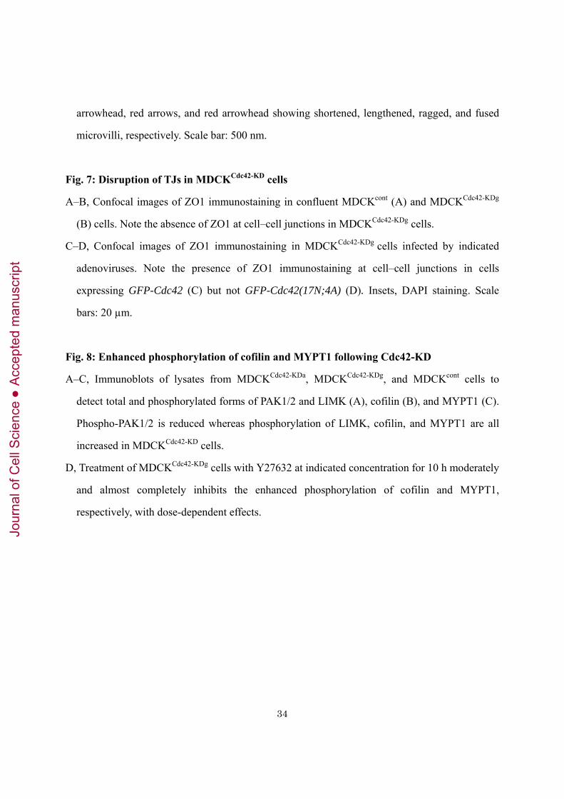
Fig. 1: Cdc42-mRNA expression in cochlea and Cdc42 localization at cochlear stereocilia
A, In situ hybridization (upper panel) detects Cdc42 mRNA expression in cochlear inner hair cell
(IHCs; arrow) and outer hair cell (OHCs; arrowheads) of P5 wild-type mice. Lower panel
shows the relative Cdc42 mRNA signal level determined by the DAB image analyzer (red was
assigned to positive). 28S rRNA complementary oligo-DNA probe was used as a positive
control. Scale bar: 50 µm.
B, Cdc42 (green) and -actin (red) immunostaining in TCA-fixed OCs from Cdc42flox/flox and
Atoh1–Cre;Cdc42flox/flox mice at P0. Note the absence of Cdc42 staining in stereocilia of
Atoh1–Cre;Cdc42flox/flox mice. Scale bars: 10 µm.
Fig. 2: Slowly progressive hearing and HC loss in Atoh1–Cre;Cdc42flox/flox mice
A, Age-related click ABR thresholds (dB SPL) in Cdc42flox/flox (cont) and Atoh1–Cre;Cdc42flox/flox
(Cdc42-KO) mice. N ≥ 4 animals; * p < 0.05
B, Age-related click and tone-burst (8, 16, 24, 32 kHz) ABR thresholds in Cdc42flox/flox and
Atoh1–Cre;Cdc42flox/flox mice. Atoh1–Cre;Cdc42flox/flox mice show a progressive and
high-frequency sound-dominant hearing loss. N ≥ 4; * p < 0.05
C, Age-related DPOAE (f2 frequency at 8, 12, 16, 20 kHz) thresholds in Cdc42flox/flox and Atoh1–
Cre;Cdc42flox/flox mice. Progressive and high-frequency sound-dominant hearing loss is
detected. N ≥ 4; * p < 0.05
D, Representative (n ≥ 4) Alexa488-phalloidin staining shows HC loss (rounds) of middle turn of
cochleae from Cdc42flox/flox and Atoh1–Cre;Cdc42flox/flox mice at the age of 2, 4, and 8 weeks.
E, The percentages of remaining IHCs and OHCs in each turn in Cdc42flox/flox and Atoh1–
Cre;Cdc42flox/flox mice at 8 weeks. Note the IHC- and basal turn (Bs)-dominant HC loss. N ≥ 3
* p < 0.05 Ap: apical Md: middle
Fig. 3: Cdc42 localization and function at the HC stereociliary membranes and AJCs
Jour
nal o
f Cel
l Sci
ence
Acc
epte
d m
anus
crip
t

31
A–B, Dissected OC from P1 wild-type mice cultured for 16 h and the indicated adenoviruses
(green) were infected. The top panels of (A) (100×; scale bar: 2 µm) show
membrane-localization of GFP-Cdc42 covering an individual stereocilum (arrows) and at
apical cell junctions in IHC (arrowhead). Lower panels of (A) and (B) show reconstructed
lateral view (in xz axis; scale bars: 10 µm) images of cochlear OHCs: GFP-Cdc42, but not
GFP-Cdc42(17N;4A), is localized at the stereociliary membranes (arrow).
C, Schematic representation of the intramolecular Cdc42 FRET biosensor: YPet and
Turquoise-GL are variants of YFP and CFP, respectively.
D–G, Dissected OC from P2 Cdc42-FRET mice were observed under a two-photon excitation
microscope. (D) (F), and (G) are obtained from different OCs. Scale bars: 10 µm. (D)
FRET/CFP ratio is highest at the stereocilia (arrowheads) and is higher at the apicolateral
membranes (large arrow) than at the basolateral membranes (small arrow). Note that the image
plane is oblique to the apical surface to show cross-sections of various depths, from the level of
stereocilia to that of the basolateral membrane (see the illustration above it). A 3D-movie is
available in Supplementary movie 1. (E) Schematic drawing shows an overview of IHCs and
OHCs1–3 in OC. (F) Composite image showing the FRET/CFP ratio of a series of five OHCs
in the row of OHC3 obtained in serial sections from the base to top of stereocilia. Note that the
five OHCs are obliquely aligned in vertical direction (see the illustration). The FRET/CFP ratio
is higher in upper parts than basal parts of stereocilia in all OHCs. (G) Representative
FRET/CFP ratio images (N ≥ 3) showing the effect of the Cdc42 inhibitor ML141 (before and
4 min after 500 µM ML141 treatment). FRET/CFP ratios at the stereocilia and AJCs are
significantly decreased by ML141. * p < 0.01
Fig. 4: Degeneration of cochlear stereocilia in Atoh1–Cre;Cdc42flox/flox mice
SEM images of the OC at the middle turn (A–J) and the apical turn (K-M) obtained from
Cdc42flox/flox (cont; A, C, G) and Atoh1–Cre;Cdc42flox/flox (B, D–F, H–M) mice at the age of P8 (E,
F), 2 (H), 4 (I), 6 (J), and 8 weeks (A–D, G, K-M).
Jour
nal o
f Cel
l Sci
ence
Acc
epte
d m
anus
crip
t

32
A, Both IHCs and OHCs are regularly aligned in a plane in Cdc42flox/flox mice at 8 weeks.
B, In Atoh1–Cre;Cdc42flox/flox mice at 8 weeks, IHCs mostly disappeared, whereas OHCs are
partially depleted and have scattered stereocilia.
C, In Cdc42flox/flox OHCs at 8 weeks, stereocilia have the characteristic W-profile.
D, In Atoh1–Cre;Cdc42flox/flox OHCs at 8 weeks, stereocilia are fewer in number and have lost
their characteristic W-profile and precise staircase pattern with consistent length of stereocilia
in each row.
E–F, The morphology of IHC (E) and OHC (F) in Atoh1–Cre;Cdc42flox/flox mice at P8 is identical
to one in Cdc42flox/flox mice (not shown).
G, Regular array of stereocilia in IHCs of Cdc42flox/flox mice at 8 weeks.
H, In the middle turn of cochlea in Atoh1–Cre;Cdc42flox/flox mice, stereocilia fusion (arrowheads)
is first observed at 2 weeks.
I–M, The stereoilia fusion (arrowheads) and IHC loss (asterisks) increase in number through 4 (I)
to 6 weeks (J). The apical turn of cochlea at 8 weeks retains some IHCs, which have frequently
fused stereocilia (K–M, arrowheads), long (L, arrows), and short stereocilia (M, double arrow).
Scale bars: 5 µm in a–b, 2 µm in c–l
Fig. 5: Disturbed ultrastructure of stereocilia and AJCs in Atoh1–Cre;Cdc42flox/flox mice
TEM images of IHCs at the middle turn of cochlea at 6 weeks of age.
A, In Cdc42flox/flox (cont) mice, IHCs and supporting cells (SCs) are alternately aligned (left) and
there is a thick actin-rich cuticular plate bordered by the arcuate-shaped AJCs with the
circumferential actin belt (arrowheads) observed as a dense layer beneath the plasma
membrane (right: magnified view). Stereocilia are located on the apical surface and each has a
single rootlet inserted into the cuticular plate (arrow).
B, Atoh1–Cre;Cdc42flox/flox (Cdc42-KO) mice are missing some IHCs, which were displaced by
SCs, characterized by the apical microvilli. Stereocilia in the remaining IHCs are often fused at
the base (black arrows) and they contain several actin cores (black arrowheads) with rootlets
Jour
nal o
f Cel
l Sci
ence
Acc
epte
d m
anus
crip
t

33
(white arrows). The right two panels are close-up views of SC with microvilli and neighboring
HC (upper) and fused stereocilia base (lower).
C, Close-up views of the AJCs in Cdc42flox/flox (upper panels) and Atoh1–Cre;Cdc42flox/flox mice
(lower panels). Each panel shows a cross section of a SC in the middle, flanked by two HCs
(asterisks). The AJCs display smooth and vertical arcs in controls but shows a range of ruffling
in Atoh1–Cre;Cdc42flox/flox mice.
D, Close-up views of the AJCs and perijunctional actin belts. HCs (asterisks) on the right side.
The actin belt (outlined by white dots) is thinner in Atoh1–Cre;Cdc42flox/flox HCs.
Scale bars: 1 µm in a–b, 500 nm in c, 200 nm in d
Fig. 6: Reduced number and shape-abnormalities of microvilli in MDCKCdc42-KD cells
A, Confocal images of MDCKGFP-Cdc42 cells cultured in Matrigel and counterstained with
Alexa568-phalloidin. Note the strong and weak GFP-Cdc42 accumulation at the apical (arrow)
and lateral surfaces of cyst-cells (arrowheads), respectively. Scale bar: 10µm
B–C, Immunoblot showing the effects of three different shRNA-plasmids targeting dog Cdc42
(sh29, sh197, sh333) in MDCK cells. Using the most effective sh197, two different clones
(MDCKCdc42-KDa and MDCKCdc42-KDg) were established.
D, SEM images showing the apical surface of MDCK cells grown on a filter insert. Both
MDCKCdc42-KDa and MDCKCdc42-KDg cell lines had substantially fewer microvilli compared
with cells expressing a control shRNA (MDCKcont). Scale bar: 5 µm.
E, SEM images of MDCKcont and MDCKCdc42-KDg cells grown on a filter insert, infected by
indicated adenoviruses. Colored insets show the GFP signal. Note the localization of
GFP-Cdc42 at cell–cell junctions (arrowheads). Scale bars: 10µm
F, Recovery of reduced microvilli in MDCKCdc42-KDg cells by the expression of
adenovirus-encoded GFP-Cdc42 but not GFP-Cdc42(17N;4A). * p < 0.01
G, High power images obtained by scanning helium-ion microscope (SHIM) showing the range
of morphological abnormalities in microvilli in MDCKCdc42-KDg cells. Black arrow, black
Jour
nal o
f Cel
l Sci
ence
Acc
epte
d m
anus
crip
t
34
arrowhead, red arrows, and red arrowhead showing shortened, lengthened, ragged, and fused
microvilli, respectively. Scale bar: 500nm.
Fig. 7: Disruption of TJs in MDCKCdc42-KD cells
A–B, Confocal images of ZO1 immunostaining in confluent MDCKcont (A) and MDCKCdc42-KDg
(B) cells. Note the absence of ZO1 at cell–cell junctions in MDCKCdc42-KDg cells.
C–D, Confocal images of ZO1 immunostaining in MDCKCdc42-KDg cells infected by indicated
adenoviruses. Note the presence of ZO1 immunostaining at cell–cell junctions in cells
expressing GFP-Cdc42 (C) but not GFP-Cdc42(17N;4A) (D). Insets, DAPI staining. Scale
bars: 20 µm.
Fig. 8: Enhanced phosphorylation of cofilin and MYPT1 following Cdc42-KD
A–C, Immunoblots of lysates from MDCKCdc42-KDa, MDCKCdc42-KDg, and MDCKcont cells to
detect total and phosphorylated forms of PAK1/2 and LIMK (A), cofilin (B), and MYPT1 (C).
Phospho-PAK1/2 is reduced whereas phosphorylation of LIMK, cofilin, and MYPT1 are all
increased in MDCKCdc42-KD cells.
D, Treatment of MDCKCdc42-KDg cells with Y27632 at indicated concentration for 10 h moderately
and almost completely inhibits the enhanced phosphorylation of cofilin and MYPT1,
respectively, with dose-dependent effects.
Jour
nal o
f Cel
l Sci
ence
Acc
epte
d m
anus
crip
t
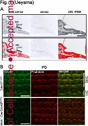
Jour
nal o
f Cel
l Sci
ence
Acc
epte
d m
anus
crip
t

Jour
nal o
f Cel
l Sci
ence
Acc
epte
d m
anus
crip
t

Jour
nal o
f Cel
l Sci
ence
Acc
epte
d m
anus
crip
t
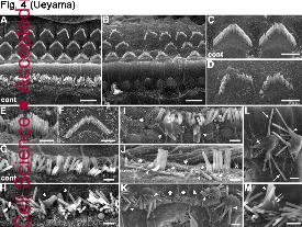
Jour
nal o
f Cel
l Sci
ence
Acc
epte
d m
anus
crip
t

Jour
nal o
f Cel
l Sci
ence
Acc
epte
d m
anus
crip
t

Jour
nal o
f Cel
l Sci
ence
Acc
epte
d m
anus
crip
t
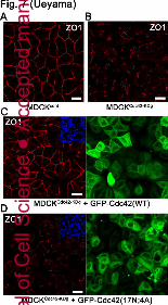
Jour
nal o
f Cel
l Sci
ence
Acc
epte
d m
anus
crip
t

Jour
nal o
f Cel
l Sci
ence
Acc
epte
d m
anus
crip
t