Embed Size (px)
Citation preview

MANAGEMENT OF ACUTE CHOLECYSTITIS
IN A MEDIUM-SIZED CENTRE
Tutor: Laia Falgueras Author: Adrià Costa i Roig
Final Degree Project
November, 2015
Faculty of Medicine
University of Girona

Management of acute cholecystitis in a medium-sized centre
2
M’agradaria agrair a la secció de cirurgia hepato-biliar de l’Hospital Dr. J. Trueta de
Girona per la seva implicació i dedicació en la meva formació.
Gràcies a la Laia Falgueras per fer possible aquest treball.
Management of acute cholecystitis in a medium-sized centre
3
INDEX
1. ABBREVIATIONS ............................................................................... 4
2. ABSTRACT ..................................................................................... 5
3. BACKGROUND ................................................................................. 6
4. JUSTIFICATION .............................................................................. 16
5. BIBLIOGRAPHY ............................................................................... 17
6. HYPOTHESIS .................................................................................. 20
7. OBJECTIVES .................................................................................. 20
7.1 Main objective ............................................................................ 20
7.2 Secondary objectives .................................................................... 20
8. METHODS ..................................................................................... 20
8.1 Study design .............................................................................. 20
8.2 Participants ............................................................................... 20
8.3 Sample size ............................................................................... 21
8.4 Sample collection ........................................................................ 21
9. VARIABLES AND MEASUREMENTS ......................................................... 23
9.1 Independent variables................................................................... 23
9.2 Dependent variables ..................................................................... 23
9.3 Co-variables .............................................................................. 24
10. STATISTICAL ANALYSIS ................................................................... 25
11. STRENGTHS AND LIMITATIONS .......................................................... 26
12. ETHICAL CONSIDERATIONS ............................................................... 27
13. FEASIBILITY ................................................................................. 28
14. IMPACT OF THE STUDY TO THE NATIONAL HEALTH SYSTEM ...................... 29
15. WORKING PLAN ............................................................................ 29
16. CHRONOGRAM .............................................................................. 31
17. BUDGET ...................................................................................... 32
18. ANNEX ....................................................................................... 33
18.1 ANNEX 1 .................................................................................. 33
18.2 ANNEX 2 .................................................................................. 34
18.3 ANNEX 3.1 ............................................................................... 35
18.4 ANNEX 3.2 ............................................................................... 36
18.5 ANNEX 4 .................................................................................. 37
Management of acute cholecystitis in a medium-sized centre
4
1. ABBREVIATIONS
γGTP Gamma-glutamyltransferase
AAC Acalculous acute cholecystitis
AC Acute Cholecystitis
AG Acute Cholangitis
ALP Alkaline Phosphatase
ALT Alanine Aminotransferase
AST Aspartate Aminotransferase
CT Computerized tomography
OC Open Cholecystectomy
LC Laparoscopic Cholecystectomy
OR Other researcher
CRP C Reactive Protein
MR Main researcher
MRI Magnetic Resonance Image
RUQ Right upper quadrant
STD Upper range standard values
SD Standard deviation
US Ultrasounds
T-Bil Total bilirubin
TG Tokyo Guidelines
WBC White blood count
Management of acute cholecystitis in a medium-sized centre
5
2. ABSTRACT
Title: Management of acute cholecystitis in a medium-sized centre
Authors: Adrià Costa, MS; Laia Falgueras, MD.
Background: Cholelithiasis is a frequent gastrointestinal disease, and acute
cholecystitis (AC) is one of its most frequent complication. Before Tokyo Guidelines
(TG) for AC management, no consensus on how to diagnose and treat these patients
was established. Despite having a low mortality rate, a meta-analysis reported a 3% of
mortality before guidelines implementation. After TG, no studies clarified its
improvement in terms of mortality and only few were carried out. Those studies
demonstrated only a reduced length of stay in comparison. Authors concluded that
further studies are needed. Additionally, no studies collecting AC data were found in
our region. Finally, due to its chronic condition, cholelithiasis is a high burden disease
all over, and AC management is expensive as well. This study may have an economic
impact if it reveals that we could be more efficient treating these patients. Thus, this
study pretends to analyse how our medium-sized centre manages AC, to compare our
results with those obtained during pre-TG era, to create a large data base and finally
to know which impact had TG on AC management.
Objective: This study aims to find the mortality rate of patients diagnosed and treated
of AC according to TG recommendations at General Surgery department of our centre.
We expect a 2’5% mortality rate, demonstrating a better survival rate than before TG
era. Length of stay and surgical-related complications will be also evaluated.
Design: Cross-sectional study conducted in a medium-sized centre between January
2016 and July 2017.
Methods: We will need a sample of 82 patients if we expect a 2’5% of mortality rate.
Non-probabilistic, consecutive method of recruitment will be used. TG diagnostic
criteria will be applied for these patients and they will be categorized into three
severity groups with different treatment per each one. Statistical analysis will be
adjusted by possible confounding variables.
Keywords ▪ acute cholecystitis ▪ mortality rate ▪ criteria ▪ hospital stay ▪ sequels
Management of acute cholecystitis in a medium-sized centre
6
3. BACKGROUND
Gallbladder stones disease, also known as cholelithiasis, is a common, chronic and
recurrent hepatobiliary disease. Due to an impaired cholesterol metabolism and
reduced gallbladder motility –among other factors- gallstones are formed in the
gallbladder or in the bile ducts. Even though it is present worldwide, its prevalence
varies by region[10,11]. In Western countries it has a prevalence of 7’9% in men and 16’6%
in women[8], with a lower rate in Asian and African countries. As a result of its chronic
and recurrent status, it has an economic impact and a sustainable burden, becoming
one of the most expensive diseases among gastroenterology pathologies, exceeding 5
billion dollars in the United States[10].
As above-commented, women suffer more from this disease as female gender is one
of the main risk factors to develop gallstones, especially during fertile age and
pregnancy, when gallbladder evacuation is impaired. However, high prevalence has
been observed among cirrhotic men as well. The bile excretion to the duodenum is
reduced by age due to motility’s reduction, becoming a common disease especially in
the elderly. Obesity also implies an impaired cholesterol metabolism, involving a high
prevalence in those patients. Drugs – like oestrogens -, diet or genetic factors are also
known as risk factors to develop gallstones[10,11].
The composition of gallstones varies: cholesterol stones are up to 75% of the total,
followed by pigment stones (less than 30% of cholesterol in their composition) and
mixed stones. Once the gallstone is formed, it may shrink, grow or remain the same
size for decades. It may have a widely range of on-set presentation, and many patients
(up to 80%) will not experience any symptom through their lives[10]. Normally, it
presents with abdominal discomfort and indigestion, but those symptoms are weakly
specific and both symptoms can be seen in other gastrointestinal diseases. It is not
unusual that the first diagnosis is made by chance during any other image exploration
Management of acute cholecystitis in a medium-sized centre
7
of the abdomen, such as a CT scan or US imaging. Among all the patients suffering
from gallstone disease, only 3-6% will develop further complications, such as acute
cholecystitis, acute cholangitis, jaundice and pancreatitis[15].
Amongst all of the complications that these patients can suffer, acute cholecystitis is
the most common and one of the most frequent diseases among gastrointestinal-
related attended visits to our emergency department. AC is an acute inflammatory
disease of the gallbladder after obstruction of the cystic duct. Up to 1-3% of patients
with abdominal pain can be account for AC, and around 10% of patients with
symptomatic gallstones will develop AC during their life, being both more common and
severer with age (20’9% above 50 years)[8,10].
Up to a 90% of the cases of AC are related with gallstones, known as
cholecystolithiasis[18], but other causes may develop it as well: helminthic infection
(ascariasis) is one of the most frequent causes of AC in developing countries. AIDS is
also a risk factor as long as patients suffering from AIDS usually have abnormal liver
functions – i.e. AF is elevated in blood tests - and dilatation of cystic is observed by US
and CT scans. Drugs may be involved in some AC such as oral contraceptives, though
its relation is discussed, ceftriaxone (only during treatment due to augmented
precipitated calcium salt in bile) and fibrates[8,11,21]. Finally, acalculous acute
cholecystitis (AAC) is also an acute condition involving gall bladder inflammation but
its causes are completely different from those of calculous AC. Normally, it’s due to a
local ischemia of the gall bladder in elderly or severe injured patients with other
illness. Diabetes and myocardial infarction have been proved as causes of AAC and
especially patients who are admitted to ICU for over a long period tend to develop this
condition[8,27].
Once a stone is impacted on the neck of the gallbladder, it goes through different
phases if treatment is not rapidly applied: till day four, only oedema is visible in its
Management of acute cholecystitis in a medium-sized centre
8
walls, evolving to haemorrhage and necrosis of the wall and vascular thrombosis after
96 hours of the on-set. After seven days, suppurated cholecystitis and pericholecystic
abscesses are present, increasing its mortality[17].
Advanced phases are avoidable with early detection using image techniques, proper
physical exploration and a administrating a correct treatment, saving a high amount
of money in further procedures[8]. Yet, they can be lethal when severe.
Further complications may occur: perforation of the gallbladder, biliary peritonitis and
biliary fistula to the duodenum, Mirizzi Syndrome1 – though infrequent - and gallstone
ileus, causing mechanical obstruction at the ileocecal valve[1,8,21], resulting in a higher
mortality.
AC prognosis may vary depending on its severity[4] though mild and moderate cases
have low mortality rate. Elderly patients and those with comorbidities tend to have
higher mortality than healthy, young patients[17]. AG findings on a AC patient are
considered as bad prognosis factors[15]. Postoperative infections were the main cause
of death after surgery before 1980 such as ascending cholangitis, hepatic abscess and
sepsis. Nowadays, mortality has decreased since the proper use of antibiotics and
general supportive care was established[9,24,26] and these patients have the same
expectancy of life as other patients have.
For decades, AC diagnosis has been established by clinical findings, local and systemic
inflammatory signs and laboratory tests[8,25]. Its typical presentation is constant pain
for hours and tenderness in the RUQ, normally with a previous history of pain on the
same region. Murphy’s sign2 may be present in these patients whereas blood tests show
elevated PCR, elevated WBC and fever. None of them were completely specific of AC,
1 Mirizzi Syndrome: Obstruction of common hepatic duct due to an impacted stone in Hartmann’s pouch 2 Murphy sign: Inspiration interruption by pain or palpation in RUQ
Management of acute cholecystitis in a medium-sized centre
9
and some of them may be present in other acute abdominal causes that have to be
ruled out. After new image techniques utilisation, such as high-resolution CT scan and
US scan, it was far easier to diagnose a gall bladder dysfunction. Actually ultrasound
scanning became a fast, non-expensive choice for these patients upon their arrival at
emergency department[2,8]. Even though it is an observational-dependent technique, it
is really useful to observe pericholecystic fluid (fluid around the gall bladder),
distended gall bladder wall and oedema. CT scan may be used to rule out other acute
abdomen causes in uncertain diagnosis. Despite having these techniques, no radiology
criteria were established to properly diagnose acute cholecystitis, leading every centre
to use their own criteria.
Before the publication of Tokyo Guidelines in 2007, there was weak consensus on
diagnosing criteria and treatment of acute cholecystitis for the above-
mentioned[3,6,19,25]. There were no international standards of managing these common
acute pathologies: open cholecystectomy used to be the best procedure to solve the
condition after the acute phase[20,26,28]. Actually, most of studies regarding AC have
always carried out in a different way that we are proposing: to prove out whether LC
was better than OC, which has been the gold standard lately[9,14,20,26,28].
LC proved to reduce the mortality and length of stay by itself due to a smaller wound
(as the intraabdominal procedure is the same for both OL and LC)[7,24].
Overall, about 20% of patients with AC need emergency surgery, especially when their
condition is deteriorated and generalised peritonitis or emphysematous cholecystitis
are present[3,5,17]. Percutaneous cholecystostomy has been proposed as a minimally
invasive procedure in moderate and severe cases. Those patients have a higher risk
from major surgery and they need to solve the acute phase before to undergo to
laparoscopy. It has been evidenced that this procedure may reduce mortality[7,9].

Management of acute cholecystitis in a medium-sized centre
10
To settle down universal criteria and management, experts of Japanese Hepatobiliary
Association proposed an international consensus in 2005: the Japanese clinical
guidelines for acute cholecystitis and acute cholangitis[19]. Then, in further revisions,
they published Tokyo Guidelines in 2007, reaching an 84’9% of sensitivity in diagnosing
AC and becoming the international reference ever since[2]. However, potential
shortcomings were present according to some authors so these guidelines needed
further evidence[6]. Thus, they carried out a new study in order to upgrade the Tokyo
guidelines, publishing TG 2013[2,3].
They finally proposed high sensitive diagnosing criteria for AC using clinical features,
blood test data and image findings. To reach that high sensitivity of diagnosing AC,
they excluded patients with AG findings or other causes of AC, i.e acalculous
cholecystitis, which is not taken into consideration in their publication either.
Table 1 Diagnostic criteria of AC. Adapted from [2]
A. Local signs of inflammation 1) Murphy’s sign 2) RUQ mass / pain / tenderness
B. Systemic signs of inflammation 1) Fever (>37’5ºC) 2) Elevated CRP (≥ 3mg/dl) 3) Abnormal WBC count
C. Imaging findings characteristic of AC*
Definite diagnosis: 1) One item in A and one item in B are positive 2) C confirms the diagnosis when AC is suspected clinically
*Imaging findings of AC: US: - Sonographic Murphy sign - Thickened gallbladder wall - Enlarged gallbladder - Incarcerated gallstone - Sonolucent layer in the gallbladder wall, striated intramural lucencies and Doppler signals MRI: - Pericholecystic high signal - Enlarged gallbladder - Thickened gallbladder wall CT: - Thickened gallbladder wall - Pericholecystic fluid collection - Enlarged gallbladder - Linear high-density areas in the pericholecystic fat tissue - Non-visualized gallbladder with normal uptake and excretion of radioactivity - Rim sign (augmentation of radioactivity around the gallbladder fossa)
Management of acute cholecystitis in a medium-sized centre
11
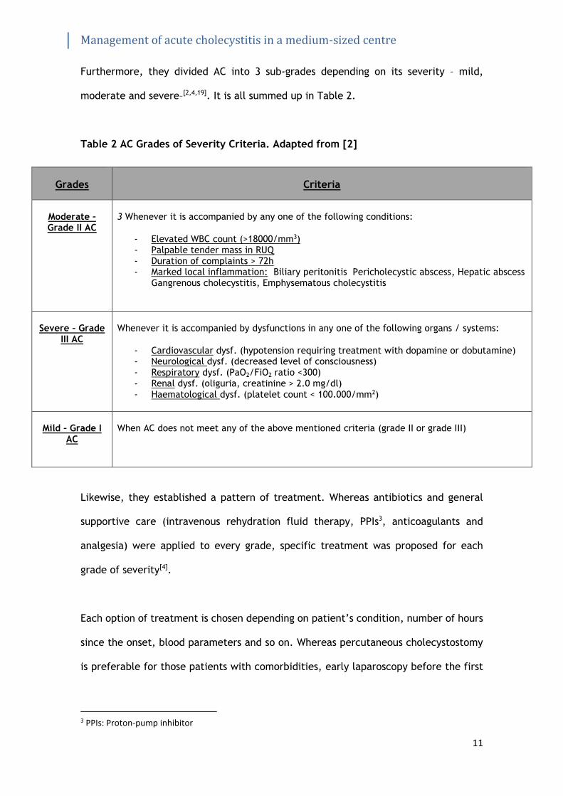
Furthermore, they divided AC into 3 sub-grades depending on its severity – mild,
moderate and severe–[2,4,19]. It is all summed up in Table 2.
Table 2 AC Grades of Severity Criteria. Adapted from [2]
Likewise, they established a pattern of treatment. Whereas antibiotics and general
supportive care (intravenous rehydration fluid therapy, PPIs3, anticoagulants and
analgesia) were applied to every grade, specific treatment was proposed for each
grade of severity[4].
Each option of treatment is chosen depending on patient’s condition, number of hours
since the onset, blood parameters and so on. Whereas percutaneous cholecystostomy
is preferable for those patients with comorbidities, early laparoscopy before the first
3 PPIs: Proton-pump inhibitor
Grades Criteria
Moderate – Grade II AC
3 Whenever it is accompanied by any one of the following conditions:
- Elevated WBC count (>18000/mm3) - Palpable tender mass in RUQ - Duration of complaints > 72h - Marked local inflammation: Biliary peritonitis Pericholecystic abscess, Hepatic abscess
Gangrenous cholecystitis, Emphysematous cholecystitis
Severe – Grade III AC
Whenever it is accompanied by dysfunctions in any one of the following organs / systems:
- Cardiovascular dysf. (hypotension requiring treatment with dopamine or dobutamine) - Neurological dysf. (decreased level of consciousness) - Respiratory dysf. (PaO2/FiO2 ratio <300) - Renal dysf. (oliguria, creatinine > 2.0 mg/dl) - Haematological dysf. (platelet count < 100.000/mm2)
Mild – Grade I AC
When AC does not meet any of the above mentioned criteria (grade II or grade III)

Management of acute cholecystitis in a medium-sized centre
12
72hrs is the best surgical choice, as it is associated with less complications, reduced
length of stay and less risk of conversion to open laparotomy[18].
To sum up all of the treatment options, a flowchart (Figure 1) from TG13 is provided.
Figure 1 Treatment options according to Grade and Response[12]
As an international consensus, it helped to settle knowledge and to start a common
way to manage these pathologies, Further studies are still needed to assess whether
the survival rate following these guidelines improved or not. Only few studies were
published after the consensus so we do not have post-Tokyo 2013 data to compare with
yet. A Japanese author weakly evidenced that only days of hospitalisation were
reduced after guidelines, but concluded that further studies to evaluate a large series
of patients were needed, also to prove a reduced mortality[13]. For that reason and to
make a good comparison between before and after TG publication, we had to focus on
Management of acute cholecystitis in a medium-sized centre
13
Kimura’s meta-analysis data which studied large series of patients[16]. It will be our
data reference. We agreed its mortality rate obtained were a good standard to
compare our results with.
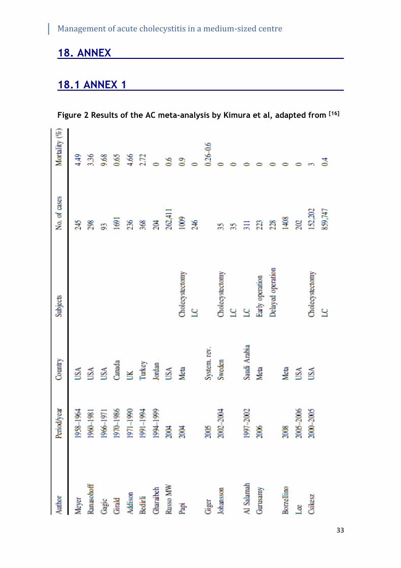
As above mentioned, Kimura et al published in 2013 a collection data of patients
diagnosed and treated of acute cholecystitis from 1958 to 2009, before the
implementation of TG (see Annex 1) [16]. Logically, we had to focus on the newest
studies as good references as both the surgical treatment and the conservative
management of these patients evolved considerably since 1958. The newest studies
got a mortality rate between 0% and 3%[16]. That value is an average of individually
obtained in each group of severity (mortality was far higher in group 3 patients than
in mild – moderate group).
To better understand how it is possible to get a 0% mortality rate in such an acute
condition, we revised those studies: we found out that their sample selection was quite
restricted and they only selected the better patients for studying. Actually, it was the
main bias of that study.
We considered that 3% should be our reference number, and we expect a reduced
mortality rate with this study. We want to prove that TG recommendations have
improved this outcome and they are useful and helpful for physicians to handle this
condition worldwide.
What studies did prove was that the diagnosing accuracy using TG 2013 were far higher
than without them [3].
Management of acute cholecystitis in a medium-sized centre
14
Something that attracted our attention was that there were not any studies evaluating
these outcomes in our centre, neither in Spain. So we had not data of how we treated
these patients and, specifically, how TG would have improved AC management in our
department. This project also aims to collect more data about AC patients in our
medium-sized centre, becoming one of the first hospitals to start a data base, along
with Tarragona’s Hospital Joan XIII, who recently started a multi-centric study to
recollect data of AC. Unfortunately, we do not have their results yet.
Additionally, as it is a common and chronic disease the total burden spent on it is really
high[8,10]. Based on last year prevalence data of our centre, we treated 84 AC with an
average of 7’65 days of hospitalisation per each patient. According to ICS financial
department, every day of hospitalisation costs 557€ (including treatments such as
antibiotics, general supportive care and personnel), daily hepatobiliary blood test costs
47’85€ and Ultrasound scan, 62€. In case an abdominal CT would be needed to rule out
other diagnoses of acute abdomen, it costs 95€. Leaving aside which grade the patient
is categorized into, those costs are equal for each patient. Then, in case a
percutaneous cholecystostomy would be necessary, total price is raised in 145€.
Finally, for those who have to undergo to a laparoscopic cholecystectomy, this
procedure is rated in 5548€ (no conversion to OL data is available).
With a mean of 7’65 days in our in-patient clinic (with general supportive care and
antibiotics, fluids, drugs), our patients will need at least a US scan to be diagnosed,
one pre-surgery blood test (supposing an invasive treatment is needed) and at least
two post-surgery/treatment blood tests to check if condition is improving. In case they
undergo to surgery - and no further complications after the early laparoscopic
cholecystectomy appear - the total cost of their stay is 10,014’6€. In that price, we
assumed diagnosis was fully accurate since the first moment using TG. We could have
got to rule out other diagnosis with a CT scan, diagnosed that acute abdomen wrongly,
Management of acute cholecystitis in a medium-sized centre
15
or to face complications during hospitalisation.
Everything results in more days of hospitalisation, more blood test and maybe other
procedures. Overall, the total cost could have been much higher. Taking into account
we treated 84 patients with AC last year, the total burden spent on it was 841,226€.
Neither complications nor readmissions were contemplated.
Furthermore, as these procedures are not risk free patients may develop potentially
complications. These complications may be related with previous comorbidity of our
patients or surgery-related[17]. For instance, one of the surgical complications – even
more frequent with LC than OC – is the biliary tract lesion. It is not frequent (0’4% to
0’6% of total LC) but it has an important reduction of health quality[23]. Thus, we
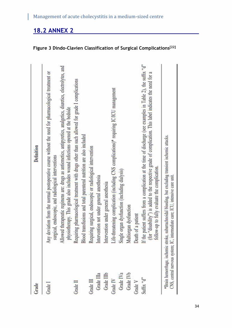
pretend to collect all of the data regarding complications during the days of
hospitalisation, and we will describe them using Dindo-Clavien Classification for
Surgical Complications (see Annex 2)[22]. We aim to be more conscious of all possible
complications in order to increase our caring towards these patients.
Management of acute cholecystitis in a medium-sized centre
16
4. JUSTIFICATION
We consider this study as the initial tool to evaluate how have been implemented the
international guidelines recommendations for an AC and how our department is
managing that pathology. With this study we will highlight what we do correctly and
especially what we can improve in further patient management.
Our first aim is to demonstrate that TG are useful for physicians and if they made an
improvement to get better outcomes since its publication. Therefore, we may point
whether we are following correctly TG or not (in terms of diagnosis and treatment).
Achieving better outcomes values is not our only aim nowadays. The total burden spent
on this condition in our centre is really high, as we have seen before. An improvement
of resources management is a latter aim if we found out that we could be as good as
we can at managing AC but also doing it with a better resources handling.
Additionally, we pretend to describe the most frequent complications our patients
suffer during hospitalisation. Having a complete list of most prevalent complications
may change our point of view of AC. In case most of the complications appeared the
first hours or days of the hospitalisation – i.e. grade 3 patients -, we should improve
our caring towards them during that time and also apply an exhaustive monitoring at
special units (UPIC)4 trying to prevent those possible complications, resulting in
reduced morbidity, better quality of life and less budget.
Therefore, being one of the first studies in our region to evaluate those outcomes, we
may set a national precedent to encourage other departments to start other projects
as well, comparing and sharing our results in further national congresses.
Our last aim is to excel in AC patient management. We may start new projects as
clinical protocols together with emergency department or radiology department,
compare results worldwide and be a national reference in its management.
4 UPIC: Unitat polivalent d’alta intenstitat de cures
Management of acute cholecystitis in a medium-sized centre
17
5. BIBLIOGRAPHY
1. Zaliekas J, Munson JL. Complications of Gallstones: The Mirizzi Syndrome,
Gallstone Ileus, Gallstone Pancreatitis, Complications of “Lost” Gallstones.
Surg Clin North Am [Internet]. Elsevier Inc.; 2008 Dec [Cited 2015 Sep
20];88(6):1345–68. Available from:
http://linkinghub.elsevier.com/retrieve/pii/S0039610908001011
2. Yokoe M, Takada T, Strasberg SM, Solomkin JS, Mayumi T, Gomi H, et al. New
diagnostic criteria and severity assessment of acute cholecystitis in revised
Tokyo guidelines. J Hepatobiliary Pancreat Sci. 2012;19(5):578–85.
3. Yokoe M, Takada T, Strasberg SM, Solomkin JS, Mayumi T, Gomi H, et al. TG13
diagnostic criteria and severity grading of acute cholecystitis (with videos). J
Hepatobiliary Pancreat Sci. 2013;20(1):35–46.
4. Yamashita Y, Takada T, Kawarada Y, Nimura Y, Hirota M, Miura F, et al.
Surgical treatment of patients with acute cholecystitis: Tokyo guidelines. J
Hepatobiliary Pancreat Surg. 2007;14(1):91–7.
5. Welch JP. Evolving treatment paradigms for acute cholecystitis: comment on
“Surgical management of acute cholecystitis at a tertiary care center in the
modern era”. Arch Surg. [Internet] 2010 May [Cited 2015 Sep 18];145(5):444.
Available from: http://www.ncbi.nlm.nih.gov/pubmed/20479341
6. Takada T, Strasberg SM, Solomkin JS, Pitt H a., Gomi H, Yoshida M, et al.
TG13: Updated Tokyo Guidelines for the management of acute cholangitis and
cholecystitis. J Hepatobiliary Pancreat Sci. [Internet] 2013 Jan [Cited 2015 Sep
12];20(1):1–7. Available from:
http://www.ncbi.nlm.nih.gov/pubmed/23307006
7. Shinya S, Yamashita Y, Takada T. The impact of the Japanese clinical
guidelines on the clinical management of patients with acute cholecystitis. J
Hepatobiliary Pancreat Sci. 2013;20(6):611–9.
8. Shaffer E a. Epidemiology and risk factors for gallstone disease: has the
paradigm changed in the 21st century? Curr Gastroenterol Rep. 2005;7:132–40.
9. Rodríguez-Sanjuán JC, Arruabarrena A, Sánchez-Moreno L, González-Sánchez
F, Herrera L a., Gómez-Fleitas M. Acute cholecystitis in high surgical risk
patients: Percutaneous cholecystostomy or emergency cholecystectomy? Am J
Surg [Internet]. Elsevier Inc.; 2012 Jul [Cited Sep 17];204(1):54–9. Available
from: http://dx.doi.org/10.1016/j.amjsurg.2011.05.013
10. Reshetnyak VI. Concept of the pathogenesis and treatment of cholelithiasis.
World J Hepatol. 2012;4(2):18–34.
Management of acute cholecystitis in a medium-sized centre
18
11. Portincasa P, Moschetta A, Palasciano G. Cholesterol gallstone disease. Lancet.
2006;368(9531):230–9.
12. Miura F, Takada T, Strasberg SM, Solomkin JS, Pitt H a., Gouma DJ, et al. TG13
flowchart for the management of acute cholangitis and cholecystitis. J
Hepatobiliary Pancreat Sci. 2013;20(1):47–54.
13. Lee SW, Yang SS, Chang C Sen, Yeh HJ. Impact of the Tokyo guidelines on the
management of patients with acute calculous cholecystitis. J Gastroenterol
Hepatol. 2009;24(12):1857–61.
14. Kortram K, van Ramshorst B, Bollen TL, Besselink MGH, Gouma DJ, Karsten T,
et al. Acute cholecystitis in high risk surgical patients: percutaneous
cholecystostomy versus laparoscopic cholecystectomy (CHOCOLATE trial):
study protocol for a randomized controlled trial. Trials [Internet]. BioMed
Central Ltd; 2012 Jan [Cited 2015 Sep 15];13(1):7. Available from:
http://www.pubmedcentral.nih.gov/articlerender.fcgi?artid=3285056&tool=p
mcentrez&rendertype=abstract
15. Kiriyama S, Takada T, Strasberg SM, Solomkin JS, Mayumi T, Pitt H a., et al.
TG13 guidelines for diagnosis and severity grading of acute cholangitis (with
videos). J Hepatobiliary Pancreat Sci. 2013;20(1):24–34.
16. Kimura Y, Takada T, Strasberg SM, Pitt H a., Gouma DJ, Garden OJ, et al.
TG13 current terminology, etiology, and epidemiology of acute cholangitis and
cholecystitis. J Hepatobiliary Pancreat Sci. 2013;20(1):8–23.
17. Kimura Y, Takada T, Kawarada Y, Nimura Y, Hirata K, Sekimoto M, et al.
Definitions, pathophysiology, and epidemiology of acute cholangitis and
cholecystitis: Tokyo Guidelines. J Hepatobiliary Pancreat Surg. 2007;14(1):15–
26.
18. Indar A a, Beckingham IJ. Acute cholecystitis. BMJ. 2002;325(September):639–
43.
19. Hirota M, Takada T, Kawarada Y, Nimura Y, Miura F, Hirata K, et al. Diagnostic
criteria and severity assessment of acute cholecystitis: Tokyo Guidelines. J
Hepatobiliary Pancreat Surg. 2007;14(1):78–82.
20. Gurusamy K, Samraj K, Gluud C, Wilson E, Davidson BR. Meta-analysis of
randomized controlled trials on the safety and effectiveness of early versus
delayed laparoscopic cholecystectomy for acute cholecystitis. Br J Surg.
[Internet] 2010 Feb [Cited 2015 Sep 20];97(2):141–50. Available from:
http://www.ncbi.nlm.nih.gov/pubmed/20035546
21. Friedman GD. Natural history of asymptomatic and symptomatic gallstones. Am
J Surg [Internet] 1993 Apr [Cited 2015 Sep 17];165(4):399–404. Available from:
http://www.ncbi.nlm.nih.gov/pubmed/8480871

Management of acute cholecystitis in a medium-sized centre
19
22. Dindo D, Demartines N, Clavien P-A. Classification of Surgical Complications.
Ann Surg [Internet]. 2004 Aug [Cited 2015 Sep 20];240(2):205–13. Available
from:
http://content.wkhealth.com/linkback/openurl?sid=WKPTLP:landingpage&an=
00000658-200408000-00003
23. Dageforde LA, Landman MP, Feurer ID, Poulose B, Pinson CW, Moore DE. A
cost-effectiveness analysis of early vs late reconstruction of iatrogenic bile
duct injuries. J Am Coll Surg [Internet]. Elsevier Inc.; 2012 Jun [Cited 2015 Sep
15];214(6):919–27. Available from:
http://www.ncbi.nlm.nih.gov/pubmed/22495064
24. Csikesz N, Ricciardi R, Tseng JF, Shah S a. Current status of surgical
management of acute cholecystitis in the United States. World J Surg.
[Internet]. 2008 Oct [Cited 2015 Sep 10];32(10):2230–6. Available from:
http://www.ncbi.nlm.nih.gov/pubmed/18668287
25. Care I, Management Q, Hospital B, Louis S, Town C, Africa S, et al. Tokyo
Guidelines for the management of acute cholangitis and cholecystitis.
Proceedings of a consensus meeting, April 2006, Tokyo, Japan. J Hepatobiliary
Pancreat Surg. 2007;14(1):1–121.
26. Borzellino G, Sauerland S, Minicozzi AM, Verlato G, Di Pietrantonj C, de
Manzoni G, et al. Laparoscopic cholecystectomy for severe acute cholecystitis.
A meta-analysis of results. Surg Endosc [Internet]. 2008 Jan [cited 2015 Sep
11];22(1):8–15. Available from:
http://www.ncbi.nlm.nih.gov/pubmed/17704863
27. Barie PS, Eachempati SR. Acute acalculous cholecystitis. Curr Gastroenterol
Rep [Internet]. 2003 Aug [cited 2015 Sep 13];5(4):302–9. Available from:
http://www.ncbi.nlm.nih.gov/pubmed/12864960
28. Agresta F, Campanile FC, Vettoretto N, Silecchia G, Bergamini C, Maida P, et
al. Laparoscopic cholecystectomy: consensus conference-based guidelines.
Langenbeck’s Arch Surg [Internet]. 2015 May [Cited Sep 17];400(4):429–53.
Available from: http://link.springer.com/10.1007/s00423-015-1300-4

Management of acute cholecystitis in a medium-sized centre
20
6. HYPOTHESIS
Patients diagnosed and treated of acute cholecystitis as TG recommends have lower
mortality than those who were not managed following these guidelines.
7. OBJECTIVES
7.1 Main objective
This study aims to find the mortality rate of patients diagnosed and treated of acute
cholecystitis according to TG recommendations at General Surgery department of our
centre in order to compare these results with those obtained from other studies before
TG implementation[16].
7.2 Secondary objectives
- To evaluate the length of stay of these patients at our department and compare
that outcome with other studies.
- To describe a list of the surgical-related complications our patients suffer
during their hospitalization using Dindo-Clavien classification (see Annex 2)[22].
8. METHODS
8.1 Study Design
It will be a cross-sectional study executed at a medium-sized centre, Hospital
Universitari Doctor Josep Trueta in Girona.
8.2 Participants
Patients admitted to Hospital Universitari Doctor Josep Trueta in Girona, from
February 2016 to February 2017 with diagnosis of acute cholecystitis (Table 1)
evaluated by an on-call surgeon or resident of general surgery department.
Management of acute cholecystitis in a medium-sized centre
21
8.3 Sample size
Patients diagnosed with AC are our reference population. Assuming an alpha risk of
0,05 and a precision of +/- 0,005 units for an estimated proportion of 0,025, our sample
size is calculated to be of 83 patients (It has been anticipated a dropout of 10%). We
estimated this sample size using an online tool - Granmo® Calculator- taking into
consideration that we treated 84 patients with AC last year. For that reason, we will
have to spend 12 months collecting our sample.
8.4 Sample collection
A non-probabilistic consecutive method of recruitment will be used. On-call
surgeons/residents of general surgery department will collect the data, who will also
give proper information to the patient or his/her relatives about the study and its
importance, the data confidentiality and the voluntary aspect of it (see Annex 3.1 and
3.2), inviting them to participate.
Surgeons will recollect data after 24hrs upon patient’s arrival (those who die within
the 24th hour will not be included) using the case report form (see Annex 4); after
discharge (to describe complications, if any) and after 30 days of the onset to register
whether he or she needed readmission within a month. We consider that readmission
after a month will not be related with the acute episode.
Inclusion and exclusion criteria will be applied for those patients (see Table 3). Only
those who come directly to our hospital will be part of our study as clinical
management may differ from one centre to another, and we only want to evaluate
how our centre is managing AC. Among exclusion criteria, patients diagnosed with
acute cholangitis will not be included in our study. AG management differs from AC,
and its mortality is higher than AC patients. It is true that some patients come with

Management of acute cholecystitis in a medium-sized centre
22
features of both conditions at the same moment. These patients will not be included
neither, as we want to focus only on acute cholecystitis management and their results.
Also, malignancy of biliary related structures is out of our interest as some procedures
cannot be applied to these patients.
Table 3 Inclusion and Exclusion criteria used in our sample collection
Inclusion criteria Exclusion Criteria
1. Patients who came to our centre directly,
not referred.
1. Death during the first 24hrs since the moment the
patient came to the emergency department and is
diagnosed with acute cholecystitis
2. Laboratory data: abnormal liver function tests
Jaundice, T-Bil>= 2 (mg/dL)
ALP (IU) >1.5 x STD
γGTP (IU) > 1.5 x STD
AST (IU)> 1.5 x STD
ALT (IU) > 1.5 x STD
3. Biliary dilatation, either observed with US or CT
Scan
4. Evidence of stricture, stone or stent in the biliary
tract
5. Diagnosis of AAC
6. Known or suspected cephalic pancreatic
malignancy
7. Known or suspected biliary tract malignancy
8. ASA PS ≥ 5
9. >7 days of the onset

Management of acute cholecystitis in a medium-sized centre
23
9. VARIABLES AND MEASUREMENTS
9.1 Independent variables
First of all, an on-call surgeon or resident will evaluate the patient at emergency
department and decide whether he or she fulfils the diagnosing criteria of AC (Table
1). Secondly, patients will be classified in three groups-grades (grade I, II and III)
according to the severity grading in the Tokyo guidelines criteria using Table 2. These
patients will be classified upon their arrival with the help of blood test performed at
emergency department (CPR, WBC count), vital signs recollected by an emergency
nurse (as temperature ≥37’5ºC), US findings and physical examination (RUQ
mass/pain/tenderness and Murphy sign) by on-call surgeon. If there is any doubt with
correct diagnosis, a CT should be performed. Once classified, specific treatment will
be administrated by surgeon (Figure 1).
Antibiotics and general supportive care will be used in every group. We expect to find
different mortality rate depending on the grade, with a higher mortality in grade 3 –
the most severe. This variable is defined as nominal, categorical variable.
9.2 Dependent variables
Our main outcome is Mortality, a categorical nominal dichotomous variable. We
defined it as death past >24hrs since the first moment a surgeon visits the patient. It
will be calculated as a percentage and we expect a different percentage in each group
(higher percentage in grade 3). It will be recollected by the responsible surgeon who
will write it down on the form (Annex 4).
A secondary dependent variable will be also recorded: Length of stay, a discrete
quantitative variable, it will be calculated with the mean ± SD of the days our patients
are hospitalized at our department. Finally, another secondary dependent variable will
be recorded: types of surgical complications during hospitalisation, a categorical
Management of acute cholecystitis in a medium-sized centre
24
qualitative variable. It will be categorized into 5 groups (from I to V) according to
Dindo-Clavien[22]. Different percentage is expected per each grade of severity.
9.3 Co-variables
Besides this, we pretend to create a data base collecting specific information of every
patient diagnosed with AC in our department for further studies.
Following variables will be also recorded using the case report form (see Annex 4):
Sociodemographic data
- Gender (Male or Female)
- Age (in years)
Anthropomorphic data
- Weight (kg) and Height (m). Both variables will be recorded with naked feet at
emergency department using a calibrated scale by a nurse.
- BMI (kg/m²).
Vital signs (data collected upon arrival at emergency department by a nurse)
o Pulse rate (beats per minute)
o Respiratory rate (number of breaths per minute)
o Blood Pressure (mm of Hg). It will be measured with an automatic,
calibrated periodically aneroid.
o Temperature (grades Celsius, armpit). Fever considered ≥ 37’5ºC
Local inflammatory signs (registered by on-call surgeon/resident after physical
examination)
o RUQ mass / pain / tenderness, recorded as presence or absence.
o Abdominal complaints over 72h, recorded as presence or absence.

Management of acute cholecystitis in a medium-sized centre
25
Systemic inflammatory signs (collected by on-call surgeon/resident according to
baseline blood test performed upon arrival at emergency department)
o Elevated CPR, recorded as presence or absence. Normal values of CPR
in our centre are from 0-0.5 mg/dL.
o Elevated WBC count, recorded as presence or absence. Normal values
of leukocytes in our centre are from 4.4-11.3 K/mcL (K as 1000 units).
o Image findings characteristics of acute cholecystitis, recorded as
presence or absence.
Type of treatment used (Observation, Cholecystostomy, Cholecystectomy). Decided by
the on-call surgeon/resident according to TG2013 and patient’s condition.
Switched grade to severer one, recorded as yes or not by the surgeon.
Open laparotomy conversion from a LC, recorded as yes or not by the surgeon
performing the surgery.
Readmission before day 30, recorded as yes or not by the surgeon who are in charge
of that patient.
American Society of Anaesthesiologists Physical Status – ASA PS -. It will be collected
by the surgeon who performs the surgery, but it is assigned by operating theatre’s
anaesthesiologist.
10. STATISTICAL ANALYSIS
Results of data collected will be presented as percentages for all categorical variables
(gender, RUQ mass, complaints >72hrs, elevated PCR, elevated WBC count, imagine
findings characteristics of AC, switched grade to severer one, OL conversion,
readmission <30 days, ASA PS, type of treatment used and our main variables: grades
of severity, mortality and type of surgical complications);
Management of acute cholecystitis in a medium-sized centre
26
The mean ± SD will be used for discrete quantitative variables (such as pulse rate and
respiratory rate) and also for continuous quantitative variables (such as days of
hospitalization, age, blood pressure, height, weight, BMI, temperature) assuming all
of them have a normal distribution.
To study the relation between categorical variables (grades of severity and mortality;
grades of severity and types of surgical complications) a Pearson’s chi-squared test will
be used. For our first secondary objective, we will compare grades of severity and days
of hospitalization. As they are a categorical variable and a quantitative variable,
ANOVA will be used.
In addition, as we will use qualitative variables for our main proposal and third
proposal, multivariate logistic regression analysis will be performed adjusting for co-
variables. General linear model adjusted for co-variables will be used for analysis
between days of hospitalisation and grades of severity. Significance for all analysis will
be set at a P value <0.05.
11. STRENGTHS AND LIMITATIONS
This study is merely descriptive so that there are some inherent limitations to this type
of design. First of all, we cannot make any direct causal association between variables
(i.e. mortality and severity grade) but we could propose further hypothesis based on
this study, especially with the large database we want to create. Secondly, our centre
were not collecting data about AC – neither before nor after TG publication - so we
had to suppose we would have got the same mortality rate as the reference studies
that we based our comparisons with. We cannot point whether we treated them better
after TG recommendations or which grade of improvement made these guidelines to
our centre. To obtain the most accurate value in our results, we selected patients
Management of acute cholecystitis in a medium-sized centre
27
according to the criteria they used in their studies.
We assumed that our population selection criteria are quite restricted, but we tried to
avoid as many confounding factors as possible. It will not be as representative as we
want to: some patients have a mix of clinical or laboratory findings between AC and
AG. Thus, this study will be representative for pure AC patients only, but not for those
who present other clinical or laboratory findings. Furthermore, the sample size of this
study is not as large as others, but we pretend to create a huge data base for further
studies to analyse their data. Additionally, as long as we are going to collect data from
only one centre our results maybe not as representative as if we were collecting data
as a multicentre study.
In order to avoid further informational bias we had various meetings discussing about
the best case report form and how to collect all the data. We tried to use hard points
like dichotomical objective variables. As AC is an acute condition, sometimes it is
complicated to register all the data when they come to emergency department, so we
accept that some data will not be completely collected. We assume that height and
weight data may not be collected for every patient as it is not a daily routine in
emergency department. Even though to answer our hypothesis - which is based on
mortality rate - we will not face that problem as death after 24hrs is easy to register
Additionally, Hawthorne effect may be present in our study as we pretend to register
how we are managing an acute condition as a department, making everyone more
prone to intensify their caring towards patients.
12. ETHICAL CONSIDERATIONS
This study will be carried out accomplishing the principles of the Helsinki Declaration
(last revision in 64th WMA General Assembly, Fortaleza, Brazil, October 2013) and it
will be presented to the Clinical Research Ethical Committee (CEIC) of the Hospital
Management of acute cholecystitis in a medium-sized centre
28
Universitari Doctor Josep Trueta who will assess whether this study fulfils the required
criteria for being approved. Furthermore, this study respects confidentiality issues as
all of the recollected data will be treated according to the Spanish Organic Law
15/1999 de 13 de Diciembre, de Protección de Datos de Carácter Personal.
According to Law 41/2002, de 14 de Noviembre, Básica reguladora de la autonomia
del paciente y de derechos y obligaciones en materia de información y documentación
clínica, every single patient – or their relatives, in case a patient would not be able to
be informed for him/herself - will be properly informed with the attached information
sheet and encourage them to sign up the informed consent (see Annex 3.1 and Annex
3.2). Personal information will not appear either in the database or in the results’
discussion. Every patient will be codified on the data base to maintain their anonymity.
Investigators of this study declare that there are no conflicts of interests.
13. FEASIBILITY
This study will take place in a medium-sized centre who has all the features we need
to carry it out. It has a 24 hours working emergency, surgery and radiology (in case a
CT would be needed) department with on-call physicians and on-call residents.
Budget will not be a huge problem to carry out this study as most of the costs are due
to translation, results publication and attendance to national congress to share our
results with other surgeons. Furthermore, no additional tests have to be performed to
register desirable data as US and blood tests are both necessary to diagnose
appropriately this condition. We do not need to buy any new equipment neither.
Additional personnel – except from a statistician – is not needed (on-call surgeons will
recollect the data). We focused on establish narrow selection criteria and specific
outcomes to avoid misunderstandings once the surgeon is asking about patient
information and fulfilling the form (Annex 4).
Management of acute cholecystitis in a medium-sized centre
29
For all above-mentioned, we considered that it is not really complicated to start this
study at our department.
14. IMPACT OF THE STUDY TO THE NATIONAL HEALTH SYSTEM
We consider that AC management needs revision. We should have a proper study
monitoring such variables and concluding firstly if TG recommendations were well
implemented in our department and secondly if it helped us to treat better these
patients. Creating a database will also help us not only to contribute to literature with
evidence but also to set a precedent for further studies.
From administrative and financial point of view, this study may have a positive
economic impact if it determines that we managing our resources well. If we follow
what evidence said about TG implementation, we should get a reduced mortality and
length of stay, which it results directly in a less budget. Yet, if it determines the
opposite, a full revision of TG in our department will be needed in order to get those
outcomes values and finally reduce the costs.
15. WORKING PLAN
Initial Phase (Phase 0)
After bibliographic research and protocol elaboration, we will review the protocol in
order to identify possible misspellings, statistical errors or other mistakes. We will
present the study to the CEIC for approval.
Coordinating phase (Phase 1)
Main researcher will organise a meeting with other involved researchers of the
department to explain the aims of this study. Methods and design will be discussed and
MR will emphasise how to properly collect patient’s data using the case report form.
Management of acute cholecystitis in a medium-sized centre
30
Also, MR will assign two responsible researchers to fulfil the data base.
We will have another meeting with emergency department nurses to explain them our
study and to ask them to collect vital signs, height and weight of every patient with
suspicion of AC.
Data Collection (Phase 1)
During 12 months, on-call surgeons and on-call residents will recollect the data of
every patient that fulfils the criteria of our study. The first time they will do it will be
twenty-four hours after arrival at emergency department. They will use the case report
form (Annex 4) that day. At the moment of the discharge, researchers will register
whether patients developed complications or not and in case they got any
complication, it will be described on the form. Within the month, if a patient was
readmitted to our department, researchers would also write it down on the form.
Every three months, we will organise following coordinating meetings where MS will
present the updated data base to all the team to follow up.
Data analysis (Phase 2)
Statistician consultant will be clearly involved in this stage. IBM Statistical Package for
the Social Sciences (SPSS) for Windows® will be used for statistical analysis.
Results interpretation, discussing and publication (Phase 3)
Everyone will be involved in this stage. Discussions among group will take place before
results publication. It will take 4 months: 1 months for results interpretation and
discussion, two for paper elaboration and revision and the last one to publish our
paper.

Management of acute cholecystitis in a medium-sized centre
31
16. CHRONOGRAM
Months (2015) September October November December
PHASE 0. Researchers: Laia Falgueras and Adrià Costa
Bibliographic research
Protocol Elaboration
Identify possible errors Evaluate the protocol
Present the protocol to the ethical committee
Months (2016-2017) J F M A M J J A S O N D J F
PHASE 1. Researchers: MR and OR
Team formation
Data collection
Months (2017) March April May June July
PHASE 2 and PHASE 3, Researchers: MR and OR
Statistical analysis
Results interpretation
Paper elaboration and
revision
Paper publication and
Results dissemination

Management of acute cholecystitis in a medium-sized centre
32
5 RNHPBP2015: Reunión Nacional de la sección de cirurgía Hepatobiliopancreática de la A.E.C. 6 IHPBA: International Hepato-Pancreato-Biliary Association 7 IJHPD: International Journal of Hepatobiliary and Pancreatic Diseases
17. BUDGET
Costs
Staff
- Meetings & Formation
- Prints
70 €
20 €
Subtotal: 90 €
Statistical Analysis
- 35€/hr; 3 hours per day, 2 days/week per 4 weeks
840 €
Subtotal: 840 €
Travel and subsistence costs
- National congress fee (RNHPBP5, Sevilla 2015)
- National congress accommodation
- National congress traveling
305 €
206 €
200 €
850 €
250 €
250 €
- International congress fee (13th World Congress of IJHPD6, Geneva 2018) - International congress accommodation
- International congress traveling
Subtotal: 2,061 €
Publication
- Paper revision
- Paper publication (IJHPD7)
200 € 450 €
Subtotal: 650 €
TOTAL: 3,641 €
Management of acute cholecystitis in a medium-sized centre
33
18. ANNEX
18.1 ANNEX 1
Figure 2 Results of the AC meta-analysis by Kimura et al, adapted from [16]
Management of acute cholecystitis in a medium-sized centre
34
18.2 ANNEX 2
Figure 3 Dindo-Clavien Classification of Surgical Complications[22]

Management of acute cholecystitis in a medium-sized centre
35
18.3 ANNEX 3.1
Full de informació sobre l’estudi
Títol de l’estudi: Maneig de la colecistitis aguda en un centre de tercer nivell Propòsit i objectiu de l’estudi
El seu metge l’encoratge a participar en l’estudi sobre el maneig de la colecistitis aguda ja que vostè compleix els requisits per participar-hi. Vostè serà avaluat per un cirurgià al departament d’urgències que decidirà quin és el seu diagnòstic i quin és el millor tractament per vostè. Aquest estudi pretén avaluar el maneig del nostre centre per la vostra patologia. Estem avaluant els dies d’ingrés de mitjana així com, per ser una patologia no lliure de risc, la seva mortalitat i les complicacions que se’n deriven durant la seva estada.
Procediments de l’estudi Un cop vostè hagi estat diagnosticat de colecistitis aguda pel cirurgià de guàrdia, aquest començarà amb les mesures necessàries per tractar-lo de la millor manera possible d’acord a les guies clíniques internacionals. En cas que fos necessari un procediment invasiu, com és una cirurgia urgent o una colecistostomia percutània, se li serà practicat un estudi preoperatori per avaluar la seva patologia i poder realitzar-la amb la màxima seguretat possible. A més, se li proporcionarà en tot moment l’atenció mèdica necessària juntament amb l’equip d’infermeria fins el moment de l’alta, així com estudis de sang de rutina per valorar la seva evolució. El cirurgià enregistrarà les dades que cregui oportunes que es derivin de la seva patologia per la futura investigació científica durant el moment de l’ingrés, així com en el moment de l’alta, per recollir si vostè ha experimentat qualsevol complicació.
Inconvenients i beneficis En cas que la nostra hipòtesis sigui correcta, els pacients tractats com ho estem avaluant tenen una supervivència major respecte aquells tractats quan no existien aquestes guies, no observant-se grans inconvenients. Participació
Ha de comprendre que la seva participació és totalment voluntària. Si decideix no
participar, això no afectarà la seva assistència mèdica. Podrà abandonar l’estudi en
qualsevol moment, així com demanar d’esborrar les seves dades recollides, sense que
això afecti en cap moment a la seva assistència mèdica i sense haver de donar
explicacions. Se li informarà de qualsevol canvi que tingui a veure amb aquest estudi,
i el cirurgià responsable pot aturar la seva participació si ho creu oportú.
Management of acute cholecystitis in a medium-sized centre
36
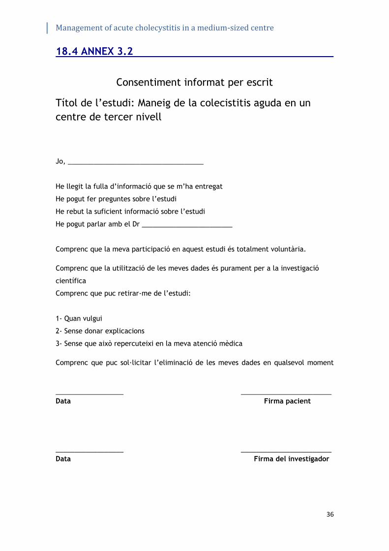
18.4 ANNEX 3.2
Consentiment informat per escrit
Títol de l’estudi: Maneig de la colecistitis aguda en un
centre de tercer nivell
Jo, ____________________________________
He llegit la fulla d’informació que se m’ha entregat
He pogut fer preguntes sobre l’estudi
He rebut la suficient informació sobre l’estudi
He pogut parlar amb el Dr ________________________
Comprenc que la meva participació en aquest estudi és totalment voluntària.
Comprenc que la utilització de les meves dades és purament per a la investigació
científica
Comprenc que puc retirar-me de l’estudi:
1- Quan vulgui
2- Sense donar explicacions
3- Sense que això repercuteixi en la meva atenció mèdica
Comprenc que puc sol·licitar l’eliminació de les meves dades en qualsevol moment
__________________ ________________________
Data Firma pacient
__________________ ________________________
Data Firma del investigador
Management of acute cholecystitis in a medium-sized centre
37
3. Imaging findings characteristics
of AC, observed either by
surgeon or radiologist
3.1 Yes
3.2 No
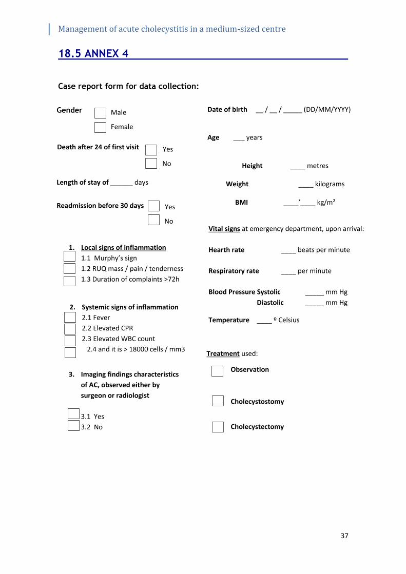
18.5 ANNEX 4
Case report form for data collection:
Gender
Death after 24 of first visit
Date of birth __ / __ / _____ (DD/MM/YYYY)
Age ___ years
Height ____ metres
Weight ____ kilograms
BMI ____’____ kg/m²
Vital signs at emergency department, upon arrival:
Hearth rate ____ beats per minute
Respiratory rate ____ per minute
Blood Pressure Systolic _____ mm Hg
Diastolic _____ mm Hg
Temperature ____ º Celsius
1. Local signs of inflammation
1.1 Murphy’s sign
1.2 RUQ mass / pain / tenderness
1.3 Duration of complaints >72h
2. Systemic signs of inflammation
2.1 Fever
2.2 Elevated CPR
2.3 Elevated WBC count
2.4 and it is > 18000 cells / mm3
Length of stay of ______ days
Readmission before 30 days
Male
Female
Yes
No
Yes
No
Treatment used:
Observation
Cholecystostomy
Cholecystectomy
Management of acute cholecystitis in a medium-sized centre
38
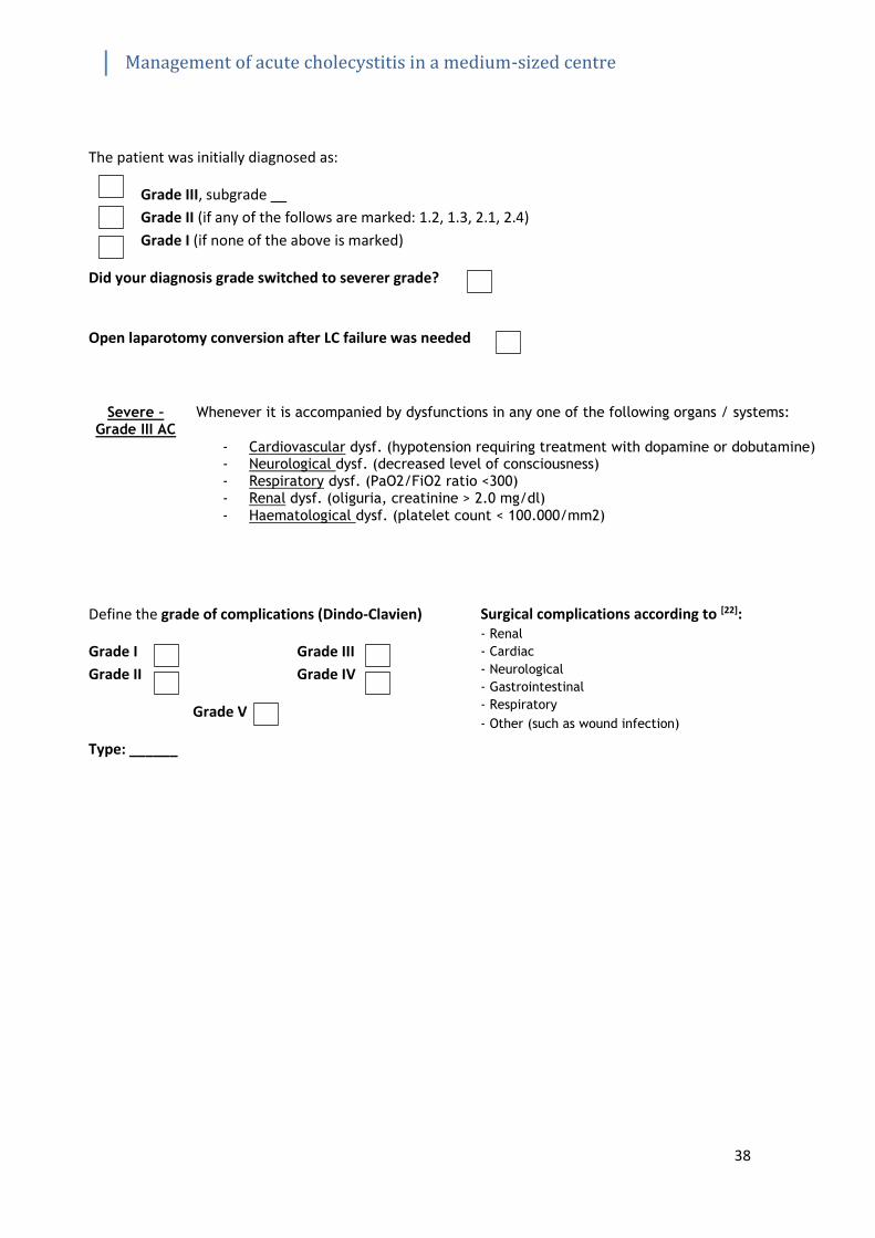
The patient was initially diagnosed as:
Grade III, subgrade __
Grade II (if any of the follows are marked: 1.2, 1.3, 2.1, 2.4)
Grade I (if none of the above is marked)
Did your diagnosis grade switched to severer grade?
Open laparotomy conversion after LC failure was needed
Define the grade of complications (Dindo-Clavien)
Grade I Grade III
Grade II Grade IV
Grade V
Type: ______
Surgical complications according to [22]: - Renal
- Cardiac
- Neurological
- Gastrointestinal
- Respiratory
- Other (such as wound infection)
Severe – Grade III AC
Whenever it is accompanied by dysfunctions in any one of the following organs / systems:
- Cardiovascular dysf. (hypotension requiring treatment with dopamine or dobutamine) - Neurological dysf. (decreased level of consciousness) - Respiratory dysf. (PaO2/FiO2 ratio <300) - Renal dysf. (oliguria, creatinine > 2.0 mg/dl)
- Haematological dysf. (platelet count < 100.000/mm2)
Management of acute cholecystitis in a medium-sized centre
39