Embed Size (px)
Citation preview

Research Article
Mechanistic aspects of maltotriose-conjugatetranslocation to the Gram-negative bacteria cytoplasmEstelle Dumont1,*, Julia Vergalli1,*, Jelena Pajovic2,3, Satya P Bhamidimarri4, Koldo Morante4 , Jiajun Wang4,Dmitrijs Lubriks5, Edgars Suna5 , Robert A Stavenger6, Mathias Winterhalter4, Matthieu Refregiers2 ,Jean-Marie Pagès1
Small molecule accumulation in Gram-negative bacteria is a keychallenge to discover novel antibiotics, because of their two mem-branes and efflux pumps expelling toxic molecules. An approach toovercome this challenge is to hijack uptake pathways so that bac-terial transporters shuttle the antibiotic to the cytoplasm. Here, wehave characterized maltodextrin–fluorophore conjugates that canpass through both the outer and inner membranes mediated bycomponents of the Escherichia coli maltose regulon. Single-channelelectrophysiology recording demonstrated that the compoundspermeate across the LamB channel leading to accumulation in theperiplasm. We have also demonstrated that a maltotriose conjugatedistributes into both the periplasm and cytoplasm. In the cytoplasm,the molecule activates the maltose regulon and triggers the ex-pression of maltose binding protein in the periplasmic space in-dicating that the complete maltose entry pathway is induced. Thismaltotriose conjugate can (i) reach the periplasmic and cytoplasmiccompartments to significant internal concentrations and (ii) auto-induce its own entry pathway via the activation of the maltoseregulon, representing an interesting prototype to deliver moleculesto the cytoplasm of Gram-negative bacteria.
DOI 10.26508/lsa.201800242 | Received 13 November 2018 | Revised 19December 2018 | Accepted 19 December 2018 | Published online 28December 2018
Introduction
The rise of multidrug resistance in pathogens is a serious and agrowing worldwide threat (1) (Centers for Disease Control and Pre-vention, https://www.cdc.gov/drugresistance/biggest_threats.html; Eu-ropean Centre for Disease Prevention and Control, https://ecdc.europa.eu/sites/portal/files/documents/AMR-surveillance-Europe-2016;
World Health Organization, http://www.who.int/medicines/areas/rational_use/antibacterial_agents_clinical_development/en/). Asignificant scientific challenge in discovering new, effective anti-bacterial agents is penetration of active molecules into bacteria toelicit activity (1, 2). This aspect is particularly difficult for Gram-negative bacteria which have two membranes, the outer (OM) andthe inner membranes, providing a barrier to the intracellular ac-cumulation of molecules (3, 4). In addition, efflux pumps can effi-ciently expel compounds (5, 6, 7, 8, 9, 10, 11), which can contribute tothe intrinsic resistance of Gram-negative bacteria to many anti-bacterials. The antibiotic concentration at the site of action mustexceed a level where it can bind and exert its antibacterial activity,and influx of the antibiotic, e.g., permeation across the Gram-negative envelope, is essential to access its target (8, 9, 11, 12, 13, 14).
With the spread of multidrug resistant bacteria, several wayshave been explored to bypass membrane-associated mechanismsof resistance including: (a) improved understanding of the porinpathways for penetration, (b) studying combinations of antibioticswith OM permeabilizers, (c) nanoparticle complexes to improvepenetration, and (d) use of “Trojan Horse” approaches to hijackactive uptake pathways and capitalize on existing bacterialreceptors-transporters (15, 16, 17, 18, 19, 20, 21, 22). Multiple “TrojanHorse” approaches have been reported, including iron uptake viasiderophore-conjugates and Opp-dependent peptide uptakepathways, but these systems are highly complex involving manyreceptors, and we are not aware of any that have been charac-terized by measuring the increase in intracellular accumulation.Another attractive strategy is to use the maltodextrin pathway,which is responsible for the internalization and degradation ofmaltodextrins in the cytoplasmic space of Gram-negative bacteria(23, 24). This system comprises the maltoporin (LamB) that providesan efficient pathway for maltose penetration through the OM, the
1Aix Marseille Univ, Institut National de la Sante et de la Recherche Medicale, Service de Sante des Armees, Institut de Recherche Biomedicale des Armees, Membranes etCibles Therapeutiques, Marseille, France 2DISCO Beamline, Synchrotron Soleil, Saint-Aubin, France 3University of Belgrade, Faculty of Physics, Belgrade, Serbia4Department of Life Sciences and Chemistry, Jacobs University Bremen, Bremen, Germany 5Latvian Institute of Organic Synthesis, Riga, Latvia 6Antibacterial DiscoveryPerformance Unit, Infectious Diseases Discovery, GlaxoSmithKline, Collegeville, PA, USA
Correspondence: [email protected] P. Bhamidimarri’s present address is Institute for Cell and Molecular Biosciences, The Medical School, Newcastle University, Newcastle, UKJiajun Wang ’s present address is Laboratory for Advanced Materials, School of Chemistry and Molecular Engineering, East China University of Science and Technology,Shanghai, China*Estelle Dumont and Julia Vergalli contributed equally to this work
© 2018 Dumont et al. https://doi.org/10.26508/lsa.201800242 vol 2 | no 1 | e201800242 1 of 13
on 13 March, 2022life-science-alliance.org Downloaded from http://doi.org/10.26508/lsa.201800242Published Online: 28 December, 2018 | Supp Info:
maltose binding protein (MalE) located in periplasm, maltodextrintransporters in the inner membrane (MalF, MalG, MalK) and variouscytoplasmic degradative enzymes; these components are under thecontrol of regulators belonging to maltose regulon (23). The in-ternalization of a maltohexaose fluorophore (25), the uptake of athiomaltose–trimethoprim conjugate (26), and the antibacterialactivity of a radezolid analog (patent WO 2016/044846) have beenrecently reported. Interestingly, labeled-maltodextrins have alsobeen used to study the localization/diagnosis of bacterial infec-tions in mouse models of infection (27, 28).
To molecularly dissect the potential of the maltodextrin transportsystem for drug transport, a collaboration between IMI TRANSLOCATIONand IMI ENABLE (www.imi.europa.eu) was established to study theuptake of maltodextrin conjugates. We report our results on both amaltohexaose–perylene conjugate (analog 1 in reference 25) and ashorter maltotriose–perylene conjugate, wherein the perylene moietyoffers a handle for conjugate detection via fluorescence while mim-icking a potential antibacterial “payload.”
To provide molecular insight into the transit of the maltodextrinconjugates, uptake was studied in purified LamB porin reconstitutedin lipid bilayers that demonstrated that the conjugates were able topass through the LamB channel. By using spectrofluorimetry andmicrospectrofluorimetry (29, 30, 31, 32), we further demonstrated aLamB-dependent uptake of one conjugate into the periplasmic andcytoplasmic space of the bacterial cells. In addition, we have shownthat the maltotriose perylene conjugate can promote the expressionof maltose-binding protein belonging to the maltose regulon, con-sistent with the uptake of the maltotriose conjugate.
Results
Accumulation of Cpd-1 and Cpd-2 is dependent on LamB expression
To study the ability of Cpd-1 (maltotriose–perylene conjugate)and Cpd-2 (maltohexaose–perylene conjugate) (see Fig 1 and the
Materials and Methods section of the Supplementary Information forsynthesis) to translocate across the Escherichia coli envelope, a seriesof well-defined isogenic strains were used, based on the parentalstrain (RAM1292) which contains an intact maltose operon (33). A lamBknock-out strain was obtained (RAM2806), as well as the lamB knock-out strain containing an empty pBAD24 plasmid (RAM2807), and thelamB knock-out strain with the pBAD24 plasmid coding for lamB underthe control of an arabinose inducible promoter (RAM2808) (Fig S1). Thestrains did not differ significantly in OmpC or OmpF content, whereasLamB expression was shown to be absent under normal growthconditions. As expected, LamB was detected in RAM2808, but not inRAM2807, after induction with arabinose (Fig S1).
In addition, in the strains RAM2808 and RAM1292, MalE proteinwas highly expressed in the presence of exogenous maltoseconsistent with induction of the maltose operon (Fig 2A). Whenbacterial cells are grown with high glucose concentrations, thecatabolic repression blocks the transcription of mal genes. Incontrast, under limited glucose concentration, the maltose regulonis expressed at elevated levels (23) (Figs S2, S3).
To study the implication of LamB andMalE in the translocation ofconjugates, strains RAM1292 and RAM2808 were grown under thefollowing conditions: RAM1292 cells were grown under conditions ofmaltose operon repression (minimal mediumwith 0.4% glucose), ormaltose operon induction (minimal medium with 0.4% maltose)(Fig 2A, left panel). RAM2808 cells were grown under conditions ofmaltose operon repression (minimal medium with 0.4% glycerol asa carbon source), LamB induction (minimal medium with 0.4%glycerol + 0.2% arabinose) or LamB and maltose operon induction(minimal medium with 0.4% maltose + 0.2% arabinose) (Fig 2A, rightpanel). Having validated these strains, they were used for accu-mulation study.
Accumulation and transport specificityUsing these isogenic strains under different expression condi-tions, the accumulation of maltodextrin conjugates Cpd-1 and
Figure 1. Chemical structure of the maltodextrin compounds studied in this work.The moieties corresponding to maltotriose of Cpd-1 and maltohexaose of Cpd-2 are boxed.
Maltocargo translocation to cytoplasm Dumont et al. https://doi.org/10.26508/lsa.201800242 vol 2 | no 1 | e201800242 2 of 13
Cpd-2 was demonstrated in both cell types and shown to beassociated with LamB expression induced by arabinose inRAM2808 or by maltose in RAM1292 as shown in Fig 2B and C. A veryweak fluorescence signal was measured in the strain RAM2807,regardless of the sugar added to the culturemedium (maltose and/or arabinose), likely corresponding to the nonspecific adsorptionof the compounds on the surface of the cells (data not shown). Toquantify conjugate accumulation, calibration curves were gen-erated to measure the number of molecules accumulated perbacterial cell (Fig S4).
To follow the intracellular accumulation of conjugates in indi-vidual bacterial cells, time-lapse experiments on a deep ultraviolet(DUV) microscope were carried out (Fig 2D and E) using methodspreviously described (29, 31). The external concentration of Cpd-1and Cpd-2 was 10 μg⋅ml−1 during incubation. Bacterial cells wereplated and observed for 30 min. Fluorescence was higher inRAM2808 cells grown in minimal medium incubated with malto-saccharide conjugates only when the cells were induced witharabinose (Fig 2D). Analyses of microspectrofluorimetric data ob-tained with RAM2808 cells incubated with the two fluorescent
compounds (at 22 μM) showed the same trend (Fig 2E). Thus,microspectrofluorimetry showed accumulation in RAM2808 cellsonly when the LamB production was induced. The box-and-whiskerplot representation shows the heterogeneity of accumulation in thecells. Similar to the accumulation measured at the population level(Fig 2B and C), individual cells showed more effective accumulationwith Cpd-1 relative to Cpd-2 (Fig 2E).
Maltose induction was also able to increase Cpd-1 and Cpd-2accumulation in the parental strain, RAM1292 (Fig 2B and C), con-sistent with induction of LamB andMalE expression (Figs 2A and S3).Moreover, the rate of accumulation was studied by using increasingconcentrations of Cpd-1 with the parental strain (RAM1292) grown inminimal media with maltose. A plateau was observed with con-centrations of Cpd-1 at ~44 μM (Fig S5). The presented plots suggestthat the uptake of Cpd-1 in E. coli cells is saturable at high con-centration for 30 min incubation.
Maltose competitionTo evaluate the selectivity of Cpd-1 transport by LamB, we in-vestigated the intracellular accumulation of Cpd-1 in individual
Figure 2. Accumulation of Cpd-1 and Cpd-2 dependson LamB expression.The strains were grown in different media to controlthe expression of the LamB porin and the MalEtransporter; 2 components of the maltose regulon (seeFigs S2 and S3). See Fig S1 for the characteristics andthe corresponding immunoblots of the strains. (A)Presence of LamB andMalE by Western blot in RAM1292and RAM2808 under different growth conditions. (B, C)Number of Cpd-1 (B) and Cpd-2 (C) moleculesaccumulated per cell in the various studied strainsfollowing analysis by spectrofluorimetry. The columnswith bars (SDs) correspond to measurements carriedout in triplicate. Calibration curves were used to obtainthe number of molecules per cell (Fig S4). (D)Microfluorimetric images obtained with DUVmicroscopy with pellets of RAM2808 with or withoutinduction of the LamB porin incubated withoutand with Cpd-1 or Cpd-2. Controls are RAM2808cells incubated without Cpd-1 and Cpd-2. (E)Microfluorimetric results obtained from (D). Data arerepresented with a box-and-whisker plots, which is away of summarizing the essential profile of aquantitative statistical series: the boxes representdata-points from the 25th to 75th percentiles; themiddle horizontal lines represent the median datapoint and the whiskers show the span of the data foreach sample. The outliers are represented by red +signs.
Maltocargo translocation to cytoplasm Dumont et al. https://doi.org/10.26508/lsa.201800242 vol 2 | no 1 | e201800242 3 of 13
bacterial cells in the absence or in the presence of maltose, a naturalsubstrate of the LamB channel (Fig S6). An excess ofmaltose reducedthe uptake of Cpd-1 in E. coli RAM1292 cells previously grown underconditions of maltose operon induction, consistent with a maltoseregulatory feedback, further supporting the hypothesis that Cpd-1crosses the outer membrane via LamB channels.
Time-course accumulation of fluorescently labeled maltodextrinconjugates
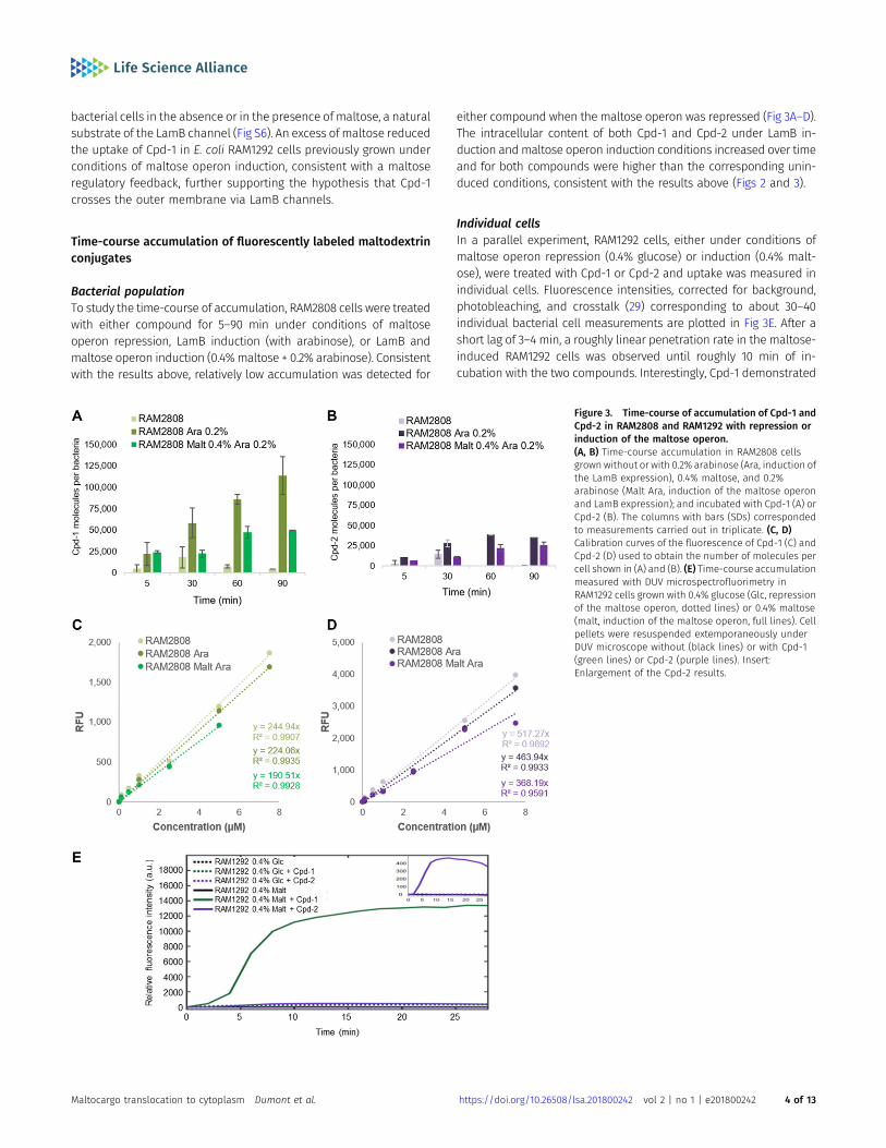
Bacterial populationTo study the time-course of accumulation, RAM2808 cells were treatedwith either compound for 5–90 min under conditions of maltoseoperon repression, LamB induction (with arabinose), or LamB andmaltose operon induction (0.4%maltose + 0.2% arabinose). Consistentwith the results above, relatively low accumulation was detected for
either compound when the maltose operon was repressed (Fig 3A–D).The intracellular content of both Cpd-1 and Cpd-2 under LamB in-duction andmaltose operon induction conditions increased over timeand for both compounds were higher than the corresponding unin-duced conditions, consistent with the results above (Figs 2 and 3).
Individual cellsIn a parallel experiment, RAM1292 cells, either under conditions ofmaltose operon repression (0.4% glucose) or induction (0.4% malt-ose), were treated with Cpd-1 or Cpd-2 and uptake was measured inindividual cells. Fluorescence intensities, corrected for background,photobleaching, and crosstalk (29) corresponding to about 30–40individual bacterial cell measurements are plotted in Fig 3E. After ashort lag of 3–4 min, a roughly linear penetration rate in the maltose-induced RAM1292 cells was observed until roughly 10 min of in-cubation with the two compounds. Interestingly, Cpd-1 demonstrated
Figure 3. Time-course of accumulation of Cpd-1 andCpd-2 in RAM2808 and RAM1292 with repression orinduction of the maltose operon.(A, B) Time-course accumulation in RAM2808 cellsgrown without or with 0.2% arabinose (Ara, induction ofthe LamB expression), 0.4% maltose, and 0.2%arabinose (Malt Ara, induction of the maltose operonand LamB expression); and incubated with Cpd-1 (A) orCpd-2 (B). The columns with bars (SDs) correspondedto measurements carried out in triplicate. (C, D)Calibration curves of the fluorescence of Cpd-1 (C) andCpd-2 (D) used to obtain the number of molecules percell shown in (A) and (B). (E) Time-course accumulationmeasured with DUV microspectrofluorimetry inRAM1292 cells grown with 0.4% glucose (Glc, repressionof the maltose operon, dotted lines) or 0.4% maltose(malt, induction of the maltose operon, full lines). Cellpellets were resuspended extemporaneously underDUV microscope without (black lines) or with Cpd-1(green lines) or Cpd-2 (purple lines). Insert:Enlargement of the Cpd-2 results.
Maltocargo translocation to cytoplasm Dumont et al. https://doi.org/10.26508/lsa.201800242 vol 2 | no 1 | e201800242 4 of 13
a rapid increase phase (4–7 min) followed by a steady-state level ofaccumulation at about 15–18 min. With Cpd-2, a slow increase wasobserved during the same period under the induced conditions andthe obtained steady state was lower (calculated rate for Cpd-1 was1250 A.U./min versus 75 for Cpd-2). In contrast, no significant accu-mulation was obtained in the non-induced strain (Fig 3E).
It should be noted that although we cannot precisely determinethe correlation between the signal intensity and the number ofmolecules inside individual bacterial cells (in these conditions, acalibration curve is not possible because of technical conditions,e.g., photobleaching (29, 31)), we were able to observe a relationshipbetween the incubation time and the accumulation rate in indi-vidual bacterial cells.
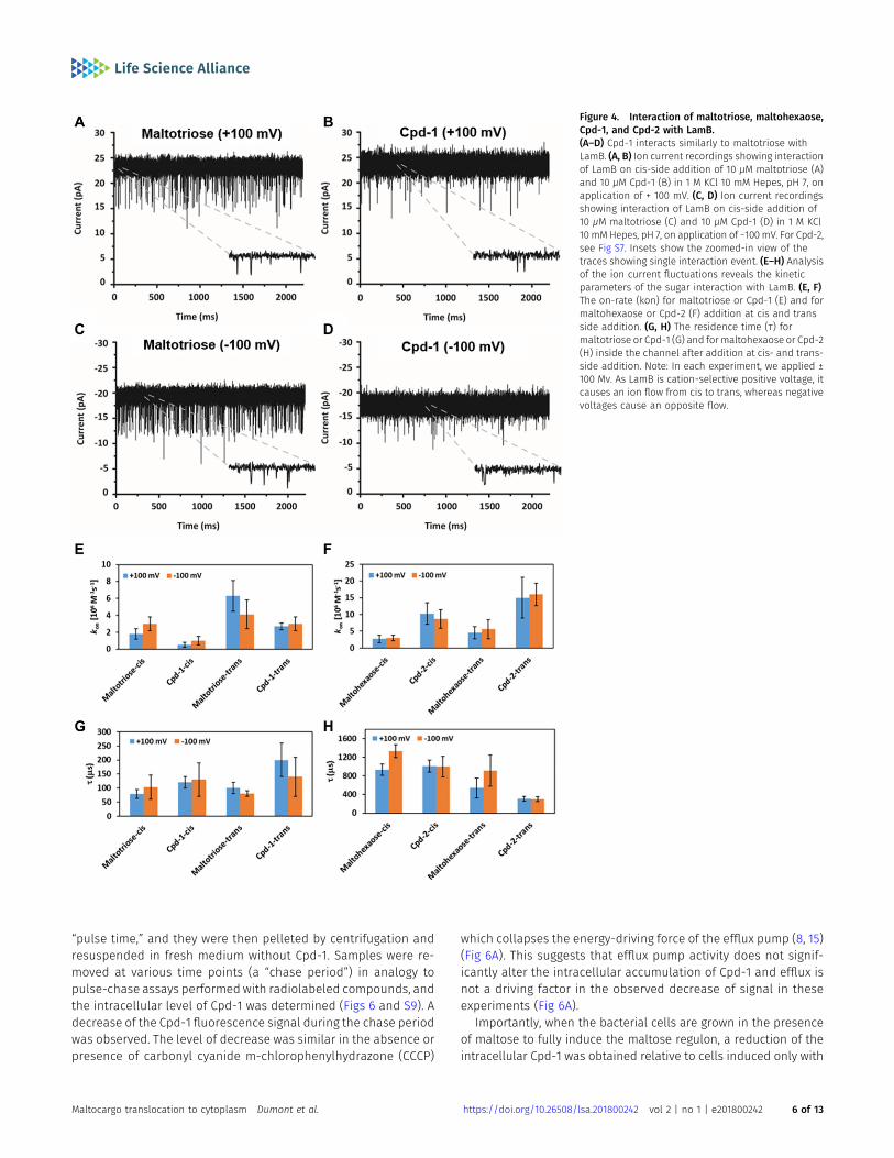
Interaction of Cpd-1 and Cpd-2 with a single LamB channel
To support the observed LamB-dependent cellular uptake of Cpd-1and Cpd-2, we exploited electrophysiology to characterize the in-teraction of fluorophore-conjugated and parent maltodextrins witha single LamB trimer reconstituted in a planar lipid bilayer. Thismethod analyzes the substrate-induced ion current fluctuationsacross a membrane channel under an applied transmembraneelectric field, providing insights on the interaction and, to someextent, the flux of substrates through the channel.
In Figs 4A–D and S7, we show examples of ion current traces inwhich ion flow (current) was transiently interrupted (blocked) by theinteraction of Cpd-1 or Cpd-2 with LamB on either the extracellular(cis) or periplasmic (trans) side of the membrane, as previouslyobserved for malto-oligosaccharides (34). Statistical analyses ofthese ion flow blockages provided both the association rate (kon),obtained from the interaction frequency, and the duration of theinteraction, or residence time (τ) (Fig 4E–H and Table S1).
Inspection of the summary of the kinetic parameters allows aqualitative conclusion on the degree to which the small (maltotrioseand Cpd-1, Fig 4E and G) or large (maltohexaose and Cpd-2, Fig 4Fand H) substrates permeate through the channel or simply bindand bounce back unproductively. Our data showed that the smallsubstrates (shown above to penetrate better than larger substratesthrough LamB in whole cells) are characterized by short τcis (≤130 ±60 μs), whereas the large substrates present long τcis (≥930 ± 120 μs)values, in agreement with enhanced interactions of the long sugarswith the inner wall of the channel (34). On the other hand, we foundthat all sugars except for Cpd-2 presented a low kcison (≤3.0 ± 0.8 ×10⁶ M−1s−1) and follow a similar trend on the enhanced trans side(ktranson ≤ 6.3 ± 1.8 M−1s−1), a behavior consistent with having a lowerenergy barrier at the trans side of LamB as observed by Danelon et al.(34). In addition, a small effect from electro-osmosis is also observed(most clearly seen with maltotriose). Electro-osmosis is revealed incation-selective channels, such as LamB, by an enhanced cationcurrent at negative applied voltage (kcis;M3
on = 3.0 ± 0.8 × 10⁶ M−1s−1) ascompared with positive voltage (kcis;M3
on = 1.8 ± 0.6 × 10⁶ M−1s−1) (35).In contrast with the other compounds tested here, Cpd-2 has a
larger kon (~10 ± 3 × 10⁶ M−1s−1). Surprisingly, a shorter overall τaverage is observed for Cpd-2 (τtrans ~290 ± 60 μs) as compared withits parent sugar maltohexaose (τtrans = 725 ± 270 μs). Our in-terpretation of this is that Cpd-2 has a substantial contribution ofshort-lived, unproductive interactions that contribute to decrease τ
as we feel the alternate explanation that Cpd-2 permeates twofoldfaster than parent maltohexaose is unlikely. This effect is moreclearly seen on the trans side, consistent with an interpretation thatthe lower energy barrier at the periplasmic opening offers thesubstrate more opportunities of binding and exit through the sameside. The fluorophore may constrain the helical conformationneeded to screw the molecule across the channel, preventing itfrom exiting through the extracellular side. This may not be the casefor Cpd-1, where the requirement of a pronounced structuralrearrangement of the sugar may be neglected given the small sizeof the sugar, allowing the molecule to diffuse through the channelwith τ values similar to those of maltotriose (M3 in equation)
�τcis;M3 ≈90±30μs; τcis; Cpd−1 ≈110±40μs
�:
Accordingly, our interpretation of this data is that Cpd-1 canpermeate LamB with a similar rate as the maltotriose sugar alone,whereas diffusion of Cpd-2 is sterically hampered relative tomaltohexaose.
Using a rough approximation of the molecular flux (ϕ = kon [c])suggests that at 10 μM concentration ~1,000 Cpd-1 moleculespermeate per second and per LamB monomer. This is in overallagreement with the trends of the whole-cell penetration datashown above wherein Cpd-1 accumulates more readily relative toCpd-2. Moreover, analysis of the electrophysiology data suggests amolecular specificity for Cpd-1 versus Cpd-2 with respect to in-teraction and permeation. This potentially suggests a limitation dueto size or diffusion efficiency inside the LamB channel for thisapproach (34, 36).
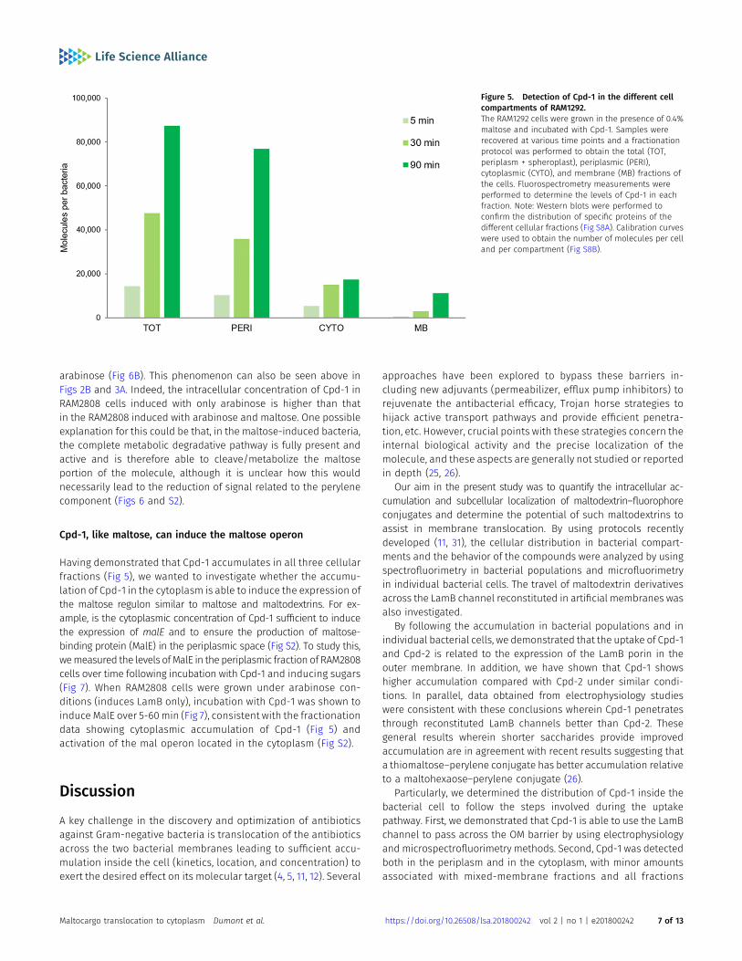
Time-course and subcellular localization of Cpd-1 and Cpd-2
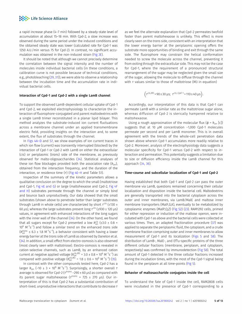
Having established that both Cpd-1 and Cpd-2 can pass the outermembrane via LamB, questions remained concerning their cellularlocalization and disposition inside the bacterial cell. Maltodextrinsare generally transported into the bacterial cytoplasm across theouter and inner membranes, via LamB/MalE and maltose innermembrane transporters (MalF,G,K), eventually to be metabolized bycytoplasmic enzymes (MalP,Q,Z) (Fig S2) (23). RAM1292 cells, primedfor either repression or induction of the maltose operon, were in-cubated with Cpd-1 as above and the bacterial cells were collected atvarious times. Then, an adapted fractionation procedure (37) wasapplied to separate the periplasmic fluid, the cytoplasm, and a crudemembrane fraction comprising outer and inner membranes to allowmeasurement of Cpd-1 and its localization (Figs 5 and S8). Thedistribution of LamB-, MalE-, and EfTu-specific proteins of the threedifferent cellular fractions (membrane, periplasm, and cytoplasm,respectively) was confirmed by immunodetection (Fig S8). The totalamount of Cpd-1 detected in the three cellular fractions increasedduring the incubation times, with the most of the Cpd-1 signal beingfound in the periplasm at all time-points (Fig 5).
Behavior of maltosaccharide conjugates inside the cell
To understand the fate of Cpd-1 inside the cell, RAM2808 cellswere incubated in the presence of Cpd-1 corresponding to a
Maltocargo translocation to cytoplasm Dumont et al. https://doi.org/10.26508/lsa.201800242 vol 2 | no 1 | e201800242 5 of 13
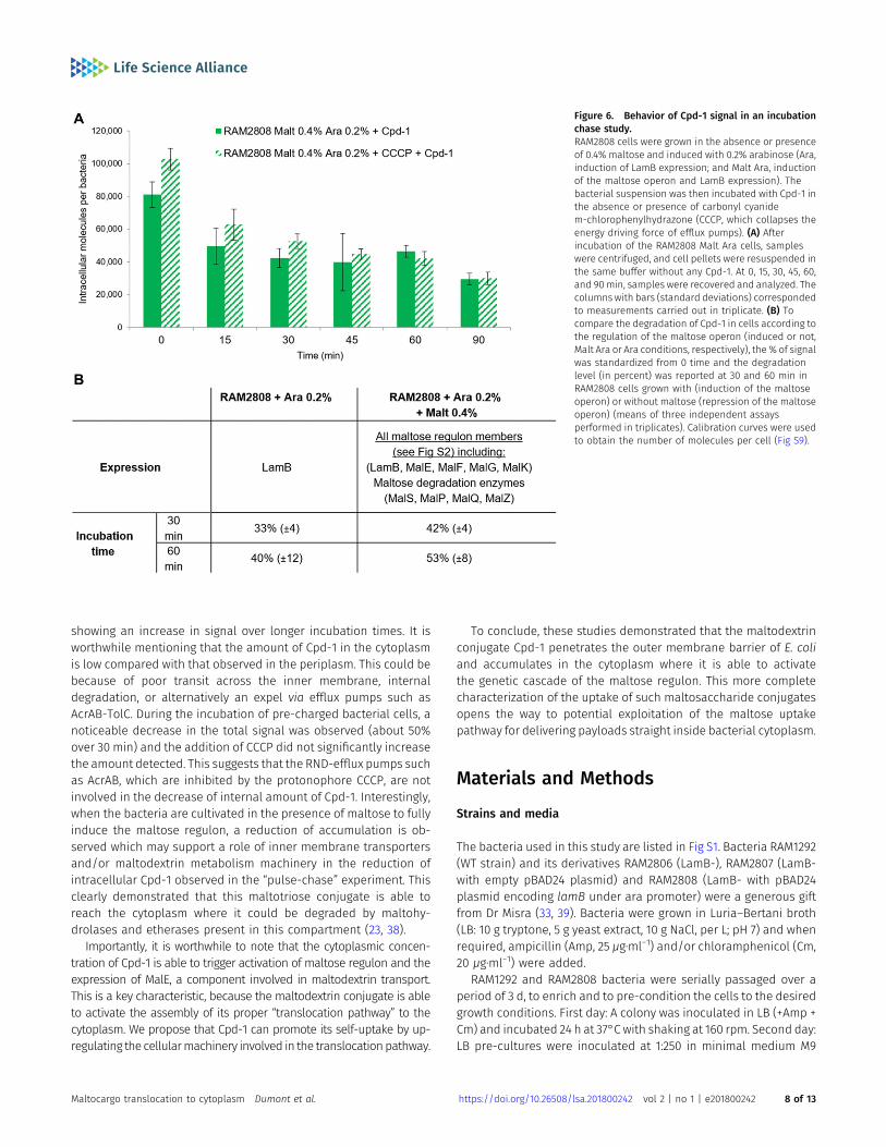
“pulse time,” and they were then pelleted by centrifugation andresuspended in fresh medium without Cpd-1. Samples were re-moved at various time points (a “chase period”) in analogy topulse-chase assays performed with radiolabeled compounds, andthe intracellular level of Cpd-1 was determined (Figs 6 and S9). Adecrease of the Cpd-1 fluorescence signal during the chase periodwas observed. The level of decrease was similar in the absence orpresence of carbonyl cyanide m-chlorophenylhydrazone (CCCP)
which collapses the energy-driving force of the efflux pump (8, 15)(Fig 6A). This suggests that efflux pump activity does not signif-icantly alter the intracellular accumulation of Cpd-1 and efflux isnot a driving factor in the observed decrease of signal in theseexperiments (Fig 6A).
Importantly, when the bacterial cells are grown in the presenceof maltose to fully induce the maltose regulon, a reduction of theintracellular Cpd-1 was obtained relative to cells induced only with
Figure 4. Interaction of maltotriose, maltohexaose,Cpd-1, and Cpd-2 with LamB.(A–D) Cpd-1 interacts similarly to maltotriose withLamB. (A, B) Ion current recordings showing interactionof LamB on cis-side addition of 10 μM maltotriose (A)and 10 μM Cpd-1 (B) in 1 M KCl 10 mM Hepes, pH 7, onapplication of + 100 mV. (C, D) Ion current recordingsshowing interaction of LamB on cis-side addition of10 μM maltotriose (C) and 10 μM Cpd-1 (D) in 1 M KCl10 mMHepes, pH 7, on application of −100 mV. For Cpd-2,see Fig S7. Insets show the zoomed-in view of thetraces showing single interaction event. (E–H) Analysisof the ion current fluctuations reveals the kineticparameters of the sugar interaction with LamB. (E, F)The on-rate (kon) for maltotriose or Cpd-1 (E) and formaltohexaose or Cpd-2 (F) addition at cis and transside addition. (G, H) The residence time (τ) formaltotriose or Cpd-1 (G) and for maltohexaose or Cpd-2(H) inside the channel after addition at cis- and trans-side addition. Note: In each experiment, we applied ±100 Mv. As LamB is cation-selective positive voltage, itcauses an ion flow from cis to trans, whereas negativevoltages cause an opposite flow.
Maltocargo translocation to cytoplasm Dumont et al. https://doi.org/10.26508/lsa.201800242 vol 2 | no 1 | e201800242 6 of 13
arabinose (Fig 6B). This phenomenon can also be seen above inFigs 2B and 3A. Indeed, the intracellular concentration of Cpd-1 inRAM2808 cells induced with only arabinose is higher than thatin the RAM2808 induced with arabinose and maltose. One possibleexplanation for this could be that, in the maltose-induced bacteria,the complete metabolic degradative pathway is fully present andactive and is therefore able to cleave/metabolize the maltoseportion of the molecule, although it is unclear how this wouldnecessarily lead to the reduction of signal related to the perylenecomponent (Figs 6 and S2).
Cpd-1, like maltose, can induce the maltose operon
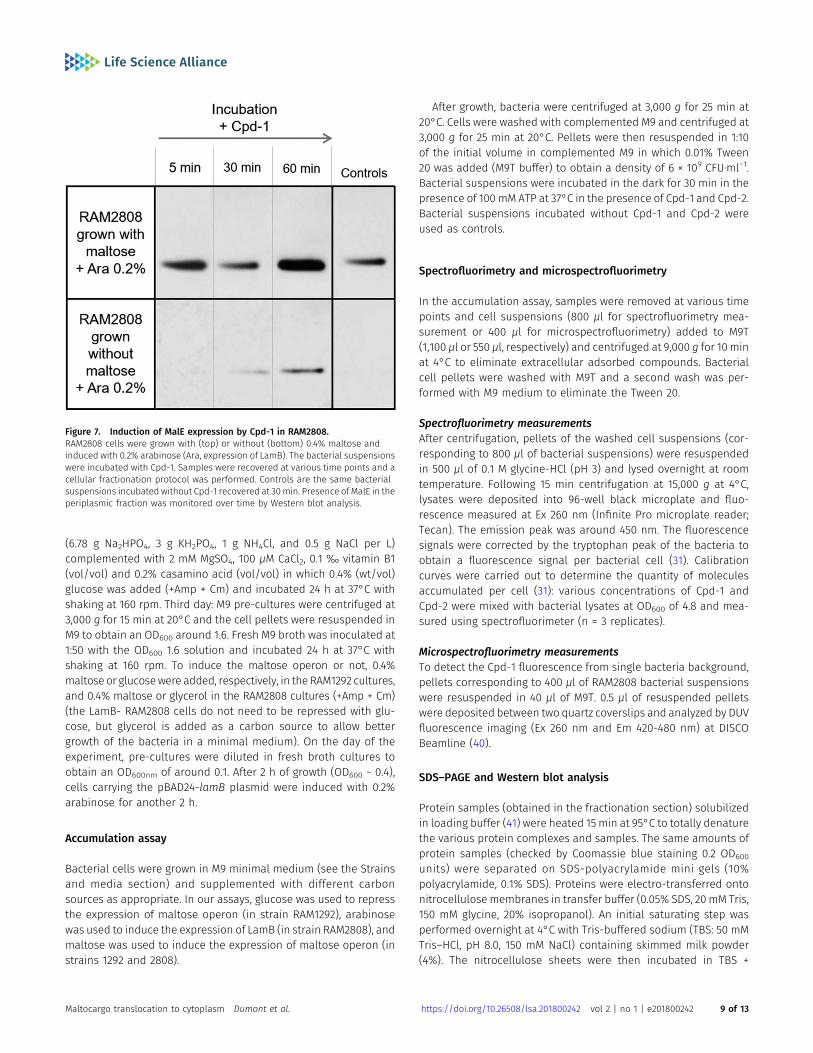
Having demonstrated that Cpd-1 accumulates in all three cellularfractions (Fig 5), we wanted to investigate whether the accumu-lation of Cpd-1 in the cytoplasm is able to induce the expression ofthe maltose regulon similar to maltose and maltodextrins. For ex-ample, is the cytoplasmic concentration of Cpd-1 sufficient to inducethe expression of malE and to ensure the production of maltose-binding protein (MalE) in the periplasmic space (Fig S2). To study this,wemeasured the levels ofMalE in the periplasmic fraction of RAM2808cells over time following incubation with Cpd-1 and inducing sugars(Fig 7). When RAM2808 cells were grown under arabinose con-ditions (induces LamB only), incubation with Cpd-1 was shown toinduce MalE over 5-60min (Fig 7), consistent with the fractionationdata showing cytoplasmic accumulation of Cpd-1 (Fig 5) andactivation of the mal operon located in the cytoplasm (Fig S2).
Discussion
A key challenge in the discovery and optimization of antibioticsagainst Gram-negative bacteria is translocation of the antibioticsacross the two bacterial membranes leading to sufficient accu-mulation inside the cell (kinetics, location, and concentration) toexert the desired effect on its molecular target (4, 5, 11, 12). Several
approaches have been explored to bypass these barriers in-cluding new adjuvants (permeabilizer, efflux pump inhibitors) torejuvenate the antibacterial efficacy, Trojan horse strategies tohijack active transport pathways and provide efficient penetra-tion, etc. However, crucial points with these strategies concern theinternal biological activity and the precise localization of themolecule, and these aspects are generally not studied or reportedin depth (25, 26).
Our aim in the present study was to quantify the intracellular ac-cumulation and subcellular localization of maltodextrin–fluorophoreconjugates and determine the potential of such maltodextrins toassist in membrane translocation. By using protocols recentlydeveloped (11, 31), the cellular distribution in bacterial compart-ments and the behavior of the compounds were analyzed by usingspectrofluorimetry in bacterial populations and microfluorimetryin individual bacterial cells. The travel of maltodextrin derivativesacross the LamB channel reconstituted in artificial membranes wasalso investigated.
By following the accumulation in bacterial populations and inindividual bacterial cells, we demonstrated that the uptake of Cpd-1and Cpd-2 is related to the expression of the LamB porin in theouter membrane. In addition, we have shown that Cpd-1 showshigher accumulation compared with Cpd-2 under similar condi-tions. In parallel, data obtained from electrophysiology studieswere consistent with these conclusions wherein Cpd-1 penetratesthrough reconstituted LamB channels better than Cpd-2. Thesegeneral results wherein shorter saccharides provide improvedaccumulation are in agreement with recent results suggesting thata thiomaltose–perylene conjugate has better accumulation relativeto a maltohexaose–perylene conjugate (26).
Particularly, we determined the distribution of Cpd-1 inside thebacterial cell to follow the steps involved during the uptakepathway. First, we demonstrated that Cpd-1 is able to use the LamBchannel to pass across the OM barrier by using electrophysiologyand microspectrofluorimetry methods. Second, Cpd-1 was detectedboth in the periplasm and in the cytoplasm, with minor amountsassociated with mixed-membrane fractions and all fractions
Figure 5. Detection of Cpd-1 in the different cellcompartments of RAM1292.The RAM1292 cells were grown in the presence of 0.4%maltose and incubated with Cpd-1. Samples wererecovered at various time points and a fractionationprotocol was performed to obtain the total (TOT,periplasm + spheroplast), periplasmic (PERI),cytoplasmic (CYTO), and membrane (MB) fractions ofthe cells. Fluorospectrometry measurements wereperformed to determine the levels of Cpd-1 in eachfraction. Note: Western blots were performed toconfirm the distribution of specific proteins of thedifferent cellular fractions (Fig S8A). Calibration curveswere used to obtain the number of molecules per celland per compartment (Fig S8B).
Maltocargo translocation to cytoplasm Dumont et al. https://doi.org/10.26508/lsa.201800242 vol 2 | no 1 | e201800242 7 of 13
showing an increase in signal over longer incubation times. It isworthwhile mentioning that the amount of Cpd-1 in the cytoplasmis low compared with that observed in the periplasm. This could bebecause of poor transit across the inner membrane, internaldegradation, or alternatively an expel via efflux pumps such asAcrAB-TolC. During the incubation of pre-charged bacterial cells, anoticeable decrease in the total signal was observed (about 50%over 30 min) and the addition of CCCP did not significantly increasethe amount detected. This suggests that the RND-efflux pumps suchas AcrAB, which are inhibited by the protonophore CCCP, are notinvolved in the decrease of internal amount of Cpd-1. Interestingly,when the bacteria are cultivated in the presence of maltose to fullyinduce the maltose regulon, a reduction of accumulation is ob-served which may support a role of inner membrane transportersand/or maltodextrin metabolism machinery in the reduction ofintracellular Cpd-1 observed in the “pulse-chase” experiment. Thisclearly demonstrated that this maltotriose conjugate is able toreach the cytoplasm where it could be degraded by maltohy-drolases and etherases present in this compartment (23, 38).
Importantly, it is worthwhile to note that the cytoplasmic concen-tration of Cpd-1 is able to trigger activation of maltose regulon and theexpression of MalE, a component involved in maltodextrin transport.This is a key characteristic, because the maltodextrin conjugate is ableto activate the assembly of its proper “translocation pathway” to thecytoplasm. We propose that Cpd-1 can promote its self-uptake by up-regulating the cellularmachinery involved in the translocationpathway.
To conclude, these studies demonstrated that the maltodextrinconjugate Cpd-1 penetrates the outer membrane barrier of E. coliand accumulates in the cytoplasm where it is able to activatethe genetic cascade of the maltose regulon. This more completecharacterization of the uptake of such maltosaccharide conjugatesopens the way to potential exploitation of the maltose uptakepathway for delivering payloads straight inside bacterial cytoplasm.
Materials and Methods
Strains and media
The bacteria used in this study are listed in Fig S1. Bacteria RAM1292(WT strain) and its derivatives RAM2806 (LamB-), RAM2807 (LamB-with empty pBAD24 plasmid) and RAM2808 (LamB- with pBAD24plasmid encoding lamB under ara promoter) were a generous giftfrom Dr Misra (33, 39). Bacteria were grown in Luria–Bertani broth(LB: 10 g tryptone, 5 g yeast extract, 10 g NaCl, per L; pH 7) and whenrequired, ampicillin (Amp, 25 μg⋅ml−1) and/or chloramphenicol (Cm,20 μg⋅ml−1) were added.
RAM1292 and RAM2808 bacteria were serially passaged over aperiod of 3 d, to enrich and to pre-condition the cells to the desiredgrowth conditions. First day: A colony was inoculated in LB (+Amp +Cm) and incubated 24 h at 37°C with shaking at 160 rpm. Second day:LB pre-cultures were inoculated at 1:250 in minimal medium M9
Figure 6. Behavior of Cpd-1 signal in an incubationchase study.RAM2808 cells were grown in the absence or presenceof 0.4% maltose and induced with 0.2% arabinose (Ara,induction of LamB expression; and Malt Ara, inductionof the maltose operon and LamB expression). Thebacterial suspension was then incubated with Cpd-1 inthe absence or presence of carbonyl cyanidem-chlorophenylhydrazone (CCCP, which collapses theenergy driving force of efflux pumps). (A) Afterincubation of the RAM2808 Malt Ara cells, sampleswere centrifuged, and cell pellets were resuspended inthe same buffer without any Cpd-1. At 0, 15, 30, 45, 60,and 90min, samples were recovered and analyzed. Thecolumns with bars (standard deviations) correspondedto measurements carried out in triplicate. (B) Tocompare the degradation of Cpd-1 in cells according tothe regulation of the maltose operon (induced or not,Malt Ara or Ara conditions, respectively), the % of signalwas standardized from 0 time and the degradationlevel (in percent) was reported at 30 and 60 min inRAM2808 cells grown with (induction of the maltoseoperon) or without maltose (repression of the maltoseoperon) (means of three independent assaysperformed in triplicates). Calibration curves were usedto obtain the number of molecules per cell (Fig S9).
Maltocargo translocation to cytoplasm Dumont et al. https://doi.org/10.26508/lsa.201800242 vol 2 | no 1 | e201800242 8 of 13
(6.78 g Na2HPO4, 3 g KH2PO4, 1 g NH4Cl, and 0.5 g NaCl per L)complemented with 2 mM MgSO4, 100 μM CaCl2, 0.1 ‰ vitamin B1(vol/vol) and 0.2% casamino acid (vol/vol) in which 0.4% (wt/vol)glucose was added (+Amp + Cm) and incubated 24 h at 37°C withshaking at 160 rpm. Third day: M9 pre-cultures were centrifuged at3,000 g for 15 min at 20°C and the cell pellets were resuspended inM9 to obtain an OD600 around 1.6. Fresh M9 broth was inoculated at1:50 with the OD600 1.6 solution and incubated 24 h at 37°C withshaking at 160 rpm. To induce the maltose operon or not, 0.4%maltose or glucosewere added, respectively, in the RAM1292 cultures,and 0.4% maltose or glycerol in the RAM2808 cultures (+Amp + Cm)(the LamB- RAM2808 cells do not need to be repressed with glu-cose, but glycerol is added as a carbon source to allow bettergrowth of the bacteria in a minimal medium). On the day of theexperiment, pre-cultures were diluted in fresh broth cultures toobtain an OD600nm of around 0.1. After 2 h of growth (OD600 ~ 0.4),cells carrying the pBAD24-lamB plasmid were induced with 0.2%arabinose for another 2 h.
Accumulation assay
Bacterial cells were grown in M9 minimal medium (see the Strainsand media section) and supplemented with different carbonsources as appropriate. In our assays, glucose was used to repressthe expression of maltose operon (in strain RAM1292), arabinosewas used to induce the expression of LamB (in strain RAM2808), andmaltose was used to induce the expression of maltose operon (instrains 1292 and 2808).
After growth, bacteria were centrifuged at 3,000 g for 25 min at20°C. Cells were washed with complemented M9 and centrifuged at3,000 g for 25 min at 20°C. Pellets were then resuspended in 1:10of the initial volume in complemented M9 in which 0.01% Tween20 was added (M9T buffer) to obtain a density of 6 × 10⁹ CFU⋅ml−1.Bacterial suspensions were incubated in the dark for 30 min in thepresence of 100 mM ATP at 37°C in the presence of Cpd-1 and Cpd-2.Bacterial suspensions incubated without Cpd-1 and Cpd-2 wereused as controls.
Spectrofluorimetry and microspectrofluorimetry
In the accumulation assay, samples were removed at various timepoints and cell suspensions (800 μl for spectrofluorimetry mea-surement or 400 μl for microspectrofluorimetry) added to M9T(1,100 μl or 550 μl, respectively) and centrifuged at 9,000 g for 10 minat 4°C to eliminate extracellular adsorbed compounds. Bacterialcell pellets were washed with M9T and a second wash was per-formed with M9 medium to eliminate the Tween 20.
Spectrofluorimetry measurementsAfter centrifugation, pellets of the washed cell suspensions (cor-responding to 800 μl of bacterial suspensions) were resuspendedin 500 μl of 0.1 M glycine-HCl (pH 3) and lysed overnight at roomtemperature. Following 15 min centrifugation at 15,000 g at 4°C,lysates were deposited into 96-well black microplate and fluo-rescence measured at Ex 260 nm (Infinite Pro microplate reader;Tecan). The emission peak was around 450 nm. The fluorescencesignals were corrected by the tryptophan peak of the bacteria toobtain a fluorescence signal per bacterial cell (31). Calibrationcurves were carried out to determine the quantity of moleculesaccumulated per cell (31): various concentrations of Cpd-1 andCpd-2 were mixed with bacterial lysates at OD600 of 4.8 and mea-sured using spectrofluorimeter (n = 3 replicates).
Microspectrofluorimetry measurementsTo detect the Cpd-1 fluorescence from single bacteria background,pellets corresponding to 400 μl of RAM2808 bacterial suspensionswere resuspended in 40 μl of M9T. 0.5 μl of resuspended pelletswere deposited between two quartz coverslips and analyzed by DUVfluorescence imaging (Ex 260 nm and Em 420-480 nm) at DISCOBeamline (40).
SDS–PAGE and Western blot analysis
Protein samples (obtained in the fractionation section) solubilizedin loading buffer (41) were heated 15 min at 95°C to totally denaturethe various protein complexes and samples. The same amounts ofprotein samples (checked by Coomassie blue staining 0.2 OD600
units) were separated on SDS-polyacrylamide mini gels (10%polyacrylamide, 0.1% SDS). Proteins were electro-transferred ontonitrocellulose membranes in transfer buffer (0.05% SDS, 20 mM Tris,150 mM glycine, 20% isopropanol). An initial saturating step wasperformed overnight at 4°C with Tris-buffered sodium (TBS: 50 mMTris–HCl, pH 8.0, 150 mM NaCl) containing skimmed milk powder(4%). The nitrocellulose sheets were then incubated in TBS +
Figure 7. Induction of MalE expression by Cpd-1 in RAM2808.RAM2808 cells were grown with (top) or without (bottom) 0.4% maltose andinduced with 0.2% arabinose (Ara, expression of LamB). The bacterial suspensionswere incubated with Cpd-1. Samples were recovered at various time points and acellular fractionation protocol was performed. Controls are the same bacterialsuspensions incubated without Cpd-1 recovered at 30min. Presence of MalE in theperiplasmic fraction was monitored over time by Western blot analysis.
Maltocargo translocation to cytoplasm Dumont et al. https://doi.org/10.26508/lsa.201800242 vol 2 | no 1 | e201800242 9 of 13
skimmed milk powder + Triton X-100 (0.2%) for 1 h at room tem-perature in the presence of polyclonal antibodies directed againstLamB, MalE, EfTu, OmpC, and OmpF proteins (1:10,000, 1:5,000, 1:30,000, 1:10,000, and 1:10,000, respectively). After washes, the de-tection of antigen–antibody complexes was performed with horse-radish peroxidase–conjugated Immune-Stargoat anti-rabbit IgGantibodies (Bio-Rad) and detection was performed with the ClarityWestern ECL kit (Bio-Rad) using a Chemidoc XRS+ (Bio-Rad).
Time-course accumulation in individual bacterial cells
The time-course accumulation in individual bacterial cells wasstudied by using a previously described protocol (29, 30). Bacterialcells were grown in complemented M9 supplemented with differentcarbon sources as appropriate. Bacteria were concentrated to obtainanOD600 of 4.8 inM9T. Then, 120 μl of the suspensionwere centrifugedat 6,000 g for 10 min at 4°C. The pellets were resuspended extem-poraneously in 40 μl of M9T containing or not containing Cpd-1 orCpd-2. 0.5 μl of resuspended pellets were deposited between twoquartz coverslips and analyzed by DUV fluorescence imaging at DISCOBeamline. The accumulation in individual bacterial cells was moni-tored directly under a DUV microscope during 30 min. Bacterial cellswere first located in brightfield before excitation in DUV through amicroscope (Zeiss Axio Observer Z-1) at Synchrotron SOLEIL (42).Emission was collected via a Zeiss ultrafluar objective at 100× withglycerin immersion. The fluorescence was recorded by exciting at260 nm through an emission bandpass filter at 420–480 nm (OMEGAOptical, Inc). For the tryptophan fluorescence, the emission wascollected through an emission bandpass filter at 327–353 nm(SEMROCK). Fluorescence images were recorded with a BUV CMOS(Prime 95B; Photometrics). The whole setup (microscope, stages,shutters, filters, and camera) was controlled by Micro-Manager (43).Bacteria were observed for 30 min with a sampling time of 2 min foreach area: 30 s with the filter 1 (Cpd-1 and Cpd-2 molecules fluo-rescence detection), 30 s with the filter 2 (tryptophan fluorescencedetection), followed by 60 s of pause. During the intervening pausein sampling, the same acquisition cycle was performed on anotherfield of view, avoiding constant UV irradiation of the same field.
The images were analyzed with Image J (Rasband, W.S., Image J,U.S. National Institutes of Health, Bethesda, MD, USA, http://imagej.nih.gov/ij/) (44). The illumination heterogeneities were correctedbefore background subtraction. First, the threshold was automat-ically adjusted using a triangle algorithm; thereafter, bacteria wereanalyzed as the remaining particles. The mean intensity comingfrom each bacterium was automatically calculated considering itspixel area. Finally, all bacteria signal taken from one image wereaveraged. For each condition, seven different localizations withminimum 30 bacteria per field of view were recorded and averaged.
Electrophysiology
Single-channel electrophysiology measurements were performedusing the Montal andMueller technique (45). In short, a 25-μm-thickTeflon film containing an aperture of approximately 100 μm di-ameter is sandwiched between the chambers of Teflon cuvette. Asolution of 1% (vol/vol) hexadecane in hexane was used to makethe area around the aperture more hydrophobic to form stable
bilayers. Membranes were formed using a solution of 5 mg⋅ml−1
DPhPC in pentane. The chambers were filled with 1 M KCl, 10 mMHepes, pH 7, that serves as an electrolyte. A pair of Ag/AgCl elec-trodes (World Precision instruments) was used to measure electriccurrent. One electrode is connected to the ground (cis side) and theother electrode is connected to the headstage (trans side) of theAxopatch 200B amplifier (Axon instruments). Protein was purifiedusing standard extraction protocol (35). Purified protein solubilizedin 1% Genapol is added to the cis side of the membrane where itreconstitutes spontaneously with the extracellular loops orientedtowards the side of protein addition, as verified by a slight voltage-dependent asymmetry of the ion current (Figs 4 and S7) previouslyobserved in other experiments (34, 46). Currentmeasurements weremade using Axopatch 200B amplifier in the voltage clampmode andwas digitized using Axon Digidata 1440A digitizer controlled bypClamp software. The current traces were filtered using an ana-logue low-pass Bessel filter of 5 kHz and sampled at 20 kHz andrecorded onto a computer hard drive. Clampfit 10.6 program wasused for data analysis. The traces were filtered at 2 kHz and plottedusing origin. The association rate and dissociation rate were cal-culated using single-channel analysis (34, 35).
To analyze the ion current fluctuation with respect to interaction,we use a two-barrier one–binding site model (34):
open channel + maltose%kon
koffclosed channel%
kon
koffopen channel
+ maltose:
For further analysis, we should note that in our experiment wework under dilute concentrations and thus the number of entries isin good approximation, proportional to the concentration of themaltose in solution. The on-rate kon (association rate) is obtainedby counting the number of ion current blockage events per timen [s-1] divided by the concentration [c] of the sugar (34). Further-more, LamB is trimeric and the ion current blockages may originateindependently from one of the three monomers:
kon = n=3½c�
On the other hand, the re-opening is a statistical event andcorrelated to the strength of the binding. A channel which is closedat t = 0 will have the probability R(t) to open at time t. Within thepreviously described simple binding model, this leads to anexponentially decaying function R(t) = e−t/τ. Fitting the life-timedistribution of a closed channel by an exponential parameter τ(called residence time) yields the off-rate (dissociation rate)koff = 1/τ (47).
Maltose competition in individual bacterial cells
The same protocol used in the time-course accumulation assaywas followed with some modifications. Bacterial cells, RAM1292,were grown in M9 (see the Strains and media section) com-plemented with 0.4% maltose. Bacteria were concentrated toobtain an OD600 of 4.8 in M9T. Then, 120 μl of the suspension wascentrifuged at 6,000 g for 10 min at 4°C. The RAM1292 pellets wereresuspended extemporaneously in 40 μl of M9T containing or not
Maltocargo translocation to cytoplasm Dumont et al. https://doi.org/10.26508/lsa.201800242 vol 2 | no 1 | e201800242 10 of 13
containing Cpd-1 (22 μM) with or without maltose 10× (220 μM).Fluorescence images were recorded with a BUV EM CCDmicroscope(Princeton PIXIS 1024 BUV) controlled by Micro-Manager (43).Bacteria were observed for 30 min with a sampling time of 2 minfor each area: 7 s with the filter 1 (Cpd-1 molecule fluorescencedetection, 435-455), 7 s with the filter 2 (tryptophan fluorescencedetection, 327–353). The same acquisition cycle was performed onother fields of view, avoiding constant UV irradiation of the samearea. For each condition, four different localizations with minimum30 bacteria per field of view were recorded and averaged.
Fractionation protocol
The cells were grown in complemented M9 (see the Strains andmedia section). RAM1292 was grown with maltose (inductionmaltose operon) (Fig 5), and RAM2808 was grown without or withmaltose 0.4% (inductionmaltose operon) and with 0.2% arabinosefor inducing LamB expression (Fig 7). Bacteria were centrifuged at3,000 g for 25 min at 20°C and washed with M9T before centri-fugation (3,000 g, 25 min, 20°C). Pellets were resuspended in 1:10 ofthe initial volume in M9T to obtain a density of 6 × 10⁹ CFU⋅ml−1. Inflasks and in the dark, the bacterial suspension was incubated for5, 30, and 90 min at 37°C with Cpd-1 at 55 μM (Fig 5) or for 5, 30, and60 min at 37°C with Cpd-1 at 22 µM (Fig 7) in presence of 100 μMATP. Bacterial suspensions incubated without Cpd-1 and Cpd-2were used as controls.
For the fractionation, a lot of bacteria were needed to observethe fluorescence signal in the different compartments. For that, fivepellets were collected and pooled together at the fractionationstep. Suspensions (5 × 800 μl) were loaded on M9T (5 × 1,100 μl) andcentrifuged at 9,000 g for 10 min at 4°C to eliminate extracellularadsorbed compounds and collected washed bacteria. A first washwas performedwith M9T and a second wash was performed with M9medium to eliminate the Tween 20.
After centrifugation, the fractionation protocol was adaptedfrom Pagès et al (37). On ice: the 5 pellets corresponding to 800 μlof bacterial suspensions were resuspended in a final volume of750 μl of 200 mM Hepes HCl, pH 7.4. Then, 750 μl of 200 mM Hepes,pH 7.4, and 1 M sucrose were added with 75 μl of 100mM EDTA, pH 6.Gentle homogenization was performed before adding 1,500 μl of500 μg⋅ml−1 lysozyme in deionized water (ratio: almost 6 × 10⁹
bacteria/ml). The mixture was incubated 20 min on ice with gentlehomogenizations. Then, 150 µl of 500 mM MgCl2 were added tostabilize the inner membrane. After 5 min of incubation on ice,500 μl of the mixture was sampled to represent the total amountof Cpd-1 accumulated in bacteria (Tot). Then, the mixture wascentrifuged for 5 min at 9,000 g and 4°C. The supernatant corre-sponded to the periplasm (Peri), and the pellet to the spheroplastsand the outer membrane.
The pellet was put, 3 times, on dry ice and after in hot water(56°C) to induce a thermal shock before resuspending in deioni-zed water containing 500 μg⋅ml−1 lysozyme. Spheroplasts werebroken by cell disruption (OneShot, CellD) at 2 kBar. The lysate wasultracentrifuged at 150,000 g for 1 h at 4°C. The supernatantcorresponded to the cytoplasm (Cyto) and the pellet to themembranes (Mb). The membranes were resuspended in 125 μl ofdeionized water.
For the fluorescence detection of Cpd-1 in different cellularcompartments (Fig 5), the solution corresponding to periplasm, cy-toplasm, andmembranes were diluted with the different used bufferto be able to compare the Cpd-1 accumulation in the differentcompartments and in total cells. The ratio 1:1 with one volume ofwater for one volume of mix solution (100 mM Hepes, pH 7.4, 500 μMsucrose, 25 mMMgCl2, and 2.5 mM EDTA) was used. The samples wereanalyzed by spectrofluorimetry (Ex 260 nm, Em 450 nm, Infinite Promicroplate reader [Tecan]). The spectra were normalized with thetryptophan peak around Em 325 nm before subtraction of controlspectra. Standard curves were obtained for the different compart-ments with the corresponding solution of bacteria without Cpd-1.
To confirm the distribution of specific proteins in the cellularcompartments (Fig S8) and to monitor the presence of MalE in theperiplasmic compartment (Fig 7), Western blots were performed.For membrane extracts, denaturing buffer (41) was added in 1:1 (vol:vol).For Tot, periplasmic, and cytoplasmic proteins, the volumes ofsample were high so a precipitation of proteins with absolute EtOH1:1 (vol:vol) was performed at −20°C overnight. Then, samples werecentrifuged at 15,000 g, 20 min at 4°C and the supernatant wasremoved. Samples were dried in speed vacuum and pellets wereresuspended in 50 μl of denaturing buffer.
Stability assays
Bacterial cells, RAM2808, were grown in complemented M9 (see theStrains and media section) with 0.4% maltose and induced with0.2% arabinose for expressing LamB. After centrifugation, cells werewashed, and the bacterial suspension concentrated at OD600
around six in M9T was incubated 30 min with Cpd-1 (50 μM) inpresence of 100 mM ATP. Then, the suspension was loaded on M9Tand centrifuged at 3,000 g for 10 min at 4°C. A first wash was per-formedwithM9T and a secondwashwas performedwithM9mediumto eliminate the Tween 20. The pellet was resuspended inM9withoutor with the efflux blocker CCCP used at 10 μM that collapses theenergy-driven force needed by the efflux pump. At 0, 15, 30, 45, 60, and90 min, samples were collected (800 μl on 1,100 μl M9) and centri-fuged (9,000 g at 4°C for 10min) before lysing cells with 500 μl of 0.1 MGlycine-HCl, pH 3, overnight at room temperature. After a centrifu-gation for 15 min at 15,000 g at 4°C, 50 μl of lysates were depositedinto 96-well black microplate and the fluorescence was measured atEx 260 nm (Infinite Pro microplate reader; Tecan). The emission peakwas around 450 nm. Calibration curves were carried out to determinethe quantity of molecules accumulated per cell. Various concen-trations of Cpd-1 were mixed with bacteria lysates at OD600 = 4.8 andmeasured with spectrofluorimeter (n = 3).
Supplementary Information
Supplementary Information is available at https://doi.org/10.26508/lsa.201800242.
Acknowledgements
We gratefully acknowledge R Misra for his generous gifts of E. coli strains. Wethank Anne Davin-Regli and Muriel Masi for their fruitful discussions, Anne-
Maltocargo translocation to cytoplasm Dumont et al. https://doi.org/10.26508/lsa.201800242 vol 2 | no 1 | e201800242 11 of 13
Marie Tran, Valerie Rouam, and Blandine Pineau for technical assistanceduring the assays and Pan Chan, Steve Baker, and Steve Rittenhouse forcritical reading of the manuscript. The research leading to these results wasconducted as part of the TRANSLOCATION and ENABLE consortia, and it hasreceived support from the Innovative Medicines Initiatives Joint Under-taking under Grant Agreement no. 115525 (TRANSLOCATION) and no. 115583(ENABLE) resources which are composed of financial contribution from theEuropean Union’s seventh framework program (FP7/2007–2013) and Euro-pean Federation of Pharmaceutical Industries and Associations companiesin kind contribution. This work was also supported by Aix-Marseille Uni-versity and Service de Sante des Armees, and by Soleil program (projects no.20151274, 20160173, 20160883, 99170096M).
Author Contributions
E Dumont: data curation, formal analysis, investigation, method-ology, and writing—original draft.J Vergalli: data curation, formal analysis, investigation, methodol-ogy, and writing—original draft.J Pajovic: data curation, investigation, and methodology.SP Bhamidimarri: data curation, investigation, and methodology.K Morante: investigation and methodology.J Wang: investigation and methodology.D Lubriks: investigation and methodology.E Suna: investigation and methodology.RA Stavenger: conceptualization, supervision, project administra-tion, and writing—original draft, review, and editing.M Winterhalter: conceptualization, supervision, project adminis-tration, and writing—original draft, review, and editing.M Refregiers: conceptualization, software, supervision, and writing—original draft, review, and editing.J-M Pagès: conceptualization, supervision, project administration,writing—original draft, review, and editing.
Conflict of Interest Statement
The author (RA Stavenger) declares competing financial interests: RAStavenger is an employee and shareholder of GlaxoSmithKline.
References
1. Boucher HW, Talbot GH, Bradley JS, Edwards JE, Gilbert D, Rice LB, ScheldM, Spellberg B, Bartlett J (2009) Bad bugs, no drugs: No ESKAPE! Anupdate from the infectious diseases Society of America. Clin Infect Dis48: 1–12. doi:10.1086/595011
2. Laxminarayan R, Matsoso P, Pant S, Brower C, Røttingen JA, Klugman K,Davies S (2016) Access to effective antimicrobials: A worldwidechallenge. Lancet 387: 168–175. doi:10.1016/s0140-6736(15)00474-2
3. Nikaido H (1989) Outer membrane barrier as a mechanism ofantimicrobial resistance. Antimicrob Agents Chemother 33: 1831–1836.doi:10.1128/aac.33.11.1831
4. Nikaido H (2003) Molecular basis of bacterial outer membranepermeability revisited. Microbiol Mol Biol Rev 67: 593–656. doi:10.1128/mmbr.67.4.593-656.2003
5. Hancock REW (1997) The bacterial outer membrane as a drug barrier.Trends Microbiol 5: 37–42. doi:10.1016/s0966-842x(97)81773-8
6. Davin-Regli A, Bolla JM, James CE, Lavigne JP, Chevalier J, Garnotel E,Molitor A, Pagès JM (2008) Membrane permeability and regulation of
drug “influx and efflux” in Enterobacterial pathogens. Curr Drug Targets9: 750–759. doi:10.2174/138945008785747824
7. Pagès JM, James CE, Winterhalter M (2008) The porin and the permeatingantibiotic: A selective diffusion barrier in Gram-negative bacteria. NatRev Microbiol 6: 893–903. doi:10.1038/nrmicro1994
8. Nikaido H, Pagès JM (2012) Broad specificity efflux pumps and their rolein multidrug resistance of Gram negative bacteria. FEMS Microbiol Rev36: 340–363. doi:10.1111/j.1574-6976.2011.00290.x
9. Li XZ, Plesiat P, Nikaido H (2015) The challenge of efflux-mediatedantibiotic resistance in Gram-negative bacteria. Clin Microbiol Rev 28:337–418. doi:10.1128/cmr.00117-14
10. Trimble MJ, Mlynarcik P, Kolar M, Hancock REW (2016) Polymyxin:Alternative mechanisms of action and resistance. Cold Spring HarbPerspect Med 6: a025288. doi:10.1101/cshperspect.a025288
11. Masi M, Refregiers M, Pos KM, Pagès JM (2017) Mechanisms of envelopepermeability and antibiotic influx and efflux in Gram-negative bacteria.Nat Microbiol 2: 17001. doi:10.1038/nmicrobiol.2017.1
12. Zgurskaya HI, Lopez CA, Gnanakaran S (2015) Permeability barrier ofGram-negative cell envelopes and approaches to bypass it. ACS InfectDis 1: 512–522. doi:10.1021/acsinfecdis.5b00097
13. Blair JMA, Webber MA, Baylay AJ, Ogbolu DO, Piddock LJV (2015) Molecularmechanisms of antibiotic resistance. Nat Rev Microbiol 13: 42–51.doi:10.1038/nrmicro3380
14. Stavenger RA, Winterhalter M (2014) TRANSLOCATION project: How to getgood drugs into bad bugs. Sci Transl Med 6: 228ed7. doi:10.1126/scitranslmed.3008605
15. Bolla JM, Alibert-Franco S, Handzlik J, Chevalier J, Mahamoud A, Boyer G,Kiec-Kononowicz K, Pagès JM (2011) Strategies for bypassing themembrane barrier in multidrug resistant Gram-negative bacteria. FEBSLett 585: 1682–1690. doi:10.1016/j.febslet.2011.04.054
16. Klahn P, Bronstrup M (2017) Bifunctional antimicrobial conjugates andhybrid antimicrobials. Nat Prod Rep 34: 832–885. doi:10.1039/c7np00006e
17. Mislin GLA, Schalk IJ (2014) Siderophore-dependent iron uptake systemsas gates for antibiotic Trojan horse strategies against Pseudomonasaeruginosa. Metallomics 6: 408–420. doi:10.1039/c3mt00359k
18. Zaidi S, Misba L, Khan AU (2017) Nano-therapeutics: A revolution ininfection control in post antibiotic era. Nanomedicine Nanotechnol BiolMed 13: 2281–2301. doi:10.1016/j.nano.2017.06.015
19. Slavin YN, Asnis J, Hafeli UO, Bach H (2017) Metal nanoparticles:Understanding the mechanisms behind antibacterial activity.J Nanobiotechnology 15: 65. doi:10.1186/s12951-017-0308-z
20. Brown D (2015) Antibiotic resistance breakers: Can repurposed drugs fillthe antibiotic discovery void? Nat Rev Drug Discov 14: 821–832.doi:10.1038/nrd4675
21. Domalaon R, Idowu T, Zhanel GG, Schweizer F (2018) Antibiotic hybrids: Thenext generation of agents and adjuvants against Gram-negativepathogens? Clin Microbiol Rev 31: e00077-e000717. doi:10.1128/cmr.00077-17
22. Schalk IJ, Mislin GLA (2017) Bacterial iron uptake pathways: Gates for theimport of bactericide compounds. J Med Chem 60: 4573–4576.doi:10.1021/acs.jmedchem.7b00554
23. Boos W, Shuman H (1998) Maltose/Maltodextrin system of Escherichiacoli: Transport, metabolism, and regulation. Microbiol Mol Biol Rev 62:204–229.
24. Schlegel A, Bohm A, Lee SJ, Peist R, Decker K, Boos W (2002) Networkregulation of the Escherichia coli maltose system. J Mol MicrobiolBiotechnol 4: 301–307.
25. Ning X, Lee S, Wang Z, Kim D, Stubblefield B, Gilbert E, Murthy N (2011)Maltodextrin-based imaging probes detect bacteria in vivo with highsensitivity and specificity. Nat Mater 10: 602–607. doi:10.1038/nmat3074
Maltocargo translocation to cytoplasm Dumont et al. https://doi.org/10.26508/lsa.201800242 vol 2 | no 1 | e201800242 12 of 13
26. Wang X, Borges CA, Ning X, Rafi M, Zhang J, Park B, Takemiya K, Sterzo CL,Taylor WR, Riley L, et al (2018) A Trimethoprim conjugate of thiomaltosehas enhanced antibacterial efficacy in vivo. Bioconjug Chem 29:1729–1735. doi:10.1021/acs.bioconjchem.8b00177
27. Gowrishankar G, Hardy J, Wardak M, Namavari M, Reeves RE, Neofytou E,Srinivasan A, Wu JC, Contag CH, Gambhir SS (2017) Specific imaging ofbacterial infection using 60-18F-Fluoromaltotriose: A second-generationPET tracer targeting the maltodextrin transporter in bacteria. J Nucl Med58: 1679–1684. doi:10.2967/jnumed.117.191452
28. Axer A, Hermann S, Kehr G, Clases D, Karst U, Fischer-Riepe L, Roth J,Fobker M, Schafers M, Gilmour R, et al (2018) Harnessing themaltodextrin transport mechanism for targeted bacterial imaging:Structural requirements for improved in vivo stability in tracer design.Chem Med Chem 13: 241–250. doi:10.1002/cmdc.201700543
29. Cinquin B, Maigre L, Pinet E, Chevalier J, Stavenger RA, Mills S, Refregiers M,Pagès JM (2015)Microspectrometric insights on the uptake of antibiotics atthe single bacterial cell level. Sci Rep 5: 17968. doi:10.1038/srep17968
30. Vergalli J, Dumont E, Cinquin B, Maigre L, Pajovic J, Bacque E, Mourez M,Refregiers M, Pagès JM (2017) Fluoroquinolone structure andtranslocation flux across bacterial membrane. Sci Rep 7:doi:10.1038/s41598-017-08775-4
31. Vergalli J, Dumont E, Pajovic J, Cinquin B, Maigre L, Masi M, Refregiers M,Pagès JM (2018) Spectrofluorimetric quantification of antibiotic drugconcentration in bacterial cells for the characterization of translocationacross bacterial membranes. Nat Protoc 13: 1348–1361. doi:10.1038/nprot.2018.036
32. Kascakova S, Maigre L, Chevalier J, Refregiers M, Pagès JM (2012)Antibiotic transport in resistant bacteria: Synchrotron UV fluorescencemicroscopy to determine antibiotic accumulation with single cellresolution. PLoS One 7: e38624. doi:10.1371/journal.pone.0038624
33. Werner J, Misra R (2005) YaeT (Omp85) affects the assembly of lipid-dependent and lipid-independent outer membrane proteins of Escherichiacoli. Mol Microbiol 57: 1450–1459. doi:10.1111/j.1365-2958.2005.04775.x
34. Danelon C, Brando T, Winterhalter M (2003) Probing the orientation ofreconstituted maltoporin channels at the single-protein level. J BiolChem 278: 35542–35551. doi:10.1074/jbc.m305434200
35. Bhamidimarri SP, Prajapati JD, van den Berg B, Winterhalter M,Kleinekathofer U (2016) Role of electroosmosis in the permeation ofneutral molecules: CymA and cyclodextrin as an example. Biophys J 110:600–611. doi:10.1016/j.bpj.2015.12.027
36. Dutzler R, Schirmer T, Karplus M, Fischer S (2002) Translocationmechanism of long sugar chains across the maltoporin membranechannel. Structure 10: 1273–1284. doi:10.1016/s0969-2126(02)00811-0
37. Pagès JM, Anba J, Bernadac A, Shinagawa H, Nakata A, Lazdunski C (1984)Normal precursors of periplasmic proteins accumulated in the
cytoplasm are not exported post-translationally in Escherichia coli. Eur JBiochem 143: 499–505. doi:10.1111/j.1432-1033.1984.tb08398.x
38. Park JT, Uehara T (2008) How bacteria consume their own exoskeletons(Turnover and recycling of cell wall peptidoglycan). Microbiol Mol BiolRev 72: 211–227. doi:10.1128/mmbr.00027-07
39. Bennion D, Charlson ES, Coon E, Misra R (2010) Dissection of β-barrelouter membrane protein assembly pathways through characterizingBamA POTRA 1 mutants of Escherichia coli. Mol Microbiol 77: 1153–1171.doi:10.1111/j.1365-2958.2010.07280.x
40. Giuliani A, Jamme F, Rouam V, Wien F, Giorgetta JL, Lagarde B, Chubar O,Bac S, Yao I, Rey S, et al (2009) DISCO: A low-energy multipurposebeamline at synchrotron SOLEIL. J Synchrotron Radiat 16: 835–841.doi:10.1107/s0909049509034049
41. Pagès JM, Lavigne JP, Leflon-Guibout V, Marcon E, Bert F, Noussair L,Nicolas-Chanoine MH (2009) Efflux Pump, the masked side of ß-Lactamresistance in Klebsiella pneumoniae clinical isolates. PLoS One 4: e4817.doi:10.1371/journal.pone.0004817
42. Jamme F, Villette S, Giuliani A, Rouam V, Wien F, Lagarde B, Refregiers M(2010) Synchrotron UV fluorescencemicroscopy uncovers new probes incells and tissues. Microsc Microanal 16: 507–514. doi:10.1017/s1431927610093852
43. Stuurman N, Edelstein AD, Amodaj N, Hoover KH, Vale RD (2010)Computer control of microscopes using μManager. Curr Protoc Mol BiolChapter 14. doi:10.1002/0471142727.mb1420s92
44. Schneider CA, Rasband WS, Eliceiri KW (2012) NIH image to ImageJ:25 years of image analysis. Nat Methods 9: 671–675. doi:10.1038/nmeth.2089
45. Montal M, Mueller P (1972) Formation of bimolecular membranes fromlipid monolayers and a study of their electrical properties. Proc NatlAcad Sci U S A 69: 3561–3566. doi:10.1073/pnas.69.12.3561
46. Kullman L, Gurnev PA, Winterhalter M, Bezrukov SM (2006) Functionalsubconformations in protein folding: Evidence from single-channelexperiments. Phys Rev Lett 96: 038101. doi:10.1103/physrevlett.96.038101
47. Colquhoun D, Hawkes AG (1995) The Principles of the stochasticinterpretation of ion-channel mechanisms. In Single-channelRecording pp 397–482. Boston, MA: Springer.
48. Dippel R, Boos W (2005) The Maltodextrin system of Escherichia coli:Metabolism and transport. J Bacteriol 187: 8322–8331. doi:10.1128/jb.187.24.8322-8331.2005
License: This article is available under a CreativeCommons License (Attribution 4.0 International, asdescribed at https://creativecommons.org/licenses/by/4.0/).
Maltocargo translocation to cytoplasm Dumont et al. https://doi.org/10.26508/lsa.201800242 vol 2 | no 1 | e201800242 13 of 13