Embed Size (px)
Citation preview

Shimura et al. BMC Cancer 2012, 12:205http://www.biomedcentral.com/1471-2407/12/205
RESEARCH ARTICLE Open Access
Nuclear translocation of the cytoplasmic domainof HB-EGF induces gastric cancer invasionTakaya Shimura1*, Michihiro Yoshida1, Shinji Fukuda2, Masahide Ebi1, Yoshikazu Hirata1, Tsutomu Mizoshita1,Satoshi Tanida1, Hiromi Kataoka1, Takeshi Kamiya1, Shigeki Higashiyama2,3 and Takashi Joh1
Abstract
Background: Membrane-anchored heparin-binding epidermal growth factor-like growth factor (proHB-EGF) yieldssoluble HB-EGF, which is an epidermal growth factor receptor (EGFR) ligand, and a carboxy-terminal fragment ofHB-EGF (HB-EGF-CTF) after ectodomain shedding. We previously reported that HB-EGF-CTF and unshed proHB-EGFwhich has the cytoplasmic domain of proHB-EGF (HB-EGF-C), translocate from the plasma membrane to thenucleus and regulate cell cycle after shedding stimuli. However, the significance of nuclear exported HB-EGF-C inhuman gastric cancer is unclear.
Methods: We investigated the relationship between intracellular localization of HB-EGF-C and clinical outcome in96 gastric cancer patients treated with gastrectomy. Moreover, we established stable gastric cancer cell linesoverexpressing wild-type HB-EGF (wt-HB-EGF) and mutated HB-EGF (HB-EGF-mC), which prevented HB-EGF-Cnuclear translocation after shedding. Cell motility between these 2 gastric cancer cell lines was investigated using atranswell invasion assay and a wound healing assay.
Results: Of the 96 gastric cancer cases, HB-EGF-C immunoreactivity was detected in both the nucleus andcytoplasm in 19 cases (19.8 %) and in the cytoplasm only in 25 cases (26.0 %). The nuclear immunoreactivity of HB-EGF-C was significantly increased in stage pT3/4 tumors compared with pT1/2 tumors (T1/2 vs. T3/4: 11.1 % vs.36.4 %, P< 0.01). The growth of wt-HB-EGF- and HB-EGF-mC-expressing cells significantly increased compared withcontrol cells, but the growth of HB-EGF-mC-expressing cells was significantly decreased compared with wt-HB-EGF-expressing cells. Gastric cancer cell invasion obviously increased in wt-HB-EGF-expressing cells, but invasion in HB-EGF-mC-expressing cells showed a slight increase compared with control cells. Moreover, wt-HB-EGF overexpressionincreased the effectiveness of wound healing, but had no significant effect in HB-EGF-mC-expressing cells.
Conclusions: Both the function of HB-EGF as an EGFR ligand and a novel signal for HB-EGF-C nuclear translocationinduce gastric cancer growth, whereas HB-EGF-C nuclear translocation independently plays a critical role in gastriccancer invasion. The present study demonstrated that HB-EGF-C nuclear translocation might be crucial in gastriccancer invasion. HB-EGF-C nuclear translocation may offer a prognostic marker and a new molecular target forgastric cancer therapy.
Keywords: EGFR, Gastric cancer, HB-EGF, Cancer invasion
* Correspondence: [email protected] of Gastroenterology and Metabolism, Nagoya City UniversityGraduate School of Medical Sciences, 1 Kawasumi, Mizuho-cho, Mizuho-ku,Nagoya, 467-8601, JapanFull list of author information is available at the end of the article
© 2012 Shimura et al.; licensee BioMed Central Ltd. This is an Open Access article distributed under the terms of the CreativeCommons Attribution License (http://creativecommons.org/licenses/by/2.0), which permits unrestricted use, distribution, andreproduction in any medium, provided the original work is properly cited.

Shimura et al. BMC Cancer 2012, 12:205 Page 2 of 10http://www.biomedcentral.com/1471-2407/12/205
BackgroundGastric cancer is the fourth most common malignancyand the second leading cause of cancer death in theworld [1]. Despite the recent development of severalnovel cytotoxic agents, the prognosis of advanced gastriccancer remains poor, and new treatments that show ac-ceptable toxicity profiles are urgently needed. No mo-lecular target agent has shown sufficient clinical effectsin gastric cancer, but trastuzumab, a monoclonal anti-body that targets human epidermal growth factor recep-tor 2 (HER2; also known as ERBB2), was recently shownin a global phase III clinical trial to confer a survivalbenefit for HER2-positive advanced gastric cancer [2].Although this molecular target agent was clinicallyapproved for use in gastric cancer therapy, further devel-opment of biomarkers should be pursued because onlyapproximately 20 % of all gastric cancers expressingHER-2. The epidermal growth factor receptor (EGFR)belongs to the ErbB receptor tyrosine kinase family,which includes erbB1 (EGFR), erbB2 (HER2), erbB3(HER3), and erbB4 (HER4). EGFR plays a key role in can-cer regulation. EGFR has 7 known ligands, all of whichare synthesized as type I transmembrane protein precur-sors and are subsequently expressed on the plasma mem-brane [3]. The increased expression of EGFR ligands,including transforming growth factor TGF-α, heparin-binding EGF-like growth factor (HB-EGF), and amphire-gulin, is associated with clinical prognosis in many can-cers such as gastric cancer [4,5].
Figure 1 Nuclear translocation of HB-EGF-C. Membrane-anchored hepayields soluble HB-EGF (sHB-EGF) and a carboxy-terminal fragment of HB-EGa ligand and activates the downstream signal pathways. HB-EGF-CTF translcytoplasmic domain of HB-EGF (HB-EGF-C) binds to some transcriptional rethe nucleus after shedding stimuli. HB-EGF-C is included in both HB-EGF-CT
Membrane-anchored HB-EGF (proHB-EGF), an EGFRligand, is cleaved from the plasma membrane in aprocess termed ectodomain shedding, which yields sol-uble HB-EGF (s-HB-EGF) and a carboxy-terminal frag-ment of HB-EGF (HB-EGF-CTF) (Figure 1). s-HB-EGFbinds to EGFR and induces activation of intracellular sig-naling cascades that are implicated in the regulation of awide variety of cellular processes, including growth, dif-ferentiation, apoptosis, adhesion, and migration. We pre-viously showed HB-EGF-CTF translocates from plasmamembrane to the nucleus after ectodomain shedding ofproHB-EGF and regulates cyclin A and cyclin D2 by thecytoplasmic domain of proHB-EGF (HB-EGF-C) bindingto transcriptional repressors, such as promyelocyticleukemia zinc finger (PLZF) and B-cell lymphoma 6 (Bcl6)[6,7]. We previously showed that BCL6 expression islinked to the downregulation of cyclin D2 in HB-EGF-positive human gastric cancer cells [8]. Moreover, we haveshown in vitro that suppression of HB-EGF-CTF nucleartranslocation may be a new molecular target for gastriccancer therapy [9]. Subsequently, we showed that not onlyHB-EGF-CTF but also unshed proHB-EGF translocate tothe nucleus after exposure to shedding stimuli and HB-EGF-C is responsible for various functions [10]. However,the details of how HB-EGF-C actually works in humangastric cancer remain unclear. Thus, we analyzed the rela-tionship between expression and localization of HB-EGFand clinical behavior by using human gastric cancer speci-mens. Furthermore, we constructed mutated HB-EGF at
rin-binding epidermal growth factor-like growth factor (proHB-EGF)F (HB-EGF-CTF) after ectodomain shedding. sHB-EGF binds to EGFR asocates from plasma membrane to the nucleus after shedding and thepressors in the nucleus. Moreover, unshed proHB-EGF translocates toF and proHB-EGF.
Shimura et al. BMC Cancer 2012, 12:205 Page 3 of 10http://www.biomedcentral.com/1471-2407/12/205
the C-terminus (HB-EGF-mC), which did not translocateto the nucleus after shedding [10], and 2 gastric cancercell lines that stably overexpressed full-length HB-EGFand HB-EGF-mC. We verified the significance of HB-EGF-C in gastric cancer by using these gastric cancer celllines.We herein demonstrate that the nuclear translocation
of HB-EGF-C is critical for the invasion and progressionof human gastric cancer.
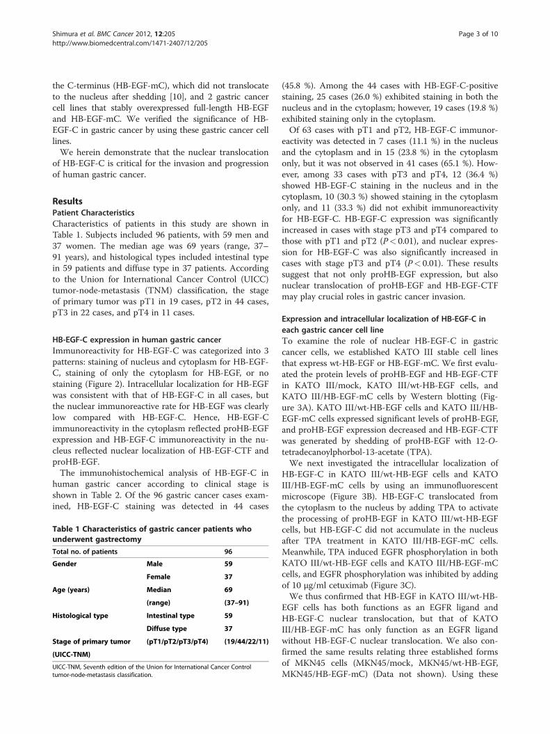
ResultsPatient CharacteristicsCharacteristics of patients in this study are shown inTable 1. Subjects included 96 patients, with 59 men and37 women. The median age was 69 years (range, 37–91 years), and histological types included intestinal typein 59 patients and diffuse type in 37 patients. Accordingto the Union for International Cancer Control (UICC)tumor-node-metastasis (TNM) classification, the stageof primary tumor was pT1 in 19 cases, pT2 in 44 cases,pT3 in 22 cases, and pT4 in 11 cases.
HB-EGF-C expression in human gastric cancerImmunoreactivity for HB-EGF-C was categorized into 3patterns: staining of nucleus and cytoplasm for HB-EGF-C, staining of only the cytoplasm for HB-EGF, or nostaining (Figure 2). Intracellular localization for HB-EGFwas consistent with that of HB-EGF-C in all cases, butthe nuclear immunoreactive rate for HB-EGF was clearlylow compared with HB-EGF-C. Hence, HB-EGF-Cimmunoreactivity in the cytoplasm reflected proHB-EGFexpression and HB-EGF-C immunoreactivity in the nu-cleus reflected nuclear localization of HB-EGF-CTF andproHB-EGF.The immunohistochemical analysis of HB-EGF-C in
human gastric cancer according to clinical stage isshown in Table 2. Of the 96 gastric cancer cases exam-ined, HB-EGF-C staining was detected in 44 cases
Table 1 Characteristics of gastric cancer patients whounderwent gastrectomy
Total no. of patients 96
Gender Male 59
Female 37
Age (years) Median 69
(range) (37–91)
Histological type Intestinal type 59
Diffuse type 37
Stage of primary tumor (pT1/pT2/pT3/pT4) (19/44/22/11)
(UICC-TNM)
UICC-TNM, Seventh edition of the Union for International Cancer Controltumor-node-metastasis classification.
(45.8 %). Among the 44 cases with HB-EGF-C-positivestaining, 25 cases (26.0 %) exhibited staining in both thenucleus and in the cytoplasm; however, 19 cases (19.8 %)exhibited staining only in the cytoplasm.Of 63 cases with pT1 and pT2, HB-EGF-C immunor-
eactivity was detected in 7 cases (11.1 %) in the nucleusand the cytoplasm and in 15 (23.8 %) in the cytoplasmonly, but it was not observed in 41 cases (65.1 %). How-ever, among 33 cases with pT3 and pT4, 12 (36.4 %)showed HB-EGF-C staining in the nucleus and in thecytoplasm, 10 (30.3 %) showed staining in the cytoplasmonly, and 11 (33.3 %) did not exhibit immunoreactivityfor HB-EGF-C. HB-EGF-C expression was significantlyincreased in cases with stage pT3 and pT4 compared tothose with pT1 and pT2 (P< 0.01), and nuclear expres-sion for HB-EGF-C was also significantly increased incases with stage pT3 and pT4 (P< 0.01). These resultssuggest that not only proHB-EGF expression, but alsonuclear translocation of proHB-EGF and HB-EGF-CTFmay play crucial roles in gastric cancer invasion.
Expression and intracellular localization of HB-EGF-C ineach gastric cancer cell lineTo examine the role of nuclear HB-EGF-C in gastriccancer cells, we established KATO III stable cell linesthat express wt-HB-EGF or HB-EGF-mC. We first evalu-ated the protein levels of proHB-EGF and HB-EGF-CTFin KATO III/mock, KATO III/wt-HB-EGF cells, andKATO III/HB-EGF-mC cells by Western blotting (Fig-ure 3A). KATO III/wt-HB-EGF cells and KATO III/HB-EGF-mC cells expressed significant levels of proHB-EGF,and proHB-EGF expression decreased and HB-EGF-CTFwas generated by shedding of proHB-EGF with 12-O-tetradecanoylphorbol-13-acetate (TPA).We next investigated the intracellular localization of
HB-EGF-C in KATO III/wt-HB-EGF cells and KATOIII/HB-EGF-mC cells by using an immunofluorescentmicroscope (Figure 3B). HB-EGF-C translocated fromthe cytoplasm to the nucleus by adding TPA to activatethe processing of proHB-EGF in KATO III/wt-HB-EGFcells, but HB-EGF-C did not accumulate in the nucleusafter TPA treatment in KATO III/HB-EGF-mC cells.Meanwhile, TPA induced EGFR phosphorylation in bothKATO III/wt-HB-EGF cells and KATO III/HB-EGF-mCcells, and EGFR phosphorylation was inhibited by addingof 10 μg/ml cetuximab (Figure 3C).We thus confirmed that HB-EGF in KATO III/wt-HB-
EGF cells has both functions as an EGFR ligand andHB-EGF-C nuclear translocation, but that of KATOIII/HB-EGF-mC has only function as an EGFR ligandwithout HB-EGF-C nuclear translocation. We also con-firmed the same results relating three established formsof MKN45 cells (MKN45/mock, MKN45/wt-HB-EGF,MKN45/HB-EGF-mC) (Data not shown). Using these

Figure 2 Immunohistochemistry in human gastric cancer. Heparin-binding epidermal growth factor-like growth factor (HB-EGF) expressionwas immunohistochemically investigated using samples of surgically resected gastric cancer cells. Cells were immunostained using anti-HB-EGFantibody and anti-HB-EGF-C antibody. Representative positive staining cells were shown by black arrows. (Upper pictures) HB-EGF and HB-EGF-Cwere detected in the cytoplasm and nucleus of gastric cancer cells. (Middle pictures) HB-EGF and HB-EGF-C were detected in only the cytoplasmof gastric cancer cells. (Lower pictures) HB-EGF and HB-EGF-C were not detected in gastric cancer cells. (Original magnification: ×400).
Shimura et al. BMC Cancer 2012, 12:205 Page 4 of 10http://www.biomedcentral.com/1471-2407/12/205
cell lines, we assessed the behaviors of nuclear HB-EGF-C in gastric cancer cells.
HB-EGF-C nuclear translocation promotes gastric cancercell growthCell growth curves in 3 cancer cell lines are shown inFigure 4A. The growth of KATO III/wt-HB-EGF and
Table 2 HB-EGF-C expression and localization accordingto clinical stage
Expression (+)* (−)
Localization C (+) N (+) C (+) N (−) C (−) N (−) Total (n)
pT1, 2 7 (11.1)** 15 (23.8) 41 (65.1) 63
n, (%)
pT3, 4 12 (36.4)** 10 (30.3) 11 (33.3) 33
n, (%)
Total 19 (19.8) 25 (26.0) 52 (54.2) 96
n, (%)
C, cytoplasm; N, nucleus; (+), positive staining; (−), negative staining.* P< 0.01, pT3, 4 vs. pT1, 2 for the rate of C (+) N (+) and C (+) N (−).** P< 0.01, pT3, 4 vs. pT1, 2 for the rate of C (+) N (+).
KATO III/HB-EGF-mC cells was significantly faster thanthat of KATOIII/mock cells, and the growth of KATOIII/HB-EGF-mC cells was significantly decreased com-pared with KATO III/wt-HB-EGF cells. The same resultswere observed in three forms of MKN45 cells (Add-itional file 1: Figure S1A). These results demonstratethat both conventional signals of HB-EGF as an EGFRligand and a novel signal for HB-EGF-C nuclear trans-location are critical for gastric cancer cell proliferation.
HB-EGF-C nuclear translocation promotes cell invasionand wound healingTo verify the present results by using clinical samples,we next investigated whether HB-EGF-C nuclear trans-location causes an increase in the migration of gastriccancer cells. Three gastric cancer cell lines were used ina transwell invasion assay. As shown in Figure 4B,KATO III/wt-HB-EGF showed a obvious increase in in-vasion compared with KATO III/mock cells, whereasKATO III/HB-EGF-mC cells did a slight increase. More-over, KATO III/HB-EGF-mC cells showed a significantdecrease in invasion compared with KATO III/wt-HB-

Figure 3 Characteristics of KATO III/wt-HB-EGF and KATO III/HB-EGF-mC cells. A) Western blot analysis of proHB-EGF and HB-EGF-CTFexpression in 3 gastric cancer cell lines. Anti-HB-EGF antibody was used to recognize the proHB-EGF ectodomain and anti-HB-EGF-C antibody wasused to recognize the cytoplasmic region of proHB-EGF. Each lane contains 100 μg of protein. Cleavage of proHB-EGF was stimulated using 200nM TPA. B) HB-EGF-C localization after TPA-inducible processing of proHB-EGF in KATO III/wt-HB-EGF and KATO III/HB-EGF-mC cells byimmunofluorescence microscopy. Nuclei were stained blue with DAPI, and HB-EGF-C were stained red with anti-HB-EGF-C antibody. Cells werestimulated with 200 nM TPA for 60 min. C) Western blot analysis of EGFR phosphorylation induced by TPA (200 nM) with or without cetuximab(10 μg/ml) in KATO III/wt-HB-EGF and KATO III/HB-EGF-mC.
Shimura et al. BMC Cancer 2012, 12:205 Page 5 of 10http://www.biomedcentral.com/1471-2407/12/205
EGF cells. Although the differences were smaller in com-parison with KATO III cells, the same results wereobserved in three forms of MKN45 cells (Additional file1: Figure S1B).Wound healing assays have been employed to study
cell polarization or tissue matrix remodeling or to esti-mate cell proliferation and migration rates. HB-EGF iswell known to be upregulated in the wound healingprocess of certain cell types, including keratinocytes[11,12]. We studied the effect of HB-EGF on woundhealing by using 3 gastric cancer cell lines (Figure 4C,D). The effectiveness of wound healing was observed inKATO III/wt-HB-EGF, but no significant effect wasobserved in KATO III/HB-EGF-mC. Although a littlewound healing effect was observed in MKN45/HB-EGF-mC, the same tendency as KATO III cells was observedin MKN45 cells (Additional file 1: Figure S1C, D). Theseresults suggest that HB-EGF-C nuclear translocation ra-ther than HB-EGF as an EGFR ligand is critical for gas-tric cancer cell migration.
DiscussionEGFR and EGFR ligands have been extensively studiedbecause the inactivation of EGFR represents a promising
strategy for the treatment of several cancers [13]. How-ever, there have been no studies regarding the actions ofproHB-EGF and HB-EGF-CTF induced by the cleavageof EGFR ligands. We previously reported that nucleartranslocation of HB-EGF-CTF results in a unique signaltransduction pathway for cell growth [6,7,10,14]. In thepresent study, we demonstrated the role of HB-EGF-Cin human gastric cancer.Of the EGFR ligands, HB-EGF is considered the most
important growth factor because HB-EGF-knockoutmice die shortly after birth, in contrast to the effects ofother EGFR ligands [15]. HB-EGF, which is an inducerof tumor growth and angiogenesis, induces resistanceto chemotherapy [16,17]. The expression of HB-EGFhas been demonstrated in many human cancers, suchas hepatocellular carcinoma [18], pancreatic cancer[19], gastric cancer [20,21], colorectal cancer [22], andovarian cancer [23]. In human gastric cancer, a previ-ous study reported that HB-EGF immunoreactivity wasdetected in 61 to 72 % of cancer cells and was gener-ally stronger in deeply invasive cancer cells than incancer cells of more superficial layers [20,21]. More-over, a recent report suggested that HB-EGF promotedperitoneal carcinomatosis from gastric cancer [24]. HB-

Figure 4 Cell proliferation and migration in KATO III/mock, KATO III/wt-HB-EGF and KATO III/HB-EGF-mC cells. A) Cell proliferation assay.Mean of 3 independent clones; bars, SD;* P< 0.01, as compared with KATO III/mock; **P< 0.05. B) Transwell invasion assay was analyzed in each cellat 48 h after 200 nM TPA stimulation. Value of KATO-III/mock cells was arbitrarily defined as 1. Mean of 3 independent clones; bars, SD;* P< 0.01, ascompared with KATO III/mock; **P< 0.05, as compared with KATO III/mock; ***P< 0.05. C) Wound healing assay. Confluent monolayers of each gastriccancer cells were mechanically wounded with a pipette tip, and photos were obtained at 0 h and 24 h after stimulation of 200 nM TPA (Originalmagnification: ×40). D) Quantification of wound healing assay in 3 independent clones. Migration rate of KATO-III/mock cells at 24 h after TPAstimulation was arbitrarily defined as 1. Mean of 3 independent clones; bars, SD* P< 0.01, as compared with KATO III/mock; **P< 0.01.
Shimura et al. BMC Cancer 2012, 12:205 Page 6 of 10http://www.biomedcentral.com/1471-2407/12/205
EGF is thus considered a potential growth factor ingastric cancer.In the present study, proHB-EGF expression was
observed in 45.8 % of human gastric cancer cases, par-ticularly in deeply invasive gastric cancer. This frequencywas slightly lower than the previous reports [20,21], butit is considered that positive for both anti-HB-EGF andanti-HB-EGF-C antibodies was defined as HB-EGF-posi-tive in the present study. Notably, we observed nuclearexpression of HB-EGF-C in deep gastric cancers (pT3and pT4).In order to confirm the relationship between invasion
and HB-EGF-C nuclear localization that had beendemonstrated in clinical experiments, we successfullyconstructed gastric cancer cell lines with and withoutHB-EGF-C nuclear translocation by overexpressing wt-HB-EGF and HB-EGF-mC. We were thus able to se-lectively analyze the fundamental effects of HB-EGF-Cnuclear translocation in vitro.The present study showed that both the function of
HB-EGF as an EGFR ligand and HB-EGF-C nucleartranslocation induce gastric cancer growth, whereas HB-
EGF-C nuclear translocation independently plays a crit-ical role in gastric cancer invasion. Hence, the presentin vitro findings support the data of clinical experiments.Recent studies have shown that the inhibition of proHB-EGF shedding promoted cell-cell interactions anddecreased cell migration [25,26]. HB-EGF-C signals inthe present study may involve these previous resultsrelated to cell migration.Nuclear staining of HB-EGF is reportedly a progressive
and poor prognostic factor in bladder cancer [27,28].Nuclear translocation of proHB-EGF and HB-EGF-CTFmight be the factor responsible for this phenomenon be-cause an antibody against HB-EGF-C was used in thesestudies. Our study showed that nuclear HB-EGF-C is animportant progressive factor in gastric cancer as well asin bladder cancer. We cannot precisely elucidate whichof proHB-EGF and HB-EGF-CTF has more responsibil-ity for gastric cancer invasion. However, we can specu-late that HB-EGF-CTF would be involved more thanproHB-EGF, because much HB-EGF-CTF yield aftershedding of proHB-EGF that is necessary for HB-EGF-Cnuclear translocation. Further investigation is necessary,

Shimura et al. BMC Cancer 2012, 12:205 Page 7 of 10http://www.biomedcentral.com/1471-2407/12/205
as the differences between proHB-EGF and HB-EGF-CTF in the nucleus and the detailed mechanisms indu-cing cell migration are not yet fully understood; clarifyingthese mechanisms may result in the development of newcancer therapies.
ConclusionsThe present study showed that HB-EGF-C nucleartranslocation is involved in gastric cancer development,in addition to the conventional function of HB-EGF asan EGFR ligand. HB-EGF-C nuclear translocation mayoffer a prognostic marker and a new molecular targetfor gastric cancer therapy.
MethodsMaterialsAnti-HB-EGF-C antibody was used to recognize HB-EGF-C, as previously described [7], and anti-HB-EGFantibody (R&D Systems, Minneapolis, MN) was used torecognize the proHB-EGF ectodomain. Cetuximab((Merk, Darmstadt, Germany)), a monoclonal antibodyto EGFR, was used to inhibit EGFR phosphorylation.
ImmunohistochemistryImmunohistochemical staining was performed usinganti-HB-EGF antibody and anti-HB-EGF-C antibody.Consecutive 4-μm-thick sections were deparaffinizedand hydrated through a graded series of alcohols. Afterinhibiting endogenous peroxidase activity by immersionin a 3 % H2O2/methanol solution, antigen retrieval wasachieved by heating the samples in 10 mM citrate buffer(pH 6.0) in a microwave oven for 10 min at 98 °C. Next,sections were incubated overnight with primary anti-bodies. After thorough washing in PBS (−), the sampleswere incubated with biotinylated secondary antibodiesand then with avidin-biotin-horseradish peroxidase com-plexes (Vectastain Elite ABC kit; Vector Laboratories,Burlingame, CA). Finally, immune complexes werevisualized by incubation in 0.01 % H2O2 and 0.05 %3,3′-diaminobenzidine tetrachloride. Nuclear counter-staining was accomplished with Mayer’s hematoxylin.
Clinical patients and assessmentUsing information from a computerized database, weobtained gastric cancer specimens from 96 patients whohad undergone surgical operations for gastric adenocar-cinoma between January 2002 and December 2006 atthe Nagoya City University Hospital. Written generalconsent that included research uses of clinical data hadbeen obtained from all patients. The study was per-formed in accordance with the Declaration of Helsinkiand Japanese ethical guidelines for epidemiological re-search. We obtained an institutional review board (IRB)waiver to conduct this study from the chairperson of the
IRB. All 96 patients were histologically diagnosed withgastric cancer. Clinical stage was determined accordingto the Seventh edition of the UICC-TNM classification[29]. All immunostained specimens were assessed by 2investigators who were blinded to all clinical informa-tion. When more than 10 % of the cancer cells in eachsection were stained for both anti-HB-EGF antibody andanti-HB-EGF-C antibody, immunostaining was definedas positive.
Cell cultureWe used KATO III and MKN45 gastric cancer cell line(Japan Health Science Research Resources Bank, Tokyo,Japan) in our investigation. KATO III and MKN45 cellswere maintained in RPMI1640 (Sigma-Aldrich Co., St.Louis, MO) medium that was supplemented with 10 %fetal bovine serum (FBS). Cells were cultured at 37 °C in5 % CO2 humidified air.
TransfectioncDNAs encoding wild-type HB-EGF (wt-HB-EGF) andthe mutant that carry a point mutation at K201A of theC-terminus (HB-EGF-mC) [10] were subcloned intopME18SIII with hygromycin-resistance gene. As previ-ously shown, HB-EGF-mC does not translocate from theplasma membrane to the nucleus after TPA stimulation.All cDNA constructs were verified by DNA sequencingusing a CEQ 8000 DNA Analysis System (BeckmanCoulter, Brea, CA).The KATO III human gastric cancer cell line was trans-
fected with wt-HB-EGF (KATO III/wt-HB-EGF) and HB-EGF-mC (KATOIII/HB-EGF-mC) by using Lipofectamine2000 (Invitrogen, Carlsbad, CA), according to the manu-facturer’s instructions, and stably transfected clones wereisolated using hygromycin B (Invitrogen). As a control,KATO III was transfected with an empty plasmid (KATOIII/mock). As well as KATO III cells, MKN45/wt-HB-EGF, MKN45/HB-EGF-mC and MKN45/mock cells wereestablished. These cells were maintained in RPMI1640medium that was supplemented with 10 % FBS and800 μg/mL hygromycin B.
Immunofluorescence microscopySamples were fixed with ethanol and acetone and incu-bated with primary antibodies against HB-EGF-C. Second-ary antibodies was Alexa FluorW 594 goat anti-rabbit IgG(H+L) (Invitrogen). All sections were counterstained withDAPI (KPL, Inc., Gaithersburg, MD). Images wereobtained using an Eclipse 80i fluorescent microscope(Nikon, Tokyo, Japan).Each gastric cancer cell in a subconfluent state was
placed in serum-free medium for 24 h and stimulatedwith 200 nM TPA (Cell Signaling Technology, Danvers,MA) for 60 min. Cells were stimulated by TPA in order

Shimura et al. BMC Cancer 2012, 12:205 Page 8 of 10http://www.biomedcentral.com/1471-2407/12/205
to investigate the localization of HB-EGF-C by sheddingproHB-EGF [30], and the intracellular localization ofHB-EGF-C was then analyzed by immunofluorescence.
Western blottingCells were washed with PBS and subsequently dissolvedin 1× cell lysis buffer (Cell Signaling Technology) con-taining 20 mM Tris–HCl (pH 7.5), 150 mM NaCl,1 mM Na2EDTA, 1 mM EGTA, 1 % Triton, 2.5 mM so-dium pyrophosphate, 1 mM β-glycerophosphate, 1 mMNa3VO4, and 1 μg/mL leupeptin. After disruption in anice bath by using a Bio-ruptor sonicator (Cosmo Bio,Tokyo, Japan) for 15 s, lysates were centrifuged at15,000 rpm for 10 min at 4°C. Each sample was normal-ized against an equal protein concentration by using aprotein assay kit (Bio-Rad Laboratories, Hercules, CA).A equal quantity of 2× sodium dodecyl sulfate-polyacryl-amide gel electrophoresis (SDS-PAGE) sample buffer(0.5 mol/L Tris–HCl, pH 7.2, 1 % SDS, 100 mmol/L β-mercaptoethanol, and 0.01 % bromophenol blue) wasadded to each sample and boiled for 5 min at 100°C. Ali-quots of sample were fractionated on 10 or 15 % SDS-PAGE and then electroblotted onto a nitrocellulosemembrane. The membrane was blocked with 5 %skimmed milk in PBS for 1 h at room temperature. Themembrane was incubated with the primary antibodiesfor HB-EGF-C, HB-EGF, EGFR (MILLIPORE, Temecula,CA) or phospho-EGFR (MILLIPORE) overnight at 4°Cand then washed with 0.05 % Tween 20 in PBS 3 timesat 5-min intervals. The membrane was incubated withsecondary antibody for 1 h at room temperature, whichwas followed by 3 washes with 0.05 % Tween 20 in PBS3 times at 5-min intervals. The membrane was treatedwith enhanced chemiluminescence detection reagents(ECL; Amersham, Arlington Heights, IL) for 1 min atroom temperature and then exposed to scientific im-aging films (Eastman Kodak, Rochester, NY). Proteinswere visualized as bands on the images. Filters werestripped and reprobed with monoclonal β-actin antibody(Abcam plc, Tokyo, Japan) as an internal control.
Cell proliferation assayProliferation assays were performed as follows. Cellswere seeded at 2.0 × 10⁴ cells in the medium with 10 %FBS on 6-cm diameter dishes. Cells were counted ondays 3, 5, and 8 by using an Automatic Cell Counter(Millipore, Billerica, MA). Each experiment was con-ducted independently in cell lines isolated from 3 inde-pendent clones.
Wound healing assayA wound healing assay was conducted in order tomeasure cell motility. Cells were grown to confluence in6-well plates and serum-starved for 24 h, and then a
cross-shaped wound was made on the monolayers byusing a sterile 200-μL pipette tip. Cells were washedwith PBS, placed in the same media with 200nM TPA,and the cross-shaped wound was photographed undermicroscope at 0 h and 24 h.
Transwell invasion assayCell migration was assessed using the Cell InvasionAssay (CULTREX, Gaithersburg, MD), which consists ofa 96-well transwell tissue culture plate with an 8-μmpore size membranes coated with matrigel (top cham-ber) and a black receiver plate compatible with a 96-wellfluorescent plate reader (bottom chamber). After 24 h ofserum starvation, cells (5000 cells/well) in serum-freemedium with 200nM TPA were placed in the top cham-ber, and medium containing 10 % FBS was added to thebottom chamber. After 48 h of incubation in CO2 at 37°C, the cells that had invaded the bottom chamber weremeasured according to the manufacturer’s instructions.Each experiment was conducted in cell lines which wasisolated from 3 independent clones.
Statistical analysisValues are expressed as the mean ± SD. Data were ana-lyzed using χ2 test as appropriate. Multiple comparisonwas done using Games-Howell’s method. P-values< 0.05were considered statistically significant. Data analyseswere performed using Dr. SPSS II for Windows release11.0.1 J software (SPSS Japan, Tokyo, Japan).
Additional file
Additional file 1: Figure S1 Cell proliferation and migration in MKN45/mock, MKN45/wt-HB-EGF and MKN45/HB-EGF-mC cells. A) Cellproliferation assay. Mean of 3 independent clones; bars, SD;* P< 0.01, ascompared with MKN45/mock; **P< 0.01. B) Transwell invasion assay wasanalyzed in each cell at 48 h after 200 nM TPA stimulation. Value ofMKN45/mock cells was arbitrarily defined as 1. Mean of 3 independentclones; bars, SD;* P< 0.01, as compared with KATO III/mock; **P< 0.05, ascompared with KATO III/mock; ***P< 0.05. C) Wound healing assay.Confluent monolayers of each gastric cancer cells were mechanicallywounded with a pipette tip, and photos were obtained at 0 h and 24 hafter stimulation of 200 nM TPA (Original magnification: ×40). D)Quantification of wound healing assay in 3 independent clones.Migration rate of MKN45/mock cells at 24 h after TPA stimulation wasarbitrarily defined as 1. Mean of 3 independent clones; bars, SD* P< 0.01,as compared with MKN45/mock; **P< 0.05, as compared with MKN45/mock; ***P< 0.01.
AbbreviationsEGFR: Epidermal growth factor receptor; proHB-EGF: Membrane-anchoredheparin-binding epidermal growth factor-like growth factor; HB-EGF-CTF: Acarboxy-terminal fragment of heparin-binding epidermal growth factor-likegrowth factor; HB-EGF-C: The cytoplasmic domain of heparin-bindingepidermal growth factor-like growth factor; s-HB-EGF: Soluble heparin-binding epidermal growth factor-like growth factor; PLZF: Promyelocyticleukemia zinc finger; Bcl6: B-cell lymphoma 6; wt-HB-EGF: Wild-type HB-EGF;HB-EGF-mC: HB-EGF at the C-terminus; PLZF: Promyelocytic leukemia zincfinger; KATO III/wt-HB-EGF: KATO III human gastric cancer cell linetransfected with wt-HB-EGF; KATOIII/HB-EGF-mC: KATO III human gastric

Shimura et al. BMC Cancer 2012, 12:205 Page 9 of 10http://www.biomedcentral.com/1471-2407/12/205
cancer cell line transfected with HB-EGF-mC; KATO III/mock: KATO III humangastric cancer cell line transfected with an empty plasmid; MKN45/wt-HB-EGF: MKN45 human gastric cancer cell line transfected with wt-HB-EGF;MKN45/HB-EGF-mC: MKN45 human gastric cancer cell line transfected withHB-EGF-mC; MKN45/mock: MKN45 human gastric cancer cell line transfectedwith an empty plasmid; FBS: Fetal bovine serum; TPA: 12-O-tetradecanoylphorbol-13-acetate.
Competing interestsAll the authors declare that we have no competing interests.
AcknowledgmentsWe wish to thank Yukimi Hashidume for technical assistance. This study wassupported by the Ministry of Education, Culture, Sports, Science andTechnology of Japan (No. 22790665).
Author details1Department of Gastroenterology and Metabolism, Nagoya City UniversityGraduate School of Medical Sciences, 1 Kawasumi, Mizuho-cho, Mizuho-ku,Nagoya, 467-8601, Japan. 2Department of Biochemistry and MolecularGenetics, Ehime University Graduate School of Medicine, Shitsukawa, Toon,Ehime, 791-0295, Japan. 3Department of Cell Growth and Tumor Regulation,Proteo-Medicine Research Center, Ehime University, Shitsukawa, Toon, Ehime,791-0295, Japan.
Author’s contributionsConception and design, TS; Acquisition of data, TS, MY, TM, and TK; Analysisand interpretation of data, TS; Drafting of the manuscript, TS; Revising itcritically for important intellectual content, SF and SH; Final approval of theversion to be published, TJ; Acquisition of funding, TS; General supervision ofresearch group, SH and TJ. All authors read and approved the finalmanuscript.
Received: 17 December 2011 Accepted: 30 May 2012Published: 30 May 2012
References1. International Agency for Research on Cancer GLOBOCAN 2008:: ; http://
globocan.iarc.fr/factsheets/populations/factsheet.asp?uno=900 (last accessedon December 16, 2011).
2. Bang YJ, Van Cutsem E, Feyereislova A, Chung HC, Shen L, Sawaki A,Lordick F, Ohtsu A, Omuro Y, Satoh T, et al: Trastuzumab in combinationwith chemotherapy versus chemotherapy alone for treatment of HER2-positive advanced gastric or gastro-oesophageal junction cancer(ToGA): a phase 3, open-label, randomised controlled trial. Lancet 2010,376:687–697.
3. Higashiyama S, Iwabuki H, Morimoto C, Hieda M, Inoue H, Matsushita N:Membrane-anchored growth factors, the epidermal growth factor family:beyond receptor ligands. Cancer Sci 2008, 99:214–220.
4. Normanno N, Bianco C, De Luca A, Maiello MR, Salomon DS: Target-basedagents against ErbB receptors and their ligands: a novel approach tocancer treatment. Endocr Relat Cancer 2003, 10:1–21.
5. Salomon DS, Brandt R, Ciardiello F, Normanno N: Epidermal growth factor-related peptides and their receptors in human malignancies. Crit RevOncol Hematol 1995, 19:183–232.
6. Kinugasa Y, Hieda M, Hori M, Higashiyama S: The carboxyl-terminalfragment of pro-HB-EGF reverses Bcl6-mediated gene repression. J BiolChem 2007, 282:14797–14806.
7. Nanba D, Mammoto A, Hashimoto K, Higashiyama S: Proteolytic release ofthe carboxy-terminal fragment of proHB-EGF causes nuclear export ofPLZF. J Cell Biol 2003, 163:489–502.
8. Hirata Y, Ogasawara N, Sasaki M, Mizushima T, Shimura T, Mizoshita T, MoriY, Kubota E, Wada T, Tanida S, et al: BCL6 degradation caused by theinteraction with the C-terminus of pro-HB-EGF induces cyclin D2expression in gastric cancers. Br J Cancer 2009, 100:1320–1329.
9. Shimura T, Kataoka H, Ogasawara N, Kubota E, Sasaki M, Tanida S, Joh T:Suppression of proHB-EGF carboxy-terminal fragment nucleartranslocation: a new molecular target therapy for gastric cancer. ClinCancer Res 2008, 14:3956–3965.
10. Hieda M, Isokane M, Koizumi M, Higashi C, Tachibana T, Shudou M, TaguchiT, Hieda Y, Higashiyama S: Membrane-anchored growth factor, HB-EGF,
on the cell surface targeted to the inner nuclear membrane. J Cell Biol2008, 180:763–769.
11. Tokumaru S, Higashiyama S, Endo T, Nakagawa T, Miyagawa JI, Yamamori K,Hanakawa Y, Ohmoto H, Yoshino K, Shirakata Y, et al: Ectodomainshedding of epidermal growth factor receptor ligands is required forkeratinocyte migration in cutaneous wound healing. J Cell Biol 2000,151:209–220.
12. Marikovsky M, Breuing K, Liu PY, Eriksson E, Higashiyama S, Farber P,Abraham J, Klagsbrun M: Appearance of heparin-binding EGF-like growthfactor in wound fluid as a response to injury. Proc Natl Acad Sci USA 1993,90:3889–3893.
13. Hynes NE, Lane HA: ERBB receptors and cancer: the complexity oftargeted inhibitors. Nat Rev Cancer 2005, 5:341–354.
14. Nanba D, Inoue H, Shigemi Y, Shirakata Y, Hashimoto K, Higashiyama S: Anintermediary role of proHB-EGF shedding in growth factor-induced c-Myc gene expression. J Cell Physiol 2008, 214:465–473.
15. Iwamoto R, Yamazaki S, Asakura M, Takashima S, Hasuwa H, Miyado K,Adachi S, Kitakaze M, Hashimoto K, Raab G, et al: Heparin-binding EGF-likegrowth factor and ErbB signaling is essential for heart function. Proc NatlAcad Sci USA 2003, 100:3221–3226.
16. Wang F, Liu R, Lee SW, Sloss CM, Couget J, Cusack JC: Heparin-bindingEGF-like growth factor is an early response gene to chemotherapy andcontributes to chemotherapy resistance. Oncogene 2007, 26:2006–2016.
17. Ongusaha PP, Kwak JC, Zwible AJ, Macip S, Higashiyama S, Taniguchi N,Fang L, Lee SW: HB-EGF is a potent inducer of tumor growth andangiogenesis. Cancer Res 2004, 64:5283–5290.
18. Inui Y, Higashiyama S, Kawata S, Tamura S, Miyagawa J, Taniguchi N,Matsuzawa Y: Expression of heparin-binding epidermal growth factor inhuman hepatocellular carcinoma. Gastroenterology 1994, 107:1799–1804.
19. Kobrin MS, Funatomi H, Friess H, Buchler MW, Stathis P, Korc M: Inductionand expression of heparin-binding EGF-like growth factor in humanpancreatic cancer. Biochem Biophys Res Commun 1994, 202:1705–1709.
20. Murayama Y, Miyagawa J, Shinomura Y, Kanayama S, Isozaki K, Yamamori K,Mizuno H, Ishiguro S, Kiyohara T, Miyazaki Y, et al: Significance of theassociation between heparin-binding epidermal growth factor-likegrowth factor and CD9 in human gastric cancer. Int J Cancer 2002,98:505–513.
21. Naef M, Yokoyama M, Friess H, Buchler MW, Korc M: Co-expression ofheparin-binding EGF-like growth factor and related peptides in humangastric carcinoma. Int J Cancer 1996, 66:315–321.
22. Kopp R, Rothbauer E, Ruge M, Arnholdt H, Spranger J, Muders M, PfeifferDG, Schildberg FW, Pfeiffer A: Clinical implications of the EGF receptor/ligand system for tumor progression and survival in gastrointestinalcarcinomas: evidence for new therapeutic options. Recent Results CancerRes 2003, 162:115–132.
23. Yagi H, Miyamoto S, Tanaka Y, Sonoda K, Kobayashi H, Kishikawa T, IwamotoR, Mekada E, Nakano H: Clinical significance of heparin-binding epidermalgrowth factor-like growth factor in peritoneal fluid of ovarian cancer. Br JCancer 2005, 92:1737–1745.
24. Yasumoto K, Yamada T, Kawashima A, Wang W, Li Q, Shterev DI, TacheuchiS, Mouri H, Yamashita K, Ohtsubo K, Yano S: The EGFR ligandsAmphiregulin and Heparin-binding EGF-like growth factor promoteperitoneal carcinomatosis in CXCR4-expressing gastric cancer. Clin CancerRes 2011, 17:3619–3630.
25. Wang F, Sloss C, Zhang X, Lee SW, Cusack JC: Membrane-bound heparin-binding epidermal growth factor like growth factor regulates E-cadherinexpression in pancreatic carcinoma cells. Cancer Res 2007, 67:8486–8493.
26. Singh AB, Tsukada T, Zent R, Harris RC: Membrane-associated HB-EGFmodulates HGF-induced cellular responses in MDCK cells. J Cell Sci 2004,117:1365–1379.
27. Kramer C, Klasmeyer K, Bojar H, Schulz WA, Ackermann R, Grimm MO:Heparin-binding epidermal growth factor-like growth factor isoforms andepidermal growth factor receptor/ErbB1 expression in bladder cancerand their relation to clinical outcome. Cancer 2007, 109:2016–2024.
28. Adam RM, Danciu T, McLellan DL, Borer JG, Lin J, Zurakowski D, Weinstein MH,Rajjayabun PH, Mellon JK, Freeman MR: A nuclear form of the heparin-binding epidermal growth factor-like growth factor precursor is a featureof aggressive transitional cell carcinoma. Cancer Res 2003, 63:484–490.
29. Sobin L, Gospodarowicz M, Wittekind C: TNM classification of malignanttumours. In. 7th editionEdited by Hoboken NJ.: John Wiley & Sons, Inc;2009.

Shimura et al. BMC Cancer 2012, 12:205 Page 10 of 10http://www.biomedcentral.com/1471-2407/12/205
30. Goishi K, Higashiyama S, Klagsbrun M, Nakano N, Umata T, Ishikawa M,Mekada E, Taniguchi N: Phorbol ester induces the rapid processing of cellsurface heparin-binding EGF-like growth factor: conversion fromjuxtacrine to paracrine growth factor activity. Mol Biol Cell 1995, 6:967–980.
doi:10.1186/1471-2407-12-205Cite this article as: Shimura et al.: Nuclear translocation of thecytoplasmic domain of HB-EGF induces gastric cancer invasion. BMCCancer 2012 12:205.
Submit your next manuscript to BioMed Centraland take full advantage of:
• Convenient online submission
• Thorough peer review
• No space constraints or color figure charges
• Immediate publication on acceptance
• Inclusion in PubMed, CAS, Scopus and Google Scholar
• Research which is freely available for redistribution
Submit your manuscript at www.biomedcentral.com/submit