Embed Size (px)
Citation preview
Mechanotransduction activates canonicalWnt/β-catenin signaling to promotelymphatic vascular patterning andthe development of lymphaticand lymphovenous valvesBoksik Cha,1 Xin Geng,1 Md. Riaj Mahamud,1,2 Jianxin Fu,1 Anish Mukherjee,3 Yeunhee Kim,4
Eek-hoon Jho,5 Tae Hoon Kim,4 Mark L. Kahn,6 Lijun Xia,1,7 J. Brandon Dixon,3 Hong Chen,8
and R. Sathish Srinivasan1,2
1Cardiovascular Biology Research Program, Oklahoma Medical Research Foundation, Oklahoma City, Oklahoma 73104, USA;2Department of Cell Biology, University of Oklahoma Health Sciences Center, Oklahoma City, Oklahoma 73104, USA; 3ParkerH. Petit Institute for Bioengineering and Bioscience,Georgia Institute of Technology, Atlanta,Georgia 30332,USA; 4Department ofBiological Sciences, Center for Systems Biology, TheUniversity of Texas at Dallas, Richardson, Texas 75080, USA; 5Department ofLife Science, University of Seoul, Seoul 130-743, Korea; 6Department of Medicine, Division of Cardiology, University ofPennsylvania, Philadelphia, Pennsylvania 19104, USA; 7Department of Biochemistry, University of Oklahoma Health SciencesCenter, Oklahoma City, Oklahoma 73104, USA; 8Vascular Biology Program, Boston Children’s Hospital, Boston, Massachusetts02115, USA
Lymphatic vasculature regulates fluid homeostasis by returning interstitial fluid to blood circulation. Lymphaticendothelial cells (LECs) are the building blocks of the entire lymphatic vasculature. LECs originate as a homoge-neous population of cells predominantly from the embryonic veins and undergo stepwise morphogenesis to becomethe lymphatic capillaries, collecting vessels or valves. Themolecular mechanisms underlying themorphogenesis ofthe lymphatic vasculature remain to be fully understood. Here we show that canonical Wnt/β-catenin signaling isnecessary for lymphatic vascularmorphogenesis. Lymphatic vascular-specific ablation of β-catenin inmice preventsthe formation of lymphatic and lymphovenous valves. Additionally, lymphatic vessel patterning is defective in thesemice, with abnormal recruitment of mural cells. We found that oscillatory shear stress (OSS), which promoteslymphatic vessel maturation, triggers Wnt/β-catenin signaling in LECs. In turn, Wnt/β-catenin signaling controlsthe expression of several molecules, including the lymphedema-associated transcription factor FOXC2. Impor-tantly, FOXC2 completely rescues the lymphatic vessel patterning defects in mice lacking β-catenin. Thus, ourwork reveals thatmechanical stimulation is a critical regulator of lymphatic vascular development via activation ofWnt/β-catenin signaling and, in turn, FOXC2.
[Keywords: FOXC2; lymphatic valves; lymphatic vascular development; lymphovenous valves; PROX1; Wnt/β-cateninsignaling]
Supplemental material is available for this article.
Received April 7, 2016; revised version accepted May 23, 2016.
The lymphatic vasculature collects and returns intersti-tial fluid to the blood. The mammalian lymphatic vascu-lature is composed of lymphatic endothelial cells (LECs)that originate predominantly from embryonic veins, al-though additional sources might exist (Srinivasan et al.2007; Martinez-Corral et al. 2015; Stanczuk et al. 2015).LEC progenitors from the vein undergo stepwise morpho-
genesis to form a hierarchical network of capillaries, col-lecting vessels, intraluminal lymphatic valves (LVs), andlymphovenous valves (LVVs). LVs within collecting ves-sels regulate the unidirectional flow of lymph, whereasLVVs at the jugular–subclavian vein junctions returnlymph to the blood circulation. Defects in lymphatic ves-sels or valves cause lymphedema, a disease in which tis-sues swell due to fluid accumulation.
Corresponding author: [email protected] published online ahead of print. Article and publication date are on-line at http://www.genesdev.org/cgi/doi/10.1101/gad.282400.116. Freelyavailable online through the Genes & Development Open Access option.
© 2016 Cha et al. This article, published in Genes & Development, isavailable under a Creative Commons License (Attribution 4.0 Internation-al), as described at http://creativecommons.org/licenses/by/4.0/.
GENES & DEVELOPMENT 30:1–16 Published by Cold Spring Harbor Laboratory Press; ISSN 0890-9369/16; www.genesdev.org 1
Cold Spring Harbor Laboratory Press on April 5, 2020 - Published by genesdev.cshlp.orgDownloaded from
LEC progenitors are specified in a subpopulation of ve-nous ECs by the activation of PROX1 expression. Mostof the LEC progenitors migrate out of the vein to formthe lymph sacs. However, a subset of LEC progenitorsstays on the veins to form four LVVs through the up-regu-lation of genes in addition to PROX1, such as FOXC2,GATA2, integrin α9 (ITGA9), and ITGA5 (Srinivasanand Oliver 2011; Geng et al. 2016). Next, lymphatic ves-sels sprout and migrate out from the lymph sacs in a ster-eotypic manner to form the primitive lymphatic plexus(Coxam et al. 2014; Martinez-Corral et al. 2015). Theprimitive lymphatic plexus undergoes maturation toform the hierarchical network of vessels. Maturation in-volves the pruning of branch points, a reduction in vesseldiameter, and the acquisition of mural cell coverage toform the collecting lymphatic vessels (Tammela et al.2007; Norrmen et al. 2009). Finally, LVs develop withinthemature collecting lymphatic vessels. At themolecularlevel, LVs and LVVs are almost identical (Geng et al.2016).
PROX1 and FOXC2 are important regulators of lym-phatic vascular development. LEC progenitors are notspecified in the absence of PROX1. Most Prox1+/− pupsdie soon after birth with severe edema. The survivingProx1+/− pups have LVs; however, LVs are absent in thosethat die soon after birth (Johnson et al. 2008). Likewise,most of the Prox1+/− embryos are completely devoid ofLVVs (Srinivasan andOliver 2011; Geng et al. 2016). How-ever, the surviving Prox1+/− pups have LVVs (X Geng,unpubl.).Mutations in FOXC2 are associatedwith humanlymphedema. The lymphatic plexuses of Foxc2−/− embry-os are mispatterned and abnormally covered with muralcells, do not undergo proper maturation, and lack LVsand LVVs (Petrova et al. 2004; Geng et al. 2016). A vari-able, strain-dependent valve phenotype is observed inFoxc2+/− embryos. Although some Foxc2+/− embryos arecompletely devoid of LVVs, other mutants have twoLVVs rather than the four that are normally observed incontrols (Geng et al. 2016). A 50% reduction in the num-ber of LVs is observed in Foxc2+/− embryos (Kanady et al.2015). Despite their critical roles, the mechanisms thatcontrol the expression of PROX1 and FOXC2, especiallyin the valves, have not yet been elucidated.
Lymphatic vessels are exposed to reversing fluid flow(more commonly known as oscillatory shear stress[OSS]) (Dixon et al. 2006, 2007; Sweet et al. 2015;Margariset al. 2016). In vitro, OSS promotes the expression ofFOXC2 in LECs (Sabine et al. 2012; Sweet et al. 2015). Fur-thermore, themesenteric lymphatic vessels do not under-go proper maturation, and LVs do not form in Clec2−/−
micewith defective lymph flow (Sweet et al. 2015). There-fore, OSS was proposed to control lymphatic vessel matu-ration and LV formation. However, howOSS is sensed andtranslated into FOXC2 expression is not known.
Intriguingly, OSS is not sufficient to promote PROX1expression in LECs (Sabine et al. 2012; Sweet et al.2015). Additional signaling pathways are likely requiredto regulate PROX1 expression in the valves. The Wnt/β-catenin pathway regulates PROX1 expression in coloncancer cells and neural stem cells (Petrova et al. 2008;
Karalay et al. 2011). Recently, activation of the Wnt/β-catenin pathway was shown to regulate PROX1 expres-sion in the lymphatic vasculature of zebrafish embryos(Nicenboim et al. 2015). Using cell-based approachesandmousemodels, we show that theWnt/β-catenin path-way regulates PROX1 expression in valves. Additionally,we identified unanticipated roles for this pathway inOSS sensing, lymphatic vascular patterning, and FOXC2expression.
Results
Wnt/β-catenin signaling is active in developing LVVs,LVs, and VVs
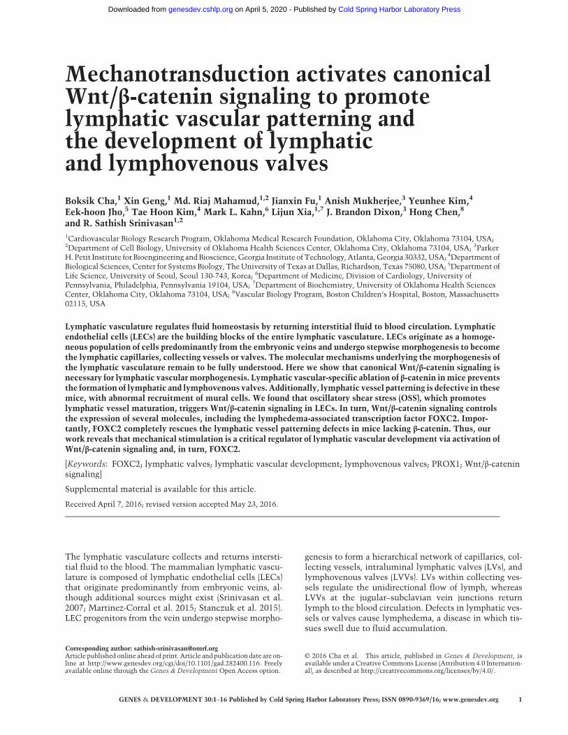
To determine whether Wnt/β-catenin signaling is activein the mouse lymphatic vasculature, we used transgenicTCF/LEF-H2B-EGFP reporter mice (Ferrer-Vaquer et al.2010). In these mice, a nuclear H2B-EGFP fusion is underthe control of TCF/LEF elements and activated by Wnt/β-catenin signaling. We analyzed 12-µm frontal sectionsof developing reporter embryos by immunohistochemis-try (IHC) and determined that ∼10% of PROX1+ LEC pro-genitors on the vein areH2B-EGFP+ at embryonic day 11.0(E11.0) (Supplemental Fig. 1A). However, LECs outsidethe vein are only rarely labeled. At E12.0 and E14.5, a sub-population of LVV-forming ECs (LVV-ECs) express nucle-ar H2B-EGFP (Fig. 1A–F). At E15.5, VV-forming ECs (VV-ECs) are H2B-EGFP+ (Supplemental Fig. 1B). To analyzeLVs, we performed whole-mount IHC of the mesenteryof E16.5–E18.5 TCF/LEF-H2B-EGFP embryos. We identi-fied modest H2B-EGFP expression in LECs at all stages(Fig. 1G–I; Supplemental Fig 1C,D). However, H2B-EGFPexpression is consistently stronger in the LV-formingECs (LV-ECs) at the three obvious stages of LV formation;i.e., up-regulation of PROX1 expression, formation of thecircular sheath, and a mature valve.
EGFP expression in the LVV-ECs is mosaic. Transgenicreporters do not fully recapitulate the activity of theWnt/β-catenin signaling pathway in vivo (Al Alam et al. 2011).Indeed, it was previously reported that a different Wnt/β-catenin signaling pathway reporter, BAT-gal, is inactivein LVs (Norrmen et al. 2009), and we did not observe anyBAT-gal+ cells in the entire lymphatic vasculature (datanot shown). Therefore, we used additional approaches toassess Wnt/β-catenin signaling activity in the valves.We performed in situ hybridization for Axin2, a well-characterized target of the Wnt/β-catenin signaling path-way, in numerous contexts. As a positive control, we eval-uated cardiac semilunar valves, which display activatedWnt/β-catenin signaling (Fig. 1J,K; Alfieri et al. 2010). Us-ing this approach, we observed strong and uniform expres-sion of Axin2 in LVVs at E14.5 (Fig. 1L–N). Furthermore,we analyzed mesenteric lymphatic vessels by IHC withantibodies that recognize total β-catenin or nonphos-phorylated (active) β-catenin and clearly detected theLVs (Fig. 1O,P).
Together, these results suggest that the Wnt/β-cateninsignaling pathway is active in LECs and particularly inthe LVs, LVVs, and VVs, in vivo.
Cha et al.
2 GENES & DEVELOPMENT
Cold Spring Harbor Laboratory Press on April 5, 2020 - Published by genesdev.cshlp.orgDownloaded from
β-Catenin is required for valve development
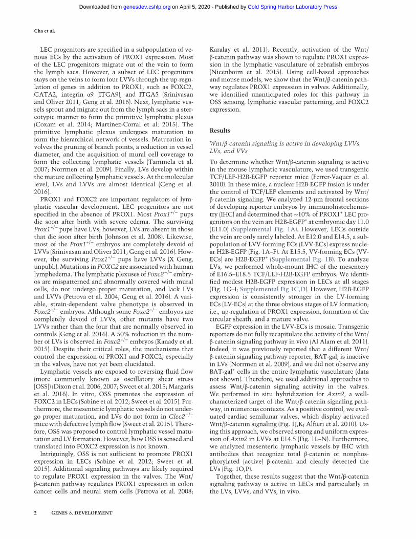
To elucidate the importance of the Wnt/β-catenin signal-ing pathway in valve development, we first conditionallydeleted Ctnnb1 (the gene encoding β-catenin) usingLyve1-Cremice (Brault et al. 2001; Pham et al. 2010). Lin-eage tracing with an R26+/tdTomato reporter revealed that
Lyve1-Cre is active in the cardinal vein as early as E9.5,and LVVs and lymphatic vessels are efficiently labeledin Cre reporter mice at E14.5 (data not shown). LVs andVVs that develop at later time points are also labeled byLyve1-Cre. We bred Lyve1-Cre;Ctnnb1+/f mice withCtnnb1+/f mice and failed to obtain any survivingLyve1-Cre;Ctnnb1f/f (Lyve1-Cre;Ctnnb1LOF) pups from>200 pups analyzed at postnatal day 0 (P0). Instead, we ob-served several dead, cyanotic, and edematous Lyve1-Cre;Ctnnb1LOF pups in the cages, suggesting perinatal lethal-ity. We collected E14.5, E16.5, and E18.5 Lyve1-Cre;Ctnnb1LOF embryos and determined that they had severeedema (Supplemental Fig. 2A,B; data not shown). Occa-sionally, some blood was observed in the peripheral skinof the mutant embryos (Supplemental Fig. 2C). We foundthat the lymph sacs of these embryos were severely dilat-ed, resulting in the constriction of the surrounding veins(Supplemental Fig. 2D–F).We recently described the stepwise morphogenesis of
LVVs and reported that the PROX1high FOXC2high
GATA2high LVV-ECs are first observed at E12.0 (Genget al. 2016). We found that LVV-ECs are absent in E12.0Lyve1-Cre;Ctnnb1LOF embryos (Supplemental Fig. 3A–
F). In scanning electron microscopy (SEM) images of con-trol embryos, LVV-ECs could be seen delaminating fromthe walls of the vein and loosely aggregating with eachother; however, these cells are absent in Lyve1-Cre;Ctnnb1LOF embryos (Supplemental Fig. 3A–F). These re-sults demonstrate that β-catenin is necessary for the dif-ferentiation of LVV-ECs. Consistently, IHC revealedthat PROX1high FOXC2high GATA2high LVV-ECs are pre-sent in E14.5 control embryos but absent in their Lyve1-Cre;Ctnnb1LOF littermates (Fig. 2A–D; data not shown).SEM confirmed that while LVVs are present in E14.5 con-trol embryos (Fig. 2E, magenta), they are absent in embry-os lacking β-catenin (Fig. 2F). Analysis of E16.5 controland Lyve1-Cre;Ctnnb1LOF embryos revealed that LVV-ECs are absent in mutant embryos at this stage as well(Fig. 2G,H). Thus, Lyve1-Cre;Ctnnb1LOF embryos displaya complete lack, and not just a delay, of LVV-ECdifferentiation.At E14.5, VV-ECs start to differentiate and could be
seen delaminating from the rim of the venous junctionin control embryos (Fig. 2E, green;Geng et al. 2016). Thesecells rapidly develop to form theVVs at E16.5 (Fig. 2G, yel-low arrows). However, VV-ECs are absent in Lyve1-Cre;Ctnnb1LOF embryos at both the E14.5 and E16.5 stages(Fig. 2F,H, respectively), demonstrating that β-catenin isalso necessary for the differentiation of VV-ECs.LV formation occurs in a stepwise manner starting at
E16.5 with the up-regulation of PROX1, FOXC2, andGATA2 in a subset of cells within the lymphatic vessels(Bazigou et al. 2009; Norrmen et al. 2009; Kazenwadelet al. 2015; Sweet et al. 2015). We determined that thePROX1high FOXC2high GATA2high LV-ECs are absent inthe mesenteric lymphatic vessels of E16.5 Lyve1-Cre;Ctnnb1LOF embryos, indicating that β-catenin is also nec-essary for the differentiation of LV-ECs (Supplemental Fig.4C–E). At E17.5, LV rudiments are more clearly visible incontrol embryos and are not observed in mutants
Figure 1. The canonical Wnt/β-catenin signaling pathway is ac-tive in the developing LVVs and LVs. (A–I ) TCF/LEF-H2BEGFPembryos were collected at E12.0 (A–C ), E14.5 (D–F ), and E18.5(G–I ). E12.0 and E14.5 embryos were frontally sectioned, andIHC was performed for GFP, PROX1, and the pan-EC markerCD31. The mesenteries of E18.5 embryos were analyzed bywhole-mount IHC for the same markers. The canonical Wnt/β-catenin signaling pathway is active in the GFP+ cells of theLVVs (A–F, yellow arrows) and LVs (G–I, yellow arrow). (J–N)The expression ofAxin2, a target ofWnt/β-catenin signaling path-way, was analyzed in E14.5 embryos by in situ hybridization. Ad-jacent sections were coimmunostained for PROX1. (J,K ) PROX1+
ECs and themesenchyme of the semilunar valves of the heart areAxin2+. (L–N) LVVs (arrows) strongly express Axin2. (O,P) Themesenteric lymphaticvesselswere immunostained for total β-cat-enin (O) or nonphosphorylated active β-catenin (P). LVs arestrongly labeled by both antibodies (red arrows). (LS) Lymphsacs; (IJV) internal jugular vein; (SCV) subclavian vein; (SVC) supe-rior vena cava. Bars: A–F,J–P, 100 µm; G–I, 50 µm. n = 4 for eachexperiment.
Wnt signaling in lymphatic vascular development
GENES & DEVELOPMENT 3
Cold Spring Harbor Laboratory Press on April 5, 2020 - Published by genesdev.cshlp.orgDownloaded from
(Supplemental Fig. 4A,B,F,G). By E18.5, oval-shaped lym-phangions are clearly distinguishable in controlmesenter-ies, and LVs with PROX1high cells are seen. In contrast,the lymphatic vessels of E18.5 Lyve1-Cre;Ctnnb1LOF
embryos are dilated, and LV-ECs are only rarely observed(Fig. 2I,J; data not shown). Consistently, LVs are signifi-cantly reduced in the mesenteries of E18.5 Lyve1-Cre;Ctnnb1LOF embryos (Supplemental Fig. 4C).
We previously reported the presence of PROX1+
FOXC2+ cells on the downstream side of cardiac valves(Srinivasan and Oliver 2011). These cells also express ad-ditional LV, LVV, and VV markers such as ITGA9 andGATA2.Wewanted to determine whether theWnt/β-cat-enin signaling pathway is necessary for the developmentof these cells as well. Cre is not expressed in the cardiacvalve cells of Lyve1-Cremice (data not shown); therefore,we used previously reported Prox1+/Cremice to condition-
ally delete Ctnnb1 in these cells. Prox1+/Cre mice areheterozygous for Prox1 and lack LVVs, LVs, and VVs (Sri-nivasan and Oliver 2011). However, PROX1+ FOXC2+
cells are observed in the cardiac valves of Prox1+/Cre em-bryos, and lineage tracing experiments revealed that Creis active in the PROX1+ cells of the cardiac valves butnot the cardiac mesenchyme (data not shown). Hence,Prox1+/Cre will specifically delete Ctnnb1 in the PROX1+
ECs of the cardiac valves. Importantly, thewell-character-ized roles of β-catenin during cardiac cushion formationare unperturbed (Liebner et al. 2004). We bred Prox1+/Cre;Ctnnb1+/f and Ctnnb1+/f mice to obtain Prox1+/Cre;Ctnnb1LOF embryos and determined that they do not sur-vive past E13.5. Analysis of the cardiac valves in thesemu-tants revealed that the PROX1+ FOXC2+ cells are absent inthe cardiac valves, although the cardiac cushion appearsunremarkable (Fig. 2K,L).
Figure 2. β-Catenin is necessary for the development of LVVs, LVs, VVs, and cardiac valves. (A–J) The gene encoding β-catenin (Ctnnb1)was deleted from LECs, LVV-ECs, LV-ECs, and VV-ECs using Lyve1-Cremice. IHC was performed for the indicated markers. Frontal sec-tions fromE14.5 (A–D) and E16.5 (G,H) embryos revealed the LVVs in controls (arrows) but not in their Lyve1-Cre; Ctnnb1LOF littermates.Arrowheads point to the valve-forming area of mutants. (G,H) At E16.5, VVs are seen in control embryos (yellow arrows) but not in mu-tants. (E,F ) SEM confirmed the presence of LVV-ECs (magenta) and VV-ECs (green) in E14.5 control embryos and their absence in Lyve1-Cre;Ctnnb1LOF littermates. (I,J) Whole-mount IHC of the mesenteric lymphatic vessels revealed the presence of LVs in E18.5 control (ar-rows) but notLyve1-Cre;Ctnnb1LOF littermates. (I ) Furthermore, PROX1 is strongly expressed in the LVs but down-regulated elsewhere inthe lymphatic vessels of controls. (J) In contrast, PROX1 expression is uniformly high in the lymphatic vessels of the mutants. (K,L)Prox1+/Crewas used to delete Ctnnb1 from the PROX1+ cells of the cardiac semilunar valves. IHC was performed for the indicated mark-ers. E13.5 Prox1+/Cre;Ctnnb1LOF embryos lacked the PROX1+FOXC2+ cells of the cardiac valves (arrows). (LS) Lymph sacs; (IJV) internaljugular vein; (SCV) subclavian vein; (SVC) superior vena cava. Bars: A–J, 200 µm; K,L, 100 µm. n = 4 for each experiment.
Cha et al.
4 GENES & DEVELOPMENT
Cold Spring Harbor Laboratory Press on April 5, 2020 - Published by genesdev.cshlp.orgDownloaded from
Combined, these results conclusively show that β-cate-nin is necessary for the differentiation of all types ofPROX1high FOXC2high GATA2high valvular ECs (VECs).
β-Catenin regulates the patterning of the lymphaticvasculature
PROX1 and VEGFR3 are expressed in a heterogeneousmanner in the mesenteric lymphatic vessels of E18.5 con-trol embryos. They are strongly expressed in the LVs,whereas their expression is much lower in the rest of thelymphatic vessels (Fig. 2I). In contrast, the valveless lym-phatic vessels of Lyve1-Cre;Ctnnb1LOF littermates ex-press uniformly high levels of PROX1 and VEGFR3 (Fig.2J). This is characteristic of lymphatic vessels that havefailed to undergo proper maturation (Norrmen et al.2009). We wanted to determine whether this phenotypeis a consequence of defects in the earlier stages of lym-phatic vascular development. We addressed this questionusing the primitive lymphatic plexus of the peripheralskin as a model.In the peripheral skin, the lymphatic vessels start mi-
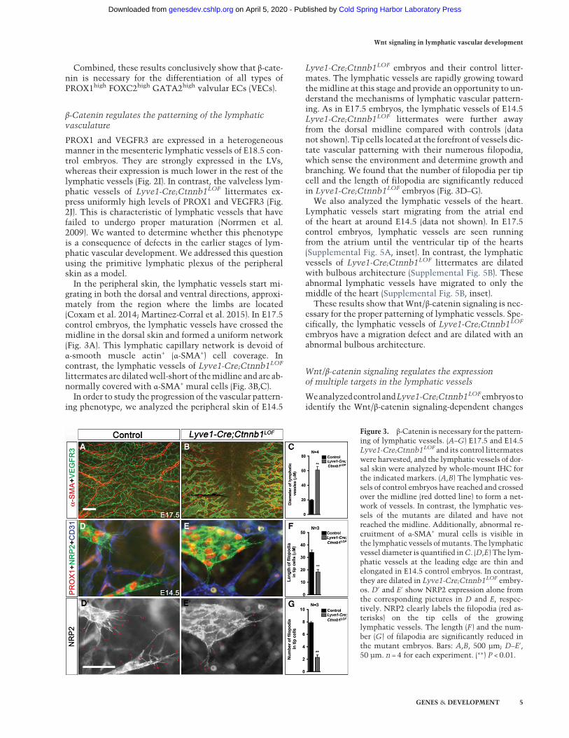
grating in both the dorsal and ventral directions, approxi-mately from the region where the limbs are located(Coxam et al. 2014; Martinez-Corral et al. 2015). In E17.5control embryos, the lymphatic vessels have crossed themidline in the dorsal skin and formed a uniform network(Fig. 3A). This lymphatic capillary network is devoid ofα-smooth muscle actin+ (α-SMA+) cell coverage. Incontrast, the lymphatic vessels of Lyve1-Cre;Ctnnb1LOF
littermates are dilatedwell-short of themidline and are ab-normally covered with α-SMA+ mural cells (Fig. 3B,C).In order to study the progression of the vascular pattern-
ing phenotype, we analyzed the peripheral skin of E14.5
Lyve1-Cre;Ctnnb1LOF embryos and their control litter-mates. The lymphatic vessels are rapidly growing towardthemidline at this stage and provide an opportunity to un-derstand the mechanisms of lymphatic vascular pattern-ing. As in E17.5 embryos, the lymphatic vessels of E14.5Lyve1-Cre;Ctnnb1LOF littermates were further awayfrom the dorsal midline compared with controls (datanot shown). Tip cells located at the forefront of vessels dic-tate vascular patterning with their numerous filopodia,which sense the environment and determine growth andbranching. We found that the number of filopodia per tipcell and the length of filopodia are significantly reducedin Lyve1-Cre;Ctnnb1LOF embryos (Fig. 3D–G).We also analyzed the lymphatic vessels of the heart.
Lymphatic vessels start migrating from the atrial endof the heart at around E14.5 (data not shown). In E17.5control embryos, lymphatic vessels are seen runningfrom the atrium until the ventricular tip of the hearts(Supplemental Fig. 5A, inset). In contrast, the lymphaticvessels of Lyve1-Cre;Ctnnb1LOF littermates are dilatedwith bulbous architecture (Supplemental Fig. 5B). Theseabnormal lymphatic vessels have migrated to only themiddle of the heart (Supplemental Fig. 5B, inset).These results show thatWnt/β-catenin signaling is nec-
essary for the proper patterning of lymphatic vessels. Spe-cifically, the lymphatic vessels of Lyve1-Cre;Ctnnb1LOF
embryos have a migration defect and are dilated with anabnormal bulbous architecture.
Wnt/β-catenin signaling regulates the expressionof multiple targets in the lymphatic vessels
WeanalyzedcontrolandLyve1-Cre;Ctnnb1LOFembryostoidentify the Wnt/β-catenin signaling-dependent changes
Figure 3. β-Catenin is necessary for the pattern-ing of lymphatic vessels. (A–G) E17.5 and E14.5Lyve1-Cre;Ctnnb1LOF and its control littermateswere harvested, and the lymphatic vessels of dor-sal skin were analyzed by whole-mount IHC forthe indicated markers. (A,B) The lymphatic ves-sels of control embryos have reached and crossedover the midline (red dotted line) to form a net-work of vessels. In contrast, the lymphatic ves-sels of the mutants are dilated and have notreached the midline. Additionally, abnormal re-cruitment of α-SMA+ mural cells is visible inthe lymphatic vessels ofmutants. The lymphaticvessel diameter is quantified inC. (D,E) The lym-phatic vessels at the leading edge are thin andelongated in E14.5 control embryos. In contrast,they are dilated in Lyve1-Cre;Ctnnb1LOF embry-os. D′ and E′ show NRP2 expression alone fromthe corresponding pictures in D and E, respec-tively. NRP2 clearly labels the filopodia (red as-terisks) on the tip cells of the growinglymphatic vessels. The length (F ) and the num-ber (G) of filapodia are significantly reduced inthe mutant embryos. Bars: A,B, 500 µm; D–E′,50 µm. n = 4 for each experiment. (∗∗) P < 0.01.
Wnt signaling in lymphatic vascular development
GENES & DEVELOPMENT 5
Cold Spring Harbor Laboratory Press on April 5, 2020 - Published by genesdev.cshlp.orgDownloaded from
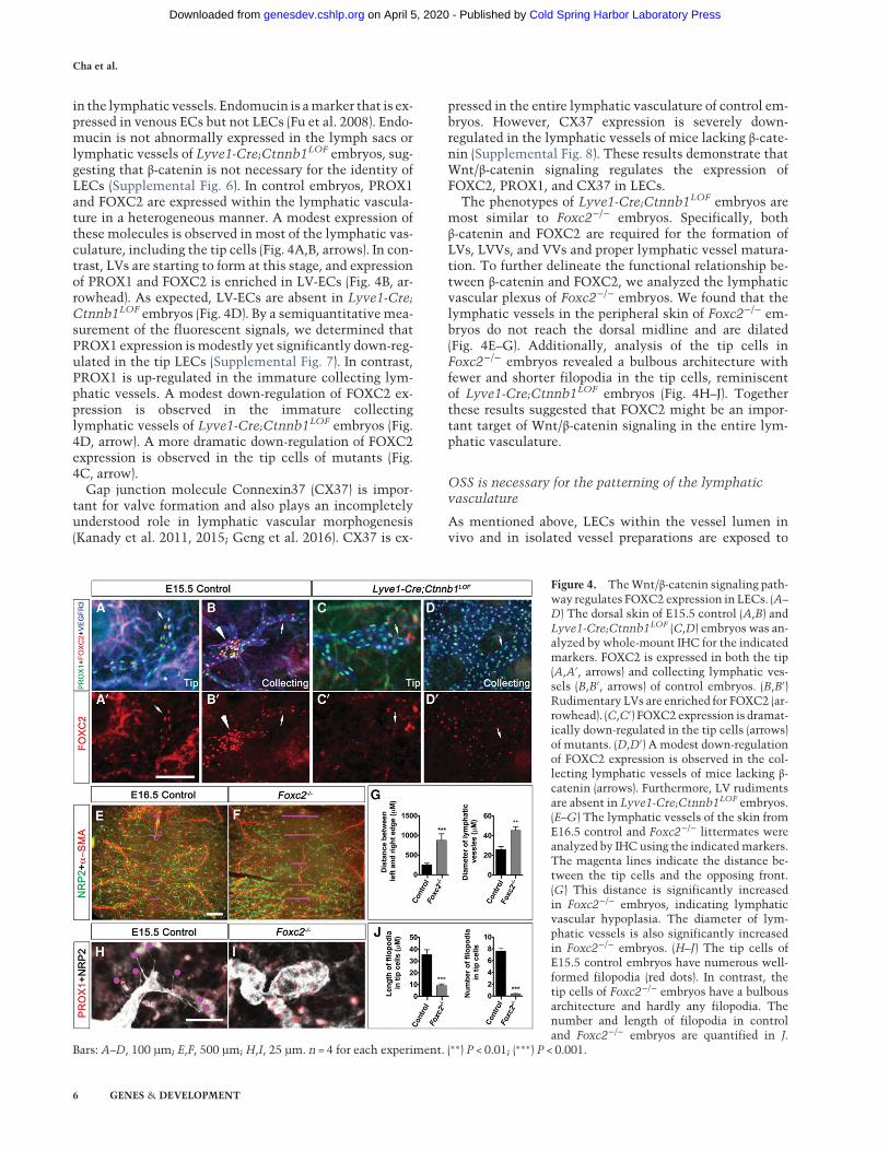
in the lymphatic vessels. Endomucin is amarker that is ex-pressed in venous ECs but not LECs (Fu et al. 2008). Endo-mucin is not abnormally expressed in the lymph sacs orlymphatic vessels of Lyve1-Cre;Ctnnb1LOF embryos, sug-gesting that β-catenin is not necessary for the identity ofLECs (Supplemental Fig. 6). In control embryos, PROX1and FOXC2 are expressed within the lymphatic vascula-ture in a heterogeneous manner. A modest expression ofthese molecules is observed in most of the lymphatic vas-culature, including the tip cells (Fig. 4A,B, arrows). In con-trast, LVs are starting to form at this stage, and expressionof PROX1 and FOXC2 is enriched in LV-ECs (Fig. 4B, ar-rowhead). As expected, LV-ECs are absent in Lyve1-Cre;Ctnnb1LOF embryos (Fig. 4D). By a semiquantitativemea-surement of the fluorescent signals, we determined thatPROX1 expression ismodestly yet significantly down-reg-ulated in the tip LECs (Supplemental Fig. 7). In contrast,PROX1 is up-regulated in the immature collecting lym-phatic vessels. A modest down-regulation of FOXC2 ex-pression is observed in the immature collectinglymphatic vessels of Lyve1-Cre;Ctnnb1LOF embryos (Fig.4D, arrow). A more dramatic down-regulation of FOXC2expression is observed in the tip cells of mutants (Fig.4C, arrow).
Gap junction molecule Connexin37 (CX37) is impor-tant for valve formation and also plays an incompletelyunderstood role in lymphatic vascular morphogenesis(Kanady et al. 2011, 2015; Geng et al. 2016). CX37 is ex-
pressed in the entire lymphatic vasculature of control em-bryos. However, CX37 expression is severely down-regulated in the lymphatic vessels of mice lacking β-cate-nin (Supplemental Fig. 8). These results demonstrate thatWnt/β-catenin signaling regulates the expression ofFOXC2, PROX1, and CX37 in LECs.
The phenotypes of Lyve1-Cre;Ctnnb1LOF embryos aremost similar to Foxc2−/− embryos. Specifically, bothβ-catenin and FOXC2 are required for the formation ofLVs, LVVs, and VVs and proper lymphatic vessel matura-tion. To further delineate the functional relationship be-tween β-catenin and FOXC2, we analyzed the lymphaticvascular plexus of Foxc2−/− embryos. We found that thelymphatic vessels in the peripheral skin of Foxc2−/− em-bryos do not reach the dorsal midline and are dilated(Fig. 4E–G). Additionally, analysis of the tip cells inFoxc2−/− embryos revealed a bulbous architecture withfewer and shorter filopodia in the tip cells, reminiscentof Lyve1-Cre;Ctnnb1LOF embryos (Fig. 4H–J). Togetherthese results suggested that FOXC2 might be an impor-tant target of Wnt/β-catenin signaling in the entire lym-phatic vasculature.
OSS is necessary for the patterning of the lymphaticvasculature
As mentioned above, LECs within the vessel lumen invivo and in isolated vessel preparations are exposed to
Figure 4. TheWnt/β-catenin signaling path-way regulates FOXC2 expression in LECs. (A–
D) The dorsal skin of E15.5 control (A,B) andLyve1-Cre;Ctnnb1LOF (C,D) embryos was an-alyzed by whole-mount IHC for the indicatedmarkers. FOXC2 is expressed in both the tip(A,A′, arrows) and collecting lymphatic ves-sels (B,B′, arrows) of control embryos. (B,B′)Rudimentary LVs are enriched for FOXC2 (ar-rowhead). (C,C′) FOXC2 expression is dramat-ically down-regulated in the tip cells (arrows)of mutants. (D,D′) A modest down-regulationof FOXC2 expression is observed in the col-lecting lymphatic vessels of mice lacking β-catenin (arrows). Furthermore, LV rudimentsare absent in Lyve1-Cre;Ctnnb1LOF embryos.(E–G) The lymphatic vessels of the skin fromE16.5 control and Foxc2−/− littermates wereanalyzed by IHC using the indicatedmarkers.The magenta lines indicate the distance be-tween the tip cells and the opposing front.(G) This distance is significantly increasedin Foxc2−/− embryos, indicating lymphaticvascular hypoplasia. The diameter of lym-phatic vessels is also significantly increasedin Foxc2−/− embryos. (H–J) The tip cells ofE15.5 control embryos have numerous well-formed filopodia (red dots). In contrast, thetip cells of Foxc2−/− embryos have a bulbousarchitecture and hardly any filopodia. Thenumber and length of filopodia in controland Foxc2−/− embryos are quantified in J.
Bars: A–D, 100 µm; E,F, 500 µm;H,I, 25 µm. n = 4 for each experiment. (∗∗) P < 0.01; (∗∗∗) P < 0.001.
Cha et al.
6 GENES & DEVELOPMENT
Cold Spring Harbor Laboratory Press on April 5, 2020 - Published by genesdev.cshlp.orgDownloaded from
OSS (Dixon et al. 2006, 2007; Sweet et al. 2015; Margariset al. 2016). Approaches to prevent lymph flow and deter-mine its role in lymphatic vascular development currentlydo not exist. However, the lymphatic vessels of Clec2−/−
mice are devoid of lymph flow (Sweet et al. 2015). LVVsand VVs that are unaffected by lymph flow form normallyin Clec2−/− mice (Hess et al. 2014). However, the mesen-teric lymphatic vessels of Clec2−/− mice do not undergoproper maturation, and LVs do not form (Sweet et al.2015). These data suggest that OSS is a critical factorinvolved in lymphatic vascular maturation and LVformation.Whether the primitive lymphatic plexus of Clec2−/−
embryos is normal is not known. To address this question,we analyzed the lymphatic vessels in the peripheral skinof Clec2−/− embryos and determined that they do not mi-grate properly and that the tip cells have a bulbous archi-tecture (Supplemental Fig. 9A,B). These phenotypes arestrikingly similar to Foxc2−/− and Lyve1-Cre;Ctnnb1LOF
embryos. Consistent with this observation, FOXC2 ex-pression is down-regulated in the LECs ofClec2−/− embry-os (Sweet et al. 2015). We propose that lymphatic vesselmigration is an aspect of lymphatic vascular maturationthat is regulated by OSS, Wnt/β-catenin signaling, andFOXC2.
Wnt/β-catenin signaling is necessary and sufficientfor OSS-mediated reprogramming of LECs
An in vitro approach has been developed to generatevalve-like cells based on the fact that LVs frequentlyform at sites exposed toOSS (Sabine et al. 2012). OSS is hy-pothesized to be important for reprogramming LECs intoan LV-EC-like identity (Sabine et al. 2012). Accordingly,primary human LECs cultured under OSS in flow cham-bers up-regulate the expression of LV-ECmarkers, includ-ing FOXC2 and GATA2 (Sabine et al. 2012; Kazenwadelet al. 2015; Sweet et al. 2015). Hence, OSS-exposed LECsare currently the best models available to study valve de-velopment in vitro. We term these cells iVECs (OSS-induced VECs) for simplicity. As mentioned in the previ-ous section, OSS contributes to lymphatic vascular pat-terning and maturation, so iVECs model this biologicalprocess as well (Sweet et al. 2015). Using this model, wedissected the relationship between OSS, Wnt/β-cateninsignaling, and FOXC2.OSS is known to activate the Wnt/β-catenin signaling
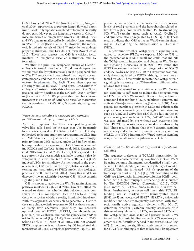
pathway in blood ECs (Li et al. 2014; Kim et al. 2015). Wewanted to determine whether this relationship is con-served in LECs. We exposed LECs to OSS by culturingthem on a rocker within a sterile, humidified incubator.With this approach, we were able to generate iVECs withthe same characteristic response to OSS as those generat-ed using flow chambers. Specifically, we observedup-regulation of FOXC2, GATA2, membrane-boundβ-catenin, VE-Cadherin, and nonphosphorylated YAP asoriginally reported (Fig. 5A–C; Kazenwadel et al. 2015;Sabine et al. 2015; Sweet et al. 2015; data not shown).PROX1 expression is not changed by OSS-mediated dif-ferentiation of LECs, as reported previously (Fig. 5C). Im-
portantly, we observed an increase in the expressionlevels of total β-catenin and the hypophosphorylated ac-tive form of β-catenin in iVECs after OSS treatment (Fig.5C). Wnt/β-catenin targets such as Axin2, Cyclin-D1,and cJun were also up-regulated by OSS (Fig. 5D). Theseresults indicate that OSS activates Wnt/β-catenin signal-ing in LECs during the differentiation of LECs intoiVECs.To determine whether Wnt/β-catenin signaling is re-
quired to generate iVECs, we exposed LECs to OSS inthe presence of iCRT3, a small molecule that inhibitsthe TCF/β-catenin interaction and abrogates Wnt/β-cate-nin signaling (Gonsalves et al. 2011). We found thatiCRT3 treatment precluded the up-regulation of FOXC2andGATA2 byOSS (Fig. 5E). PROX1 expressionwasmod-estly down-regulated by iCRT3, although it was not af-fected by OSS. These results indicate that Wnt/β-cateninsignaling is necessary for OSS-mediated differentiationof LECs into iVECs.Finally, we wanted to determine whether Wnt/β-cate-
nin signaling is sufficient to induce the reprogrammingof LECs into iVECs.We treated LECs with Bio, a chemicalcompound that inhibits GSK3β, stabilizes β-catenin, andactivatesWnt/β-catenin signaling (Sato et al. 2004). As ex-pected, Bio stabilized β-catenin in LECs and enhanced theexpression of known targets of Wnt/β-catenin signalingsuch as Axin2 and cMyc (Fig. 5F,G,I). In addition, the ex-pression of genes such as FOXC2, GATA2, and CX37was also enhanced by Bio without OSS treatment (Fig.5F–I). PROX1 expression was also modestly enhanced byBio. These results indicate that Wnt/β-catenin signalingis necessary and sufficient to promote the reprogrammingof LECs into iVECs. Importantly, Wnt/β-catenin signalingis upstream of all known regulators of VEC fate.
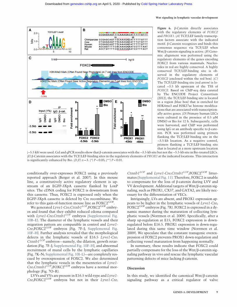
FOXC2 and PROX1 are direct targets of Wnt/β-cateninsignaling
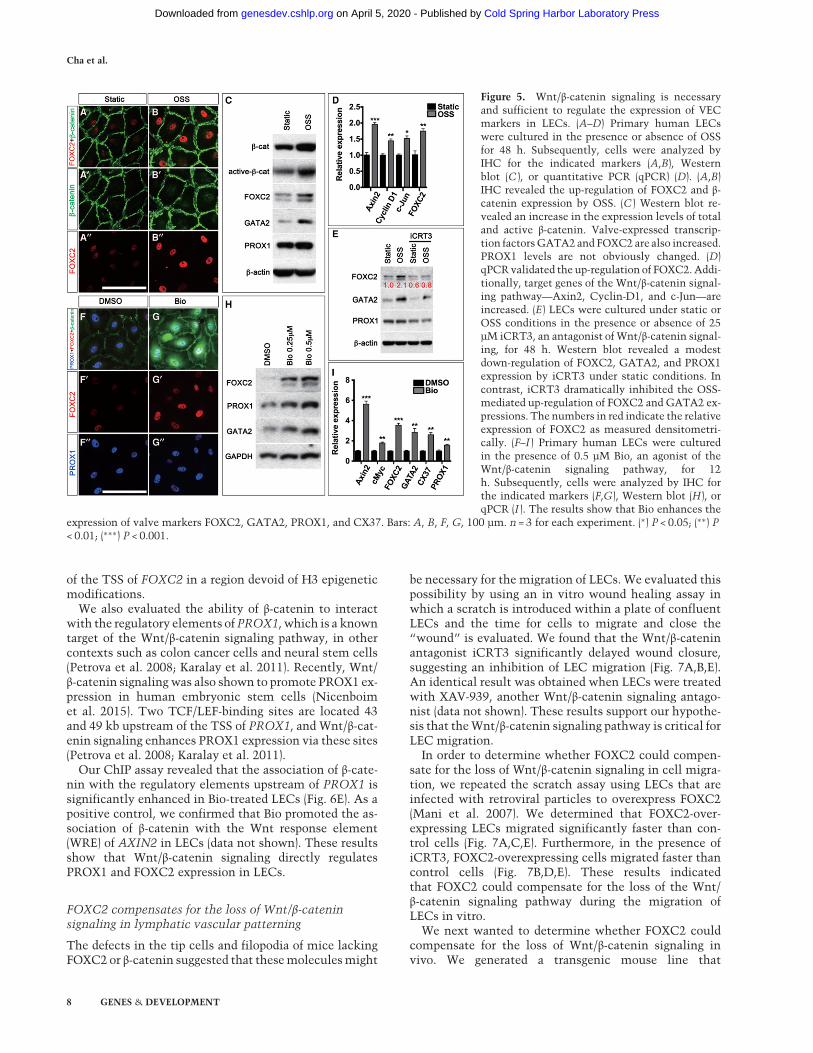
The sequence preference of TCF/LEF transcription fac-tors is well characterized (Fig. 6A; Korinek et al. 1997).By using genomic alignments, we identified a highly con-served TCF/LEF-binding site in the regulatory element ofFOXC2. This site is located ∼3.5 kb upstream of thetranscription start site (TSS) (Fig. 6B). According to theChIP-seq (chromatin immunoprecipitation [ChIP] com-bined with high-throughput sequencing) data curatedby The ENCODE Project Consortium (2012), TCF4(also known as TCF7L2) binds to this site in two celllines. Furthermore, in seven cell lines, this TCF/LEF-binding site is marked with trimethylated H3K4(H3K4me3) and acetylated H3K27 (H3K27ac), epigeneticmodifications that are frequently associated with tran-scriptionally active regulatory elements (Fig. 6C). Todetermine whether β-catenin directly associates withthis regulatory element in LECs, we treated LECs withthe Wnt/β-catenin agonist Bio and performed ChIP. Wefound that β-catenin binding to the FOXC2 regulatory el-ement is significantly enhanced in Bio-treated LECs (Fig.6D). In contrast, no significant enrichment is observedfor a TCF/LEF-binding site that is located 5 kb upstream
Wnt signaling in lymphatic vascular development
GENES & DEVELOPMENT 7
Cold Spring Harbor Laboratory Press on April 5, 2020 - Published by genesdev.cshlp.orgDownloaded from
of the TSS of FOXC2 in a region devoid of H3 epigeneticmodifications.
We also evaluated the ability of β-catenin to interactwith the regulatory elements of PROX1, which is a knowntarget of the Wnt/β-catenin signaling pathway, in othercontexts such as colon cancer cells and neural stem cells(Petrova et al. 2008; Karalay et al. 2011). Recently, Wnt/β-catenin signaling was also shown to promote PROX1 ex-pression in human embryonic stem cells (Nicenboimet al. 2015). Two TCF/LEF-binding sites are located 43and 49 kb upstream of the TSS of PROX1, and Wnt/β-cat-enin signaling enhances PROX1 expression via these sites(Petrova et al. 2008; Karalay et al. 2011).
Our ChIP assay revealed that the association of β-cate-nin with the regulatory elements upstream of PROX1 issignificantly enhanced in Bio-treated LECs (Fig. 6E). As apositive control, we confirmed that Bio promoted the as-sociation of β-catenin with the Wnt response element(WRE) of AXIN2 in LECs (data not shown). These resultsshow that Wnt/β-catenin signaling directly regulatesPROX1 and FOXC2 expression in LECs.
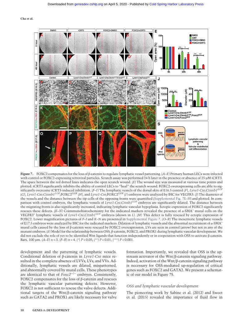
FOXC2 compensates for the loss of Wnt/β-cateninsignaling in lymphatic vascular patterning
The defects in the tip cells and filopodia of mice lackingFOXC2 or β-catenin suggested that thesemoleculesmight
be necessary for the migration of LECs. We evaluated thispossibility by using an in vitro wound healing assay inwhich a scratch is introduced within a plate of confluentLECs and the time for cells to migrate and close the“wound” is evaluated. We found that the Wnt/β-cateninantagonist iCRT3 significantly delayed wound closure,suggesting an inhibition of LEC migration (Fig. 7A,B,E).An identical result was obtained when LECs were treatedwith XAV-939, another Wnt/β-catenin signaling antago-nist (data not shown). These results support our hypothe-sis that theWnt/β-catenin signaling pathway is critical forLEC migration.
In order to determine whether FOXC2 could compen-sate for the loss of Wnt/β-catenin signaling in cell migra-tion, we repeated the scratch assay using LECs that areinfected with retroviral particles to overexpress FOXC2(Mani et al. 2007). We determined that FOXC2-over-expressing LECs migrated significantly faster than con-trol cells (Fig. 7A,C,E). Furthermore, in the presence ofiCRT3, FOXC2-overexpressing cells migrated faster thancontrol cells (Fig. 7B,D,E). These results indicatedthat FOXC2 could compensate for the loss of the Wnt/β-catenin signaling pathway during the migration ofLECs in vitro.
We next wanted to determine whether FOXC2 couldcompensate for the loss of Wnt/β-catenin signaling invivo. We generated a transgenic mouse line that
Figure 5. Wnt/β-catenin signaling is necessaryand sufficient to regulate the expression of VECmarkers in LECs. (A–D) Primary human LECswere cultured in the presence or absence of OSSfor 48 h. Subsequently, cells were analyzed byIHC for the indicated markers (A,B), Westernblot (C ), or quantitative PCR (qPCR) (D). (A,B)IHC revealed the up-regulation of FOXC2 and β-catenin expression by OSS. (C ) Western blot re-vealed an increase in the expression levels of totaland active β-catenin. Valve-expressed transcrip-tion factorsGATA2 and FOXC2 are also increased.PROX1 levels are not obviously changed. (D)qPCRvalidated the up-regulation of FOXC2. Addi-tionally, target genes of the Wnt/β-catenin signal-ing pathway—Axin2, Cyclin-D1, and c-Jun—areincreased. (E) LECs were cultured under static orOSS conditions in the presence or absence of 25µM iCRT3, an antagonist of Wnt/β-catenin signal-ing, for 48 h. Western blot revealed a modestdown-regulation of FOXC2, GATA2, and PROX1expression by iCRT3 under static conditions. Incontrast, iCRT3 dramatically inhibited the OSS-mediated up-regulation of FOXC2 and GATA2 ex-pressions. The numbers in red indicate the relativeexpression of FOXC2 as measured densitometri-cally. (F–I ) Primary human LECs were culturedin the presence of 0.5 µM Bio, an agonist of theWnt/β-catenin signaling pathway, for 12h. Subsequently, cells were analyzed by IHC forthe indicated markers (F,G), Western blot (H), orqPCR (I ). The results show that Bio enhances the
expression of valve markers FOXC2, GATA2, PROX1, and CX37. Bars: A, B, F, G, 100 µm. n = 3 for each experiment. (∗) P < 0.05; (∗∗) P< 0.01; (∗∗∗) P < 0.001.
Cha et al.
8 GENES & DEVELOPMENT
Cold Spring Harbor Laboratory Press on April 5, 2020 - Published by genesdev.cshlp.orgDownloaded from
conditionally over-expresses FOXC2 using a previouslyreported approach (Berger et al. 2007). In this mouseline, a constitutively active regulatory element is up-stream of an EGFP-3XpA cassette flanked by LoxPsites. The cDNA coding for FOXC2 is downstream fromthis cassette. Thus, FOXC2 is expressed only when theEGFP-3XpA cassette is deleted by Cre recombinase. Werefer to this gain-of-function mouse line as FOXC2GOF.We generated Lyve1-Cre;Cttnb1LOF;FOXC2GOF embry-
os and found that they exhibit reduced edema comparedwith Lyve1-Cre;Cttnb1LOF embryos (Supplemental Fig.10B–E). The diameter of the lymphatic vessels and theirmigration pattern are comparable in control and Lyve1-Cre;FOXC2GOF embryos (Fig. 7F–J; Supplemental Fig.10F–H). Further analysis revealed that the morphologicaldefects in the lymphatic vessels of E16.5 Lyve1-Cre;Ctnnb1LOF embryos—namely, the dilation, growth retar-dation (Fig. 7F–J; Supplemental Fig. 10F–H), and abnormalrecruitment of mural cells by the lymphatic capillaries(Fig. 7K–N; Supplemental Fig. 10I–L)—are completely res-cued by overexpression of FOXC2. We also determinedthat the lymphatic vessels in the mesenteries of Lyve1-Cre;Ctnnb1LOF;FOXC2GOF embryos have a normal mor-phology (Fig. 7O–R).LVVs and VVs are present in E16.5 wild-type and Lyve1-
Cre;FOXC2GOF embryos but not in their Lyve1-Cre;
Ctnnb1LOF and Lyve1-Cre;Cttnb1LOF;FOXC2GOF litter-mates (Supplemental Fig. 11). Therefore, FOXC2 is unableto compensate for the loss of β-catenin during LVV andVV development. Additional targets of Wnt/β-catenin sig-naling, such as PROX1, CX37, andGATA2, are likely nec-essary for the differentiation of VECs.Intriguingly, LVs are absent, and PROX1 expression ap-
pears to be higher in the lymphatic vessels of Lyve1-Cre;FOXC2GOF embryos (Fig. 7R). FOXC2 is expressed in a dy-namic manner during the maturation of collecting lym-phatic vessels (Norrmen et al. 2009). Specifically, after asharp up-regulation at E15, FOXC2 expression is down-regulated before E16.5. PROX1 expression is down-regu-lated during this same time window (Norrmen et al.2009). We speculate that the constant transgenic overex-pression of FOXC2 prevents PROX1 down-regulation andcollecting vessel maturation from happening normally.In summary, these results indicate that FOXC2 could
partially compensate for the loss of theWnt/β-catenin sig-naling pathway in vivo and rescue the lymphatic vascularpatterning defects of mice lacking β-catenin.
Discussion
In this study, we identified the canonical Wnt/β-cateninsignaling pathway as a critical regulator of valve
Figure 6. β-Catenin directly associateswith the regulatory elements of FOXC2and PROX1. (A) TCF/LEF family transcrip-tion factors associate with the indicatedmotif. β-Catenin recognizes and binds thisconsensus sequence via TCF/LEF whenWnt/β-catenin signaling is active. (B) Geno-mic alignment was performed using theregulatory elements of the genes encodingFOXC2 from various mammals. Nucleo-tides in red are highly conserved. A highlyconserved TCF/LEF-binding site is ob-served in the regulatory elements ofFOXC2 (enclosed within the red box). (C )The TCF/LEF-binding site (red arrow) is lo-cated ∼3.5 kb upstream of the TSS ofFOXC2. Based on ChIP-seq data curatedby The ENCODE Project Consortium(2012), the TCF/LEF-binding site is locatedin a region (blue box) that is enriched forH3K4me3 and H3K27ac histone modifica-tions that are associatedwith transcription-ally active genes. (D) Primary human LECswere cultured in the presence of 0.5 µMDMSO or Bio for 12 h. Subsequently, cellswere harvested, and ChIP was performedusing IgG or an antibody specific to β-cate-nin. PCR was performed using primersflanking the TCF/LEF-binding site in the−3.5-kb location. As a negative control,primers flanking a TCF/LEF-binding sitethat is located at a more upstream location
(−5.5 kb)were used.Gel and qPCR results show that β-catenin associateswith the−3.5-kb site but not the−5.5-kb site in Bio-treated LECs.(E) β-Catenin associates with the TCF/LEF-binding sites in the regulatory elements of PROX1 at the indicated locations. This interactionis significantly enhanced by Bio. (D,E) n = 3. (∗) P < 0.05; (∗∗) P < 0.01.
Wnt signaling in lymphatic vascular development
GENES & DEVELOPMENT 9
Cold Spring Harbor Laboratory Press on April 5, 2020 - Published by genesdev.cshlp.orgDownloaded from
development and the patterning of lymphatic vessels.Conditional deletion of β-catenin in Lyve1-Cre mice re-sulted in the complete absence of LVVs, LVs, and VVs. Ad-ditionally, lymphatic vessels are dilated, mispatterned,and abnormally covered bymural cells. These phenotypesare identical to that of Foxc2−/− embryos. Consistently,FOXC2 compensates for the loss of β-catenin and rescuesthe lymphatic vascular patterning defects. However,FOXC2 is not sufficient to rescue the valve defects. Addi-tional targets of the Wnt/β-catenin signaling pathwaysuch as GATA2 and PROX1 are likely necessary for valve
formation. Importantly, we revealed that OSS is the up-stream activator of the Wnt/β-catenin signaling pathway.Indeed, activation of theWnt/β-catenin signaling pathwayis necessary for OSS-mediated up-regulation of criticalgenes such as FOXC2 and GATA2.We present a schemat-ic of our model in Figure 7S.
OSS and lymphatic vascular development
The pioneering work by Sabine et al. (2012) and Sweetet al. (2015) revealed the importance of fluid flow in
Figure 7. FOXC2 compensates for the loss of β-catenin to regulate lymphatic vessel patterning. (A–E) Primary humanLECswere infectedwith control or FOXC2-expressing retroviral particles. Scratch assay was performed 24 h later in the presence or absence of 25 µM iCRT3.The space between the red dotted lines indicates the open scratch wound. (E) The wound size was measured at various time points andplotted. iCRT3 significantly inhibits the ability of control LECs to “heal” the scratch wound. FOXC2-overexpressing cells are able to sig-nificantly overcome iCRT3-induced inhibition. (F–I ) The lymphatic vessels of the dorsal skin of E16.5 control (F ), Lyve1-Cre;Ctnnb1LOF
(G), Lyve1-Cre;Ctnnb1LOF;FOXC2GOF (H), and Lyve1-Cre;FOXC2GOF (I ) embryos were analyzed by IHC for VEGFR3. (J) The diameter ofthe vessels and the distance between the tip cells of the opposing fronts were quantified (Supplemental Fig. 7E–H) and plotted. In com-parison with control embryos, the lymphatic vessels of Lyve1-Cre;Cttnb1LOF embryos are significantly dilated. The distance betweenthe migrating fronts is also significantly increased, indicating lymphatic vascular hypoplasia. Ectopic expression of FOXC2 significantlyrescues these defects. (K–N) Coimmunohistochemistry for the indicated markers revealed the presence of α-SMA+ mural cells on theVEGFR3+ lymphatic vessels of Lyve1-Cre;Cttnb1LOF embryos (shown in L). (M ) This defect is fully rescued by ectopic expression ofFOXC2. Lower magnification pictures of F–I and K–N are presented in Supplemental Figure 7. (O–R) The mesenteric lymphatic vesselsof E17.5 embryos were analyzed by IHC for the indicatedmarkers. Dilation of lymphatic vessels and the abnormal recruitment of α-SMA+
mural cells caused by the loss of β-catenin were rescued by FOXC2 overexpression. LVs are seen in control (arrow) but not in any of themutant embryos. (S) Model for the relationship betweenOSS, β-catenin, FOXC2, and PROX1 during lymphatic vascular development.Wedid not exclude the role of yet to be identified Wnt ligands that function independently or in cooperation with OSS to activate β-catenin.Bars, 100 µm. (A–E) n = 3. (F–R) n = 4. (∗) P < 0.05; (∗∗) P < 0.01; (∗∗∗) P < 0.001.
Cha et al.
10 GENES & DEVELOPMENT
Cold Spring Harbor Laboratory Press on April 5, 2020 - Published by genesdev.cshlp.orgDownloaded from

lymphatic vascular development. These investigators ex-posed LECs to OSS of 0.5 dynes/cm2 in flow chambers togenerate valve-like cells from LECs.We used a simple testtube rocker-based approach to generate iVECs. Accordingto our calculations, LECs are exposed to anOSS that peaksat 0.2 dynes/cm2 for the six-well plates with a time aver-age mean shear stress of 0 dynes/cm2 for all of the condi-tions (see the Materials and Methods for a detailedexplanation of the analytical determination of wall shearstress). A similar result was obtained when the experi-ments were performed using 24-well plates with a shearstress of 0.09 dynes/cm2 (data not shown). Despite the ap-parent difference in the shear stress values, we were ableto precisely recapitulate the results of Sabine et al.(2012). This observation has interesting implications re-garding the mechanisms of iVEC differentiation. TheOSS value used by Sabine et al. (2012) was similar to val-uesmeasured in themesenteric lymphatic vessels of adultrats (Dixon et al. 2006). Due to technical limitations,we currently do not know the OSS values in the develop-ing lymphatic vessels of mice. However, given the appar-ent conservation in the mechanisms, we reason thatthe flow pattern rather than the absolute shear stressvalue is important for lymphatic vascular development.Our in vitro data are consistent with this hypothesis. Fur-thermore, our finding provides an inexpensive and easilyaccessible approach that will accelerate the field of lym-phatic research.
OSS and PROX1 expression
OSS was originally proposed to be critical for LV develop-ment (Sabine et al. 2012). Although PROX1 is strongly ex-pressed in the VECs, its expression is not promoted byOSS. However, the Wnt agonist Bio promotes PROX1 ex-pression in LECs (Fig. 4). Why does activation of the Wnt/β-catenin signaling pathway but not OSS have the capaci-ty to promote PROX1 expression in LECs? Signaling path-ways activate their target genes in a signal intensity-dependent and time-dependent manner (Wolpert 2016).Therefore, it is conceivable that the strength of the Wnt/β-catenin signaling pathway activated by OSS in vitro isinsufficient to activate PROX1 expression. The three-di-mensional architecture of the valve-forming regions like-ly enhances and sustains OSS-mediated signals in vivo.Valves are composed of upstream and downstream por-tions with distinct molecular profiles (Kanady et al.2011; Sabine et al. 2012; Munger et al. 2013). The “OSSmodel” recapitulates the cells on the downstream sideof valves by up-regulating markers such as FOXC2 andCX37. It is possible that the cells on the upstream sideof the valve, which aremissing in this assay,might be nec-essary to promote PROX1 expression and valve morpho-genesis. Consistent with this possibility, whereas VECshave an elongated architecture in vivo, iVECs are cuboidal(Sabine et al. 2012; Tatin et al. 2013; Geng et al. 2016).Cell–cell and cell–matrix interactions between the up-stream and downstream sides of the valves could modu-late the flow-mediated mechanical force experienced bythe cells. Alternatively, the cells located in the upstream
side of the valves might provide signals to modulate OSSand the Wnt/β-catenin signaling pathway. In mammals,there are 19 Wnt ligands that are frequently coexpressedand could compensate for each other (Logan and Nusse2004). At least five Wnt ligands (Wnt2, Wnt3a, Wnt4,Wnt7b, and Wnt9b) are expressed in the cardiac valveswhere the Wnt/β-catenin signaling pathway is known tobe necessary for cardiac cushion formation and matura-tion (Alfieri et al. 2010). Whether these ligands play anyrole during lymphatic vessel growth and valve formationremains to be investigated.
OSS, lymphatic vascular maturation, and valvedevelopment
Our results show that OSS promotes Wnt/β-catenin sig-naling and FOXC2 expression, both of which are neces-sary in turn for valve formation and lymphatic vascularmaturation. While FOXC2 compensates for the loss ofβ-catenin in lymphatic vessels, other targets ofWnt/β-cat-enin signaling appear to be necessary for valve formation.What are the factors that modulate the LEC responseto OSS and determine whether they differentiate intovalves or mature lymphatic vessels? We speculate thatthe OSS response could be regulated by whether PROX1is up-regulated at a specific site. Accordingly, OSS andOSS+PROX1 will promote lymphatic vascular matura-tion and valve formation, respectively.In the future, it will be important to determine theOSS-
specific effects on LECs. Valve-forming cells undergocomplex migratory events such as delamination, realign-ment with respect to flow, condensation, and elongationto form valves (Geng et al. 2016). Lymphatic vascular pat-terning involves the growth of the lymphatic plexus(Coxam et al. 2014). Therefore, cell migration might bethe cellular event that is conserved between these twoprocesses. Our work has revealed that the tip cells, whichare important for cell migration, are defective in micelacking FOXC2. We also provided evidence that FOXC2is a target of the Wnt/β-catenin signaling pathway duringlymphatic vascular development. It will be importantto mechanistically dissect the relationship betweenFOXC2 and cell migration.
Sensing OSS
Despite its elegance, a definitive proof for the accuracy ofthe “OSS model” is still missing. Identifying the mecha-nosensory molecules in the lymphatic vasculature willbring us a step closer to testing thismodel in vivo. Primarycilia function as mechanosensors in many cell types(Ingber 2006). Using SEM, we failed to observe any prima-ry cilia in the developing LVV-ECs (Supplemental Fig. 12).Based on the published literature, we discuss a few alter-nate mechanisms that may be involved in sensing andtranslating OSS into Wnt/β-catenin signals.
Integrins Integrins arewell-knownmechanosensors in nu-merous contexts (Ingber 2006). Integrin α9 and integrin α5are both critical for LVV and LV development (Bazigou
Wnt signaling in lymphatic vascular development
GENES & DEVELOPMENT 11
Cold Spring Harbor Laboratory Press on April 5, 2020 - Published by genesdev.cshlp.orgDownloaded from
et al. 2009; Turner et al. 2014). Integrin α5β1 was shownrecently to enhance Wnt/β-catenin signals in osteoblastsvia the PI3K–AKT pathway (Saidak et al. 2015).
Wnt receptors Frizzled receptors might function as mecha-nosensors, as reported recently (Rotherham and El Haj2015). According to this model, shear activated Frizzledreceptors could interact with LRP5/6 and activate thedownstream signals.
Receptor tyrosine kinases In tumor cells, mechanical strainactivates a receptor tyrosine kinase (Ret) that phosphory-lates and promotes the nuclear translocation of β-cateninby inhibiting catenin–cadherin interaction at cell junc-tions (Fernandez-Sanchez et al. 2015). VEGFR2 andVEGFR3 are mechanoresponsive and may play the roleof Ret in ECs (Coon et al. 2015).
Ion channels Mutations in the mechanically activated ionchannel PIEZO1 are associated with human lymphedema(Fotiou et al. 2015; Lukacs et al. 2015). Hence, PIEZO1 andits paralog, PIEZO2, might be involved in sensing OSS.
Evolutionarily divergent roles of the Wnt/β-cateninsignaling pathway in lymphatic vascular development
TheWnt/β-catenin signaling pathwaywas shown recentlyto promote PROX1 expression and asymmetric division ofspecialized angioblast cells in the venous niche of zebra-fish. These cells give rise to the lymphatic vessels of zebra-fish (Nicenboim et al. 2015). As shown above, conditionaldeletion of β-catenin using Lyve1-Cre in mice did not re-veal any obvious defects in LEC progenitor specification.Conditional deletion of β-catenin using Tie2-Cre in theentire vascular network resulted in embryonic death atE12.5, as reported previously (Cattelino et al. 2003). Anal-ysis of these mutants did not reveal any obvious lymphat-ic vascular defects (data not shown). PROX1+ LECs appearto be normally specified in these mutants.
Despite numerous similarities, some important differ-ences between mammalian and zebrafish lymphatics ex-ist. Relevant to this work, the lymphatic vasculature ofzebrafish is devoid of LVs (Koltowska et al. 2013). Further-more, the site of lymph return to blood circulation is cur-rently unknown in these animals. Therefore, it is possiblethat Wnt/ β-catenin signaling pathway is playing species-specific roles in lymphatic vascular development.
Clinical implications
Approaches to treat lymphedema currently do not exist.We identified an important signaling pathway that regu-lates lymphatic vascular morphogenesis. Numerous ago-nists and antagonists of the Wnt/ β-catenin signalingpathway are currently available. Some of these moleculesare in clinical trials for various diseases such as cancer andAlzheimer’s disease (MacDonald et al. 2009). A better un-derstanding of the spatial and temporal regulations of the
Wnt/ β-catenin signaling pathway within the lymphaticvasculature will provide exciting opportunities to treatlymphedema, potentially by repurposing existing drugs.
Materials and methods
Mouse models
TCF/LEF-H2B-EGFP, Lyve1-Cre, Ctnnb1+/f, ProxTom,Prox1+/Cre, Foxc2+/−, andClec2−/−mice were reported previously(Brault et al. 2001; Bertozzi et al. 2010; Ferrer-Vaquer et al. 2010;Pham et al. 2010; Srinivasan et al. 2010; Truman et al. 2012;Geng et al. 2016). We generated the FOXC2GOF mouse line by in-serting a cDNA coding for Foxc2 into the JOJO plasmid (Bergeret al. 2007). Linearized plasmid was electroporated into blasto-cysts, and the transgenic founders were visually selected basedon the expression of EGFP. All mice were housed and handled ac-cording to the Institutional Animal Care and Use Committeeprotocols.
Antibodies
Primary antibodies used for immunostainingwere as follows: rab-bit anti-PROX1 (AngioBio), goat anti-human PROX1 (R&D Sys-tems), sheep anti-mouse FOXC2 (R&D Systems), goat anti-mouse VEGFR3 (R&D Systems), goat anti-mouse LYVE1 (R&DSystems), rat anti-mouse CD31 (BD Pharmingen), goat anti-mouse GATA2 (R&D Systems), rabbit anti-total β-catenin andrabbit anti-active β-catenin antibodies (both from Cell SignalingTechnologies), goat anti-mouse NRP2 (R&D Systems), chickenanti-GFP (Abcam), rat anti-mouse Endomucin (eBioscience), rab-bit anti-CX37 (Invitrogen), and Cy3-conjugated monoclonal anti-α-SMA (Sigma-Aldrich). The following secondary antibodies wereused: Cy3-conjugated donkey anti-rabbit, Cy3-conjugated don-key anti-sheep, and Cy5-conjugated donkey anti-rat (all pur-chased from Jackson ImmunoResearch Laboratories). Alexa488-conjugated donkey anti-goat, Alexa 488-conjugated goatanti-chicken, andAlexa 488-conjugated donkey anti-rat were pur-chased from Life Technologies.Primary antibodies used for Western blotting were as follows:
mouse anti- β-Actin (Sigma), mouse anti-mouse β-catenin (BDPharmingen), rabbit anti-active β-catenin (Cell Signaling Tech-nologies), goat anti-human PROX1 (R&D Systems), sheep anti-mouse FOXC2 (R&D Systems), and goat anti-mouse GATA2(R&D Systems). The following HRP-conjugated secondary anti-bodies from Santa Cruz Biotechnology were used: goat anti-mouse IgG, goat anti-rabbit IgG, donkey anti-goat IgG, and don-key anti-sheep IgG.
IHC
IHC on sections was done according to our detailed protocolsthat we published recently (Geng et al. 2016). Whole-mountIHC using the skin, gut, or heart was performed using a modifiediDISCO protocol (Renier et al. 2014). Briefly, isolated sampleswere fixed in 4% PFA for 4 h at 4°C and washed profusely withPBST (0.2% Triton X-100 in PBS). The samples were incubatedin PBSTD (0.2% Triton X-100, 20% DMSO in PBS) overnightand incubated in PBSTTDND (0.1% Tween X-100, 0.1% TritonX-100, 0.1% deoxycholate, 0.1%NP40, 20%DMSO in PBS) over-night at room temperature. Pretreated samples were incubatedwith PBSTDM (0.2% Triton X-100, 20% DMSO, 0.3 M glycinein PBS) overnight and blocked with PBSTD (0.2% Triton X-100/10% DMSO/6% donkey serum in PBS) overnight. Samples
Cha et al.
12 GENES & DEVELOPMENT
Cold Spring Harbor Laboratory Press on April 5, 2020 - Published by genesdev.cshlp.orgDownloaded from
were incubated overnight with primary antibodies diluted inPTwHD (0.2% Tween-20, 10 µg/mL heparin, 10% DMSO, 3%donkey serum in PBS). After profuse washing, samples were incu-bated overnight with secondary antibodies diluted in PTwH(0.2% Tween-20, 10 µg/mL heparin, 3% donkey serum in PBS).A lighter fixation protocol (1% PFA for 1 h at 4°C) was usedfor whole-mount IHC with anti CX37 antibody. Samples werevisualized and analyzed as described previously (Geng et al.2016).
In situ hybridization
In situ hybridization and coimmunohistochemistry experimentswere performed as described previously (Srinivasan et al. 2010).Information regarding the Axin2 probe will be provided onrequest.
Western blot
Cells were lysed in lysis buffer (20 mM Tris–HCl at pH 7.5, 150mM NaCl, 1.0% Triton X-100, 20 mM NaF, 2 mM EDTA, 2mM Na-orthovanadate, 1 mM phenylmethylsulfonyl fluoride[PMSF], 5 mg/mL leupeptin A). Concentration of proteins wasmeasured using Bradford reagent (Bio-Rad). Western blot was car-ried out according to standard protocols.
SEM
SEM was done according to our previous protocol (Geng et al.2016).
RNA isolation and quantitative real-time PCR
Total RNA was isolated from cells using Trizol reagent (Invitro-gen) according to themanufacturer’s protocol. cDNAwas synthe-sized from total RNA using the SuperScript III first strandsynthesis system (Invitrogen) following the manufacturer’s in-structions. Quantitative PCR (qPCR) was performed usingSYBR Green PCR master mix reagent (Bio-Rad) in a CFX96 real-time system (Bio-Rad). Primer sequences will be provided on re-quest. The threshold cycle (Ct) value for each gene was normal-ized to the Ct value for β-actin.
ChIP
ChIP assays were performed using 8 × 10⁶ to 10 × 106 human pri-mary LECs (Lonza) as described previously (Kim et al. 2005). Cellsat 90% confluency were treated with 0.5 µM DMSO or Bio for12 h. Subsequently, LECs were fixed in 1% formaldehyde for30 min at room temperature, and glycine at a final concentrationof 0.125 M was added for 10 min. Cells were washed, harvestedwith cold PBS, resuspended in lysis buffer 1 (50 mM Hepes-KOH at pH 7.5, 140mM NaCl, 1.0 µM EDTA, 10% glycerol,0.5% NP-40, 0.25% Triton X-100, protease inhibitor cocktail),and rotated for 5 min at 4°C. Cells were spun down at 2000rpm for 3 min, and supernatant was removed. The cell pelletwas resuspended in lysis buffer 2 (200 mM NaCl, 1.0 µMEDTA, 0.5 µM EGTA, 10 µMTris-HCl at pH 8, protease inhibitorcocktail) for 5 min at 4°C with rotation. After centrifugation at2000 rpm for 3 min, the supernatant was removed by aspiration.The pellet was resuspended using lysis buffer 3 (1.0 µM EDTA,0.5 µM EGTA, 10 µM Tris-HCl at pH 8, protease inhibitor cock-tail), sonicated on ice, and centrifuged at 13,000 rpm for 10 min.The supernatant was diluted in immunoprecipitation dilution
buffer (20 mM Tris-HCl at pH 8.0, 150 mM NaCl, 1 mMEDTA, 1% Triton X-100, protease inhibitors) and used for down-stream analysis. The cross-linked protein–DNA complexes wereimmunoprecipitated using 3.0 µg of mouse anti-β-catenin anti-body (BD Pharmingen) or 1.0 µg of mouse IgG antibody (SantaCruz Biotechnology). qPCRwas performed as described above us-ing primers flanking the predicted TCF/LEF sites or control sites.Primer sequences will be provided on request.
Wound healing assay
Primary human LECs were plated in 24-well plates and infect-ed with retroviral particles expressing control GFP or humanFOXC2. When cells were near confluency (>90% density),scratch wounds were made at the center of the well using asterile 1000-µL pipette tip. Loose cells were removed by aPBS wash, and DMSO-containing (Sigma) or iCRT3-containing(Sigma) medium was added. Images were recorded at t = 0, t =24, and t = 48 h using an inverted microscope to measure thewound size.
OSS
Human LECs were cultured to confluency in six-well plates andexposed to OSS using a test tube rocker (Thermolyne Speci-Mixaliquot mixer model M71015, Barnstead International) with apreset frequency (18 rpm). The entire setupwas kept inside a ster-ile humidified incubator with 5% CO2 for 48 h. The rocker wentthrough an angle of 48o in ∼2 sec. One milliliter of medium wasused for the 24-well plates, and 6 mL of medium was used forthe six-well plates. In both the cases, the amount of mediumwas sufficient to cover the center of the wells at all times, al-though the medium did lose contact with the sides of the wellsat high angles of oscillation.An analytical formulation for a rectangular Petri dish was
presented in Zhou et al. (2010), where the investigators derivedthe shear stress at the bottom of the dish as a function of timeand position. The derivation involved the use of lubrication ap-proximation, which is justified when low volumes of fluids areused compared with the cross-sectional area and assumed noslip boundary condition at the bottom of the dish and 0 veloc-ity gradient at the free surface. With these conditions, thewall shear stress at the bottom of the well can be describedby the equation
|tw | = 3pmumaxx(L− x)T h0cotu+ L
2 − x[ ]2sin2u
cos2ptT
, u ≤ u0
= 3pmumaxx
T������������2h0Lcotu
√− x
[ ]2sin2u
cos2ptT
, u ≤ u0
,
where µ is the dynamic viscosity of the fluid, L is the length of thedish along thedirectionofmotionof the rocker,x is the distanceofthe point of interest from a side of the dish, h0 is the height of thefluid surfacewhen the dish is horizontal, θ is the angle of the rock-er, θmax is themaximum angle of the rocker, θ0 is the “critical flipangle” (defined as the angle at which the fluid just leaves contactwith the bottomof the dish), t is the time, andT is the time periodof the sinusoidal motion of the rocker.The aforementioned analytical formulationwas adapted for the
present scenario, since the lubrication approximation holds forthe small volume of fluid used, and the region of interest is thecenter of the wells, which was always covered with fluid. Forthe medium, µ is considered as the same as that of water, whichis 1 centiPoise. The rocker had a θmax of 48
o and T of ∼2 sec. The
Wnt signaling in lymphatic vascular development
GENES & DEVELOPMENT 13
Cold Spring Harbor Laboratory Press on April 5, 2020 - Published by genesdev.cshlp.orgDownloaded from
dish had an L of ∼3.48 cm for the six-well plates and 1.56 cm forthe 24-well plates. The volume of fluid used was 6 mL and 1 mL,and the h0 was ∼0.63 cm for the six-well plates and ∼0.53 cm forthe 24-well plates, respectively.With these values, themaximumshear stress is calculated to be ∼0.3 dynes/cm2 for the six-wellplates and ∼0.09 dynes/cm2 for the 24-well plates at the centerof the wells.
Acknowledgments
We thank Dr. Rodger McEver and Dr. Andrew McMahon for in-sightful comments, Ms. LisaWhitworth (Microscopy Laboratory,Oklahoma State University, Stillwater) for SEM, Dr. AngelaAndersen (Life Science Editors) for editorial assistance, andMs. Lijuan Chen for mouse colony management. We thankDr. Guillermo Oliver and St. Jude Children’s research hospitalfor generous support in generating the FOXC2GOF line. R.S.S. issupported by National Institutes of Health/National Heart,Lung, and Blood Institute (R01HL131652), institutional funds ofthe Oklahoma Medical Research Foundation, Oklahoma Centerfor Adult Stem Cell Research (OCASCR, 4340), and AmericanHeart Association (15BGIA25710032). B.C. is supported by apost-doctoral fellowship from the American Heart Associa-tion (15POST25080182). E.-h.J. is supported by National Re-search Foundation grants funded by the Ministry of Science,ICT, and Future Planning of the Republic of Korea(2012M3A9C6050109). Work in the laboratory of T.H.K is sup-ported by grants fromNational Institute of Allergy and InfectiousDiseases (R21AI107067) and National Cancer Institute(R01CA140485).
References
Al Alam D, Green M, Tabatabai Irani R, Parsa S, Danopoulos S,Sala FG, Branch J, El Agha E, Tiozzo C, Voswinckel R, et al.2011. Contrasting expression of canonical Wnt signaling re-porters TOPGAL, BATGAL and Axin2(LacZ) during murinelung development and repair. PLoS One 6: e23139.
Alfieri CM,Cheek J, Chakraborty S, YutzeyKE. 2010.Wnt signal-ing in heart valve development and osteogenic gene induc-tion. Dev Biol 338: 127–135.
Bazigou E, Xie S, Chen C, Weston A, Miura N, Sorokin L, AdamsR, Muro AF, Sheppard D, Makinen T. 2009. Integrin-α9 is re-quired for fibronectinmatrix assembly during lymphatic valvemorphogenesis. Dev Cell 17: 175–186.
Berger J, Berger S, Tuoc TC, D’Amelio M, Cecconi F, Gorski JA,Jones KR, Gruss P, Stoykova A. 2007. Conditional activationof Pax6 in the developing cortex of transgenicmice causes pro-genitor apoptosis. Development 134: 1311–1322.
Bertozzi CC, Schmaier AA, Mericko P, Hess PR, Zou Z, ChenM,ChenCY, Xu B, LuMM,ZhouD, et al. 2010. Platelets regulatelymphatic vascular development through CLEC-2–SLP-76signaling. Blood 116: 661–670.
Brault V, Moore R, Kutsch S, Ishibashi M, Rowitch DH, McMa-honAP, Sommer L, BoussadiaO, Kemler R. 2001. Inactivationof the β-catenin gene by Wnt1-Cre-mediated deletion resultsin dramatic brain malformation and failure of craniofacial de-velopment. Development 128: 1253–1264.
Cattelino A, Liebner S, Gallini R, Zanetti A, Balconi G, Corsi A,Bianco P, Wolburg H, Moore R, Oreda B, et al. 2003. The con-ditional inactivation of the β-catenin gene in endothelial cellscauses a defective vascular pattern and increased vascular fra-gility. J Cell Biol 162: 1111–1122.
Coon BG, BaeyensN, Han J, BudathaM, Ross TD, Fang JS, Yun S,Thomas JL, Schwartz MA. 2015. Intramembrane binding ofVE-cadherin to VEGFR2 and VEGFR3 assembles the endothe-lial mechanosensory complex. J Cell Biol 208: 975–986.
Coxam B, Sabine A, Bower NI, Smith KA, Pichol-Thievend C,Skoczylas R, Astin JW, Frampton E, Jaquet M, Crosier PS,et al. 2014. Pkd1 regulates lymphatic vascular morphogenesisduring development. Cell Rep 7: 623–633.
Dixon JB, Greiner ST, Gashev AA, Cote GL, Moore JE, ZawiejaDC. 2006. Lymph flow, shear stress, and lymphocyte velocityin rat mesenteric prenodal lymphatics. Microcirculation 13:597–610.
Dixon JB, Gashev AA, Zawieja DC, Moore JE Jr, Cote GL. 2007.Image correlation algorithm for measuring lymphocyte veloc-ity and diameter changes in contracting microlymphatics.Ann Biomed Eng 35: 387–396.
The ENCODE Project Consortium. 2012. An integrated encyclo-pedia of DNA elements in the human genome. Nature 489:57–74.
Fernandez-Sanchez ME, Barbier S, Whitehead J, Bealle G, MichelA, Latorre-Ossa H, Rey C, Fouassier L, Claperon A, Brulle L,et al. 2015. Mechanical induction of the tumorigenic β-cate-nin pathway by tumour growth pressure. Nature 523: 92–95.
Ferrer-Vaquer A, Piliszek A, Tian G, Aho RJ, Dufort D, Hadjanto-nakis AK. 2010. A sensitive and bright single-cell resolutionlive imaging reporter of Wnt/ss-catenin signaling in themouse. BMC Dev Biol 10: 121.
Fotiou E, Martin-Almedina S, Simpson MA, Lin S, Gordon K,Brice G, Atton G, Jeffery I, Rees DC, Mignot C, et al. 2015.Novel mutations in PIEZO1 cause an autosomal recessivegeneralized lymphatic dysplasia with non-immune hydropsfetalis. Nat Commun 6: 8085.
Fu J, Gerhardt H, McDaniel JM, Xia B, Liu X, Ivanciu L, Ny A,Hermans K, Silasi-Mansat R, McGee S, et al. 2008. Endotheli-al cell O-glycan deficiency causes blood/lymphatic miscon-nections and consequent fatty liver disease in mice. J ClinInvest 118: 3725–3737.
Geng X, Cha B, Mahamud MR, Lim KC, Silasi-Mansat R, UddinMK, Miura N, Xia L, Simon AM, Engel JD, et al. 2016. Multi-plemousemodels of primary lymphedema exhibit distinct de-fects in lymphovenous valve development. Dev Biol 409:218–233.
Gonsalves FC, Klein K, Carson BB, Katz S, Ekas LA, Evans S,Nagourney R, Cardozo T, Brown AM, DasGupta R. 2011. AnRNAi-based chemical genetic screen identifies three small-molecule inhibitors of the Wnt/wingless signaling pathway.Proc Natl Acad Sci 108: 5954–5963.
Hess PR, Rawnsley DR, Jakus Z, Yang Y, Sweet DT, Fu J, HerzogB, LuM, Nieswandt B, Oliver G, et al. 2014. Platelets mediatelymphovenous hemostasis to maintain blood–lymphatic sep-aration throughout life. J Clin Invest 124: 273–284.
Ingber DE. 2006. Cellular mechanotransduction: putting all thepieces together again. FASEB J 20: 811–827.
Johnson NC, Dillard ME, Baluk P, McDonald DM, Harvey NL,Frase SL, Oliver G. 2008. Lymphatic endothelial cell identityis reversible and its maintenance requires Prox1 activity.Genes Dev 22: 3282–3291.
Kanady JD, Dellinger MT, Munger SJ, Witte MH, Simon AM.2011. Connexin37 and Connexin43 deficiencies in mice dis-rupt lymphatic valve development and result in lymphaticdisorders including lymphedema and chylothorax. Dev Biol354: 253–266.
Kanady JD, Munger SJ, Witte MH, Simon AM. 2015. CombiningFoxc2 and Connexin37 deletions in mice leads to severe
Cha et al.
14 GENES & DEVELOPMENT
Cold Spring Harbor Laboratory Press on April 5, 2020 - Published by genesdev.cshlp.orgDownloaded from
defects in lymphatic vascular growth and remodeling. DevBiol 405: 33–46.
Karalay O, Doberauer K, Vadodaria KC, Knobloch M, Berti L,Miquelajauregui A, Schwark M, Jagasia R, Taketo MM, Tara-bykin V, et al. 2011. Prospero-related homeobox 1 gene (Prox1)is regulated by canonical Wnt signaling and has a stage-specif-ic role in adult hippocampal neurogenesis. Proc Natl Acad Sci108: 5807–5812.
Kazenwadel J, Betterman KL, Chong CE, Stokes PH, Lee YK,Secker GA, Agalarov Y, Demir CS, Lawrence DM, SuttonDL, et al. 2015. GATA2 is required for lymphatic vessel valvedevelopment and maintenance. J Clin Invest 125: 2979–2994.
Kim TH, Barrera LO, ZhengM, Qu C, Singer MA, Richmond TA,WuY,GreenRD, Ren B. 2005. A high-resolutionmap of activepromoters in the human genome. Nature 436: 876–880.
Kim PG, Nakano H, Das PP, Chen MJ, Rowe RG, Chou SS, RossSJ, Sakamoto KM, Zon LI, Schlaeger TM, et al. 2015. Flow-in-duced protein kinaseA–CREB pathway acts via BMP signalingto promote HSC emergence. J Exp Med 212: 633–648.
Koltowska K, Betterman KL, Harvey NL, Hogan BM. 2013. Get-ting out and about: the emergence and morphogenesis ofthe vertebrate lymphatic vasculature. Development 140:1857–1870.
Korinek V, Barker N, Morin PJ, van Wichen D, de Weger R, Kin-zler KW, Vogelstein B, Clevers H. 1997. Constitutive tran-scriptional activation by a β-catenin–Tcf complex in APC−/−
colon carcinoma. Science 275: 1784–1787.Li R, Beebe T, Jen N, Yu F, Takabe W, Harrison M, Cao H, Lee J,
Yang H, Han P, et al. 2014. Shear stress-activated Wnt–angio-poietin-2 signaling recapitulates vascular repair in zebrafishembryos. Arterioscler Thromb Vasc Biol 34: 2268–2275.
Liebner S, Cattelino A, Gallini R, Rudini N, Iurlaro M, Piccolo S,Dejana E. 2004. β-Catenin is required for endothelial-mesen-chymal transformation during heart cushion development inthe mouse. J Cell Biol 166: 359–367.
LoganCY,Nusse R. 2004. TheWnt signaling pathway in develop-ment and disease. Annu Rev Cell Dev Biol 20: 781–810.
Lukacs V,Mathur J,MaoR, Bayrak-Toydemir P, ProcterM, Caha-lan SM, KimHJ, BandellM, LongoN, Day RW, et al. 2015. Im-paired PIEZO1 function in patients with a novel autosomalrecessive congenital lymphatic dysplasia. Nat Commun 6:8329.
MacDonald BT, Tamai K, He X. 2009. Wnt/β-catenin signaling:components, mechanisms, and diseases. Dev Cell 17: 9–26.
Mani SA, Yang J, Brooks M, Schwaninger G, Zhou A, Miura N,Kutok JL, Hartwell K, Richardson AL, Weinberg RA. 2007.Mesenchyme Forkhead 1 (FOXC2) plays a key role inmetasta-sis and is associated with aggressive basal-like breast cancers.Proc Natl Acad Sci 104: 10069–10074.
Margaris KN, Nepiyushchikh Z, Zawieja DC, Moore J Jr, BlackRA. 2016. Microparticle image velocimetry approach to flowmeasurements in isolated contracting lymphatic vessels. JBiomed Opt 21: 25002.
Martinez-Corral I, Ulvmar MH, Stanczuk L, Tatin F, Kizhatil K,John SW, Alitalo K, Ortega S, Makinen T. 2015. Nonvenousorigin of dermal lymphatic vasculature. Circ Res 116:1649–1654.
Munger SJ, Kanady JD, Simon AM. 2013. Absence of venousvalves in mice lacking Connexin37. Dev Biol 373: 338–348.
Nicenboim J, Malkinson G, Lupo T, Asaf L, Sela Y, Mayseless O,Gibbs-Bar L, Senderovich N, Hashimshony T, Shin M, et al.2015. Lymphatic vessels arise from specialized angioblastswithin a venous niche. Nature 522: 56–61.
Norrmen C, Ivanov KI, Cheng J, Zangger N, Delorenzi M, JaquetM, Miura N, Puolakkainen P, Horsley V, Hu J, et al. 2009.
FOXC2 controls formation and maturation of lymphatic col-lecting vessels through cooperation with NFATc1. J CellBiol 185: 439–457.
Petrova TV, Karpanen T, Norrmen C, Mellor R, Tamakoshi T,Finegold D, Ferrell R, Kerjaschki D, Mortimer P, Yla-Hert-tuala S, et al. 2004. Defective valves and abnormal muralcell recruitment underlie lymphatic vascular failure in lymph-edema distichiasis. Nat Med 10: 974–981.
Petrova TV, Nykanen A, Norrmen C, Ivanov KI, Andersson LC,HaglundC, Puolakkainen P,Wempe F, vonMelchnerH,Grad-wohlG, et al. 2008. Transcription factor PROX1 induces coloncancer progression by promoting the transition from benign tohighly dysplastic phenotype. Cancer Cell 13: 407–419.
Pham TH, Baluk P, Xu Y, Grigorova I, Bankovich AJ, Pappu R,Coughlin SR, McDonald DM, Schwab SR, Cyster JG. 2010.Lymphatic endothelial cell sphingosine kinase activity is re-quired for lymphocyte egress and lymphatic patterning. JExp Med 207: 17–27.
Renier N, Wu Z, Simon DJ, Yang J, Ariel P, Tessier-Lavigne M.2014. iDISCO: a simple, rapid method to immunolabel largetissue samples for volume imaging. Cell 159: 896–910.
Rotherham M, El Haj AJ. 2015. Remote activation of the Wnt/β-catenin signalling pathwayusing functionalisedmagnetic par-ticles. PLoS One 10: e0121761.
Sabine A, Agalarov Y,Maby-El Hajjami H, JaquetM, Hagerling R,Pollmann C, Bebber D, Pfenniger A, Miura N, Dormond O,et al. 2012. Mechanotransduction, PROX1, and FOXC2 coop-erate to control connexin37 and calcineurin during lymphat-ic-valve formation. Dev Cell 22: 430–445.
Sabine A, Bovay E, Demir CS, Kimura W, Jaquet M, Agalarov Y,Zangger N, Scallan JP, Graber W, Gulpinar E, et al. 2015.FOXC2 and fluid shear stress stabilize postnatal lymphaticvasculature. J Clin Invest 125: 3861–3877.
SaidakZ, LeHenaff C, Azzi S,Marty C, DaNascimento S, SonnetP, Marie PJ. 2015. Wnt/β-catenin signaling mediates osteo-blast differentiation triggered by peptide-induced α5β1 integ-rin priming in mesenchymal skeletal cells. J Biol Chem 290:6903–6912.
Sato N, Meijer L, Skaltsounis L, Greengard P, Brivanlou AH.2004. Maintenance of pluripotency in human and mouse em-bryonic stem cells through activation of Wnt signaling by apharmacological GSK-3-specific inhibitor. Nat Med 10:55–63.
Srinivasan RS, Oliver G. 2011. Prox1 dosage controls the numberof lymphatic endothelial cell progenitors and the formation ofthe lymphovenous valves. Genes Dev 25: 2187–2197.
Srinivasan RS, Dillard ME, Lagutin OV, Lin FJ, Tsai S, Tsai MJ,Samokhvalov IM, Oliver G. 2007. Lineage tracing demon-strates the venous origin of the mammalian lymphatic vascu-lature. Genes Dev 21: 2422–2432.
Srinivasan RS, Geng X, Yang Y, Wang Y, Mukatira S, Studer M,PortoMP, LagutinO,OliverG. 2010. The nuclear hormone re-ceptor Coup-TFII is required for the initiation and early main-tenance of Prox1 expression in lymphatic endothelial cells.Genes Dev 24: 696–707.
Stanczuk L, Martinez-Corral I, Ulvmar MH, Zhang Y, Lavina B,Fruttiger M, Adams RH, Saur D, Betsholtz C, Ortega S, et al.2015. cKit lineage hemogenic endothelium-derived cells con-tribute to mesenteric lymphatic vessels. Cell Rep 10:1708–1721.
Sweet DT, Jimenez JM, Chang J, Hess PR,Mericko-Ishizuka P, FuJ, Xia L, Davies PF, KahnML. 2015. Lymph flow regulates col-lecting lymphatic vessel maturation in vivo. J Clin Invest 125:2995–3007.
Wnt signaling in lymphatic vascular development
GENES & DEVELOPMENT 15
Cold Spring Harbor Laboratory Press on April 5, 2020 - Published by genesdev.cshlp.orgDownloaded from
Tammela T, Saaristo A, Holopainen T, Lyytikka J, Kotronen A,Pitkonen M, Abo-Ramadan U, Yla-Herttuala S, Petrova TV,Alitalo K. 2007. Therapeutic differentiation and maturationof lymphatic vessels after lymph node dissection and trans-plantation. Nat Med 13: 1458–1466.
Tatin F, Taddei A, Weston A, Fuchs E, Devenport D, Tissir F,Makinen T. 2013. Planar cell polarity protein Celsr1 regulatesendothelial adherens junctions and directed cell rearrange-ments during valve morphogenesis. Dev Cell 26: 31–44.
Truman LA, Bentley KL, Smith EC, Massaro SA, Gonzalez DG,Haberman AM, Hill M, Jones D, Min W, Krause DS, et al.2012. ProxTom lymphatic vessel reporter mice reveal Prox1
expression in the adrenalmedulla,megakaryocytes, and plate-lets. Am J Pathol 180: 1715–1725.
Turner CJ, Badu-Nkansah K, Crowley D, van der Flier A, HynesRO. 2014. Integrin-α5β1 is not required for mural cell func-tions during development of blood vessels but is required forlymphatic-blood vessel separation and lymphovenous valveformation. Dev Biol 392: 381–392.
Wolpert L. 2016. Positional information and pattern formation.Curr Top Dev Biol 117: 597–608.
Zhou X, Liu D, You L, Wang L. 2010. Quantifying fluidshear stress in a rocking culture dish. J Biomech 43: 1598–1602.
Cha et al.
16 GENES & DEVELOPMENT
Cold Spring Harbor Laboratory Press on April 5, 2020 - Published by genesdev.cshlp.orgDownloaded from
10.1101/gad.282400.116Access the most recent version at doi: published online June 16, 2016Genes Dev.
Boksik Cha, Xin Geng, Md. Riaj Mahamud, et al. lymphatic and lymphovenous valvespromote lymphatic vascular patterning and the development of
-catenin signaling toβMechanotransduction activates canonical Wnt/
Material
Supplemental
http://genesdev.cshlp.org/content/suppl/2016/06/16/gad.282400.116.DC1
Published online June 16, 2016 in advance of the full issue.
License
Commons Creative
.http://creativecommons.org/licenses/by/4.0/License (Attribution 4.0 International), as described at
, is available under a Creative CommonsGenes & DevelopmentThis article, published in
ServiceEmail Alerting
click here.right corner of the article or
Receive free email alerts when new articles cite this article - sign up in the box at the top
Published by © 2016 Cha et al.; Published by Cold Spring Harbor Laboratory Press
Cold Spring Harbor Laboratory Press on April 5, 2020 - Published by genesdev.cshlp.orgDownloaded from

![GeneralizedBayesian Canonical Correlation Analysis ...openaccess.thecvf.com/content_ECCVW_2018/papers/... · 3.2 Semi Canonical Correlation Analysis Semi CCA [13] is a method that](https://img.pdfslide.tips/doc/110x75/5f6c4d5d3572aa0fb1165380/generalizedbayesian-canonical-correlation-analysis-32-semi-canonical-correlation.jpg)