Embed Size (px)
Citation preview

Research ArticleMibefradil and Flunarizine, Two T-Type Calcium ChannelInhibitors, Protect Mice against Lipopolysaccharide-InducedAcute Lung Injury
Limei Wan ,1 Weibin Wu ,2 Shunjun Jiang ,3 Shanhe Wan ,4 Dongmei Meng ,3
Zhipeng Wang ,3 Jiajie Zhang ,4 Li Wei ,3 and Pengjiu Yu 3
1The First Affiliated Hospital of Guangdong Pharmaceutical University, Guangzhou 510080, China2Department of Basic Medicine, Zhaoqing Medical College, Zhaoqing 526020, China3Department of Pharmacy, The First Affiliated Hospital of Guangzhou Medical University, Guangzhou 510120, China4Guangdong Provincial Key Laboratory of New Drug Screening, School of Pharmaceutical Science, Southern Medical University,Guangzhou 510515, China
Correspondence should be addressed to Pengjiu Yu; [email protected]
Received 20 July 2020; Revised 4 October 2020; Accepted 21 October 2020; Published 10 November 2020
Academic Editor: Fumio Tsuji
Copyright © 2020 Limei Wan et al. This is an open access article distributed under the Creative Commons Attribution License,which permits unrestricted use, distribution, and reproduction in any medium, provided the original work is properly cited.
Recent studies have illuminated that blocking Ca2+ influx into effector cells is an attractive therapeutic strategy for lung injury. Wehypothesize that T-type calcium channel may be a potential therapeutic target for acute lung injury (ALI). In this study, thepharmacological activity of mibefradil (a classical T-type calcium channel inhibitor) was assessed in a mouse model oflipopolysaccharide- (LPS-) induced ALI. In LPS challenged mice, mibefradil (20 and 40mg/kg) dramatically decreased the totalcell number, as well as the productions of TNF-α and IL-6 in bronchoalveolar lavage fluid (BALF). Mibefradil also suppressedtotal protein concentration in BALF, attenuated Evans blue extravasation, MPO activity, and NF-κB activation in lung tissue.Furthermore, flunarizine, a widely prescripted antimigraine agent with potent inhibition on T-type channel, was also found toprotect mice against lung injury. These data demonstrated that T-type calcium channel inhibitors may be beneficial for treatingacute lung injury. The important role of T-type calcium channel in the acute lung injury is encouraged to be further investigated.
1. Introduction
Acute lung injury (ALI) is a life-endangering syndrome fea-tured by serious lung inflammation and noncardiogenic pul-monary edema; acute respiratory distress syndrome (ARDS)presents the most severe form of ALI [1]. Severe bacterialinfection is one of the most common contributors ofALI/ARDS [2]. Although various protective strategies includ-ing extracorporeal membrane oxygenation (ECMO), proneposition ventilation (PPV), and continuous high-volumehemofiltration (CHVH) have been wildly used, the mortalityof ALI/ARDS is still unacceptable [3, 4]. Thus, novel effectivemedicines and a more meaningful intelligence of the underly-ing pathogenic mechanisms are urgently required.
Recent studies have illuminated the crucial role of cal-cium in the occurrence and development of ALI [5–7]. An
increase in intracellular Ca2+ gives rise to transformationsin endothelial cell morphology and the expanding of adher-ent junctions, leading to increasing of endothelial perme-ability [8, 9]. The Ca2+ oscillations are also involved incontrolling neutrophil activation and endothelial cellularinflammatory responses, including regulation of geneexpression and cell death, which are mainly modulated byNF-κB [10–12]. Therefore, blocking calcium influx intoeffector cells is an attractive therapeutic strategy for lunginjury, since it yields remission in both increases of endo-thelial permeability and neutrophilic inflammation.
T-type calcium channels are low-voltage-activated chan-nels, which contain three different subunits: α1G, α1H, andα1I, also known as Cav3.1, Cav3.2, and Cav3.3, respectively[13]. The physical roles of T-type channels have beenreported in different tissues, such as smooth muscle
HindawiMediators of InflammationVolume 2020, Article ID 3691701, 10 pageshttps://doi.org/10.1155/2020/3691701

contraction [14], fertilization [15], pain neurotransmission[16], pacing of the heart [17], or adrenal steroid biosynthesis[18]. The effects of T-type calcium channels in pulmonarymicrovascular endothelium have been also investigated [19,20]. Wu et al. demonstrated that Cav3.1 channel is expressedin lung microvascular endothelial cells, while lung macrovas-cular endothelial cells do not express it [21]. Importantly,Cav3.1 channel has been reported to regulate the expressionsof P-selectin and vWF in pulmonary microvascular endothe-lial cells [22, 23].
In this study, we hypothesize that T-type calcium channelis a potential target for treating ALI. A lipopolysaccharide-(LPS-) induced ALI mice model was used because of itsreproducibility and handleability characteristics. LPS expo-sure causes a rapid influx of neutrophils, overwhelmingrelease of inflammatory cytokines, and severe protein leakagein the lung, which admirably mimic the pathophysiologicalterations observed in ALI/ARDS patients [24]. Mibefradilis an acknowledged T-type calcium channel inhibitor thatwas first launched on the market as antihypertensive andantianginal agent [25]. We explored the potentially protec-tive role of mibefradil on LPS-induced lung injury model.In addition, the protective effect of flunarizine, an antimi-graine agent with potent inhibition of T-type calcium chan-nel, was further evaluated.
2. Materials and Methods
Mibefradil dihydrochloride (Purity: 98.49% by LC-MS) waspurchased from MedChem Express (Shanghai, China). Flu-narizine hydrochloride and LPS (Escherichia coli 055:B5)were purchased from Sigma-Aldrich (St, Louis, MO, USA).ELISA kits for examination of mouse TNF-α and IL-6 werepurchased from Dakewe Biotech Co. Ltd (Beijing, China).Antibodies for phosphorylated p65 and β-actin were pur-chased from Cell Signaling Technology (Danvers, Massachu-setts, USA).
2.1. Animals and Procedures. All animal care and experimen-tal procedures were abided by the National Institutes ofHealth Guidelines for the Care and Use of Laboratory Ani-mals and were approved by The Medical Ethics Committeeof The First Affiliated Hospital of Guangzhou MedicalUniversity.
Male BALB/c mice (6-8 weeks old; 18-22 g) wereobtained from Experimental Animal Center of Guangdongprovince (Foshan city, China) and were housed in standard-ized conditions in animal facilities at 20 ± 2°C room temper-ature, 40 ± 5% relative humidity with a 12h light/dark cycle.LPS-induced ALI was processed as described in our previousstudy [26]. Mice were placed in a plexiglass chamber(20 × 30 × 40 cm) throughout the LPS exposure (30min).LPS solution (0.5mg/mL) was aerosolized through an ultra-sonic nebulizer (NB-150U, Omron Co., Kyoto, Japan).
Mibefradil was dissolved in saline. In a set of experimentsto investigate the effects of flunarizine, the solvent is distilledwater. Drugs were freshly prepared and intraperitoneallyinjected 30min before or after LPS exposure. The dosagesof mibefradil (20 and 40mg/kg) [27, 28] and flunarizine
(30mg/kg) [29] were according to the previous studies. Micewere sacrificed 6h after end of LPS exposure.
Bronchoalveolar lavage fluid (BALF) collection for totalcell count, as well as measurements of total protein concen-tration and cytokines level, BALF collection was performedas our previously described [26]. Briefly, after tracheostomywas processed, a cannula was placed into the trachea andtightened with surgical silks; the lungs were lavaged 3 timeswith cold PBS (0.5mL for each time). A part of BALF(0.1mL) was used for the total cell counting by using a hemo-cytometer; the rest was centrifuged at 500 g for 10min at 4°C.Total protein concentration and the levels of TNF-α and IL-6in the supernatant were measured.
2.2. Evans Blue Assay. To further test the protein leakage,Evans blue dye- (EBD-) albumin conjugate (0.5% EBD/4%BSA solution in saline) was injected through the tail vein(30mg/kg) 30min before sacrifice. Mice were killed by anoverdose of pentobarbitone (200mg/kg, i.p); then, the EBDin the systemic circulation system was rinsed with saline.After that, lungs were excised then placed in 2mL formamideto extract EBD (72h, 42°C). Optical density was examined at620 nm, and the EBD concentration was calculated withexpression as μg/g of tissue.
2.3. Histological Evaluation and MPO Activity Measurement.Left lobe was fixed with 10% formalin for 48 h and thenembedded in paraffin. Sections with 5μm thick were stainedwith hematoxylin and eosin. Lung injury score was per-formed as described by previous study [30]: (1) alveolar con-gestion, (2) hemorrhage, (3) infiltration or aggregation ofneutrophils in airspace or vessel wall, and (4) thickness ofthe alveolar wall. For each subject, a five-point scale wasapplied: 0, minimal (little) damage; 1+, mild damage; 2+,moderate damage; 3+, severe damage; and 4+, maximal dam-age. Points were added up and are expressed as median ±range of injury score.
The rest of lung lobes were homogenized in PBS; MPOactivity in the homogenate was measured according to themanufacturer’s instruction (Nanjing JianCheng Bioengineer-ing Institute, Nanjing, China) and was expressed as units pergram of protein.
2.4. Western Blot. The total protein was extracted from lungtissues, and protein concentration was measured by theBCA method. Protein samples were solubilized in SDS bufferand separated on SDS-PAGE gels and then transferred toPVDF membranes. The membranes were blocked with 5%nonfat milk and then incubated with primary antibody(phosphorylated p65, p65, IκB-α or β-actin) and conjugatedsecondary antibody in succession. ECL detection kit (Milli-pore, Billerica, USA) was used to detect protein bands, andthe protein signals were quantified.
2.5. Statistical Analysis. The SPSS 13.0 software was used fordata analysis. All values are expressed as means ± standarderror of themean (SEM). Data were analyzed by using one-way analysis of variance followed by LSD test. Two-tailed pvalues < 0.05 were considered statistically significant.
2 Mediators of Inflammation
0
200
400
600
800
1000 #
Tota
l cel
ls (1
03 /mL)
Con LPS
LPS+M20 LPS+M40
⁎⁎
⁎⁎
(a)
0.0
0.2
0.4
0.6
0.8
1.0#
MPO
activ
ity (U
/g)
Con LPS
LPS+M20 LPS+M40
⁎⁎
⁎⁎
(b)
0
2
4
6
8 #
Con LPS
LPS+M20 LPS+M40
TNF-𝛼
(ng/
mL)
⁎⁎
⁎⁎
(c)
0.0
0.2
0.4
0.6
0.8
1.0
1.2
1.4 #
IL-6
(ng/
mL)
Con LPS
LPS+M20 LPS+M40
⁎⁎
⁎⁎
(d)
Figure 1: Mibefradil decreased cell counts and inflammatory cytokines level in BALF of LPS-induced ALI mice. Mibefradil (20 and 40mg/kg)was administrated 30min before LPS exposure. Mice were sacrificed 6 h after LPS exposure and bronchoalveolar lavage was processed. Thetotal cell number (a), TNF-α (c), and IL-6 (d) levels in BALF were measured. (b) Six hours after LPS exposure, mice were sacrificed and theright lung tissues were homogenized with PBS for MPO assay. All values are mean ± SEM (n = 6). #p < 0:05, significant compared withvehicle-treated control; ∗p < 0:05, significant compared with LPS alone; ∗∗p < 0:01, significant compared with LPS alone.
0.0
0.1
0.2
0.3
0.4 #
Tota
l pro
tein
(mg/
mL)
⁎⁎
⁎⁎
Con LPS
LPS+M20 LPS+M40
(a)
0
10
20
30
40
50
60
Evan
s blu
e (ng
/mg
of ti
ssue
)
#
⁎⁎
⁎⁎
Con LPS
LPS+M20 LPS+M40
(b)
Figure 2: Mibefradil decreased total protein concentration in BALF and inhibited Evans blue extravasation in lung tissue. Mibefradil (20 and40mg/kg) was administrated 30min before LPS exposure. Mice were sacrificed 6 h after LPS challenge and bronchoalveolar lavage wasprocessed. (a) The concentration of total protein in BALF was measured. (b) Evans blue dye (30mL/kg, i/v) was injected 0.5 h beforesacrifice. Evans blue accumulation in the lung tissue was examined to test pulmonary vascular permeability. All values are mean ± SEM(n = 6). #p < 0:05, significant compared with vehicle-treated control; ∗p < 0:05, significant compared with LPS alone; ∗∗p < 0:01, significantcompared with LPS alone.
3Mediators of Inflammation
3. Results
3.1. Mibefradil Decreased Cell Counts and InflammatoryCytokines Level in BALF of LPS Challenged Mice. Inflamma-
tory cell influx is a key event at the early stage of ALI. Asshown in Figure 1, LPS exposure caused a remarkable cellinflux into BALF. Pretreatment of 20 and 40mg/kg mibefra-dil markedly suppressed LPS-induced cell influx. In addition,
Con LPS
LPS+M20 LPS+M40
(a)
0
2
4
6
8
10
12#
Inju
ry sc
ore
ConLPS
LPS+M20LPS+M40
⁎⁎
⁎⁎
(b)
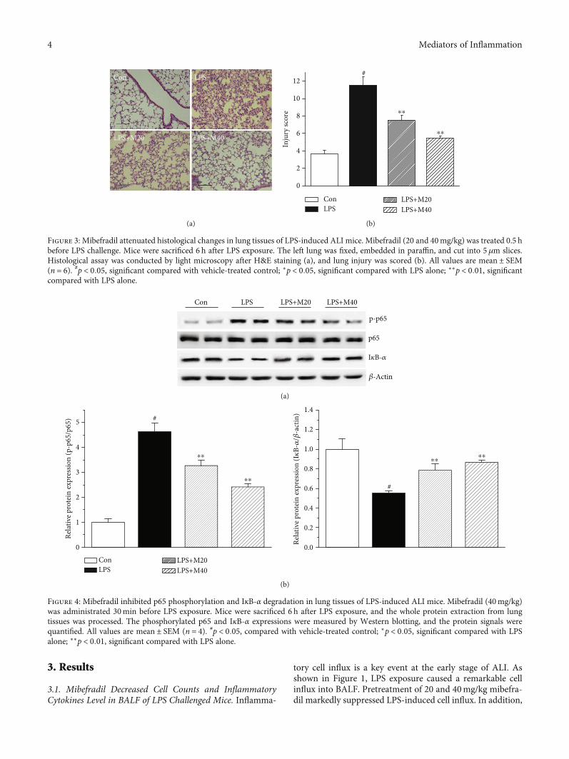
Figure 3: Mibefradil attenuated histological changes in lung tissues of LPS-induced ALI mice. Mibefradil (20 and 40mg/kg) was treated 0.5 hbefore LPS challenge. Mice were sacrificed 6 h after LPS exposure. The left lung was fixed, embedded in paraffin, and cut into 5 μm slices.Histological assay was conducted by light microscopy after H&E staining (a), and lung injury was scored (b). All values are mean ± SEM(n = 6). #p < 0:05, significant compared with vehicle-treated control; ∗p < 0:05, significant compared with LPS alone; ∗∗p < 0:01, significantcompared with LPS alone.
𝛽-Actin
I𝜅B-𝛼
p65
p-p65
Con LPS LPS+M20 LPS+M40
(a)
0
1
2
3
Relat
ive p
rote
in ex
pres
sion
(p-p
65/p
65)
4
5
0.0
0.2
0.4
0.6
0.8
1.0
#
#1.2
1.4
Relat
ive p
rote
in ex
pres
sion
(I𝜅
B-𝛼
/𝛽-a
ctin
)
⁎⁎
⁎⁎ ⁎⁎⁎⁎
Con LPS
LPS+M20 LPS+M40
(b)
Figure 4: Mibefradil inhibited p65 phosphorylation and IκB-α degradation in lung tissues of LPS-induced ALI mice. Mibefradil (40mg/kg)was administrated 30min before LPS exposure. Mice were sacrificed 6 h after LPS exposure, and the whole protein extraction from lungtissues was processed. The phosphorylated p65 and IκB-α expressions were measured by Western blotting, and the protein signals werequantified. All values are mean ± SEM (n = 4). #p < 0:05, compared with vehicle-treated control; ∗p < 0:05, significant compared with LPSalone; ∗∗p < 0:01, significant compared with LPS alone.
4 Mediators of Inflammation
mibefradil also significantly lowered LPS-induced MPOactivity in lung tissue, which is a key indicator of neutrophilsinfiltration in tissue.
We also examined the inflammatory cytokine levels inBALF. LPS exposure resulted in obviously increased levelsof TNF-α and IL-6 in BALF, whereas these rises were dose-dependently inhibited by mibefradil.
3.2. Mibefradil Decreased Protein Concentration in BALF andInhibited Evans Blue Extravasation in Lung Tissue. Vascularleakage is a crucial event of lung injury; therefore, we mea-
sured the total protein level in BALF. As shown in Figure 2,LPS exposure caused a dramatic elevation of protein concen-tration in BALF, from 0:133 ± 0:007 to 0:376 ± 0:024mg/mL.Pretreatment with 20 and 40mg/kg mibefradil significantlyinhibited total protein level in BALF of LPS challenged mice.In parallel with the total protein levels, pretreatment withmibefradil also suppressed LPS-induced increase in Evansblue extravasation.
3.3. Mibefradil Attenuated LPS-Induced PathologicalAlterations in Lung Tissues. The pulmonary histopathology
Con LPS LPS+M200
200
400
600
Tota
l cel
ls (1
03 /mL)
800
#
⁎⁎
(a)
Con LPS LPS+M200.0
0.2
0.4
0.6
0.8 #
MPO
activ
ity (U
/g) ⁎
(b)
0.0
0.1
0.2
0.3 #
Con LPS LPS+M20
Tota
l pro
tein
(mg/
mL)
⁎⁎
(c)
Con LPS LPS+M200
10
20
30
40 #
⁎⁎
Extr
avas
atio
n of
evan
s blu
e dye
(𝜇g/
g)
(d)
Con LPS LPS+M200
1
2
3
4
5
6 #TN
F-𝛼
(ng/
mL)
⁎
(e)
Con LPS LPS+M200.0
0.2
0.4
IL-6
(ng/
mL)
0.6
0.8 #
⁎⁎
(f)
Con LPS LPS+M200
2
4
6
8
10
12
Lung
inju
ry sc
ore
#
LPS+M20
Con LPS
⁎⁎
(g)
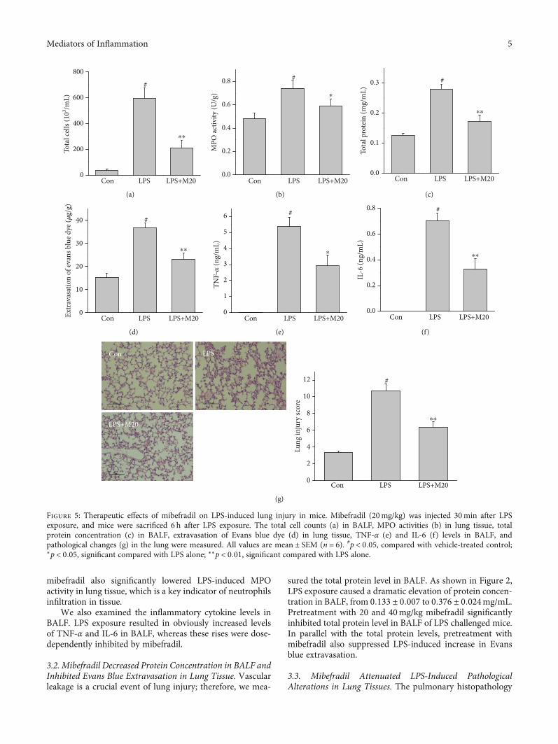
Figure 5: Therapeutic effects of mibefradil on LPS-induced lung injury in mice. Mibefradil (20mg/kg) was injected 30min after LPSexposure, and mice were sacrificed 6 h after LPS exposure. The total cell counts (a) in BALF, MPO activities (b) in lung tissue, totalprotein concentration (c) in BALF, extravasation of Evans blue dye (d) in lung tissue, TNF-α (e) and IL-6 (f) levels in BALF, andpathological changes (g) in the lung were measured. All values are mean ± SEM (n = 6). #p < 0:05, compared with vehicle-treated control;∗p < 0:05, significant compared with LPS alone; ∗∗p < 0:01, significant compared with LPS alone.
5Mediators of Inflammation
was evaluated by HE staining and lung injury score system.Compared with control group, lung sections in mice treatedwith LPS showed notable neutrophils infiltration, alveolarhemorrhage, and interalveolar septal thickening. Treatmentwith mibefradil improved pulmonary histological changesin LPS challenged mice (Figure 3).
3.4. Mibefradil Inhibited LPS-Induced NF-κB Activation inLung Tissues. NF-κB plays a center role in the regulation ofinflammation, and phosphorylation of p65 and degradativeIκB-α are key signs of NF-κB activation. We measured phos-phorylated p65 and IκB-α levels in lung tissue by Westernblot method. As shown in Figure 4, mibefradil inhibited
phosphorylation of p65 and degradation of IκB-α, whichdemonstrated that mibefradil suppressed NF-κB pathwayactivation in lung tissues of LPS challenged mice.
3.5. Therapeutic Effects of Mibefradil on LPS-Induced LungInjury. To additionally evaluate the therapeutic effects ofmibefradil on LPS-induced lung injury, mice were treatedwith mibefradil (20mg/kg) 30min after LPS exposure. AsFigure 5 shown, mibefradil attenuated the cell influx, proteinleakage, and inflammatory cytokines release in ALI mice.
3.6. Flunarizine Protected Mice from LPS-Induced ALI. Toadditionally verify the protective properties of T-type
Con Flu LPS LPS+Flu0
200
400
600
800
1000
1200
Tota
l cel
ls (1
03 /mL)
#
⁎⁎
(a)
0.0
0.2
0.4
0.6
0.8
1.0 #
Con Flu LPS LPS+Flu
MPO
activ
ity (U
/g)
⁎⁎
(b)
Con Flu LPS LPS+Flu0.0
0.1
0.2
0.3
0.4
0.5#
Tota
l pro
tein
(mg/
mL)
⁎⁎
(c)
Con Flu LPS LPS+Flu0
10
20
30
40
50
60 #
Extr
avas
atio
n of
evan
s blu
e dye
(𝜇g/
g)
⁎⁎
(d)
Con Flu LPS LPS+Flu0123456789
10#
TNF-𝛼
(ng/
mL)
⁎⁎
(e)
Con Flu LPS LPS+Flu0.0
0.2
0.4
0.6
0.8
1.0
1.2 #
IL-6
(ng/
mL)
⁎⁎
(f)
Con Flu LPS LPS+Flu0
2
4
6
8
10
12
14
16 #Con Flu
LPS+FluLPS
Lung
inju
ry sc
ore
⁎⁎
(g)
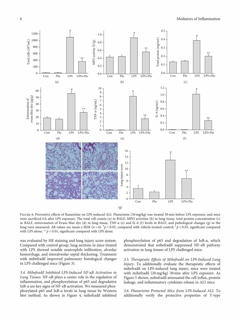
Figure 6: Preventive effects of flunarizine on LPS-induced ALI. Flunarizine (30mg/kg) was treated 30min before LPS exposure, and micewere sacrificed 6 h after LPS exposure. The total cell counts (a) in BALF, MPO activities (b) in lung tissue, total protein concentration (c)in BALF, extravasation of Evans blue dye (d) in lung tissue, TNF-α (e) and IL-6 (f) levels in BALF, and pathological changes (g) in thelung were measured. All values are mean ± SEM (n = 6). #p < 0:05, compared with vehicle-treated control; ∗p < 0:05, significant comparedwith LPS alone; ∗∗p < 0:01, significant compared with LPS alone.
6 Mediators of Inflammation
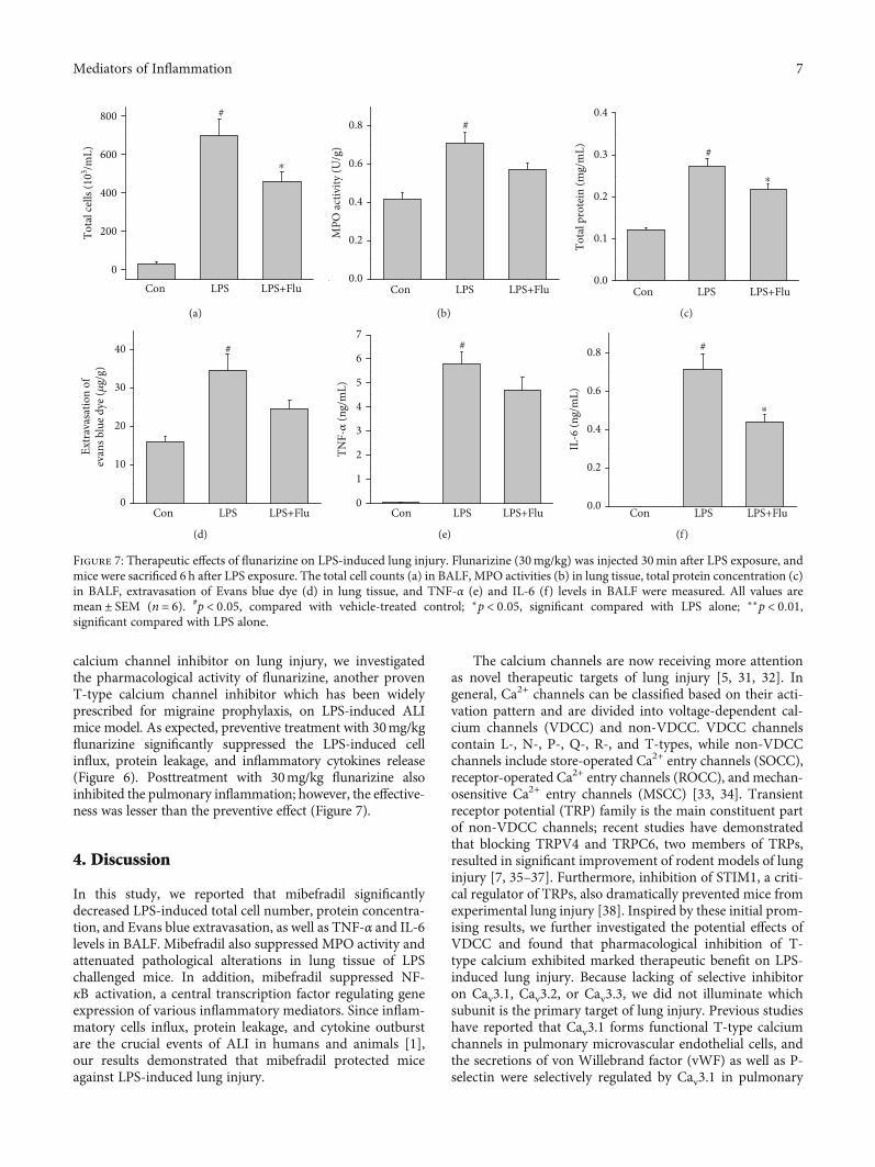
calcium channel inhibitor on lung injury, we investigatedthe pharmacological activity of flunarizine, another provenT-type calcium channel inhibitor which has been widelyprescribed for migraine prophylaxis, on LPS-induced ALImice model. As expected, preventive treatment with 30mg/kgflunarizine significantly suppressed the LPS-induced cellinflux, protein leakage, and inflammatory cytokines release(Figure 6). Posttreatment with 30mg/kg flunarizine alsoinhibited the pulmonary inflammation; however, the effective-ness was lesser than the preventive effect (Figure 7).
4. Discussion
In this study, we reported that mibefradil significantlydecreased LPS-induced total cell number, protein concentra-tion, and Evans blue extravasation, as well as TNF-α and IL-6levels in BALF. Mibefradil also suppressed MPO activity andattenuated pathological alterations in lung tissue of LPSchallenged mice. In addition, mibefradil suppressed NF-κB activation, a central transcription factor regulating geneexpression of various inflammatory mediators. Since inflam-matory cells influx, protein leakage, and cytokine outburstare the crucial events of ALI in humans and animals [1],our results demonstrated that mibefradil protected miceagainst LPS-induced lung injury.
The calcium channels are now receiving more attentionas novel therapeutic targets of lung injury [5, 31, 32]. Ingeneral, Ca2+ channels can be classified based on their acti-vation pattern and are divided into voltage-dependent cal-cium channels (VDCC) and non-VDCC. VDCC channelscontain L-, N-, P-, Q-, R-, and T-types, while non-VDCCchannels include store-operated Ca2+ entry channels (SOCC),receptor-operated Ca2+ entry channels (ROCC), and mechan-osensitive Ca2+ entry channels (MSCC) [33, 34]. Transientreceptor potential (TRP) family is the main constituent partof non-VDCC channels; recent studies have demonstratedthat blocking TRPV4 and TRPC6, two members of TRPs,resulted in significant improvement of rodent models of lunginjury [7, 35–37]. Furthermore, inhibition of STIM1, a criti-cal regulator of TRPs, also dramatically prevented mice fromexperimental lung injury [38]. Inspired by these initial prom-ising results, we further investigated the potential effects ofVDCC and found that pharmacological inhibition of T-type calcium exhibited marked therapeutic benefit on LPS-induced lung injury. Because lacking of selective inhibitoron Cav3.1, Cav3.2, or Cav3.3, we did not illuminate whichsubunit is the primary target of lung injury. Previous studieshave reported that Cav3.1 forms functional T-type calciumchannels in pulmonary microvascular endothelial cells, andthe secretions of von Willebrand factor (vWF) as well as P-selectin were selectively regulated by Cav3.1 in pulmonary
Con LPS LPS+Flu0
200
400
Tota
l cel
ls (1
03 /mL) 600
800 #
⁎
(a)
0.0
0.2
0.4
0.6
MPO
activ
ity (U
/g)
0.8 #
Con LPS LPS+Flu
(b)
Tota
l pro
tein
(mg/
mL)
Con LPS LPS+Flu0.0
0.1
0.2
0.3
0.4
#
⁎
(c)
Con LPS LPS+Flu0
10
20
30
40
Extr
avas
atio
n of
evan
s blu
e dye
(𝜇g/
g)
#
(d)
Con LPS LPS+Flu
TNF-𝛼
(ng/
mL)
0
1
2
3
4
5
6
7#
(e)
Con LPS LPS+Flu
IL-6
(ng/
mL)
0.2
0.0
0.4
0.6
0.8 #
⁎
(f)
Figure 7: Therapeutic effects of flunarizine on LPS-induced lung injury. Flunarizine (30mg/kg) was injected 30min after LPS exposure, andmice were sacrificed 6 h after LPS exposure. The total cell counts (a) in BALF, MPO activities (b) in lung tissue, total protein concentration (c)in BALF, extravasation of Evans blue dye (d) in lung tissue, and TNF-α (e) and IL-6 (f) levels in BALF were measured. All values aremean ± SEM (n = 6). #p < 0:05, compared with vehicle-treated control; ∗p < 0:05, significant compared with LPS alone; ∗∗p < 0:01,significant compared with LPS alone.
7Mediators of Inflammation
capillary endothelium [19, 22, 23]. These data reminded usthat inhibition of Cav3.1 may be the contributor against lunginjury. However, more studies of experimental lung injurymodels based on transgenic animal technology are needed.
Identifying new uses for existing drugs is one way toavoid the current costly and time-consuming status of drugdiscovery. Because existing drugs have known pharmacoki-netics and safety profiles, the pharmacokinetic and toxico-logical experiments could probably be eliminated [39].Although mibefradil has been withdrawn in 1998 becauseof severe drug interactions, there are still a few drugs withpotential inhibitory effect on T-type calcium channel in themarket, such as flunarizine, penfluridol, and ethosuximide[40, 41]. Flunarizine is one of the most widely prescribedmedicine for migraine prevention. Additionally, flunarizineis used as a first-line medication for migraine prophylaxisin children and adolescents because of its satisfactory safetyand efficacy profiles [42]. Previous studies reported that flu-narizine potently inhibited T-type calcium channel in ven-tricular myocytes [43, 44], aorta smooth muscle cells [45],granulosa cells [46], pulmonary microvascular endothelialcells [21], and spermatogenic cells [47]. In this study, wefound that preventive treatment with flunarizine significantlyinhibited LPS-induced protein leakage, cell influx, andinflammatory cytokine release in BALF and improved thepathologic changes in lung tissues. These results verified theprotective effect of T-type calcium channel inhibitors on lunginjury. What is more, since flunarizine has acceptable safetyand tolerability for long-term usage, further clinical worksare warranted to explore the potential of this drug in the pre-vention of ALI.
5. Conclusion
In summary, our study demonstrated that T-type calciumchannel inhibitors may be beneficial for treating lung injury.The key role of T-type calcium channel in the acute lunginjury is encouraged to be further investigated.
Data Availability
The data used to support the findings of this study are avail-able from the corresponding author upon request.
Conflicts of Interest
The authors report no conflicts of interest.
Acknowledgments
This study was supported by grants from the EducationBureau of Guangzhou (No.1201581610), Natural ScienceFoundation of Guangdong Province (No.2017A030313773),and National Natural Science Foundation of China(No.81402992).
Supplementary Materials
Supplement figures: mibefradil (40mg/kg) or saline wastreated 30min before aerosolized saline exposure, and mice
were sacrificed 6h after aerosol inhalation of saline. The totalcell counts (a) in BALF, MPO activities (b) in lung tissue,total protein concentration (c) in BALF, extravasation ofEvans blue dye (d) in lung tissue, TNF-α (e) and IL-6 (f)levels in BALF, pathological changes (g), and NF-κB activa-tion in the lung were measured. All values are mean ± SEM(n = 6). #p < 0:05, compared with vehicle-treated control;∗p < 0:05, significant compared with LPS alone; ∗∗p < 0:01,significant compared with LPS alone. (SupplementaryMaterials)
References
[1] Y. Butt, A. Kurdowska, and T. C. Allen, “Acute lung injury: aclinical and molecular review,” Archives of Pathology & Labo-ratory Medicine, vol. 140, no. 4, pp. 345–350, 2016.
[2] L. Papazian, C. S. Calfee, D. Chiumello et al., “Diagnosticworkup for ARDS patients,” Intensive Care Medicine, vol. 42,no. 5, pp. 674–685, 2016.
[3] S. Liu, Z. Zhao, L. Tan et al., “Optimal mean airway pressureduring high-frequency oscillatory ventilation in an experimen-tal model of acute respiratory distress syndrome: EIT-basedmethod,” Annals of Intensive Care, vol. 10, no. 1, p. 31, 2020.
[4] C. Karagiannidis, T. Joost, S. Strassmann et al., “Safety andEfficacy of a novel Pneumatically Driven Extracorporeal Mem-brane Oxygenation device,” The Annals of Thoracic Surgery,vol. 109, no. 6, pp. 1684–1691, 2020.
[5] R. E. Morty and W. M. Kuebler, “TRPV4: an exciting new tar-get to promote alveolocapillary barrier function,” AmericanJournal of Physiology. Lung Cellular and Molecular Physiology,vol. 307, no. 11, pp. L817–L821, 2014.
[6] E. J. Seeley, P. Rosenberg, and M. A. Matthay, “Calcium fluxand endothelial dysfunction during acute lung injury: a STIM-ulating target for therapy,” The Journal of Clinical Investiga-tion, vol. 123, no. 3, pp. 1015–1018, 2013.
[7] M. Tauseef, N. Knezevic, K. R. Chava et al., “TLR4 activationof TRPC6-dependent calcium signaling mediates endotoxin-induced lung vascular permeability and inflammation,” TheJournal of Experimental Medicine, vol. 209, no. 11, pp. 1953–1968, 2012.
[8] G. Wang, J. Zhang, C. Xu, X. Han, Y. Gao, and H. Chen, “Inhibi-tion of SOCs attenuates acute lung injury induced by severe acutepancreatitis in rats and PMVECs injury induced by lipopolysac-charide,” Inflammation, vol. 39, no. 3, pp. 1049–1058, 2016.
[9] K. Suresh, L. Servinsky, J. Reyes et al., “Hydrogen peroxide-induced calcium influx in lung microvascular endothelial cellsinvolves TRPV4,” American Journal of Physiology. Lung Cellu-lar and Molecular Physiology, vol. 309, no. 12, pp. L1467–L1477, 2015.
[10] K. Kandasamy, L. Bezavada, R. B. Escue, and K. Parthasarathi,“Lipopolysaccharide induces endoplasmic store Ca2+-depen-dent inflammatory responses in lung microvessels,” PLoSOne, vol. 8, no. 5, article e63465, 2013.
[11] C. Lee, D. Z. Xu, E. Feketeova et al., “Store-operated calciumchannel inhibition attenuates neutrophil function and post-shock acute lung injury,” The Journal of Trauma, vol. 59,no. 1, pp. 56–63, 2005, 63.
[12] J. Yin, L. Michalick, C. Tang et al., “Role of transient receptorpotential vanilloid 4 in neutrophil activation and acute lunginjury,” American Journal of Respiratory Cell and MolecularBiology, vol. 54, no. 3, pp. 370–383, 2016.
8 Mediators of Inflammation
[13] T. P. Snutch and G. W. Zamponi, “Recent advances in thedevelopment of T-type calcium channel blockers for painintervention,” British Journal of Pharmacology, vol. 175,no. 12, pp. 2375–2383, 2018.
[14] L. L. Cribbs, “T-type Ca2+ channels in vascular smooth muscle:multiple functions,” Cell Calcium, vol. 40, no. 2, pp. 221–230,2006.
[15] M. L. Bernhardt, Y. Zhang, C. F. Erxleben et al., “CaV3.2 T-type channels mediate Ca2+ entry during oocyte maturationand following fertilization,” Journal of Cell Science, vol. 128,no. 23, pp. 4442–4452, 2015.
[16] S. M. Todorovic and V. Jevtovic-Todorovic, “T-type voltage-gated calcium channels as targets for the development of novelpain therapies,” British Journal of Pharmacology, vol. 163,no. 3, pp. 484–495, 2011.
[17] M. E. Mangoni, A. Traboulsie, A. L. Leoni et al., “Bradycardiaand slowing of the atrioventricular conduction in mice lackingCaV3.1/alpha1G T-type calcium channels,” CirculationResearch, vol. 98, no. 11, pp. 1422–1430, 2006.
[18] M. F. Rossier, “T-type calcium channel: a privileged gate forcalcium entry and control of adrenal steroidogenesis,” Fron-tiers in Endocrinology (Lausanne), vol. 7, p. 43, 2016.
[19] C. Zhou and S. Wu, “T-type calcium channels in pulmonaryvascular endothelium,” Microcirculation, vol. 13, no. 8,pp. 645–656, 2006.
[20] Z. Zheng, H. Chen, P. Xie et al., “α1GT-type calcium channeldetermines the angiogenic potential of pulmonary microvas-cular endothelial cells,” American Journal of Physiology-CellPhysiology, vol. 316, no. 3, pp. C353–C364, 2019.
[21] S. Wu, J. Haynes Jr., J. T. Taylor et al., “Cav3.1 (alpha1G) T-type Ca2+ channels mediate vaso-occlusion of sickled erythro-cytes in lung microcirculation,” Circulation Research, vol. 93,no. 4, pp. 346–353, 2003.
[22] C. Zhou, H. Chen, F. Lu et al., “Cav3.1 (alpha1G) controls vonWillebrand factor secretion in rat pulmonary microvascularendothelial cells,” American Journal of Physiology. Lung Cellu-lar and Molecular Physiology, vol. 292, no. 4, pp. L833–L844,2007.
[23] C. Zhou, H. Chen, J. A. King et al., “α1GT-type calcium channelselectively regulates P-selectin surface expression in pulmonarycapillary endothelium,” American Journal of Physiology. LungCellular and Molecular Physiology, vol. 299, no. 1, pp. L86–L97, 2010.
[24] H. Chen, C. Bai, and X. Wang, “The value of thelipopolysaccharide-induced acute lung injury model in respi-ratory medicine,” Expert Review of Respiratory Medicine,vol. 4, no. 6, pp. 773–783, 2014.
[25] P. Mulder, V. Richard, P. Compagnon et al., “Increased survivalafter long-term treatment with mibefradil, a selective T-channelcalcium antagonist, in heart failure,” Journal of the AmericanCollege of Cardiology, vol. 29, no. 2, pp. 416–421, 1997.
[26] W. Li-Mei, T. Jie, W. Shan-He, M. Dong-Mei, and Y. Peng-Jiu,“Anti-inflammatory and anti-oxidative effects of dexpanthe-nol on lipopolysaccharide induced acute lung injury in mice,”Inflammation, vol. 39, no. 5, pp. 1757–1763, 2016.
[27] D. Bilici, Z. Nur Banoğlu, A. Kiziltunç, B. Avci, A. Çiftçioğlu,and S. Bilici, “Antioxidant effect of T-type calcium channelblockers in gastric injury,” Digestive Diseases and Sciences,vol. 47, no. 4, pp. 850–855, 2002.
[28] C. Qiu, P. Bruneval, A. Roeckel, D. Heudes, J. P. D. van Huyen,and S. Roux, “Mibefradil prevents L-NAME-exacerbated
nephrosclerosis in spontaneously hypertensive rats,” Journalof Hypertension, vol. 17, no. 10, pp. 1489–1495, 1999.
[29] N. Egashira, R. Okuno, M. Abe et al., “Calcium-channelantagonists inhibit marble-burying behavior in mice,” Jour-nal of Pharmacological Sciences, vol. 108, no. 1, pp. 140–143, 2008.
[30] U. Schingnitz, K. Hartmann, C. F. MacManus et al., “Signalingthrough the A2B adenosine receptor dampens endotoxin-induced acute lung injury,” Journal of Immunology, vol. 184,no. 9, pp. 5271–5279, 2010.
[31] U. Simonsen, C. Wandall-Frostholm, A. Olivan-Viguera, andR. Kohler, “Emerging roles of calcium-activated K channelsand TRPV4 channels in lung oedema and pulmonary circula-tory collapse,” Acta Physiologica, vol. 219, no. 1, pp. 176–187,2017.
[32] D. Andres, B. Keyser, B. Benton et al., “Transient receptorpotential (TRP) channels as a therapeutic target for interven-tion of respiratory effects and lethality from phosgene,” Toxi-cology Letters, vol. 244, pp. 21–27, 2016.
[33] G. W. Zamponi, J. Striessnig, A. Koschak, and A. C. Dolphin,“The physiology, pathology, and pharmacology of voltage-gated calcium channels and their future therapeutic potential,”Pharmacological Reviews, vol. 67, no. 4, pp. 821–870, 2015.
[34] A. B. Parekh and J. W. Putney Jr., “Store-operated calciumchannels,” Physiological Reviews, vol. 85, no. 2, pp. 757–810,2005.
[35] S. Balakrishna, W. Song, S. Achanta et al., “TRPV4 inhibitioncounteracts edema and inflammation and improves pulmo-nary function and oxygen saturation in chemically inducedacute lung injury,” American Journal of Physiology. Lung Cel-lular and Molecular Physiology, vol. 307, no. 2, pp. L158–L172, 2014.
[36] D. F. Alvarez, J. A. King, D. Weber, E. Addison, W. Liedtke,and M. I. Townsley, “Transient receptor potential vanilloid4-mediated disruption of the alveolar septal barrier: a novelmechanism of acute lung injury,” Circulation Research,vol. 99, no. 9, pp. 988–995, 2006.
[37] N. Weissmann, A. Sydykov, H. Kalwa et al., “Activation ofTRPC6 channels is essential for lung ischaemia-reperfusioninduced oedema in mice,” Nature Communications, vol. 3,no. 1, p. 649, 2012.
[38] R. K. Gandhirajan, S. Meng, H. C. Chandramoorthy et al.,“Blockade of NOX2 and STIM1 signaling limitslipopolysaccharide-induced vascular inflammation,” The Jour-nal of Clinical Investigation, vol. 123, no. 2, pp. 887–902, 2013.
[39] C. R. Chong and D. J. Sullivan Jr., “New uses for old drugs,”Nature, vol. 448, no. 7154, pp. 645-646, 2007.
[40] C. M. Santi, F. S. Cayabyab, K. G. Sutton et al., “Differentialinhibition of T-type calcium channels by neuroleptics,” TheJournal of Neuroscience, vol. 22, no. 2, pp. 396–403, 2002.
[41] J. C. Gomora, A. N. Daud, M. Weiergraber, and E. Perez-Reyes, “Block of cloned human T-type calcium channels bysuccinimide antiepileptic drugs,” Molecular Pharmacology,vol. 60, no. 5, pp. 1121–1132, 2001.
[42] B. P. Mohamed, P. J. Goadsby, and P. Prabhakar, “Safety andefficacy of flunarizine in childhood migraine: 11 years’ experi-ence, with emphasis on its effect in hemiplegic migraine,”Developmental Medicine and Child Neurology, vol. 54, no. 3,pp. 274–277, 2012.
[43] J. Tytgat, J. Vereecke, and E. Carmeliet, “Differential effects ofverapamil and flunarizine on cardiac L-type and T-type Ca
9Mediators of Inflammation
channels,” Naunyn-Schmiedeberg's Archives of Pharmacology,vol. 337, no. 6, pp. 690–692, 1988.
[44] J. Tytgat, J. Vereecke, and E. Carmeliet, “Mechanism of L- andT-type Ca2+ channel blockade by flunarizine in ventricularmyocytes of the guinea-pig,” European Journal of Pharmacol-ogy, vol. 296, no. 2, pp. 189–197, 1996.
[45] T. Kuga, J. Sadoshima, H. Tomoike, H. Kanaide, N. Akaike,and M. Nakamura, “Actions of Ca2+ antagonists on two typesof Ca2+ channels in rat aorta smooth muscle cells in primaryculture,” Circulation Research, vol. 67, no. 2, pp. 469–480,1990.
[46] A. Agoston, L. Kunz, A. Krieger, and A. Mayerhofer, “Twotypes of calcium channels in human ovarian endocrine cells:involvement in steroidogenesis,” The Journal of Clinical Endo-crinology and Metabolism, vol. 89, no. 9, pp. 4503–4512, 2004.
[47] C. S. Wang, X. H. Gao, H. Cheng et al., “Effects of flunarizineon T-type calcium channels in mouse spermatogenic cells,”Zhonghua Nan Ke Xue, vol. 12, pp. 594–7, 601, 2006.
10 Mediators of Inflammation