Embed Size (px)
Citation preview
8/2/2019 Microscop Examination
http://slidepdf.com/reader/full/microscop-examination 1/5
Microscopical Examination of the Faeces.
Part 3
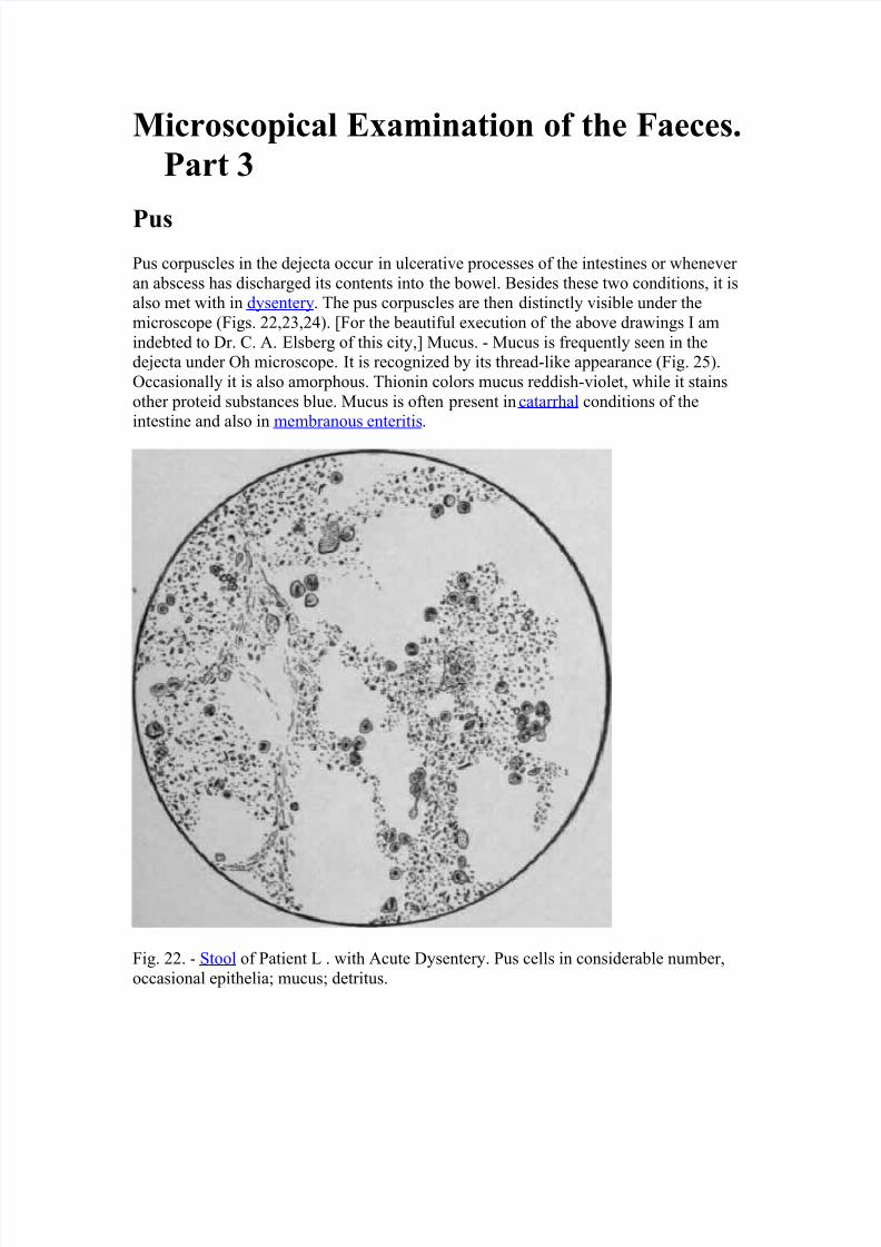
Pus
Pus corpuscles in the dejecta occur in ulcerative processes of the intestines or whenever
an abscess has discharged its contents into the bowel. Besides these two conditions, it isalso met with in dysentery. The pus corpuscles are then distinctly visible under the
microscope (Figs. 22,23,24). [For the beautiful execution of the above drawings I am
indebted to Dr. C. A. Elsberg of this city,] Mucus. - Mucus is frequently seen in thedejecta under Oh microscope. It is recognized by its thread-like appearance (Fig. 25).
Occasionally it is also amorphous. Thionin colors mucus reddish-violet, while it stains
other proteid substances blue. Mucus is often present in catarrhal conditions of theintestine and also in membranous enteritis.
Fig. 22. - Stool of Patient L . with Acute Dysentery. Pus cells in considerable number,
occasional epithelia; mucus; detritus.
8/2/2019 Microscop Examination
http://slidepdf.com/reader/full/microscop-examination 2/5
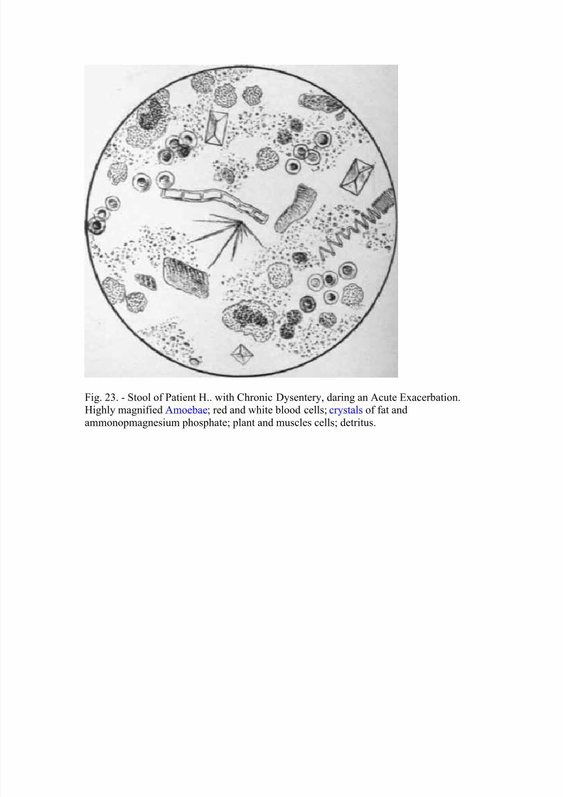
Fig. 23. - Stool of Patient H.. with Chronic Dysentery, daring an Acute Exacerbation.Highly magnified Amoebae; red and white blood cells; crystals of fat and
ammonopmagnesium phosphate; plant and muscles cells; detritus.
8/2/2019 Microscop Examination
http://slidepdf.com/reader/full/microscop-examination 3/5
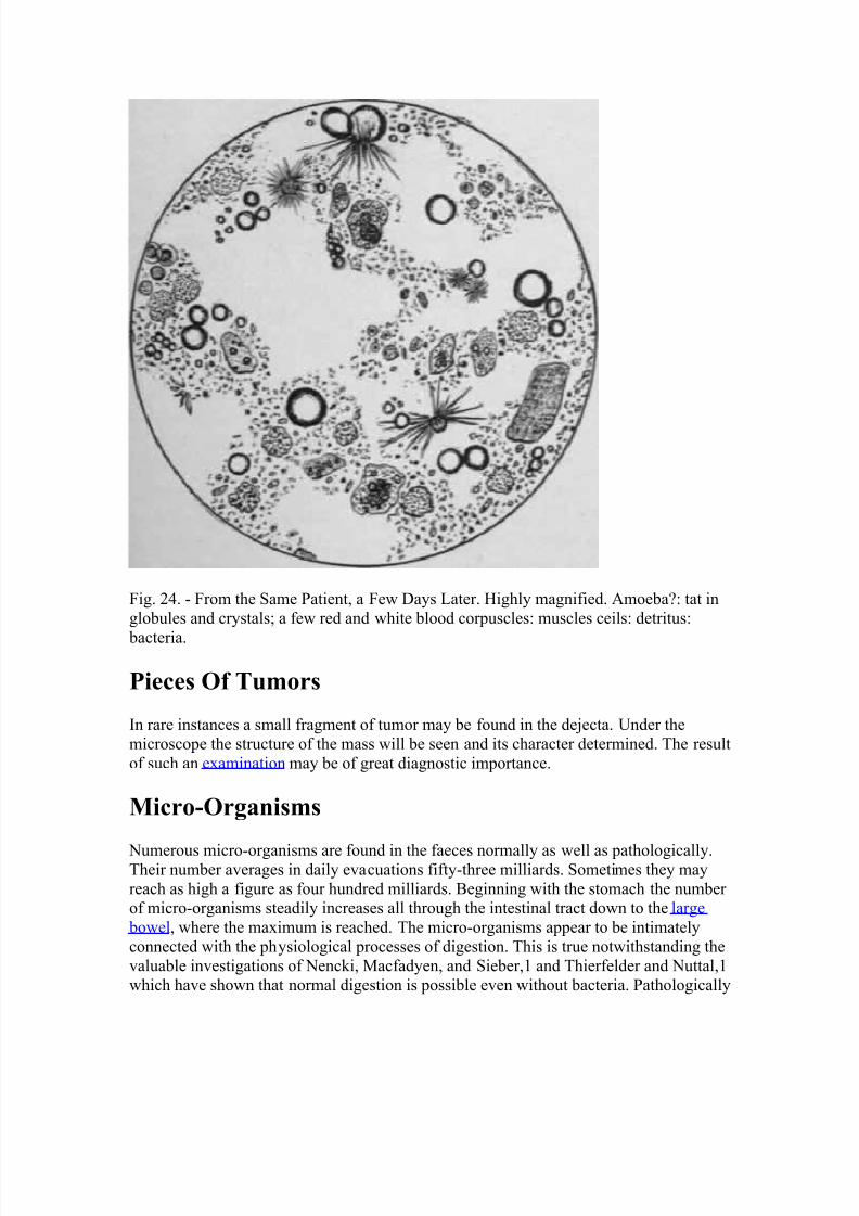
Fig. 24. - From the Same Patient, a Few Days Later. Highly magnified. Amoeba?: tat in
globules and crystals; a few red and white blood corpuscles: muscles ceils: detritus:
bacteria.
Pieces Of Tumors
In rare instances a small fragment of tumor may be found in the dejecta. Under the
microscope the structure of the mass will be seen and its character determined. The result
of such an examination may be of great diagnostic importance.
Micro-Organisms
Numerous micro-organisms are found in the faeces normally as well as pathologically.Their number averages in daily evacuations fifty-three milliards. Sometimes they may
reach as high a figure as four hundred milliards. Beginning with the stomach the number of micro-organisms steadily increases all through the intestinal tract down to the large
bowel, where the maximum is reached. The micro-organisms appear to be intimately
connected with the physiological processes of digestion. This is true notwithstanding thevaluable investigations of Nencki, Macfadyen, and Sieber,1 and Thierfelder and Nuttal,1
which have shown that normal digestion is possible even without bacteria. Pathologically
8/2/2019 Microscop Examination
http://slidepdf.com/reader/full/microscop-examination 4/5
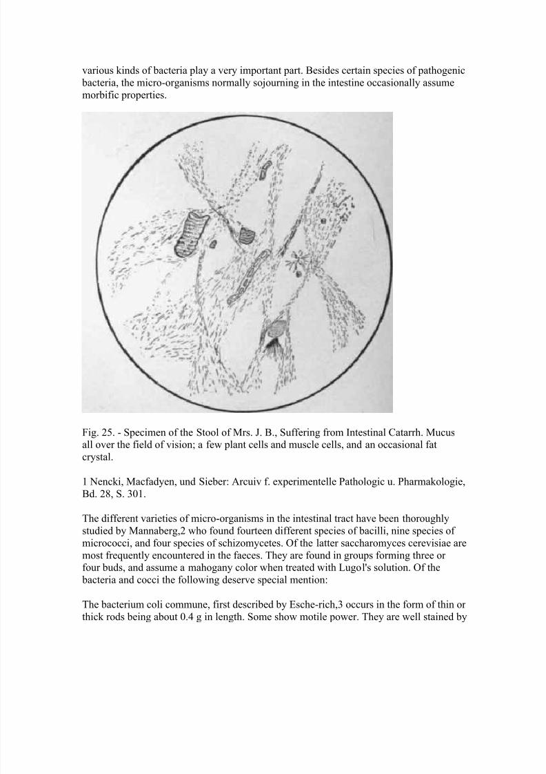
various kinds of bacteria play a very important part. Besides certain species of pathogenic
bacteria, the micro-organisms normally sojourning in the intestine occasionally assume
morbific properties.
Fig. 25. - Specimen of the Stool of Mrs. J. B., Suffering from Intestinal Catarrh. Mucusall over the field of vision; a few plant cells and muscle cells, and an occasional fat
crystal.
1 Nencki, Macfadyen, und Sieber: Arcuiv f. experimentelle Pathologic u. Pharmakologie,
Bd. 28, S. 301.
The different varieties of micro-organisms in the intestinal tract have been thoroughly
studied by Mannaberg,2 who found fourteen different species of bacilli, nine species of
micrococci, and four species of schizomycetes. Of the latter saccharomyces cerevisiae aremost frequently encountered in the faeces. They are found in groups forming three or four buds, and assume a mahogany color when treated with Lugol's solution. Of the
bacteria and cocci the following deserve special mention:
The bacterium coli commune, first described by Esche-rich,3 occurs in the form of thin or
thick rods being about 0.4 g in length. Some show motile power. They are well stained by

8/2/2019 Microscop Examination
http://slidepdf.com/reader/full/microscop-examination 5/5
the ordinary anilin dyes and decolorized by Gram's solution. Their colonies growing upon
gelatin resemble those of the bacillus of typhoid fever .
The bacterium lactis aerogenes (Escherich) greatly resembles the bacterium colicommune. It is frequently found in the stools of infants, and is now and then met with in
those of adults. It is found in thick rods frequently lying in pairs. They are non-motile andhave the property of causing fermentation of milk, producing coagulation and formation
of gas within sixty hours.
1 Thierfelder u. Nuttal . Zeitscbrift f. phys. Chemie, Bd. 21, S. 109, u. Bd. 22, S. 62.
2 Mannaberg: " Die Bacterien des Darms " - Notbnagel 's Erkrankun-gen des Darms,
Wien, 1895.
3Escbericb: "Beitrage zur Kenntniss der Darmbacterien. " Milnch-ener med.
Wochenscbr., 1886. No i., 43-45.
Bacillus putrificus coli (Bienstock 1) forms slender rods 3 m in length. This bacillus
energetically decomposes proteid substances in presence of air under the formation of
ammonia, amin bases, fatty acids, tyrosin, phenol, indol.
While all the above-mentioned micro-organisms give a mahogany or brown color withsolutions of iodine, there are a few varieties which give a blue color with this substance.
To the latter belongs the bacillus butyricus described by Nothnagel." It is rod-shaped, 3 to
10 μ long and 1 μ thick. It is often lemon-shaped. This bacillus is anaerobic and producesfermentation of starch, sugar, and cellulose, forming butyric acid and gas. The bacillus
butyricus is often found in pathological conditions of the intestine, but occurs in small
numbers also in normal faeces.
Of the pathogenic micro-organisms, cholera, typhoid, and tubercle bacilli are found in thefeces. The cholera and typhoid bacilli causing infectious diseases do not belong, strictly
speaking, to the micro-organisms producing diseases of the intestine alone. The tubercle
bacilli, occasionally producing intestinal tuberculosis, are recognized in the faeces by the
same methods which are employed in the examination of the sputum.
http://jp.zooomr.com/z/photos/zoom/5802136/size-32/
http://www.pediatriatropical.com/helmintos.html