Embed Size (px)
Citation preview

MID TERM REVIEW

2
Structural Levels of Organization
• Organization is the most important characteristic of body structure
• The body as a whole is a unit constructed of the following smaller units:• Atoms and molecules—chemical level• Cells—the smallest structural units; organizations of
various chemicals• Tissues—organizations of similar cells• Organs—organizations of different kinds of tissues• Systems—organizations of many different kinds of
organs

Assume the Position
•Anatomical Position – Standing up with arms at the side, and palms facing forward.
•Supine Position – Laying horizontal on your back (face up)
•Prone Position – Laying horizontal on your stomach (face down position)

4
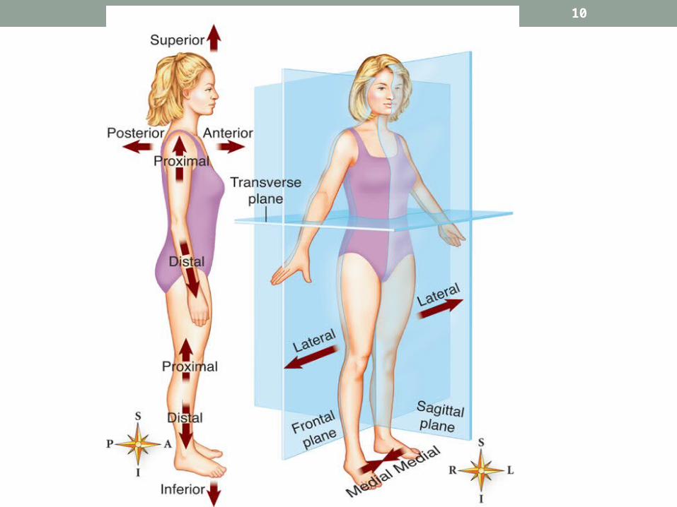
Anatomical Directions
•Superior—toward the head, upper, above
•Inferior—toward the feet, lower, below

5
Anatomical Directions
•Anterior—front, in front of (same as ventral in humans)
•Posterior—back, in back of (same as dorsal in humans)

6
Anatomical Directions
•Medial—toward the midline of a structure
•Lateral—away from the midline or toward the side of a structure

7
Anatomical Directions
•Proximal—toward or nearest the trunk, or nearest the point of origin of a structure
•Distal—away from or farthest from the trunk, or farthest from a structure’s point of origin

8
Anatomical Directions
•Superficial—nearer the body surface
•Deep—farther away from the body surface

9
Planes or Body Sections
• Sagittal plane—lengthwise plane that divides a structure into right and left sections
• Midsagittal—sagittal plane that divides the body into two equal halves
• Frontal (coronal) plane—lengthwise plane that divides a structure into anterior and posterior sections
• Transverse plane—horizontal plane that divides a structure into upper and lower sections
10

CHAPTER 6The Integumentary System and Body Membranes

12
Tissues
• Epithelial tissue• Covers body and lines body cavities• Cells packed closely together with little matrix
• Classified by shape of cells • Squamous• Cuboidal• Columnar• Transitional

13

14
Tissues
• Epithelial tissue • Classified by arrangement of cells
• Simple• Stratified
• Simple squamous epithelium• Single layer of scalelike cells• Transport (e.g., absorption) is function
• Stratified squamous epithelium • Several layers of closely packed cells• Protection is primary function

15
Classification of Body Membranes• Epithelial membranes
• Cutaneous membrane—the skin• Mucous membrane – Lines the inside of the body• Hollow organs – digestive system
• Serous membranes—simple squamous epithelium on a connective tissue basement membrane• Parietal—line walls of body cavities• Visceral—cover organs found in body cavities

16
Classification of Body Membranes
• Examples• Pleura (serous membranes )—parietal and visceral layers line walls of thoracic cavity and cover the lungs
• Peritoneum (serous membranes )—parietal and visceral layers line walls of abdominal cavity and cover the organs in that cavity

17
Classification of Body Membranes
•Diseases•Pleurisy—inflammation of the serous membranes that line the chest cavity and cover the lungs
•Peritonitis—inflammation of the serous membranes in the abdominal cavity that line the walls and cover the abdominal organs

18
Classification of Body Membranes
•Mucous membranes•Line body surfaces that open directly to the exterior
•Produce mucus, a thick secretion that keeps the membranes soft and moist

19
The Skin
•Structure—two primary layers called epidermis and dermis•Epidermis
•Outermost and thinnest primary layer of skin
•Composed of several layers of stratified squamous epithelium

20
The Skin• Structure
• Epidermis• Stratum germinativum—innermost (deepest) layer of
cells that continually reproduce; new cells move toward the surface• Sometimes called the pigment layer• Pigment cells called melanocytes, which produce
the brown pigment melanin • As cells approach the surface, they are filled with a
tough, waterproof protein called keratin and eventually flake off
• Stratum corneum—outermost layer of keratin-filled cells

21
The Skin• Structure
•Dermis• Deeper and thicker of the two primary skin
layers and composed largely of connective tissue
• Upper area of dermis characterized by parallel rows of peglike dermal papillae
• Thick skin has parallel friction ridges and no hairs
• Thin skin has irregular, shallow grooves and hair • Deeper area of dermis is filled with network of
tough collagenous and stretchable elastic fibers

22
The Skin
•Skin glands—two main types•Sweat, or sudoriferous•Sebaceous

23
The Skin
• Skin glands• Sweat, or sudoriferous, glands
• Eccrine sweat gland• Most numerous, important, and widespread of the sweat glands
• Produce perspiration or sweat, which flows out through pores on skin surface
• Function throughout life and assist in body heat regulation

24
The Skin• Skin glands
• Sweat or sudoriferous glands• Apocrine sweat glands
• Found primarily in axilla and around genitalia
• Secrete a thicker, milky secretion quite different from eccrine perspiration
• Breakdown of secretion by skin bacteria produces odor

25
The Skin
• Skin glands• Sebaceous glands
• Secrete oil or sebum for hair and skin• Secretion increases during adolescence• Amount of secretion regulated by sex hormones
• Sebum in sebaceous gland ducts may darken to form a blackhead
• Acne vulgaris—inflammation of sebaceous gland ducts

26
Disorders of the Skin (Dermatoses)
• Skin lesions—any measurable variation from the normal structure • Elevated lesions—cast a shadow outside their edges
• Papule—small, firm raised lesion• Plaque—large raised lesion• Vesicle—blister• Pustule—pus-filled lesion• Crust—scab• Wheal (hive)—raised, firm lesion with a light center

27
Burns
• Treatment and recovery or survival depend on total area involved and severity or depth of the burn
• Classification of burns • First-degree (partial-thickness) burns—only surface layers of epidermis involved
• Second-degree (partial-thickness) burns—involve deep epidermal layers; always cause injury to upper layers of the dermis
28
Burns
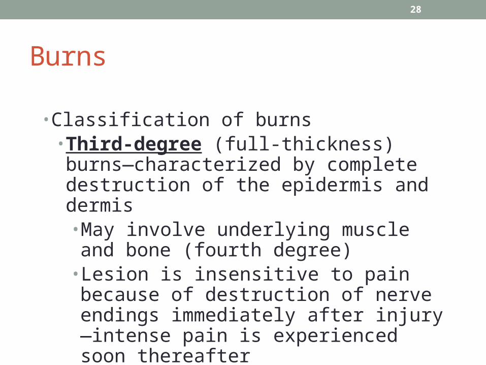
• Classification of burns• Third-degree (full-thickness) burns—characterized by complete destruction of the epidermis and dermis• May involve underlying muscle and bone (fourth degree)
• Lesion is insensitive to pain because of destruction of nerve endings immediately after injury—intense pain is experienced soon thereafter

29

30

31
Burns
•Estimating body surface area using the “rule of nines” in adults•Body divided into 11 areas of 9% each
•Additional 1% of body surface area around genitals

32
Skin Infections
•Boils• furuncles; staphylococcal infection in hair follicles

34
Skin Cancer
• Three common types• Squamous cell carcinoma—the most common type, characterized by hard, raised tumors
• Basal cell carcinoma—characterized by papules with a central crater; rarely spreads
• Melanoma—malignancy in a nevus (mole); the most serious type

35
Skin Cancer
•The most important causative factor in common skin cancers is exposure to sunlight
•Kaposi sarcoma, characterized by purple lesions, is associated with AIDS and other immune deficiencies

36

CHAPTER 7The Skeletal System

38
Functions of Skeletal System•Provides internal framework that supports the body
•Protects internal organs and helps fight disease by producing white blood cells
•Makes movement possible by working in concert with muscle contraction and relaxation
•Stores calcium, a vital resource •Forms blood cells—process is called hematopoiesis

39
Microscopic Structure of Bone and Cartilage• Two major types of connective tissue: bone and cartilage
• Bone types • Spongy
• Texture from needlelike threads of bone called trabeculae surrounded by network of open spaces
• Found in epiphyses of bones• Spaces contain red bone marrow

40
Microscopic Structure of Bone and Cartilage
• Bone types• Compact
• Structural unit is an osteon-calcified matrix arranged in multiple layers or rings called concentric lamella
• Bone cells, called osteocytes, are found inside spaces called lacunae, which are connected by tiny tubes called canaliculi
• Covered by periosteum

Parts of Bone
•Epiphysis – Ends of long bones
•Diaphysis – Long middle part of long bones

42
Microscopic Structure of Bone and Cartilage
•Cartilage •Cell type called chondrocytes located in lacunae
•Matrix is flexible gel-like substance and lacks blood vessels

43
Microscopic Structure of Bone and Cartilage
• Structural unit called osteon or Haversian system composed of:• Concentric lamella—circular layers of calcified
matrix• Lacunae—spaces in matrix between lamella
containing osteocytes• Canaliculi—canals/passageways that connect
lacunae and all nutrients to reach osteocytes• Central canal of osteon contains blood vessel

44
Bone Formation and Growth
•New bone-forming cells are called osteoblasts and bone resorbing cells are called osteoclasts
•The ability of bone to ossify, grow, change shape, heal after injury, and respond to stress occurs because of continuous “sculpting” by osteoblasts and osteoclasts

45
Bone Formation and Growth
•Bone is formed by two processes•Most bones develop from a process called endochondral ossification
•Some flat bones (e.g., skull bones) form in connective tissue membranes by another process

46
Bone Formation and Growth
•Endochondral bone formation•Epiphyseal plate of cartilage between epiphyses and diaphysis remains until skeletal maturity
•Epiphyseal line (bone) replaces epiphyseal plate (cartilage) when growth ceases

47
Bone Formation and Growth
•Early bone development (before birth) consists of cartilage and fibrous structures
•Cartilage models gradually replaced by calcified bone matrix—process called endochondral ossification
•Osteoblasts form new bone, and osteoclasts resorb bone

48
Divisions of Skeleton
• Axial skeleton (80 bones)• Skull• Spine, or vertebral column • Thorax
• Appendicular skeleton (126 bones)• Upper extremities, including shoulder (pectoral) girdle
• Lower extremities, including hip (pelvic) girdle

49
Skeletal Disorders
•Metabolic bone diseases• Osteoporosis
• Characterized by loss of calcified bone matrix and reduction in number of trabeculae in spongy bone
• Bones fracture easily, especially in wrists, hips, and vertebrae
• Treatment includes drug therapy, exercise, and dietary supplements of calcium and vitamin D

50
Skeletal Disorders
• Metabolic bone diseases• Rickets and osteomalacia—both diseases characterized by loss of bone minerals related to vitamins • Rickets
• Loss of bone minerals occurs in infants and young children before skeletal maturity
• Lack of bone rigidity causes gross skeletal changes (bowing of legs)
• Treated with vitamin D
51
Skeletal Disorders
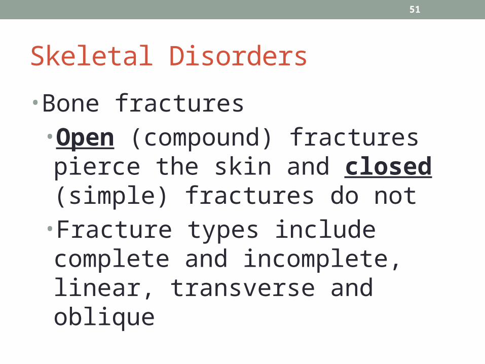
•Bone fractures •Open (compound) fractures pierce the skin and closed (simple) fractures do not
•Fracture types include complete and incomplete, linear, transverse and oblique

Dislocation
•Subluxation•When there is a partial dislocation of a bone or organ.

53

54
Joint (Articulations)
• Joint types • Diarthrosis (free movement)—most joints belong
to this class• Structures of freely movable joints—joint capsule
and ligaments hold adjoining bones together but permit movement at joint
• Articular cartilage—covers joint ends of bones and absorbs jolts
• Synovial membrane—lines joint capsule and secretes lubricating fluid
• Joint cavity—space between joint ends of bones


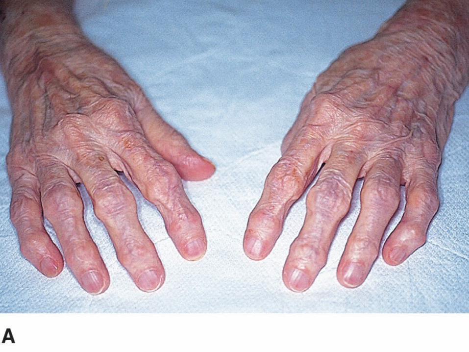
Arthritis
•Types of arthritis. •Osteoarthritis. Note the presence of nodes in the proximal interphalangeal joints (Bouchard nodes) and distal interphalangeal joints (Heberden nodes).

CHAPTER 13The Heart and Heart Disease
59
Anatomy of the Heart
•Heart chambers •Two upper chambers are called atria (receiving chambers)—right and left atria
•Two lower chambers called ventricles (discharging chambers)—right and left ventricles

Know the Stars…..
61
Anatomy of the Heart
•Heart valves and valve disorders•Valves keep blood flowing through the heart; prevent backflow
•Atrioventricular (AV) valves•Tricuspid: at the opening of the right atrium into the ventricle
•Bicuspid (mitral): at the opening of the left atrium into the ventricle

62
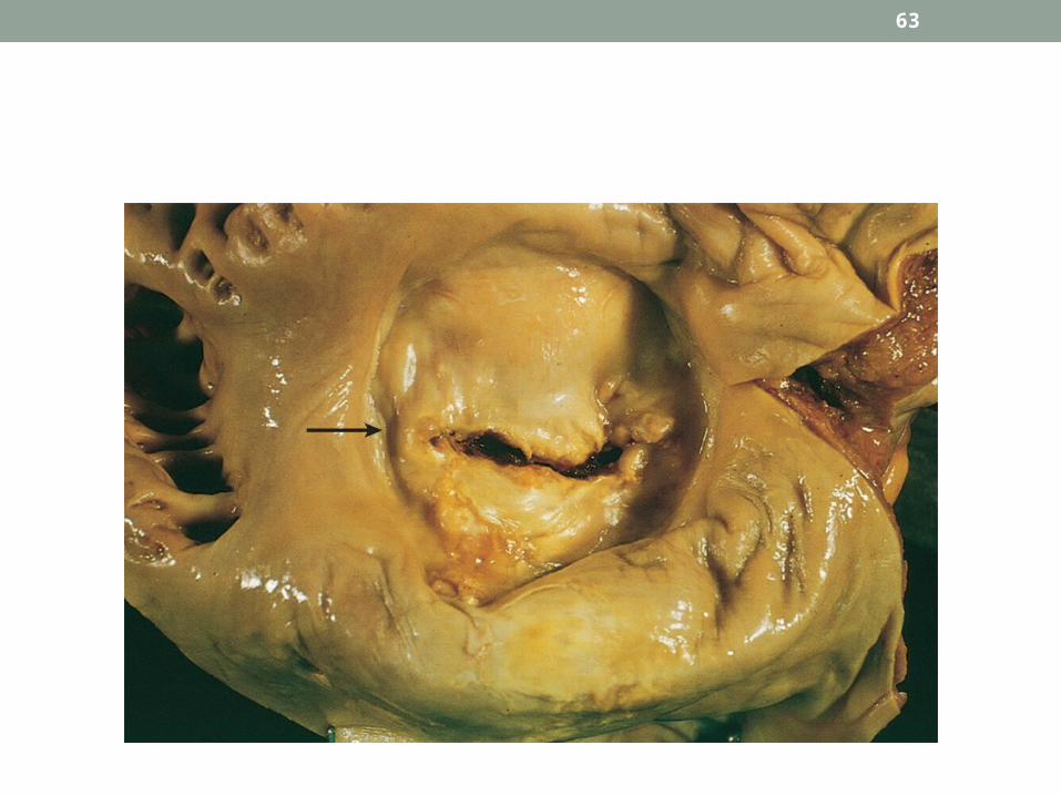
Anatomy of the Heart
•Heart valves and valve disorders•Semilunar (SL) valves
•Pulmonary semilunar: at the beginning of the pulmonary artery
•Aortic semilunar: at the beginning of the aorta
63

64
Heart Sounds• Two distinct heart sounds in every heartbeat or cycle—“lub-dup”
• First sound (lub) caused by the vibration and closure of AV valves during contraction of the ventricles
• Second sound (dup) caused by the closure of the semilunar valves during relaxation of the ventricles
• Heart murmurs—abnormal heart sounds often caused by abnormal valves

65
Conduction System of the Heart • Normal structure and function
• SA (sinoatrial) node, (the pacemaker)—located in the wall of the right atrium near the opening of the superior vena cava
• AV (atrioventricular) node—located in the right atrium along the lower part of the interatrial septum
• AV bundle (bundle of His)—located in the septum of the ventricle
• Purkinje fibers—located in the walls of the ventricles allows the ventricles to contract

66
Conduction System of the Heart
•Electrocardiography• The normal ECG has three deflections or waves • P wave—associated with depolarization of the atria
• QRS complex—associated with depolarization of the ventricles
• T wave—associated with repolarization of the ventricles

67
Conduction System of the Heart• Bradycardia—slow heart rate (less than 60 beats/min)
• Tachycardia—rapid heart rate (more than 100 beats/min)
• Sinus dysrhythmia—variation in heart rate during breathing cycle
• Premature contraction (extrasystole)—contraction that occurs sooner than expected in a normal rhythm
• Fibrillation—condition in which cardiac muscle fibers are “out of step,” producing no effective pumping action