Embed Size (px)
Citation preview
Module 3 HWSoléy ValenciaCBNS 130L
Friday, August 16, 2013

Abstract
By using, “My First Neuron,” I was able to observe: A, L, C, AHP, M, and T Currents. By varying voltage and current clamp in some instances as well as, ionic concentrations and conductances of different ions, I
was able to define each current’s behavior under both altered and normal circumstances.
Friday, August 16, 2013

Introduction
Connor and Stevens in 1971, discovered what is now known as the A-Current by conducting experiments on gastropods neuronal somata. They discovered this
potassium an exponential increase in action potentials as voltage clamp changed in increasing steps, where potassium flowed out of the cell. In 1980, Brown and Adams
found M-Current while studying frog sympathetic neurons and identified this potassium current as separate from the delayed rectifier. L-Current brings Calcium
into the cell. C, AHP, and T-Current are calcium dependent potassium currents.
Friday, August 16, 2013

Methods and MaterialsMy First Neuron Software
The A-Current: Observed the difference between default settings graph to a graph set to maximum conductance of iA=0. Ran a voltage clamp and then compared it to a graph with a voltage clamp series while having extracellular potassium concentration reduced to 25mM.
The L-Current and C-Current: Compared a graph with regular settings to a graph with extracellular calcium concentration set to 0.1mM and observed the changes in both L and C currents.
The AHP-Current: Compared a graph with regular default settings to a graph with maximum conductance of AHP-Current to 0. Then reduced extracellular [Ca++] to 0.1mM and compared it with the original graph.
The M-Current: Graphed default settings then compared this original graph another containing settings with maximum conductance of M-Current equal to 0. I then reduced [Ca++] out to 0.1mM and compared it with the original graph. I ran a voltage clamp on the M-Current while changing the extracellular concentration of potassium from 3.1mM to 0mM.
The T-Current: I graphed the first of my graphs with regular settings. I then set a graph with maximum conductance of sodium set to 0 and compared it with my original default graph. I then, changed the base current from -0.27nA to 0.18nA and changed the initial voltage from -85mV to -60mV. I compared these graphed changed with the graph set with extracellular calcium set to 0.1mM.
Friday, August 16, 2013
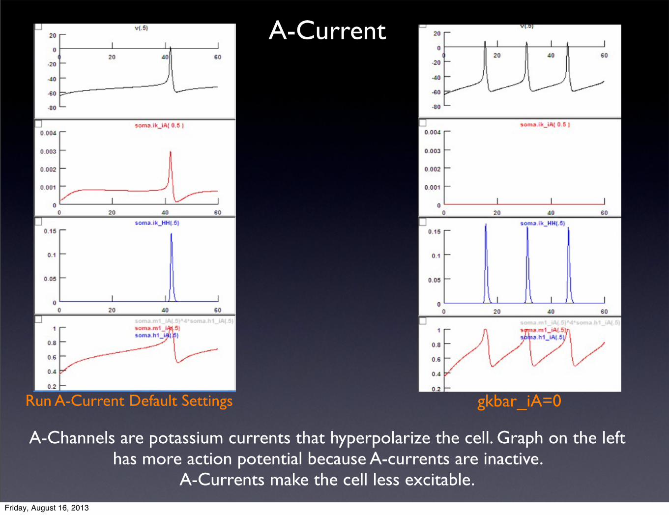
Run A-Current Default Settings gkbar_iA=0
A-Channels are potassium currents that hyperpolarize the cell. Graph on the left has more action potential because A-currents are inactive.
A-Currents make the cell less excitable.
A-Current
Friday, August 16, 2013

Run Voltage Clamp Series: A-Current
iA Default Settings [K+]out from 3.1mM to 25mM
A-Current is voltage dependent; the more voltage that is applied the more action potentials are observed. On the right, the applied voltage changes from -100mV to 0mV so that potassium moves out of the cell because it wants to reach the
equilibrium potential of potassium.
Friday, August 16, 2013
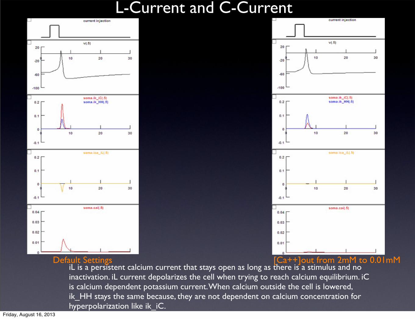
L-Current and C-Current
Default Settings [Ca++]out from 2mM to 0.01mMiL is a persistent calcium current that stays open as long as there is a stimulus and no inactivation. iL current depolarizes the cell when trying to reach calcium equilibrium. iC is calcium dependent potassium current. When calcium outside the cell is lowered, ik_HH stays the same because, they are not dependent on calcium concentration for hyperpolarization like ik_iC.
Friday, August 16, 2013

AHP Current
Default Settings
kgbar_iAHP=0 [Ca++]out from 2mM to 0mM
AHP current is calcium dependent potassium current that is in charge of spike frequencyadaptation. So, when iAHP=0, then we will see more action potentials because there isless potassium within the cell triggering hyperpolarization, in spite of having small amount of calcium within the cell. When calcium concentration outside the cell is lowered, there will still be a lot of action potential because less calcium inside thecell prevents potassium to come and hyperpolarize the cell leading to more action potentials.
Friday, August 16, 2013
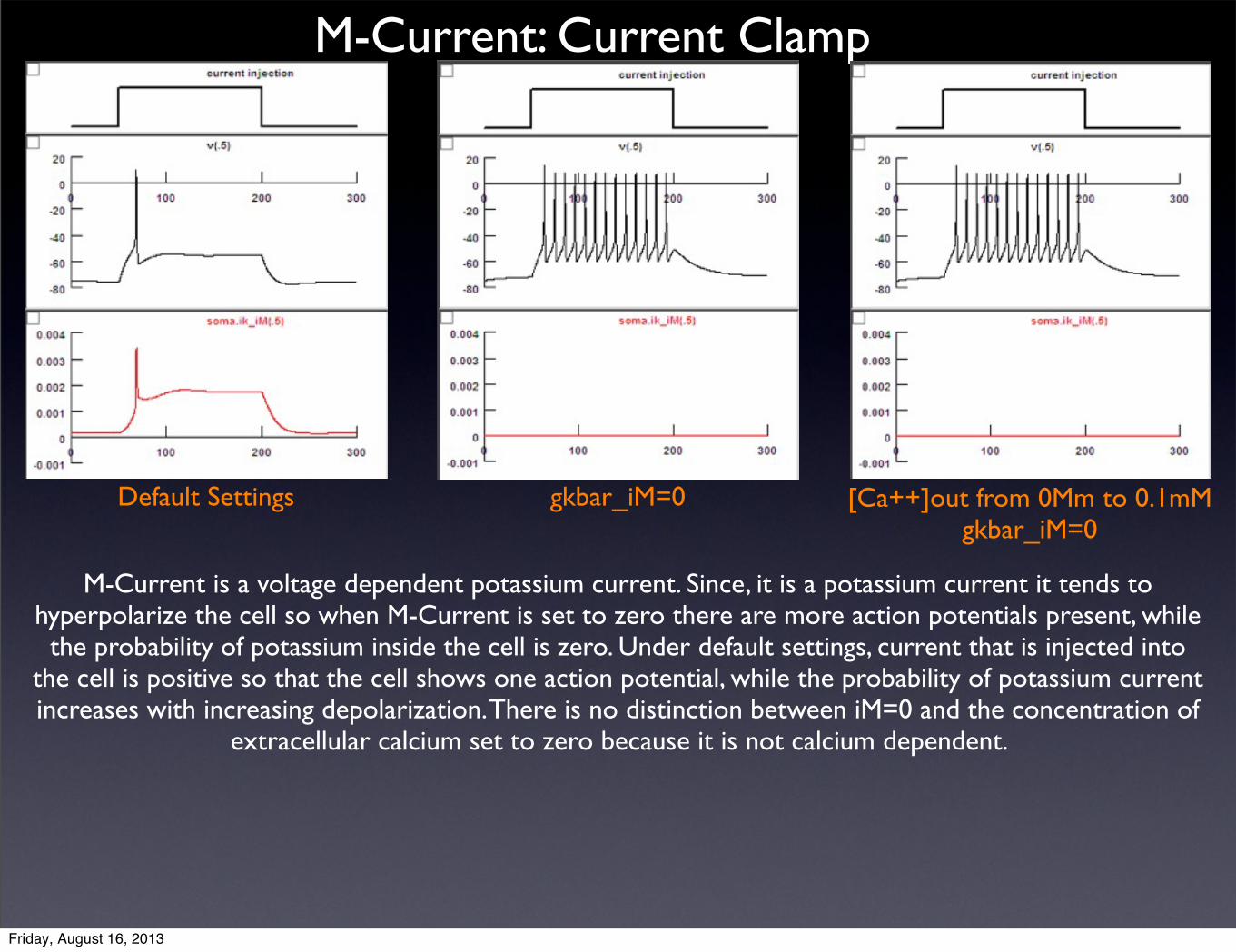
M-Current: Current Clamp
Default Settings gkbar_iM=0 [Ca++]out from 0Mm to 0.1mMgkbar_iM=0
M-Current is a voltage dependent potassium current. Since, it is a potassium current it tends to hyperpolarize the cell so when M-Current is set to zero there are more action potentials present, while the probability of potassium inside the cell is zero. Under default settings, current that is injected into
the cell is positive so that the cell shows one action potential, while the probability of potassium current increases with increasing depolarization. There is no distinction between iM=0 and the concentration of
extracellular calcium set to zero because it is not calcium dependent.
Friday, August 16, 2013
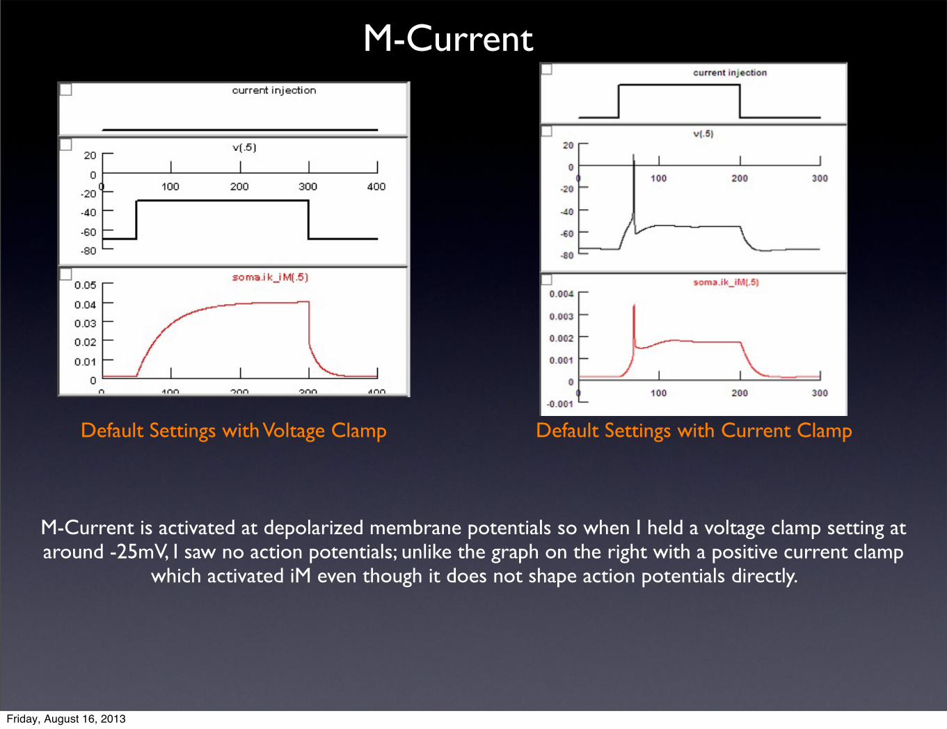
M-Current
Default Settings with Voltage Clamp Default Settings with Current Clamp
M-Current is activated at depolarized membrane potentials so when I held a voltage clamp setting at around -25mV, I saw no action potentials; unlike the graph on the right with a positive current clamp
which activated iM even though it does not shape action potentials directly.
Friday, August 16, 2013
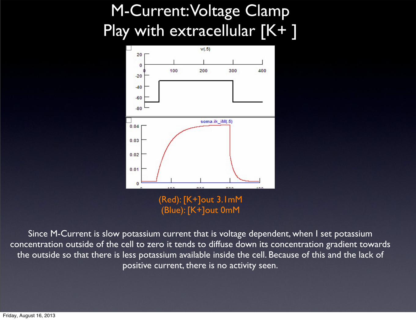
(Red): [K+]out 3.1mM(Blue): [K+]out 0mM
M-Current: Voltage ClampPlay with extracellular [K+ ]
Since M-Current is slow potassium current that is voltage dependent, when I set potassium concentration outside of the cell to zero it tends to diffuse down its concentration gradient towards
the outside so that there is less potassium available inside the cell. Because of this and the lack of positive current, there is no activity seen.
Friday, August 16, 2013

Current voltage is at -85mV a hyperpolarized state so T-Currents become activated. There is a Ca++ influx into the cell that helps the cell reach threshold where Na+ then undergoes
positive feedback leading to the burst of AP, influx of Ca++Leads to activation of Ca++ dependent Potassium Channels that hyperpolarize the cell afterwards.
Default T-Current Settings
Friday, August 16, 2013

These Action Potential bursts usually take place when Ca++ reaches close to threshold and sodium positive feedback of channels causes Burst of Action Potentials.Since, T-currents are not activated due to the initial voltage at -60mV, there are no action potential bursts. In the absence of sodium conductance the presence of these Action Potential bursts would be absent.
Changed gnabar_HH from 0.052 to 0
(Black): T-Current Default Settings(Red): gnabar_HH=0
(Organge): T-Current Default (Green): gnabar_HH=0
(Red & Blue): T-Current Default( Yellow): gnabar_HH=0
Friday, August 16, 2013
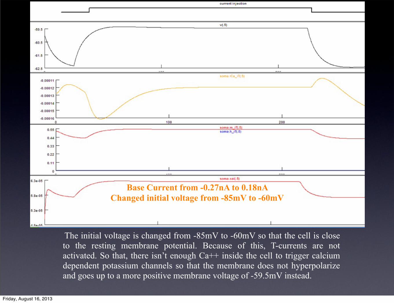
Base Current from -0.27nA to 0.18nA Changed initial voltage from -85mV to -60mV
The initial voltage is changed from -85mV to -60mV so that the cell is close to the resting membrane potential. Because of this, T-currents are not activated. So that, there isn’t enough Ca++ inside the cell to trigger calcium dependent potassium channels so that the membrane does not hyperpolarize and goes up to a more positive membrane voltage of -59.5mV instead.
Friday, August 16, 2013

The cell is at a resting state of around -63.5mV. By decreasing Ca++ concentration outside the cell, there will be a smaller driving force for Ca++ influx into the cell when
T-Currents become activated.
Reduce external concentration of calcium from 2mM to 0.1mM
Friday, August 16, 2013
Discussion- In A-Currents are potassium currents so if iA=0 there is more action potentials. Even if I were to block the channel, activation and deactivation gates would remain present.
- L-Current is a persistent current that lets calcium inside the cell. Once calcium is inside the cell, it can then help modulate other channels such as, C and AHP currents. AHP is calcium dependent potassium current.
- C-Current is calcium dependent potassium channel. Once calcium is inside the cell it tries to reach calcium equilibrium which pushes the membrane potential close to threshold where sodium channels act as positive feedback and start firing. At this more depolarized membrane potential potassium channels are activated leading to the hyperpolarization of the cell.
- iK_HH does not depend on calcium, but it does reflect the presence of other potassium currents because its a voltage dependent and other potassium currents bring the membrane potential to a more hyperpolarized state, making it harder for iK_HH to activate.
- When M-Current is blocked there is a lot of action potentials. M-Current, acetylcholine binds, which activates muscarinic receptors that block M-Current. iM activated when current shows depolarization for a long time in which time, iM can modulate long term neuronal states.
- When T-Current is equal to zero, we don’t have Ca++ spikes but, we still get depolarization even though it does not reach threshold so that we observe no bursts.
Friday, August 16, 2013

Literature Cited
"Result Filters." National Center for Biotechnology Information. U.S. National Library of Medicine, n.d. Web. 16 Aug. 2013.
"Ionic Currents in Molluscan Soma." - Annual Review of Neuroscience, 3(1):141. N.p., n.d. Web. 16 Aug. 2013.
Friday, August 16, 2013