Embed Size (px)
Citation preview
This document is downloaded at: 2019-04-09T13:57:04Z
Title Probable Noonan syndrome in a boy without PTPN11 mutation,manifesting unusual complications.
Author(s) Sumi, Muneichiro; Ohno, Yasuharu; Sasaki, Rie; Kondoh, Tatsuro;Tagawa, Masato; Masuzaki, Hideaki; Moriuchi, Hiroyuki
Citation Pediatrics international, 51(1), pp.138-140; 2009
Issue Date 2009-02
URL http://hdl.handle.net/10069/23340
Right Journal compilation © 2010 Japan Pediatric Society
NAOSITE: Nagasaki University's Academic Output SITE
http://naosite.lb.nagasaki-u.ac.jp
PED-00163-2005.R2
Title page
A case of probable Noonan syndrome boy without PTPN11 mutation, manifesting
unusual complications
Muneichiro Sumi, MD.1 ,Yasuharu Ohno, MD,PhD.2 Rie Sasaki, MD, PhD.3,
Tatsuro Kondoh, MD,PhD.1, Masato Tagawa, MD.1, Hideaki Masuzaki, MD, PhD.4,
Hiroyuki Moriuchi, MD, PhD.1
1 Department of Pediatrics, Nagasaki University School of Medicine, Nagasaki, Japan
2 Department of Surgery II, Nagasaki University School of Medicine, Nagasaki, Japan
3 Department of Endocrinology and Metabolism, National Research Institute for Child
Health and Development, Tokyo, Japan
4 Department of Obstetrics and Gynecology, Nagasaki University School of Medicine,
Nagasaki, Japan
Corresponding to; Muneichiro Sumi MD.
Department of Pediatrics, National Hospital Organization Nagasaki Medical Center,
2-1101-1 Kubara, Ohmura, Nagasaki 856-8562, Japan.
Tel: +81-957-52-3121 Fax: +81-957-54-0292 e-mail:[email protected]

Introduction
Noonan syndrome (NS) (MIM No.163950) is autosomal dominant disorder
characterized by short stature, cardiovascular defects, and dysmorphological features,
including hypertelorism, a webbed neck, a pectus excavatum and cuvitus valgus. Mental
retardation and hearing difficulty are often observed in affected individuals.
Approximately 40% of NS patients have Protein-Tyrosine Phosphatase, Nonreceptor
type 11 (PTPN11) mutations.1 The genotypic-phenotypic correlation and phenotypical
heterogeneity between the two genotypes have been reported.1 Pulmonary valve stenosis
is more frequent in PTPN11 mutation-positive patients than mutation-negative patients,1
whereas hypertrophic cardiomyopathy is more frequent in PTPN11 mutation-negative
patients than mutation-positive patients. Gastrointestinal complications are very few
among NS patients. Double outlet right ventricle (DORV) and congenital rectal atresia
have never been associated with the PTPN11 mutation –negative NS patients.
We here report a Japanese boy with DORV and rectal atresia. He fulfilled clinical
criteria for NS and had no PTPN11 gene mutation.
Case presentation
The patient, a newborn Japanese boy, is the first child of non-consanguineous parents.
There are no other patients with mental retardation or dysmorphological problems in his
family.Both parents were 33 years old, when the boy was born. The mother was
primipara. Ultrasound examination of the fetus revealed intrauterine growth retardation,
distended digestive tract, cardiac dilatation and pericardial effusion at 33 weeks of his
gestational age. The mother underwent emergent caesarian section at 34 gestational
weeks due to non re-assuring fetal states.
Apgar scores were 7 and 9 at 1and 5 minutes after birth, respectively. His birth weight
was 1,806 g (below the 10th percentile), length 40.5 cm (below the 10th percentile), and
occipitofrontal head circumference 29.0 cm (the 10-50th percentile). Physical
examinations revealed broad forehead, downward eye slants, epicanthal folds, ptosis,
low set posterior rotated ears, cleft velum, small jaw, a short neck without webbing of
skin, pectus carinatus, distended abdomen, small anal dimple with sphincter ani and
bilateral hydrocele. He had no bleeding diasthesis, choanal atresia and coloboma.
His complete blood cell counts and biochemical tests showed as follows: WBC 14.2
x103/ul (normal range; 5.0-19.0) with Ba 1 Eo 2 St 2 Seg 57 Ly 32 and Mo 6,
erythroblasts 10/100wbc, RBC 4.63 x10⁶/ul (4.77-5.93), hemoglobin 14.2g/dl (14-22),
hematocrit 53.1% (53.5-62.3), platelets 239 x103/ul (80-356), Na 140 mEq/l (133-146),
K 6.5 mEq/l (4.6-6.7), Cl 100 mEq/l (100-117), Ca 10.6 mg/dl (6.1-11.6) , inorganic
phosphate 5.2 mg/dl (5.4-10.9), BUN 8 mg/dl (3.1-25.5), CRP 0.04mg/dl (<0.30), total
bilirubin 0.3mg/dl (3.8-17.7), AST 59 IU/l (11-59), ALT 23 IU/l (4-21), LDH 384 IU/l
(364-1120), and γ-glutamayl transpeptidase 152 IU/l (1-45).
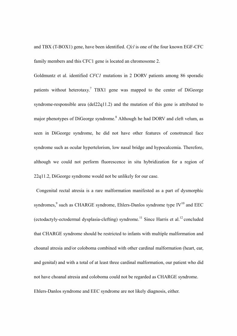
Chest X-ray showed widening of cardiac shadow and normal pulmonary blood flow.
Plain abdominal X-ray showed small amount of digestive tract air with no gases in the
pelvic region (Fig.1). Echocardiography found DORV with patent ductus arteriosus
(PDA). Ultrasound examination of the abdomen revealed enlarged colon with massive
calcificated debris. His abdominal distention was so severe, as to require emergent
colostomy on day 0, and further investigation for his large intestinal obstruction was
performed thereafter. Barium contrast fluoroscopy found interruption of his rectum
(Fig.2) and abdominal magnetic resonance imaging (MRI) revealed no tumors or
masses around the rectum (Fig.3), indicating simple congenital rectal atresia.
He is now 18 months of age. His body weight is 5,320 g (below the 3rd percentile),
length 63.6 cm (below the 3rd percentile), and occipitofrontal head circumference 42.0
cm (below the 3rd percentile). He has no hematological problems. He cannot sit without
support, nor speak at all. His present developmental quotient is around 40.
As he had typical facial anomalies, cardiac defects, short stature, pectus carinatus and
mental retardation, he was diagnosed as NS from the clinical criteria by van der Burgt et
al.2
Since DORV and rectal atresia were quite unusual complications of NS, molecular
analysis of PTPN11 gene was performed with an informed consent from his parents.
The leukocyte genomic DNA of the patient was amplified by PCR for all 15 exons and
flanking introns of the PTPN11 gene Subsequently, the PCR products were subjected to
direct sequencing from both directions on a CEQ 8000 autosequencer (Beckman Coulter,
Fullerton, CA; www.beckman.com). The primer sequences and the PCR condition were
previously described by Yoshida et al.3
Direct sequencing results revealed no mutations in all 15 exons or intronic regions
adjacent to splicing junctions.Therefore, he was classified into the PTPN11
mutation-negative NS patient.
Discussion
In this report, we presented a Japanese boy who was diagnosed as NS based on the
clinical criteria by van der Burgt et al.2, however, he was unique in that he was also
complicated with DORV and rectal atresia.
PTPN11, which encodes the non-receptor type protein tyrosine phosphatase SHP-2
(src homology region 2-domain phosphatase-2), was identified as NS1 disease gene.1
Epidermal growth factor (EGF) signaling is important for semilunar valve development
and SHP-2 is a component of the EGF-mediated signal transduction pathway.4 A ptpn11
knockout allele in heterozygosity results in a higher prevalence and increased severity
of thickened aortic and pulmonary valve leaflets. 1
According to the previous reports of Japanese NS patients, pulmonary vulvar stenosis
(PS) and atrial septum defect (ASD) were frequently, and hematological abnormalities
were exclusively present in mutation-positive patients.3,5,6 Since our patient was not
complicated with PS or ASD and had no hematological abnormality, his clinical features
were not incompatible of PTPN11 mutation-negative NS. Curiously, however, his
cardiac defect was DORV, not hypertrophic cardiomyopathy that has been frequently
associated with PTPN11 mutation-negative NS. Recently, cardiac genes for human
conotruncal heart malformations, the EGF-CFC (Cripto, Frll, and Cryptic) gene family
and TBX (T-BOX1) gene, have been identified. Cfcl is one of the four known EGF-CFC
family members and this CFC1 gene is located an chromosome 2.
Goldmuntz et al. identified CFC1 mutations in 2 DORV patients among 86 sporadic
patients without heterotaxy.7 TBX1 gene was mapped to the center of DiGeorge
syndrome-responsible area (del22q11.2) and the mutation of this gene is attributed to
major phenotypes of DiGeorge syndrome.8 Although he had DORV and cleft velum, as
seen in DiGeorge syndrome, he did not have other features of conotruncal face
syndrome such as ocular hypertelorism, low nasal bridge and hypocalcemia. Therefore,
although we could not perform fluorescence in situ hybridization for a region of
22q11.2, DiGeorge syndrome would not be unlikely for our case.
Congenital rectal atresia is a rare malformation manifested as a part of dysmorphic
syndromes,9 such as CHARGE syndrome, Ehlers-Danlos syndrome type IV10 and EEC
(ectodactyly-ectodermal dysplasia-clefting) syndrome.11 Since Harris et al.12 concluded
that CHARGE syndrome should be restricted to infants with multiple malformation and
choanal atresia and/or coloboma combined with other cardinal malformation (heart, ear,
and genital) and with a total of at least three cardinal malformation, our patient who did
not have choanal atresia and coloboma could not be regarded as CHARGE syndrome.
Ehlers-Danlos syndrome and EEC syndrome are not likely diagnosis, either.
Our patients fulfilled clinical criteria for NS but had rare complications such as DORV
and congenital rectal atresia. Very few malformation syndrome manifest both of the
above two conditions, however, none of them is consistent with our patient.
Furthermore, DORV and rectal atresia has never been associated with each other from
molecular and cytogenetic aspects. Accumulation and investigation of similar cases
from clinical, molecular and cytogenetic aspects will be needed for better understanding
of possible association of NS-like phenotypes with DORV and rectal atresia.
References
1. Tartaglia M, Kalidas L, Shaw A, et al. PTPN11 mutations in Noonan syndrome:
Molecular spectrum, Genotype-phenotype correlation, and phenotypic heterogeneity.
Am J Hum Genet 2002; 70:1555-1563
2. van der Burgt I, Berends E, Lommen E, van Beersum S, Hamel B, Mariman E.
Clinical and molecular studies in a large Dutch family with Noonan syndrome. Am J
Med Genet 1994; 53:187-191
3. Yoshida R, Hasegawa T, Hasegawa Y, et al. Protein-tyrosine phosphatase,
nonreceptor type 11 mutation analysis and clinical assessment in 45 patients with
Noonan syndrome. J Clin Endocrinol Metab 2004; 89:3359-3364
4. Qu C-K, Yu W-M, Azzarelli B, et al. Genetic evidence that Shp-2 tyrosine
phosphatase is a signal enhancer of the epidermal growth factor receptor in mammals.
Proc Natl Acad Sci USA 1999; 96:8528-8533
5. Kosaki K, Suzuki T, Muroya K, et al. PTPN11 (Protein-tyrosine phosphatase,
nonreceptor type 11) mutations in seven Japanese patients with Noonan syndrome. J
Clin Endocrinol Metab 2002; 87:3529-3533
6. Niihori T, Aoki Y, Ohashi H, et al. Function analysis of PTPN11/SHP-2 identified in
Noonan syndrome and childhood leukemia. J Hum Genet 2005;50:192-202

7. Goldmuntz E, Bamford R, Karkera JD, et al. CFC1 mutation in patients with
Transposition of the Great Arteries and Double-Outlet Right Ventricle. Am J Hum Genet
2002; 70:776-780
8. Chieffo C, Garvey N, Gong W, et al. Isolation and characterization of gene from
DiGeorge chromosomal region homologous to the mouse Tbx1 gene. Genomics 1997;
43:267-277
9. Endo A, Hayashi A, Ishihara I, et al. Analysis of 1,992 patients with anorectal
malformations over the past to decades in Japan. J Pediatr Surg 1999; 34: 435-441
10. Fuchs JR, Fishman SJ. Management of spontaneous colonic perforation in
Ehlers-Danlos syndrome Type IV. J Pediatr Surg 2004; 39 (Suppl 3):E13
11. Mjewski F, Goecke T. Rectal atresia as rare manifestation in ECC syndrome. Am J
Med Genet 1996; 63:190-192
12. Harris J, Robert E, Kallen B. Epidemiology of choanal atresia with special reference
to the CHARGE association. Pediatrics 1997; 99:363-367
Figure legends
Fig.1 Plain X-lay of the chest and the abdomen, showing widening of cardiac shadow
with normal pulmonary blood flow, and small amount of digestive tract air with no gass
in the pelvic region.
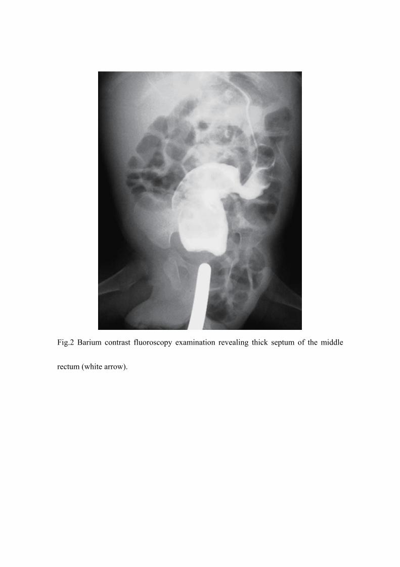
Fig.2 Barium contrast fluoroscopy examination revealing thick septum of the middle
rectum (white arrow).
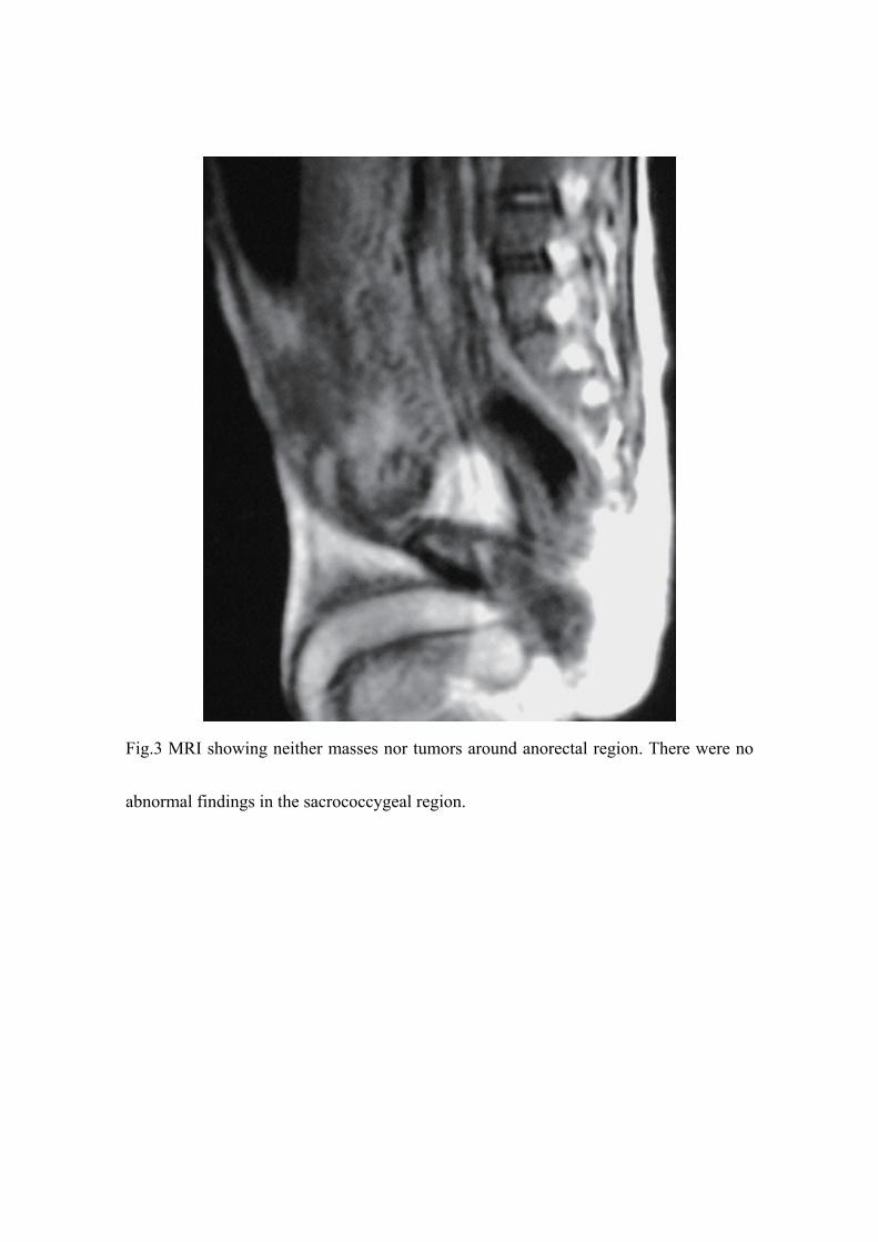
Fig.3 MRI showing neither masses nor tumors around anorectal region. There were no
abnormal findings in the sacrococcygeal region.


![Quantification of systemic right ventricle by echocardiography · 2017-02-26 · of systemic right ventricle by echocardiography ... with dobutamine stress [17]. These data were confirmed](https://img.pdfslide.tips/doc/110x75/5ecb2f51d4cb202a22168cb3/quantification-of-systemic-right-ventricle-by-echocardiography-2017-02-26-of-systemic.jpg)