Embed Size (px)
Citation preview
This document is downloaded at: 2018-09-01T03:12:03Z
Title Mannose-recognition mutant of the galactose/N-acetylgalactosamine-specific C-type lectin CEL-I engineered by site-directed mutagenesis
Author(s) Moriuchi, Hiromi; Unno, Hideaki; Goda, Shuichiro; Tateno, Hiroaki;Hirabayashi, Jun; Hatakeyama, Tomomitsu
Citation Biochimica et Biophysica Acta (BBA) - General Subjects, 1850(7),pp.1457-1465; 2015
Issue Date 2015-07
URL http://hdl.handle.net/10069/35445
Right© 2015 Elsevier B.V. Licensed under the Creative Commons Attribution-NonCommercial-NoDerivatives 4.0 Internationalhttp://creativecommons.org/licenses/by-nc-nd/4.0/
NAOSITE: Nagasaki University's Academic Output SITE
http://naosite.lb.nagasaki-u.ac.jp
1
Mannose-recognition mutant of the galactose/N-acetylgalactosamine-specific C-type lectin
CEL-I engineered by site-directed mutagenesis
Hiromi Moriuchi a, Hideaki Unno a, Shuichiro Goda a, Hiroaki Tateno b, Jun Hirabayashi b,
and Tomomitsu Hatakeyama a,*
a Laboratory of Biomolecular Chemistry, Graduate School of Engineering, Nagasaki University, 1-14
Bunkyo-machi, Nagasaki 852-8521, Japan b Research Center for Stem Cell Engineering, National Institute of Advanced Industrial Science and
Technology, Tsukuba 305-8568, Japan
Keywords: carbohydrate; C-type lectin; site-directed mutagenesis; X-ray crystallography
-----
Abbreviations: CD, circular dichroism; CRD, carbohydrate-recognition domain; CTLD, C-type
lectin-like domain; EDTA, ethylenediamine tetraacetate; PAA, polyacrylamide; PD, polyamidoamine
dendrimer; MBL, mannose-binding lectin; TBS, Tris-buffered saline; WT, wild-type.
The atomic coordinates and structure factors for the CEL-I/mannose complex have been
deposited in the Protein Data Bank, www.pdb.org (PDB ID: 4WQQ).
* Corresponding author: Biomolecular Chemistry Laboratory, Graduate School of Engineering,
Nagasaki University, 1-14 Bunkyo-machi, Nagasaki 852-8521, Japan. Tel: +81-95-819-2686; Fax:
+81-95-819-2686
E-mail address: [email protected]
2
Abstract
Background: CEL-I is a galactose/N-acetyl-galactosamine-specific C-type lectin isolated from the sea
cucumber Cucumaria echinata. Its carbohydrate-binding site contains a QPD (Gln-Pro-Asp) motif,
which is generally recognized as the galactose specificity-determining motif in the C-type lectins. In
our previous study, replacement of the QPD motif by an EPN (Glu-Pro-Asn) motif led to a weak
binding affinity for mannose. Therefore, we examined the effects of an additional mutation in the
carbohydrate-binding site on the specificity of the lectin.
Methods: Trp105 of EPN-CEL-I was replaced by a histidine residue using site-directed mutagenesis,
and the binding affinity of the resulting mutant, EPNH-CEL-I, was examined by sugar-
polyamidoamine dendrimer assay, isothermal titration calorimetry, and glycoconjugate microarray
analysis. Tertiary structure of the EPNH-CEL-I/mannose complex was determined by X-ray
crystallographic analysis.
Results: Sugar-polyamidoamine dendrimer assay and glycoconjugate microarray analysis revealed a
drastic change in the specificity of EPNH-CEL-I from galactose/N-acetyl-galactosamine to mannose.
The association constant of EPNH-CEL-I for mannose was determined to be 3.17 × 10³ M−1 at 25°C.
Mannose specificity of EPNH-CEL-I was achieved by stabilization of the binding of mannose in a
correct orientation, in which the EPN motif can form proper hydrogen bonds with 3- and 4-hydroxy
groups of the bound mannose.
Conclusions: Specificity of CEL-I can be engineered by mutating a limited number of amino acid
residues in addition to the QPD/EPN motifs.
General significance: Versatility of the C-type carbohydrate-recognition domain structure in the
recognition of various carbohydrate chains could become a promising platform to develop novel
molecular recognition proteins.
1. Introduction
Lectins (carbohydrate-binding proteins) are known to play important roles in molecular and
cellular recognition systems in organisms. While many plant lectins have been isolated from storage
tissues such as seeds and tubers, animal lectins have been found in various tissues and body fluids,
reflecting their diverse structures and functions. Among the animal lectin families, C-type lectins,
which were named because of their Ca2+ dependence, contain C-type carbohydrate-recognition
3
domains (CRDs)2 usually composed of 110–130 amino acid residues, which share a common amino
acid sequence homology as well as similar tertiary structures [1-3]. While the C-type CRDs often
function as carbohydrate-recognition modules in cooperation with distinct domains having different
biological activities [3], particularly in immune systems [4-8], invertebrate organisms also utilize C-
type lectins in the innate immune system [9]. We have been investigating three Ca2+-dependent lectins,
CEL-I, CEL-III, and CEL-IV, from the sea cucumber Cucumaria echinata [10]. Among these, CEL-III
is a unique ricin-type (R-type) lectin that exerts strong hemolytic and cytotoxic activities by forming
membrane pores in target cells [11], whereas CEL-I and CEL-IV belong to the C-type lectins [12, 13].
These lectins may play important roles in defense against foreign organisms through their cytotoxicity
or opsonization by binding to surface carbohydrates.
The carbohydrate-binding site of many C-type CRDs contains the sequence motifs QPD (Gln-
Pro-Asp) and EPN (Glu-Pro-Asn), which are known to be closely related to galactose or mannose
specificities. Strong evidence for this has been demonstrated by the site-directed mutagenesis study on
the mannose-binding lectin MBP-A [14], in which its EPN motif was changed to a QPD motif. The
resulting mutant exhibited galactose specificity, although it was relatively weak. In addition, the
replacement of the residues on the basis of the structures of other galactose-binding CRDs resulted in a
much increased affinity for galactose or N-acetylgalactosamine [15, 16], suggesting the importance of
stabilization of the bound carbohydrates by surrounding residues in the binding site. Other reports
have also shown alterations in the specificity of the C-type CRDs in surfactant proteins from mannose
to galactose by changing the EPN motif to a QPD motif [17, 18]. However, to our knowledge, no
report has described a specificity change from galactose to mannose in the C-type CRDs. We have
previously described that the site-directed mutant EPN-CEL-I, in which the EPN motif (residues 101–
103) was replaced by a QPD motif, exhibited relatively weak mannose specificity, although there was
a substantial affinity for galactose [19]. This observation also suggested that the specificities of the C-
type CRDs are not only exclusively specified by the EPN or QPD motifs but also largely supported by
other residues situated nearby in the binding site, as suggested by the MBP-A mutants [15]. In the
present study, we have prepared another CEL-I mutant, in which Trp105 in the binding site of EPN-
CEL-I was replaced by a histidine residue (EPNH-CEL-I), to further develop an engineered CEL-I
with enhanced mannose specificity. X-ray crystallographic analysis of the EPNH-CEL-I/mannose
complex also revealed the mannose-recognition mechanism of EPNH-CEL-I.
2. Materials and methods
4
2.1. Materials
Oligonucleotides used in this study were purchased from Sigma-Aldrich. The plasmid vectors
pTAC-2 and pET-3a were obtained from DynaExpress and Novagen, respectively. Polyamidoamine
dendrimers (PDs) with 64 amino surface groups (ethylenediamine core, generation 4.0, M.W. 14,214)
(amino-PD) were obtained from Sigma-Aldrich. All the other chemicals were of analytical grade for
biochemical use. Carbohydrate-immobilized Cellufine (mannose-Cellufine and GalNAc-Cellufine)
columns were prepared by attaching carbohydrates to Cellufine gels (JNC Corp., Tokyo, Japan) using
the cross-linking reagent divinyl sulfone (Sigma-Aldrich), as described previously [10].
2.2. Expression and purification of the recombinant CEL-I
The gene for EPNH-CEL-I (Supplementary figure) was synthesized by polymerase chain reaction
(PCR) using the EPN-CEL-I gene [19] as a template and the primers listed in Supplementary table.
PCR amplification was performed using the primers W105H-F and W105H-R, which contained the
mutation site His105, and the primers corresponding to the 5′- and 3′-ends of the EPN-CEL-I gene
(CEL-I-F and CEL-I-R), which contained the restriction sites NdeI and BamHI. After amplification of
the 5′-terminal and 3′-terminal DNA fragments using combinations of the primers CEL-I-F/W105H-R
and W105H-F/CEL-I-R, the fragments were ligated and amplified using CEL-I-F and CEL-I-R in a
similar manner as that reported previously for EPN-CEL-I [19]. The resulting DNA encoding EPNH-
CEL-I was cloned into Escherichia coli JM109 cells using the pTAC2 vector. The inserted EPNH-
CEL-I gene was digested with the restriction enzymes NdeI and BamHI and then ligated with the pET-
3a vector previously digested with the same enzymes. The resulting plasmid was introduced into E.
coli BL21(DE3)pLysS cells (Novagen), and expression of the protein was induced with 0.4 mM
isopropylthiogalactoside. Because the recombinant proteins were obtained as inclusion bodies after
disrupting the cells by sonication, they were solubilized in the solubilization buffer [50 mM Tris–HCl,
pH 8.0; 0.2 M NaCl; 1 mM ethylenediamine tetraacetate (EDTA); and 6 M guanidine hydrochloride]
and the protein was refolded in the refolding buffer (0.1 M Tris–HCl, pH 8.0; 0.4 M L-arginine; 2 mM
EDTA; 5 mM reduced glutathione; 0.5 mM oxidized glutathione; and 0.1 mM phenylmethylsulfonyl
fluoride). After dialysis of the refolded proteins in Tris-buffered saline (TBS; 10 mM Tris–HCl, pH
7.5; 0.15 M NaCl) containing 10 mM CaCl2, the protein was purified by affinity chromatography
5
using the mannose-Cellufine column (3 × 10 cm). Wild-type- (WT-) and EPN-CEL-I were also
expressed in an essentially similar manner, although purification was performed using GalNAc-
Cellufine (for WT-CEL-I) or ion-exchange chromatography (for EPN-CEL-I) [19].
2.3. N-Terminal amino acid sequence analysis
The N-terminal amino acid sequence of the expressed protein was determined using a protein
sequencer, PPSQ-21 (Shimadzu, Kyoto, Japan).
2.4. Circular dichroism (CD) measurements
Far-UV CD measurements of the proteins were performed within the range of 200–250 nm using
a J-720 spectropolarimeter (JASCO). The spectra were measured using a quartz cell of 1-mm path
length at 20°C in TBS containing 10 mM CaCl2.
2.5. Measurement of carbohydrate-binding activity using sugar-PDs
Mannan and mannotriose were prepared as reported previously [20]. Sugar-PDs containing
mannan or mannotriose were prepared by a reductive amination reaction between an aldehyde group
of the sugars and primary amino groups of amino-PD, as described previously [21]; carbohydrates
(110 µM) were incubated with amino-PD (0.17 µM) in 1 ml of 0.2 M sodium phosphate buffer (pH
8.0) in the presence of 110 µM NaBH3CN for 24 h at 45°C. The solution was then dialyzed against
water to remove the residual reagents, and the resulting sugar-PDs were collected after freeze-drying.
The carbohydrate-binding ability was evaluated by the increase in Rayleigh scattering on the basis of
the complex formation between the lectin and sugar-PD. After recording the initial scattering intensity
of the lectin solution at 420 nm (20 µg/ml, 1 ml) in TBS containing 10 mM CaCl2 using a fluorescence
spectrophotometer (Hitachi F-3010) at 25°C, small volumes of the sugar-PD solution in the same
buffer were serially added, and the changes in the scattering intensity were recorded. Values were
corrected for dilution by the addition of the sugar-PD solution. For the competitive experiments,
various sugars were added to the pre-formed complex between EPNH-CEL-I (25 μg/mL) and
mannotriose-PD (1.2 μg/mL). Dissociation of the EPNH-CEL-I/mannotriose-PD complex was
evaluated by the changes in the light scattering intensity at 420 nm.
6
2.6. Isothermal titration calorimetry (ITC)
ITC for analyzing the interaction between EPNH-CEL-I and mannose was performed at 25°C
using iTC 200 (MicroCal iTC200, GE Healthcare). Aliquots of the mannose solution (3.8 mM) were
injected into the EPNH-CEL-I (6.1 mg/ml, 0.19 mM) solution in TBS containing 10 mM CaCl2. The
data were analyzed using ORIGIN software, version 7.0.
2.7. Glycoconjugate microarray analysis
Binding specificities of WT-CEL-I and EPNH-CEL-I for various glycoconjugates were examined
by glycoconjugate microarray analysis, as described previously [22]. In brief, Cy3-labeled lectins were
incubated overnight with the microarray, on which various glycoconjugates had been immobilized, in
TBS containing 10 mM CaCl2. After washing with the same buffer, the fluorescence intensity of the
bound lectins was recorded.
2.8. Crystallization and X-ray crystallographic analysis of the EPNH-CEL-I/mannose complex
EPNH-CEL-I was crystallized by the hanging-drop and sitting-drop vapor diffusion methods. The
initial screening was performed using the screening kits Crystal Screen 1 and 2 (Hampton Research,
Aliso Viejo, CA, USA) and Wizard I and II (Emerald BioSystems, Inc., Bedford, MA, USA). In the
initial screening of crystallization, 1 µl of the protein solution (10 mg/ml) in TBS containing 10 mM
CaCl2 was mixed with an equal volume of reservoir solution and allowed to equilibrate against 100 µl
of the reservoir solution at 20°C. X-ray diffraction data were collected at −140°C using the Rigaku
MicroMax-007 HF/Raxis IV++ System. Data indexing, integration, and scaling were performed with
the CCP4 [23] programs Mosflm [24] and SCALA [25]. Data collection statistics are summarized in
Table 1. All the data sets belonged to the P21 space group, with two EPNH-CEL-I molecules per
asymmetric unit. The structures of the EPNH-CEL-I/mannose complex were determined by the
molecular replacement method using native CEL-I (PDB ID: 1WMY) [12] as the search model.
Molecular replacement was performed using the program Phaser [26]. The model was refined using
the program Refmac [27] from the CCP4 suite. Manual fitting of the model was performed by the
program Coot [28]. The quality of the final model was assessed by Ramachandran plot and the
7
program Procheck [29]. The refinement statistics are summarized in Table 1. Figures illustrating the
protein models were prepared by PyMOL [30].
2.9. Affinity chromatography of CEL-I mutants having different amino acid residues at position 105
Genes for two additional EPN-CEL-I mutants, in which Trp105 was replaced by tyrosine (EPNY-
CEL-I) or alanine (EPNA-CEL-I), were prepared using the QuikChange site-directed mutagenesis kit
(Agilent, Santa Clara, CA) using the primers listed in Supplementary Table and the pET-3a vector
containing the EPN-CEL-I gene as a template. Expression and refolding were performed in the same
manner as those for EPNH-CEL-I. The resulting mutants, as well as WT-, EPN-, and EPNH-CEL-I,
were applied to mannose-Cellufine and GalNA-Cellufine columns (2 × 2 cm) in TBS containing 10
mM CaCl2, and after washing the columns with the same buffer, adsorbed proteins were eluted with
TBS containing 20 mM EDTA.
3. Results
3.1. Expression and purification of EPNH-CEL-I
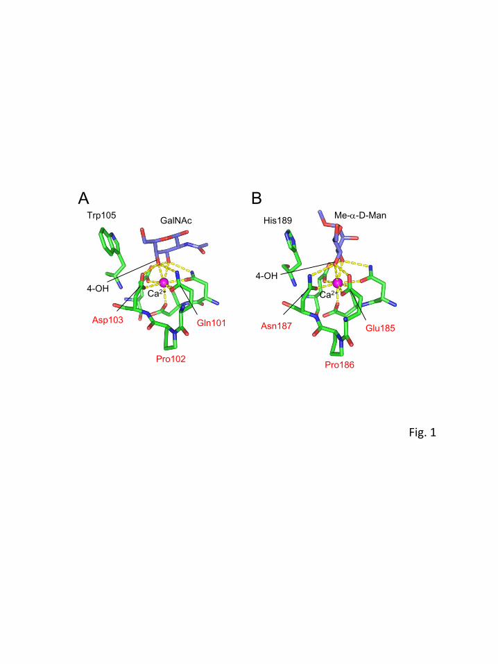
Figure 1 shows the structures of the carbohydrate-binding sites of CEL-I/N-acetylgalactosamine
and MBP-A/methyl α-D-mannoside complexes as examples for galactose- and mannose-specific C-
type lectins, respectively [12, 31]. They exhibit completely different specificities toward galactose and
mannose despite possessing very similar structures around Ca2+. This can be partly explained by the
recognition of the orientation of 4-hydroxy groups (axial in galactose and equatorial in mannose) by
the QPD or EPN motifs. However, even after changing the motif to EPN, the mutant CEL-I (EPN-
CEL-I) showed very weak affinity, suggesting that additional interactions by other residue(s) are
necessary for the recognition of mannose [19]. Therefore, we tried to replace Trp105 in CEL-I with a
histidine residue on the basis of the comparison with MBP-A. The gene for the mutant CEL-I (EPNH-
CEL-I) was synthesized by PCR using the primers containing the mutation site His105, as described in
the Materials and methods. Because the expressed protein was recovered in inclusion bodies, they
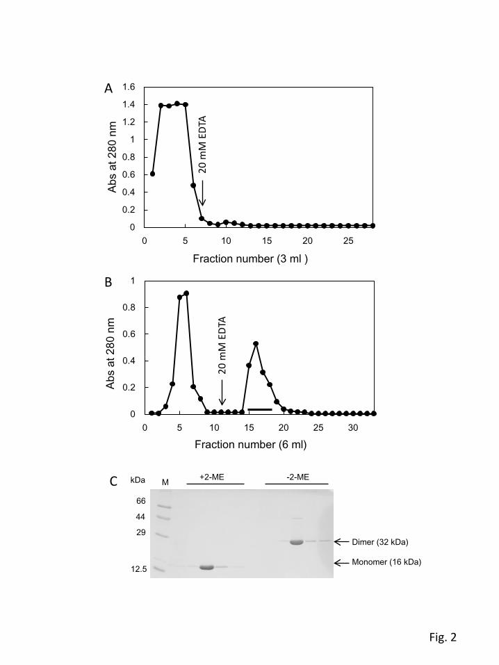
were refolded after complete unfolding using guanidine hydrochloride. After the refolding process,
EPNH-CEL-I was purified by affinity chromatography using a sugar-immobilized column. Although
WT-CEL-I can be purified using the GalNAc-Cellufine column, EPNH-CEL-I was not able to bind to
8
GalNAc-Cellufine (Fig. 2A). However, it could successfully bind to the mannose-Cellufine column
and be eluted with EDTA (Fig. 2B). The purified protein showed single bands at 16 kDa and 32 kDa in
the presence and absence of 2-mercaptoethanol, respectively (Fig. 2C), corresponding to the monomer
and dimer linked by a disulfide bond. These results suggested that the expressed protein was correctly
folded to adopt the native protein-like dimer structure with a Ca2+-dependent mannose-binding ability.
N-terminal amino acid analysis confirmed the sequence of the first five residues (Met-Asn-Gln-X-Pro)
of the purified protein, indicating that the initiator methionine residue remained attached to the N-
terminus in the recombinant protein. The unidentified residue X was assumed to be a cysteine (or half-
cystine) residue from the designed sequence. To examine the secondary structure, the far-UV CD
spectrum of the purified EPNH-CEL-I was measured along with that of WT-CEL-I. As shown in Fig. 3.
EPNH-CEL-I showed a CD spectrum with a broad negative peak around the region of 210–240 nm,
which is similar to that of WT-CEL-I, confirming that EPNH-CEL-I had a correctly folded structure.
However, a slight difference in the intensity was also observed between the 205–220-nm and 220–
240-nm regions. This may be caused by the replacement of a tryptophan residue (Trp105), which has a
large UV absorption around this region, by a histidine residue.
3.2. Carbohydrate-binding specificity of EPNH-CEL-I
Binding abilities of EPNH-CEL-I for different sugars were examined by an assay using sugar-PD
containing reducing sugar attached to the primary amino groups at the surface of PD [21]. Figure 4A
shows the change in the light-scattering intensity measured at 420 nm. When mannan-PD was added
to the solutions of WT-CEL-I, EPN-CEL-I, or EPNH-CEL-I, EPNH-CEL-I showed a large increase in
the light-scattering intensity at 420 nm, while EPN-CEL-I showed a slight increase and WT-CEL-I
showed a very little change. These results suggest that EPNH-CEL-I has a significant binding affinity
for mannose, although EPN-CEL-I also has a weak affinity. To compare the affinities for different
sugars, a competitive assay for binding of EPNH-CEL-I with mannotriose-PD was conducted as
shown in Fig. 4B. After the formation of a complex between EPN-CEL-I and mannotriose-PD, several
simple carbohydrates were added to compete for binding to the protein. Consequently, the strongest
binding was observed for mannose and methyl α-D-mannoside, confirming its definite affinity for
mannose and its related carbohydrates. Glucose and maltose also showed moderate affinity for EPNH-
CEL-I because glucose has the same configuration of 3- and 4-hydroxy groups as mannose. It seems
interesting that EPNH-CEL-I still retained a considerable degree of affinity for N-acetylgalactosamine,
9
while galactose showed a very weak affinity. This suggests that the acetamido group of N-
acetylgalactosamine has significant interactions with the carbohydrate-binding site in EPNH-CEL-I.
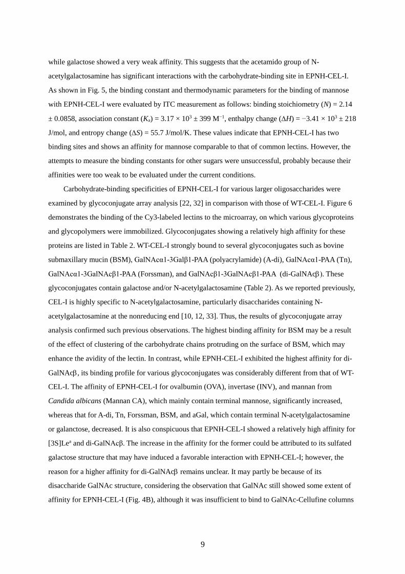
As shown in Fig. 5, the binding constant and thermodynamic parameters for the binding of mannose
with EPNH-CEL-I were evaluated by ITC measurement as follows: binding stoichiometry (N) = 2.14
± 0.0858, association constant (Ka) = 3.17 × 10³ ± 399 M−1, enthalpy change (∆H) = −3.41 × 10³ ± 218
J/mol, and entropy change (∆S) = 55.7 J/mol/K. These values indicate that EPNH-CEL-I has two
binding sites and shows an affinity for mannose comparable to that of common lectins. However, the
attempts to measure the binding constants for other sugars were unsuccessful, probably because their
affinities were too weak to be evaluated under the current conditions.
Carbohydrate-binding specificities of EPNH-CEL-I for various larger oligosaccharides were
examined by glycoconjugate array analysis [22, 32] in comparison with those of WT-CEL-I. Figure 6
demonstrates the binding of the Cy3-labeled lectins to the microarray, on which various glycoproteins
and glycopolymers were immobilized. Glycoconjugates showing a relatively high affinity for these
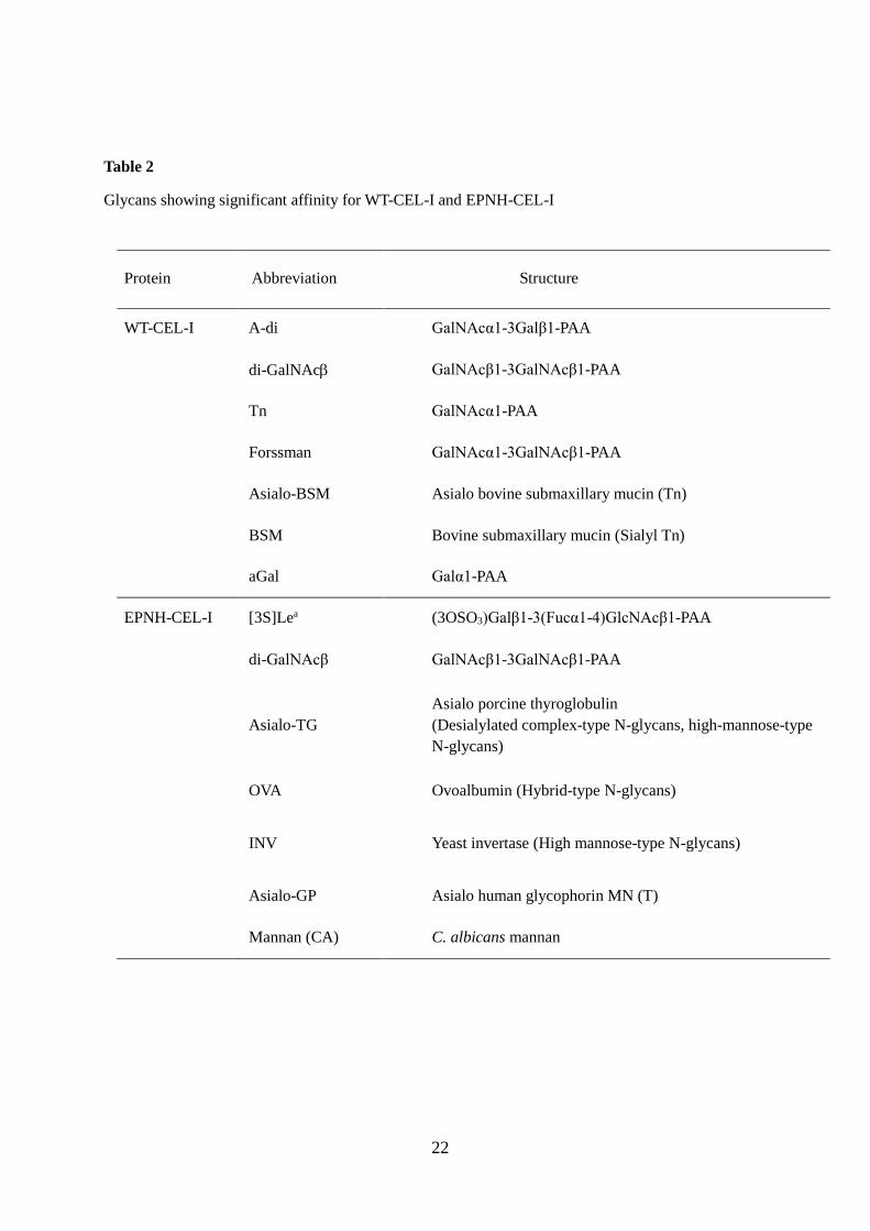
proteins are listed in Table 2. WT-CEL-I strongly bound to several glycoconjugates such as bovine
submaxillary mucin (BSM), GalNAcα1-3Galβ1-PAA (polyacrylamide) (A-di), GalNAcα1-PAA (Tn),
GalNAcα1-3GalNAcβ1-PAA (Forssman), and GalNAcβ1-3GalNAcβ1-PAA (di-GalNAcβ). These
glycoconjugates contain galactose and/or N-acetylgalactosamine (Table 2). As we reported previously,
CEL-I is highly specific to N-acetylgalactosamine, particularly disaccharides containing N-
acetylgalactosamine at the nonreducing end [10, 12, 33]. Thus, the results of glycoconjugate array
analysis confirmed such previous observations. The highest binding affinity for BSM may be a result
of the effect of clustering of the carbohydrate chains protruding on the surface of BSM, which may
enhance the avidity of the lectin. In contrast, while EPNH-CEL-I exhibited the highest affinity for di-
GalNAcβ, its binding profile for various glycoconjugates was considerably different from that of WT-
CEL-I. The affinity of EPNH-CEL-I for ovalbumin (OVA), invertase (INV), and mannan from
Candida albicans (Mannan CA), which mainly contain terminal mannose, significantly increased,
whereas that for A-di, Tn, Forssman, BSM, and aGal, which contain terminal N-acetylgalactosamine
or galanctose, decreased. It is also conspicuous that EPNH-CEL-I showed a relatively high affinity for
[3S]Lea and di-GalNAcβ. The increase in the affinity for the former could be attributed to its sulfated
galactose structure that may have induced a favorable interaction with EPNH-CEL-I; however, the
reason for a higher affinity for di-GalNAcβ remains unclear. It may partly be because of its
disaccharide GalNAc structure, considering the observation that GalNAc still showed some extent of
affinity for EPNH-CEL-I (Fig. 4B), although it was insufficient to bind to GalNAc-Cellufine columns
10
(Fig. 2A).
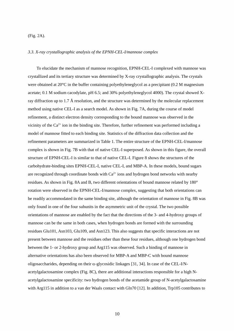
3.3. X-ray crystallographic analysis of the EPNH-CEL-I/mannose complex
To elucidate the mechanism of mannose recognition, EPNH-CEL-I complexed with mannose was
crystallized and its tertiary structure was determined by X-ray crystallographic analysis. The crystals
were obtained at 20°C in the buffer containing polyethyleneglycol as a precipitant (0.2 M magnesium
acetate; 0.1 M sodium cacodylate, pH 6.5; and 30% polyethyleneglycol 4000). The crystal showed X-
ray diffraction up to 1.7 Å resolution, and the structure was determined by the molecular replacement
method using native CEL-I as a search model. As shown in Fig. 7A, during the course of model
refinement, a distinct electron density corresponding to the bound mannose was observed in the
vicinity of the Ca2+ ion in the binding site. Therefore, further refinement was performed including a
model of mannose fitted to each binding site. Statistics of the diffraction data collection and the
refinement parameters are summarized in Table 1. The entire structure of the EPNH-CEL-I/mannose
complex is shown in Fig. 7B with that of native CEL-I superposed. As shown in this figure, the overall
structure of EPNH-CEL-I is similar to that of native CEL-I. Figure 8 shows the structures of the
carbohydrate-binding sites EPNH-CEL-I, native CEL-I, and MBP-A. In these models, bound sugars
are recognized through coordinate bonds with Ca2+ ions and hydrogen bond networks with nearby
residues. As shown in Fig. 8A and B, two different orientations of bound mannose related by 180°
rotation were observed in the EPNH-CEL-I/mannose complex, suggesting that both orientations can
be readily accommodated in the same binding site, although the orientation of mannose in Fig. 8B was
only found in one of the four subunits in the asymmetric unit of the crystal. The two possible
orientations of mannose are enabled by the fact that the directions of the 3- and 4-hydroxy groups of
mannose can be the same in both cases, when hydrogen bonds are formed with the surrounding
residues Glu101, Asn103, Glu109, and Asn123. This also suggests that specific interactions are not
present between mannose and the residues other than these four residues, although one hydrogen bond
between the 1- or 2-hydroxy group and Arg115 was observed. Such a binding of mannose in
alternative orientations has also been observed for MBP-A and MBP-C with bound mannose
oligosaccharides, depending on their α-glycosidic linkages [31, 34]. In case of the CEL-I/N-
acetylgalactosamine complex (Fig. 8C), there are additional interactions responsible for a high N-
acetylgalactosamine specificity: two hydrogen bonds of the acetamide group of N-acetylgalactosamine
with Arg115 in addition to a van der Waals contact with Gln70 [12]. In addition, Trp105 contributes to
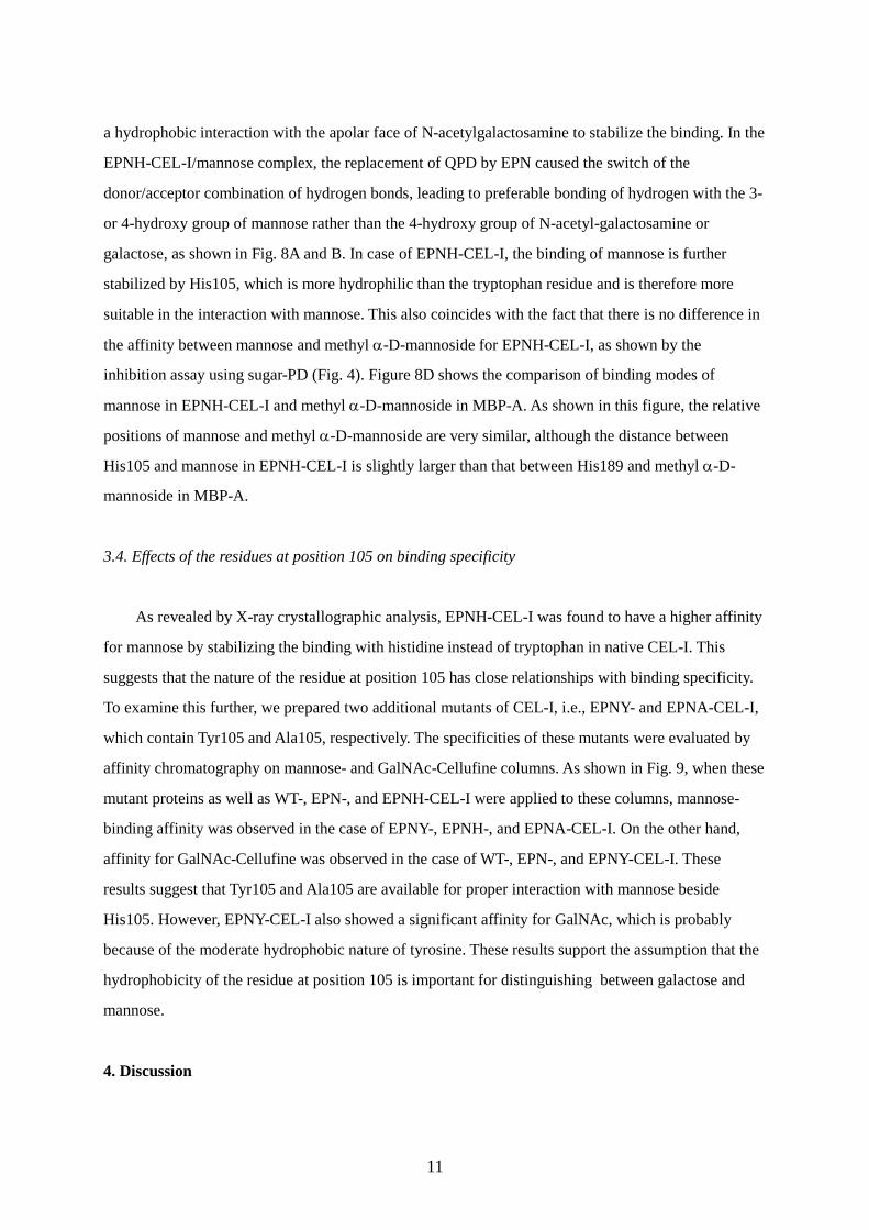
11
a hydrophobic interaction with the apolar face of N-acetylgalactosamine to stabilize the binding. In the
EPNH-CEL-I/mannose complex, the replacement of QPD by EPN caused the switch of the
donor/acceptor combination of hydrogen bonds, leading to preferable bonding of hydrogen with the 3-
or 4-hydroxy group of mannose rather than the 4-hydroxy group of N-acetyl-galactosamine or
galactose, as shown in Fig. 8A and B. In case of EPNH-CEL-I, the binding of mannose is further
stabilized by His105, which is more hydrophilic than the tryptophan residue and is therefore more
suitable in the interaction with mannose. This also coincides with the fact that there is no difference in
the affinity between mannose and methyl α-D-mannoside for EPNH-CEL-I, as shown by the
inhibition assay using sugar-PD (Fig. 4). Figure 8D shows the comparison of binding modes of
mannose in EPNH-CEL-I and methyl α-D-mannoside in MBP-A. As shown in this figure, the relative
positions of mannose and methyl α-D-mannoside are very similar, although the distance between
His105 and mannose in EPNH-CEL-I is slightly larger than that between His189 and methyl α-D-
mannoside in MBP-A.
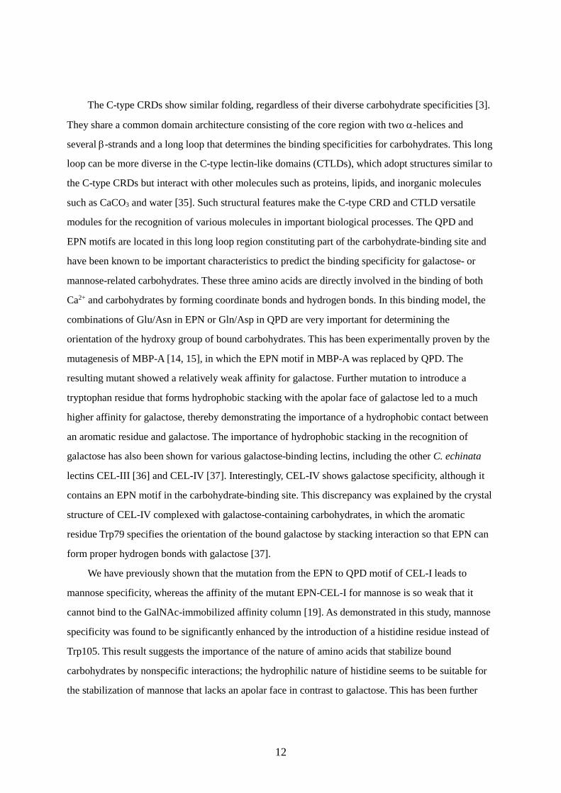
3.4. Effects of the residues at position 105 on binding specificity
As revealed by X-ray crystallographic analysis, EPNH-CEL-I was found to have a higher affinity
for mannose by stabilizing the binding with histidine instead of tryptophan in native CEL-I. This
suggests that the nature of the residue at position 105 has close relationships with binding specificity.
To examine this further, we prepared two additional mutants of CEL-I, i.e., EPNY- and EPNA-CEL-I,
which contain Tyr105 and Ala105, respectively. The specificities of these mutants were evaluated by
affinity chromatography on mannose- and GalNAc-Cellufine columns. As shown in Fig. 9, when these
mutant proteins as well as WT-, EPN-, and EPNH-CEL-I were applied to these columns, mannose-
binding affinity was observed in the case of EPNY-, EPNH-, and EPNA-CEL-I. On the other hand,
affinity for GalNAc-Cellufine was observed in the case of WT-, EPN-, and EPNY-CEL-I. These
results suggest that Tyr105 and Ala105 are available for proper interaction with mannose beside
His105. However, EPNY-CEL-I also showed a significant affinity for GalNAc, which is probably
because of the moderate hydrophobic nature of tyrosine. These results support the assumption that the
hydrophobicity of the residue at position 105 is important for distinguishing between galactose and
mannose.
4. Discussion
12
The C-type CRDs show similar folding, regardless of their diverse carbohydrate specificities [3].
They share a common domain architecture consisting of the core region with two α-helices and
several β-strands and a long loop that determines the binding specificities for carbohydrates. This long
loop can be more diverse in the C-type lectin-like domains (CTLDs), which adopt structures similar to
the C-type CRDs but interact with other molecules such as proteins, lipids, and inorganic molecules
such as CaCO3 and water [35]. Such structural features make the C-type CRD and CTLD versatile
modules for the recognition of various molecules in important biological processes. The QPD and
EPN motifs are located in this long loop region constituting part of the carbohydrate-binding site and
have been known to be important characteristics to predict the binding specificity for galactose- or
mannose-related carbohydrates. These three amino acids are directly involved in the binding of both
Ca2+ and carbohydrates by forming coordinate bonds and hydrogen bonds. In this binding model, the
combinations of Glu/Asn in EPN or Gln/Asp in QPD are very important for determining the
orientation of the hydroxy group of bound carbohydrates. This has been experimentally proven by the
mutagenesis of MBP-A [14, 15], in which the EPN motif in MBP-A was replaced by QPD. The
resulting mutant showed a relatively weak affinity for galactose. Further mutation to introduce a
tryptophan residue that forms hydrophobic stacking with the apolar face of galactose led to a much
higher affinity for galactose, thereby demonstrating the importance of a hydrophobic contact between
an aromatic residue and galactose. The importance of hydrophobic stacking in the recognition of
galactose has also been shown for various galactose-binding lectins, including the other C. echinata
lectins CEL-III [36] and CEL-IV [37]. Interestingly, CEL-IV shows galactose specificity, although it
contains an EPN motif in the carbohydrate-binding site. This discrepancy was explained by the crystal
structure of CEL-IV complexed with galactose-containing carbohydrates, in which the aromatic
residue Trp79 specifies the orientation of the bound galactose by stacking interaction so that EPN can
form proper hydrogen bonds with galactose [37].
We have previously shown that the mutation from the EPN to QPD motif of CEL-I leads to
mannose specificity, whereas the affinity of the mutant EPN-CEL-I for mannose is so weak that it
cannot bind to the GalNAc-immobilized affinity column [19]. As demonstrated in this study, mannose
specificity was found to be significantly enhanced by the introduction of a histidine residue instead of
Trp105. This result suggests the importance of the nature of amino acids that stabilize bound
carbohydrates by nonspecific interactions; the hydrophilic nature of histidine seems to be suitable for
the stabilization of mannose that lacks an apolar face in contrast to galactose. This has been further
13
supported by affinity chromatography of the two additional mutants EPNY- and EPNA-CEL-I (Fig. 9).
Although EPNY-CEL-I exhibited affinity for both mannose and N-acetylgalactosamine, EPNA-CEL-I
only bound to the mannose column, similar to EPNH-CEL-I. These results strongly suggest that the
hydrophobicity of the residue stabilizing the orientation of the bound sugars significantly influences
binding specificities; a more hydrophobic nature is favorable for the binding of galactose.
Although EPNH-CEL-I did not bind to the GalNAc-Cellufine column, some degree of affinity for
N-acetylgalactosamine was observed in the inhibition experiments (Fig. 4B). As revealed by X-ray
crystallographic analysis [12], native CEL-I shows strong N-acetylgalactosamine specificity through
two hydrogen bonds with Arg115 and van der Waals interaction with Gln70 (Fig. 8C). Therefore,
possibly, N-acetylgalactosamine can also bind to EPNH-CEL-I with moderate affinity through these
interactions with Arg115 and Gln70. Glycoconjugate microarray analysis also proved definite affinity
for N-acetyl-galactosamine-containing oligosaccharides with an even higher intensity than that for
mannose-containing oligosaccharides. This may be due to additional interactions with the
oligosaccharides rather than with the N-acetylgalactosamine monosaccharide. In the EPNH-CEL-
I/mannose complex, Arg115 was found to form a hydrogen bond with the 1- or 2-hydroxy group of
mannose in different conformations (Fig. 8A and B), depending on two alternative orientations of the
bound mannose, suggesting that Arg115 has some extent of contribution in mannose recognition by
EPNH-CEL-I. In the competition assay using sugar-PD (Fig. 4), glucose also exhibited a moderately
high affinity for EPNH-CEL-I. As apparent from Fig. 8A, the 2- and 6-hydroxy groups of mannose are
not necessary for the binding to EPNH-CEL-I, while the 3- and 4-hydroxy groups are essential.
Therefore, it seems reasonable that glucose, which has the same configuration regarding 3- and 4-
hydroxy groups as mannose, can bind to EPNH-CEL-I with a moderate affinity. Along with the
findings of the preceding studies on engineering of MBP-A to change its specificity to galactose
binding [14, 15] and its applications for the identification of cell surface glycoconjugates [38-40], a
reversal change in the specificity of CEL-I from galactose (N-acetylgalactosamine) to mannose shown
in the present study suggests the versatility of C-type CRDs. As observed in glycoconjugate
microarray analysis, mutations in CEL-I seem to have affected the recognition of not only
monosaccharides but also oligosaccharides. Such alterations in the recognition of oligosaccharides or
glycolipids have also been reported for other C-type lectins [31, 41, 42], which may involve
interaction with additional sites of protein (subsites) extending from the primary binding site
containing Ca2+. The versatility of C-type CRDs appears to be closely related to the structural features
of the C-type CRDs that are composed of two parts: a stable core structure with a common fold and a
14
long loop region that can potentially accommodate various molecules. The binding of CTLDs to
various molecules strongly suggests that their molecular architecture could be useful in the
development of novel molecular recognition proteins.
Acknowledgments
We are grateful to Ms. Jinko Murakami and Ms. Keiko Hiemori for help in glycoconjugate
microarray analysis. This work was supported by a Grants-in-Aid for Scientific Research (C) to TH
(26450128) and Grants-in-Aid for Young Scientists (B) to HU (10452872) and SG (00346587) from
the Japan Society for the Promotion of Science (JSPS).
15
References
[1] K. Drickamer, Two distinct classes of carbohydrate-recognition domains in animal lectins, J Biol
Chem, 263 (1988) 9557-9560.
[2] D.C. Kilpatrick, Animal lectins: a historical introduction and overview., Biochim Biophys Acta,
1572 (2002) 187-197.
[3] A.N. Zelensky, J.E. Gready, The C-type lectin-like domain superfamily., FEBS J, 272 (2005) 6179-
6217.
[4] S. Mukherjee, H. Zheng, M.G. Derebe, K.M. Callenberg, C.L. Partch, D. Rollins, D.C. Propheter, J.
Rizo, M. Grabe, Q.X. Jiang, L.V. Hooper, Antibacterial membrane attack by a pore-forming
intestinal C-type lectin, Nature, 505 (2014) 103-107.
[5] E. Riboldi, R. Daniele, C. Parola, A. Inforzato, P.L. Arnold, D. Bosisio, D.H. Fremont, A. Bastone,
M. Colonna, S. Sozzani, Human C-type lectin domain family 4, member C (CLEC4C/BDCA-
2/CD303) is a receptor for asialo-galactosyl-oligosaccharides., J Biol Chem, 286 (2011) 35329-
35333.
[6] D. Mourão-Sá, M.J. Robinson, S. Zelenay, D. Sancho, P. Chakravarty, R. Larsen, M. Plantinga, N.
Van Rooijen, M.P. Soares, B. Lambrecht, C. Reis e Sousa, CLEC-2 signaling via Syk in myeloid
cells can regulate inflammatory responses., Eur J Immunol, 41 (2011) 3040-3053.
[7] D.C. Kilpatrick, Mannan-binding lectin and its role in innate immunity., Transfus Med, 12 (2002)
335-352.
[8] A. Cambi, C.G. Figdor, Dual function of C-type lectin-like receptors in the immune system, Curr
Opin Cell Biol, 15 (2003) 539-546.
[9] F. Liu, J. Li, J. Fu, Y. Shen, X. Xu, Two novel homologs of simple C-type lectin in grass carp
(Ctenopharyngodon idellus): Potential role in immune response to bacteria., Fish Shellfish
Immunol, 31 (2011) 765–773.
[10] T. Hatakeyama, H. Kohzaki, H. Nagatomo, N. Yamasaki, Purification and characterization of four
Ca2+-dependent lectins from the marine invertebrate, Cucumaria echinata., J Biochem, 116
(1994) 209-214.
[11] H. Unno, S. Goda, T. Hatakeyama, Hemolytic lectin CEL-III heptamerizes via a large structural
transition from α-helices to a β-barrel during the transmembrane pore formation process, J Biol
Chem, 289 (2014) 12805-12812.
[12] H. Sugawara, M. Kusunoki, G. Kurisu, T. Fujimoto, H. Aoyagi, T. Hatakeyama, Characteristic
recognition of N-acetylgalactosamine by an invertebrate C-type Lectin, CEL-I, revealed by X-ray
16
crystallographic analysis., J Biol Chem, 279 (2004) 45219-45225.
[13] T. Hatakeyama, K. Ohuchi, M. Kuroki, N. Yamasaki, Amino acid sequence of a C-type lectin
CEL-IV from the marine invertebrate Cucumaria echinata, Biosci Biotech Biochem, 59 (1995)
1314-1317.
[14] K. Drickamer, Engineering galactose-binding activity into a C-type mannose-binding protein.,
Nature, 360 (1992) 183-186.
[15] A.R. Kolatkar, A.K. Leung, R. Isecke, R. Brossmer, K. Drickamer, W.I. Weis, Mechanism of N-
acetylgalactosamine binding to a C-type animal lectin carbohydrate-recognition domain., J Biol
Chem, 273 (1998) 19502-19508.
[16] S.T. Iobst, K. Drickamer, Binding of sugar ligands to Ca2+-dependent animal lectins. II.
Generation of high-affinity galactose binding by site-directed mutagenesis., J Biol Chem, 269
(1994) 15512-15519.
[17] Y. Ogasawara, D.R. Voelker, Altered carbohydrate recognition specificity engineered into
surfactant protein D reveals different binding mechanisms for phosphatidylinositol and
glucosylceramide, J Biol Chem, 270 (1995) 14725-14732.
[18] F.X. McCormack, Y. Kuroki, J.J. Stewart, R.J. Mason, D.R. Voelker, Surfactant protein A amino
acids Glu195 and Arg197 are essential for receptor binding, phospholipid aggregation, regulation
of secretion, and the facilitated uptake of phospholipid by type II cells, J Biol Chem, 269 (1994)
29801-29807.
[19] T. Hatakeyama, T. Ishimine, T. Baba, M. Kimura, H. Unno, S. Goda, Alteration of the
Carbohydrate-Binding Specificity of a C-type Lectin CEL-I Mutant with an EPN Carbohydrate-
Binding Motif, Protein Pept Lett, 20 (2013) 796-801.
[20] Y. Kato, K. Kochi, H. Unno, S. Goda, T. Hatakeyama, Manno-oligosaccharide-binding ability of
mouse RegIV/GST-fusion protein evaluated by complex formation with the carbohydrate-
containing polyamidoamine dendrimer, Biosci Biotechnol Biochem, (2014) 1-4.
[21] T. Hatakeyama, R. Karino, Y. Terai, M. Kimura, H. Unno, S. Goda, An assay for carbohydrate-
binding activity of lectins using polyamidoamine dendrimer conjugated with carbohydrates,
Biosci Biotechnol Biochem, 76 (2012) 1999-2001.
[22] H. Tateno, A. Mori, N. Uchiyama, R. Yabe, J. Iwaki, T. Shikanai, T. Angata, H. Narimatsu, J.
Hirabayashi, Glycoconjugate microarray based on an evanescent-field fluorescence-assisted
detection principle for investigation of glycan-binding proteins, Glycobiology, 18 (2008) 789-798.
[23] N. Collaborative Computational Project, The CCP4 suite: programs for protein crystallography,
17
Acta Crystallogr. D Biol. Crystallogr., 50 (1994).
[24] A.G. Leslie, The integration of macromolecular diffraction data, Acta Crystallogr D Biol
Crystallogr, 62 (2006) 48-57.
[25] P. Evans, Scaling and assessment of data quality, Acta Crystallogr D Biol Crystallogr, 62 (2006)
72-82.
[26] A.J. McCoy, R.W. Grosse-Kunstleve, L.C. Storoni, R.J. Read, Likelihood-enhanced fast
translation functions, Acta Crystallogr D Biol Crystallogr, 61 (2005) 458-464.
[27] G.N. Murshudov, A.A. Vagin, E.J. Dodson, Refinement of macromolecular structures by the
maximum-likelihood method, Acta Crystallogr D Biol Crystallogr, 53 (1997) 240-255.
[28] P. Emsley, B. Lohkamp, W.G. Scott, K. Cowtan, Features and development of Coot, Acta
Crystallogr D Biol Crystallogr, 66 (2010) 486-501.
[29] R. Laskowski, M. MacArthur, D. Moss, J. Thornton, PROCHECK: a program to check the
stereochemical quality of protein structures, J. Appl. Crystallogr., 26 ( 1993) 283–291.
[30] W.L. DeLano, The PyMOL Molecular Graphics Systems, DeLano Scientific, San Carlos, CA,
2002.
[31] K.K. Ng, A.R. Kolatkar, S. Park-Snyder, H. Feinberg, D.A. Clark, K. Drickamer, W.I. Weis,
Orientation of bound ligands in mannose-binding proteins. Implications for multivalent ligand
recognition., J Biol Chem, 277 (2002) 16088-16095.
[32] H. Tateno, Evaluation of glycan-binding specificity by glycoconjugate microarray with an
evanescent-field fluorescence detection system, Methods Mol Biol, 1200 (2014) 353-359.
[33] T. Hatakeyama, K. Shiba, N. Matsuo, T. Fujimoto, T. Oda, H. Sugawara, H. Aoyagi,
Characterization of recombinant CEL-I, a GalNAc-specific C-type lectin, expressed in
Escherichia coli using an artificial synthetic gene., J Biochem, 135 (2004) 101-107.
[34] K.K. Ng, K. Drickamer, W.I. Weis, Structural analysis of monosaccharide recognition by rat liver
mannose-binding protein., J Biol Chem, 271 (1996) 663-674.
[35] K. Drickamer, C-type lectin-like domains., Curr Opin Struct Biol, 9 (1999) 585-590.
[36] T. Hatakeyama, H. Unno, Y. Kouzuma, T. Uchida, S. Eto, H. Hidemura, N. Kato, M. Yonekura, M.
Kusunoki, C-type lectin-like carbohydrate recognition of the hemolytic lectin CEL-III containing
ricin-type -trefoil folds., J Biol Chem, 282 (2007) 37826-37835.
[37] T. Hatakeyama, T. Kamiya, M. Kusunoki, S. Nakamura-Tsuruta, J. Hirabayashi, S. Goda, H.
Unno, Galactose recognition by a tetrameric C-type lectin, CEL-IV, containing the EPN
carbohydrate recognition motif., J Biol Chem, 286 (2011) 10305-10315.
18
[38] A.S. Powlesland, P.G. Hitchen, S. Parry, S.A. Graham, M.M. Barrio, M.T. Elola, J. Mordoh, A.
Dell, K. Drickamer, M.E. Taylor, Targeted glycoproteomic identification of cancer cell
glycosylation, Glycobiology, 19 (2009) 899-909.
[39] A.S. Powlesland, A. Quintero-Martinez, P.G. Lim, Z. Pipirou, M.E. Taylor, K. Drickamer,
Engineered carbohydrate-recognition domains for glycoproteomic analysis of cell surface
glycosylation and ligands for glycan-binding receptors, Methods Enzymol, 480 (2010) 165-179.
[40] A.S. Powlesland, M.M. Barrio, J. Mordoh, P.G. Hitchen, A. Dell, K. Drickamer, M.E. Taylor,
Glycoproteomic characterization of carriers of the CD15/Lewisx epitope on Hodgkin's Reed-
Sternberg cells, BMC Biochem, 12 (2011) 13.
[41] H. Wang, J. Head, P. Kosma, H. Brade, S. Müller-Loennies, S. Sheikh, B. McDonald, K. Smith, T.
Cafarella, B. Seaton, E. Crouch, Recognition of heptoses and the inner core of bacterial
lipopolysaccharides by surfactant protein d, Biochemistry, 47 (2008) 710-720.
[42] T.K. Carlson, J.B. Torrelles, K. Smith, T. Horlacher, R. Castelli, P.H. Seeberger, E.C. Crouch, L.S.
Schlesinger, Critical role of amino acid position 343 of surfactant protein-D in the selective
binding of glycolipids from Mycobacterium tuberculosis, Glycobiology, 19 (2009) 1473-1484.
[43] M. Shimokawa, A. Fukudome, R. Yamashita, Y. Minami, F. Yagi, H. Tateno, J. Hirabayashi,
Characterization and cloning of GNA-like lectin from the mushroom Marasmius oreades,
Glycoconj J, 29 (2012) 457-465.
19
Figure legends
Fig. 1. Comparison of the carbohydrate-binding sites of CEL-I and MBP-A. (A) CEL-I/N-
acetylgalactosamine complex (PDB ID: 1WMY) [12]. (B) MBP-A/methyl α-D-mannoside complex
(PDB ID: 1KWU) [31]. The coordinate bonds and hydrogen bonds are depicted by yellow dashed lines.
The amino acid residues consisting of the QPD and EPN motifs are shown in red.
Fig. 2. Affinity chromatography of EPNH-CEL-I on the carbohydrate-immobilized Cellufine columns.
Refolded EPNH-CEL-I was applied to the GalNAc-Cellufine column (A) or mannose-Cellufine column
(B). After elution of the nonadsorbed proteins with TBS containing 10 mM CaCl2, the adsorbed protein
was eluted with TBS containing 20 mM EDTA. (C) SDS-PAGE of EPNH-CEL-I eluted from the
mannose-Cellufine column indicated by the bar in panel B in the presence and absence of 2-
mercaptoethanol (2-ME).
Fig. 3. Far-UV CD spectra of EPNH-CEL-I and WT-CEL-I. Measurement was performed with 0.28
mg/ml protein in TBS containing 10 mM CaCl2 at 20°C. The values [θ] are expressed as the mean
residue molar ellipticity.
Fig. 4. Carbohydrate-binding specificity of the lectins examined by complex formation with sugar-PD.
(A) The changes in the light-scattering intensity induced by complex formation between the lectins and
mannan-PD were measured using a fluorescence spectrophotometer at 25°C. (B) The solutions of
indicated sugars were added to the pre-formed complex between EPNH-CEL-I and mannotriose-PD.
Dissociation of the EPNH-CEL-I/mannotriose-PD complex was evaluated by the changes in the light
scattering intensity. The initial light scattering intensity of the pre-formed complex was considered as
100%.
Fig. 5. ITC for the binding of mannose to EPNH-CEL-I. The mannose solution was titrated into a
temperature-controlled sample cell containing the EPNH-CEL-I solution. The change in the heat
accompanying the binding (upper panel) was integrated and plotted against the mannose/EPNH-CEL-I
20
molar ratio (lower panel).
Fig. 6. Glycoconjugate microarray analysis of WT-CEL-I and EPNH-CEL-I. The binding specificities
of Cy3-labeled WT-CEL-I and EPNH-CEL-I for various glycoconjugates were measured.
Glycoconjugates used in this analysis are designated by the abbreviations, as listed in ref. [43].
Fig. 7. The crystal structure of the EPNH-CEL-I/mannose complex. (A) The binding site with the bound
mannose is shown with a 2Fo-Fc electron density map contoured at 1σ. (B) Main chain structure of the
EPNH-CEL-I/mannose complex (yellow) is shown by a ribbon model in comparison with that of native
CEL-I (pink).
Fig. 8. Comparison of the carbohydrate-binding sites of EPNH-CEL-I, native CEL-I, and MBP-A. (A
and B) Two different binding modes of EPNH-CEL-I with the bound mannose rotated by 180°. (C) The
binding site of the CEL-I/N-acetylgalactosamine complex [12]. (D) Comparison of the EPNH-CEL-
I/mannose complex (green) with the MBP-A/methyl α-D-mannoside complex (yellow). Only residue
numbers for the MBP-A/methyl α-D-mannoside complex are indicated.
Fig. 9. Affinity chromatography of WT and CEL-I mutants. Each protein was applied to mannose-
Cellufine (left panels) or GalNAc-Cellufine (right panels) columns in TBS containing 10 mM CaCl2.
After washing the columns with the same buffer, bound proteins were eluted with TBS containing 20
mM EDTA at the positions indicated by vertical arrows.
21
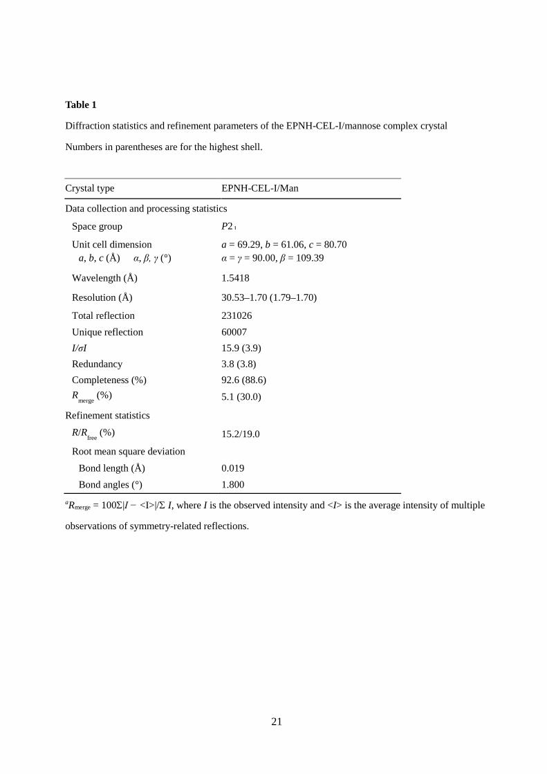
Table 1
Diffraction statistics and refinement parameters of the EPNH-CEL-I/mannose complex crystal
Numbers in parentheses are for the highest shell.
Crystal type EPNH-CEL-I/Man
Data collection and processing statistics
Space group P2₁
Unit cell dimension a, b, c (Å) α, β, γ (°)
a = 69.29, b = 61.06, c = 80.70 α = γ = 90.00, β = 109.39
Wavelength (Å) 1.5418
Resolution (Å) 30.53–1.70 (1.79–1.70)
Total reflection 231026 Unique reflection 60007 I/σI 15.9 (3.9) Redundancy 3.8 (3.8) Completeness (%) 92.6 (88.6) R
merge (%) 5.1 (30.0)
Refinement statistics
R/Rfree
(%) 15.2/19.0
Root mean square deviation
Bond length (Å) 0.019 Bond angles (°) 1.800 aRmerge = 100Σ|I − <I>|/Σ I, where I is the observed intensity and <I> is the average intensity of multiple
observations of symmetry-related reflections.
22
Table 2
Glycans showing significant affinity for WT-CEL-I and EPNH-CEL-I
Protein Abbreviation Structure
WT-CEL-I A-di GalNAcα1-3Galβ1-PAA
di-GalNAcβ GalNAcβ1-3GalNAcβ1-PAA
Tn GalNAcα1-PAA
Forssman GalNAcα1-3GalNAcβ1-PAA
Asialo-BSM Asialo bovine submaxillary mucin (Tn)
BSM Bovine submaxillary mucin (Sialyl Tn)
aGal Galα1-PAA
EPNH-CEL-I [3S]Lea (3OSO3)Galβ1-3(Fucα1-4)GlcNAcβ1-PAA
di-GalNAcβ GalNAcβ1-3GalNAcβ1-PAA
Asialo-TG Asialo porcine thyroglobulin (Desialylated complex-type N-glycans, high-mannose-type N-glycans)
OVA Ovoalbumin (Hybrid-type N-glycans)
INV Yeast invertase (High mannose-type N-glycans)
Asialo-GP Asialo human glycophorin MN (T)
Mannan (CA) C. albicans mannan
A B
Fig. 1
Trp105 GalNAc
Asp103
Pro102
Gln101
Ca2+4-OH
His189 Me-α-D-Man
Asn187
Pro186
Glu185
Ca2+
4-OH
0
0.2
0.4
0.6
0.8
1
1.2
1.4
1.6
0 5 10 15 20 25
Abs
at 2
80 n
m
Fraction number (3 ml )
0
0.2
0.4
0.6
0.8
1
0 5 10 15 20 25 30
Abs
at 2
80 n
m
Fraction number (6 ml)
A
Fig. 2
20 m
MED
TA
20 m
MED
TA
B
C
12.5
66
44
29
kDa +2-ME -2-MEM
Dimer (32 kDa)
Monomer (16 kDa)
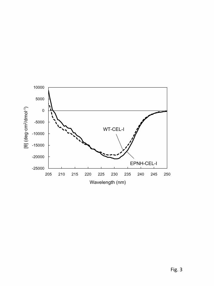
Fig. 3
-25000
-20000
-15000
-10000
-5000
0
5000
10000
205 210 215 220 225 230 235 240 245 250
[θ](
deg·
cm2 /d
mol
-1)
Wavelength (nm)
WT-CEL-I
EPNH-CEL-I
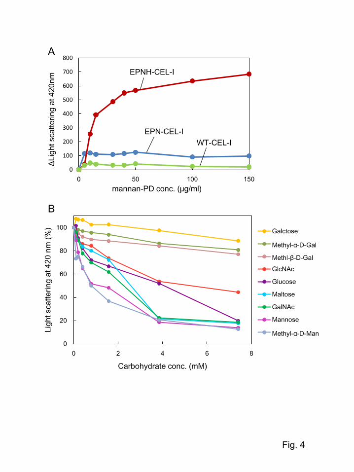
Fig. 4
0
100
200
300
400
500
600
700
800
0 50 100 150
ΔLight
scat
terin
g at
420
nm
mannan-PD conc. (μg/ml)
0
20
40
60
80
100
0 2 4 6 8
Ligh
t sca
tterin
g at
420
nm
(%)
Carbohydrate conc. (mM)
Galctose
Methyl-α-D-Gal
Methl-β-D-GalGlcNAc
Glucose
Maltose
GalNAc
Mannose
Methyl-α-D-Man
EPNH-CEL-I
EPN-CEL-IWT-CEL-I
A
B
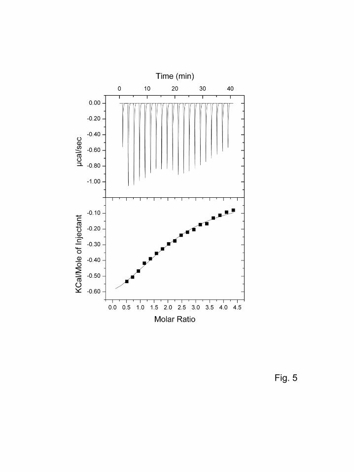
Fig. 5
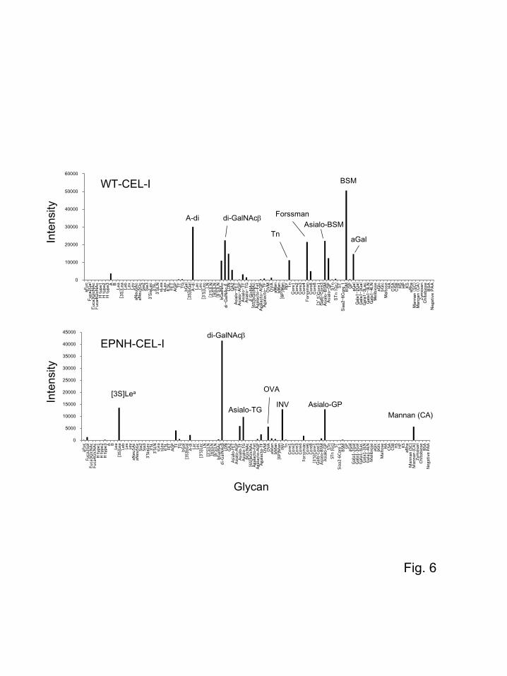
Fig. 6
0
5000
10000
15000
20000
25000
30000
35000
40000
45000
aFuc
Fuca
2Gal
Fuca
3Glc
NAc
Fuca
4Glc
NAc
H ty
pe1
H ty
pe2
H ty
pe3 A B
Lea
[3S]
Lea
Leb
Lex
Ley
aNeu
5Ac
aNeu
5Gc
Sia2
Sia3
3'Si
aLec
3'SL
3'SL
NsL
easL
ex6'
SL FET
AGP TF TG
bGal
[3S]
bGal
A-di Lac
Lec
[3'S
]Lec LN
[3'S
]LN
[6S]
LN[6
'S]L
NbG
alN
Acdi
-Gal
NAc
βLD
NGA
2As
ialo
-FET
Asia
lo-A
GP
Asia
lo-T
FAs
ialo
-TG
βGlc
NAc
[6S]
bGlc
NAc
Agal
acto
-Fet
Agal
acto
-AGP
Agal
acto
-TF
OVM OVA
aMan
bMan
[6P]
Man INV Tn
Core
1Co
re2
Core
3Co
re4
Fors
sman
Core
6Co
re8
[3’S
]Cor
e1Ga
lb-C
ore3
Asia
lo-B
SMAs
ialo
-GP
STn
STn
(Gc) ST
Siaa
2-6C
ore
1BS
M GPaG
alGa
la1-
2Gal
Gala
1-3G
alGa
la1-
3Lac
Gala
1-3L
NGa
la1-
4LN
Mel
ibio
seaG
lcbG
lcM
alto
se HA CSA
CSB HS HP KS
aRha
Man
nan
(SC)
Man
nan
(CA)
Zym
osan
Chito
bios
eBS
AN
egat
ive
PAA
0
10000
20000
30000
40000
50000
60000
aFuc
Fuca2
Gal
Fuca3
Glc
NA
cFuca4
Glc
NA
cH
typ
e1H
typ
e2H
typ
e3 A BLea
[3S]L
ea
Leb
Lex
Ley
aNeu
5A
caN
eu5G
cSia
2Sia
33'S
iaLec
3'S
L3'S
LN
sLea
sLex
6'S
LFET
AG
PTF
TG
bG
al[3
S]b
Gal
A-di
Lac
Lec
[3'S
]Lec
LN
[3'S
]LN
[6S]L
N[6
'S]L
NbG
alN
Ac
di-
Gal
NA
cβ
LD
NG
A2
Asi
alo-FET
Asi
alo-A
GP
Asi
alo-TF
Asi
alo-TG
βG
lcN
Ac
[6S]b
Glc
NA
cA
gala
cto
-Fet
Aga
lacto
-A
GP
Aga
lacto
-TF
OV
MO
VA
aMan
bM
an[6
P]M
anIN
V Tn
Core
1C
ore
2C
ore
3C
ore
4Fors
sman
Core
6C
ore
8[3
’S]C
ore
1G
alb-C
ore
3A
sial
o-B
SM
Asi
alo-G
PSTn
STn (G
c)
ST
Sia
a2-6C
ore
1B
SM
GP
aGal
Gal
a1-2G
alG
ala1
-3G
alG
ala1
-3L
acG
ala1
-3L
NG
ala1
-4L
NM
elib
iose
aGlc
bG
lcM
alto
se HA
CSA
CSB
HS
HP
KS
aRha
Man
nan (
SC
)M
anna
n (
CA
)Zym
osa
nC
hitobio
seB
SA
Nega
tive
PA
A
Glycan
WT-CEL-I
EPNH-CEL-I
Inte
nsity
Inte
nsity
BSM
A-di Forssman
Tn
di-GalNAcβ
Asialo-GPINV
di-GalNAcβ
Mannan (CA)
[3S]Lea
Asialo-TG
aGal
Asialo-BSM
OVA
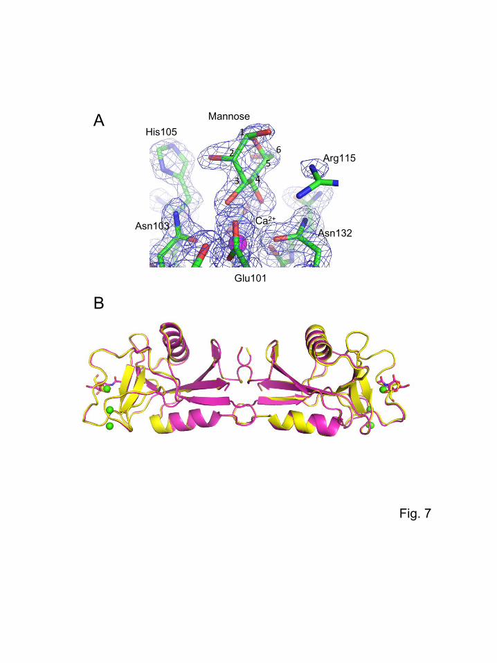
Fig. 7
A
B
1
25
43
Ca2+
6
His105
Arg115
Glu101
Asn103 Asn132
Mannose
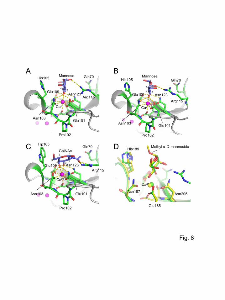
Fig. 8
A B
C D
His105
Arg115
Gln70
Asn103
Pro102
Glu101
Trp105His189
Asn187
Glu185
Mannose
GalNAc
Mannose
Methyl α-D-mannoside
Ca2+
Ca2+
His105 Gln70
Arg115
Asn103
Pro102
Glu101
Ca2+
Arg115
Asn103
Pro102
Glu101
Ca2+
Asn205
Glu109 Asn123
Gln70
Glu109 Asn123
Glu109 Asn123
1
34
1
3 4
1
34 2
5
6
252
Fraction number (3 ml)
Abs
at 2
80 n
m
Fig. 9
0
0.5
1
1.5
2
0 10 20 30
WT
0
0.5
1
1.5
0 10 20 30
WT
0
0.5
1
1.5
2
0 10 20 30
EPN (EPNW)
0
0.5
1
1.5
0 10 20 30
EPN (EPNW)
0
0.5
1
1.5
0 10 20 30
EPNY
0
0.5
1
0 10 20 30
EPNY
0
0.5
1
1.5
0 10 20 30
EPNH
0
0.5
1
1.5
0 10 20 30
EPNH
0
0.5
1
1.5
0 10 20 30
EPNA
0
0.5
1
1.5
0 10 20 30
EPNA
Mannose-Cellufine GalNAc-Cellufine