Embed Size (px)
Citation preview
Automatic Region Growing Method forSegmentation of Tumor on Mammogram
Image
0
0
Nayab ShaikMSRIT, Bangalore
abdul . nayab c^r gmail.com
Abstract - Breast cancer is a common female
malignancy , does not show any symptoms in its
early stage. Screening tests are therefore important
to reduce the death rates. By far, mammography
test proves to be the test for early detection of breast
cancer . However , this test has limitations such as
darkness and the existence of unwanted noise on the
breast image , which can obscure breast ` tumors.
Many image processing techniques have been
introduced in order to detect the edges or segment
these breast cancer morphologies including the seed
based region growing (SBRG) algorithm . However,
two parameters , namely the seed point and the
threshold value of the conventional SBRG algorithm
need to be determined manually. It is time
consuming process. This paper proposes automatic
region growing algorithm , which find these two
parameters automatically . It also detects and
distinguish breast tumor automatically from the
background.
Keywords-Mammography, Moving K-means
Clustering, GUI, Region growing, seed point and
Threshold value.
1. INTRODUCTION
Cancer is the second leading cause of death in
World. Cancer does not show any symptoms in its early
stage. Especially breast cancer does not show any
Megha.P.ArakeriMSRIT, Bangalore
rnegha_u c(r^yahoo.co.in
trouble in its early stage. Therefore screening tests are
important to reduce the death rates. They are many
screening tests (Imaging Techniques) are available for
different type of cancers. The main imaging techniques
are MRI, CT scan, X-ray (radiography) and
Mammography. In radiography and CT scan, X-ray
photons are transmitting through the body onto an X-
ray sensitive detector forms images. Using this image
doctors find abnormities. In MRI, magnetic and radio
waves are used. This technique used to find brain
abnormities. In same way mammography used for early
detection of breast cancer.
Mammography is a specific type of imaging
technique, which use low-dose x-ray waves to examine
breast. A mammography exam, called a mammogram,
is used to aid in the early detection and diagnosis of
breast diseases in women. Image segmentation
technique is required to extract tumor area on
mammogram image. Image Segmentation subdivides an
image into its constituent parts or objects. The level to
which this subdivision is carried out depends on the
problem being solved. That is, segmentation should
stop when the objects of interest in an application have
been isolated. The Exiting segmentation techniques are
Histogram-based segmentation, Edge detection
segmentation, Region growing and Region splitting
segmentation. In general, autonomous image
segmentation is one of the most difficult'tasks in image
processing. In this paper, the region growing
segmentation technique is used. Because it has

advantages like stability with respecti . e noise and
edges founded are thin and connected . In this method
seed point is input . The regions are iteratively grown by
comparing all unallocated neighboring pixels with
using threshold value . But the drawback of this method
is seed point and threshold values are calculated
manually. These are done on a trial and error basis and
must be repeated until satisfactory results are obtained.
This leads to time-consuming issue. Furthermore, the
results obtained from the processes are highly
subjective to the user . This paper presents a new
automatic region growing method to segment tumor.
This method combines Moving K-means Clustering
algorithm and Proposed Region growing algorithm.
2. LITERATURE SURVEY
In this following section, we present a brief
summary of earlier work carried out in the field
Edward A Sickles [3] discuss about
Mammographic Features of Early Breast Cancer. The
Strength of mammography is its ability to detect breast
cancers before they grow large enough to be palpable.
However, to identify cancers at the earliest possible
stage, when they are small and difficult to recognize,
the radiologist must learn to search not only for the
conventional mammographic features of carcinoma,
albeit on a smaller than usual scale, but also for the
more subtle, indirect signs of malignancy.
P K sinha and Q H hong[4] discuss about an
Improved Median Filter. Median filter is frequently
chosen for image smoothing. However, the level of
noise reduction offered by the median filter may not be
sufficiently high for some applications. Here a modified
median filtering technique • which offers improved
smoothing performance.
i N Otsu [5] discuss about A Threshold
Selection Method from Gray-Level Histograms. A
nonparametric and unsupervised method of automatic
threshold selection for picture segmentation is
presented . An optimal threshold is selected by the
discriminant criterion , namely, so as to maximize the
separability of the resultant classes in gray levels. The
procedure is very simple , utilizing only the zeroth- and
the first-order cumulative moments of the gray-level
histogram. It is straightforward to extend the method to
multi threshold problems. Several experimental results
are also presented to support the validity of the method.
Rolf Adams and Leanne Bischof [6] Discuss
about seeded region growing the segmentation of
intensity images in which the individual objects or
regions in the image are characterized by connected
pixels of similar value. Thus, the method presented may
not be applicable to highly textured images or to range
images . It may be applied to images affected by lighting
variation but only after suitable preprocessing. The
new algorithm for segmentation of intensity images
which is robust, rapid , and free of tuning parameters.
The method, however, requires the input of a number of
seeds , either individual pixels or regions , which will
control the formation of regions into which the image
tivili be segmented.
%' N. Ikonomakis, K.N. Plataniotis, M. Zervakis,
A.N. Venetsanopoulos [7] Discuss about Region
growing and region merging image, segmentation.
Image segmentation refers to partitioning an image into
different regions that are homogeneous or similar in
some image characteristics. It is usually the first task of
any image analysis process module and thus,
subsequent tasks rely strongly on the quality of
segmentation. Here a seeded region growing and
merging algorithm was created to segment grey scale

i
•
and colour images. The approach starts with a set of
seed pixels and from these grows regions by appending
to each seed pixel those neighboring pixels that satisfy
a certain predicate. Small regions of far away values
were merged to neighboring regions while regions of
similar value were also merged. Homogeneity functions
are introduced for both grey scale and color images.
Ahmad Fadzil Mohd Han, Umi Kalthum
Ngah, Venkatachalam'Lim Eng Eng [8] Discuss about
Processing of Abdominal Ultrasound Images Using
Seed based Region Growing Method.• They are many
diseases relating to abdomen. Patients suffering by
abdominal diseases will be experiencing chronic or
acute abdominal pain or suspects of an abdominal mass.
Abdomen has two major ports: liver and gall bladder.
Gallbladder and liver diseases are very common in all
over the globe. An abdominal ultrasound image is a
useful way of examining internal organs, including the
liver, gallbladder, spleen and kidneys. In general raw
ultrasound images contain lot of imbedded noises. So
digital processing can improve the quality of raw
ultrasound images. In this work a software tool called
Ultrasound Processing Tool (UPT) has been developed
by employing the histogram equalization and region
growing approach to give a clearer view of the affected
regions in the abdomen.
Tien Dzung Nguyen, Viet Dzung Nguyen,
Thuan Duong Ba Hong, Nam Chul Kim [9] Discuss
about Fast segmentation based on a hybrid of
clustering and morphological approaches. The
clustering is to partition an input image into a number
of clusters such that the gray levels within each cluster
are similar. The clustered image is further processed by
using morphological segmentation approach, in which a
seeded region growing however plays a role of the
decision tool instead of a watershed algorithm for a..r
remarkable improvement of processing time. The
performance of the proposed method is evaluated by
comparing its region-based coding results with those of
the morphological watershed-based segmentation
method and the split-and-merge algorithm. The
experiments results showed that region-based coding
using the proposed algorithm yields PSNR
improvement of about 1.5 dB over the morphological
watershed-based method. Especially, the total time
elapsed to segment an image using the proposed
method is reduced about 116 and 113 compared with
those of the watershed-based segmentation and the
split-and-merge methods, respectively.
3. ANALYSIS AND DESIGN
3.1 ANALYSIS
The purpose of analysis phase in the project
development is to find out what services the system
should provide, the required performance of the system,
application domain, and so on. Analysis is an important
process. The acceptance of the system depends on how
well it provides expected functionality and meets the
requirements that were defined in the analysis phase.
The analysis phase mainly focuses on
determining which technique or which algorithms are
suitable for solving given problem efficiently. The
existing systems successfully detect the edges or
segment the tumour. But, the two parameters, namely
seed point and threshold value are determined manually
by the user. These are done on a trial and error basis
and must be repeated until satisfactory results are
obtained from the process. But it is highly subjective to
the user. This project mainly focuses on calculating
these two parameters and segments the tumour
automatically.

For determining the threshold value, the data
mining technique like clustering is used . M any existing
s"stems use clustering algorithm, mainly k-means
clustering algorithm for classifying set of clusters. But
this k-means clustering algorithm did not always
produce good performance due to centre redundancy
and trapped centre at local minima problem. To
overcome this problem , in this project Moving K-means
algorithm is used to find the threshold value.
The automatic image segmentation is one of the
most difficult tasks in image processing . Many edge
detection techniques are used to detect the tumor edges.
The main techniques are threshold technique , boundary
based methods and region based methods. Threshold
technique uses only gray level information. They
neglect all the spatial information of the image and do
not manage well with noise or blurring at boundaries
which generally encountered in ultrasound images.
Boundary based methods use the pixel values that
varies rapidly at the boundary between adjacent
regions . in this method the edge pixels are identified
and then these pixels are modified to produce closed
curves . But to convert the edge pixels into close
boundary is difficult for the ultrasound image
segmentation Region based segmentation is based on
the principle that neighboring pixels within the one
region have similar value . Split and merge algorithm is
best known region based category for the segmentation.
In this project , the region growing segmentation
technique is used . Because it has advantages like
stability with respective noise and edges founded are
thin and connected . Modifications are made to the
region growing algorithm to find the seed point
automatically . This project mainly involves two stages.
In the first stage, clustering algorithm is applied to the
image to find the threshold value . This threshold value
will be used in the modified region growing algorithm
to segment the tumor. The graphical user interface is
needed to help the user to interact with the application
3.2 DESIGN
In the design phase the architecture is
established. This phase starts with the requirement
document and maps the requirements into architecture.
The architecture defines the components, their
interfaces and behaviors. The deliverable design
document is the architecture. The design document
describes a complete plan • to implement the
requirements. The figure 3.1 shows class diagram for
automatic region growing method for segmentation of
tumor on mammogram image.
6etlmage
+Fderare
as
-Attrbutel
+RHSS^
+uge!tietqua[)
+cGdata()-«tl
Q
Clusterag
+uEs2
+aa
sur
Figure 3.1 class diagram
Repo,Gr wing
IFno rrrr
i*ca1
1
N
H
^ +t
' -Att^^Ue1. ;Atttia;;;2t
cusne()
j ^t".mcr^fO
i tndl)
sae()+brl
-axes )
4. PROPOSED APPROACH
The proposed region growing algorithm involves
two stages. in the first stage, clustering algorithm is
applied to the image to find the threshold value. The
number of clusters depends on the number of region to

a4
a
a
i
a
r
.o►
0
0
Step 7: Compare each neighbor pixel with the initial
seed pixel. Add a pixel to a region if it
qualified for the region through either one of
the two conditions listed below
(a) If the gradient of the neighbor pixel is less
than 95% of the equalized histogram and
its grey level value is more than or equal
to the preselected threshold, b.
(b) If the gradient of the neighbor pixel is
more than or equal to 95% of the
equalized histogram and the grey level of
the pixel is not more than or equal to one
standard deviation away from the region
mean.
Step 8: Set the neighbor pixel, which is added to the
region, as the new seed location.
Step 9: Repeat Steps (6) to (8) until the region cannot
be grown or all the pixels have been
considered.
Step 10: Change the grey level of the pixel that cannot
be grown with the value of O or 255.
Step I l: Set next pixel with grey level more than a as
the new initial seed location, po(x, y) if the
pixel is not been grown yet. Repeat Steps (6)
to (10) until all the pixels of the image have
been considered.
4.3 GUI
For display the results user interface was developed
here. For developing User Interface GUI Components
like buttons and image display components was used.
The outputs of each algorithm showed on user
interface.
5. EXPERIMENTAL RESULTS
The software used for the implementation of the
above method is Mat lab. It can be run on any Windows
Operating system, with a minimum 512MB of RAM
and 680MB of free disk space.
Here also implemented a GUI (Graphical User
Interface) for display output images . This GUI work
with mat lab GUIDE and call functions and other GUI
components . Mainly 3 buttons and 3 image display
components were used. The buttons are Get Image, K
Means Clustering and Segmentation. When click I
button , it display all original mammogram image
names. Here I selected patientl image and it displayed
on User Interface. When click button 2, it shows
clustered image of original mammogram image and
when click 3rd button, it shows segmented tumor image
for original mammogram image.
Auto iiatic Peon Cr:win j Method for S .c,n e^tafion of T umour
on Mommog^am Image
Fig I : GUI for This paper
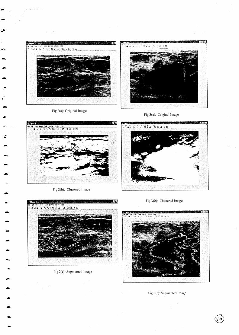
The figures 2, 3 show automatic region growing
method and its propose algorithm outputs. Here Fig2
(a) and Fig3 (a) shows Original Mammogram Images.
Fig2(b) and Fig3(b) are outputs for K Means Clustering
algorithm for given original mammogram images and
Fig2(c)and Fig3(c) shows segmented tumor images by
proposed region growing algorithm.
0

be segmented on the image. In the second stage, this
threshold value will be used in the Region Growing
algorithm. Modifications are made to the conventional
Region Growing algorithm where the seed point and
threshold values are automatically determined. The
block diagram for automatic region growing method for
segmentation of tumor on mammogram image is shown
in figure 4.1.
Original Image
a pixel to be clustered and C is the j-th cluster (centre)i
(x = 1, 2, ....NI , y = 1, 2, ..., N and j =1, 2, _.., n). The
moving K-means clustering alggorithm finds the
threshold value . This algorithm is implemented as:
Step 1: Start
Step 2: Initialize the cluster centers and a , and set a =(1 u
a= a (where a is a small constant value, 0h U 0
< a <1 /3nd should be chosen to be inversely0
proportional to the number of centers).
IMoving K-means
Clustering
Al gorithmv
Modified Region
Growing Algorithm
Threshold
Value
Segmented Tumor
Image
1
Figure 4.1 Block Diagram for automatic region
growing method for segmentation of tumor on
mammogram image
4.1 Moving K means Cluster Algorithm
The clustering algorithm is implemented on the
mammogram image to automatically find the threshold .
value for classifying two regions of clusters, i.e. the
object of interest (tumor) and the background.
Consider an image with MxN pixels (where hI and
N are number of row and column of the image
respectively) to be clustered into n clusters . Let p(x,y) is
Step 3: Assign all pixels to the nearest cluster and
calculate the centre positions using
1C = - ^yeC >x E c p(x, y) ............ (I)
1 ° j J J
Where j = 1, 2... n
x=1,2...1\1
y=1, 2...N
Step 4: Check the fitness of each cluster using equation:
f(C)= ^y^X(^ p(x,y) - C I)2 ......(2)
Step 5: Find C and C, the cluster that has the smallest.s i
and the largest value off (•).
Step 6: Iff(C) < a f(C^,s^ a
(i) Assign the members (pixels) of C to C if
(
1
P(x,y) < C , Where x,y E C ,and leavei r
the rest of the members (pixels) to C.i
i) Recalculate the positions of C and C
according to:
C = Il (n )^yEc LIxEC p(x, y) .......... (3).^^ s S S
D
I"
D
Fig 2(a): Original Image
Fig 2(b): Clustered Image
Fig 3(a): Original Image
Fig 3(c): Segmented image

6. CONCLUSION
The aim of this work is to develop an
automatic algorithm for segmentation of tumor on
mammogram image which is unique challenge in
mammogram image segmentation . The above result
shows that our algorithm is one of the best automatic
methods of segmenting tumor on mammogram images.
The proposed method automatically determines the
seed point and threshold value. The results obtained are
favorable as the proposed Region Growing algorithm
provides more meaningful images. The region of tumor
is successfully detected . The size and shape of tumor
region has also been preserved.
7. REFERENCES
[1] Reifael C. Gonzalez and Richard E. Woods,"Digital
Image Processing' Pearson Edition,
2002.
[2] B Chanda, D Dutta Majumder, "Digital Image
Processing and Analysis", Prentice Hall, 1st
Edition 2004.
[3] Edward A Sickles, Mammographic Features of
Early Breast Cancer , the annual meeting of the
American Roentgen Ray Society, Las Vegas, April
1984.
[4] P K sinha and Q H hong , An Improved Median
Filter., IEEE Transactions on Medical Imaging,
Vol. 9, Sep 1990, pp 51-54.
[5] N Otsu , A Threshold Selection Method from Gray-
Level Histograms, IEEE Transactions on systems,
Vol.l, Jan 1979, pp 149-151.
[6] Rolf Adams and Bischof L, Seeded region
growing , IEEE Transactions on Pattern Analysis
and Machine Intelligence, Vol 16(6):641-647,Oct
1994. pp 34-38.
[7] N. Ikonomakis, K.N. Plataniotis , M. Zervakis, A.N.
Venetsanopoulos , Region growing and region
merging image segmentation , IEEE Transactions
on Medical Imaging, Vol 13, April 1997,pp 82-87.
[8J
[9J
Ahmad Fadzil Mohd Han , Umi Kaithum Ngah,
Venkatachalam ' Lim Eng Eng , Processing of
Abdominal Ultrasound Images Using Seed based
Region Growing Method , IEEE Transactions, Vol
33, May 2004,pp 52-60.
Tien Dzung Nguyen, Viet Dzung Nguyen, Thuan
Duong Ba Hong, Nam Chul Kim , Fast
segmentation based on a hybrid of clustering and
morphological approaches , IEEE Transactions, Vol
23 , June 2008, pp 315-319.
[I0] w'vw.wikipedia.org