Embed Size (px)
Citation preview
Abstract A distinctive histopathological feature of sev-eral neurodegenerative diseases, including corticobasal de-generation, argyrophilic grain disease, progressive supra-nuclear palsy, and Pick’s disease, are achromatic nervecells that express small heat-shock proteins, such as αB-crystallin or hsp-27, and develop in specific telencephaliccortical areas and subcortical nuclei. Here, we point to theconsistent presence of such cells in Parkinson’s disease.In this disorder, the neurons under consideration remainimmunonegative for phosphorylated neurofilaments or forubiquitin, thus exhibiting an immunocytochemical profiledifferent from that shown by αB-crystallin-positive neu-rons in other neurodegenerative disorders. In severe casesof Parkinson’s disease, the αB-crystallin-positive neuronsare dispersed throughout the cerebral cortex, amygdala,and ventral claustrum. In cases showing relatively mildinvolvement of the telecephalon, these neurons occurchiefly within the reaches of the anterior temporal and in-sular mesocortex. These telencephalic predilection sitesare nearly identical with those of the α-synuclein pathol-ogy. Nevertheless, most of the telencephalic αB-crys-tallin-immunopositive neurons refrain from developingLewy bodies and Lewy neurites and, vice versa, most ofthe nerve cells containing Lewy bodies do not accumulateαB-crystallin.
Keywords Anterior mesocortex · Ballooned neurons ·Heat-shock proteins · Parkinson’s disease
Introduction
Parkinson’s disease (PD) is characterized morphologicallyby intraneuronal inclusion bodies containing α-synuclein
(α-SN) known as Lewy bodies (LBs) in nerve cell somataand as Lewy neurites (LNs) in cellular processes [1, 16,23, 25, 39, 41]. This disease-specific pathology evolves inonly a few neuronal types in functionally related corticalareas and subcortical nuclei [6, 7, 8].
Apart from the LBs and LNs that contain α-SN, PDcases almost consistently present a supplementary pathol-ogy, which, to our knowledge, has not been described insufficient detail previously. Because its detection is labo-rious in sections stained for general overview (H&E), itusually escapes recognition. Immunoreactions, however,for the small heat-shock or stress proteins αB-crystallinand/or hsp-27 unequivocally demonstrate this supplemen-tary pathology.
Many heat-shock proteins (HSPs) function as molecu-lar chaperones in that they prevent deleterious protein-protein interactions and assist in the refolding of dena-tured proteins [10, 19, 21, 26, 32, 37]. The HSP αB-crys-tallin is normally expressed solely by macroglial cells andnot nerve cells. Up-regulation of HSPs is interpreted asone of many cellular responses to stress [17, 19, 20, 24].Concentrations of HSPs high enough to be detected in im-munoreactions during light microscopy are encounteredfrequently in activated macroglial cells. Similarly, impres-sive examples of up-regulated HSPs in neurons rarely areseen, but when they do occur it is only in a few types oftelencephalic projection cells located in specific corticallayers and areas, as well as in some subcortical nuclei.The intraneuronal appearance of hsp-27 and/or αB-crys-tallin is often accompanied by telling changes in both thesizes and shapes of the involved neurons, which eventu-ally display considerably bloated or “ballooned” cell bod-ies together with a few noticeably altered cellular pro-cesses [24, 29, 42].
Such neurons are found in other degenerative diseases,among them Pick’s disease (PID), corticobasal degenera-tion (CBD), argyrophilic grain disease (AGD), and pro-gressive supranuclear palsy (PSP) [5, 11, 26, 27, 29, 31,33, 34, 38, 40, 43]. In PD, up-regulation of αB-crystallinhas been identified heretofore in astrocytes and oligoden-drocytes [35]. The present study is aimed at supplement-
Heiko Braak · Kelly Del Tredici ·Daniele Sandmann-Keil · Udo Rüb · Christian Schultz
Nerve cells expressing heat-shock proteins in Parkinson’s disease
Acta Neuropathol (2001) 102 :449–454DOI 10.1007/s004010100395
Received: 29 January 2001 / Revised, accepted: 19 March 2001 / Published online: 10 August 2001
REGULAR PAPER
H. Braak (✉ ) · K. Del Tredici · D. Sandmann-Keil · U. Rüb ·C. SchultzDepartment of Clinical Neuroanatomy, J.W. Goethe University,Theodor Stern Kai 7, 60590 Frankfurt/Main, Germanye-mail: [email protected], Tel.: +49-69-63016900, Fax: +49-69-63016425
© Springer-Verlag 2001

ing this notion by showing that αB-crystallin-immunore-active neurons (αBCINs) consistently develop in thecourse of PD.
Material and methods
We examined brains removed at autopsy from 19 individuals withclinically documented and neuropathologically confirmed idio-pathic PD (11 females, 8 males, ages 72.8±7.0 years, Hoehn andYahr stages III–V, concomitant Alzheimer’s disease (AD)-relatedneurofibrillary changes rated at less than stage IV, amyloid load 0-C; Table1). In addition, 8 brains obtained at autopsy from per-sons lacking a history of neurological disorders were used for con-trol and comparison (4 females, 4 males, ages 62.1±16.4 years,Hoehn and Yahr stage 0, concomitant AD-related neurofibrillarychanges rated at less than stage IV, amyloid load 0-C, and tissuevirtually free of other pathological changes; Table 1). The clinicaland post-mortem neuropathological diagnoses of idiopathic PDwere established using standard published criteria: The clinicalprotocols of all of the PD cases noted the presence of tremor, rigid-ity, and bradykinesia. Moreover, the brain tissue exhibited nigralLBs, loss of nigral neuromelanin-laden neurons, and an associatedgliosis [11, 12]. Concomitant AD-related alterations were classi-fied according to a published staging procedure [4]. Cytoskeletal
changes related to AGD were classified according to published cri-teria [5]. None of the cases exhibited AGD-related lesions with theexception of cases 11 and 15, both of which demonstrated a milddegree of AGD pathology.
Following fixation by immersion in a 4% buffered solution offormaldehyde for at least 3 weeks, one hemisphere was cut in thefrontal plane into three blocks. These blocks and the brain stems ofall of the cases were embedded in polyethylene glycol (PEG 1000,Merck). Each central block and brain stem was then cut perpen-dicularly to the intercommissural axis of Forel (central block) or atright angles to Meynert’s axis (brain stem) into uninterrupted se-ries of 100 µm sections. Ten sets of free-floating sections, each cutserially 1 mm equidistant from the other, were collected.
The first series of sections was stained for both lipofuscin pig-ment (aldehyde-fuchsin) and Nissl material (Darrow red) to facili-tate topographic orientation and identification of specific neuronaltypes classifiable by virtue of their respective pigment deposits [3].The following two collections were processed with (1) a silver-pyridine method [9] for detection of LBs/LNs as well as β-amyloiddeposits and neuromelanin granules [2, 36], and (2) a silver-iodidemethod [15] to assess the possible presence of neurofibrillary tan-gles (NFTs) and neuropil threads (NTs) [4, 22].
The fourth collection was immunostained for α-SN. Sectionswere pre-treated according to a standard protocol designed to in-hibit endogenous peroxidase and prevent nonspecific binding. In-cubation for 48 h in the affinity-purified α-SN antiserum (AFshp)
450
Case Age Gender Diagnosis H+Y DSM-III-R NFP-AT β-Amy αB-crys hsp-27
1 61 M PD III D I 0 1 n.e.2 62 F PD V D I B 1 13 63 F PD IV ND I B 1 n.e.4 67 M PD V D I B 1 n.e.5 68 F PD III D II B 1 16 68 M PD IV ND III C 1 17 69 F PD IV D I A 1 n.e.8 70 F PD V D II B 3 n.e.9 75 F PD IV ND II C 1 1
10 75 M PD V D II 0 1 111 75 M PD III D I B 1 n.e.12 76 F PD IV ND II 0 1 n.e.13 76 M PD V D II A 1 114 77 F PD III D II C 2 115 77 M PD IV D II B 2 116 78 F PD IV D II C 2 n.e.17 79 M PD IV D II C 1 n.e.18 80 F PD V D II C 2 n.e.19 86 F PD V D III C 3 220 33 F Control 0 ND 0 0 0 021 45 M Control 0 ND II A 0 022 59 M Control 0 ND I 0 0 023 62 F Control 0 ND I 0 0 024 69 F Control 0 ND I B 0 025 69 M Control 0 ND II B 0 026 80 F Control 0 ND II B 0 027 80 M Control 0 ND II B 0 0
Table 1 Occurrence of nerve cells immunoreactive for heat-shockproteins hsp-27 and/or αB-crystallin in cases of PD and controls(PD Parkinson’s disease, H+Y Hoehn and Yahr stage, DSM-III-Rcognitive status of cases according to the Diagnostic and StatisticalManual criteria for dementia, D dementia, ND no dementia. Theseverity of the concomitant Alzheimer’s disease-related pathology[4] appears in Roman numerals under NFP-AT (cortical neurofi-brillary pathology of the Alzheimer type: stages I–VI), and upper
case letters refer to β-amyloid deposition (β-Amy: A–C, 0 no amy-loid). αB-crys indicates the average density of αB-crystallin-im-munoreactive nerve cells in the anterior mesocortex. hsp-27 refersto the average density of hsp-27-immunoreactive neurons (0 noimmunopositivity, 1 presence of a few isolated immunoreactiveneurons, 2 moderate numbers, 3 dense accumulation of immunore-active neurons, n.e. not evaluated)

at a dilution of 1:2,000–4,000 followed. This antiserum, which isspecific to α-SN, was generated in sheep by W.P. Gai (see Ac-knowledgments) using a peptide corresponding to the amino acidresidues 116–131 of the human α-SN [14]. After processing withbiotinylated secondary antibodies (anti-sheep IgG, 2 h), the reac-tions were visualized with the avidin-biotin-peroxidase complex(ABC, Vectastain, Vector) and 3,3-diaminobenzidine-tetra-HCl/H2O2 (D7679 Sigma). Omission of the primary antiserum resultedin non-staining.
Varying numbers of sections from the fifth collection wereused for immunoreactions with antibodies against hsp-27 (1:1,000,StressGen), αB-crystallin (1:2,000, Novocastra), ubiquitin (1:500,DAKO), and phosphorylated neurofilaments (1:5,000, SMI 31,Sternberger). Some of the sections were stained initially for lipo-fuscin deposits, subsequently immunostained, and then cover-slipped or counterstained a second time for Nissl material to permitrecognition of immunopositive material in specific cortical layersand to identify select types of nerve cells. Others were silver-stained initially for LB/LNs and then underwent immunoreactionswith an antibody against αB-crystallin, and still other sectionswere immunostained first with an antibody against αB-crystallinand subsequently with a second antibody against α-SN. Finally, allof the sections were cleared and mounted in a synthetic resin (Per-mount, Fischer).
Results
All of the PD cases examined exhibited LBs/LNs in neu-romelanin-laden projection neurons of the substantia ni-gra and moderate to severe neuronal loss in this nucleargray. Furthermore, the cases displayed the characteristicextranigral pathology that consistently develops in thecourse of this disorder. Varying densities of LBs and/orLNs were found within select nuclei in the brain stem,thalamus, hypothalamus, amygdala, and claustrum, aswell as in select areas of the hippocampal formation, en-torhinal region, meso- and neocortex. The anterior meso-cortex, in particular the periallocortical transentorhinaland the proneocortical ectorhinal regions, showed a par-ticular proclivity to develop the PD-specific lesions [6, 8].All controls were free of this pathology. Many PD casesand controls contained concomitant mild AD-related neu-rofibrillary alterations corresponding to stages I–III. ThePD cases either were devoid of telencephalic β-amyloidprecipitates or contained only a few such deposits (Table 1).
Notably, all of the PD cases displayed the supplemen-tary pathology under consideration here. The PD-associ-ated αBCINs usually elude detection light microscopi-cally in sections stained with conventional techniques orwith the Gallyas silver-staining method but were easilyidentified in immunoreactions directed against αB-crys-tallin or hsp-27 (Fig.1). The severity of this pathologyvaried among cases. None of the controls containedαBCINs (Table 1).
The PD-associated αBCINs were dispersed preferen-tially throughout the deep layers V–VI of the anteriortemporal and insular neo- and mesocortex. Only on rareoccasion were a few isolated α-BCINs seen in layer IIIc.Isolated or small groups of αBCINs were also encoun-tered in both the amygdala and the ventral claustrum butnot in other subcortical nuclear grays. The involvement ofthe hippocampal formation and entorhinal cortex was
comparably mild. Allocortical predilection sites were thefirst Ammon’s horn sector and layer pri-α of the entorhi-nal cortex (Fig.1d). The highest density of αBCINs wasusually attained in the anterior mesocortex, i.e., within astretch of cortex encompassing, among other areas, theperiallocortical transentorhinal region, the proneocorticalectorhinal region (Fig.1a–c), and the insular mesocortex.Cases in which the anterior mesocortex was very severelyinvolved likewise displayed remarkably high densities ofαBCINs in the amygdala and ventral claustrum. CorticalαBCINs gradually decreased in number and maintainedhigher intervals from each other in an imaginary line ex-tending from the anterior mesocortex into the neocorticalprefrontal areas or temporal sensory association areas.
All of the neurons under consideration were intenselyimmunoreactive with antibodies against αB-crystallin(Fig.1). A subset of these nerve cells also displayed aslightly less pronounced immunoreaction with antibodiesdirected against hsp-27 (Table 1). Notably, immunoreac-tions for the possible presence of phosphorylated neurofil-aments within the somatodendritic compartments of theαBCINs proved negative, and the abnormal material contained in these neurons was also ubiquitin negative.Most of the αBCINs contained no α-SN-immunoreactiveinclusion bodies and most of the immediately surroundingnon-ballooned nerve cells containing LBs/LNs or AD-re-lated NFTs/NTs lacked immunocytochemically detectableamounts of hsp-27 or αB-crystallin. In addition, no obvi-ous relationship existed between αBCINs and extracellu-lar deposits of β-amyloid protein. The overall packingdensity and topographic distribution of the αBCINs, how-ever, reflected the overall severity and distribution patternof both the PD-specific LBs/LNs and the AD-relatedNFTs/NTs.
Despite severe distortion of their somata and the cellu-lar processes, αBCINs could be readily distinguishedfrom activated astrocytes containing HSPs chiefly owingto their rounded and sharply drawn contours (compareFig.1e–j with k). In general, the αBCINs exhibited con-spicuously bloated cell bodies. Such cells usually have re-duced numbers of distorted cellular processes that appearto be considerably reduced in length. Often, two stout cel-lular processes emerge from the cell body at oppositepoles and, in cortical αBCINs, these neurites are alignedat right angles to the surface. Similarly, the main neuritesof amygdalar or claustral αBCINs tended to run counterto each other but with their longitudinal axes lying criss-cross or in every which direction.
Immunoreactions for αB-crystallin either revealed ho-mogeneous somatodendritic distributions of the protein(Fig.1e, f, i) or exhibited a fine, granular substance withglobular and very intensely immunoreactive condensationof the material (Fig.1h). Occasionally, clear vacuoles ofvarying sizes and shapes were seen scattered in the neu-ronal perikarya and in the proximal portions of their cel-lular processes, with the pale and cap-like immunonega-tive nuclei regularly assuming a peripheral position withinthe cell (Fig.1i, j). In sections stained for lipofuscin gran-ules and basophilic material, the α-BCINs were remark-
451

452

ably depleted of their Nissl substance but they containednumerous eccentrically positioned lipofuscin granules.
In addition to the highly altered nerve cells, less se-verely affected αBCINs occurred, albeit in small numbers.A morphological spectrum could be established rangingfrom immunoreactive neurons of nearly normal appear-ance to extremely bizarre-looking cells. Neurons with themildest changes in size and shape were recognizablypyramid-shaped projection neurons possessing apical andbasal dendrites that emerged from the somata with conicalstems and gradually tapered off distally. Often, an axonwas also identifiable as a thread-like cellular process ofunvarying diameter, usually aligned radially and headeddirectly for the white substance. In sections stained forlipofuscin pigment and Nissl material, such cells showedfeatures closely resembling either those of the large pyra-midal cells located in layer Vb or those of the multipolarprojection neurons in the claustrum and amygdala.
Discussion
In the course of specific neurodegenerative diseases, afew types of nerve cells in select telencephalic cortical ar-eas and subcortical nuclei register characteristic stress re-sponses by up-regulating small HSPs, such as αB-crys-tallin and/or hsp-27, in their somata and neuritic processes[24]. None of these reactions occur in healthy neurons.The neurons in question mostly appear in the form ofachromatic ballooned cells and have been described in de-tail in cases of PID, AGD, PSP, and CBD [5, 11, 26, 27,28, 29, 30, 31, 33, 34, 38, 40, 43]. The present studydemonstrates their consistent presence in 19 cases with id-iopathic PD.
Immunoreactivity to αB-crystallin is a feature com-mon to such neurons in each of the disorders mentionedabove. Immunoreations for phosphorylated neurofila-ments have frequently been employed with success toidentify αBCINs in PID, PSP, and CBD. Notably, the PD-associated αBCINs do not contain conspicuous amountsof phosphorylated neurofilaments in their somatodendriticcompartments. This peculiarity corroborates the notionthat αBCINs associated with a spectrum of neurodegener-ative diseases react differently to the presence of phos-phorylated neurofilaments [40]. PD-associated αBCINs,like those occurring in PID [25] and CBD [17], show theabsence of immunoreactions with antibodies directedagainst ubiquitin [43]. The αBCINs that appear in thetauopathy AGD exhibit remarkably large amounts of ab-normally phosphorylated tau protein distributed evenlythroughout their somatodendritic compartments [40]. Asimilar co-occurrence of α-SN aggregates is not regularlyfound in the PD-associated αBCINs.
Most of the Lewy body-bearing nerve cells seen in thevicinities of αBCINs do not show remarkable up-regula-tion of the HSPs under consideration here, thus suggest-ing that the existing pathogenic mechanisms for the de-velopment of PD-associated αBCINs differ from thoseunderlying the formation of LBs and LNs. At the sametime, however, the overall amounts of αBCINs, togetherwith their regional predilection sites in idiopathic PD, ap-pear to overlap with the severity and predilection sites ofthe PD-specific LBs/LNs. It should be noted in this con-text that the PD-related neuronal devastation observed inthe substantia nigra is always accompanied by extranigralalterations which, in the telencephalon, usually most se-verely affect the simply organized frontal, insular, andtemporal mesocortical transitional zones. From there, thedensity of nerve cells containing LBs/LNs and α-BCINsdecreases in inverse proportion to the evolutionary trajec-tories of increasing differentiation and hierarchical refine-ment on the part of the various neocortical areas [6, 8].
Acknowledgements This study was supported by the Bundes-ministerium für Bildung, Wissenschaft, Forschung und Technolo-gie and the Deutsche Forschungsgemeinschaft. The contribution ofDr. W.P. Gai was supported by funding from the NHMRC (De-partment of Human Physiology, Flinders Medical Centre, BedfordPark, Australia). Our thanks to Ms. A. Biczysko and Ms. M. Lazarfor their skillful laboratory technical assistance.
References
1.Baba M, Nakajo S, Tu PH, Tomita T, Nakaya K, Lee VMY,Trojanowski JQ, Iwatsubo T (1998) Aggregation of α-synu-clein in Lewy bodies of sporadic Parkinson’s disease and de-mentia with Lewy bodies. Am J Pathol 152:879–884
2.Braak E, Braak H (1999) Silver staining method for demon-strating Lewy bodies in Parkinson’s disease and argyrophilicoligodendrocytes in multiple system atrophy. J Neurosci Meth-ods 87:111–115
3.Braak H (1980) Architectonics of the human telencephalic cor-tex. Springer, Berlin Heidelberg New York
4.Braak H, Braak E (1991) Neuropathological stageing of Alz-heimer-related changes. Acta Neuropathol 82:239–259
453
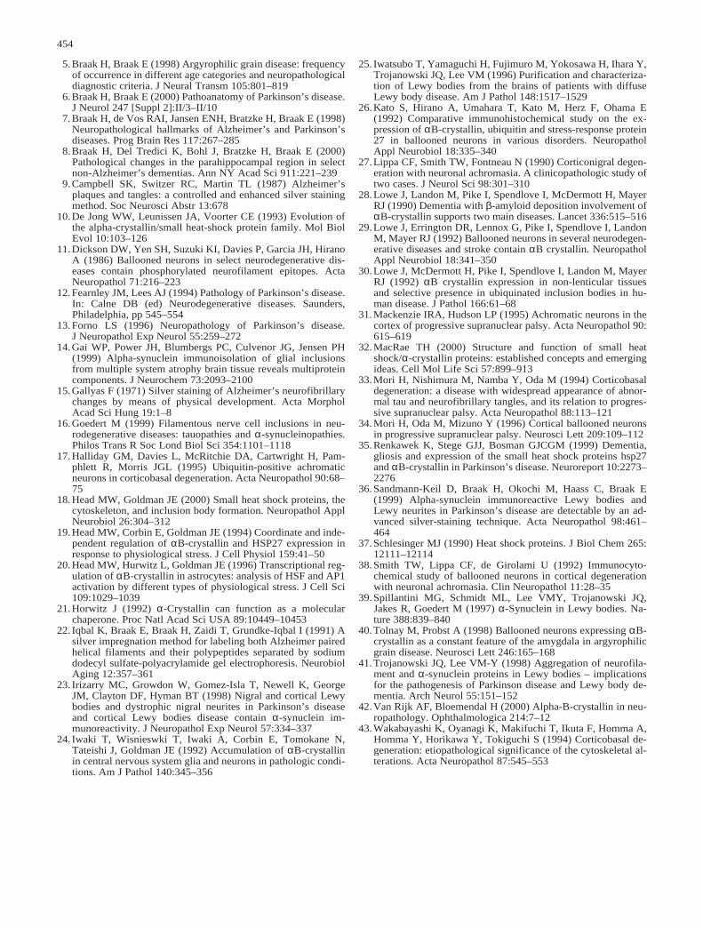
Fig. 1a–k Parkinson’s disease (case 14, Table 1), anteromedialportion of the temporal lobe cut coronally at the level of the uncus.The micrographs show a portion of the anterior mesocortex, in-cluding the proneocortical ectorhinal region and periallocorticaltransentorhinal region. a Considerable numbers of αBCINs areseen in infragranular layers of the anterior mesocortex: ect ectorhi-nal region (proneocortex), cs collateral sulcus, tre transentorhinalregion (periallocortex). The frames indicate the position of the mi-crographs seen in b and c at higher magnification. b, c Note therelatively high packing density of bloated and achromatic αBCINsin the infragranular layers. d Co-staining of intraneuronal lipofus-cin granules with aldehyde-fuchsin permits recognition of the lay-ers harboring the αBCINs. The αBCINs, seen in this micrograph,are located in layer pri-α of the entorhinal cortex (arrow). Thelamination pattern of the entorhinal cortex is indicated at the rightmargin. e αBCINs often exhibit two radially aligned dendritesemerging from opposite poles of the cell body. f αBCIN with rel-atively evenly distributed immunoreactive material. Note the club-shaped swelling at the tip of one of the basal dendrites. This cellu-lar process is greatly reduced in size. g The cellular processes of-ten show irregularly arranged swellings and constrictions. h Theimmunoreactive material is frequently partially condensed into aglobular and centrally placed mass. i, j Clear vacuoles often are en-countered in the cell body and in the proximal dendrites. k. αB-crystallin-immunoreactive astrocyte in neocortical layer III. PEG-embedded material, 100 µm, immunoreaction for αB-crystallin.Bar in h also applies to i–k. (αBCIN αB-crystallin-immunoreac-tive neuron)
�
5.Braak H, Braak E (1998) Argyrophilic grain disease: frequencyof occurrence in different age categories and neuropathologicaldiagnostic criteria. J Neural Transm 105:801–819
6.Braak H, Braak E (2000) Pathoanatomy of Parkinson’s disease.J Neurol 247 [Suppl 2]:II/3–II/10
7.Braak H, de Vos RAI, Jansen ENH, Bratzke H, Braak E (1998)Neuropathological hallmarks of Alzheimer’s and Parkinson’sdiseases. Prog Brain Res 117:267–285
8.Braak H, Del Tredici K, Bohl J, Bratzke H, Braak E (2000)Pathological changes in the parahippocampal region in selectnon-Alzheimer’s dementias. Ann NY Acad Sci 911:221–239
9.Campbell SK, Switzer RC, Martin TL (1987) Alzheimer’splaques and tangles: a controlled and enhanced silver stainingmethod. Soc Neurosci Abstr 13:678
10.De Jong WW, Leunissen JA, Voorter CE (1993) Evolution ofthe alpha-crystallin/small heat-shock protein family. Mol BiolEvol 10:103–126
11.Dickson DW, Yen SH, Suzuki KI, Davies P, Garcia JH, HiranoA (1986) Ballooned neurons in select neurodegenerative dis-eases contain phosphorylated neurofilament epitopes. ActaNeuropathol 71:216–223
12.Fearnley JM, Lees AJ (1994) Pathology of Parkinson’s disease.In: Calne DB (ed) Neurodegenerative diseases. Saunders,Philadelphia, pp 545–554
13.Forno LS (1996) Neuropathology of Parkinson’s disease. J Neuropathol Exp Neurol 55:259–272
14.Gai WP, Power JH, Blumbergs PC, Culvenor JG, Jensen PH(1999) Alpha-synuclein immunoisolation of glial inclusionsfrom multiple system atrophy brain tissue reveals multiproteincomponents. J Neurochem 73:2093–2100
15.Gallyas F (1971) Silver staining of Alzheimer’s neurofibrillarychanges by means of physical development. Acta MorpholAcad Sci Hung 19:1–8
16.Goedert M (1999) Filamentous nerve cell inclusions in neu-rodegenerative diseases: tauopathies and α-synucleinopathies.Philos Trans R Soc Lond Biol Sci 354:1101–1118
17.Halliday GM, Davies L, McRitchie DA, Cartwright H, Pam-phlett R, Morris JGL (1995) Ubiquitin-positive achromaticneurons in corticobasal degeneration. Acta Neuropathol 90:68–75
18.Head MW, Goldman JE (2000) Small heat shock proteins, thecytoskeleton, and inclusion body formation. Neuropathol ApplNeurobiol 26:304–312
19.Head MW, Corbin E, Goldman JE (1994) Coordinate and inde-pendent regulation of αB-crystallin and HSP27 expression inresponse to physiological stress. J Cell Physiol 159:41–50
20.Head MW, Hurwitz L, Goldman JE (1996) Transcriptional reg-ulation of αB-crystallin in astrocytes: analysis of HSF and AP1activation by different types of physiological stress. J Cell Sci109:1029–1039
21.Horwitz J (1992) α-Crystallin can function as a molecularchaperone. Proc Natl Acad Sci USA 89:10449–10453
22. Iqbal K, Braak E, Braak H, Zaidi T, Grundke-Iqbal I (1991) Asilver impregnation method for labeling both Alzheimer pairedhelical filaments and their polypeptides separated by sodiumdodecyl sulfate-polyacrylamide gel electrophoresis. NeurobiolAging 12:357–361
23. Irizarry MC, Growdon W, Gomez-Isla T, Newell K, GeorgeJM, Clayton DF, Hyman BT (1998) Nigral and cortical Lewybodies and dystrophic nigral neurites in Parkinson’s diseaseand cortical Lewy bodies disease contain α-synuclein im-munoreactivity. J Neuropathol Exp Neurol 57:334–337
24. Iwaki T, Wisnieswki T, Iwaki A, Corbin E, Tomokane N,Tateishi J, Goldman JE (1992) Accumulation of αB-crystallinin central nervous system glia and neurons in pathologic condi-tions. Am J Pathol 140:345–356
25. Iwatsubo T, Yamaguchi H, Fujimuro M, Yokosawa H, Ihara Y,Trojanowski JQ, Lee VM (1996) Purification and characteriza-tion of Lewy bodies from the brains of patients with diffuseLewy body disease. Am J Pathol 148:1517–1529
26.Kato S, Hirano A, Umahara T, Kato M, Herz F, Ohama E(1992) Comparative immunohistochemical study on the ex-pression of αB-crystallin, ubiquitin and stress-response protein27 in ballooned neurons in various disorders. NeuropatholAppl Neurobiol 18:335–340
27.Lippa CF, Smith TW, Fontneau N (1990) Corticonigral degen-eration with neuronal achromasia. A clinicopathologic study oftwo cases. J Neurol Sci 98:301–310
28.Lowe J, Landon M, Pike I, Spendlove I, McDermott H, MayerRJ (1990) Dementia with β-amyloid deposition involvement ofαB-crystallin supports two main diseases. Lancet 336:515–516
29.Lowe J, Errington DR, Lennox G, Pike I, Spendlove I, LandonM, Mayer RJ (1992) Ballooned neurons in several neurodegen-erative diseases and stroke contain αB crystallin. NeuropatholAppl Neurobiol 18:341–350
30.Lowe J, McDermott H, Pike I, Spendlove I, Landon M, MayerRJ (1992) αB crystallin expression in non-lenticular tissuesand selective presence in ubiquinated inclusion bodies in hu-man disease. J Pathol 166:61–68
31.Mackenzie IRA, Hudson LP (1995) Achromatic neurons in thecortex of progressive supranuclear palsy. Acta Neuropathol 90:615–619
32.MacRae TH (2000) Structure and function of small heatshock/α-crystallin proteins: established concepts and emergingideas. Cell Mol Life Sci 57:899–913
33.Mori H, Nishimura M, Namba Y, Oda M (1994) Corticobasaldegeneration: a disease with widespread appearance of abnor-mal tau and neurofibrillary tangles, and its relation to progres-sive supranuclear palsy. Acta Neuropathol 88:113–121
34.Mori H, Oda M, Mizuno Y (1996) Cortical ballooned neuronsin progressive supranuclear palsy. Neurosci Lett 209:109–112
35.Renkawek K, Stege GJJ, Bosman GJCGM (1999) Dementia,gliosis and expression of the small heat shock proteins hsp27and αB-crystallin in Parkinson’s disease. Neuroreport 10:2273–2276
36.Sandmann-Keil D, Braak H, Okochi M, Haass C, Braak E(1999) Alpha-synuclein immunoreactive Lewy bodies andLewy neurites in Parkinson’s disease are detectable by an ad-vanced silver-staining technique. Acta Neuropathol 98:461–464
37.Schlesinger MJ (1990) Heat shock proteins. J Biol Chem 265:12111–12114
38.Smith TW, Lippa CF, de Girolami U (1992) Immunocyto-chemical study of ballooned neurons in cortical degenerationwith neuronal achromasia. Clin Neuropathol 11:28–35
39.Spillantini MG, Schmidt ML, Lee VMY, Trojanowski JQ,Jakes R, Goedert M (1997) α-Synuclein in Lewy bodies. Na-ture 388:839–840
40.Tolnay M, Probst A (1998) Ballooned neurons expressing αB-crystallin as a constant feature of the amygdala in argyrophilicgrain disease. Neurosci Lett 246:165–168
41.Trojanowski JQ, Lee VM-Y (1998) Aggregation of neurofila-ment and α-synuclein proteins in Lewy bodies – implicationsfor the pathogenesis of Parkinson disease and Lewy body de-mentia. Arch Neurol 55:151–152
42.Van Rijk AF, Bloemendal H (2000) Alpha-B-crystallin in neu-ropathology. Ophthalmologica 214:7–12
43.Wakabayashi K, Oyanagi K, Makifuchi T, Ikuta F, Homma A,Homma Y, Horikawa Y, Tokiguchi S (1994) Corticobasal de-generation: etiopathological significance of the cytoskeletal al-terations. Acta Neuropathol 87:545–553
454


![Dr. Ruby Kevala, DC, DACNB., Chiropractic Neurologist · Multiple Sclerosis Mumps Osteoporosis Pacemaker [2 Yes C] Yes C) Yes C] Yes C] Yes Parkinson's Disease C] Yes Pinched Nerve](https://img.pdfslide.tips/doc/110x75/5f437b6d8b35c93990049cb4/dr-ruby-kevala-dc-dacnb-chiropractic-neurologist-multiple-sclerosis-mumps-osteoporosis.jpg)