Embed Size (px)
Citation preview

Neuroscience Update: Executive Functions
DyslexiaDTI
Vincent P. Culotta Ph.D. ABNNeuroBehavioral Associates
www.nbatests.comColumbia, MD

Brain-Behavior Assumptions
Neurodevelopemental Maturation underlies and Drives Behavior and CognitionPrefrontal Cortex is the Seat of Reasoning and Last Region to Reach Structural Maturity
Moral Reasoning, Judgment, Impulse Control, Planning, Character and Behavior are directly related to the Biological Maturation of the Brain
Adolescent Brain is not Structurally Comparable to the Competent Adult Brain
Maturation of Executive Functions in Adolescence is Critical for Self-Regulation, Judgment, and Reasoning

• Must be 25 years old
• Must Have a Valid Credit Card

Executive FunctionsOverview
What are they? How do they develop? Developmental risk factors Academic issues Social issues Role in Dyslexia

Executive Functions Unique Set of Mental Functions Regionally specific to the Prefrontal cortex Mediated by flexible and dynamic neural
networks Direct Cognitive and Emotional Functions Include Cognitive Initiation and Inhibition, Self
Regulation, Problem Solving, Flexibility, Error Detection, Organization, Self Monitoring, & Motor Output
Orchestrate the Domains of Thought and Action“The Conductor of the Orchestra”

Executive Function Difficulties
Forgetfulness, can’t keep several things in mind
Distractibility and inattention
Have difficulty following instructions
Disorganization Difficulty using sense of
time to prepare for upcoming events and the future
Cannot accurately estimate how much time it will take to finish a task
Emotional reactivity Low frustration tolerance
Lack of Initiation Impulsivity Decreased self awareness Diminished working
memory Lack of anticipation Inflexibility Hyperfocus Temper Dyscontrol Weak self-calming skills Difficulty reading social
cues Poor follow through Low tolerance for boredom Impatience

Executive Function
Anatomic Neurologic Neuropsychological
Prefrontal Cortex and Striatum
Working Memory
Inhibition
Self Regulation
Organization and Planning
Use of Self-Directed Speech, Rules, and Plans
Levels of Analysis
Initiate, Sustain, Inhibit, Shifts
Delayed Gratification
Goal Directed, Future Oriented Actions
NeuroBehavioral Associates

Biological Maturation and EF
• Develops in Fits and Starts• Vulnerable to Early Insult and
Chronic Stressors• Adolescent Brain is Structurally
Closer to a Young Child’s Brain than an Adult’s Brain• Prefrontal Cortex or Executive
Control Centers are Last to Mature
Ontogeny Recapitulates Phylogeny
Embryonic Development of an Organism follows the same Path as the Evolutionary History of its Species
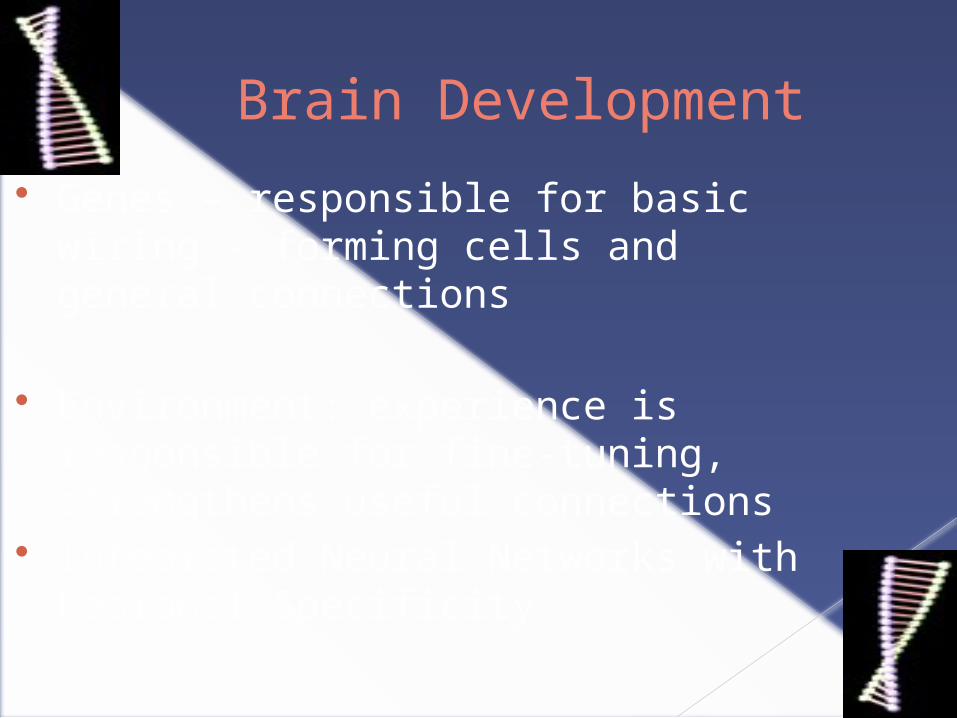
Brain Development
Genes – responsible for basic wiring – forming cells and general connections
Environment: experience is responsible for fine-tuning, strengthens useful connections
Integrated Neural Networks with Regional Specificity

Environmental Influences
Early mother-infant interaction is important for the development of the orbitofrontal cortex during the first months of life.
Early life stressful experiences may permanently damage the orbitofrontal cortex, predisposing the individual to later life neurobehavioral deficits
Severe early stress induced by deprivation and abuse induces changes in the developing brain.

Environmental Influences
Corticotrophin – releasing factor (CRF) hypersecretion throughout life is a consequence of severe abuse in childhood and may underlie the psychopathology that follows abuse
Abuse leads to a state of chronic hyper-arousal and specific neurochemical changes occur in the brains of abused children.
The memory loss of disassociative amnesia induced by psychological stress may be the result of the toxic action of high, prolonged levels of glucosteriods on the hippocampus

Risk Factors for Executive Dysfunction
Contributors to the risk for ED: Pregnancy complications Drug/alcohol prenatal Prematurity Low birth weight Toxin exposure Post-natal injury (prefrontal)
ED possibly related to a birth defect due to a nutritional deficiency of Omega-3 fatty acids during pregnancy and while nursing (Ottoboni & Ottoboni, 2003.)
ED/ADHD does not arise from increased sugar intake, food additives, excessive viewing of television, or poor child management by parents.

Executive Functions Brain Development
UCLA study: Comparison of MRI scans of young adults, 23-30, with those of teens, 12-16:› Areas of the frontal lobe showed the largest
difference between young adults and teens. › Parietal and temporal areas appeared largely
mature in the teen brain. › Increased myelination in the adult frontal
cortex related to the maturation of cognitive processing and other executive functions.

The shades of blue symbolize maturing brain functions.
Maturation culminates in the prefrontal cortex, the area just behind the brow. This is the seat of Executive Functions the area that controls judgment and the weighing of risks and consequences.
Previously this area was thought to be mature by 18 but studies suggest this area is not fully developed until 25 or later .
Executive FunctionsCortical Maturation

Patient Profile: Premature Birth
Age: 16Gender: FemaleGrade: 10th
Presenting Problem: Grades don’t reflect her ability, organizational difficulties, problems with recall and retention, slow to complete assignments, mild social skill difficulties
Strengths: Caring, empathetic, artistic, well behaved
Birth History: Premature birth at 32 weeks, respiratory problems requiring intubation/ventilation, episodes of apnea,
Developmental History: Motor and language delays, OT/SP prior to Kindergarten, socially immature, escalating academic difficulties thru middles and high school
Family History: Unremarkable

Premature Birth The survival of very low birth weight infants has increased dramatically
during the past decade
Studies examining infants born at less than 32 gestational weeks yield consistent evidence of long term neurocognitive deficits impacting executive function
Recent studies reveal a very high prevalence of higher-order neurodevelopmental impairment evident when such youngsters reach school age
Approximately 50% of ex-preterm infants experience deficits in executive functioning and other areas cognitive development which require special academic support.
Unexpectedly high risk of autism. Autism spectrum features may be an unrecognized feature of very low birth weight infants. Children born before 31 weeks gestation have a doubled risk for developing an autism spectrum disorder.
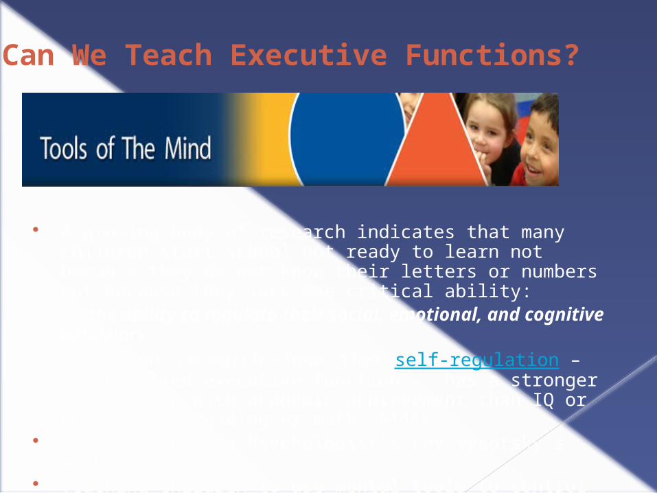
A growing body of research indicates that many children start school not ready to learn not because they do not know their letters or numbers but because they lack one critical ability:
the ability to regulate their social, emotional, and cognitive behaviors. Current research shows that self-regulation – often called executive
function -- has a stronger association with academic achievement than IQ or entry-level reading or math skills.
Based on Russian Psychologist’s Lev Vygotsky’s work Teaching children to use mental tools to control cognition, emotions, and
behavior.
Can We Teach Executive Functions?

Academic Affairs : Extended Campus
Research on Tools of the Mind (Diamond, Barnett, Thomas, & Munroe, 2007) showed that children who attended Tools classrooms had higher rates of self-regulation than closely matched pairs and that the level of self-regulation correlated with child achievement in literacy and mathematics.
“Masters of Their Own Behavior”

NeuroBehavioral Associates
Executive Dysfunction

Executive Dysfunction
Problem of the “Mind” not the “Body”
Problem of “Motivation”
Engenders Moralistic Judgments rather than a Readiness to Help
Misunderstandings & Misassumptions


ADHD & Executive Dysfunction
Snap the Whip – Winslow Homer 1872

Role of Genes
ADHD most likely polygenetic in nature. May involve genes governing DA and NE systems. “The 7-repeat allele:” associated with novelty seeking
(more impulsive, excitable, and exploratory). – Steven Pliszka, MD.

Brain Volume Differences in ADHD
ADHD children show 3-4 percent smaller brain volumes - NIMH, 2008

ADHD:Delay in Cortical Maturation
2007 NIMH Study: Compared brain scans of 446 children with and without
ADHD. Brains of children with ADHD develop normally but lagging
behind approximately 3 years. Biological differences most evident in the frontal lobes,
temporal grey matter, CN, and cerebellum. Shaw et al., 2007

NeuroBehavioral Associates
• Children with ADHD had significantly smaller brain volumes (3%)
• Unmedicated youngsters with ADHD had strikingly smaller volumes (5.8%) than controls
• Children with ADHD had developmental trajectories that paralleled controls but on a lower trajectory
Castellanos, F. (2002) JAMA
Structural Differences
in Brain Volume


Abnormal Brain Anatomy Found With ADHD : Diffusion tensor imaging shows abnormalities in pathways between the frontal lobe, cerebellum. Kate Johnson March 2005
Children with and without ADHD “Abnormalities in fiber pathways in the frontal cortex, basal ganglia,
brain stem, and cerebellum in the ADHD patients” “The circuit that connects the frontal lobe and cerebellum is not
efficient in ADHD” “Fiber pathways abnormalities are less pronounced in children who
have been treated with stimulant medication, compared with those who have not.”
“Our hope is, in the future, to be able to diagnose ADHD with this technique.”
Manzar Ashtari, Ph.D. North Shore-Long Island Jewish Health System
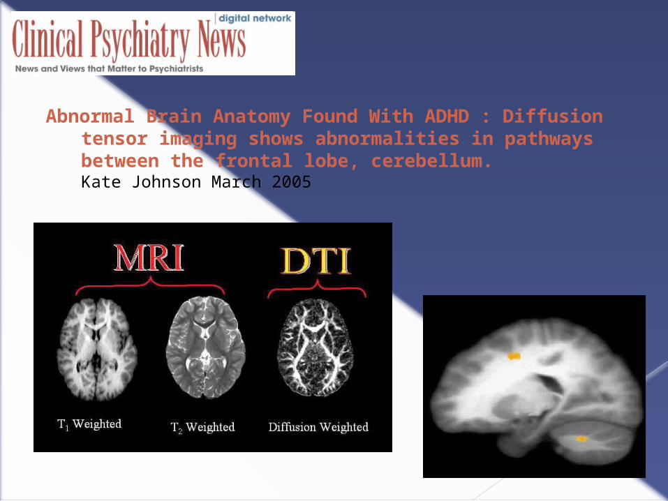
Abnormal Brain Anatomy Found With ADHD : Diffusion tensor imaging shows abnormalities in pathways between the frontal lobe, cerebellum. Kate Johnson March 2005
Social ConsiderationsExecutive Functions & Adolescents

Adolescent Cognition and Behavior
Executive Functions
Driven by Rapid Development in Prefrontal Cortex
Self-ControlInhibitionFlexibilityInitiation
PlanningOrganizeReasoning Judgment
Self-MonitorDelay GratificationAssess ConsequencesPerspective-Taking
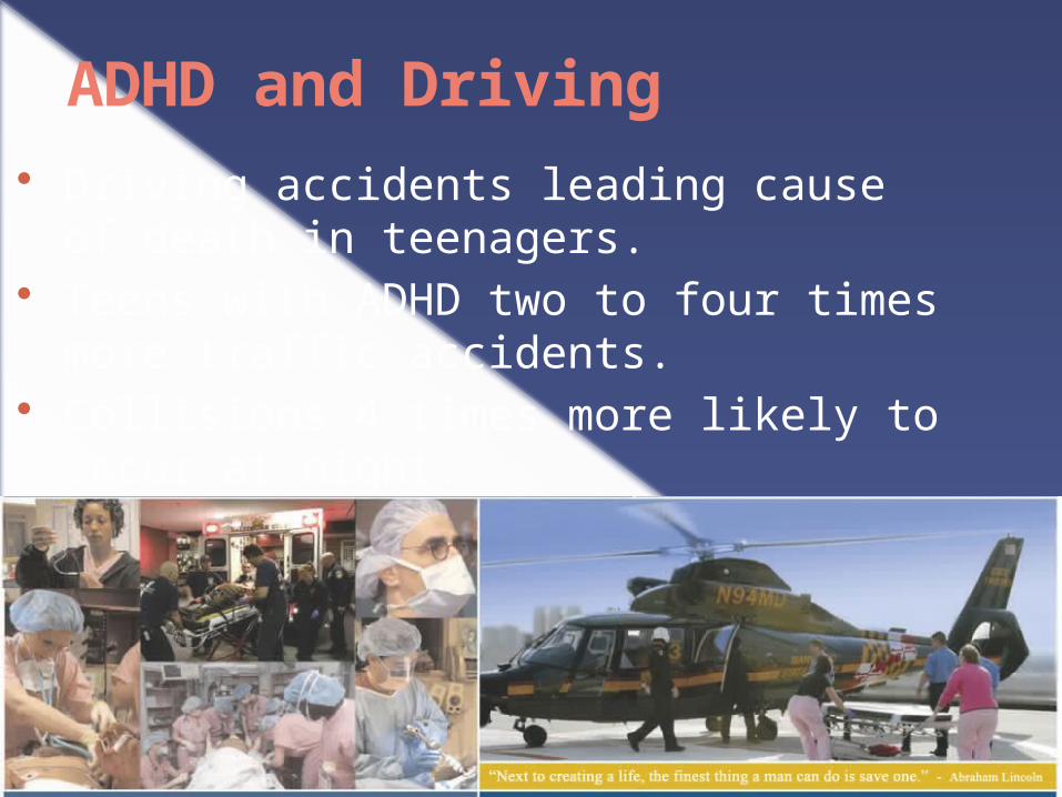
ADHD and Driving Driving accidents leading cause of
death in teenagers. Teens with ADHD two to four times
more traffic accidents. Collisions 4 times more likely to occur
at night.

ADHD and Driving Compared Teens on Concerta, Adderall
& Placebo Stimulants improved Driving
Performance Concerta was more effective late in the
evening when most adolescent accidents occur.
2005 Pediatric Academic Societies by Daniel Cox, Ph.D
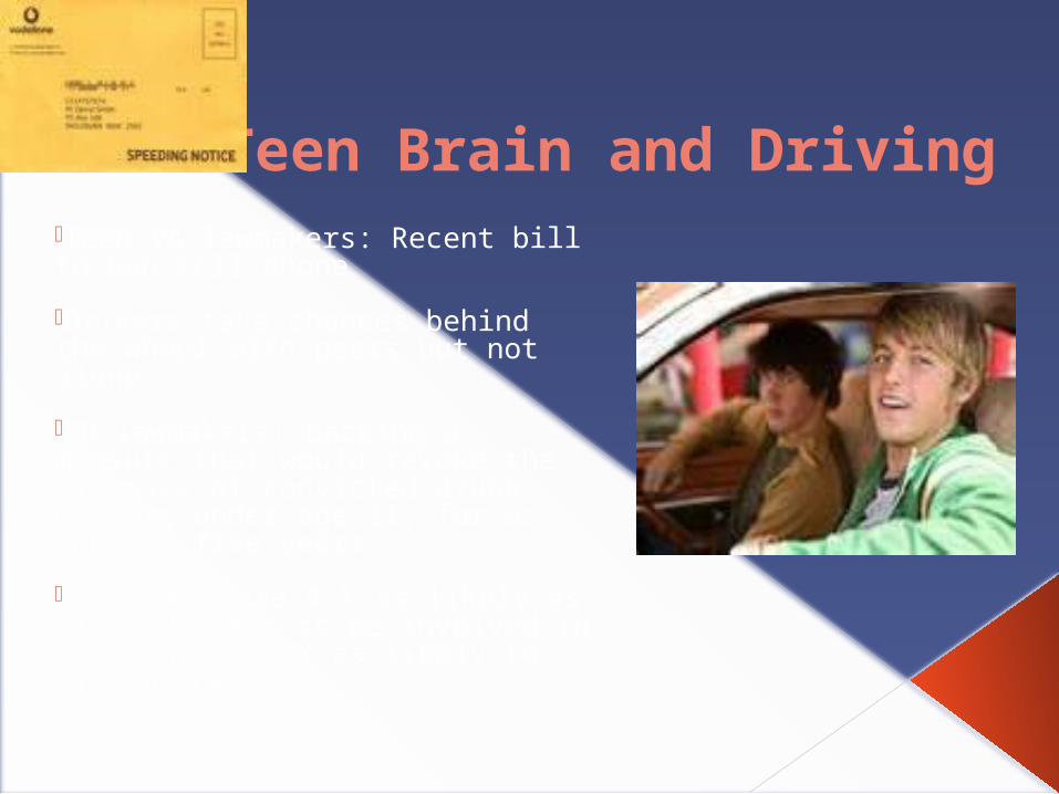
Teen Brain and Driving•Teen VA lawmakers: Recent bill to ban cell phone
•Drivers take chances behind the wheel with peers but not alone.
•MD lawmakers: backing a measure that would revoke the licenses of convicted drunk drivers under age 21, for as long as five years. •Teenagers are 4 X as likely as older drivers to be involved in a crash and 3 X as likely to die in one

"Teenagers' brains are not broken; they're just still under construction." -Giedd

Violent Video Games 44 teen brains examined via fMRI after teens
played violent video game Less activation of prefrontal portions of the brain. More activity in the amygdala. Lingering effects of heightend emotional
arousal and suppressed self-control and concentration remained.

Consequences of Poor Executive Functions in AdolescenceEmotional
Difficulties Aggression Mood Swings Depression & Anxiety
Compulsive Behaviors
Alcohol and Drug Abuse
Preoccupation with Appearance
Self Mutilation
Risk Taking Behavior Alcohol and Drug
Abuse Unprotected Sex
Inattention Distractibility
Poor Academic Performance
Planning Difficulties Test-Taking Difficulties

Plasticity
“Maturity is not simply a matter of slipping software (learning) into existing equipment. Instead, the hardware changes. Those changes partly reflect signals from the world outside, and seem to be a peculiarly human adaptation. Think of it as nature’s way of giving us a second chance.”
Giedd, 2002

The Brain and Reading

Left Temporal Anatomy
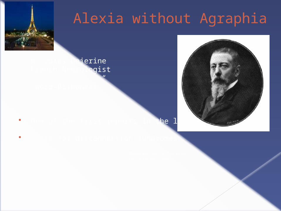
Alexia without Agraphia
M. Jules DejerineFrench Neurologist
“Word-Blindness”
One of the first reports in the literature
Basis for disconnection syndromes
Memoires de la Societe
De Bologie, 1891

Dejerine’s Patient
51 year old store keeper Left Cerebral Vascular Accident Following CVA
› Could not read words› Color naming deficit› R homonymous hemianopsia› He could write, but not read what he wrote› He could identify letters› He could only identify words spelled aurally or
tacitly
“Linguistic Blindfold”


Samuel T. Orton, M.D. - 1928 Neurologist Wife was a Reading Teacher Soft Neurologic signs Explanation of “Word Blindness” Problems with laterality Twisted Symbols & Mirror-Writing were
common in poor reading
“Strephosymbolia”
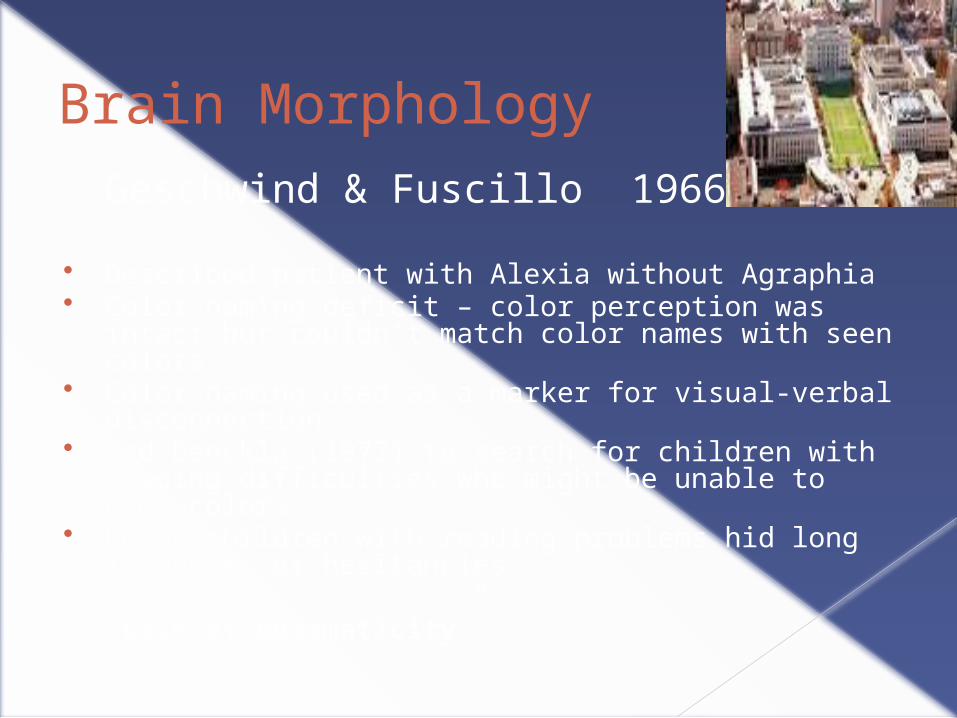
Brain MorphologyGeschwind & Fuscillo 1966
Described patient with Alexia without Agraphia Color naming deficit – color perception was intact but
couldn’t match color names with seen colors Color naming used as a marker for visual-verbal
disconnection Led Denckla (1972) to search for children with reading
difficulties who might be unable to name colors Found children with reading problems hid long latencies
or hesitancies…
“Lack of Automaticity”

Brain Morphology
Galaburda & Kemper - Cellular Abnormalities:› Clusters of Ectopic neurons in outside layer of the
cerebral neocortex› Ectopias are cellular migration between different
cortical layers› Ectopias prevalent in frontal and perisylvan
language regions of left hemisphere› Ectopias are produced before 6 months of
gestation› Found polymicrogyria – multiple narrow, short,
curved gyri

The Neocortex and Cortical Migration
The cerebral cortex is a thin layer of cells about 1.5 to 4 mm thick.
The cortex provides the connections and pathways for the highest cognitive functions, such as language and abstract thinking.
The cerebral cortex contains about 25 billion neurons, more than 62,000 miles of axons, and 300,000,000,000,000 synapses.
Neocortex layer
The thin layer of the neocortex is dense with neurons.

Brain Morphology
Galaburda & Kemper 1979:› Post-mortem structure of severely dyslexic
individuals› Brains were structurally different› Planum Temporale were symmetrical› Found cellular abnormalities
Archives of Neurology, 1978Archives of Neurology, 1979

Brain Morphology
Beaton, 1997:“It is uncritically accepted...
that Dyslexia is associated with a pattern of planum asymmetry more often reversed in direction or reduced in size among Dyslexic’s.”
Brain and Language, 1997

Morphology of Temporal Lobe

Functional Neuroimaging
Shaywitz et al. - PET Study:
… brain activation patterns provide evidence of an imperfectly functioning system for segmenting words into their phonological constituents.
… pattern of underactivation in left posterior brain regions contrasted with relative overactivation in anterior regions may provide a neural signature for the phonological difficulties characterizing dyslexia.

Dyslexia
Anatomic Neurologic Functional
§ Left Superior Temporal Gyrus
§ Heschel’s Gyrus
§ Broca’s Area
Phonemic Awareness
Rapid Naming
Levels of Analysis
NeuroBehavioral Associates
Phonological Processing
Spelling Difficulties
Weak Writing Skills
Diminished Verbal Fluency
Mispronunciations
Decoding Difficulties
Diminished Reading Fluency

Fluency
Denckla & Rudel, 1972, 1974, 1976
Rapid Automatized Naming Test – RAN Traced to Geschwind’s description of Dejerine’s: “Alexia
without Agraphia” “Lack of Automaticity” Strong prediction of reading success Taxes executive control of language system Reid Lyon recommended as Kindergarten screening tool
Annals of Dyslexia, 1999
Fluency
RAN represents separate function from phonological processing
Unique contribution to reading beyond phonological awareness
Poor readers can be subtyped Phonological Deficit only RAN Deficits only Phonological & RAN Deficits
Double – Deficit Model Bowers & Wolf, 1993

Fluency
RAN discriminates between good and poor readers with ADHD
Deficits in letter word fluency and RAN characterized ADHD – RD children
LWF is executive task requires a rule-governed, self-monitored search of the lexicon
Zone of convergence linking ADHD to RD concerns the executive aspects of language- Overlap Zone
Learning
DisabilitiesAttention Deficit/
Hyperactivity Disorder


Dyslexia and Adults

Dyslexia and Adults


Diffusion Tensor Imaging
DTI is a MRI technique that allows visualization and characterization of the brain’s white matter tracts.
DTI measures the diffusion of water molecules in brain tissue
Diffusion is isotropic (equal in all directions) in CSF and cell bodies but anisotropic (greater in one direction than another) in axons that comprise white matter
White matter tracts are myelinated neuronal fiber tracts that connect one brain region with another
Myelin prevents the diffusion of water through the walls of the axon

DTI can measure the orientation and direction of white matter tracts
DTI allows assessment of the coherence or strength of white matter tracts
DTI provides a quantitative index of the organization of large white matter tracts connecting brain regions

The goal of diffusion tensor imaging is to image the microstructure of the brain’s white matter tracts
Red is left to right
Green is front to back
Blue is top to bottom
Diffusion Tensor Imaging
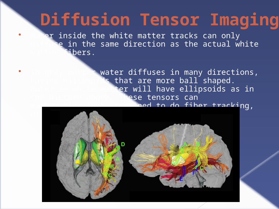
Diffusion Tensor Imaging Water inside the white matter tracks can only diffuse in the
same direction as the actual white matter fibers.
In gray matter water diffuses in many directions, having ellipsoids that are more ball shaped. Water in white matter will have ellipsoids as in the picture above. These tensors can mathematically be combined to do fiber tracking, also called tractography.
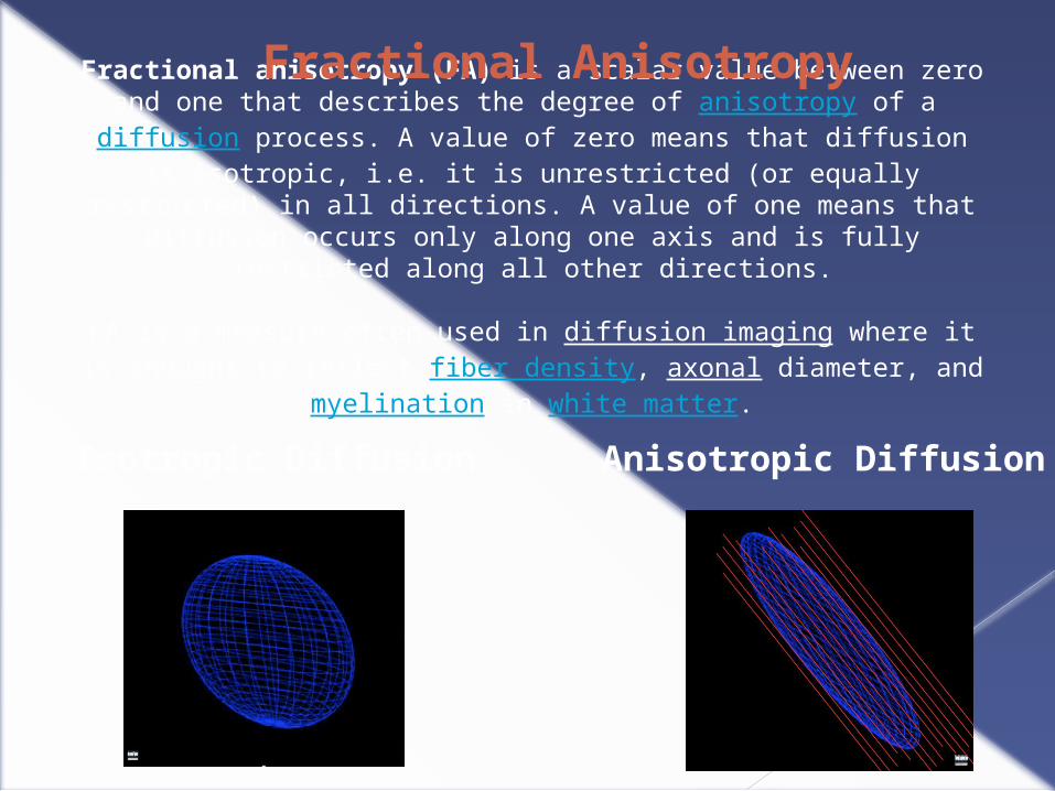
Fractional anisotropy (FA) is a scalar value between zero and one that describes the degree of anisotropy of a diffusion process. A value of zero
means that diffusion is isotropic, i.e. it is unrestricted (or equally restricted) in all directions. A value of one means that diffusion occurs only along one axis
and is fully restricted along all other directions.
FA is a measure often used in diffusion imaging where it is thought to reflect fiber density, axonal diameter, and myelination in white matter.
Isotropic Diffusion Anisotropic Diffusion
Fractional Anisotropy

There is a rapidly expanding body of literature addressing the capability of diffusion-tensor imaging to depict normal white matter and subtle age- and pediatric disease state–related perturbations in white matter that are not visible at routine MR imaging .
FA and ADC are two of the most widely used diffusion-tensor indices. FA is considered a marker of axonal integrity: White matter maturational changes are expressed in part as increases in FA
Diffusion Tensor Imaging

ADVANCES IN KNOWLEDGE
• Diffusion-tensor imaging reveals differences in white matter structure between dyslexic and age-matched normal-reading children.
• Age-related maturational changes in white matter depicted at diffusion-tensor imaging in dyslexic children differ from these changes in normal-reading children.
Diffusion Tensor Imaging

Microstructure of Temporo-Parietal White Matter as a Basis for Reading Ability: Evidence from Diffusion Tensor Magnetic Resonance Imaging
Torkel Klingberg, Maj Hedehus, Elise Temple, Talya Salz, John D.E Gabrieli, Michael E Moseley, Russell A PoldrackDepartment of Psychology, Stanford University, California 94305, USANeuron, Vol. 25, 493-500, February, 2000
Compared Adults with and without Dyslexia
White matter diffusion anisotropy in the temporo-parietal region of the left hemisphere was significantly correlated with reading scores within both the reading impaired and control groups
Greater anisotropy may reflect greater communication between cortical area involved in visual. auditory, and language processing

Neural Changes following Remediation in Adult Developmental Dyslexia
Guinevere F. Eden, Karen M. Jones,Katherine Cappell, Lynn Gareau, Frank B. Wood, Thomas A. Zeffiro, Nicole A.E. Dietz, John A. Agnew, and D. Lynn FlowersNeuron, Vol. 44, 411-422, October 28, 2004
Examined adults with and without dyslexia
Utilized fMRI
Behavioral changes in adults receiving reading intervention correlated with:
-- increased activity in the left hemisphere regions engaged by normal readers --compensatory activity in the right perisylvan cortex.
Behavioral plasticity involves two distinct neural mechanisms
Children’s Reading Performance is Correlated with White Matter Structure Measured by Diffusion Tensor ImagingGayle K. Deutsch, Robert F. Dougherty, Roland Bammer, Wai Ting Siok, John D.E. Gabrieli, Brian WandellCortex, Vol. 4, 354-363, 2005
Examined children with a wide range of reading performance levels
Utilized DTI
White matter structure as measured by FA and CI significantly correlated with behavioral measures or reading, spelling, and rapid naming
Lower FA, reflecting diminished white matter organization, was associated with lower performance scores
Findings support importance of the left temporo-parietal neural pathways in the development of reading skills
Functional and Morphometric Brain Dissociation between Dyslexia and Reading Ability Fumiko Hoeft,*†‡ Ann Meyler,§ Arvel Hernandez,* Connie Juel,¶ Heather Taylor-Hill,* Jennifer L. Martindale,* Glenn
McMillon,* Galena Kolchugina,* Jessica M. Black,*¶ Afrooz Faizi,* Gayle K. Deutsch,* Wai Ting Siok,*‖ Allan L. Reiss,†
Susan Whitfield-Gabrieli,*** and John D. E. Gabrieli Examined adolescents with and without dyslexia
Utilized fMRI techniques
Found patterns of both hypoactivation and hyperactivation
Hypoactivation reflected functional atypicalities related to dyslexia itself
Hyperactivation reflected processes related to current reading levels independent of dyslexia
Hypoactivation is related to the cause of dyslexia
Hyperactivation is associated with the consequence of dyslexia-compensatory mechanisms?

Prediction of Children's Reading Skills using Behavioral, Functional, and Structural Neuroimaging MeasuresFumiko Hoeft, Takefumi Ueno, Allan L. Reiss, Ann Meyler, Susan Whitfield-Gabrieli, Gary H. Glover, Timothy A. Keller, Nobuhisa Kobayashi, Paul Mazaika, Booil Jo, Marcel Adam Just, John D.E. GabrieliBehavioral Neuroscience, Vol 121(3), 602-613, Jun 2007
Examined children of varying reading abilities at both the beginning and end of the school year
Utilized fMRI while performing a phonemic awareness task, behavioral measures, and structural brain measures
Specific patterns of brain activation during phonological processing and white matter densities predicted decoding skills at the end of the year
Combined behavioral and brain imaging techniques predicted outcome better than either alone
Neuroimaging may be useful in identifying those children at risk for poor decoding and reading skills

Tract-based spatial statistics of diffusion tensor imaging in adults with dyslexia.
Richards T, Stevenson J, Crouch J, Johnson LC, Maravilla K, Stock P, Abbott R, Berninger V.Department of Radiology, University of Washington, Seattle, WA 98195, USA. Am J of Neuroradiology 2008 June; 29(6) : 1134-1139
Compared 7 normal adult readers with 14 adults with dyslexia
Utilized DTI
Higher FA values in adult normal readers versus adults with dyslexia
Stronger functional connectivity in the bilateral inferior frontal gyrus of adult normal readers
Expands past studies demonstrating left temporal-parietal differences
Supports disconnections in structural, as well as functional, connectivity in the development of dyslexia

Simple Developmental Dyslexia in Children: Alterations in Diffusion-Tensor Metrics of White Matter Tracts at 3 T1
Radiology. 2009 Jun;251(3):882-91. Epub 2009 Apr 3
CONCLUSION: Findings at 3.0-T DT imaging suggest that white matter differences in
dyslexic children are not limited to the portion of the brain traditionally considered to be integral to word recognition and processing.
1. Nancy K. Rollins, MD, 2. Behroze Vachha, MD, PhD, 3. Priya Srinivasan, MS, 4. Jonathon Chia, MS, 5. Joyce Pickering, PhD, 6. Carrol W. Hughes, PhD and 7. Barjor Gimi, PhD

A dual DTI approach to analyzing white matter in children with dyslexia
Psychiatry Res. 2009 Jun 30;172(3):215-9. Epub 2009 Apr 5 John C. Carter,a Diane C. Lanham,b Laurie E. Cutting,bcd Amy M. Clements-Stephens,b Xuejing Chen,a Muhamed Hadzipasic,a Joon Kim,a Martha B. Denckla,bcef and Walter E. Kaufmann
Used voxel-based (VBA) and region-of-interest (ROI) diffusion tensor imaging (DTI) analyses,
Examined white matter (WM) organization in 7 children with dyslexia and 6 age-matched controls. Both methods demonstrated reduced fractional anisotropy (FA) in the left superior longitudinal fasciculus (SLF) and abnormal orientation in the right SLF in dyslexics.
Application of this complementary dual DTI approach to dyslexia, which included novel analyses of fiber orientation, demonstrates its usefulness for analyzing mild and complex WM abnormalities.
Children with Dyslexia Lack Multiple Specializations Along the Visual Word-Form (VWF) System
Sanne van der Mark, Kerstin Bucher, Urs Maurer, Enrico Schulz, Silvia Brem, Jsabelle Buckelmüller, Martin Kronbichler, Thomas Loenneker, Peter Klaver, Ernst Martin, Daniel BrandeisNeuroimage, vol. 47(4), 1940-9. Oct 2009
Examined children with and without dyslexia
Utilized fMRI to examine activation of the left inferior occipito-temporal cortex (VWF area)
Presented real word, pseudowords, and false fonts
Children with dyslexia showed impaired specialization for both print and orthography

Brain connectivity in non-reading impaired children and children diagnosed with developmental dyslexia.
Odegard TN, Farris EA, Ring J, McColl R, Black J.University of Texas Arlington, Arlington, TX 76019-0528, United States. [email protected], 2009
Diffusion Tensor Imaging (DTI) was used to investigate the relationship between white matter and reading abilities in reading impaired and non-reading impaired children. Seventeen children (7 non-reading impaired, 10 reading impaired) participated in this study.
The data replicated previous results seen across multiple studies and extended findings to include measures of both real word and pseudoword decoding.
Negative correlations were observed in the left posterior corpus callosum between fractional anisotropy (FA) values and both measures of decoding.
Positive correlations between FA values and real word and pseudoword decoding were observed in the left superior corona radiata.
This extension of findings regarding correlations between the corona radiata and reading skills suggests an important direction for future research into the neurological substrates of reading.

White Matter Microstructural Differences Linked to Left Perisylvian Language Network in Children with DyslexiaSheryl L, Rimrodt, Daniel J. Peterson, Martha B. Denckla, Walter E. Kaufmann, Laurie E. CuttingDepartment of Developmental Cognitive Neurology, Kennedy Krieger Institute, 707 N Broadway, Baltimore, MD 21205, USA
Cortex, vol. 46(6):739-49, June 2010
Examined children with and without dyslexia
Utilized DTI to examine white matter structure
FA decreases in dyslxia in LIFG and left temporo-parietal white matter
Positive corelation of FA to reading speed in a left posterior cicuit
Found differences in fiber orientation in Left anterior perisylvan language pathway
Links an atypical white matter structure in dyslexia to atypical fiber orientation in reading circuits of the left perisylvan language network
“To find a convergence of MRI evidence… linked to an identifiable structure …Brings us closer to understanding how dyslexia happens”
Altering cortical connectivity: remediation-induced changes in the white matter of poor readers.Keller TA, Just MA.Center for Cognitive Brain Imaging, Department of Psychology, Carnegie Mellon University, Pittsburgh, PA 15213, USA. [email protected]
Examined whether 100 hr of intensive remedial instruction affected the white matter of 8- to 10-year-old poor readers utilizing DTI.
Prior to instruction, poor readers had significantly lower FA than good readers in a region of the left anterior cerebral white matter
The instruction resulted in a change in white matter (significantly increased FA), and in the very same region.
The FA increase was correlated with improvement in phonological decoding ability, clarifying the cognitive locus of the effect.
The results demonstrate the capability of a behavioral intervention to bring about a positive change in cortico-cortical white matter tracts.
PMID: 20005820 [PubMed - indexed for MEDLINE]PMCID: PMC2796260 [Available on 2010/12/10]

Neural Systems Predicting Long-Term Outcome in DyslexiaFumiko Hoeft, Bruce D. McCandliss, Jessica M. Black, Alexander Gantman, Nahal Zakerani, Charles Hulme, Heikki Lyytinen, Susan Whitfield-Gabrieli, Gary H. Glover, Allan L. Reiss, John D. E. GabrieliJ. Neurosci. Vol. 31 (26) 9641-9648, 2011
Examined children with and without dyslexia over a 2.5 year period to determine if brain imaging (fMRI & DTI) can predict future long term gains in dyslexia
Greater right prefrontal activation during a reading task that demanded phonological awareness and right superior longitudinal fasciculus white matter organization significantly predicted future reading gains in dyslexia
This method predicted significantly above chance (72% accuracy) which child would or would not improve reading skills in dyslexia.
Behavioral measures (testing) were at chance
Right prefrontal mechanisms may be critical for reading improvement in dyslexia, perhaps identifying structures necessary for compensation
What do we know…What do we think we know…
Dyslexia and ADHD involve both structural and functional connectivity abnormalities and regional specificity
Connectivity abnormalities are developmental rather than acquired or the consequence of reduced reading practice
Left temporo-parietal hypoactivation and reduced connectivity is related to etiology of dyslexia
White matter organization is weaker in left posterior brain regions
Right prefrontal activation during reading and right superior longitudinal fasciculus white matter organization predicted future reading gains in dyslexics-Neuroprognosis

What do we know…What do we think we know…
Greater than normal white matter connectivity in the corpus callosum which may reflect an atypical reliance on right hemishphere regiions for reading
Greater preintervntion activation in the right IFG and greater white matter intergrity in the right SLF, on rhyme task, predicts greaterimprovement in reading over the next 2.5 years-Neuroprognosis
DTI may be helpful in measuring response to reading intervention
Remediation is associated with increased activation and connectivity in left tempopro-partietal and frontal regions

What do we know…What do we think we know…
Psychometric testing predicted gains in decoding, accounting for 65% of variance
Fuunctional and structural imaging predicted gains in decoding accounting for 57% of variance
Combined behavioral testing and brain imaging accounted for 81% of variance-Neuroprognosis
Event-Related Potentials (ERP’s) may predict fuure language and reading problems in infants and children before reading instruction
ERP response to language sounds within 36 hours of birth predict children who will go on to become dyslexic by age 8 with 81% accuracy-Neuroprognosis

“The New Revolution”
Educational Neuroscience- Mind Brain and Education Laura-Ann Petitto Kurt Fischer
Neuroprognosis Combination of Brain Imaging and Behavioral
Measures for Diagnosis Better Prediction of At Risk Children Predict capacity for Response to Intervention Possibility of Prevention Measure the Response to Intervention Shape Educational Policy and Practice Shape Health Care Policy-Insurance Guide Family Decision-Making

“An Interpretation of Michelangelo's Creation of Adam” JAMA 1990
The Frontal Lobes : The Seat of Civilization

The Creation of Adam (1508-1512) ceiling of the Sistine Chapel ... Meshberger, M.D. described an anatomically accurate image of the human brain portrayed behind God.
On close examination, borders in the painting correlate with sulci in the inner and outer surface of the brain, the brain stem, the basilar artery, the pituitary gland and the optic chiasm. God's hand does not touch Adam, yet Adam is already alive as if the spark of life is being transmitted across a synaptic cleft.*
Below the right arm of God is a sad angel in an area of the brain that is activated on PET scans when someone experiences a sad thought. God is superimposed over the limbic system, the emotional center of the brain and possibly the anatomical counterpart of the human soul. God's right arm extends to the prefrontal cortex, the most creative and most uniquely human region of the brain.
*Frank Lynn Meshberger, M.D., JAMA #14 October 1990
NeuroBehavioral Associates
Vincent P. Culotta, Ph.D. ABN
Phone: 410-772-7155Fax: 410-772-7156
Email: [email protected]
Across from The Mall in Columbia


NeuroBehavioral Associates